Article

Identification of Listeria Isolates by Using a Pragmatic Multilocus Phylogenetic Analysis

by
Antonio Martínez-Murcia
1,2,*,
Aaron Navarro
2 and
Caridad Miró-Pina
2
1
Department of Microbiology, University Miguel Hernández, Orihuela, 03312 Alicante, Spain
2
Genetic PCR Solutions™, Orihuela, 03300 Alicante, Spain
*
Author to whom correspondence should be addressed.
Microbiol. Res. 2024, 15(4), 2114-2128; https://doi.org/10.3390/microbiolres15040142
Submission received: 3 September 2024 / Revised: 3 October 2024 / Accepted: 10 October 2024 / Published: 14 October 2024

Abstract

:
Species identification of Listeria isolates remained a tedious process still based on culturing methods that, in recent years, have led to the description of many species that are not even part of the genus Listeria. It is advisable to provide new precise techniques since this taxon includes two pathogens that are usually transmitted through the food chain, Listeria monocytogenes and L. ivanovii. The approach, so-called multilocus phylogenetic analysis (MLPA) that uses several concatenated housekeeping gene sequences, provides accurate and affordable classification frameworks to easily identify Listeria species by simple Sanger sequencing. Fragments of seven housekeeping genes (gyrA, cpn60, parE, recA, rpoB, atpA, and gyrB) from 218 strains of all Listeria species currently described were used to build an MLPA of the concatenated sequence, a total of 4375 bp. All isolates subjected to identification were clustered within the species of Listeria sensu stricto, L. monocytogenes, L. innocua, and L. welshimeri, and some reference strains were reclassified as L. ivanovii and L. seeligeri. Housekeeping-gene sequencing has been demonstrated to represent a pragmatic tool that can be firmly considered in food control.

1. Introduction

Listeria is a ubiquitous bacterium that can be found in a wide range of environments, from the soil, water, or decaying vegetation to food or animal digestive tracts [1,2]. This is due to its ability to withstand and multiply at a broad range of pH (pH 4.5–9.2), temperature (−1 °C to 45 °C), and high salt concentrations (10% NaCl) [3]. The Listeria genus is formed by facultative anaerobic, Gram-positive, non-spore forming, rod-shaped bacteria with low G + C content [4]. Only two species, L. monocytogenes and L. ivanovii, are considered pathogens, although rare cases of infections caused by other species have been reported [5,6,7]. Listeria ivanovii is particularly pathogenic in ruminants but rarely affects humans [8]. Listeria monocytogenes is the main culprit of listeriosis, which, although not the most common zoonosis in the EU, is the deadliest, with a hospitalization rate of over 90% and a fatality rate of over 10% [9]. It affects particularly vulnerable individuals like the elderly, pregnant women, infants, and immunocompromised individuals. It was not until the 80s when several large common-source outbreaks of listeriosis highlighted the significance of food as the primary path of transmission for human infection by L. monocytogenes; to date, it is generally considered that most cases involve foodborne transmission [10]. Outbreaks occur generally in developed countries due to processed, refrigerated, ready-to-eat products of animal origin that have a long shelf life and are consumed without further listericidal treatments. The ability of Listeria to grow at a wide range of conditions allows it to proliferate in food-processing environments, making it challenging to ensure food safety [11]. L. monocytogenes is divided into four phylogenetic lineages with distinct evolutionary and phenotypic characteristics [12,13,14,15,16]. Most of the strains isolated worldwide belong to lineages I and II [17]. Lineage I strains are more frequently associated with human clinical cases than those from lineage II and are considered more virulent, while lineage II strains are more frequently isolated from food and environmental sources [18,19]. Lineage III and IV strains are predominantly isolated from animals [20]. Therefore, the assignation of isolates to L. monocytogenes lineages may help to ascertain virulence levels.
The genus currently comprises twenty-seven recognized species (L. monocytogenes, L. grayi, L. innocua, L. seeligeri, L. welshimeri, L. ivanovii (2 subspecies), L. marthii, L. rocourtiae, L. fleischmannii (2 subspecies), L. weihenstephanensis, L. aquatica, L. cornellensis, L. floridensis, L. grandensis, L. riparia, L. booriae, L. newyorkensis, L. costaricensis, L. goaensis, L. thailandensis, L. valentina, L. cossartiae (2 subspecies), L. farberi, L. immobilis, L. portnoyi, L. rustica, and L. ilorinensis), twenty-one of which have only been described since 2010 [21,22,23,24,25,26,27,28,29,30,31,32,33]. Recently, a strain isolated from soil collected in the Great Smoky Mountains National Park (US) was proposed as a new species named “Listeria swaminathanii” but has not been validly published yet [34,35]. All Listeria species so far described have been divided into two large clades due to genomic and phenotypic differences: Listeria sensu stricto (L. monocytogenes, L. innocua, L. seeligeri, L. welshimeri, L. ivanovii, L. marthii, L. cossartiae, L. farberi, L. immobilis, and “L. swaminathanii” [28]), and Listeria sensu lato, comprising species for which three new genera, “Murraya”, “Mesolisteria” and “Paenilisteria”, have been proposed [36,37]. During the last decades, some species of Listeria have also been subjected to misclassification. For instance, J. Rocourt et al., in 1987 [38], established a new taxa, Jonesia denitrificans, from a previously classified strain of Listeria, Listeria denitrificans. Also, J. Rocourt et al., in 1992 [39], using molecular techniques, established that Listeria murrayi and Listeria grayi, are, in fact, the same species, which led to the reclassification of this species as Listeria grayi.
Current methods for the identification of Listeria isolates according to EN ISO 11290-1:2017 [40] are based on classical cultures, which are tedious, take a long time to perform, and may show wrong or unconclusive results. Commonly used methods for Listeria detection are antibody-based lateral flow or enzyme immunoassays (EIA) as well as polymerase chain reaction (PCR)-based methods [10]. PCR is highly specific and sensitive and is more reliable than phenotypic methods [10] but does not characterize differences in the DNA sequence. Multilocus phylogenetic analysis (MLPA) is a highly discriminative approach to characterizing microbial isolates, which has been demonstrated to be very useful for species identification. This method uses the concatenated nucleotide sequences of several housekeeping genes to construct a phylogeny, which allows for a robust hierarchical classification of the species [41,42,43,44]. MLPA technology cannot be confused with MLST (multilocus sequence typing), which analyzes allelic differences for intra-species typing [45,46,47,48]. The concatenation of housekeeping genes allows for a more reliable evaluation of the evolution of the species, minimizing the weight of recombination events in the final phylogenetic tree [49]. Various studies have stated the capacity of MLPA as a tool to provide a robust phylogenetic frame and resolve the phylogeny of closely related species [42,48,50,51,52,53,54,55] and its capacity as a pragmatic technique for species identification.
In the present study, an MLPA of the genus Listeria that uses seven housekeeping gene fragments (gyrA, cpn60, parE, recA, rpoB, atpA, and gyrB; a total of 4375 bp) of representative strains belonging to all Listeria species described so far, has been developed. This MLPA scheme has been demonstrated to be a practical tool to rapidly identify Listeria isolates and may help in the control of Listeria pathogens in food and related environments.

2. Materials and Methods

2.1. Bacterial Strains

Listeria strains used in this study were received from the Colección Española de Cultivos Tipo (CECT, Valencia, Spain) (Table S1). Strains were maintained on TSA plates at 4 °C and stored in 30% glycerol in 0.9% NaCl with 10% TSB at −80 °C.

2.2. DNA Extraction and Purification

A single colony isolated from a fresh plate culture was resuspended in 100 μL TE buffer by vortexing. The tube was incubated at 95 °C for 5 min, vortexed, and kept for 5 min in dry ice. This process was repeated three times consecutively. The sample was centrifuged at 9500 rpm for 10 min, and 30 μL of the supernatant was transferred to a fresh tube and stored at −20 °C.

2.3. MLPA Genes and Primers

The MLPA was performed with housekeeping genes whose sequences were available on public databases. Most of them are involved in DNA/RNA processing functions. PCR and sequencing primers were designed for the seven housekeeping genes: DNA gyrase subunit A (gyrA); chaperonin-60 (cpn60); DNA topoisomerase IV subunit B (parE); homologous recombination factor (recA); DNA-directed RNA polymerase subunit beta (rpoB); F0F1 ATP synthase subunit alpha (atpA); and the DNA gyrase subunit B (gyrB) (Table 1). Primers were designed based on preliminary alignments of available gene sequences from different species of Listeria and related genera.

2.4. PCR Amplification and Sequencing

Fragments of the selected genes, with lengths ranging from 640 to 1020 bp, were amplified by PCR using a peqSTAR 2X Thermocycler. PCR reaction mixtures contained 12.5 μL HotStart Mix Y (Peqlab Biotechnology GmbH, Erlangen, Germany), 1 μL of each primer (10 μM), 1 μL DNA, and 9.5 μL sterile ultrapure water, in a total volume of 25 μL. DNA amplification was performed with denaturation at 94 °C for 3 min, 35 cycles of amplification that consisted of denaturation at 94 °C for 15 s, annealing at 45 °C for 30 s, extension at 72 °C for 1 min, and a final extension step at 72 °C for 5 min. Amplified fragments were analyzed by electrophoresis on 1% agarose-TAE gels. PCR products were purified using the peqGOLD Cycle-Pure Kit (Peqlab Biotechnology GmbH, Germany), following the manufacturer’s instructions. PCR fragments were sequenced by GATC (Eurofins Genomics) using the forward primers (5 μL of purified PCR product + 5 μL of 5 μM primer). Gene sequences from additional Listeria isolates and 16S rRNA coding-gene sequences from reference strains were downloaded from NCBI (National Center for Biotechnology Information) Nucleotide and Whole-genome shotgun contigs databases using BLAST (Basic Local Alignment Search Tool, NCBI) to identify sequences from our seven selected genes.

2.5. Single Gene and Multilocus Sequence Analysis

Pairwise sequence comparisons were performed for each individual gene sequence to obtain single-gene phylogenetic trees. For each analyzed strain, sequence fragments of the seven selected genes, beginning and ending at the same positions, were concatenated. Matrices of nucleotide differences and phylogenetic trees were obtained from the alignment of the concatenated sequences. Alignments were obtained with the MEGA5 program [56] by Clustal W [57]. Evolutionary distances were obtained using the Kimura 2-parameter method [58], and phylogenetic trees were constructed by the neighbor-joining [59] with the MEGA5 program [56] with 1000 bootstrap replicates. The same protocol was followed to obtain a reference 16S rDNA phylogenetic tree using downloaded sequences from type strains when available.
Ranges of intra-species and inter-species nucleotide substitutions were calculated by determining the maximum and minimum values (excluding identical sequences) of nucleotide substitutions from pairwise comparisons of strains for each Listeria species (intra-species) or between strains of different Listeria species (inter-species), including all isolates. Nucleotide substitutions corresponded to the percentage of base differences per position between each pair of compared sequences. All positions containing gaps were excluded from the analysis. Intra-species and inter-species phylogenetic depth was calculated by summing the nucleotide substitution percentages from pairwise comparisons and dividing by the total number of comparisons.

3. Results

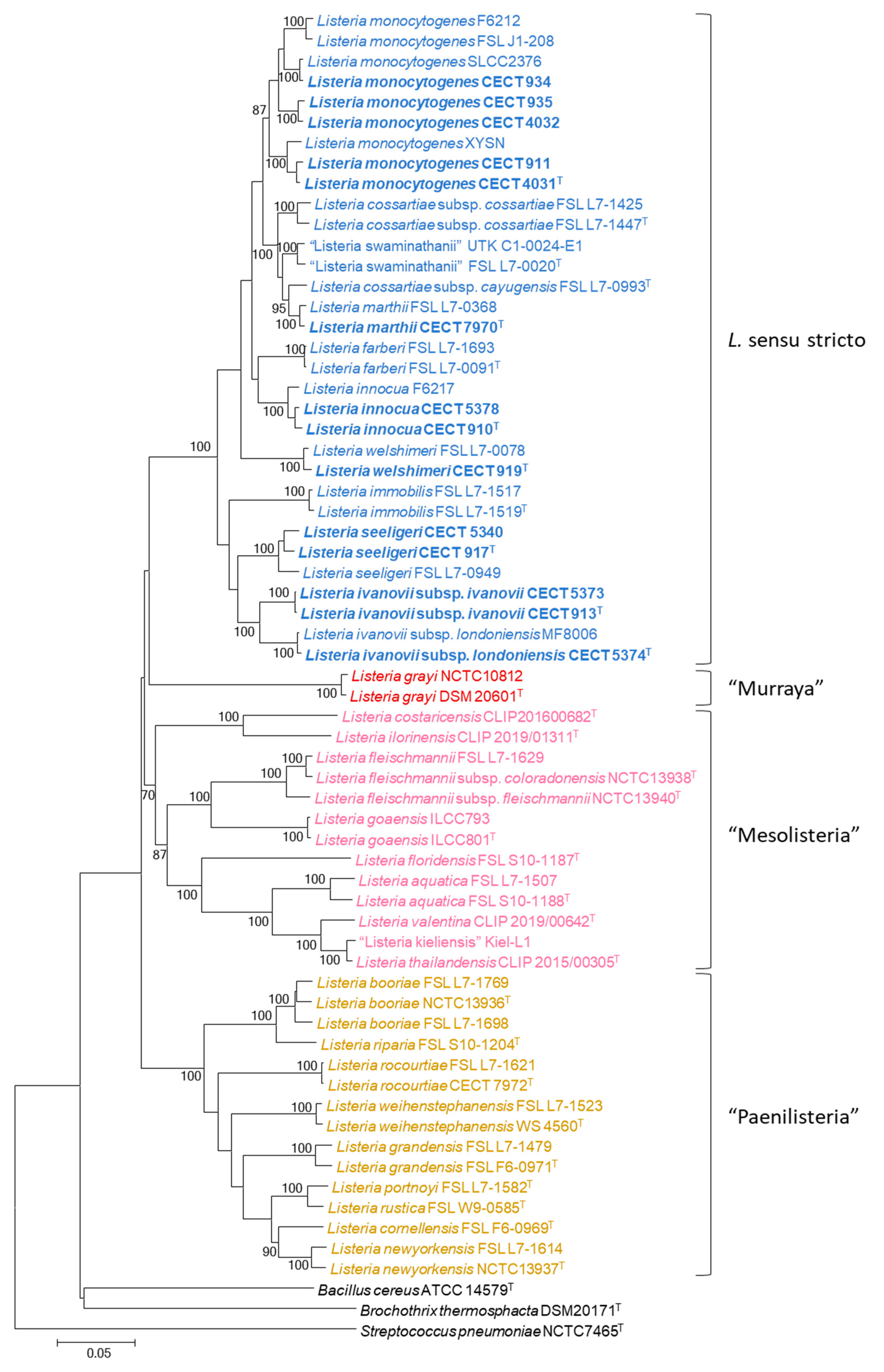
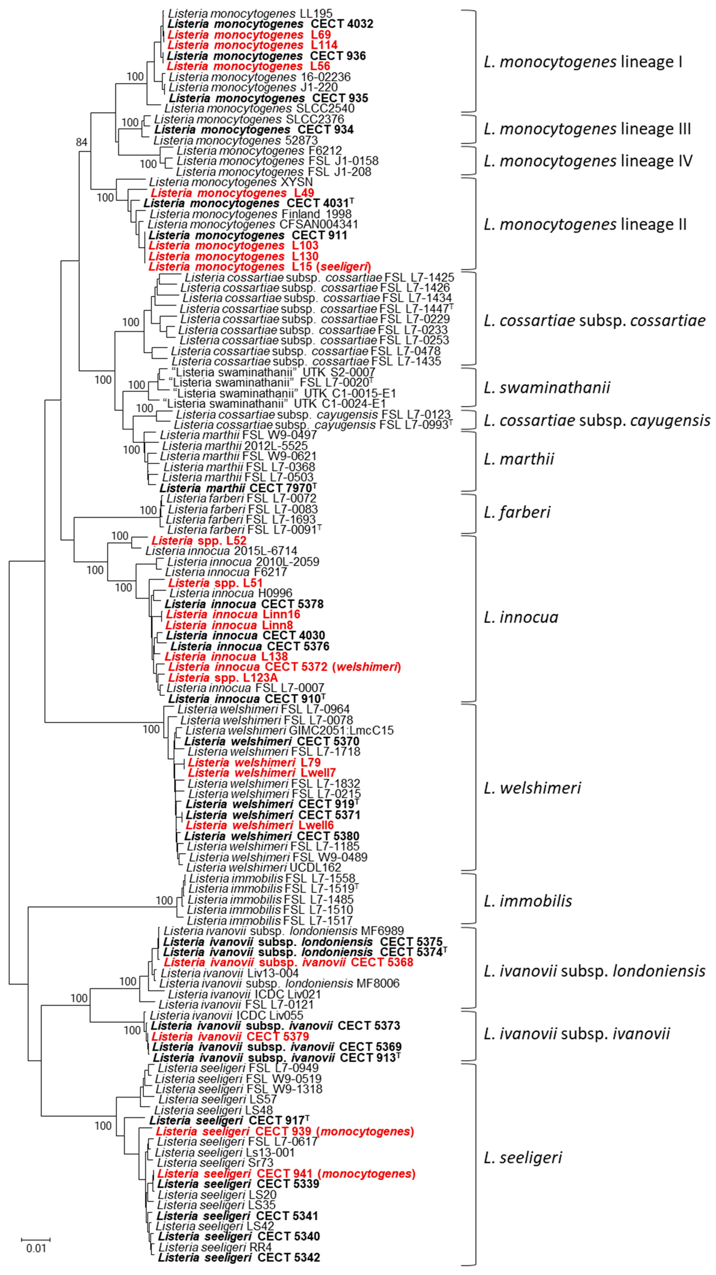
Fragments from the seven selected genes, with lengths ranging from 458 bp to 833 bp, were amplified by PCR with primers listed in Table 1, using DNA extracted from 16 Listeria isolates from foods and 30 CECT Listeria strains listed in Table S1. Additionally, 172 sequences of other isolates from all Listeria species previously identified using BLAST were downloaded from the NCBI database. The fragments of the seven genes for each isolate were concatenated in the same order (gyrAcpn60parErecArpoBatpAgyrB), giving rise to concatenated sequences of ca. 4375 bp. Sequences from each gene were aligned, and nucleotide differences for each pairwise comparison were obtained using the MEGA5 program [56] by Clustal W [57]. Percentages of sequence similarity values were calculated. The fragments of the seven genes, concatenated in the same order (gyrA—cpn60parErecArpoBatpAgyrB, 4375 bp), showed a sequence similarity ranging from 77.51% to 99.89%, which corresponded to 5–981 nucleotide differences and there was a total of 1805 variable positions (41.23% of the sequence). Sequences of the seven concatenated gene fragments were identical for L. ivanovii subsp. londoniensis CECT 5374T and CECT 5375; L. monocytogenes CECT 936 and L56; L. monocytogenes L69 and L114; L. monocytogenes CECT 911, L103, L130 and L15; L. seeligeri CECT 941 and CECT 5339; L. welshimeri CECT 5371 and Lwell6, and L. welshimeri L79 and Lwell7. Those identical sequences were excluded from calculations of intra-species and inter-species nucleotide substitution ranges and intra-species and inter-species phylogenetic depths. Phylogenetic trees were obtained by the neighbor-joining method for the 16S rDNA sequences (Figure 1) and the concatenated housekeeping-gene sequences (Figure 2).
The phylogenetic trees obtained from the concatenated protein-coding gene sequences showed four large clusters, which is in agreement with the previous phylogenetic studies based on whole-genome sequencing data [21,22,23,34,37]. All species considered Listeria sensu stricto appeared in a cluster that included L. monocytogenes, L. innocua, L. seeligeri, L. welshimeri, L. ivanovii, L. marthii, L. cossartiae, L. farberi, L. immobilis, and non-yet validated, “L. swaminathanii”. The rest of the Listeria species, most described since 2010, were distributed into three clusters clearly separated from each other and, altogether, were previously so-called Listeria sensu lato [37]. This result was in agreement with the previous proposal of three additional genera: “Murraya”; “Mesolisteria”; and “Paenilisteria” [21]. The phylogenetic tree obtained from the reference 16S rDNA sequences was in agreement with this distribution of species into four large clusters.
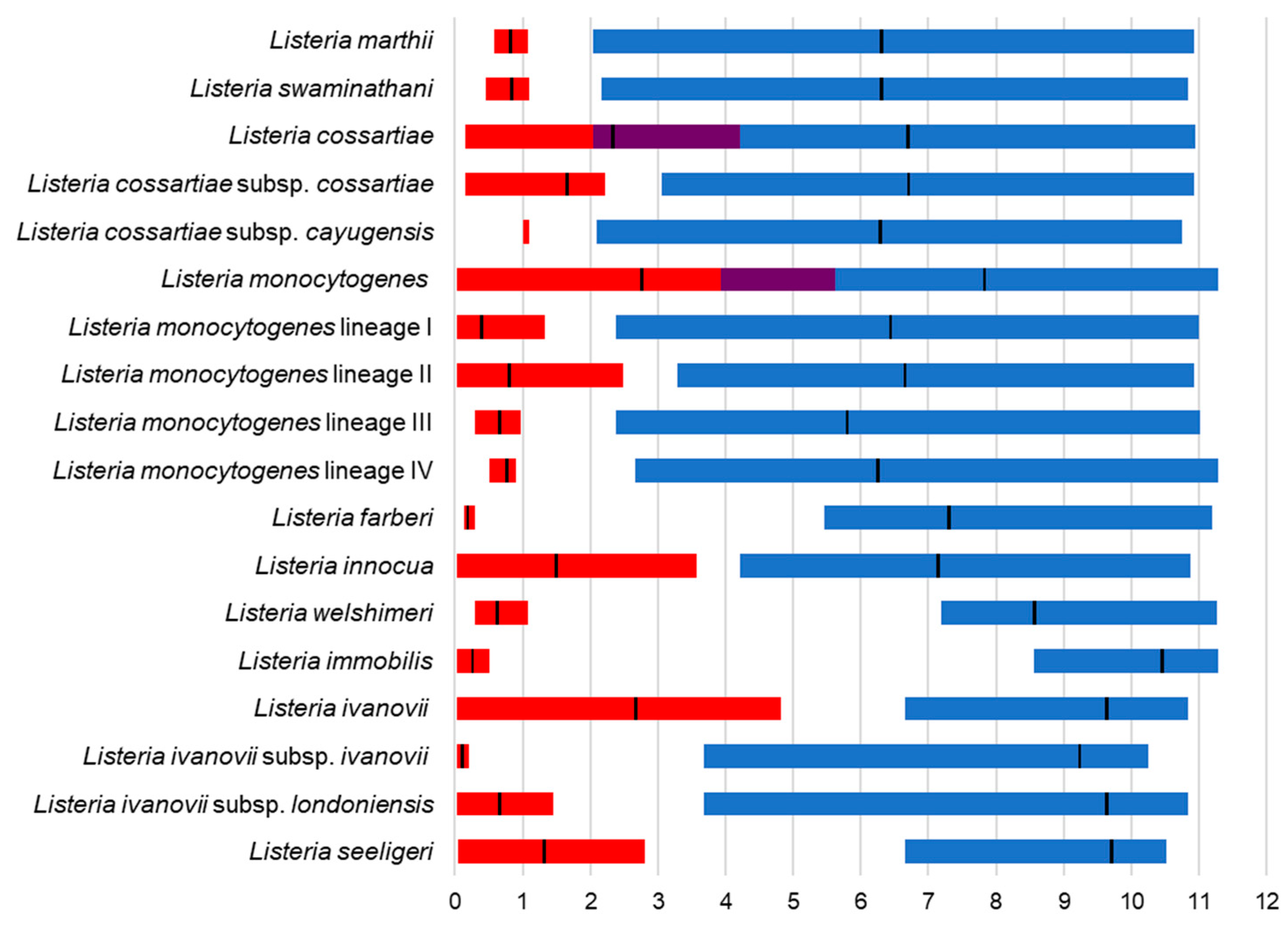
All isolates sequenced in the present study belonged to Listeria sensu stricto, which included the pathogenic species L. monocytogenes and L. ivanovii, and thus, this group was analyzed in greater depth. For this purpose, additional sequences of Listeria sensu stricto were selected and retrieved for the NCBI data. The concatenated fragments of the seven genes (gyrAcpn60parErecArpoBatpAgyrB; a total of 4375 bp) showed a sequence similarity ranging from 88.71% to 99.98%, which corresponded to 1–492 nucleotide differences, and there was a total of 1093 variable positions (24.98% of the sequence). For each Listeria sensu stricto species, ranges of intra-species and inter-species nucleotide differences were determined, and intra-species and inter-species phylogenetic depth values were calculated (Figure 3), as detailed in Materials and Methods. Ranges of intra- and inter-species nucleotide similarities (%) for the concatenated sequence only overlapped for the species L. monocytogenes and L. cossartiae. Overall, a similar picture was obtained from each single-gene phylogeny (Figures S1–S7), indicating that the obtained clusters all showed similar phylogenetic relationships, or, in other words, the genes evolved in concert. The MLPA tree derived from the alignment of the concatenated sequence (Figure 4) showed clearly defined clusters for the Listeria sensu stricto species with bootstrap values of 100%, except for L. monocytogenes that was divided into four subclusters also sustained by 100% bootstrap, which corresponded to the four genetic lineages previously described.
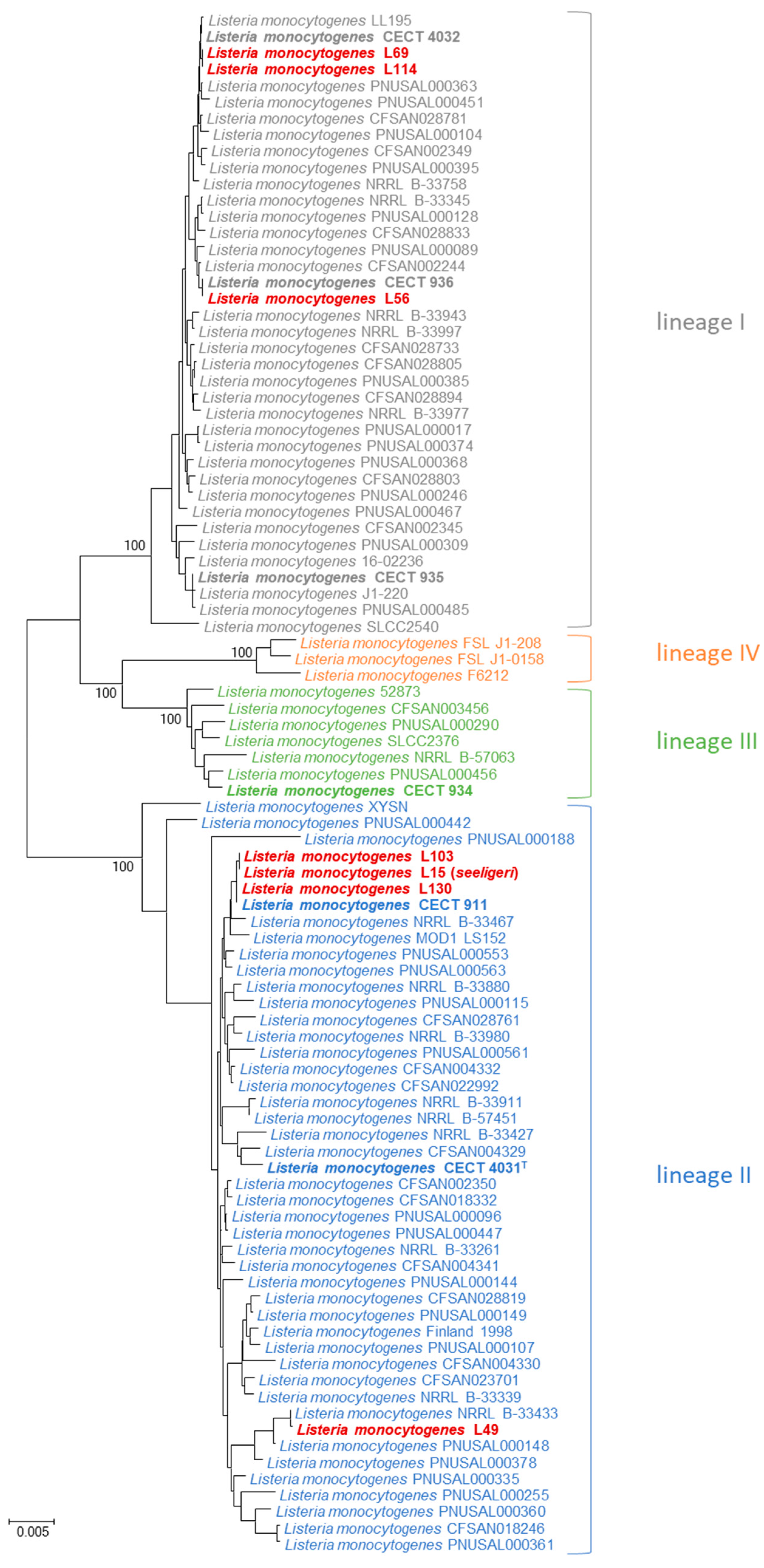
The concatenated sequence of the seven housekeeping genes was downloaded from the NCBI database for 169 L. monocytogenes strains, the genetic lineage of which had been previously determined [60]. The phylogenetic tree showed that all four L. monocytogenes lineages formed clearly defined clusters with bootstraps of 100%, confirming again that all strains were correctly clustered according to their lineage (Figure 5; only non-redundant sequences are shown). Ranges of intra- and inter-sequence similarity for each of the four lineages did not overlap (Figure 3). For L. monocytogenes strains of the four genetic lineages, the concatenated sequence showed an overall sequence similarity ranging from 94.37% to 99.98%, which corresponded to 1–245 nucleotide differences, and there was a total of 434 variable positions (9.92% of the sequence). L. ivanovii subsp. ivanovii and L. ivanovii subsp. londoniensis formed two different clusters that were quite divergent from each other. However, L. cossartiae, L. marthii, and “L. swaminathanii” were very closely related, finding that L. cossartiae subsp. cayugensis was closer to L. marthii than to L. cossartiae subsp. cossartiae.
The MLPA allowed for the identification of Listeria isolates of food origin obtained from the CECT (Figure 4). Isolates L56, L69, and L114 clustered in the MLPA with L. monocytogenes lineage I and L15, L49, L103, and L130 with L. monocytogenes lineage II. Isolates L51, L52, L123A, L138, Linn8, Linn16, and CECT 5372 clustered with L. innocua strains. CECT 5372 had been identified as L. welshimeri by the CECT. Isolates L79, Lwell6, and Lwell7 clustered with L. welshimeri. Strains CECT 939 and CECT 941, which were previously classified as L. monocytogenes, clustered with L. seeligeri. Nevertheless, CECT 939 has lately been discontinued. L. ivanovii CECT 5379 clustered with Listeria ivanovii subsp. ivanovii. L. ivanovii subsp. ivanovii CECT 5368 clustered with Listeria ivanovii subsp. londoniensis.

4. Discussion

The MLPA strategy to infer phylogenetic bacterial identification based on several housekeeping loci assumes a synchronized evolutionary pattern in genomes, where fundamental genes evolve in concert. Consequently, the concatenated multigene phylogeny can serve as a ‘mirror’ reflecting the overall relationships within the entire genomic content [42,44,48,50,51,52,53,54]. Sequences of housekeeping genes coding for proteins, particularly those involved in DNA processing (i.e., replication, transcription, translation, etc.), are phylogenetic markers suitable for species discrimination with a high resolution [55,61,62,63]. Mainly, this is because they are subjected to a degenerative code when translated into proteins, and, therefore, nucleotide changes can occur throughout the gene without consequences in the amino acid sequence. This mode of evolution contrasts with that of rRNAs, which exhibit a mosaic of discrete variation at some signature regions, making the protein-coding genes the best candidates for distinguishing closely related strains.

4.1. MLPA of the Genus Listeria

A comprehensive multilocus phylogenetic analysis (MLPA) of a diverse representation of the genus Listeria has been approached to provide a framework for simple, easy, and fast species identification from isolated colonies, as has been performed for other genera [64,65,66]. Both concatenated seven-gene MLPA and 16S rDNA phylogenetic trees showed that species of Listeria sensu stricto clustered together in a group separated from other species currently recognized as Listeria sensu lato (Figure 1 and Figure 2). This result was in agreement with previous studies using genomic data [36,37] and proposed new genera, “Murraya”, “Mesolisteria”, and “Paenilisteria”, to accommodate Listeria sensu lato species. “Murraya” only contained the species Listeria grayi. The group named “Mesolisteria” was formed by a very diverse species not able to grow at 4 °C [37], L. costaricensis, L. ilorinensis, L. fleischmannii, L. goaensis, L. floridensis, L. aquatica, L. valentina, L. thailandensis, and the non-validated species “L. kieliensis” [67], which was found highly related to L. thailandensis. And finally, the cluster containing L. booriae, L. riparia, L. rocourtiae, L. weihenstephanensis, L. grandensis, L. portnoyi, L. rustica, L. cornellensis, and L. newyorkensis corresponded to this previously proposed to be reclassified in the genus “Paenilisteria” [37]. The mere fact that the majority of species described in recent decades do not belong to the genus Listeria in the strict sense reaffirms the need to provide a framework for identifying isolates based on a tool as decisive as the MLPA. Additionally, this MLPA can also identify L. monocytogenes genetic lineages (Figure 5), being useful as an intra-species typing method, similar to MLST. The four genetic lineages have different characteristics but do not allow us to determine the serotype of the strains, as each lineage can correspond to different serotypes [12]. All cultures, isolates, and reference strains used in this study were identified by MLPA as species of Listeria sensu stricto (Figure 4 and Figure 5) and, therefore, were subjected to a deeper analysis.

4.2. MLPA Clustering of Listeria Sensu Stricto

The clustering of Listeria species based on single-gene phylogenies of gyrA, cpn60, parE, recA, rpoB, atpA, and gyrB were consistent with each other. However, the topology of the phylogenetic trees at the deep branching points (or nodes) varied, likely due to differences in mutation rates between genes. For instance, the parE-based tree showed the type strain L. monocytogenes 4031T more affiliated to the L. cossartiae, although it was branched at the expected position in all the other single-gene phylogenies. However, for a pragmatic taxonomy of the Listeria species, the MLPA approach, which grouped strains into well-defined clusters with high bootstrap support (close to 100%), was considered highly valuable. Although the concatenated sequence for some species displayed slight variations in nucleotide substitution ranges, intra-species phylogenetic depths, and inter-species phylogenetic distances (Figure 3), most species showed non-overlapping ranges of intra- and inter-species sequence similarities, indicating that they were well-delimited species, clearly separated from each other. These range values overlapped for the species L. monocytogenes and L. cossartiae, indicating that intra-species diversity was higher than inter-species relationships; for example, some strains of L. cossartiae may be more related to other species. However, if the analysis of intra- and inter-species values is performed for the two subspecies of L. cossartiae (L. cossartiae subsp. cossartiae and L. cossartiae subsp. cayugensis) or the four lineages of L. monocytogenes, no overlapping was observed indicating that they were entities clearly separated from the other species of Listeria sensu stricto. Indeed, these lineages are quite divergent and can be considered different subspecies, as proposed by Orsi et al. in 2011 [12]. L. monocytogenes XYSN strain, which has been classified as belonging to the hybrid sub-lineage II [68], clustered borderline of lineage II but was distantly related. Moreover, the two sequences available of L. cossartiae subsp. cayugensis were phylogenetically closer to L. marthii than to L. cossartiae subsp. cossartiae. L. ivanovii subsp. ivanovii and L. ivanovii subsp. londoniensis are relatively divergent (3.67–4.82% inter-cluster nucleotide substitutions in the 4375 bp concatenate sequence) and form two well-delineated clusters, supported by 100% bootstrap values in our MLPA (Figure 4). These two subspecies of L. ivanovii could be close to being considered as different species. However, L. marthii, L. cossartiae, and “L. swaminathanii” were found to be very closely related and, perhaps, in future studies, should be reclassified into a single species.

4.3. Identification of Listeria Isolates

The MLPA performed in the present study allowed for the identification of Listeria isolates subjected to analysis (Figure 4 and Figure 5). Isolates L56, L69, and L114 clustered in the MLPA with L. monocytogenes lineage I and L15, L49, L103, and L130 with L. monocytogenes lineage II. None were identified as L. monocytogenes lineages III and IV. This finding was in agreement with previous reports, indicating that most strains isolated worldwide belonged to lineages I and II [17], the first more frequently isolated from humans, while the second usually recovered from food and environmental samples [18,19]. The capacity of MLPA to discriminate between the four L. monocytogenes lineages seems of good value in ascertaining if they belong to different levels of virulence in future studies. We should remark that isolate L15 (L. monocytogenes lineage II) was received as L. seeligeri according to partial 16S sequencing. Isolates L51, L52, L123A, L138, Linn8, Linn16, and strain CECT 5372 clustered with the cluster corresponding to L. innocua. Strain L52, together with closely related L. innocua 2015L-6714, clustered at a border-line position of the cluster containing the rest of L. innocua strains; however, they clustered close to some strains of L. monocytogenes in cpn60, rpoB, and gyrB single-gene phylogenies. Strain CECT 5372, obtained from CECT as L. welshimeri clearly clustered within the L. innocua species. Isolates L79, Lwell6, and Lwell7 clustered with L. welshimeri. Strains CECT 939 and CECT 941, classified as L. monocytogenes, clustered with L. seeligeri. As a consequence, CECT 939 is no longer available. L. ivanovii CECT 5379, whose subspecies was not identified previously, clustered with L. ivanovii subsp. ivanovii. L. ivanovii subsp. ivanovii CECT 5368 clustered with L. ivanovii subsp. londoniensis. Almost the same identification results from MLPA can be obtained when using some of these single-gene phylogenies, for instance, when using recA or gyrA phylogenies. Whole-genome sequencing (WGS) is a powerful tool for new species description and phylogenetic analysis, but it is more expensive and time-consuming than MLPA for the identification of Listeria isolates. However, WGS also allows us to determine the serotype, antimicrobial-resistant genes, mobile genetic element, and virulence markers and perform clustering methods (MLPA, MLST) [10,69,70,71]. Once this MLPA framework shows all these genes evolve in concert (i.e., the same phylogenetic relationships were obtained), single-gene sequencing may be very useful as a fast, cheap, and simple identification approach for preliminary screening of isolates. The simultaneous sequencing of a second housekeeping gene, as confirmatory identification, is highly recommended.

5. Conclusions

Listeria currently comprises a complex group of species, some of which are highly virulent, often transmitted in the food chain, and of concern to public health. During the last decades, a number of new taxa have been wrongly described as belonging to the genus Listeria. Therefore, unequivocal and fast identification of isolates represents a challenge, particularly in food control. Once Listeria spp. has been identified and isolated following the international norm EN ISO 11290-1:2017 [40], this MLPA approach could be employed for species identification, as it has demonstrated that simple housekeeping gene sequencing represented a powerful and affordable methodology for accurate species identification. All isolates identified in the present study belonged to species of Listeria sensu stricto; in particular, some of them were classified as L. monocytogenes lineages I and II, both considered virulent phylogenetic groups typically found in clinical and food samples.

Supplementary Materials

The following supporting information can be downloaded at https://www.mdpi.com/article/10.3390/microbiolres15040142/s1, Figures S1–S7: Single-gene phylogenetic neighbor-joining trees of Listeria sensu stricto species; Table S1: List of Listeria strains used for sequencing.

Author Contributions

Conceptualization, A.M.-M., A.N. and C.M.-P.; methodology, A.M.-M., A.N. and C.M.-P.; formal analysis, A.M.-M., A.N. and C.M.-P.; writing—original draft, A.M.-M., A.N. and C.M.-P.; writing—review and editing, A.M.-M., A.N. and C.M.-P. All authors contributed equally to this work. All authors have read and agreed to the published version of the manuscript.

Funding

This research was partially funded by Instituto Valenciano de Competitividad Empresarial (IVACE), program PIDI-CV, grant number IMIDTA/2017/53.

Institutional Review Board Statement

Not applicable.

Informed Consent Statement

Not applicable.

Data Availability Statement

Gene sequences are publicly available at GenBank with accession numbers PP970580-PP970901.

Acknowledgments

Thanks to GPS™ for their financial support. Authors are grateful to Patricia Elizaquivel and Rosa Aznar (University of Valencia, Spain) for sharing Listeria isolates.

Conflicts of Interest

A.M.-M. is a scientific advisor at Genetic PCR Solutions™. A.N. and C.M.-P. are employees of Genetic PCR Solutions™. The research was conducted as an academic R&D collaboration between university and Genetic PCR Solutions™. No commercial products or kits are associated with this study, and no commercial interest is perceived.

References

  1. Freitag, N.E.; Port, G.C.; Miner, M.D. Listeria Monocytogenes—from Saprophyte to Intracellular Pathogen. Nat. Rev. Microbiol. 2009, 7, 623. [Google Scholar] [CrossRef] [PubMed]
  2. Linke, K.; Rückerl, I.; Brugger, K.; Karpiskova, R.; Walland, J.; Muri-Klinger, S.; Tichy, A.; Wagner, M.; Stessl, B. Reservoirs of Listeria Species in Three Environmental Ecosystems. Appl. Environ. Microbiol. 2014, 80, 5583–5592. [Google Scholar] [CrossRef] [PubMed]
  3. Hain, T.; Chatterjee, S.S.; Ghai, R.; Kuenne, C.T.; Billion, A.; Steinweg, C.; Domann, E.; Kärst, U.; Jänsch, L.; Wehland, J.; et al. Pathogenomics of Listeria spp. Int. J. Med. Microbiol. IJMM 2007, 297, 541–557. [Google Scholar] [CrossRef] [PubMed]
  4. McLauchlin, J.; Rees, C.E.D. Listeria. In Bergey’s Manual of Systematics of Archaea and Bacteria; John Wiley & Sons, Ltd.: Oxford, UK, 2015; pp. 1–29. ISBN 978-1-118-96060-8. [Google Scholar]
  5. Favaro, M.; Sarmati, L.; Sancesario, G.; Fontana, C. First Case of Listeria Innocua Meningitis in a Patient on Steroids and Eternecept. JMM Case Rep. 2014, 1, e003103. [Google Scholar] [CrossRef]
  6. Perrin, M.; Bemer, M.; Delamare, C. Fatal Case of Listeria Innocua Bacteremia. J. Clin. Microbiol. 2003, 41, 5308–5309. [Google Scholar] [CrossRef]
  7. Rapose, A.; Lick, S.D.; Ismail, N. Listeria Grayi Bacteremia in a Heart Transplant Recipient. Transpl. Infect. Dis. 2008, 10, 434–436. [Google Scholar] [CrossRef]
  8. Guillet, C.; Join-Lambert, O.; Monnier, A.L.; Leclercq, A.; Mechaï, F.; Mamzer-Bruneel, M.-F.; Bielecka, M.K.; Scortti, M.; Disson, O.; Berche, P.; et al. Human Listeriosis Caused by Listeria ivanovii. Emerg. Infect. Dis. 2010, 16, 136. [Google Scholar] [CrossRef]
  9. European Food Safety Authority. European Centre for Disease Prevention and Control (ECDC) The European Union One Health 2022 Zoonoses Report. EFSA J. 2023, 21, e8442. [Google Scholar] [CrossRef]
  10. Food and Agriculture Organization; World Health Organization. Listeria Monocytogenes in Ready-to-Eat (RTE) Foods: Attribution, Characterization and Monitoring: Meeting Report; FAO; WHO: Rome, Italy, 2022; ISBN 978-92-5-136983-8.
  11. Ferreira, V.; Wiedmann, M.; Teixeira, P.; Stasiewicz, M.J. Listeria Monocytogenes Persistence in Food-Associated Environments: Epidemiology, Strain Characteristics, and Implications for Public Health. J. Food Prot. 2014, 77, 150–170. [Google Scholar] [CrossRef]
  12. Orsi, R.H.; den Bakker, H.C.; Wiedmann, M. Listeria Monocytogenes Lineages: Genomics, Evolution, Ecology, and Phenotypic Characteristics. Int. J. Med. Microbiol. IJMM 2011, 301, 79–96. [Google Scholar] [CrossRef]
  13. den Bakker, H.C.; Didelot, X.; Fortes, E.D.; Nightingale, K.K.; Wiedmann, M. Lineage Specific Recombination Rates and Microevolution in Listeria Monocytogenes. BMC Evol. Biol. 2008, 8, 277. [Google Scholar] [CrossRef] [PubMed]
  14. Gray, M.J.; Zadoks, R.N.; Fortes, E.D.; Dogan, B.; Cai, S.; Chen, Y.; Scott, V.N.; Gombas, D.E.; Boor, K.J.; Wiedmann, M. Listeria Monocytogenes Isolates from Foods and Humans Form Distinct but Overlapping Populations. Appl. Environ. Microbiol. 2004, 70, 5833–5841. [Google Scholar] [CrossRef] [PubMed]
  15. Nightingale, K.K.; Windham, K.; Wiedmann, M. Evolution and Molecular Phylogeny of Listeria Monocytogenes Isolated from Human and Animal Listeriosis Cases and Foods. J. Bacteriol. 2005, 187, 5537–5551. [Google Scholar] [CrossRef] [PubMed]
  16. Ragon, M.; Wirth, T.; Hollandt, F.; Lavenir, R.; Lecuit, M.; Le Monnier, A.; Brisse, S. A New Perspective on Listeria Monocytogenes Evolution. PLoS Pathog. 2008, 4, e1000146. [Google Scholar] [CrossRef]
  17. Chenal-Francisque, V.; Lopez, J.; Cantinelli, T.; Caro, V.; Tran, C.; Leclercq, A.; Lecuit, M.; Brisse, S. Worldwide Distribution of Major Clones of Listeria Monocytogenes. Emerg. Infect. Dis. 2011, 17, 1110–1112. [Google Scholar] [CrossRef]
  18. Balandyté, L.; Brodard, I.; Frey, J.; Oevermann, A.; Abril, C. Ruminant Rhombencephalitis-Associated Listeria Monocytogenes Alleles Linked to a Multilocus Variable-Number Tandem-Repeat Analysis Complex. Appl. Environ. Microbiol. 2011, 77, 8325–8335. [Google Scholar] [CrossRef]
  19. Maury, M.M.; Tsai, Y.-H.; Charlier, C.; Touchon, M.; Chenal-Francisque, V.; Leclercq, A.; Criscuolo, A.; Gaultier, C.; Roussel, S.; Brisabois, A.; et al. Uncovering Listeria Monocytogenes Hypervirulence by Harnessing Its Biodiversity. Nat. Genet. 2016, 48, 308–313. [Google Scholar] [CrossRef]
  20. Kuenne, C.; Billion, A.; Mraheil, M.A.; Strittmatter, A.; Daniel, R.; Goesmann, A.; Barbuddhe, S.; Hain, T.; Chakraborty, T. Reassessment of the Listeria Monocytogenes Pan-Genome Reveals Dynamic Integration Hotspots and Mobile Genetic Elements as Major Components of the Accessory Genome. BMC Genom. 2013, 14, 47. [Google Scholar] [CrossRef]
  21. Raufu, I.A.; Moura, A.; Vales, G.; Ahmed, O.A.; Aremu, A.; Thouvenot, P.; Tessaud-Rita, N.; Bracq-Dieye, H.; Krishnamurthy, R.; Leclercq, A.; et al. Listeria ilorinensis sp. Nov., Isolated from Cow Milk Cheese in Nigeria. Int. J. Syst. Evol. Microbiol. 2022, 72, 005437. [Google Scholar] [CrossRef]
  22. Carlin, C.R.; Liao, J.; Weller, D.; Guo, X.; Orsi, R.; Wiedmann, M. Listeria cossartiae sp. Nov., Listeria immobilis sp. Nov., Listeria portnoyi sp. Nov. and Listeria rustica sp. Nov., Isolated from Agricultural Water and Natural Environments. Int. J. Syst. Evol. Microbiol. 2021, 71, 004795. [Google Scholar] [CrossRef]
  23. Quereda, J.J.; Leclercq, A.; Moura, A.; Vales, G.; Gómez-Martín, Á.; García-Muñoz, Á.; Thouvenot, P.; Tessaud-Rita, N.; Bracq-Dieye, H.; Lecuit, M. Listeria valentina sp. Nov., Isolated from a Water Trough and the Faeces of Healthy Sheep. Int. J. Syst. Evol. Microbiol. 2020, 70, 5868–5879. [Google Scholar] [CrossRef] [PubMed]
  24. Leclercq, A.; Moura, A.; Vales, G.; Tessaud-Rita, N.; Aguilhon, C.; Lecuit, M. Listeria thailandensis sp. Nov. Int. J. Syst. Evol. Microbiol. 2019, 69, 74–81. [Google Scholar] [CrossRef] [PubMed]
  25. Doijad, S.P.; Poharkar, K.V.; Kale, S.B.; Kerkar, S.; Kalorey, D.R.; Kurkure, N.V.; Rawool, D.B.; Malik, S.V.S.; Ahmad, R.Y.; Hudel, M.; et al. Listeria goaensis sp. Nov. Int. J. Syst. Evol. Microbiol. 2018, 68, 3285–3291. [Google Scholar] [CrossRef] [PubMed]
  26. Núñez-Montero, K.; Leclercq, A.; Moura, A.; Vales, G.; Peraza, J.; Pizarro-Cerdá, J.; Lecuit, M. Listeria costaricensis sp. Nov. Int. J. Syst. Evol. Microbiol. 2018, 68, 844–850. [Google Scholar] [CrossRef]
  27. Weller, D.; Andrus, A.; Wiedmann, M.; den Bakker, H.C. Listeria booriae sp. Nov. and Listeria newyorkensis sp. Nov., from Food Processing Environments in the USA. Int. J. Syst. Evol. Microbiol. 2015, 65, 286–292. [Google Scholar] [CrossRef]
  28. den Bakker, H.C.; Warchocki, S.; Wright, E.M.; Allred, A.F.; Ahlstrom, C.; Manuel, C.S.; Stasiewicz, M.J.; Burrell, A.; Roof, S.; Strawn, L.K.; et al. Listeria floridensis sp. Nov., Listeria Aquatica sp. Nov., Listeria cornellensis sp. Nov., Listeria Riparia sp. Nov. and Listeria grandensis sp. Nov., from Agricultural and Natural Environments. Int. J. Syst. Evol. Microbiol. 2014, 64, 1882–1889. [Google Scholar] [CrossRef]
  29. Lang Halter, E.; Neuhaus, K.; Scherer, S. Listeria weihenstephanensis sp. Nov., Isolated from the Water Plant Lemna Trisulca Taken from a Freshwater Pond. Int. J. Syst. Evol. Microbiol. 2013, 63, 641–647. [Google Scholar] [CrossRef]
  30. Bertsch, D.; Rau, J.; Eugster, M.R.; Haug, M.C.; Lawson, P.A.; Lacroix, C.; Meile, L. Listeria fleischmannii sp. Nov., Isolated from Cheese. Int. J. Syst. Evol. Microbiol. 2013, 63, 526–532. [Google Scholar] [CrossRef]
  31. den Bakker, H.C.; Manuel, C.S.; Fortes, E.D.; Wiedmann, M.; Nightingale, K.K. Genome Sequencing Identifies Listeria fleischmannii Subsp. Coloradonensis Subsp. Nov., Isolated from a Ranch. Int. J. Syst. Evol. Microbiol. 2013, 63, 3257–3268. [Google Scholar] [CrossRef]
  32. Graves, L.M.; Helsel, L.O.; Steigerwalt, A.G.; Morey, R.E.; Daneshvar, M.I.; Roof, S.E.; Orsi, R.H.; Fortes, E.D.; Milillo, S.R.; den Bakker, H.C.; et al. Listeria marthii sp. Nov., Isolated from the Natural Environment, Finger Lakes National Forest. Int. J. Syst. Evol. Microbiol. 2010, 60, 1280–1288. [Google Scholar] [CrossRef]
  33. Leclercq, A.; Clermont, D.; Bizet, C.; Grimont, P.A.D.; Le Flèche-Matéos, A.; Roche, S.M.; Buchrieser, C.; Cadet-Daniel, V.; Le Monnier, A.; Lecuit, M.; et al. Listeria rocourtiae sp. Nov. Int. J. Syst. Evol. Microbiol. 2010, 60, 2210–2214. [Google Scholar] [CrossRef] [PubMed]
  34. Carlin, C.R.; Liao, J.; Hudson, L.K.; Peters, T.L.; Denes, T.G.; Orsi, R.H.; Guo, X.; Wiedmann, M. Soil Collected in the Great Smoky Mountains National Park Yielded a Novel Listeria sensu stricto species, L. swaminathanii. Microbiol. Spectr. 2022, 10, e00442-22. [Google Scholar] [CrossRef] [PubMed]
  35. Hudson, L.K.; Chaggar, H.K.; Schamp, C.N.; Claxton, M.L.; Bryan, D.W.; Peters, T.L.; Song, Y.; Carlin, C.R.; den Bakker, H.C.; Denes, T.G. Phenotypic Characterization and Analysis of Complete Genomes of Two Distinct Strains of the Proposed Species “L. swaminathanii”. Sci. Rep. 2022, 12, 9137. [Google Scholar] [CrossRef]
  36. Chiara, M.; Caruso, M.; D’Erchia, A.M.; Manzari, C.; Fraccalvieri, R.; Goffredo, E.; Latorre, L.; Miccolupo, A.; Padalino, I.; Santagada, G.; et al. Comparative Genomics of Listeria Sensu Lato: Genus-Wide Differences in Evolutionary Dynamics and the Progressive Gain of Complex, Potentially Pathogenicity-Related Traits through Lateral Gene Transfer. Genome Biol. Evol. 2015, 7, 2154–2172. [Google Scholar] [CrossRef]
  37. Orsi, R.H.; Wiedmann, M. Characteristics and Distribution of Listeria spp., Including Listeria Species Newly Described since 2009. Appl. Microbiol. Biotechnol. 2016, 100, 5273–5287. [Google Scholar] [CrossRef]
  38. Rocourt, J.; Wehmeyer, U.; Stackebrandt, E. Transfer of Listeria dentrificans to a New Genus, Jonesia gen. Nov., as Jonesia denitrificans Comb. Nov. Int. J. Syst. Evol. Microbiol. 1987, 37, 266–270. [Google Scholar] [CrossRef]
  39. Rocourt, J.; Boerlin, P.; Grimont, F.; Jacquet, C.; Piffaretti, J.-C. Assignment of Listeria Grayi and Listeria Murrayi to a Single Species, Listeria Grayi, with a Revised Description of Listeria Grayi. Int. J. Syst. Evol. Microbiol. 1992, 42, 171–174. [Google Scholar] [CrossRef]
  40. ISO 11290-1:2017(En); Microbiology of the Food Chain—Horizontal Method for the Detection and Enumeration of Listeria Monocytogenes and of Listeria spp.—Part 1: Detection Method. ISO: Geneva, Switzerland, 2017. Available online: https://www.iso.org/obp/ui/en/#iso:std:iso:11290:-1:ed-2:v1:en (accessed on 11 June 2024).
  41. Gevers, D.; Cohan, F.M.; Lawrence, J.G.; Spratt, B.G.; Coenye, T.; Feil, E.J.; Stackebrandt, E.; de Peer, Y.V.; Vandamme, P.; Thompson, F.L.; et al. Re-Evaluating Prokaryotic Species. Nat. Rev. Microbiol. 2005, 3, 733–739. [Google Scholar] [CrossRef]
  42. Martinez-Murcia, A.J.; Monera, A.; Saavedra, M.J.; Oncina, R.; Lopez-Alvarez, M.; Lara, E.; Figueras, M.J. Multilocus Phylogenetic Analysis of the Genus Aeromonas. Syst. Appl. Microbiol. 2011, 34, 189–199. [Google Scholar] [CrossRef]
  43. Glaeser, S.P.; Kämpfer, P. Multilocus Sequence Analysis (MLSA) in Prokaryotic Taxonomy. Syst. Appl. Microbiol. 2015, 38, 237–245. [Google Scholar] [CrossRef]
  44. Soler, L.; Yáñez, M.A.; Chacon, M.R.; Aguilera-Arreola, M.G.; Catalán, V.; Figueras, M.J.; Martínez-Murcia, A.J. Phylogenetic Analysis of the Genus Aeromonas Based on Two Housekeeping Genes. Int. J. Syst. Evol. Microbiol. 2004, 54, 1511–1519. [Google Scholar] [CrossRef] [PubMed]
  45. Maiden, M.C.J. Multilocus Sequence Typing of Bacteria. Annu. Rev. Microbiol. 2006, 60, 561–588. [Google Scholar] [CrossRef] [PubMed]
  46. Maiden, M.C.J.; Bygraves, J.A.; Feil, E.; Morelli, G.; Russell, J.E.; Urwin, R.; Zhang, Q.; Zhou, J.; Zurth, K.; Caugant, D.A.; et al. Multilocus Sequence Typing: A Portable Approach to the Identification of Clones within Populations of Pathogenic Microorganisms. Proc. Natl. Acad. Sci. USA 1998, 95, 3140–3145. [Google Scholar] [CrossRef]
  47. Sullivan, C.B.; Diggle, M.A.; Clarke, S.C. Multilocus Sequence Typing. Mol. Biotechnol. 2005, 29, 245–254. [Google Scholar] [CrossRef] [PubMed]
  48. Navarro, A.; Martínez-Murcia, A. Phylogenetic Analyses of the Genus Aeromonas Based on Housekeeping Gene Sequencing and Its Influence on Systematics. J. Appl. Microbiol. 2018, 125, 622–631. [Google Scholar] [CrossRef]
  49. Gadagkar, S.R.; Rosenberg, M.S.; Kumar, S. Inferring Species Phylogenies from Multiple Genes: Concatenated Sequence Tree versus Consensus Gene Tree. J. Exp. Zoolog. B Mol. Dev. Evol. 2005, 304B, 64–74. [Google Scholar] [CrossRef]
  50. Figueras, M.; Hidalgo, R.; Collado, L.; Martinez-Murcia, A. Recommendations for a New Bacterial Species Description Based on Analyses of the Unrelated Genera Aeromonas and Arcobacter. Bull. BISMiS 2011, 2, 1–16. [Google Scholar]
  51. Labeda, D.P. Multilocus Sequence Analysis of Phytopathogenic Species of the Genus Streptomyces. Int. J. Syst. Evol. Microbiol. 2011, 61, 2525–2531. [Google Scholar] [CrossRef] [PubMed]
  52. Margos, G.; Hojgaard, A.; Lane, R.S.; Cornet, M.; Fingerle, V.; Rudenko, N.; Ogden, N.; Aanensen, D.M.; Fish, D.; Piesman, J. Multilocus Sequence Analysis of Borrelia Bissettii Strains from North America Reveals a New Borrelia Species, Borrelia Kurtenbachii. Ticks Tick-Borne Dis. 2010, 1, 151–158. [Google Scholar] [CrossRef] [PubMed]
  53. Pascual, J.; Macián, M.C.; Arahal, D.R.; Garay, E.; Pujalte, M.J. Multilocus Sequence Analysis of the Central Clade of the Genus Vibrio by Using the 16S rRNA, recA, pyrH, rpoD, gyrB, rctB and toxR Genes. Int. J. Syst. Evol. Microbiol. 2010, 60, 154–165. [Google Scholar] [CrossRef] [PubMed]
  54. Richter, D.; Postic, D.; Sertour, N.; Livey, I.; Matuschka, F.-R.; Baranton, G. Delineation of Borrelia Burgdorferi Sensu Lato Species by Multilocus Sequence Analysis and Confirmation of the Delineation of Borrelia spielmanii sp. Nov. Int. J. Syst. Evol. Microbiol. 2006, 56, 873–881. [Google Scholar] [CrossRef] [PubMed]
  55. Yáñez, M.A.; Catalán, V.; Apráiz, D.; Figueras, M.J.; Martínez-Murcia, A.J. Phylogenetic Analysis of Members of the Genus Aeromonas Based on gyrB Gene Sequences. Int. J. Syst. Evol. Microbiol. 2003, 53, 875–883. [Google Scholar] [CrossRef]
  56. Tamura, K.; Peterson, D.; Peterson, N.; Stecher, G.; Nei, M.; Kumar, S. MEGA5: Molecular Evolutionary Genetics Analysis Using Maximum Likelihood, Evolutionary Distance, and Maximum Parsimony Methods. Mol. Biol. Evol. 2011, 28, 2731–2739. [Google Scholar] [CrossRef] [PubMed]
  57. Thompson, J.D.; Higgins, D.G.; Gibson, T.J. CLUSTAL W: Improving the Sensitivity of Progressive Multiple Sequence Alignment through Sequence Weighting, Position-Specific Gap Penalties and Weight Matrix Choice. Nucleic Acids Res. 1994, 22, 4673–4680. [Google Scholar] [CrossRef] [PubMed]
  58. Kimura, M. A Simple Method for Estimating Evolutionary Rates of Base Substitutions through Comparative Studies of Nucleotide Sequences. J. Mol. Evol. 1980, 16, 111–120. [Google Scholar] [CrossRef] [PubMed]
  59. Saitou, N.; Nei, M. The Neighbor-Joining Method: A New Method for Reconstructing Phylogenetic Trees. Mol. Biol. Evol. 1987, 4, 406–425. [Google Scholar] [CrossRef]
  60. Pirone-Davies, C.; Chen, Y.; Pightling, A.; Ryan, G.; Wang, Y.; Yao, K.; Hoffmann, M.; Allard, M.W. Genes Significantly Associated with Lineage II Food Isolates of Listeria Monocytogenes. BMC Genom. 2018, 19, 708. [Google Scholar] [CrossRef]
  61. Yamamoto, S.; Harayama, S. PCR Amplification and Direct Sequencing of gyrB Genes with Universal Primers and Their Application to the Detection and Taxonomic Analysis of Pseudomonas Putida Strains. Appl. Environ. Microbiol. 1995, 61, 1104–1109. [Google Scholar] [CrossRef]
  62. Huang, W.M. Bacterial Diversity Based on Type II DNA Topoisomerase Genes. Annu. Rev. Genet. 1996, 30, 79–107. [Google Scholar] [CrossRef]
  63. Stackebrandt, E.; Frederiksen, W.; Garrity, G.M.; Grimont, P.A.D.; Kämpfer, P.; Maiden, M.C.J.; Nesme, X.; Rosselló-Mora, R.; Swings, J.; Trüper, H.G.; et al. Report of the Ad Hoc Committee for the Re-Evaluation of the Species Definition in Bacteriology. Int. J. Syst. Evol. Microbiol. 2002, 52, 1043–1047. [Google Scholar] [CrossRef]
  64. Stevanović, O.; Milanov, D.; Prošić, I.; Gajdov, V.; Nedić, D.; Sladojević, Ž.; Radalj, A. Multilocus Sequence Analysis (MLSA) of a Nocardia Cyriacigeorgica Strain Causing Severe Bovine Mastitis in Bosnia and Herzegovina. Acta Vet. Hung. 2023, 71, 65–70. [Google Scholar] [CrossRef] [PubMed]
  65. Rahman, M.S.; Currò, S.; Carraro, L.; Cardazzo, B.; Balzan, S.; Novelli, E.; Fontana, F.; Caburlotto, G.; Manfrin, A.; Fasolato, L. Retrospective Analysis of Vibrio spp. Isolated from Marketed Crustaceans Using Multilocus Sequence Analysis. Ital. J. Food Saf. 2023, 12, 11045. [Google Scholar] [CrossRef] [PubMed]
  66. Meghdadi, H.; Khosravi, A.D.; Hashemzadeh, M.; Tabandeh, M.R. New Design of Multilocus Sequence Analysis of rpoB, ssrA, Tuf, atpE, Ku, and dnaK for Identification of Mycobacterium Species. Mol. Biol. Rep. 2022, 49, 7967–7977. [Google Scholar] [CrossRef] [PubMed]
  67. Schardt, J. Characterization of Listeria Sensu Stricto Specific Genes Involved in Colonization of the Gastrointestinal Tract by Listeria monocytogenes. Doctoral Dissertation, Technische Universität München, München, Germany, 2018. [Google Scholar]
  68. Yin, Y.; Yao, H.; Doijad, S.; Kong, S.; Shen, Y.; Cai, X.; Tan, W.; Wang, Y.; Feng, Y.; Ling, Z.; et al. A Hybrid Sub-Lineage of Listeria Monocytogenes Comprising Hypervirulent Isolates. Nat. Commun. 2019, 10, 4283. [Google Scholar] [CrossRef] [PubMed]
  69. Daza Prieto, B.; Pietzka, A.; Martinovic, A.; Ruppitsch, W.; Zuber Bogdanovic, I. Surveillance and Genetic Characterization of Listeria Monocytogenes in the Food Chain in Montenegro during the Period 2014–2022. Front. Microbiol. 2024, 15, 1418333. [Google Scholar] [CrossRef] [PubMed]
  70. Nouws, S.; Bogaerts, B.; Verhaegen, B.; Denayer, S.; Crombé, F.; De Rauw, K.; Piérard, D.; Marchal, K.; Vanneste, K.; Roosens, N.H.C.; et al. The Benefits of Whole Genome Sequencing for Foodborne Outbreak Investigation from the Perspective of a National Reference Laboratory in a Smaller Country. Foods 2020, 9, 1030. [Google Scholar] [CrossRef]
  71. Mispelaere, M.; De Rop, A.-S.; Hermans, C.; De Maeseneire, S.L.; Soetaert, W.K.; De Mol, M.L.; Hulpiau, P. Whole Genome–Based Comparative Analysis of the Genus Streptomyces Reveals Many Misclassifications. Appl. Microbiol. Biotechnol. 2024, 108, 453. [Google Scholar] [CrossRef]
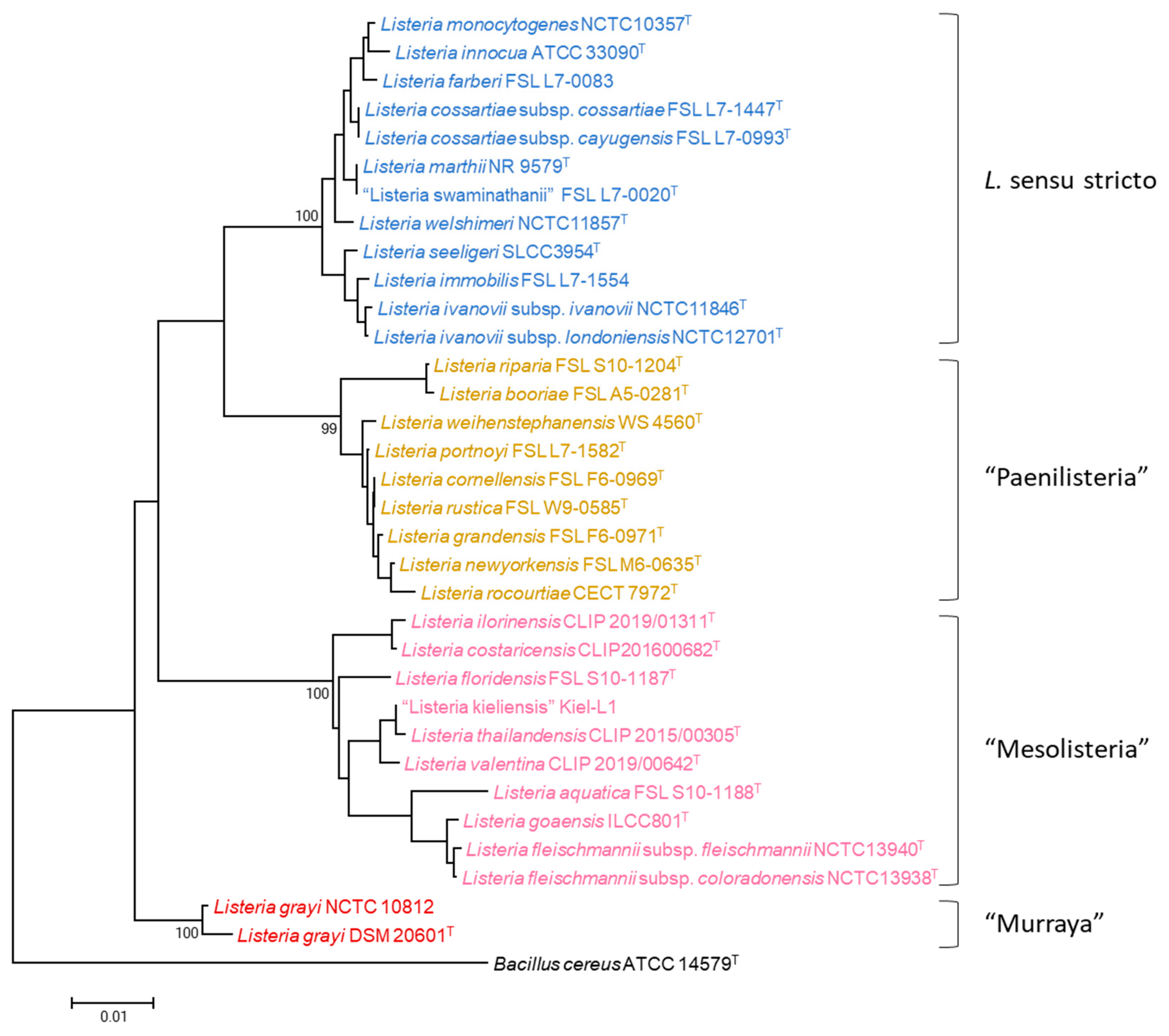
Figure 1. Neighbor-joining phylogenetic tree based on the analysis of 16S rRNA gene sequences (935 bp) of all described Listeria sensu stricto and Listeria sensu lato species, routed using Bacillus cereus. Numbers at nodes indicate bootstrap values (percentage of 1000 replicates). T—type strains.
Microbiolres 15 00142 g001
Figure 2. Neighbor-joining phylogenetic tree based on the MLPA from seven concatenated housekeeping genes (gyrA, cpn60, parE, recA, rpoB, atpA and gyrB; a total of 4375 bp) of strains of all described Listeria sensu stricto and sensu lato species, Bacillus cereus, Brochothrix thermosphacta, and Streptococcus pneumoniae. Numbers at nodes indicate bootstrap values (percentage of 1000 replicates). Strains sequenced in this study are shown in bold. T—type strains.
Microbiolres 15 00142 g002
Figure 3. Graphical representation of the ranges of intra- and inter-species nucleotide substitution percentages in red and blue bars, respectively, and intra- and inter-species phylogenetic depth (black) for the concatenated seven-gene sequence, calculated for all Listeria sensu stricto species and subspecies and L. monocytogenes genetic lineages.
Microbiolres 15 00142 g003
Figure 4. Neighbor-joining phylogenetic tree based on the MLPA from seven concatenated housekeeping genes (gyrA, cpn60, parE, recA, rpoB, atpA, and gyrB; a total of 4375 bp) of strains of all described Listeria sensu stricto, including non-characterized isolates. Numbers at nodes indicate bootstrap values (percentage of 1000 replicates). Strains sequenced in this study are shown in bold and Listeria isolates identified in this study are shown in red. T—type strains.
Microbiolres 15 00142 g004
Figure 5. Neighbor-joining phylogenetic tree based on the MLPA from seven concatenated housekeeping genes (gyrA, cpn60, parE, recA, rpoB, atpA, and gyrB; a total of 4375 bp) of strains from the four L. monocytogenes genetic lineages. Numbers at nodes indicate bootstrap values (percentage of 1000 replicates). Strains sequenced in this study are shown in bold and Listeria isolates identified in this study are shown in red. T—type strains.
Microbiolres 15 00142 g005
Table 1. Name of the genes used in this MLPA study, length of the amplified fragments, and primer sequences.
LocusEncoded ProteinAmplified FragmentPrimers CodeSequence (5′–3′)
gyrADNA gyrase subunit A971 bpLis-gyrA-3FGATGGACATGGTAACTTTG
Lis-gyrA-13R2CGCATATCTAAAATGGCTTG
cpn60chaperonin-60931 bpLis-cpn60-2FGAATTAGAAGACCCATTTGA
Lis-cpn60-11RGTTTTGCTAAACGTTCTTGT
parEDNA topoisomerase IV subunit B928 bpLis-parE-0F’AATGATGATTCTATTCAGGTGC
Lis-parE-9R’CGCGAATATCGCTACCYTC
recAhomologous recombination factor757 bpLis-recA-1FTGAAAAACAATTCGGTAAAGG
Lis-recA-8RTGARATACCTTCWCCGTACA
rpoBDNA-directed RNA polymerase subunit beta1021 bpLis-rpoB-23FAGGATGCGATCATCATGAG
Lis-rpoB-33RGCTTCGTAAGTTTTCACACG
atpAF0F1 ATP synthase subunit alpha947 bpLis-atpA-2FTATGGCCCAAAACTTAGAA
Lis-atpA-11RTTTTCATTGCTTTAATTTGCG
gyrBDNA gyrase subunit B646 bpLis-gyrB-4FGCGGCGGCGGATATAAAGTA
Lis-gyrB-10RCCTTCACGAACATCTTCACC
Disclaimer/Publisher’s Note: The statements, opinions and data contained in all publications are solely those of the individual author(s) and contributor(s) and not of MDPI and/or the editor(s). MDPI and/or the editor(s) disclaim responsibility for any injury to people or property resulting from any ideas, methods, instructions or products referred to in the content.