Article

Candida auris Antifungal Resistance, Virulence and Susceptibility to a Novel Nitric Oxide-Releasing Microparticle and Its Correlations to Clade Identification

by
Alessandro F. Valdez
1,2,†,
Flora Bohner
1,†,
Joshua P. Goldman
1,
Ali B. Jaquiery
1,
Eduardo V. C. do Amaral
2,
Dario Correa-Junior
1,3,
Andrew Draganski
4,
Leonardo Nimrichter
2,
Joshua D. Nosanchuk
1 and
Daniel Zamith-Miranda
1,*
1
Departments of Medicine (Division of Infectious Diseases) and Microbiology and Immunology, Albert Einstein College of Medicine, Bronx 10461, NY, USA
2
Departamento de Microbiologia Geral, Instituto de Microbiologia Paulo de Goés, Universidade Federal do Rio de Janeiro, Rio de Janeiro 21941-902, RJ, Brazil
3
Biologia Celular e Parasitologia, Instituto de Biofisica Carlos Chagas Filho, Universidade Federal do Rio de Janeiro, Rio de Janeiro 21941-902, RJ, Brazil
4
Zylö Therapeutics, Greenville, SC 29607, USA
*
Author to whom correspondence should be addressed.
These authors contributed equally to this work.
Microbiol. Res. 2025, 16(1), 15; https://doi.org/10.3390/microbiolres16010015
Submission received: 8 November 2024 / Revised: 3 January 2025 / Accepted: 9 January 2025 / Published: 11 January 2025

Abstract

:
Candida auris is a globally emerging pathogenic fungus described in Japan in 2009. This fungus has been identified mainly in nosocomial environments, associated with a high frequency of multidrug-resistant strains, and mortality rates reach 60%. C. auris is divided into 6 biogeographic clades, and there is a correlation between the clades and resistance against antifungals. In the current report, 8 strains of C. auris isolated in the Montefiore Medical Center, Bronx were analyzed to assess their clade (via ClaID) and common molecular determinants of antifungal resistance. We assessed antifungal resistance as well as the efficacy of a novel nitric oxide-donating microparticle as an alternative approach against C. auris in vitro through microplate susceptibility tests. Virulence was also determined in a Galleria mellonella model. Our results indicate that 7 out of 8 strains, belonging to clade 1, were resistant to fluconazole, while clade 2 was susceptible. Additionally, the clade 2 strain was more susceptible to treatment with the microparticle, while also being more virulent in an invertebrate model of infection. Our findings were then correlated to visualize parallels between clade identification and resistance/virulence patterns.

1. Introduction

Candida auris is a multidrug-resistant pathogenic fungus first described in 2009 [1]. C. auris has emerged globally with four major geographic-specific clades—clade 1 (South Asian), clade 2 (East Asian), clade 3 (South African), and clade 4 (South American) [2], and recently, the fifth (Iran) [3] and sixth (Singapore) [4] clades were described. Notably, these clades also present different resistance profiles [5,6,7,8,9,10]. Misidentification of this pathogen due to its phenotypic similarities to C. haemulonii has led to its relatively late description and unclear distribution data [11,12,13]. For 2022, the CDC reports 2377 C. auris cases in the United States, but these numbers are potentially underreported, since the reporting of C. auris is not compulsory [14]. Although New York state reported only 326 cases in 2022, the state is a hot spot for C. auris, as it has reported 1325 out of 5654 (~23%) total cases in the U.S. since 2013, when the CDC started tracking the disease [14].
C. auris has a wide range of disease manifestations, often identified as a severe bloodstream infection [13,15,16], but also causing disease in other sites such as the urinary tract [17,18,19] and the ear canal [1,20]. C. auris disease occurs primarily in immunocompromised individuals, with mortality rates up to 60% [16,21,22]. The pathogen is transmitted through direct contact with an infected person or environmental sources, being able to survive on surfaces and contaminating them for prolonged periods of time [23,24]. In the U.S., recent outbreaks appear to be transmitted nosocomially, beginning in healthcare facilities [21,25,26]. Unfortunately, the high resistance profile of C. auris against conventional antifungal therapies [1,27] and many common hospital sanitizing agents [28] makes it difficult to control the pathogen’s spread, resulting in subsequent infections and disease development.
Conventional treatment approaches for fungal infections are usually limited to polyenes, azoles, and echinocandins, but the ever-increasing reports of antifungal resistance and pharmacological limitations of these drugs prompt research for alternative therapeutic options [29,30,31]. Recently, novel formulations of nitric oxide (NO)-releasing microparticles have been investigated with successful antifungal activity in vitro not only against C. auris [11] and C. albicans [32,33] but also against species of Trichophyton [34,35], demonstrating the potential of this kind of formulation for antifungal treatment.
With this in mind, we performed the clade identification of eight clinical strains collected at the Montefiore Medical Center (MMC; Bronx, NY, USA) and analyzed the genome sequence of ERG3, ERG11, and TAC1b, genes that are commonly linked to triazole resistance. We also characterized their antifungal susceptibility profiles and assessed the antifungal activity of a novel NO-releasing microparticle (SNO-MP) against the C. auris strains. Additionally, we assessed virulence profiles in an in vivo infection model using Galleria mellonella. These findings will help enhance our understanding of this emerging pathogen as we explore the development of more effective treatments and infection control measures, which will ultimately reduce the impact of C. auris.

2. Materials and Methods

2.1. Candida Auris Strains

A total of eight C. auris strains from patients with fungemia were collected over a 5-year period at the MMC and analyzed in this study (MMC1-MMC8). The samples were gathered with consent from all eight patients under the guidelines approved by the institutional review board (IRB Number: 2016–7455 approved on 2 July 2017) of the Albert Einstein College of Medicine and MMC. C. auris CDC type strains AR0381 and AR0387 were also used as controls. All strains were maintained at −80 °C. Prior to use, they were first plated onto Sabouraud (SAB)-agar media (Becton Dickinson and Co, Franklin Lakes, NJ, USA), and then colonies were picked and grown in liquid SAB for 24 h at 30 °C in an orbital shaker at 150 rpm.

2.2. DNA Extraction and Clade Classification

Genomic DNA was extracted using a phenol-chloroform isoamyl alcohol (PCI—25:24:1 v/v) method with minor modifications [36]. The MMC1-MMC8 and CDC type isolates AR0381 and AR0387 [37] were grown in yeast-peptone-dextrose (YPD—Becton Dickinson and Co, Franklin Lakes, NJ, USA) media. For each isolate, yeast cell pellets were suspended in lysis buffer, and cells were lysed by vigorous shaking with zirconia/silica beads. First ammonium-acetate, and then PCI were added to each tube. After centrifugation, the upper aqueous phase from each sample was collected and added to isopropanol at a 1:1 ratio. Samples were incubated at −20 °C for 45 min. The tubes were centrifuged, and the supernatants were removed. The pellets were washed with 70% EtOH and dried at room temperature. RNase-supplemented nuclease-free water was used to suspend samples, and DNA integrity was verified by gel electrophoresis.
A recently described PCR-based clade-identification system (ClaID) was used to classify the clinical isolates [38]. For each isolate, the extracted genomic DNA was mixed with DreamTaq buffer (20 mM MgCl2), DreamTaq DNA Polymerase (Thermo Scientific, Waltham, MA, USA), dNTPs mix (Roche, Basel, Switzerland), and the clade-specific forward and reverse primer pairs (Integrated DNA Technologies, Coralville, IA, USA), and nuclease-free water. PCR reactions were set up as established by Narayanan et al. [38], and the resulting amplicons were analyzed by electrophoresis on a 2% agarose gel. Verified isolates were also used as a control; therefore, PCR bands from the unclassified isolates were compared to the results of the known isolates from the CDC.

2.3. Sequence Analysis

Isolated genomic DNA was further used for targeted genome sequencing. Phusion Green master mix (Thermo Scientific, Waltham, MA, USA) was used to amplify ERG3, ERG11 and TAC1b with primer pairs listed in Supplementary Table S1. Automated reaction cleanup with (Beckman Genomics, Brea, CA, USA) and PCR purification with Ampure (Beckman Genomics, Brea, CA, USA) were performed before Sanger sequencing each amplicons using sequencing primers. Sequence fragments were aligned and compared to type isolates from their corresponding clades (clade I: B8441; clade II: B11220).

2.4. Antifungal Microplate Susceptibility Test

The susceptibility test was performed according to the CLSI M27 4th Ed protocol [39]. Briefly, stock solutions of amphotericin B (AmB), caspofungin (Cas), and fluconazole (FCZ) were serially diluted in round-bottom 96-well plates (in RPMI 1640 buffered with MOPS—RPMI/MOPS) to curves starting at 32 µg/mL, 16 µg/mL, and 256 µg/mL, respectively. DMSO at 1% was used as a solvent control. C. auris suspensions in RPMI/MOPS (Corning, NY, USA) were adjusted to 2.5 × 10³ cells/mL and added to the plate containing the antifungals. Following an incubation at 37 °C for 24 h, the plates were visually analyzed to determine the minimal inhibitory concentration (MIC). Resistance was established according to the tentative antifungal cutoff values provided by CDC [37]. Three experiments were performed with three replicates per condition.

2.5. SNO-MP Synthesis and Nitrosation

Nitric oxide-releasing macroparticles, SNO-MPs, were synthesized as previously described [40,41]. The first step is synthesis of thiolated microparticles (Thiol-MP): briefly, this process consists of (i) acid-catalyzed hydrolysis of two silicate precursors, tetraethylorthosilicate (TEOS) and mercaptopropyltrimethoxysilicate (MPTS); (ii) co-condensation of hydrolyzed TEOS and MPTS to form a highly porous thiolated sol–gel monolith; (iii) a series of proprietary steps to remove any unreacted monomer and nano-sized particulate matter; and (iv) a drying step to evaporate residual water and ethanol. The second step is the nitrosation of Thiol-MP to form S-nitrosothiol-MP (SNO-MP): Thiol-MP is suspended in methanol, and HCl is added and mixed, followed by the addition of sodium nitrite. Residual HCl is then neutralized with NaOH. SNO-MPs were spun down and suspended in cold RPMI/MOPS.

2.6. SNO-MP Microplate Susceptibility Test

Working solutions of activated SNO-MP were serially diluted in round-bottom 96-well plates (using RPMI/MOPS) starting at 40 mg/mL, and C. auris suspensions were prepared as described above. The plates were incubated at 37 °C for 48 h. To address fungal viability, a solution of XTT and menadione was added to the plates and incubated at 37 °C for 2 h. Color development was monitored by absorbance reading at 490 nm and MICs were labeled as the lowest concentration that dampened metabolic activity by at least 80% in comparison to the control group. The experiments were performed with two replicates per concentration in each experiment, for a total of three independent experiments.

2.7. Hemolysis by SNO-MP

Hemolytic activity was assessed using 5% sheep blood agar medium (Colorado Serum Company, Denver, CO, USA), and enzymatic activity was evaluated on 90 mm Petri dishes using the Pz value (Pz = A/B), where A is the diameter of the colony and B is the sum of the colony’s and the halo’s diameters. The test was performed using mSNO nanoparticles at concentrations of 40, 20, and 10 mg/mL. Candida albicans (ATCC SC5314) suspension of 1 × 10⁶ yeasts/mL served as the positive control. Inoculations of 5 µL were performed in triplicate and incubated at 37 °C for 48 h. Hemolytic activity was categorized as follows: Pz = 1 (negative), 0.70–0.99 (low), 0.40–0.69 (moderate), and <0.39 (high).

2.8. Galleria Mellonella Infection

A G. mellonella infection model was used to investigate the virulence of the selected C. auris strains in vivo. Ten larvae (0.3 g each) were used per experimental condition. Larvae were inoculated with 10 μL of fungal suspensions totaling 5 × 10⁵ yeasts/larva into the hemocoel through the last proleg using a 31 G insulin syringe. Prior to inoculation, yeasts were washed 3x with PBS, counted, and immediately used to infect the larvae. PBS buffer was used as a negative control of infection. All larvae were placed in sterile Petri dishes and kept in the dark at 37 °C. Larval mortality was monitored daily for 10 days. Death was assessed by the lack of movement in response to stimulation. Data were analyzed with GraphPad Prism 8 software (GraphPad, San Diego, CA, USA), and statistical analysis was performed using Mantel–Cox log-rank tests for Kaplan–Meier survival curve. A total of three independent experiments were performed.

3. Results

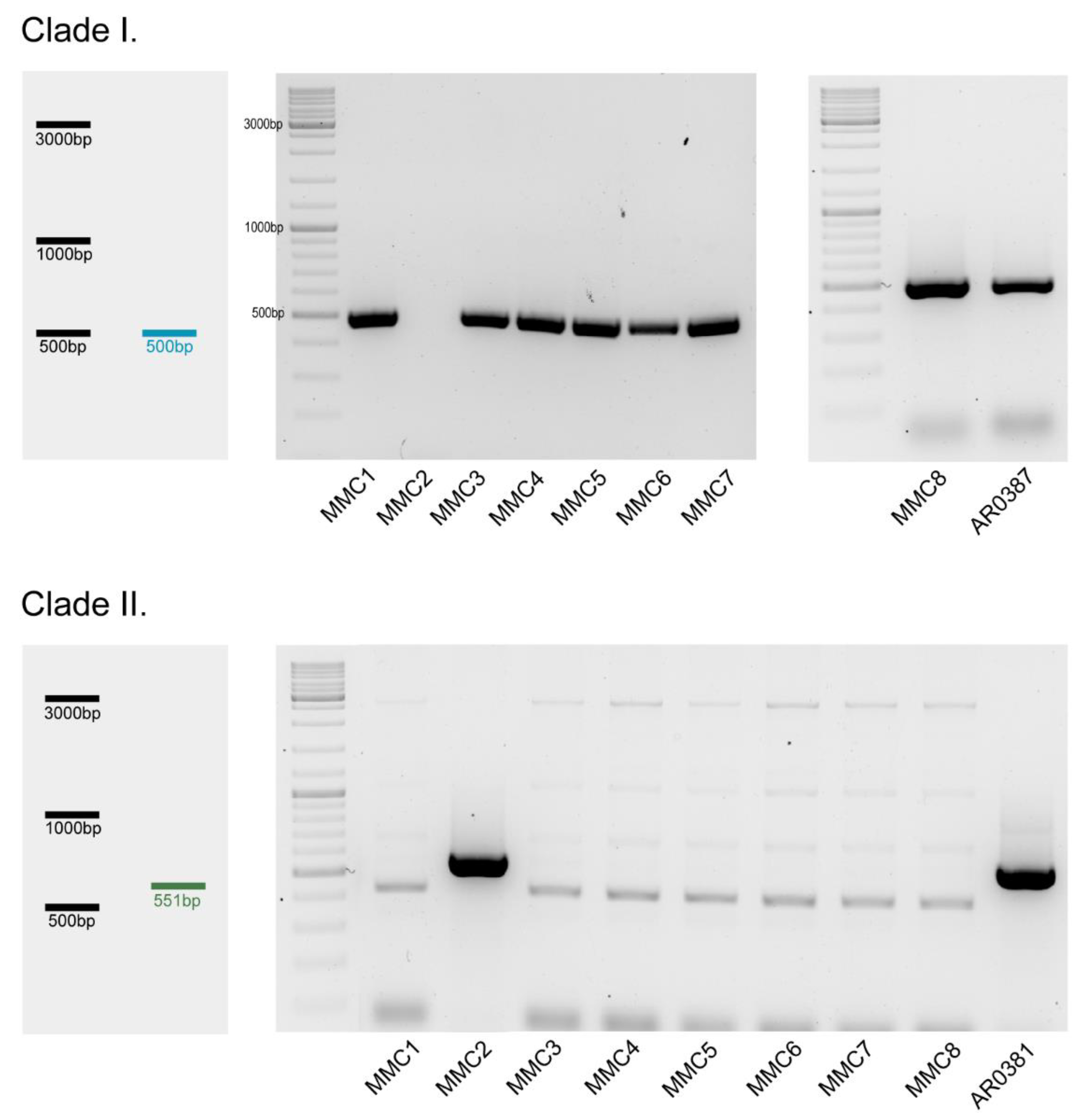
3.1. Clade Classification

The clades of the clinical isolates were classified based on a previously described ClaID system [38]. MMC1 and MMC3–8 all belonged to clade 1, along with the CDC’s already characterized AR0387. Only MMC2 diverged from the isolate set; similarly to the control isolate AR0381, it belonged to clade 2 (East Asia) (Figure 1).

3.2. Antifungal Susceptibility

The antifungal microplate susceptibility test showed that the tested C. auris strains were all resistant to fluconazole with MICs > 256 μg/mL except for MMC2, which had an MIC of 4 μg/mL (Table 1). MMC1–8 were susceptible to caspofungin (0.25 μg/mL). The MIC for amphotericin B was 1 μg/mL for all isolates except MMC2, which had an MIC of 0.25 μg/mL.

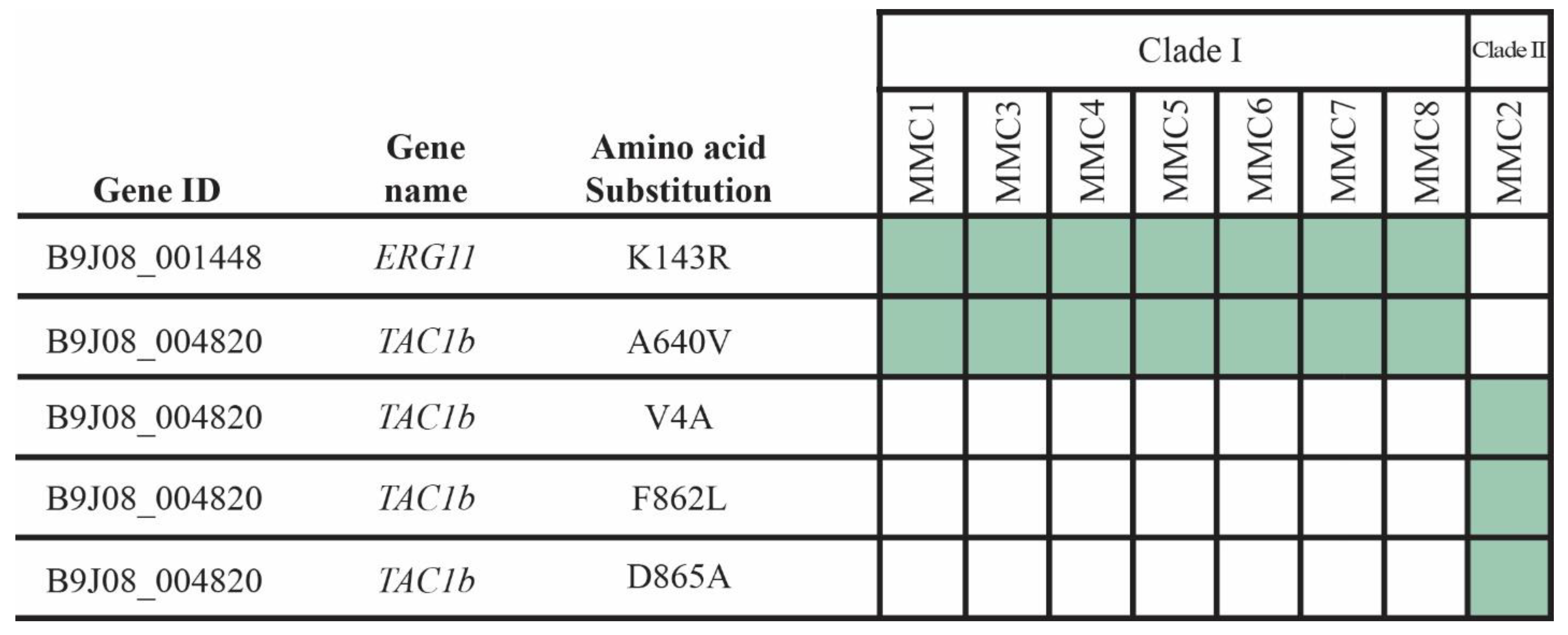
3.3. Detection of SNPs

Compared to the CDC standard strain, all fluconazole-resistant isolates carried non-synonymous mutations in both ERG11 (K143R) and TAC1b (A640V) genes. Three additional SNPs were also identified in the TAC1b gene of the MMC2 isolate, but the effects of these mutations remain unknown. Although ERG3 was also analyzed, no amino acid changes were identified in this gene in either isolate (Figure 2).

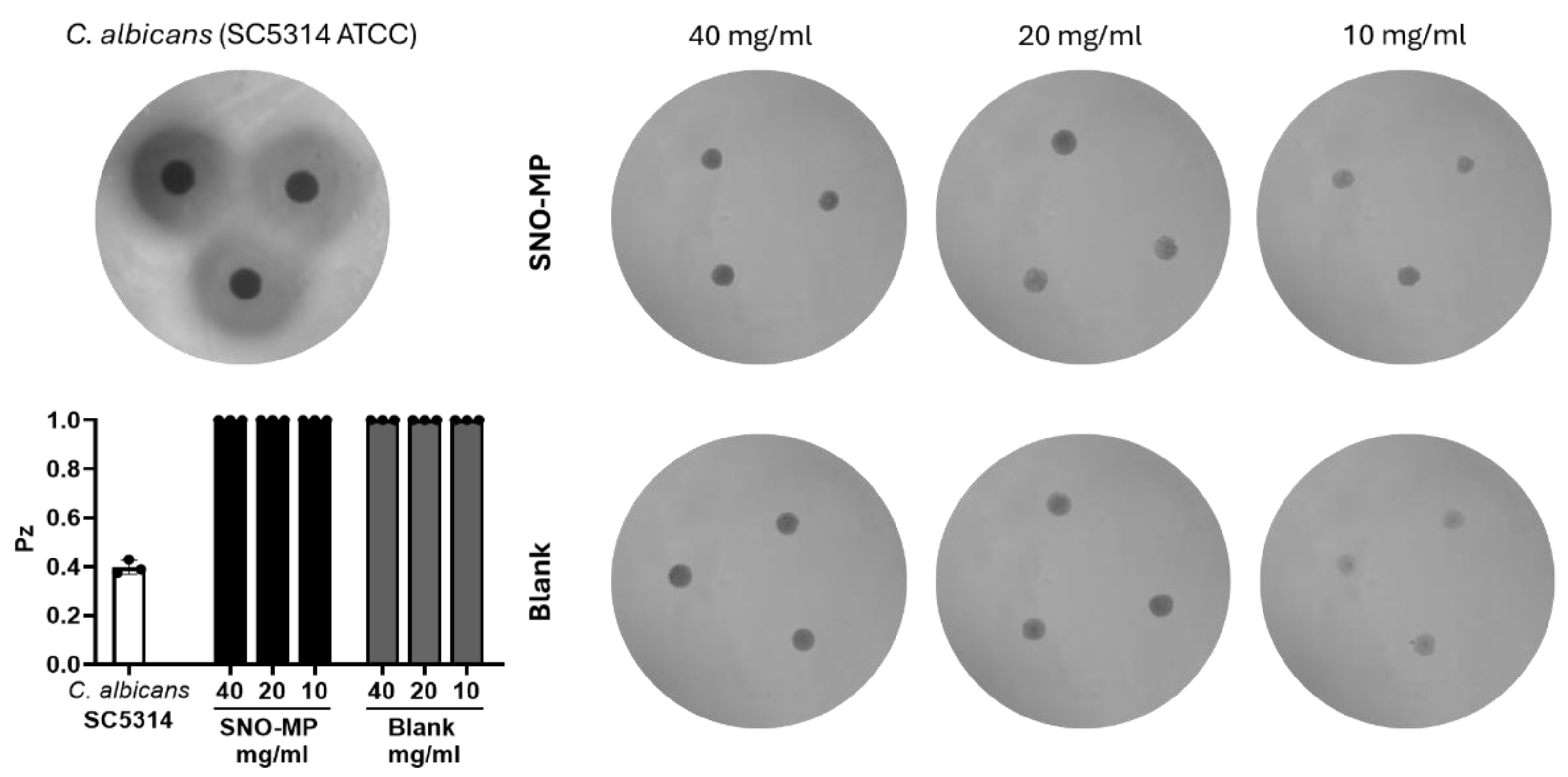
3.4. SNO-MP Microplate Susceptibility Test and Hemolytic Activity

The microplate susceptibility test with the SNO-MP showed a similar pattern to the one with the antifungals, as the NO-releasing microparticles had antifungal activity against all 8 C. auris strains. The MIC for all strains was 20 mg/mL, except for MMC2, with an MIC of 5 mg/mL (Table 2). No hemolytic activity was detected, as seen by the absence of halo formation in blood-agar plates inoculated with the particles in different concentrations even higher than the MICs (Figure 3). In contrast, the positive control, C. albicans (ATCC SC5314), showed a halo formation (Pz = 0.397), confirming enzymatic activity.

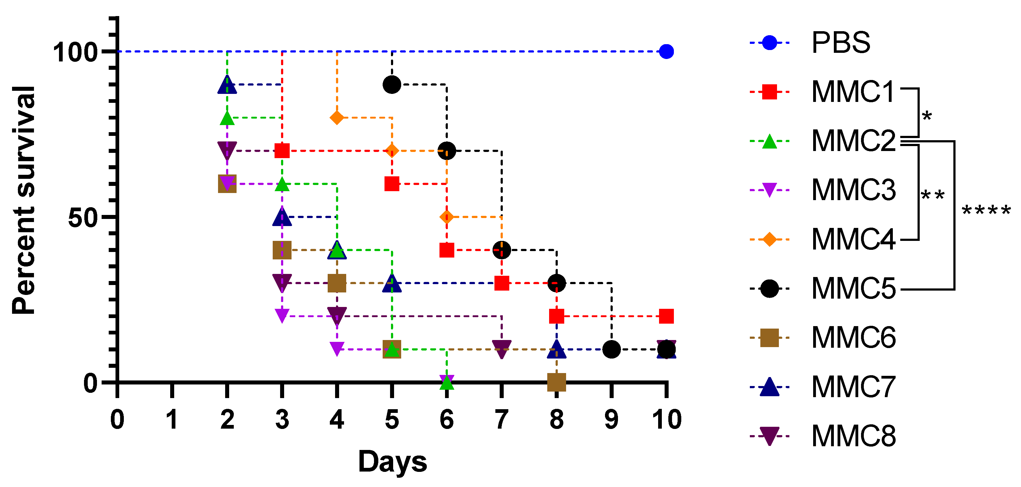
3.5. Virulence in Galleria mellonella

G. mellonella injected with a lethal inoculum of C. auris were observed up to 10 days. Survival curves demonstrate that the clade 2 strain MMC2 was significantly more virulent than the clade 1 strains MMC1, MMC4, and MMC5. Other clade 1 strains were not significantly different than MMC2. Control larvae injected with PBS displayed a 100% survival rate for the experimental timeframe (Figure 4).

4. Discussion

With its rapid emergence, C. auris has attracted intense attention from the medical community for its multidrug-resistant profile and nosocomial pattern of transmission. In fact, the threat of C. auris infections was recently recognized by the World Health Organization 2022 Fungal Priority Pathogen List, with C. auris being added to the critical group alongside Cryptococcus neoformans, Candida albicans, and Aspergillus fumigatus [42].
In this study, we characterized clades and antifungal resistance profiles for eight different strains isolated in one healthcare facility in the Bronx. In line with previous studies, clade classification further verifies that clade 1 is the most common type in New York state, as seven out of the eight MMC isolates (87.5%) showed this genotype [8,43]. The only diverging strain, MMC2, belonged to the rarely isolated clade 2 (Figure 1). Generally, isolates from clade 2 are associated with sporadic cases, while clade 1 has been linked to excessive outbreaks of systemic candidiasis [8,44]. This difference in clinical manifestations may explain the differences in isolation frequencies between the clades.
Our results also show a resistance pattern that correlates to the literature reports of the initial four clades of C. auris that showed 93% resistance to fluconazole, 35% resistance to amphotericin B, and 7% resistance to echinocandins (including caspofungin) [2]. Seven out of eight strains (87.5%) tested in this study showed resistance to fluconazole (MMC1 and MMC3–8), and no strains resistant to AmB and caspofungin were found (Table 1). The seven FCZ-resistant isolates belong to clade 1, a clade that often exhibits a high frequency of antifungal resistance [8,43]. The only identified strain from clade 2, MMC2, was susceptible to FCZ, Cas, and AmB (Table 1). It is also important to highlight that MMC2, the only strain from clade 2, was not only susceptible to fluconazole but also the most susceptible to AmB, in accordance with previous data from our group [45] and the literature [7,8].
Genomic analysis from Chow et al. indicated that in clade 1 isolates, the resistant phenotype is predominantly associated with single nucleotide polymorphisms (Y132F, K143R) in the coding region (ERG11) of the 14-α-demethylase enzyme. Since 96% of the 118 studied clade 1 isolates contained either of these mutations, we can hypothesize that they might also be present in the MMC strains [8]. With that in mind, we verified the genomic background of the resistant phenotypes in our isolates and found out that resistance profiles corresponded with sequencing results, as all resistant strains harbored both one of the most common resistance-inducing amino acid substitution (K143R) in Erg11 and the likewise commonly present mutation A640V in Tac1b [46]. Additional non-synonymous mutations were also identified in the MMC2 strain (V4A; F862L; D865A), but they seem to have no definitive effect on antifungal resistance (Figure 2).
There are several pharmacological limitations associated with conventional antifungal therapy, such as low bioavailability, lack of selectivity, and high toxicity [30,31]. This, in addition to C. auris’s high resistance to antifungal drugs, makes alternative treatments in high demand. As one of the few novel platforms for antifungal delivery currently in development, micro- and nano-drug delivery systems (MiNaDDS) offer an interesting alternative to counter both pharmacological limitations and antifungal resistance [47].
Our results demonstrate that the microparticle has antifungal activity similar to what is described for other NO-releasing particles that were recently tested with MMC1 and MMC2 [11]. Other than that, the susceptibility pattern was also similar to what was observed for the conventional antifungal agents, with MMC2 being more susceptible than strains belonging to clade 1 (Table 2). Moreover, the microparticle did not promote hemolysis (Figure 3), suggesting that mSNO nanoparticles may exhibit antifungal properties without promoting hemolytic-associated toxicity, making them promising candidates for further investigation in therapeutic applications. The microparticle treatment approach for C. auris could be applied to systemic approaches for candidemia as well as topically for either ear infections or as part of a colonization eradication method [48,49,50]. The latter is especially notable as there currently is no markedly effective de-colonization method, which is essential for mitigating risks for outbreaks [51].
Clade 2 isolates have been correlated to a less virulent profile, mainly because they are rarely isolated and usually not linked to invasive disease, as most isolates originate from ear infections, while isolates from clade 1 are usually associated with invasive disease and large outbreaks and are therefore considered more virulent [8,44]. However, this correlation was not observed in our in vivo model of infection (Figure 4), with clade 2 strain MMC2 not only being as virulent as four clade 1 strains in our wax moth model but also being significantly more virulent than 3 clade 1 strains (MMC1, 4, and 5). It is important to highlight that even though G. mellonella has been increasingly used as an infection model for fungal pathogenesis due to its multiple advantages [52,53,54], this model also has its shortcomings, like the lack of an adaptive immune system, and experiments with more complex in vivo models and a larger variety of strains are needed in order to achieve more definitive results.
Taken together, our data corroborates the correlation between clades and the resistance profiles of the tested strains. The findings also support the need for more studies on MiNaDDS, as NO-releasing platforms have significant potential as alternative treatments for C. auris infections, but further development of the platform, in vivo experiments, and full characterization of the microparticle formulation are still necessary. Additionally, as far as the model limitations go, our present data suggests that clade 2 isolates can be at the very least as virulent as clade 1 isolates, going against initial correlations between these two clades and virulence profiles.

5. Conclusions

In conclusion, our study highlights the complex interplay between clade classification, antifungal resistance profiles, and virulence in C. auris. While clade 1 remains predominant at our institution, our findings demonstrate that clade 2 isolates, often considered less virulent, can exhibit comparable or even greater virulence in an invertebrate model of infection. Our study also emphasizes the potential of innovative treatment strategies like MiNaDDS, particularly NO-releasing platforms, to address the challenges posed by C. auris. Although further experiments featuring more sophisticated in vivo models and a broader pool of strains are needed, our findings highlight the importance of continued research into alternative therapeutic options.

Supplementary Materials

The following supporting information can be downloaded at: https://www.mdpi.com/article/10.3390/microbiolres16010015/s1. Table S1: Primers used for the amplification of the selected resistance related genes.

Author Contributions

Conceptualization, J.D.N. and D.Z.-M.; Methodology, A.F.V., F.B., J.P.G., A.B.J., A.D., L.N., D.Z.-M. and D.C.-J.; Formal analysis, A.F.V., F.B., J.P.G., A.B.J., E.V.C.d.A., D.Z.-M. and D.C.-J.; Investigation, A.F.V., F.B., A.B.J., E.V.C.d.A., D.Z.-M. and D.C.-J.; Resources, A.D. and J.D.N.; Data curation, A.F.V., L.N., J.D.N. and D.Z.-M.; Writing—original draft, A.F.V. and F.B.; Writing—review and editing, A.D., L.N., J.D.N. and D.Z.-M.; Visualization, A.F.V., F.B., J.P.G., A.B.J. and E.V.C.d.A.; Supervision, A.F.V., F.B., L.N., J.D.N. and D.Z.-M.; Project administration, J.D.N. and D.Z.-M.; Funding acquisition, J.D.N. All authors have read and agreed to the published version of the manuscript.

Funding

AFV was supported by Fundação de Amparo a Pesquisa do Estado do Rio de Janeiro (FAPERJ—SEI-260003/000751/2024) and Coordenação de Aperfeiçoamento de Pessoal de Nível Superior (CAPES—Grant 001). The National Institutes of Health (NIH) R41 AI165204 partially supported A.F.V., A.D., J.D.N. and D.Z.-M. NIH R21AI156104 partially supported F.B., J.D.N. and D.Z.-M.

Institutional Review Board Statement

Study was performed according to guidelines supported by IRB Number: 2016–7455 (approved on 2 July 2017) of the Albert Einstein College of Medicine and Montefiore Medical Center. All in vivo experiments were performed in a Galleria mellonella model of infection and therefore exempt from legal and ethical considerations.

Informed Consent Statement

C. auris strains were gathered with informed consent from all eight patients under the guidelines approved by the institutional review board (IRB Number: 2016–7455 approved on 2 July 2017) of the Albert Einstein College of Medicine and Montefiore Medical Center.

Data Availability Statement

The original sequencing data presented in the study is openly available in NCBI SRA database under the following reference number: PRJNA1189548.

Conflicts of Interest

A.D. is employed by Zylo Therapeutics. All remaining authors have no conflicts of interest to declare.

References

  1. Satoh, K.; Makimura, K.; Hasumi, Y.; Nishiyama, Y.; Uchida, K.; Yamaguchi, H. Candida auris sp. nov., a novel ascomycetous yeast isolated from the external ear canal of an inpatient in a Japanese hospital. Microbiol. Immunol. 2009, 53, 41–44. [Google Scholar] [CrossRef] [PubMed]
  2. Lockhart, S.R.; Etienne, K.A.; Vallabhaneni, S.; Farooqi, J.; Chowdhary, A.; Govender, N.P.; Colombo, A.L.; Calvo, B.; Cuomo, C.A.; Desjardins, C.A.; et al. Simultaneous Emergence of Multidrug-Resistant Candida auris on 3 Continents Confirmed by Whole-Genome Sequencing and Epidemiological Analyses. Clin. Infect. Dis. Off. Publ. Infect. Dis. Soc. Am. 2017, 64, 134–140. [Google Scholar] [CrossRef] [PubMed]
  3. Spruijtenburg, B.; Badali, H.; Abastabar, M.; Mirhendi, H.; Khodavaisy, S.; Sharifisooraki, J.; Taghizadeh Armaki, M.; de Groot, T.; Meis, J.F. Confirmation of fifth Candida auris clade by whole genome sequencing. Emerg. Microbes Infect. 2022, 11, 2405–2411. [Google Scholar] [CrossRef]
  4. Suphavilai, C.; Ko, K.K.K.; Lim, K.M.; Tan, M.G.; Boonsimma, P.; Chu, J.J.K.; Goh, S.S.; Rajandran, P.; Lee, L.C.; Tan, K.Y.; et al. Discovery of the sixth Candida auris clade in Singapore. medRxiv 2023. [Google Scholar] [CrossRef]
  5. Chatterjee, S.; Alampalli, S.V.; Nageshan, R.K.; Chettiar, S.T.; Joshi, S.; Tatu, U.S. Draft genome of a commonly misdiagnosed multidrug resistant pathogen Candida auris. BMC Genomics 2015, 16, 686. [Google Scholar] [CrossRef] [PubMed]
  6. Du, H.; Bing, J.; Hu, T.; Ennis, C.L.; Nobile, C.J.; Huang, G. Candida auris: Epidemiology, biology, antifungal resistance, and virulence. PLoS Pathog. 2020, 16, e1008921. [Google Scholar] [CrossRef] [PubMed]
  7. Muñoz, J.F.; Gade, L.; Chow, N.A.; Loparev, V.N.; Juieng, P.; Berkow, E.L.; Farrer, R.A.; Litvintseva, A.P.; Cuomo, C.A. Genomic insights into multidrug-resistance, mating and virulence in Candida auris and related emerging species. Nat. Commun. 2018, 9, 5346. [Google Scholar] [CrossRef]
  8. Chow, N.A.; Muñoz, J.F.; Gade, L.; Berkow, E.L.; Li, X.; Welsh, R.M.; Forsberg, K.; Lockhart, S.R.; Adam, R.; Alanio, A.; et al. Tracing the Evolutionary History and Global Expansion of Candida auris Using Population Genomic Analyses. mBio 2020, 11, e03364-19. [Google Scholar] [CrossRef] [PubMed]
  9. Maphanga, T.G.; Mpembe, R.S.; Naicker, S.D.; Govender, N.P.; Germs-Sa, F. In Vitro Antifungal Activity of Manogepix and Other Antifungal Agents against South African Candida auris Isolates from Bloodstream Infections. Microbiol. Spectr. 2022, 10, e01717-21. [Google Scholar] [CrossRef]
  10. Szekely, A.; Borman, A.M.; Johnson, E.M. Candida auris Isolates of the Southern Asian and South African Lineages Exhibit Different Phenotypic and Antifungal Susceptibility Profiles In Vitro. J. Clin. Microbiol. 2019, 57, e02055-18. [Google Scholar] [CrossRef] [PubMed]
  11. Cleare, L.G.; Li, K.L.; Abuzeid, W.M.; Nacharaju, P.; Friedman, J.M.; Nosanchuk, J.D. NO Candida auris: Nitric Oxide in Nanotherapeutics to Combat Emerging Fungal Pathogen Candida auris. J. Fungi 2020, 6, 85. [Google Scholar] [CrossRef] [PubMed]
  12. Jeffery-Smith, A.; Taori, S.K.; Schelenz, S.; Jeffery, K.; Johnson, E.M.; Borman, A.; Candida auris Incident Management Team; Manuel, R.; Brown, C.S. Candida auris: A Review of the Literature. Clin. Microbiol. Rev. 2018, 31, e00029-17. [Google Scholar] [CrossRef] [PubMed]
  13. Chowdhary, A.; Sharma, C.; Duggal, S.; Agarwal, K.; Prakash, A.; Singh, P.K.; Jain, S.; Kathuria, S.; Randhawa, H.S.; Hagen, F.; et al. New Clonal Strain of Candida auris, Delhi, India. Emerg. Infect. Dis. 2013, 19, 1670–1673. [Google Scholar] [CrossRef] [PubMed]
  14. Tracking Candida Auris|Candida Auris|Fungal Diseases|CDC. Available online: https://www.cdc.gov/fungal/candida-auris/tracking-c-auris.html (accessed on 14 August 2023).
  15. Calvo, B.; Melo, A.S.A.; Perozo-Mena, A.; Hernandez, M.; Francisco, E.C.; Hagen, F.; Meis, J.F.; Colombo, A.L. First report of Candida auris in America: Clinical and microbiological aspects of 18 episodes of candidemia. J. Infect. 2016, 73, 369–374. [Google Scholar] [CrossRef] [PubMed]
  16. Lee, W.G.; Shin, J.H.; Uh, Y.; Kang, M.G.; Kim, S.H.; Park, K.H.; Jang, H.-C. First Three Reported Cases of Nosocomial Fungemia Caused by Candida auris. J. Clin. Microbiol. 2020, 49, 3139–3142. [Google Scholar] [CrossRef]
  17. Kathuria, S.; Singh, P.K.; Sharma, C.; Prakash, A.; Masih, A.; Kumar, A.; Meis, J.F.; Chowdhary, A. Multidrug-Resistant Candida auris Misidentified as Candida haemulonii: Characterization by Matrix-Assisted Laser Desorption Ionization–Time of Flight Mass Spectrometry and DNA Sequencing and Its Antifungal Susceptibility Profile Variability by Vitek 2, CLSI Broth Microdilution, and Etest Method. J. Clin. Microbiol. 2015, 53, 1823–1830. [Google Scholar] [CrossRef]
  18. Chowdhary, A.; Voss, A.; Meis, J.F. Multidrug-resistant Candida auris: ‘new kid on the block’ in hospital-associated infections? J. Hosp. Infect. 2016, 94, 209–212. [Google Scholar] [CrossRef]
  19. Morales-López, S.E.; Parra-Giraldo, C.M.; Ceballos-Garzón, A.; Martínez, H.P.; Rodríguez, G.J.; Álvarez-Moreno, C.A.; Rodríguez, J.Y. Invasive Infections with Multidrug-Resistant Yeast Candida auris, Colombia. Emerg. Infect. Dis. 2017, 23, 162–164. [Google Scholar] [CrossRef]
  20. Schwartz, I.S.; Hammond, G.W. First reported case of multidrug-resistant Candida auris in Canada. Can. Commun. Dis. Rep. Releve Mal. Transm. Au Can. 2017, 43, 150–153. [Google Scholar] [CrossRef]
  21. Vallabhaneni, S. Investigation of the First Seven Reported Cases of Candida auris, a Globally Emerging Invasive, Multidrug-Resistant Fungus—United States, May 2013–August 2016. MMWR Morb. Mortal. Wkly. Rep. 2016, 65, 1234–1237. [Google Scholar] [CrossRef] [PubMed]
  22. Sarma, S.; Kumar, N.; Sharma, S.; Govil, D.; Ali, T.; Mehta, Y.; Rattan, A. Candidemia caused by amphotericin B and Fluconazole resistant Candida auris. Indian J. Med. Microbiol. 2013, 31, 90–91. [Google Scholar] [CrossRef] [PubMed]
  23. Welsh, R.M.; Bentz, M.L.; Shams, A.; Houston, H.; Lyons, A.; Rose, L.J.; Litvintseva, A.P. Survival, Persistence, and Isolation of the Emerging Multidrug-Resistant Pathogenic Yeast Candida auris on a Plastic Health Care Surface. J. Clin. Microbiol. 2017, 55, 2996–3005. [Google Scholar] [CrossRef] [PubMed]
  24. Piedrahita, C.T.; Cadnum, J.L.; Jencson, A.L.; Shaikh, A.A.; Ghannoum, M.A.; Donskey, C.J. Environmental Surfaces in Healthcare Facilities are a Potential Source for Transmission of Candida auris and Other Candida Species. Infect. Control Hosp. Epidemiol. 2017, 38, 1107–1109. [Google Scholar] [CrossRef] [PubMed]
  25. Tsay, S. Notes from the Field: Ongoing Transmission of Candida auris in Health Care Facilities—United States, June 2016–May 2017. MMWR Morb. Mortal. Wkly. Rep. 2017, 66, 514–515. [Google Scholar] [CrossRef] [PubMed]
  26. Dahiya, S.; Chhillar, A.K.; Sharma, N.; Choudhary, P.; Punia, A.; Balhara, M.; Kaushik, K.; Parmar, V.S. Candida auris and Nosocomial Infection. Curr. Drug Targets 2020, 21, 365–373. [Google Scholar] [CrossRef] [PubMed]
  27. Sanyaolu, A.; Okorie, C.; Marinkovic, A.; Abbasi, A.F.; Prakash, S.; Mangat, J.; Hosein, Z.; Haider, N.; Chan, J. Candida auris: An Overview of the Emerging Drug-Resistant Fungal Infection. Infect. Chemother. 2022, 54, 236–246. [Google Scholar] [CrossRef]
  28. Fu, L.; Le, T.; Liu, Z.; Wang, L.; Guo, H.; Yang, J.; Chen, Q.; Hu, J. Different efficacies of common disinfection methods against candida auris and other candida species. J. Infect. Public Health 2020, 13, 730–736. [Google Scholar] [CrossRef]
  29. Kischkel, B.; Rossi, S.A.; Santos, S.R.; Nosanchuk, J.D.; Travassos, L.R.; Taborda, C.P. Therapies and Vaccines Based on Nanoparticles for the Treatment of Systemic Fungal Infections. Front. Cell. Infect. Microbiol. 2020, 10, 463. [Google Scholar] [CrossRef]
  30. Nett, J.E.; Andes, D.R. Antifungal Agents: Spectrum of Activity, Pharmacology, and Clinical Indications. Infect. Dis. Clin. N. Am. 2016, 30, 51–83. [Google Scholar] [CrossRef]
  31. Voltan, A.R.; Quindós, G.; Alarcón, K.P.M.; Fusco-Almeida, A.M.; Mendes-Giannini, M.J.S.; Chorilli, M. Fungal diseases: Could nanostructured drug delivery systems be a novel paradigm for therapy? Int. J. Nanomedicine 2016, 11, 3715–3730. [Google Scholar] [CrossRef]
  32. Ahmadi, M.S.; Lee, H.H.; Sanchez, D.A.; Friedman, A.J.; Tar, M.T.; Davies, K.P.; Nosanchuk, J.D.; Martinez, L.R. Sustained Nitric Oxide-Releasing Nanoparticles Induce Cell Death in Candida albicans Yeast and Hyphal Cells, Preventing Biofilm Formation In Vitro and in a Rodent Central Venous Catheter Model. Antimicrob. Agents Chemother. 2016, 60, 2185–2194. [Google Scholar] [CrossRef]
  33. Macherla, C.; Sanchez, D.; Ahmadi, M.; Vellozzi, E.; Friedman, A.; Nosanchuk, J.; Martinez, L. Nitric Oxide Releasing Nanoparticles for Treatment of Candida Albicans Burn Infections. Front. Microbiol. 2012, 3, 193. [Google Scholar] [CrossRef]
  34. Costa-Orlandi, C.B.; Martinez, L.R.; Bila, N.M.; Friedman, J.M.; Friedman, A.J.; Mendes-Giannini, M.J.S.; Nosanchuk, J.D. Nitric Oxide-Releasing Nanoparticles Are Similar to Efinaconazole in Their Capacity to Eradicate Trichophyton rubrum Biofilms. Front. Cell. Infect. Microbiol. 2021, 11, 684150. [Google Scholar] [CrossRef] [PubMed]
  35. Costa-Orlandi, C.B.; Mordorski, B.; Baltazar, L.M.; Mendes-Giannini, M.J.S.; Friedman, J.M.; Nosanchuk, J.D.; Friedman, A.J. Nitric Oxide Releasing Nanoparticles as a Strategy to Improve Current Onychomycosis Treatments. J. Drugs Dermatol. JDD 2018, 17, 717–720. [Google Scholar] [PubMed]
  36. Biss, M.; Hanna, M.D.; Xiao, W. Isolation of Yeast Nucleic Acids. In Yeast Protocols; Xiao, W., Ed.; Springer: New York, NY, USA, 2014; pp. 15–21. ISBN 978-1-4939-0799-1. [Google Scholar]
  37. Panel Details|Antimicrobial Resistance Isolate Bank|Antibiotic/Antimicrobial Resistance | CDC. Available online: https://wwwn.cdc.gov/ARIsolateBank/Panel/PanelDetail?ID=2 (accessed on 18 September 2023).
  38. Narayanan, A.; Selvakumar, P.; Siddharthan, R.; Sanyal, K. ClaID: A Rapid Method of Clade-Level Identification of the Multidrug Resistant Human Fungal Pathogen Candida auris. Microbiol. Spectr. 2022, 10, e0063422. [Google Scholar] [CrossRef]
  39. M27 Ed4 Broth Dilution Antifungal Susceptibility, Yeasts. (n.d.). Clinical & Laboratory Standards Institute. Available online: https://clsi.org/standards/products/microbiology/documents/m27/ (accessed on 13 July 2023).
  40. Tar, M.T.; Friedman, J.M.; Draganski, A.; Davies, K.P. Topically delivered nitric oxide acts synergistically with an orally administered PDE5 inhibitor in eliciting an erectile response in a rat model of radical prostatectomy. Int J Impot Res. 2022, 34, 573–580. [Google Scholar] [CrossRef] [PubMed]
  41. Li, K.L.; Miranda, D.Z.; Cleare, L.G.; Akbar, N.A.; Friedman, J.M.; Draganski, A.; Nosanchuk, J.D.; Abuzeid, W.M. Nitric oxide-generating microparticles: An in vitro evaluation of anti-biofilm efficacy and sinonasal epithelial cell cytotoxicity. Int. Forum Allergy Rhinol. 2022, 13, 954–957. [Google Scholar] [CrossRef] [PubMed]
  42. WHO Fungal Priority Pathogens List to Guide Research, Development and Public Health Action. Available online: https://www.who.int/publications-detail-redirect/9789240060241 (accessed on 23 May 2023).
  43. Adams, E.; Quinn, M.; Tsay, S.; Poirot, E.; Chaturvedi, S.; Southwick, K.; Greenko, J.; Fernandez, R.; Kallen, A.; Vallabhaneni, S.; et al. Candida auris in Healthcare Facilities, New York, USA, 2013-2017. Emerg. Infect. Dis. 2018, 24, 1816–1824. [Google Scholar] [CrossRef] [PubMed]
  44. Welsh, R.M.; Sexton, D.J.; Forsberg, K.; Vallabhaneni, S.; Litvintseva, A. Insights into the Unique Nature of the East Asian Clade of the Emerging Pathogenic Yeast Candida auris. J. Clin. Microbiol. 2019, 57, e00007-19. [Google Scholar] [CrossRef] [PubMed]
  45. Zamith-Miranda, D.; Heyman, H.M.; Cleare, L.G.; Couvillion, S.P.; Clair, G.C.; Bredeweg, E.L.; Gacser, A.; Nimrichter, L.; Nakayasu, E.S.; Nosanchuk, J.D. Multi-omics Signature of Candida auris, an Emerging and Multidrug-Resistant Pathogen. mSystems 2019, 4, e00257-19. [Google Scholar] [CrossRef] [PubMed]
  46. Rybak, J.M.; Cuomo, C.A.; Rogers, P.D. The molecular and genetic basis of antifungal resistance in the emerging fungal pathogen Candida auris. Curr Opin Microbiol. 2022, 70, 102208. [Google Scholar] [CrossRef]
  47. Valdez, A.F.; Zamith-Miranda, D.; Nimrichter, L.; Nosanchuk, J.D. Micro- and nanoparticles as platforms for the treatment of fungal infections: Present and future perspectives. Futur. Microbiol. 2023, 18, 1007–1011. [Google Scholar] [CrossRef] [PubMed]
  48. Cabrales, P.; Han, G.; Nacharaju, P.; Friedman, A.J.; Friedman, J.M. Reversal of hemoglobin-induced vasoconstriction with sustained release of nitric oxide. American Journal of Physiology. Heart Circ. Physiol. 2011, 300, H49–H56. [Google Scholar] [CrossRef] [PubMed]
  49. Jani, V.P.; Friedman, J.M.; Cabrales, P. Nitric oxide releasing nanoparticles reduce inflammation in a small animal model of ARDS. Biomed. Pharmacother. 2022, 148, 112705. [Google Scholar] [CrossRef]
  50. Tar, M.; Cabrales, P.; Navati, M.; Adler, B.; Nacharaju, P.; Friedman, A.J.; Friedman, J.; Davies, K.P. Topically applied NO-releasing nanoparticles can increase intracorporal pressure and elicit spontaneous erections in a rat model of radical prostatectomy. J. Sex. Med. 2014, 11, 2903–2914. [Google Scholar] [CrossRef] [PubMed]
  51. Rossow, J.; Ostrowsky, B.; Adams, E.; Greenko, J.; McDonald, R.; Vallabhaneni, S.; Forsberg, K.; Perez, S.; Lucas, T.; A Alroy, K.; et al. Factors Associated With Candida auris Colonization and Transmission in Skilled Nursing Facilities With Ventilator Units, New York, 2016–2018. Clin Infect Dis. 2021, 72, e753–e760. [Google Scholar] [CrossRef] [PubMed]
  52. Honorato, L.; de Araujo, J.F.D.; Ellis, C.C.; Piffer, A.C.; Pereira, Y.; Frases, S.; Araújo, G.R.d.S.; Pontes, B.; Mendes, M.T.; Pereira, M.D.; et al. Extracellular Vesicles Regulate Biofilm Formation and Yeast-to-Hypha Differentiation in Candida albicans. mBio 2022, 13, e0030122. [Google Scholar] [CrossRef] [PubMed]
  53. Vargas, G.; Rocha, J.D.B.; Oliveira, D.L.; Albuquerque, P.C.; Frases, S.; Santos, S.S.; Nosanchuk, J.D.; Gomes, A.M.O.; Medeiros, L.C.A.S.; Miranda, K.; et al. Compositional and immunobiological analyses of extracellular vesicles released by Candida albicans. Cell Microbiol. 2015, 17, 389–407. [Google Scholar] [CrossRef] [PubMed]
  54. Bonilla, J.J.A.; Honorato, L.; de Oliveira, D.F.C.; Gonçalves, R.A.; Guimarães, A.; Miranda, K.; Nimrichter, L. Silver chitosan nanocomposites as a potential treatment for superficial candidiasis. Med. Mycol. 2021, 59, 993–1005. [Google Scholar] [CrossRef]
Figure 1. Clade identification. Schematic images of the expected clade I and clade II-specific fragment sizes, compared to the agarose gels, containing the clade-specific PCR fragments of the unclassified strains (MMC1–8) and the already classified clinical isolates (AR0387, AR0381).
Microbiolres 16 00015 g001
Figure 2. Identified nonsynonymous amino acid substitutions. According to the targeted sequencing of three antifungal resistance-associated genes (ERG3, ERG11, TAC1b). Compared to the CDC isolates on the corresponding clades (B8441; B11220) non-synonymous amino acid substitution conferring SNPs were found in the ERG11 and TAC1b genes, while no mutation was found in ERG3. Colored boxes indicate the presence of the listed substitutions.
Microbiolres 16 00015 g002
Figure 3. Hemolytic activity by SNO-MP. Hemolytic activity was evaluated using. C. albicans SC5314 was used as a positive control. Different concentrations of SNO-MP and blank particles were inoculated on plates containing sheep blood agar and incubated for 48 h at 37 °C until development of halo indicating hemolytic activity. Pz values were calculated as Pz = A/B, where A is the colony diameter and B is the total diameter of the colony plus the enzymatic activity halo (Pz = 1 indicates no hemolytic activity).
Microbiolres 16 00015 g003
Figure 4. Galleria mellonella survival rates following infection with Candida auris. G. mellonella were infected with a lethal inoculum of C. auris yeasts, and survival was monitored for 10 days. Infection with the clade 2 strain, MMC2, resulted in a significantly higher mortality rate compared to the clade 1 isolates MMC1, 4, and 5. Infection with other clade 1 isolates had no significant survival rates compared to MMC2. Statistical analysis was performed with log-rank Mantel–Cox, * p < 0.05, ** p < 0.01, **** p < 0.0001.
Microbiolres 16 00015 g004
Table 1. Antifungal MIC for 8 Candida auris strains were determined (µg/mL). Values that are bolded indicate resistance to the specific antifungal, according to CDC’s tentative MIC breakpoints for C. auris.
Minimal Inhibitory Concentration (μg/mL)
StrainAmphotericin BCaspofunginFluconazole
MMC110.25>256
MMC20.250.254
MMC310.25>256
MMC410.25>256
MMC510.25>256
MMC610.25>256
MMC710.25>256
MMC810.25>256
Table 2. SNO-MP MIC for 8 Candida auris strains was determined in mg/mL.
Minimal Inhibitory Concentration (mg/mL)
StrainSNO-MPStrainSNO-MP
MMC120MMC520
MMC25MMC620
MMC320MMC720
MMC420MMC820
Disclaimer/Publisher’s Note: The statements, opinions and data contained in all publications are solely those of the individual author(s) and contributor(s) and not of MDPI and/or the editor(s). MDPI and/or the editor(s) disclaim responsibility for any injury to people or property resulting from any ideas, methods, instructions or products referred to in the content.

