Review

Pathogenic and Non-Pathogenic Microbes in the Wound Microbiome—How to Flip the Switch

by
Sophie Charlotte Liegenfeld
1,
Svenja Stenzel
1,
Julian-Dario Rembe
2,
Mandy Dittmer
1,
Paulo Ramos
3,4 and
Ewa Klara Stuermer
1,*
1
Department of Vascular Medicine, Translational Wound Research, University Medical Center Hamburg-Eppendorf, 20251 Hamburg, Germany
2
Department for Vascular and Endovascular Surgery, University Hospital Duesseldorf (UKD), Heinrich Heine University Duesseldorf, 40225 Düsseldorf, Germany
3
Unidade de Saúde Familiar Corino de Andrade, 4490-602 Póvoa de Varzim, Portugal
4
Nursing Department, Institute of Health Science, Oporto Campus, Universidade Católica Portuguesa, 4169-005 Porto, Portugal
*
Author to whom correspondence should be addressed.
Microbiol. Res. 2025, 16(2), 39; https://doi.org/10.3390/microbiolres16020039
Submission received: 20 December 2024 / Revised: 29 January 2025 / Accepted: 4 February 2025 / Published: 6 February 2025

Abstract

:
The wound microbiome refers to the specific community of microbes, including bacteria, fungi, and other microorganisms, that are present in and around a wound. This microbiome plays a crucial role in wound healing, as it includes both healing-promoting and pathogenic microbes. The balance between these microbes significantly influences the healing process; a balanced microbial colonization can support wound healing and prevent infections, while an overgrowth of pathogenic microbes can lead to delayed healing processes and complications. The composition of the wound microbiome can vary depending on the type of wound, cause, genetic predisposition, and (social) environment. In this scope review, the complex interactions in the wound microbiome will be highlighted and the importance of non-pathogenic microbes for wound healing will be discussed. In addition, possible therapeutic approaches to restore a healthy microbiome and prevent infections will be addressed. A deeper understanding of these dynamics could open up new perspectives for the treatment of wounds and the development of strategies to combat wound infections.

1. Introduction

Infected wounds represent a widespread and complex problem in modern healthcare, posing significant challenges in both stationary and ambulatory settings. The prevalence of hard-to-heal wounds has increased in recent decades due to several factors, including the rise in chronic diseases such as diabetes mellitus, vascular disease, and the increasing number of surgical procedures [1,2]. A central aspect of wound pathology is the wound microbiome—the diversity of microorganisms present in and around infected wounds. This microbiome includes both pathogenic and commensal bacteria, the composition and interactions of which can have a significant impact on the healing process [3]. Studies show that a healthy microbiome can promote the healing process, whereas an imbalance in the microbial community can lead to chronic wounds and increased infection rates [4]. The study of the wound microbiome has become increasingly important in recent years, particularly with regard to the development of more targeted therapeutic strategies [5]. Innovative approaches to modulate the microbiome, such as probiotic therapies or the use of antimicrobial substances, may help to improve wound healing and reduce the need for systemic antibiotics. This review summarizes the current evidence on the prevalence of infected wounds, the role of the wound microbiome, and its relevance to wound care practice. The aim is to highlight the relevance of these topics for improving patient care and optimizing therapeutic strategies.

2. Materials and Methods

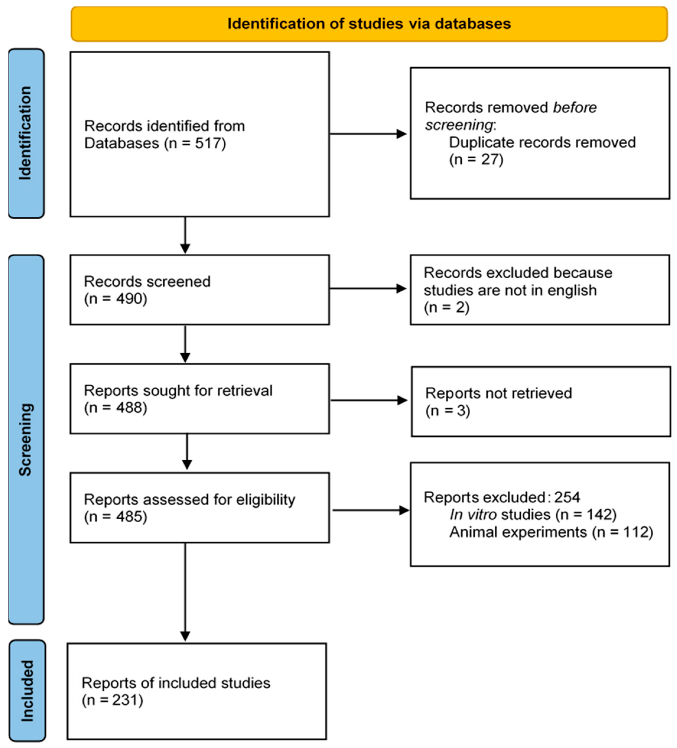
2.1. Literature Search Strategy

A literature review was performed, employing the electronic database Medline (PubMed). The search included studies published in the last ten years and was conducted in September 2024. The following keywords were included: “wound microbiome”, “complex wounds” and microbiome, “acute wounds” and microbiome, probiotics and “wound microbiome”, probiotics and “chronic wound”, probiotics and “wound healing”, and “chronic wounds” and antiseptics. Keywords were combined using the Boolean operators “and” and “or”. The review was performed by two researchers. Titles and abstracts were screened independently for relevance to the research question. If there was a disagreement, both researchers debated the relevance of the specific literature and decided whether it should be included in this review. Out of the 517 articles obtained after entering the keywords into the search engine, 27 were duplicates and 259 were excluded based on the exclusion criteria (Figure 1).

2.2. Inclusion and Exclusion Criteria

The inclusion criteria included peer-reviewed articles investigating the relationship between wounds and bacterial colonization in human and translational studies. Studies published between January 2014 and September 2024 were included in the review. The exclusion criteria encompassed non-peer-reviewed articles and articles not published in English. Articles focusing solely on animal and experimental studies were excluded, as these analyses lack human skin and the wound environment, which significantly influence bacterial interactions and interactions between human cells, antimicrobials, and pathogenic and commensal bacteria.

3. Results

3.1. Wound Microbiome: An Overview of Wound Entities

The human microbiome, particularly the skin microbiome, plays a crucial role in health and well-being. It consists of a multitude of microorganisms that interact with each other in a dynamic balance. Both pathogenic and non-pathogenic microbes can be found on the skin (e.g., Staphylococcus epidermidis, Cutibacterium acnes, Staphylococcus aureus, Corynebacterium spp., and Micrococcus spp.) and their interactions are critical for wound healing [3,6,7]. While non-pathogenic microbes can modulate the immune system [8] and prevent the formation of biofilm, pathogenic microbes contribute to inflammation and tissue damage [4]. The diversity and species composition of the microbes that colonize a wound are influenced by several factors [3]. Certain types of bacteria that are normally present on the skin can invade and colonize the wound. The immune status of the host plays a decisive role, as a compromised immune system (e.g., in the case of diabetes or HIV) leads to increased colonization by pathogenic microbes. Other factors such as genetic predisposition, environmental factors, and lifestyle also play a role in the colonization of wounds by microbes. Different types of wounds have different patterns of microbial colonization (Table 1).
Acute wounds have a different microbial colonization to chronic wounds (e.g., diabetic foot wounds or pressure ulcers) [9]. Infection occurs in 5–26% of acute wounds resulting from trauma or burns. Common microbes found in acute wounds are Gram-negative (Pseudomonas aeruginosa, Acinetobacter baumannii, Enterobacteriaceae, Escherichia coli, Klebsiella pneumoniae, Serratia marcescens, Enterobacter spp., Proteus spp., and Bacteroides spp.), Gram-positive (Staphylococcus aureus, Streptococcus spp., Enterococcus spp., Micrococcus spp., Corynebacterium spp., Streptococcus pyogens, and Cornybacterium diphteria) and coagulase-negative (Staphylococci) [10,11].
Chronic wounds tend to harbor a more stable, often pathogenic microbial population. The most commonly found pathogens in chronic wounds are S. aureus and P. aeruginosa. Other bacterial genera such as Corynebacterium, Prevotella, Finegoldia, and Anaerococcus also colonize chronic wounds [11]. Previous research on the microbiome of chronic wounds has also observed that anaerobic bacteria play a specific role in delayed healing. Verbanic et al. reported an association between non-healing wounds and the presence of facultative anaerobes [12]. In addition, the formation and presence of biofilm is often observed in chronic wounds. A biofilm matrix of aggregated polysaccharides, lipids, nucleic acids, and proteins allows for cross-signaling/communication between the different microbes that can promote inflammatory immune responses. As a result, the healing of these wounds is compromised and delayed [5].
Table 1. Overview of the five most prevalent bacterial species identified on intact skin, acute, and chronic wound environments 1.
Intact SkinAcute WoundsChronic Wounds
Staphylococcus epidermidisStaphylococcus aureusStaphylococcus aureus
Cutibacterium acnesStreptococcus spp.Pseudomonas aeruginosa
Staphylococcus aureusPseudomonas aeruginosaEnterobacter spp.
Corynebacterium spp.Escherichia coliProteus mirabilis
Micrococcus spp.Enterococcus spp.Corynebacterium spp.
1 According to the literature [6,7,10,11,13,14].

3.2. Influence of Bacterial Colonization on Wound Healing: The Role of Commensal Bacteria

The interaction of bacteria on the skin is a complex and dynamic system that is critical to skin health. The skin harbors a variety of microbes, including bacteria, fungi, and viruses, in a symbiotic relationship with each other. The skin is not only home to pathogens, but also to commensal organisms that have a beneficial effect on the skin barrier and health and inhibit pathogenic microbes [15]. Some microbes can regulate the skin’s immune response and wound healing through competitive growth or direct inhibition. S. epidermidis is one of these pro-healing microbes, as it can downregulate the inflammatory immune response of the wound and upregulate the production of human antimicrobial peptides. Together with its own produced antimicrobial peptides, pathogens such as S. aureus can be inhibited [16]. Cells stimulated with S. epidermidis show the ability to kill intracellular methicillin-resistant Staphylococcus aureus (MRSA) [17]. Previous research has also demonstrated the ability of S. epidermidis to inhibit S. aureus biofilm formation by degrading the key proteins necessary for biofilm formation and epithelial adhesion in a host, using the serine protease glutamyl endopeptidase (Esp) and thereby preventing a major factor driving wound chronicity [18].
Acinetobacter lwoffii is another commensal microbe on human skin that helps maintain the balance of the skin microbiome and mitigates potential allergic reactions by playing a protective role against allergic sensitization and inflammatory responses [19]. Staphylococcus lugdunensis is a commensal bacterium that produces lugdunin. Lugdunin is a peptide that has antimicrobial properties specifically against S. aureus, and prevents colonization of the wound with this pathogen. Colonization of the human nose with S. lugdunensis has been associated with a lower S. aureus transmission rate [3]. Another important mechanism of skin protection is provided by the Corynebacterium jeikeium. This bacterium plays a critical role in protecting the host’s epidermis from harmful reactive oxygen species (ROS). This is primarily achieved by producing superoxide dismutase, an enzyme that fends off oxidative damage, and by acquiring manganese, which serves as a cofactor for several antioxidant enzymes [20]. Propionibacteria spp. contributes to skin health by stimulating the expression of the Toll-like receptors TLR2 and TLR4 in keratinocytes. Such pattern recognition receptors (PRRs) are crucial for specific and non-specific immune responses and help to recognize pathogenic microbes via pathogen-associated molecular patterns (PAMPs), such as lipopolysaccharides (LPSs) [21]. They also produce antibacterial substances [20]. Several other commensal species (e.g., Corynebacterium striatum and Corynebacterium accolens) have been identified so far that can influence and alter bacterial behavior, shifting the balance from pathogenic to commensal organisms and thus protecting skin health and integrity [18].

3.3. The Skin Microbiome in Dysbiosis and the Resulting Consequences

A diverse skin microbiome is therefore important to stabilize the skin barrier and protect it from pathogens. Dysbiosis of the skin microbiome has been reported in inflammatory skin diseases (hidradenitis suppurativa, atopic dermatitis, and acne vulgaris) as well as in chronic wounds and diabetic patients [16].
In hidradenitis suppurativa (HS), a chronic, inflammatory skin disease, microbial colonization of the skin is of great importance. Certain Gram-negative bacteria, particularly the genera Prevotella and Porphyromonas, are commonly found in this disease [16]. These bacteria may contribute to the development and progression of the disease by promoting inflammatory processes in the tissue. Compared to healthy skin, the presence of C. acnes is significantly reduced in HS lesions. This bacterium is normally part of the skin microflora and plays a role in maintaining skin health. However, an altered bacterial composition is observed in patients with HS, which may contribute to the severity of the disease. In addition, there is a correlation between C. acnes skin colonization and disease severity in other dermatologic conditions, such as atopic dermatitis and acne vulgaris. A higher density of C. acnes often correlates with more severe symptoms in these conditions. This aspect underlines the importance of the microbiota for the understanding and treatment of inflammatory skin diseases [16].
A significant dysbiosis of the skin microbiome has been observed in diabetic patients, characterized by a significantly lower diversity of microbial communities compared to healthy individuals [19]. This reduced diversity can weaken the skin barrier and increase the risk of infection. In particular, studies show that Staphylococci, a group of bacteria commonly found on the skin, are the most abundant microbes in diabetic patients. This altered colonization of the skin may be caused by the diabetes itself, as well as associated factors such as reduced blood flow and an impaired immune response. The alteration in microbial composition, particularly a reduction in diversity, may not only affect skin health, but also increase the risk and severity of several diseases [19]. Verbanic et al. found that the healthy skin microbiome is much more diverse compared to the wound microbiome, with species found in chronic wounds that were not present in skin samples (Proteus spp. and Enterobacter spp.) [12]. However, several species showed a distinct overrepresentation in chronic wounds compared to skin samples (e.g., Staphylococcus aureus and capitis, P. aeruginosa), while previously mentioned ‘protective’ species such as Corynebacterium were underrepresented. This supports the hypothesis of a significant shift towards pathogenic bacteria that are responsible for wound infection, and biofilm formation establishing a stable bacterial community, which is proving difficult to resolve. Studies of the relationship between bacterial community stability in chronic wounds and healing rates have shown faster healing and positive clinical outcomes for unstable communities and, conversely, delayed healing for stable, established communities that are more difficult to eradicate (e.g., due to biofilm formation) [18,22].
Some clinical studies of the gut microbiome in diabetic patients have shown that it contributes to pro-inflammatory status and metabolic syndrome [23,24]. On the other hand, there is some evidence to suggest that there is a direct link between the gut microbiome and inflammatory (immunological) skin diseases, such as psoriasis or atopic dermatitis [25]. Similarly, a link can be postulated between the chronic inflammation of hard-to-heal wounds and gut dysbiosis, which is often associated with diabetes; bacteria and toxins enter the submucosal layers through the leaky gut and contribute to the stimulation of the response of various T cells [26], which also has holistic effects in the context of the gut–skin axis [25,27]. Overall, there is growing evidence for the complex relationship between the skin and the gut microbiome, known as the ‘gut–skin axis’ (Figure 2). The gut–skin axis is the complex relationship between the gut microbiome and how the gut and its absorption can influence skin health through its immunological and metabolic properties [28]. Although not obvious, several studies have shown a link between the two, with certain skin conditions and gastrointestinal disorders having a reciprocal relationship. For example, 10–25% of patients with inflammatory bowel disease (e.g., Crohn’s disease or ulcerative colitis) also have immunological skin diseases such as psoriasis and ulcers [27], and vice versa [29]. The exact mechanisms of gut–skin microbial interactions are not fully understood, but the immune and endocrine systems are likely to be involved. There is also preliminary evidence that oral probiotics, prebiotics, and dietary changes have the potential to influence the skin microbiome, particularly in the setting of dysbiosis [30,31].

3.4. The Switch from Commensal to Pathogenic Bacteria

As previously discussed, a balance of different microbes is important for a healthy skin barrier. These so-called commensal bacteria vary significantly depending on the body site. While a mixed population of bacterial species colonizes dry skin with a higher abundance of Proteobacteria and Flavobacteriales, Propionibacteria and Staphylococcus species predominate the sebaceous sites (Figure 3), and Corynebacteria dominate in the more moist skin regions such as the axilla or groin [7]. Although these different regions represent distinct ecological niches, they are in close proximity, and can lose their well-orchestrated cooperation and stability through external triggers of imbalance. These include immunological skin diseases such as psoriasis and atopic dermatitis, as well as antibiotic creams or antiseptics. The current omnipotence of antiseptics to treat only colonized wounds to prevent infection ignores the fact that the unselective mechanism of action of antiseptics does not discriminate between commensal and pathogenic bacteria, and therefore also eradicates the ‘good’ skin microbiome. This can be problematic in the context of chronic wounds, e.g., due to peripheral arterial disease (PAD), with inadequate blood supply to the affected limb and correspondingly reduced ‘local’ immune competence. Under selection pressure, common pathogens such as S. aureus, P. aeruginosa, and Enterobacteriaceae tend to prevail and dominate due to higher virulence, effective bacteriotoxins, and acquired adaptation mechanisms compared to commensal microbes. In the case of continuous, non-reevaluated antiseptic overuse, such pathogens therefore have an advantage, because they can recolonize more easily. However, after the eradication of commensal regulative bacteria, growth can occur selectively, unchecked and unbalanced.
When the skin barrier is disrupted by injuries, cuts, burns, or chronic disease, bacteria can penetrate the deeper layers of the skin. There, the skin’s ability to protect itself is reduced, and generally harmless (commensal) bacteria can cause infections. A weakened immune system can also allow commensal bacteria to proliferate and cause infections. The skin environment can also be affected by external environmental conditions such as high temperatures and excessive sweating. Antiseptics and antiseptic soaps can also alter the skin microbiome through a variety of factors. One such specific factor is the pH of the skin’s naturally mildly acidic barrier. For example, excessive hygiene practices can affect the skin’s natural pH value, which ranges between 4.5 and 5.5. This acidic environment inhibits the growth of most human pathogenic bacteria and thus provides a simple but effective protective barrier [32,33]. An increase in the skin’s natural pH is associated with several particularly inflammatory skin conditions, such as atopic dermatitis and acne, which cause the environment to turn alkaline [34]. Disruption of the skin barrier’s pH can also be observed in chronic wounds (with pH values of 7.3 and higher), most likely caused due to exposed dermal tissue and structures, and is associated with a reduction in wound healing capacity. Pathogens such as S. aureus, P. aeruginosa, or other Enterobacteriaceae require pH values of above 6 for optimal growth [35], and therefore thrive in the presence of a disrupted skin barrier, such as that found in wounds, e.g., after shaving the axillary area (Figure 3). Metabolic products such as ammonia can also lead to necrotic areas by impairing the oxygen supply to a wound, thereby promoting an alkaline wound environment. Increased biofilm formation has also been reported under alkaline conditions [33]. In contrast, a mildly acidic environment promotes healing by altering protease activities, degrading bacterial products, promoting angiogenesis and re-epithelialization, and increasing oxygen availability through the Bohr effect [35]. The use of acidification as a treatment strategy has therefore been employed for several decades, using wound dressings or acidic solutions. The treatment of chronic wounds with pH-lowering dressings has been shown to reduce wound size within 3 weeks in previously refractory wounds. However, in terms of therapeutic implications, recent reviews such as that by Derwin et al. from 2022 have concluded that there is currently insufficient evidence to recommend specific therapeutic strategies or product groups for manipulation of the pH, and thus its influence on wound healing. This is especially true since data on which degree of acidification could be beneficial and what duration of a potential acidifying treatment should be aimed for are missing. Current therapeutics that have the potential to modulate the pH level in wounds, as they are themselves acidic or alkaline solutions (e.g., hypochlorous solutions), range between a pH of 6.5 and 9.5 [36]. Other experimental approaches used therapeutics with a pH as low as 3 and demonstrated somewhat beneficial results. Nonetheless, the current literature base is insufficient to recommend any specific ‘target pH’ for wound healing or therapeutic products [37]. However, Derwin et al. and further studies demonstrate that it is possible to therapeutically influence the pH of a wound and that initial studies have shown positive results in terms of improving the healing of chronic wounds [38].

3.5. The Impact of Antiseptics and Antibiotics

Antiseptics and antibiotics play a critical role in the management of wound infections, particularly in wounds with biofilm [39]. Antiseptics, such as iodine solutions, octenidine di-hydrochloride or polyhexamethylene-biguanide (polyhexanide/PHMB), or antimicrobial hypochlorous wound irrigation solutions are applied locally to the wound to kill bacteria and other microbes, thereby reducing the number of microbes on the wound surface to prevent the risk of infection [40]. The clinically approved solutions subsumed within the group ‘sodium hypochlorite/hypochlorous acid solutions’ (NaClO/HClO) vary in their antimicrobial potential depending on the NaClO- or HClO concentration [36]. There are generally no randomized controlled trials (RCTs) on the use of the latter or other antiseptics, so no evidence-based recommendation can be made on the use of iodine, octenidine di-hydrochloride, PHMB, or hypochlorous solutions, for example, in guidelines [41]. Additionally, the variety of any available antiseptic or antimicrobial solution in terms of active agent, agent concentration, and composition is broad, which in turn complicates the recommendation of products in specific situations. Therefore, there is also no clear or uniform (expert) recommendation regarding their use in acute or chronic wound infections, even though expert panels have done their best to formulate national and international best-practice guidelines [41,42]. However, some fundamental aspects can be recognized. For example, the lack of residual effect (<5 min) in hypochlorous solutions makes them more controllable than the other antiseptics mentioned above. Also, in this context, the different exposure times until the complete eradication of planktonic bacteria (e.g., iodine or octenidine di-hydrochloride: 1–3 min, PHMB: >10 min) and their different biocompatibility based on their active agent concentration should be emphasized [41,42].
Several studies have successfully demonstrated that biofilm-forming microbes can be killed using antiseptics such as octenidine di-hydrochloride or hypochlorous acid solutions [43,44,45,46]. In most cases, long exposure times are required, but the efficacy of these agents is high. The biofilm models with pathogens such as E. coli, P. aeruginosa, various MRSA, and even Candida spp. could be sterilized [43]. In addition to the use of antiseptic solutions, mechanical and other forms of debridement can have a positive impact on bioburden reduction [47]. Another role of debridement is to allow antiseptic solutions to be more effective [41,48,49] by combining a structure disruption of the biofilms’ extracellular polymeric substance (EPS) with the subsequent penetration of the biofilm by an antiseptic.
Antibiotics are designed to inhibit the growth of bacteria or kill microbes directly. Most antibiotics that are administered are broad-spectrum antibiotics that can kill a large number of different bacteria. A specific type of bacteria cannot be targeted. According to the current research, there is no evidence that probiotic bacteria affect or interfere with the effects of antibiotics in any way; it is assumed that they will be eliminated similarly to pathogenic bacteria [50,51]. Due to the high prescription rate of antibiotics, many pathogens have already developed resistance to these drugs [9]. Thus, the use of antibiotics and antiseptics does not specifically target pathogens, but also targets commensal bacteria in the human body. These microbes play an important role in maintaining the microbial balance and immune defenses described above. An imbalance, which can result from the use of antibiotics as well as antiseptics, could lead to an overgrowth of pathogenic germs, which in turn can lead to serious secondary infections [52]. As discussed previously, the unselected prolonged use of local antiseptics and antimicrobial solutions can induce selective overgrowth of pathogenic bacteria. In addition, as with under-dosed systemic antibiotics that are not administered for an adequate period of time (either too short or too long), the inappropriate use of low-potency antimicrobial solutions (e.g., preserved wound irrigation solutions) can affect susceptible commensal bacteria more selectively than resilient pathogenic microbes, promoting a form of selective advantage. Careful selection and the use of both agents are therefore essential to promote healing and prevent complications.

3.6. Probiotics as Allies in the Wound Environment

The use of probiotics in infected wounds is becoming increasingly important because of their potential to aid the healing process. Probiotics are live microorganisms that can promote beneficial health effects, particularly by stabilizing the microbial balance in the body [53]. One of the key properties of probiotics is their ability to compete with pathogenic microorganisms for nutrients and binding sites on the host’s mucosa. Through this competition, they prevent harmful pathogens from colonizing this ecological niche. Their species-specific antagonistic activity plays a crucial role in maintaining a healthy microbial balance. In addition, probiotics contribute to the fight against pathogens through their antimicrobial effect. This occurs primarily through the production of lactic acid, which creates an unfavorable mildly acidic condition for the growth of many pathogenic species [31]. Some probiotic strains can also produce bacteriocins—special proteins that act specifically against other bacteria and can inhibit their growth. Probiotics generally act as immunomodulatory triggers that actively support the inflammatory process at the wound site. They specifically promote the influx of various inflammatory cells, particularly macrophages and polymorphonuclear leukocytes (PMNs), which play a central role in the immune response. These immune cells are critical for fighting infection and promoting wound healing. An important mechanism through which probiotics exert their immunomodulatory effect is the production of exopolysaccharides [53]. These biochemical compounds are immunostimulatory and can stimulate the activation of cytokines and chemokines. These signaling substances are critical for recruiting immune cells to the wound site. As a result, there is an increased influx of neutrophils and macrophages into the affected tissue, which can fight harmful microbes and induce tissue repair. In addition, probiotics stimulate the process of phagocytosis, a mechanism through which immune cells ingest and eliminate foreign particles, bacteria, and other pathogenic microbes. This increased phagocytosis significantly reduces the bacterial load of the wound by effectively reducing the growth and adherence of pathogens. These broad immunological effects of probiotics help to promote wound healing and minimize the risk of infection. Studies show that the use of probiotics in wound dressings can accelerate wound healing and prevent infection [54]. By strengthening the immune response and promoting tissue repair, probiotics help to optimize the regeneration of damaged skin. Examples of commonly used probiotics include Lactobacillus spp. (such as Lactobacillus rhamnosus and Lactobacillus casei), Bifidobacterium spp. (such as Bifidobacterium longum), and Saccharomyces boulardii, a yeast culture. A study investigating the use of Lactobacillus plantarum species to treat patients with second-degree burns showed fewer secondary infections and graft rejections compared to a control group treated with 1% silver sulfadiazine [55]. Thus, the targeted use of these microorganisms may not only support wound healing, but also promote overall health and reduce the risk of infection.
The effect of probiotics on wound healing in humans has only been studied to a limited extent so far. A study by Mohseni et al. (2018) investigated the effect of probiotics on wound healing in patients with diabetic foot ulcers. A significant improvement in wound length, width, and height was demonstrated compared to the control groups [56]. In another study by Esposito et al. (2018), patients who received probiotics had significantly fewer dressing changes than patients who received a placebo or antibiotics [57]. Pediatric burn patients treated with prophylactic antibiotics had a significantly reduced need for transplantation [58]. This observation is particularly relevant, as transplants are often associated with additional risks and longer healing times. It was also found that in cases where grafts were not used, the time to complete wound healing was significantly reduced. This suggests that probiotic therapy may not only reduce the need for invasive procedures, but may also make the healing process more efficient overall [59].

4. Conclusions for Wound Healing in Practice

Understanding the wound microbiome and its complex interactions is critical to the practice of wound care. The diversity and composition of the microbes that colonize a wound have a direct impact on the healing process. In particular, the balance between commensal and pathogenic bacteria plays a central role. Dysbiosis, as observed in diabetes or chronic wounds, can significantly delay healing and increase the risk of infection. It is therefore important to choose treatment methods carefully to support and not damage the microbiota. The targeted use of antiseptics and antibiotics can help to optimize wound healing and prevent complications. However, these target and kill the pathogenic bacteria as well as the beneficial part of the skin microbiome.
Future research efforts should examine the role of the microbiome in wound healing to develop evidence-based strategies that promote both skin health and patient recovery. The integration of advanced methods, such as next-generation sequencing (NGS), particularly 16S rRNA gene sequencing, holds significant potential to provide a more comprehensive and nuanced understanding of the wound microbiome, ultimately enhancing diagnostic accuracy and guiding more effective, personalized treatment strategies [60]. In contrast to conventional swabbing and culture-based techniques, which are widely used in clinical practice but have limitations in capturing the full spectrum of microbial populations, NGS enables a more comprehensive and accurate analysis of both the aerobic and anaerobic bacterial communities present within wound sites. This approach is particularly valuable for identifying microbial communities that may be difficult to detect using the traditional methods, such as those residing in deeper tissue layers or within biofilms, which are common in chronic wounds [57]. However, despite its promising potential, the widespread adoption of NGS in clinical routine is still uncertain, due to challenges related to its availability, high cost, and technical complexity.
If probiotic bacteria could be used as microbiome stimulants, as in gut health, they could be a new therapeutic option. In clinical practice, it is critical to develop an individualized treatment approach that takes into account the specific needs of each patient and does not ignore the importance of the microbiome in wound care.

Author Contributions

Conceptualization, E.K.S. and J.-D.R.; methodology, S.C.L., M.D. and S.S.; resources, E.K.S.; data collection, S.C.L., S.S., J.-D.R., M.D. and P.R.; writing—original draft preparation, S.C.L., S.S., J.-D.R. and E.K.S.; writing—review and editing, P.R., M.D. and E.K.S.; supervision, P.R. and E.K.S.; project administration; E.K.S. All authors have read and agreed to the published version of the manuscript.

Funding

This research received no external funding.

Institutional Review Board Statement

The study was conducted in accordance with the Declaration of Helsinki. Procedures of blood product collection are in accordance with “good clinical practice” standards.

Informed Consent Statement

Not applicable.

Data Availability Statement

The data presented in this study are available upon request from the corresponding author.

Conflicts of Interest

The authors declare no conflicts of interest with regard to this project. JDR received fees for consulting, travel expenses, and or lectures from Serag-Wiessner and DEBx medical. EKS received fees for consulting, lectures, and/or studies from Institut AllergoSan, BEMER Inc., Curea medical, DEBx medical, Essity, Schülke and Mayr, Serag-Wiessner, and Smith and Nephew.

Abbreviations

The following abbreviations are used in this manuscript:
EPSExtracellular polymeric substance
HSHidradenitis suppurativa
LPSLipopolysaccharides
MRSAMethicillin-resistant Staphylococcus aureus
NGSNext-generation sequencing
PADPeripheral arterial disease
PAMPPathogen-associated molecular pattern
PHMBPolyhexamethylene-biguanide
PMNPolymorphonuclear leukocytes
PRRPattern recognition receptors
RCTRandomized controlled trials
ROSReactive oxygen species

References

  1. Martinengo, L.; Olsson, M.; Bajpai, R.; Soljak, M.; Upton, Z.; Schmidtchen, A.; Car, J.; Järbrink, K. Prevalence of chronic wounds in the general population: Systematic review and meta-analysis of observational studies. Ann. Epidemiol. 2019, 29, 8–15. [Google Scholar] [CrossRef] [PubMed]
  2. Carter, M.J.; Frykberg, R.G.; Oropallo, A.; Sen, C.K.; Armstrong, D.G.; Nair, H.K.; Serena, T.E. Efficacy of Topical Wound Oxygen Therapy in Healing Chronic Diabetic Foot Ulcers: Systematic Review and Meta-Analysis. Adv. Wound Care 2023, 12, 177–186. [Google Scholar] [CrossRef]
  3. White, E.K.; Grice, E.A. The Wound Microbiome. Cold Spring Harb. Perspect. Biol. 2023, 15, a041218. [Google Scholar] [CrossRef] [PubMed]
  4. Canchy, L.; Kerob, D.; Demessant, A.; Amici, J.-M. Wound healing and microbiome, an unexpected relationship. J. Eur. Acad. Dermatol. Venereol. 2023, 37 (Suppl. 3), 7–15. [Google Scholar] [CrossRef]
  5. Malone, M.; Bjarnsholt, T.; McBain, A.J.; James, G.A.; Stoodley, P.; Leaper, D.; Tachi, M.; Schultz, G.; Swanson, T.; Wolcott, R.D. The prevalence of biofilms in chronic wounds: A systematic review and meta-analysis of published data. J. Wound Care 2017, 26, 20–25. [Google Scholar] [CrossRef] [PubMed]
  6. Schommer, N.N.; Gallo, R.L. Structure and function of the human skin microbiome. Trends Microbiol. 2013, 21, 660–668. [Google Scholar] [CrossRef]
  7. Grice, E.A.; Kong, H.H.; Conlan, S.; Deming, C.B.; Davis, J.; Young, A.C.; NISC Comparative Sequencing Program; Bouffard, G.G.; Blakesley, R.W.; Murray, P.R.; et al. Topographical and Temporal Diversity of the Human Skin Microbiome. Science 2009, 324, 1190–1192. [Google Scholar] [CrossRef] [PubMed]
  8. Luna, P.C. Skin Microbiome as Years Go By. Am. J. Clin. Dermatol. 2020, 21 (Suppl. 1), 12–17. [Google Scholar] [CrossRef] [PubMed]
  9. Ding, X.; Tang, Q.; Xu, Z.; Xu, Y.; Zhang, H.; Zheng, D.; Wang, S.; Tan, Q.; Maitz, J.; Maitz, P.K.; et al. Challenges and innovations in treating chronic and acute wound in-fections: From basic science to clinical practice. Burns Trauma 2022, 10, tkac014. [Google Scholar] [CrossRef]
  10. Jockenhöfer, F.; Chapot, V.; Weindorf, M.S.; Körber, A.; Klode, J.; Buer, J.; Küpper, B.; Roesch, A.; Dissemond, J. Bacterial spectrum colonizing chronic leg ulcers: A 10-year comparison from a German wound care center. JDDG J. Dtsch. Dermatol. Ges. 2014, 12, 1121–1127. [Google Scholar] [CrossRef] [PubMed]
  11. Morsli, M.; Salipante, F.; Magnan, C.; Dunyach-Remy, C.; Sotto, A.; Lavigne, J.-P. Direct metagenomics investigation of non-surgical hard-to-heal wounds: A review. Ann. Clin. Microbiol. Antimicrob. 2024, 23, 39. [Google Scholar] [CrossRef]
  12. Verbanic, S.; Shen, Y.; Lee, J.; Deacon, J.M.; Chen, I.A. Microbial predictors of healing and short-term effect of debridement on the microbiome of chronic wounds. NPJ Biofilms Microbiomes 2020, 6, 21. [Google Scholar] [CrossRef] [PubMed]
  13. Pitocco, D.; Spanu, T.; Di Leo, M.; Vitiello, R.; Rizzi, A.; Tartaglione, L.; Fiori, B.; Caputo, S.; Tinelli, G.; Zaccardi, F.; et al. Diabetic foot infections: A comprehensive overview. Eur. Rev. Med. Pharmacol. Sci. 2019, 23, 26–37. [Google Scholar] [CrossRef]
  14. Stuermer, E.K.; Bang, C.; Giessler, A.; Smeets, R.; Janke, T.M.; Seki, F.D.; Debus, E.S.; Franke, A.; Augustin, M. Effect of oral multispecies probiotic on wound healing, periodontitis and quality of life on patients with diabetes. J. Wound Care 2024, 33, 394–407. [Google Scholar] [CrossRef] [PubMed]
  15. Belkaid, Y.; Segre, J.A. Dialogue between skin microbiota and immunity. Science 2014, 346, 954–959. [Google Scholar] [CrossRef] [PubMed]
  16. Bay, L.; Ring, H.C. Human skin microbiota in health and disease: The cutaneous communities’ interplay in equilibrium and dysbiosis: The cutaneous communities’ interplay in equilibrium and dysbiosis. APMIS 2022, 130, 706–718. [Google Scholar] [CrossRef]
  17. Pastar, I.; O’neill, K.; Padula, L.; Head, C.R.; Burgess, J.L.; Chen, V.; Garcia, D.; Stojadinovic, O.; Hower, S.; Plano, G.V.; et al. Staphylococcus epidermidis Boosts Innate Immune Response by Activation of Gamma Delta T Cells and Induction of Perforin-2 in Human Skin. Front. Immunol. 2020, 11, 550946. [Google Scholar] [CrossRef] [PubMed]
  18. Byrd, A.L.; Belkaid, Y.; Segre, J.A. The human skin microbiome. Nat. Rev. Microbiol. 2018, 16, 143–155. [Google Scholar] [CrossRef]
  19. Gardiner, M.; Vicaretti, M.; Sparks, J.; Bansal, S.; Bush, S.; Liu, M.; Darling, A.; Harry, E.; Burke, C.M. A longitudinal study of the diabetic skin and wound micro-biome. PeerJ 2017, 5, e3543. [Google Scholar] [CrossRef] [PubMed]
  20. Zielińska, M.; Pawłowska, A.; Orzeł, A.; Sulej, L.; Muzyka-Placzyńska, K.; Baran, A.; Filipecka-Tyczka, D.; Pawłowska, P.; Nowińska, A.; Bogusławska, J.; et al. Wound Microbiota and Its Impact on Wound Healing. Int. J. Mol. Sci. 2023, 24, 17318. [Google Scholar] [CrossRef] [PubMed]
  21. Li, D.; Wu, M. Pattern recognition receptors in health and diseases. Signal Transduct. Target. Ther. 2021, 6, 291. [Google Scholar] [CrossRef] [PubMed]
  22. Loesche, M.; Gardner, S.E.; Kalan, L.; Horwinski, J.; Zheng, Q.; Hodkinson, B.P.; Tyldsley, A.S.; Franciscus, C.L.; Hillis, S.L.; Mehta, S.; et al. Temporal Stability in Chronic Wound Microbiota Is Associated With Poor Healing. J. Investig. Dermatol. 2017, 137, 237–244. [Google Scholar] [CrossRef] [PubMed]
  23. Scheithauer, T.P.M.; Rampanelli, E.; Nieuwdorp, M.; Vallance, B.A.; Verchere, C.B.; van Raalte, D.H.; Herrema, H. Gut Microbiota as a Trigger for Metabolic Inflammation in Obesity and Type 2 Diabetes. Front. Immunol. 2020, 11, 571731. [Google Scholar] [CrossRef]
  24. Yang, G.; Wei, J.; Liu, P.; Zhang, Q.; Tian, Y.; Hou, G.; Meng, L.; Xin, Y.; Jiang, X. Role of the gut microbiota in type 2 diabetes and related diseases. Metabolism 2021, 117, 154712. [Google Scholar] [CrossRef] [PubMed]
  25. Sinha, S.; Lin, G.; Ferenczi, K. The skin microbiome and the gut-skin axis. Clin. Dermatol. 2021, 39, 829–839. [Google Scholar] [CrossRef] [PubMed]
  26. Salem, I.; Ramser, A.; Isham, N.; Ghannoum, M.A. The Gut Microbiome as a Major Regulator of the Gut-Skin Axis. Front. Microbiol. 2018, 9, 1459. [Google Scholar] [CrossRef] [PubMed]
  27. Saarialho-Kere, U. The gut-skin axis. J. Pediatr. Gastroenterol. Nutr. 2004, 39 (Suppl. 3), S734–S735. [Google Scholar] [CrossRef]
  28. O’Neill, C.A.; Monteleone, G.; McLaughlin, J.T.; Paus, R. The gut-skin axis in health and disease: A paradigm with therapeutic implications. BioEssays 2016, 38, 1167–1176. [Google Scholar] [CrossRef] [PubMed]
  29. Huang, B.L.; Chandra, S.; Shih, D.Q. Skin Manifestations of Inflammatory Bowel Disease. Front. Physiol. 2012, 3, 13. [Google Scholar] [CrossRef] [PubMed]
  30. Ellis, S.R.; Nguyen, M.; Vaughn, A.R.; Notay, M.; Burney, W.A.; Sandhu, S.; Sivamani, R.K. The Skin and Gut Microbiome and Its Role in Common Dermatologic Conditions. Microorganisms 2019, 7, 550. [Google Scholar] [CrossRef]
  31. Fijan, S.; Frauwallner, A.; Langerholc, T.; Krebs, B.; ter Haar née Younes, J.A.; Heschl, A.; Turk, D.M.; Rogelj, I. Efficacy of Using Probiotics with Antagonistic Activity against Pathogens of Wound Infections: An Integrative Review of Literature. BioMed Res. Int. 2019, 2019, 7585486. [Google Scholar] [CrossRef]
  32. Dissemond, J.; Witthoff, M.; Brauns, T.C.; Haberer, D.; Goos, M. pH values in chronic wounds. Evaluation during modern wound therapy. Hautarzt 2003, 54, 959–965. [Google Scholar] [CrossRef]
  33. Schreml, S.; Szeimies, R.-M.; Karrer, S.; Heinlin, J.; Landthaler, M.; Babilas, P. The impact of the pH value on skin integrity and cu-taneous wound healing. J. Eur. Acad. Dermatol. Venereol. 2010, 24, 373–378. [Google Scholar] [CrossRef] [PubMed]
  34. Proksch, E. pH in nature, humans and skin. J. Dermatol. 2018, 45, 1044–1052. [Google Scholar] [CrossRef] [PubMed]
  35. Schneider, L.A.; Korber, A.; Grabbe, S.; Dissemond, J. Influence of pH on wound-healing: A new perspective for wound-therapy? Arch. Dermatol. Res. 2007, 298, 413–420. [Google Scholar] [CrossRef]
  36. Severing, A.-L.; Rembe, J.-D.; Koester, V.; Stuermer, E.K. Safety and efficacy profiles of different commercial sodium hypo-chlorite/hypochlorous acid solutions (NaClO/HClO): Antimicrobial efficacy, cytotoxic impact and physicochemical parameters in vitro. J. Antimicrob. Chemother. 2019, 74, 365–372. [Google Scholar] [CrossRef]
  37. Sim, P.; Strudwick, X.L.; Song, Y.; Cowin, A.J.; Garg, S. Influence of Acidic pH on Wound Healing In Vivo: A Novel Perspective for Wound Treatment. Int. J. Mol. Sci. 2022, 23, 13655. [Google Scholar] [CrossRef] [PubMed]
  38. Derwin, R.; Patton, D.; Strapp, H.; Moore, Z. Wound pH and temperature as predictors of healing: An observational study. J. Wound Care 2023, 32, 302–310. [Google Scholar] [CrossRef] [PubMed]
  39. Alves, P.J.; Barreto, R.T.; Barrois, B.M.; Gryson, L.G.; Meaume, S.; Monstrey, S.J. Update on the role of antiseptics in the management of chronic wounds with critical colonisation and/or biofilm. Int. Wound J. 2020, 18, 342–358. [Google Scholar] [CrossRef] [PubMed]
  40. Cwajda-Białasik, J.; Mościcka, P.; Szewczyk, M.T. Antiseptics and antimicrobials for the treatment and management of chronic wounds: A systematic review of clinical trials. Adv. Dermatol. Allergol. 2022, 39, 141–151. [Google Scholar] [CrossRef]
  41. Nair, H.K.; Mrozikiewicz-Rakowska, B.; Sanches Pinto, D.; Stuermer, E.; Matiasek, J.; Sander, J.; Lázaro-Martínez, J.L.; Ousey, K.; Assadian, O.; Kim, P.J.; et al. Use of Wound Antiseptics in Practice; Wounds Intern: London, UK, 2024. [Google Scholar]
  42. Kramer, A.; Dissemond, J.; Kim, S.; Willy, C.; Mayer, D.; Papke, R.; Tuchmann, F.; Assadian, O. Consensus on Wound Antisepsis: Update 2018. Ski. Pharmacol. Physiol. 2017, 31, 28–58. [Google Scholar] [CrossRef] [PubMed]
  43. Harriott, M.M.; Bhindi, N.; Kassis, S.; Summitt, B.; Perdikis, G.; Wormer, B.A.; Rankin, T.M.; Kaoutzanis, C.; Samaha, M.; Stratton, C.; et al. Comparative Antimicrobial Activity of Commercial Wound Care Solutions on Bacterial and Fungal Biofilms. Ann. Plast. Surg. 2019, 83, 404–410. [Google Scholar] [CrossRef] [PubMed]
  44. Besser, M.; Dietrich, M.; Weber, L.; Rembe, J.D.; Stuermer, E.K. Efficacy of antiseptics in a novel 3-dimensional human plasma biofilm model (hpBIOM). Sci. Rep. 2020, 10, 4792. [Google Scholar] [CrossRef]
  45. Roche, E.D.; Woodmansey, E.J.; Yang, Q.; Gibson, D.J.; Zhang, H.; Schultz, G.S. Cadexomer iodine effectively reduces bacterial biofilm in porcine wounds ex vivo and in vivo. Int. Wound J. 2019, 16, 674–683. [Google Scholar] [CrossRef] [PubMed]
  46. Rembe, J.-D.; Huelsboemer, L.; Plattfaut, I.; Besser, M.; Stuermer, E.K. Antimicrobial Hypochlorous Wound Irrigation Solutions Demonstrate Lower Anti-biofilm Efficacy Against Bacterial Biofilm in a Complex in-vitro Human Plasma Biofilm Model (hpBIOM) Than Common Wound Antimicrobials. Front. Microbiol. 2020, 11, 564513. [Google Scholar] [CrossRef] [PubMed]
  47. Moelleken, M.; Krimphove, S.H.; Krefting, F.; Benson, S.; Rammos, C.; Cyrek, A.E.; Dissemond, J. How effective is simple mechanical wound debridement in reducing bacterial colonisation? Results of a prospective clinical study. Int. Wound J. 2024, 21, e14824. [Google Scholar] [CrossRef] [PubMed]
  48. Mayer, D.; Tettelbach, W.H.; Ciprandi, G.; Downie, F.; Hampton, J.; Hodgson, H.; Lazaro-Martinez, J.L.; Probst, A.; Schultz, G.; Stürmer, E.K.; et al. Best practice for wound debridement. J. Wound Care 2024, 33, S1–S32. [Google Scholar] [CrossRef]
  49. Atkin, L.; Bućko, Z.; Montero, E.C.; Cutting, K.; Moffatt, C.; Probst, A.; Romanelli, M.; Schultz, G.S.; Tettelbach, W. Implementing TIMERS: The race against hard-to-heal wounds. J. Wound Care 2019, 28, S1–S50. [Google Scholar] [CrossRef] [PubMed]
  50. Kim, D.-H. Gut Microbiota-Mediated Drug-Antibiotic Interactions. Drug Metab. Dispos. 2015, 43, 1581–1589. [Google Scholar] [CrossRef]
  51. Neut, C.; Mahieux, S.; Dubreuil, L.J. Antibiotic susceptibility of probiotic strains: Is it reasonable to combine probiotics with anti-biotics? Med. Mal. Infect. 2017, 47, 477–483. [Google Scholar] [CrossRef] [PubMed]
  52. Johnson, T.R.; Gómez, B.I.; McIntyre, M.K.; Dubick, M.A.; Christy, R.J.; Nicholson, S.E.; Burmeister, D.M. The Cutaneous Microbiome and Wounds: New Molecular Targets to Promote Wound Healing. Int. J. Mol. Sci. 2018, 19, 2699. [Google Scholar] [CrossRef]
  53. Bădăluță, V.A.; Curuțiu, C.; Dițu, L.M.; Holban, A.M.; Lazăr, V. Probiotics in Wound Healing. Int. J. Mol. Sci. 2024, 25, 5723. [Google Scholar] [CrossRef] [PubMed]
  54. Xu, H.; Li, Y.; Song, J.; Zhou, L.; Wu, K.; Lu, X.; Zhai, X.; Wan, Z.; Gao, J. Highly active probiotic hydrogels matrixed on bacterial EPS accelerate wound healing via maintaining stable skin microbiota and reducing inflammation. Bioact. Mater. 2024, 35, 31–44. [Google Scholar] [CrossRef]
  55. Soleymanzadeh Moghadam, S.; Momeni, M.; Mazar Atabaki, S.; Mousavi Shabestari, T.; Boustanshenas, M.; Afshar, M.; Roham, M. Topical Treatment of Second-Degree Burn Wounds with Lactobacillus plantarum Supernatant: Phase I Trial. Iran J. Pathol. 2022, 17, 460–468. [Google Scholar] [CrossRef]
  56. Mohseni, S.; Bayani, M.; Bahmani, F.; Tajabadi-Ebrahimi, M.; Bayani, M.A.; Jafari, P.; Asemi, Z. The beneficial effects of probiotic admin-istration on wound healing and metabolic status in patients with diabetic foot ulcer: A randomized, double-blind, place-bo-controlled trial. Diabetes Metab. Res. Rev. 2018, 34, e2970. [Google Scholar] [CrossRef] [PubMed]
  57. Esposito, C.; Roberti, A.; Turrà, F.; Cerulo, M.; Severino, G.; Settimi, A.; Escolino, M. Frequency of Antibiotic-Associated Diarrhea and Related Complications in Pediatric Patients Who Underwent Hypospadias Repair: A Comparative Study Using Probiotics vs. Placebo. Probiotics Antimicrob Proteins 2018, 10, 323–328. [Google Scholar] [CrossRef]
  58. El-Ghazely, M.H.; Mahmoud, W.H.; A Atia, M.; Eldip, E.M. Effect of probiotic administration in the therapy of pediatric thermal burn. Ann. Burns Fire Disasters 2016, 29, 268–272. [Google Scholar]
  59. Togo, C.; Zidorio, A.P.; Gonçalves, V.; Botelho, P.; de Carvalho, K.; Dutra, E. Does Probiotic Consumption Enhance Wound Healing? A Systematic Review. Nutrients 2021, 14, 111. [Google Scholar] [CrossRef]
  60. Nayak, N.; Kapaettu, S.; Sreepathy, M.T. Next-generation sequencing technology for the diagnosis of microbial infections in hard-to-heal wounds. J. Wound Care 2023, 32 (Suppl. 6a), xcvii–cix. [Google Scholar] [CrossRef] [PubMed]
Figure 1. PRISMA flow diagram for the study selection process.
Microbiolres 16 00039 g001
Figure 2. Schematic overview of gut dysbiosis (left) and skin dysbiosis (right) showing the bidirectional cross-talk in the gut–skin axis. In a healthy state, the immune balance is maintained, resulting in a stable skin and gut microbiome. Impairment of either the skin barrier or the gut barrier can lead to disturbance of the immune balance between the gut and skin, leading to the dysbiotic state of both (©Institut AllergoSan).
Microbiolres 16 00039 g002
Figure 3. (a) Postoperative view of a right axillary abscess in a 28-year-old woman. Microbiological detection of S. aureus in the intraoperative swab. (b) Stimulation of red autofluorescence of metabolic degradation products (e.g., porphyrins) of S. aureus using UV-near light (MolecuLight®, Toronto, ON, Canada). Verification of the dominance of this bacterial species in the peri-incisional wound and skin microbiome.
Microbiolres 16 00039 g003
Disclaimer/Publisher’s Note: The statements, opinions and data contained in all publications are solely those of the individual author(s) and contributor(s) and not of MDPI and/or the editor(s). MDPI and/or the editor(s) disclaim responsibility for any injury to people or property resulting from any ideas, methods, instructions or products referred to in the content.

