Article

The Optimization of In Vitro Culture Establishment and Shoot Proliferation of “GiSelA 17” (Prunus canescens × Prunus avium): A Novel Cherry Rootstock

1
Division of Fruit Science, Faculty of Horticulture, Sher-e-Kashmir University of Agricultural Sciences & Technology of Kashmir, Shalimar, Srinagar 190025, India
2
Commercial Tissue Culture & Nursery, Seven Star Fruits Private Limited—A Unit of Maharashtra Hybrid Seeds Co. (MahycoGrow), Jalna 431203, India
3
Division of Agricultural Statistics, Faculty of Horticulture, Sher-e-Kashmir University of Agricultural Sciences & Technology of Kashmir, Shalimar, Srinagar 190025, India
4
Instituto de Bioingeniería, Universidad Miguel Hernandez, 03202 Elche, Spain
*
Authors to whom correspondence should be addressed.
Int. J. Plant Biol. 2025, 16(1), 33; https://doi.org/10.3390/ijpb16010033
Submission received: 20 January 2025 / Revised: 21 February 2025 / Accepted: 28 February 2025 / Published: 7 March 2025
(This article belongs to the Section Plant Reproduction)

Abstract

:
“GiSelA 17” (Prunus canescens × Prunus avium) is a novel cherry clonal rootstock with the ability to bear fruit early and resist replant situations, and it has a high tolerance to the menaces of Prunus dwarf virus (PDV) and Prunus necrotic ring spot virus (PNRSV). In this study, two kinds of explants were taken, i.e., shoot tip (E1) (10 mm) and nodal segment (E2) (15 mm) explants. Five different sterilant regimes using sodium hypochlorite, mercuric chloride, and ethyl alcohol were employed to assess surface sterilization. Two types of media, namely Murashige and Skoog (MS) and Woody Plant Medium (WPM), and twelve and six plant growth regulator combinations with benzyl amino purine (BAP) and indole-3-butyric acid (IBA) were used, respectively, for the establishment and proliferation steps. The results show that maximum culture asepsis (75.33%) was obtained with shoot tips (E1) using 0.05% HgCl2 for 5 min + 70% ethanol for 10 s (S4), and maximum explant survival (80.33%) was observed in 0.1% HgCl2 for 5 min (S1) for shoot tips (E1). The maximum establishment rate (83.33%) was found in shoot tips (E1) in MS medium with BAP + IBA (1 + 0.01 mg/L) during the establishment step, with a maximum proliferation rate of 92.00% obtained in MS and BAP (0.75 mg/L). Inferior establishment results (26.66%) were obtained in nodal segments (E2) using WPM and BAP + IBA (1.50 + 0.01 mg/L), with a low proliferation rate (68.66%) in WPM and BAP + IBA (0.25 + 0.01 mg/L). Nonetheless, our research is the first in vitro study on “GiSelA 17” rootstock that focuses on generating the best quality planting material for commercial cherry production.

1. Introduction

A novel cherry rootstock belonging to the GiSelA family, known as “GiSelA 17”, which has a parentage of Prunus canescens × Prunus avium, bears exquisite features such as a semi-dwarfing growth habit; rapid fruit bearing with a better fruit size; reduced soil and climate requirements; resistance to replant situations; controlled production aspects, particularly with self-compatible cultivars; and high tolerance to the menaces of Prunus dwarf virus (PDV) and Prunus necrotic ring spot virus (PNRSV) [1]. “GiSelA 17”, a novel and recently introduced rootstock, has been brought into commercial evaluation due to its clonal nature, high productivity, horizontal branching (suitable for high-density orchards), strong anchorage, and low sucker production [2]. The GiSelA rootstock family, also known as the Giessen series, was developed in Germany by crossing Prunus cerasus with Prunus canescens for Prunus avium. The name GiSelA stands for Giessen’s Selection for Prunus avium [3]. In the past few decades, the breeding of rootstocks in many fruit crops has been carried out with the aim of selecting less vigorous plants which can be grafted with elite cultivars for commercial high-density plantations. The main characteristic aspects of cherry tree clonal rootstocks are early bearing, better productivity with an optimum cost–benefit ratio, and easy orchard management practices [4]. The selection of rootstocks provides a better understanding of sustainability evaluation in the production of fruit crops by intensifying the avenues of commercialization, with adaptable natures for variable temperature and soil gradients, cold hardiness, drought tolerance, waterlogging resistance, efficient use of resources, and the capability to resist insects, pests, and diseases [5,6]. By introducing clonal rootstocks, effective and vigorous production and breeding goals for fruit crop cultivars with prominent traits can be achieved for commercial orcharding [7]. The establishment of an orchard requires both a suitable rootstock and an elite cultivar with better yield and quality attributes. Many rootstocks have the ability to reduce vigor, but the exact control of the size has not yet been achieved. Many researchers have elucidated the vigor and quality aspects of fruit crops based on the type of rootstock used over many years [8,9,10,11,12,13,14,15]. The reduced vigor of cultivars grafted onto clonal rootstocks may result from the rootstock’s influence on water and nutrient conduction as well as the metabolism of phytohormones [10,16]. Numerous studies have examined the impact of rootstocks on cherry fruit yield and quality [17,18,19,20,21,22,23]. Cultivars grafted onto clonal rootstocks generally produce higher yields compared to those grafted onto seedling rootstocks [16].
In recent years, the vigor of grafted cherry trees has been reduced due to the use of rootstocks obtained from the crossing of sour cherry and European cherry [24,25]. Sour cherry rootstocks reduce vigor [26], modify biochemical aspects [26], enhance drought tolerance [27], and improve yield and quality [28]. Cherries of European origin can be used per se or can contribute to breeding programs for rootstock production [29,30]. They also increase tolerance to drought conditions, transmitting this trait both in vegetative and generative cells [27]. During the selection and rigorous evaluation of rootstocks, substantial amounts of space, practical and technical know-how, and financing are needed; time is also necessary to determine the influence of rootstocks on scions [31].
Traditional propagation methods in clonal rootstocks have fewer yield benefits [32]. The endeavor of micropropagation provides various advantages compared to other propagation methods under natural scenarios [33,34]. This technique increases the production of novel varieties of crops with elite features since it takes up less time and space [35]. Using artificial media under aseptic conditions, plants of in vitro origin produce true-to-type and quality planting material [36,37,38,39]. The nursery business needs to carry out the in vitro propagation of rootstock material, which is quite effective and feasible compared to other vegetative propagation methods, as micropropagation develops innumerable clones that are otherwise hard to root, such as the “GiSelA 5” (Prunus cerasus × Prunus canescens) cherry rootstock, peach varieties grafted on two rootstocks of Prunus spp., and the “Saint Julien” (Prunus domestica subsp. insititia) plum rootstock [40,41,42,43,44]. Many research strategies have been employed in India and abroad to propagate the GiSelA series under in vitro conditions, producing varieties like “GiSelA 5” (Prunus cerasus × Prunus canescens) cherry rootstock and “GiSelA12” [45,46,47,48,49,50]. To date, there are no studies on the in vitro propagation of the cherry clonal rootstock “GiSelA 17”.
The current investigation aims to develop a systematic and reliable protocol for in vitro culture establishment and proliferation of the cherry clonal rootstock “GiSelA 17”.

2. Materials and Methods

2.1. Collection of Planting Material

The current study was carried out at the tissue culture unit of Seven Star Fruits Pvt. Ltd., (MahycoGrow) Dawalwadi, Aurangabad, India. During the month of May in 2023 from the current season of growth of the “GiSelA17” rootstock of cherry, two kinds of explant material were taken, i.e., shoot tips (E1) and nodal segments (E2), from the farm of Seven Star Fruits Pvt. Ltd., Jalna, Maharashtra (19°50′48.5196″ N and 75°53′26.2788″ E). The explant material was kept in flasks containing tap water to prevent desiccation.

2.2. Sterilization of Planting Material

In this study, two kinds of explants were taken, i.e., shoot tip (E1) (10 mm) and nodal segments (E2) (15 mm). The explants were washed thoroughly and kept under running tap water for an hour, followed by treatment with 2–3 drops of Labolene (cleaning agent, Thermofisher, Waltham, MA, USA) for 10 min and then washed again with water and placed in laminar air flow. After this, the explant material was disinfected using five different types of sterilant, i.e., 0.1% HgCl2 for 5 min (S1), 10% NaClO for 10 min (S2), 70% ethyl alcohol for 10 s (S3), 0.05% HgCl2 for 5 min + 70% ethanol for 10 s (S4), and 5% NaClO for 10 min + 70% ethyl alcohol for 10 s (S5). After four weeks following the sterilization process, the following observations were taken as percentage data and then analyzed using a two-factor ANOVA in OPSTAT (version 6.8) at C.D (p ≤ 0.05):
C u l t u r e   a s e p s i s   % = t h e   n u m b e r   o f   a s e p t i c   c u l t u r e s   t a k e n t h e   t o t a l   n u m b e r   o f   s t e r i l i z e d   e x p l a n t s   t a k e n × 100
E x p l a n t   s u r v i v a l   % = t h e   n u m b e r   o f   e x p l a n t s   t h a t   s u r v i v e d t h e   t o t a l   n u m b e r   o f   s t e r i l i z e d   e x p l a n t s   t a k e n × 100

2.3. Culture Media Preparation

After sterilization with five different types of sterilants, the explants were rinsed with distilled water, properly dried using sterilized tissue paper, and then kept on two types of media, namely MS [51] and WPM [52], for the establishment of aseptic cultures. Both media types were enriched with macro- and micro-nutrients, vitamins, 3% sucrose (carbon source), myo-inositol (100 mg/L), and PGRs, i.e., IBA and BAP (HiMedia, Mumbai, India), into different combinations of BAP (0.25 mg/L; 0.50 mg/L; 0.75 mg/L; 1.00 mg/L; 1.25 mg/L; and 1.50 mg/L) and BAP + IBA (0.25 + 0.01 mg/L; 0.50 + 0.01 mg/L; 0.75 + 0.01 mg/L; 1.00 + 0.01 mg/L; 1.25 + 0.01 mg/L; and 1.50 + 0.01 mg/L). Agar (8 g/L) was used as a solidifying agent, with the media adjusted to a pH of 5.7. The explants were placed in three culture bottles (250 mL) each (10 explants/bottle) supplemented with media and PGRs, which were then kept for sterilization into an autoclave at a temperature of 121 °C and a pressure of 15 psi for 15–20 min.

2.4. Culture Establishment and Conditions

Four weeks after inoculation, the cultures were noticed, and the following parameters were recorded. During this step, the best type of explant was selected for further experimentation. The percentage data were taken and then analyzed using a three-factor ANOVA in OPSTAT (version 6.8) at C.D (p ≤ 0.05) as follows:
E x p l a n t   s u r v i v a l   % = t h e   n u m b e r   o f   e x p l a n t   c u l t u r e s   s u r v i v e d t h e   t o t a l   n u m b e r   o f   c u l t u r e s   t a k e n   f o r   i n o c u l a t i o n × 100
E s t a b l i s h m e n t   % = t h e   n u m b e r   o f   e x p l a n t   c u l t u r e s   e s t a b l i s h e d t h e   t o t a l   n u m b e r   o f   c u l t u r e s   t a k e n   f o r   i n o c u l a t i o n × 100
N e c r o s i s   % = t h e   n u m b e r   o f   d e a d   e x p l a n t   c u l t u r e s t h e   t o t a l   n u m b e r   o f   c u l t u r e s   t a k e n   f o r   i n o c u l a t i o n × 100
The inoculated cultures were kept in culture room at the temperature of 24 °C with a light/dark cycle of 16:8 h, and the illumination source was a light-emitting diode (LED) with PPFD (34.5–69 µmol/s/m2) [53].

2.5. Shoot Proliferation

The established shoot tip cultures were put on new and fresh media, MS and WPM, respectively, containing PGRs, i.e., BAP and BAP + IBA, to induce the proliferation of shoots. Three different concentrations of BAP, i.e., 0.25 mg/L, 0.50 mg/L, and 0.75 mg/L, along with three different combinations of BAP + IBA (0.25 + 0.01 mg/L, 0.50 + 0.01 mg/L, and 0.75 + 0.01 mg/L) were used. Four weeks following establishment, various observations were recorded, viz., shoot proliferation (%), the regenerated shoot number, average length of shoots (mm), and longest shoot length (mm). During this entire process, the culture bottles (three glass bottles (250 mL) each containing 10 explants/bottle/replication) were given a temperature of 25 °C, an illumination intensity of (46 µmol/s/m2) PPFD, and light/dark cycles of 16:8 h. This research provides reliable information about the growth rate of culture material under variable conditions of shoot proliferation. The percentage data were taken and then analyzed using a two-factor ANOVA in OPSTAT (version 6.8) at C.D (p ≤ 0.05) as follows:
S h o o t   P r o l i f e r a t i o n   % = t h e   n u m b e r   o f   p r o l i f e r a t e d   c u l t u r e s t h e   t o t a l   n u m b e r   o f   c u l t u r e s   t a k e n   f o r   i n o c u l a t i o n × 100

2.6. Rooting

To establish a root system, de novo-formed shoots were transferred to a medium containing ½ MS salt + 2% sucrose supplemented with 0–3 mg/L IBA. After 7 days, the number of roots per shoot and total root length were counted.

2.7. Hardening

After 35 days of rooting, the superior rooted plantlets were transferred to a customized ellepot medium containing 100% cocopeat moss (Ezgrow, Langton, ON, Canada) for hardening. The pots were treated with three different types of ready-made bio-inoculants for comparison to determine the most effective one. The bio-inoculant tested included Rallis Gold, which is a mycorrhizal-based product (1.0 mg/10.0 mL distilled water); Phosphate Solubilizing Bacteria (1.0 mL/10.0 mL distilled water) + Trichoderma viride (1.0 mg/10.0 mL distilled water); and Prerak (a product containing a consortium of bacteria containing Azospirillium, Azotobacter, Phosphate Solubilizing Bacteria, and Potassium Mobilizing Bacteria (1.0 mL/10.0 mL distilled water)) (Table 1).
To maintain high humidity levels, ellepots were placed under glass jars and transferred to the greenhouse (100 μmol/s/m2, 16/8 h day/night). After 15 days, with the emergence of new leaves (with establishment signs shown by plantlets), the humid environment decreased slowly by removing the jars every day in the morning/evening for 30 min to one hour and progressively increasing the exposure time after one week. After 40 days of hardening, ellepots were completely uncovered, and observations of different parameters, including survival (%) and the average length of roots (mm), were recorded as follows:
%   S u r v i v a l = N u m b e r   o f   r o o t l e t s   w h i c h   s u r v i v e d   d u r i n g   h a r d e n i n g T o t a l   n u m b e r   o f   r o o t l e t s   t a k e n   d u r i n g   h a r d e n i n g
A v e r a g e   r o o t   l e n g t h   ( m m )   = T h e   t o t a l   l e n g t h   o f   r o o t s N u m b e r   o f   r o o t s

2.8. Statistical Analysis

A completely randomized design (CRD) under controlled environmental conditions with three replications was used. Each replication contained three glass bottles (250 mL) each containing 10 explants/bottle. The various observations collected throughout the entire research underwent statistical analysis using two-factori and three-factor data via OPSTAT (version 6.8). This rigorous statistical analysis ensured robust and reliable conclusions by systematically evaluating the observed results. Significant statistical differences determined using a two-way ANOVA (Treatment × Explant) and (Treatment × Media) and three-way ANOVA (Treatment × Media × Explant) followed by Tukey’s HSD test using R software. The results are presented as bar plots showing the mean ± SD at a 95% confidence level (p ≤ 0.05), with different letters above the bars indicating significantly different treatment groups.

3. Results and Discussion

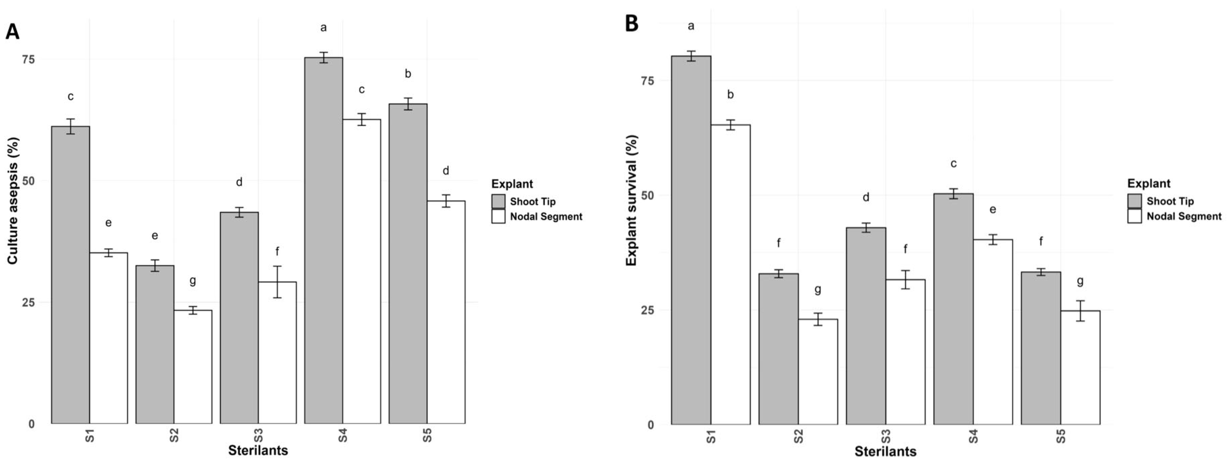
3.1. Sterilization Regime

Culture Asepsis and Explant Survival

The results of this study show that there are significant differences when using five different types of sterilants. Maximum culture asepsis for shoot tips (75.33%) was achieved after treatment with 0.05% HgCl2 for 5 min + 70% ethanol for 10 s. Similarly, in nodal segments, maximum culture asepsis (62.60%) was observed after treatment with 0.05% HgCl2 for 5 min + 70% ethanol for 10 s. In this study, the maximum explant survival (80.33%) was obtained after using shoot tips in 0.1% HgCl2 for 5 min. In addition, the maximum explant survival (65.33%) in nodal segments was obtained after using 0.1% HgCl2 for 5 min. In contrast, the lowest culture asepsis (23.33%) and explant survival (22.96%) were attained by nodal segments with 10% NaClO for 10 min (Table S1). The significant statistical differences were determined using a two-way ANOVA (Treatment and Explant) followed by Tukey’s HSD test. The results are presented as bar plots showing the mean ± SD at a 95% confidence level (p ≤ 0.05), with different letters above the bars indicating significantly different treatment groups, as shown in (Figure 1 and Figure 2A,B).
Previous research supports our findings, demonstrating that culture asepsis (77.78%) was maximized using HgCl2 along with ethyl alcohol in garden Rauvolfia (Rauvolfia tetraphylla L.) [54]. Similarly, the highest asepsis percentage was achieved using 70% ethyl alcohol for 30 s in the Arka Vaibhav cultivar of tuberose, aligning with our findings [55]. For explant sterilization, 70% ethyl alcohol and HgCl2 were used in finger lime (Citrus australasica F. Muell), as 70% ethyl alcohol exhibits superior osmotic activity and polarity compared to 100% ethanol [56]. Treating Pterocarpus santalinus L. explants with HgCl2 for 3 min reduced contamination to 18% [57]. In seed sterilization, immersing seeds in distilled water followed by immediate ethanol sterilization resulted in 87.5% sterilized seeds [58]. A sterilization treatment involving ethyl alcohol (96%), hydrogen peroxide (38%), and water in a 1:1:2 ratio, with a soaking duration of 5 min, resulted in 62.6% live and sterile nodal explants [59]. Various studies confirmed that HgCl2 is an effective sterilant against soil-borne pathogens [60,61]. Among different sterilization regimes, HgCl2 (0.1%) for 3 min yielded the highest survival rate across five Eucalyptus varieties [62]. In Gerbera (Gerbera jamesonii Bolus), a survival rate of 62.8% was achieved with HgCl2 (0.1%) for 4 min [63]. The highest survival rate (73.33 ± 11.55%) in the Bhagwa cultivar of pomegranate (Punica granatum L.) was attained using HgCl2 (0.1%) for 2 min [64]. In strawberry (Fragaria × ananassa Duch.), the maximum survival rate (72.05 ± 8.41%) was observed after HgCl2 treatment for 1 min [65]. Consistent results were reported for Pterocarpus santalinus L., where HgCl2 treatment for 3 min yielded the highest survival rate (68%) [57]. Moreover, sterilization with HgCl2 (0.1%) for 5 min resulted in a 100% survival rate in the Arka Kiran cultivar of guava [66].

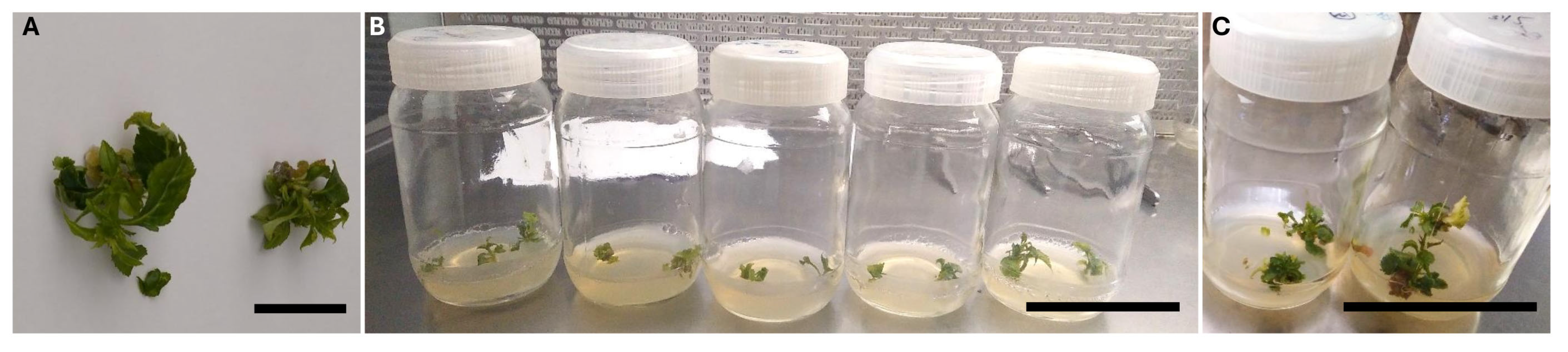
3.2. Culture Establishment

Explant Survival, Establishment, and Necrosis

In vitro culture establishment facilitates the differentiation of explants into new shoots and axillary branches from various explant types. The present study on the “GiSelA 17” rootstock of cherry involves two types of media, i.e., MS and WPM, along with two explant materials (E1 and E2), and twelve PGR regimes have been used, including BAP (0.25; 0.50; 0.75; 1.00; 1.25; and 1.50 mg/L) and BAP + IBA (0.25 + 0.01 mg/L; 0.50 + 0.01 mg/L; 0.75 + 0.01 mg/L; 1.00 + 0.01 mg/L; 1.25 + 0.01 mg/L; and 1.50 + 0.01 mg/L), for the standardizing establishment step by analyzing the mean values using a three-factor ANOVA in OPSTAT (version 6.8) at C.D (p ≤ 0.05) (see Supplementary Tables S2a,b, S3a,b and S4a,b).
This study reveals that using explants (E1) supplemented with MS medium and a plant growth regulator (PGR) regime of BAP + IBA (1.00 + 0.01 mg/L) resulted in the highest explant survival rate (93.33%). This was followed by MS medium supplemented with BAP + IBA (0.75 + 0.01 mg/L) and MS medium with BAP (1 mg/L) for explants (E1), achieving a survival rate of 91.00%. In contrast, lower survival rates (36.66%) were observed in nodal segments (E2) cultured on WPM supplemented with BAP + IBA (1.5 + 0.01 mg/L) (see Supplementary Table S2a,b).
The highest culture establishment rate (83.33%) was observed with explants (E1) cultured on MS medium supplemented with BAP + IBA (1 + 0.01 mg/L). This was followed by MS medium supplemented with BAP + IBA (0.75 + 0.01 mg/L) and MS medium with BAP (1.00 mg/L), achieving an establishment rate of 81% for explants (E1). In contrast, the lowest establishment rate (26.66%) was observed in nodal segments (E2) cultured on WPM supplemented with BAP + IBA (1.50 + 0.01 mg/L) (Supplementary Table S3a,b) (Figure 2C and Figure 3A).
Minimal necrosis (6.66%) was observed in explants (E1) cultured on MS medium supplemented with BAP + IBA (1.00 + 0.01 mg/L). This was followed by MS medium with BAP + IBA (0.75 + 0.01 mg/L), which resulted in a necrosis rate of 9.33%, and MS medium with BAP (1.00 mg/L), which showed a necrosis rate of 9.00% for explants (E1). In contrast, higher necrosis (63.33%) was recorded in nodal segments (E2) cultured on WPM supplemented with BAP + IBA (1.5 + 0.01 mg/L) (Supplementary Table S4a,b).
Statistical differences were analyzed using a three-way ANOVA (Treatment, Media, and Explant), followed by Tukey’s HSD test. The results are presented as bar plots showing the mean ± SD at a 95% confidence level (p ≤ 0.05), with different letters above the bars indicating significantly different treatment groups, as depicted in Figure 4A–C.
The establishment of culture material facilitates the differentiation of explants into new shoots and axillary branches. Similarly to our study, the highest survival rate of meristem explants (79.20%) was observed in strawberry cv. Toro, which was cultured on MS medium during the establishment phase [67]. Likewise, two banana varieties, Amritasagar and Sabri, exhibited maximum survival rates of 90% and 80%, respectively, when cultured on MS medium supplemented with BAP [68]. In the cherry rootstock “GiSelA5” (Prunus cerasus × Prunus canescens), Thakur et al. [69] reported that the maximum bud establishment occurred on MS medium supplemented with BAP (0.5 mg/L). Our findings align with previous research, which demonstrated that the highest establishment rate (88.88 ± 1.01%) was achieved using BAP + IBA (3.00 + 1.00 mg/L) with no necrotic cultures in Sumac (Rhus coriaria L.) [70]. BAP, a cytokinin, plays a crucial role in plant development by promoting cell differentiation and lateral shoot formation. Using BA + IBA (0.5 + 0.2 µM), a maximum bud establishment rate of 58.95% was observed in the Frontier cultivar of plum (Prunus salicina L.) [71]. Additionally, a lower BAP concentration (1.00 mg/L) minimized necrosis while promoting callus establishment in Artemisia annua L. [72]. The highest establishment rate (76.67%) was observed in Ardisia crenata var. bicolor using MS medium supplemented with BAP + IBA (1.00 + 0.50 mg/L) [73]. In Cenchrus ciliaris L., suspension cell cultures achieved maximum establishment on MS medium [74]. Moreover, a 100% survival rate for healthy plants was recorded in Aloe elegans Tod when cultured on MS medium with BAP (0.25 mg/L) [75].

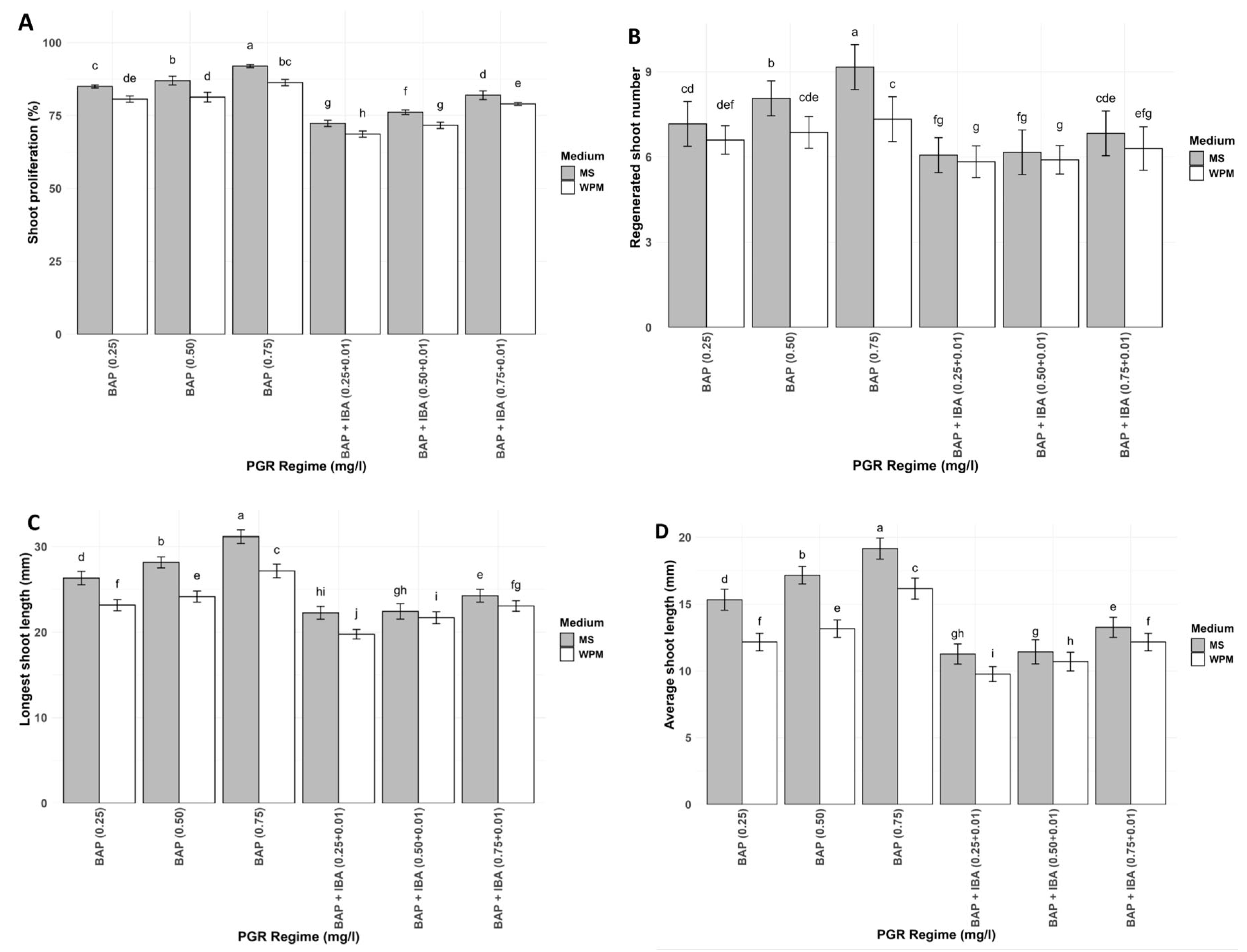
3.3. Shoot Regeneration

Upon subculturing, the established cultures of GiSelA 17 shoot tips, which showed promising results during the establishment phase, were transferred to a fresh medium comprising MS and WPM. The shoots were divided into stem segments measuring 0.5–1.0 cm in length. Mini-shoots were treated with six different PGR regimes to induce shoot proliferation: BAP (0.25 mg/L, 0.50 mg/L, and 0.75 mg/L) and BAP + IBA (0.25 + 0.01 mg/L, 0.50 + 0.01 mg/L, and 0.75 + 0.01 mg/L). The mean values were analyzed using a two-factor ANOVA in OPSTAT (version 6.8) at a critical difference (C.D.) of p ≤ 0.05. The highest shoot proliferation rate (92.00%) and greatest number of regenerated shoots (9.16) were achieved using MS medium with BAP (0.75 mg/L). This was followed by MS medium with BAP (0.50 mg/L), which resulted in 87.00% shoot proliferation with 8.06 shoots per explant. In contrast, the lowest proliferation rate (68.66%) and shoot number (5.85) were recorded in WPM with BAP + IBA (0.25 + 0.01 mg/L) (Supplementary Table S5a) (Figure 2D and Figure 3B,C). The average shoot length (19.16 mm) and the longest shoot (31.16 mm) were the highest in the MS medium with BAP (0.75 mg/L). This was followed by MS with BAP (0.50 mg/L), which resulted in an average shoot length of 17.16 mm and the longest shoot length (28.16 mm). Conversely, the shortest average shoot length (9.76 mm) was recorded in WPM with BAP + IBA (0. 25 + 0.01 mg/L), while the longest shoot length (17.16 mm) was observed in WPM and BAP (0.75 mg/L) (Supplementary Table S5b) (Figure 2D and Figure 3C).
Significant statistical differences were determined using a two-way ANOVA (Treatment and Media) followed by Tukey’s HSD test. The results are presented as bar plots showing the mean ± SD at a 95% confidence level (p ≤ 0.05), with different letters above the bars indicating significantly different treatment groups (Figure 5A–D).
Our findings align with previous research, where the maximum shoot proliferation (63.6% ± 0.63) and shoot number (13 ± 0.54) were observed in the S1 ecotype of Moroccan almond (Prunus dulcis Mill.) cultured on MS medium [76]. Additionally, a shoot proliferation index of 3.93 was reported using MS medium supplemented with 6-BA (2.0 mg/L), while the longest shoots were recorded with MS medium containing 6-BA (1.8 mg/L) in the M9T337 rootstock of apple (Malus × domestica Borkh) [77].
The MS medium consistently demonstrated a higher shoot proliferation rate. For instance, an average of 7.1 shoots per explant was recorded on MS medium supplemented with BAP (3 mg/L), while a proliferation rate of 8.0 was achieved in the Poovan cultivar of banana under the same conditions [78]. Similarly, MS medium supplemented with BAP (1 mg/L) supported optimal shoot initiation in the Gudiene variety, whereas the Belete variety exhibited increased shoot production at the same concentration. Additionally, MS combined with BAP has been shown to enhance shoot proliferation rates by 2–3 times in potato (Solanum tuberosum L.) [79].
Leaf explants demonstrated a high proliferation rate (92.73%) with an average of 53.5 ± 0.47 shoots and an average shoot length of 11.2 ± 0.53 cm on MS medium in Solanum khasianum Clarke [80]. In marigold (Dendranthema grandiflora T.), shoot numbers of 5.25, 8.65, and 11.90 per explant and shoot lengths of 4.45, 5.55, and 7.45 cm were observed using MS medium supplemented with BAP (1.5 mg/L) [81].
Furthermore, maximum proliferation (82.2%) and a shoot number of 2.1 were reported in Tilia mandshurica (Rupr. and Maxim) using MS medium with BAP (1.0 mg/L) [82]. Enhanced shoot growth was also noted in apple when cultured on MS medium supplemented with BAP (0.5 mg/L) [83]. In Vigna radiata L. a maximum shoot length of 4.51 cm was recorded after inoculating shoot tips into MS medium [84].
In Passiflora caerulea, complete shoot regeneration (100%) was achieved with BAP (0.77 mg/L) [85]. Additionally, in the Philodendron erubescens variety “Pink Princess”, an average of 11.2 shoots per explant was obtained using MS medium fortified with BAP (1 mg/L) [86].
These findings collectively highlight the efficacy of MS medium, particularly when combined with optimized BAP concentrations, in enhancing shoot proliferation, shoot numbers, and shoot length across various plant species. MS medium’s rich nutrient composition supports the growth and regeneration of a diverse range of plant species, including both herbaceous and woody plants. In contrast, WPM is specifically formulated for woody plants and may not provide a suitable nutrient balance for other plant types.
Traditional propagation methods for clonal rootstocks offer limited yield benefits. In contrast, micropropagation presents several advantages over conventional techniques. This method involves cultivating plants in a nutrient-rich medium supplemented with an exogenous carbon source and plant growth regulators, thereby enhancing shoot vigor and accelerating the production of novel crop varieties with elite traits while requiring less time and space. By utilizing artificial media under aseptic conditions, in vitro propagation ensures true-to-type, high-quality planting material.
The nursery industry greatly benefits from the in vitro propagation of rootstocks as it is more efficient and feasible than other vegetative propagation methods. Micropropagation enables the mass production of clones that are otherwise difficult to root, making it a valuable technique for commercial plant propagation.
“GiSelA 17” (Prunus canescens × Prunus avium), a novel cherry clonal rootstock with a precocious bearing, resistance to replant situations, and a high tolerance to the menaces of Prunus dwarf virus (PDV) and Prunus necrotic ring spot virus (PNRSV). Nonetheless, our research is the first in vitro study on the “GiSelA 17” rootstock that focuses on generating the best quality planting material for commercial cherry production.

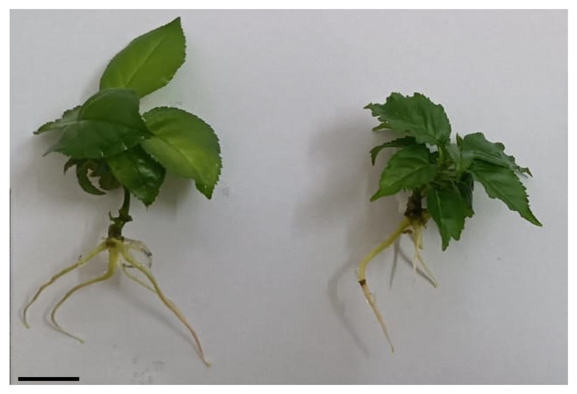
3.4. Rooting Process

The rooting process is a key step in the micropropagation protocol, essential for successful soil acclimatization. Roots can be considered as uni-polar auxin canalization of shoot-derived auxin, while exogenous auxin acts as an inducer of de novo root primordia from competent cells in the newly formed hypocotyl/stem [87]. In our protocol, we used IBA as an exogenous auxin in a concentration of up to 3 mg/L. The results demonstrate that the most effective concentration was 1.5 mg/L (Figure 6, Supplementary Table S6).
Seedlings with well-developed root systems can be readily transferred to the soil to complete micropropagation.

3.5. Hardening Process

The hardening and acclimatization of in vitro-generated plantlets are essential steps for ensuring better survival and the successful establishment of plant in soil. In this study, different substrates were used for hardening. An adaptation rate of approximately 80% was achieved under natural conditions (Table 2; Figure 7), confirming that micropropagation methods are economically feasible and can be used in agricultural practices.
Moreover, the main reason for using in vitro propagation is that, in traditional cutting methods, one shoot can produce an average of five plants. However, in vitro, a single explant can produce an average of 100 plants through up to five subcultures.

4. Conclusions

The current study represents the first approach to depicting and highlighting the aspects of micropropagation on the culture establishment and shoot proliferation of the novel clonal rootstock of cherry “GiSelA 17” (Prunus canescens × Prunus avium) within a 2-month period. The promising results demonstrate that the proliferation rate is 92.00%, which clearly paves the way for the production of disease-free and quality planting material for the commercialization of the “GiSelA 17” clonal rootstock of cherry for propagation and breeding purposes in temperate cherry-growing regions worldwide. Currently, there are no reports on the development of virus-free planting material that is resistant to replant disease for cherry orchards through conventional propagation methods. In this context, our research represents a breakthrough for developing high-density cherry orchards in the shortest possible amount of time.

Supplementary Materials

The following supporting information can be downloaded at https://www.mdpi.com/article/10.3390/ijpb16010033/s1, Table S1: Influence of sterilant type and explant origin on culture asepsis (%) and explant survival (%) in cherry rootstock “GiSelA 17” (Prunus canescens × Prunus avium); Table S2: (a,b) Effect of explant origin, plant growth regulator regime, and media type on explant survival (%) in cherry rootstock “GiSelA 17” (Prunus canescens × Prunus avium); Table S3: (a,b) Effect of explant origin, plant growth regulator, and media type on establishment (%) in cherry rootstock “GiSelA 17” (Prunus canescens × Prunus avium); Table S4: (a,b) Effect of explant origin, plant growth regulator regime, and media type on necrosis (%) in cherry rootstock “GiSelA 17” (Prunus canescens × Prunus avium); Table S5: (a) Effect of plant growth regulator regime and media type on shoot proliferation (%) and regenerated shoot number in cherry rootstock “GiSelA 17” (Prunus canescens × Prunus avium). (b) Effect of plant growth regulator regime and media type on average shoot length (mm) and longest shoot length (mm) in cherry rootstock “GiSelA 17” (Prunus canescens × Prunus avium); Table S6: Influence of plant growth regulators on rooting (%), number of roots (%), and average root length (mm) in cherry rootstock “GiSelA 17” (Prunus canescens × Prunus avium).

Author Contributions

Conceptualization, I.M., K.M.B. and M.A.M.; methodology, I.M., N.M.N. and A.H.P.; formal analysis, I.M., K.M.B. and U.K.; investigation, I.M.; resources, N.M.N.; data curation, I.M., U.K. and M.A.M.; writing—original draft preparation, I.M., S.Q., S.D. and K.M.B.; writing—review and editing, I.M.; visualization, K.M.B.; supervision, A.H.P.; project administration, I.M.; acquisition of funds and final writing and editing of text, T.P. All authors have read and agreed to the published version of the manuscript.

Funding

This research received no external funding.

Data Availability Statement

The data are contained within the article.

Acknowledgments

The authors are thankful to the Science & Engineering Research Board (SERB), the Department of Science and Technology, (GOI) in association with the Confederation of Indian Industry (CII) for fellowship and the Commercial Tissue Culture Laboratory, Seven Star Fruits Private Limited, Maharashtra Hybrid Seeds Co. (MahycoGrow), Dawalwadi, Jalna, Auranagabad (Maharashtra), for providing the research facilities, fellowship, and plant material to conduct this research.

Conflicts of Interest

Authors Narendran M. Nair, Shoeb Quadri, and Smithal Deshmukh were employed by the company Commercial Tissue Culture & Nursery, Seven Star Fruits Private Limited. The remaining authors declare that the research was conducted in the absence of any commercial or financial relationships that could be construed as a potential conflict of interest.

References

  1. GiSelA®17. Available online: https://battistinivivai.com/en/products/gisela-17-rootstock (accessed on 29 November 2024).
  2. Milkovich, M.; Mullinax, T.J. Good Fruit Grower: A Lot of Choices for Cherry Rootstocks. Available online: https://www.goodfruit.com/a-lot-of-choices-for-cherry-rootstocks/ (accessed on 28 November 2024).
  3. Wolfe, D.E.; Strang, J.; Becker, D.; Wright, S. Rootstocks for Kentucky Fruit Trees. University of Kentucky. College of Agriculture, Food and Environment. Cooperative Extension Service. Available online: http://www2.ca.uky.edu/agc/pubs/HO/HO82/HO82.pdf. (accessed on 2 December 2024).
  4. Lang, G.A. Critical concepts for sweet cherry training systems. Compact Fruit Tree 2001, 34, 70–75. [Google Scholar]
  5. Gregory, P.J.; Atkinson, C.J.; Bengough, A.G.; Else, M.A.; Fernandez-Fernandez, F.; Harrison, R.J.; Schmidt, S. Contributions of roots and rootstocks to sustainable, intensified crop production. J. Exp. Bot. 2013, 64, 1209–1222. [Google Scholar] [CrossRef] [PubMed]
  6. Hrotko, K. Development in fruit trees production systems. AgroLife Sci. J. 2013, 2, 28–35. [Google Scholar]
  7. Ljubojevic, M. Horticulturalization of the 21st century cities. Sci. Hortic. 2021, 288, 110350. [Google Scholar] [CrossRef]
  8. Atkinson, C.; Else, M. Understanding how rootstocks dwarf fruit trees. Compact Fruit Tree 2001, 34, 46–49. [Google Scholar]
  9. Webster, A.D. Vigour mechanisms in dwarfing rootstocks for temperate fruit trees. Acta Hortic. 2002, 658, 29–41. [Google Scholar] [CrossRef]
  10. Hrotko, K. Progress in cherry rootstock research. Acta Hortic 2008, 795, 171–178. [Google Scholar] [CrossRef]
  11. Gjamovski, V.; Kiprijanovski, M. Influence of nine dwarfing apple rootstocks on vigour and productivity of apple cultivar ‘Granny Smith’. Sci. Hortic. 2011, 129, 742–746. [Google Scholar] [CrossRef]
  12. Marra, F.P.; Bianco, R.L.; La Mantia, M.; Caruso, T. Growth, yield and fruit quality of ‘Tropic Snow’ peach on size-controlling rootstocks under dry Mediterranean climates. Sci. Hortic. 2013, 160, 274–282. [Google Scholar] [CrossRef]
  13. Tworkoski, T.; Fazio, G. Hormone and growth interactions of scions and size-controlling rootstocks of young apple trees. Plant Growth Regul. 2016, 78, 105–119. [Google Scholar] [CrossRef]
  14. Yahmed, J.B.; Ghrab, M.; Mimoun, M.B. Eco-physiological evaluation of different scion-rootstock combinations of almond grown in Mediterranean conditions. Fruits 2016, 71, 185–193. [Google Scholar] [CrossRef]
  15. Bujdoso, G.; Magyar, L.; Hrotko, K. Long term evaluation of growth and cropping of sweet cherry (Prunus avium L.) varieties on different rootstocks under Hungarian soil and climatic conditions. Sci. Hortic. 2019, 256, 108613. [Google Scholar] [CrossRef]
  16. Webster, T. Dwarfing rootstocks: Past, present and future. Compact Fruit Tree 2002, 35, 67–72. [Google Scholar]
  17. Santos, A.; Santos-Ribeiro, R.; Cavalheiro, J.; Cordeiro, V.; Lousada, J.L. Initial growth and fruiting of ‘Summit’ sweet cherry (Prunus avium) on five rootstocks. N. Z. J. Crop Hortic. Sci. 2006, 34, 269–277. [Google Scholar] [CrossRef]
  18. Wocior, S. The effect of rootstock on the growth and yielding of ‘Regina’ cherry trees. Folia Hortic. 2008, 20, 15–22. [Google Scholar] [CrossRef]
  19. Lopez-Ortega, G.; Garcia-Montiel, F.; Bayo-Canha, A.; Frutos-Ruiz, C.; Frutos-Tomas, D. Rootstock effects on the growth, yield and fruit quality of sweet cherry cv. ‘Newstar’ in the growing conditions of the region of Murcia. Sci. Hortic. 2016, 198, 326–335. [Google Scholar] [CrossRef]
  20. Dziedzic, E.; Błaszczyk, J.; Kaczmarczyk, E. Postharvest properties of sweet cherry fruit depending on rootstock and storage conditions. Folia Hortic. 2017, 29, 113–121. [Google Scholar] [CrossRef]
  21. Pal, M.D.; Mitre, I.; Asanica, A.C.; Sestraș, A.F.; Peticila, A.G.; Mitre, V. The influence of rootstock on the growth and fructification of cherry cultivars in a highdensity cultivation system. Not. Bot. Horti Agrobot. 2017, 45, 451–457. [Google Scholar] [CrossRef]
  22. Balducci, F.; Capriotti, L.; Mazzoni, L.; Medori, I.; Albanesi, A.; Giovanni, B.; Giampieri, F.; Mezzetti, B.; Capocasa, F. The rootstock effects on vigor, production and fruit quality in sweet cherry (Prunus avium L.). J. Berry Res. 2019, 9, 249–265. [Google Scholar] [CrossRef]
  23. Morandi, B.; Manfrini, L.; Lugli, S.; Tugnoli, A.; Boini, A.; Perulli, G.D.; Bresilla, K.; Venturi, M.; Grappadelli, L.C. Sweet cherry water relations and fruit production efficiency are affected by rootstock vigor. J. Plant Physiol. 2019, 237, 43–50. [Google Scholar] [CrossRef]
  24. Callesen, O. Recent developments in cherry rootstock research. Acta Hortic. 1998, 468, 219–228. [Google Scholar] [CrossRef]
  25. Wertheim, S.J. Rootstock Guide: Apple, Pear, Cherry, European Plum; Fruit Research Station: Wilhelminadorp, The Netherlands, 1998. [Google Scholar]
  26. Milosevic, T.; Milosevic, N.; Mladenovic, J. Combining fruit quality and main antioxidant attributes in the sour cherry: The role of new clonal rootstock. Sci. Hortic. 2020, 265, 109236. [Google Scholar] [CrossRef]
  27. Solonkin, A.; Nikolskaya, O.; Seminchenko, E. The effect of low-growing rootstocks on the adaptability and productivity of sour cherry varieties (Prunus cerasus L.) in Arid Conditions. Horticulturae 2022, 8, 400. [Google Scholar] [CrossRef]
  28. Cline, J.A. Planting density and size-controlling rootstocks influence the performance of Montmorency tart cherry (Prunus cerasus L.). Can. J. Plant Sci. 2019, 100, 16–28. [Google Scholar] [CrossRef]
  29. Barac, G.; Ognjanov, V.; Vidakovic, D.O.; Doric, D.; Ljubojevic, M.; Dulic, J.; Miodragovic, M.; Gasic, K. Genetic diversity and population structure of European ground cherry (Prunus fruticose Pall.) using SSR markers. Sci. Hortic. 2017, 224, 374–383. [Google Scholar] [CrossRef]
  30. Ljubojevic, M.; Sebolt, A.; Ognjanov, V.; Iezzoni, A. Heritability of anatomical characteristics in cherry interspecific hybrids. J. Plant Growth Regul. 2022, 41, 965–982. [Google Scholar] [CrossRef]
  31. Hrotko, K. Potentials in Prunus mahaleb L. for cherry rootstock breeding. Sci. Hortic. 2016, 205, 70–78. [Google Scholar] [CrossRef]
  32. Bosnjak, A.M.; Keresa, S.; Jercic, I.H.; Baric, M. The effect of cytokinin type and explant orientation on axillary shoot proliferation and in vitro rooting of ‘GiSelA 5’ cherry rootstock. J. Food Agric. Environ. 2012, 10, 616–620. [Google Scholar]
  33. Bhojwani, S.S.; Dantu, P.K. Micropropagation. In Plant Tissue Culture: An Introductory Text; Bhojwani, S.S., Dantu, P.K., Eds.; Springer: New Delhi, India, 2013. [Google Scholar]
  34. Chandran, H.; Meena, M.; Barupal, T.; Sharma, K. Plant tissue culture as a perpetual source for production of industrially important bioactive compounds. Biotechnol. Rep. 2020, 26, e00450. [Google Scholar] [CrossRef]
  35. Krasteva, G.; Georgiev, V.; Pavlov, A. Recent applications of plant cell culture technology in cosmetics and foods. Eng. Life Sci. 2020, 21, 68–76. [Google Scholar] [CrossRef]
  36. George, E.F.; Hall, M.A.; De Klerk, G.J. Plant Propagation by Tissue Culture; Springer Science & Business Media: Berlin/Heidelberg, Germany, 2007; Volume 1. [Google Scholar]
  37. Cardoso, J.C.; Lee Tseng Sheng, G.; Teixeira da Silva, J.A. Micropropagation in the Twenty-First Century. In Plant Cell Culture Protocols; Springer: Berlin/Heidelberg, Germany, 2018. [Google Scholar]
  38. Boonsnongcheep, P.; Benyakan, P. Factors affecting micropropagation of Cannabis sativa L.: A review. Pharm. Sci. Asia. 2020, 47, 21–29. [Google Scholar] [CrossRef]
  39. Adhikary, D.; Kulkarni, M.; El-Mezawy, A.; Mobini, S.; Elhiti, M.; Gjuric, R.; Ray, A.; Polowick, P.; Slaski, J.J.; Jones, M.; et al. Medical cannabis and industrial hemp tissue culture: Present status and future potential. Front. Plant Sci. 2021, 12, 627240. [Google Scholar] [CrossRef]
  40. Borsai, O.; Clapa, D.; Magdea, A.; Harta, M.; Andrecan, A.; Mitre, V. Effects of different culture media and plant growth regulators on micropropagation of ‘GiSelA 5’ cherry rootstock. Sci. Papers Ser. B Hortic. 2020, 64, 33–40. [Google Scholar]
  41. Marin, J.A.; Garcia, E.; Lorente, P.; Andreu, P.; Arbeloa, A. Assessing effect of rootstock micropropagation on field performance of grafted peach varieties by fitting mixed-effects models: A longitudinal study. Plants 2023, 12, 674. [Google Scholar] [CrossRef]
  42. Lacuzzi, N.; Salamone, F.; Farruggia, D.; Tortorici, N.; Vultaggio, L.; Tuttolomondo, T. Development of a new micropropagation protocol and transfer of in vitro plants to in vivo conditions for cascade hop. Plants 2023, 12, 2877. [Google Scholar] [CrossRef] [PubMed]
  43. Guo, M.; Yu, Q.; Li, D.; Xu, K.; Di, Z.; Zhang, Y.; Yu, Y.; Zheng, J.; Zhang, Y. In vitro propagation, shoot regeneration, callus induction, and suspension from lamina explants of Sorbus caloneura. For. Res. 2023, 3, 7. [Google Scholar] [CrossRef]
  44. Nacheva, L.; Dimitrova, N.; Koleva-Valkova, L.; Stefanova, M.; Ganeva, T.; Nesheva, M.; Tarakanov, I.; Vassilev, A. In vitro multiplication and rooting of plum rootstock ‘Saint Julien’ (Prunus domestica subsp. insititia) under fluorescent light and different LED spectra. Plants 2023, 12, 2125. [Google Scholar]
  45. Tariverdi, Z.; Nughabi, K.A.; Piri, S. Propagation of rootstocks of ‘GiSelA 5’ based on tissue culture method. Biosci. Biotech. Res. Asia 2017, 14, 1395–1399. [Google Scholar] [CrossRef]
  46. Kumar, A.; Sharma, V.; Thakur, M. In vitro rooting and hardening of clonal cherry rootstock “GiSelA 5” (Prunus cerasus × Prunus canescens). Int. J. Agr. Sci. 2020, 90, 1032–1035. [Google Scholar] [CrossRef]
  47. Sharma, K.; Thakur, M.; Sharma, V. Cost effective in vitro propagation of GiSelA 5 cherry rootstock. Indian J. Hortic. 2020, 77, 597–602. [Google Scholar] [CrossRef]
  48. Jafarlou, A.M.; Pirivatlo, S.P.; Salehi, B.; Mogbli, A.H.H. Establishing an efficient in vitro propagation system for sweet cherry rootstocks “GiSelA 12” and “Maxma14” and assessment of genetic homogeneity by ISSR markers. J. Crop Sci. Biotechnol. 2021, 24, 449–460. [Google Scholar] [CrossRef]
  49. Nicolae, I.; Venat, O.; Peticila, A.; Ștefanut, M.M.; Hoza, D. Use of different hormones on in vitro propagation of “GiSela 5” cherry rootstock. Sci. Papers Ser. B Hortic. 2022, 66, 407–412. [Google Scholar]
  50. Sarropoulou, V.; Sperdouli, I.; Adamakis, I.D.; Grigoriadou, K. The use of different LEDs wavelength and light intensities for in vitro proliferation of cherry rootstock: Influence on photosynthesis and photomorphogenesis. Plant Cell Tiss. Organ. Cult. 2023, 152, 317–330. [Google Scholar] [CrossRef]
  51. Murashige, T.; Skoog, F. A revised medium for rapid growth and bio assays with tobacco tissue cultures. Plant Physiol. 1962, 15, 473–497. [Google Scholar] [CrossRef]
  52. Lloyd, G.; McCown, B. Commercially-feasible micropropagation of mountain laurel, Kalmia latifolia, by use of shoot-tip culture. Comb. Proc. Int. Plant Propag Soc. 1980, 30, 421–427. [Google Scholar]
  53. Zhang, A.; Wang, H.; Shao, Q.; Xu, M.; Zhang, W.; Li, M. Large-scale in vitro propagation of Anoectochilus roxburghii for commercial application: Pharmaceutically important and ornamental plant. Ind. Crop. Prod. 2015, 70, 158–162. [Google Scholar] [CrossRef]
  54. Mamatha, D.S.; Nishani, S.; Mahantesha, B.N.; Hegde, L.; Shet, R.; Prasad, D.S. Tissue culture studies on garsden Rauvolfia (Rauvolfia tetraphylla L.). J. Pharm. Innov. 2021, 10, 1205–1210. [Google Scholar]
  55. Patil, M.; Bharathi, T.U.; Usharani, T.R.; Kumar, R.; Kulkarni, B.S. Standardization of sterilization protocol for explants and its suitability for direct organogenesis in tuberose cv. Arka Vaibhav. J. Hortic. Sci. 2023, 18, 173–180. [Google Scholar] [CrossRef]
  56. Supreetha, B.G.; Prakasha, D.P.; Kulapati Hipparagi, P.G. Optimizing sterilization protocols for in vitro culture establishment of finger lime (Citrus australasica F. Muell): A comprehensive investigation. J. Pharm. Innov. 2023, 12, 355–361. [Google Scholar]
  57. Shaheen, S.; Ankanna, S.; Pattanaik, S.; Savithramma, N. Standardization of explant sterilization for in vitro propagation of Pterocarpus Santalinus L.F.-An endemic, endangered multipurpose medicinal tree taxon. Int. J. Creat. Res. Thoughts 2023, 11, 307–317. [Google Scholar]
  58. Chokheli, V.; Bakulin, S.; Ermolaeva, O.; Rajput, V.; Azarov, A.S.; Kumari, A.; Stepanenko, V.; Bushkova, A.; Dmitriev, P.; Martinez-Montero, M.E.; et al. An Efficient Method for Micropropagation of Red-List Herbaceous Plant Species (Hedysarum cretaceum). OBM Genet. 2023, 7, 1–14. [Google Scholar] [CrossRef]
  59. Sereda, M.; Petrenko, V.; Kapralova, O.; Chokheli, V.; Varduni, T.; Dmitriev, P.; Minkina, T.; Sushkova, S.; Barbashev, A.; Dudnikova, T.; et al. Establishment of an In Vitro Micropropagation Protocol for Hibiscus moscheutos L. ‘Berry Awesome’. Horticulturae 2023, 10, 21. [Google Scholar] [CrossRef]
  60. Ansar, S.; Iqbal, M.J.T.R. Effect of dietary antioxidant on mercuric chloride induced lung toxicity and oxidative stress. Toxin Rev. 2015, 34, 168–172. [Google Scholar] [CrossRef]
  61. Palei, S.; Das, A.K.; Dash, D.K.; Rout, G.R. Effect of surface sterilant for reducing microbial contamination of field grown strawberry explants intended for in vitro culture. Int. Chem. Stud. 2017, 5, 1476–1479. [Google Scholar]
  62. Kuppusamy, S.; Ramanathan, S.; Sengodagounder, S.; Senniappan, C.; Shanmuganathan, R.; Brindhadevi, K.; Kaliannan, T. Optimizing the sterilization methods for initiation of the five different clones of the Eucalyptus hybrid species. Biocatal. Agric. Biotechnol. 2019, 22, 101361. [Google Scholar] [CrossRef]
  63. Kumar, M.; Prasad, Y.; Yadav, A.; Kumar, A. Effects of two different surface sterilization (sodium hypochlorite and mercuric chloride) agents under in-vitro leaf explant in Gerbera (Gerbera jamesonii Bolus). J. Pharm. Innov. 2021, 10, 1346–1349. [Google Scholar]
  64. Pal, D.; Kumar, M.; Kumar, A.; Sengar, R.S.; Yadav, M.K.; Pal, A.; Singh, K.P.; Pandey, V. Standardization of Surface Sterilization for in vitro Cloning of Pomegranate (Punica granatum L.) cv. Bhagwa. Biol. Forum. 2022, 14, 653–656. [Google Scholar]
  65. Pandey, V.; Kumar, A.; Kumar, V.; Prakash, S.; Gangwar, L.K.; Sengar, R.S.; Kumar, M.; Pal, A.; Pal, D.; Kumar, A. Standardize the aseptic environment protocol for strawberry in vitro cloning (Fragaria × ananassa Duch.). J. Pharm. Innov. 2023, 12, 1443–1449. [Google Scholar]
  66. Bhyravi, B.M.; Athani, S.I.; Prakasha, D.P.; Anil, I.S.; MS, K.; Nagesh Naik, D.A.; Puspha, T.N.; Yallesh Kumar, H.S. Effect of different sterilization agents on in vitro culture establishment of guava cv. Arka Kiran. J. Pharm. Innov. 2023, 12, 1761–1766. [Google Scholar]
  67. Munir, M.; Iqbal, S.; Baloch, J.U.D.; Khakwani, A.A. In vitro explant sterilization and bud initiation studies of four strawberry cultivars. J. Appl. Hortic. 2015, 17, 192–198. [Google Scholar] [CrossRef]
  68. Ferdous, M.H.; Billah, A.M.; Mehraj, H.; Taufique, T.; Uddin, A.F.M.J. BAP and IBA pulsing for in vitro multiplication of banana cultivars through shoot-tip culture. J. Biosci. Agric. Res. 2015, 3, 87–95. [Google Scholar] [CrossRef]
  69. Thakur, M.; Sharma, V.; Sharma, D.P.; Kumari, G.; Vivek, M. In Vitro Propagation of Virus Indexed Gisela-5 (Prunus cerasus × Prunus canescens)-Clonal Cherry Rootstock. Int. J. Crop Sci. Tech. 2016, 2, 87–99. [Google Scholar]
  70. Amiri, S.; Mohammadi, R. Establishment of an efficient in vitro propagation protocol for Sumac (Rhus coriaria L.) and confirmation of the genetic homogeneity. Sci. Rep. 2021, 11, 173. [Google Scholar] [CrossRef]
  71. Thakur, M.; Sharma, V.; Luharch, R. Propagation of plum (Prunus salicina L.) cultivar Frontier in vitro through control of shoot tip necrosis (STN) and validation of genetic integrity using ISSR markers. Plant Physiol. Rep. 2021, 26, 238–246. [Google Scholar] [CrossRef]
  72. Bahgat, H.; Hassan, S.A.M.; Salaheldin, S.; Abou Ellail, M. Tissue culture protocol establishment of Artemisia annua L. plant and artemisinin production. SVU-Int. J. Agric. Sci. 2021, 3, 73–83. [Google Scholar] [CrossRef]
  73. Ai, X.; Wen, Y.; Wang, B. Assessment of genetic stability on in vitro propagation of Ardisia crenata var. bicolorusing ISSR markers. Int. J. Plant Biol. 2023, 14, 218–227. [Google Scholar] [CrossRef]
  74. Goyal, S.; Chatterjee, V.; Kulkarni, V.M.; Bhat, V. Plant regeneration through somatic embryogenesis in cell suspensions of Cenchrus ciliaris L. Plant Methods. 2023, 19, 110. [Google Scholar] [CrossRef]
  75. Welehaweria, M.; Sbhatu, D.B. In vitro micropropagation of Aloe elegans Tod. using offshoot cuttings. BMC Res. Notes 2023, 16, 215. [Google Scholar] [CrossRef]
  76. Kodad, S.; Melhaoui, R.; Hano, C.; Addi, M.; Sahib, N.; Elamrani, A.; Abid, M.; Mihamou, A. Effect of culture media and plant growth regulators on shoot proliferation and rooting of internode explants from Moroccan native Almond (Prunus dulcis Mill.) genotypes. Int. J. Agron. 2021, 2021, 9931574. [Google Scholar] [CrossRef]
  77. Shi, J.L.; Dong, Z.D.; Song, C.H.; Xie, B.Y.; Zheng, X.B.; Song, S.W.; Jiao, J.; Wang, M.; Bai, T.H. Establishment of an efficient micropropagation system in enhancing rooting efficiency via stem cuttings of apple rootstock M9T337. Hort. Sci. 2021, 48, 63–72. [Google Scholar] [CrossRef]
  78. Sivakumar, P.; Visalakshi, M. In vitro micropropagation of banana cv. Poovan (AAB). J. Appl. Hortic. 2021, 23, 37–41. [Google Scholar] [CrossRef]
  79. Hajare, S.T.; Chauhan, N.M.; Kassa, G. Effect of growth regulators on in vitro micropropagation of potato (Solanum tuberosum L.) Gudiene and Belete varieties from Ethiopia. Sci. World J. 2021, 2021, 5928769. [Google Scholar] [CrossRef] [PubMed]
  80. Chirumamilla, P.; Gopu, C.; Jogam, P.; Taduri, S. Highly efficient rapid micropropagation and assessment of genetic fidelity of regenerants by ISSR and SCoT markers of Solanum khasianum Clarke. Plant Cell Tissue Organ. Cult. 2021, 144, 397–407. [Google Scholar] [CrossRef]
  81. Sushmarani, Y.S.; Venkatesha Murthy, P.; Deeksha Raj, N. Influence of BAP with TDZ growth regulators on in vitro regeneration in chrysanthemum (Dendranthema grandiflora T.) cv. Marigold. J. Pharmcog. Phytochem. 2021, 10, 1171–1176. [Google Scholar]
  82. Kim, T.D.; Kim, N.H.; Park, E.J.; Lee, N.N. High-frequency regeneration of plants in vitro from seedling-derived apical bud explants of Tilia mandshurica Rupr. & Maxim. J. Plant Biotechnol. 2021, 48, 54–61. [Google Scholar]
  83. Cabral-Miramontes, J.P.; Chavez-Simental, J.A.; Pulido-Díaz, C.; González-Portillo, M.; Goche-Télles, J.R.; Barragán-Hernández, V.M. In vitro propagation of apple tree from mature zygotic embryos. Rev. Mexicanacienc. Agríc. 2022, 13, 603–616. [Google Scholar]
  84. Iqbal, Z.; Javad, S.; Naz, S.; Shah, A.A.; Shah, A.N.; Paray, B.A.; Gulnaz, N.; Abdelsalam, N.R. Elicitation of the in vitro cultures of selected varieties of Vigna radiata L. with zinc oxide and copper oxide nanoparticles for enhanced phytochemicals production. Front. Plant Sci. 2022, 13, 908532. [Google Scholar] [CrossRef]
  85. Jafari, M.; Daneshvar, M.H. Prediction and optimization of indirect shoot regeneration of Passiflora caerulea using machine learning and optimization algorithms. BMC Biotechnol. 2023, 23, 27. [Google Scholar] [CrossRef]
  86. Klanrit, P.; Kitwetcharoen, H.; Thanonkeo, P.; Thanonkeo, S. In vitro propagation of Philodendron erubescens ‘Pink Princess’ and Ex Vitro Acclimatization of the Plantlets. Horticulturae 2023, 9, 688. [Google Scholar] [CrossRef]
  87. Pasternak, T.P.; Steinmacher, D. Plant growth regulation in cell and tissue culture in vitro. Plants 2024, 13, 327. [Google Scholar] [CrossRef]
Figure 1. The influence of sterilant type and explant origin on (A) Culture asepsis (%). (B) Explant survival (%). (S1) 0.1% HgCl2 for 5 min; (S2) 10% NaClO for 10 min, (S3) 70% ethyl alcohol for 10 s, (S4) 0.05% HgCl2 for 5 min + 70% ethanol for 10 s; (S5) 5% NaClO for 10 min + 70% ethyl alcohol for 10 s. The error bars represent the mean ± SD with small constant (±0.5) added for better visualization. This adjustment does not affect the statistical analysis or interpretation of the results. The letters (e.g., “de” and “klm”) in the plots represent statistical groupings obtained from Tukey’s Honest Significant Difference (HSD) test. Treatments sharing at least one letter are not significantly different from each other at the chosen significance level (e.g., 0.05). Conversely, treatments with entirely different letters indicate a significant difference. These groupings help in identifying statistically similar and distinct treatments based on their means.
Ijpb 16 00033 g001
Figure 2. Visualization of in vitro protocol for culture establishment and shoot proliferation of “GiSelA 17” (Prunus canescens × Prunus cerasus) novel cherry clonal rootstock. (A) Explants after sterilization of shoot tips with 0.05% HgCl2 for 5 min + 70% ethanol for 10 s; (B) explant survival using 0.1% HgCl2 for 5 min with shoot tips; (C) establishment with shoot tips on MS medium with BAP + IBA (1 + 0.01 mg/L); (D) shoot proliferation on MS medium with BAP (0.75 mg/L). Scale bar—10 mm.
Ijpb 16 00033 g002
Figure 3. Images of nodal segments (E2) with WPM with various PGRs. (A) Establishment with (E2) explant material and WPM with BAP + IBA (1.5 + 0.01 mg/L). (B) Shoot proliferation using WPM with BAP (0.25 mg/L). (C) Shoot elongation. Scale bars: (A) 10 mm; (B,C) 80 mm.
Ijpb 16 00033 g003
Figure 4. Effect of plant growth regulators, media type, and explant origin on (A) explant survival (%); (B) establishment (%); and (C) necrosis (%). Graphs with error bars represent mean ± SD with small constant (±0.5) added for improved images without affecting statistical interpretation. Letters (e.g., “de”, “klm”) in plots represent statistical groupings obtained from Tukey’s Honest Significant Difference (HSD) test. Treatments sharing at least one letter are not significantly different from each other at chosen significance level (e.g., 0.05). Conversely, treatments with entirely different letters indicate significant difference. These groupings help in identifying statistically similar and distinct treatments based on their means.
Ijpb 16 00033 g004
Figure 5. Effect of plant growth regulators and media type on (A) shoot proliferation (%); (B) de novo-generated shoot number; (C) average shoot length (mm); and (D) longest shoot length (mm). Graphs showing statistical differences in data have error bars representing mean ± SD plus small constant (±0.5) to depict visuals better without affecting statistical analysis. Letters (e.g., “de” and “klm”) in plots represent statistical groupings obtained from Tukey’s Honest Significant Difference (HSD) test. Treatments sharing at least one letter are not significantly different from each other at chosen significance level (e.g., 0.05). Conversely, treatments with entirely different letters indicate significant difference. These groupings help in identifying statistically similar and distinct treatments based on their means.
Ijpb 16 00033 g005
Figure 6. Plantlets after rooting on the medium with 1.5 mg/L IBA (10 days). Scale bar—1 cm.
Ijpb 16 00033 g006
Figure 7. Plantlets after hardening (30 days after adaptation; scale bar—5 cm).
Ijpb 16 00033 g007
Table 1. Details of different types of bio-inoculants.
S. No.Bio-InoculantsDetails
1PrerakPrerak is a product containing a consortium of bacteria containing Azospirillium, Azotobacter, Phosphate Solubilizing Bacteria, and Potassium Mobilizing Bacteria that fixes Nitrogen (N), solubilizes Phosphorus (P) and zinc (Zn) and helps in the mobilization of Potassium (K). It is a product of Grow Indigo company.
2PSBPhosphate Solubilizing Bacteria is a product which solubilizes Phosphorus (P) for improving the growth of plantlets. It is a product of Grow Indigo company.
3Rallis GoldRallis Gold is a bio-inoculant of Rallis India Ltd. containing a mycorrhizal-based product which helps in the uptake of Phosphorus (P) and increases the root surface area of plantlets. It is a product of Rallis India Pvt. Ltd. company.
Table 2. Influence of cocopeat and bio-inoculants on hardening of rooted plantlets of GiSelA 17.
TreatmentsSurvival (%)Average Root Length (mm)
Ralligold (1.0 mg/10.0 mL distilled water)70.33 +/− 0.36113.01 +/− 1.23
Phosphate Solubilizing Bacteria (1.0 mL/10.0 mL distilled water) + Trichoderma viride (1.0 mg/10.0 mL distilled water)78.33 +/− 0.47122.94 +/− 1.74
Prerak (1.0 mL/10.0 mL distilled water)54.66 +/− 0.2987.75 +/− 1.59
Disclaimer/Publisher’s Note: The statements, opinions and data contained in all publications are solely those of the individual author(s) and contributor(s) and not of MDPI and/or the editor(s). MDPI and/or the editor(s) disclaim responsibility for any injury to people or property resulting from any ideas, methods, instructions or products referred to in the content.

