Article

Anterior Cervical and Upper Thoracic Column Reconstruction Using an Expandable Poly-Ether-Ether-Ketone Vertebral Body Replacement: A Retrospective Single Center Cohort Analysis

by
Martin Štefanides
1,
Katharina A. C. Oswald
1,
Anaïs K. Luyet
1,2,
Christoph E. Albers
1,
Lorin M. Benneker
1,3 and
Moritz C. Deml
1,*
1
Department of Orthopaedic Surgery and Traumatology, Inselspital, Bern University Hospital, University of Bern, 3010 Bern, Switzerland
2
Spine Surgery, EHC Hospital Morges, 1110 Morges, Switzerland
3
Spine-Center Sonnenhofspital Bern, 3006 Bern, Switzerland
*
Author to whom correspondence should be addressed.
Surg. Tech. Dev. 2024, 13(2), 107-121; https://doi.org/10.3390/std13020008
Submission received: 11 February 2024 / Revised: 12 March 2024 / Accepted: 7 April 2024 / Published: 12 April 2024

Abstract

:
This study aimed to evaluate the safety and efficacy of a novel Poly-Ether-Ether-Ketone (PEEK) expandable vertebral body replacement (VBR) for anterior cervico-thoracic vertebral column reconstruction in patients with metastatic, traumatic, or degenerative diseases. Radiographic and clinical outcomes, as well as complication rates, were analyzed in a retrospective analysis of 28 patients (61 ± 13 years; 64% female) who underwent an anterior cervical corpectomy and fusion (ACCF) with the Expandable Corpectomy Device (ECD) from DePuy/Synthes (2011–2020). Correction of the bisegmental kyphotic angle (BKA) was chosen as the primary outcome. Bony fusion, loss of device height, and implant subsidence were evaluated additionally. Clinical outcome was assessed using Odom’s criteria, the numerical pain rating scale (NRS), the American Spinal Injury Association Impairment Scale (AIS), and the Karnofsky Performance Status Scale (KPSS). Our study found a significant improvement in the BKA (12.3° ± 9.6°; p = 0.0002) at the last follow-up with no statistically relevant loss of device height (p = 0.96) or implant subsidence (p = 0.99). Successful bony fusion was observed in all patients. The KPSS significantly improved in patients with a tumorous disease at the time of discharge (p = 0.0009), and the sensation of pain showed significant improvement at six months post-operatively and at the final follow-up (p = 0.004; p = 0.021). However, four patients needed further secondary posterior stabilization, and one ECD was explanted due to a severe surgical site infection after an accidental esophageal lesion. In conclusion, the ECD proofed the radiographic stability for the anterior column reconstruction of the cervico-thoracic spine with significantly improved clinical outcome.

1. Introduction

The reconstruction and stabilization of the anterior cervico-thoracic spinal column have remained challenging since Denis’s first description of the three-column model in 1983 [1]. After Robinson and Smith first described the anterior approach to the cervical spine in 1958, the anterior cervical corpectomy and fusion procedure (ACCF) was established as a favorable and safe surgical procedure to stabilize the anterior cervical and upper thoracic spinal column [2]. An ACCF with a vertebral body replacement device (VBR) shows reliable spinal alignment, stability, and clinical outcomes in patients suffering from neoplasms, degenerative diseases, or trauma [3,4]. In former years, non-expandable and expandable VBRs replaced traditionally used bony auto- or allografts, which are accompanied by non-negligible morbidity at the donor site and higher rates of pseudoarthrosis [5,6,7].
In recent years, implants made of Poly-Ether-Ether-Ketone (PEEK), an organic polymer thermoplastic, gained popularity in spine surgery [8]. PEEK is chemically inert and resistant to thermal, chemical, and post-irradiation degradation [9]. It is not cytotoxic and even increases the amount of osteoblast cell protein, which stimulates bone formation around the implant [10,11]. Furthermore, intervertebral PEEK implants exhibit similar rates of bony fusion when compared to those of titanium implants [12]. Being radiolucent, PEEK allows for good visualization of bony structures in post-operative imaging, the assessment of fusion, precise CT planning for irradiation, and reliable radiographic follow-ups for tumorous diseases [9,13,14]. Moreover, PEEK has an elastic module comparable to that of spongious bone, allowing for better stress distribution across the implant-adjacent vertebral bodies and less implant subsidence [13,15].
The first study reporting on non-expandable PEEK-VBRs was published in 2014 [16]. Since then, expandable PEEK-VBRs such as the Expandable Corpectomy Device (ECD) by DePuy/Synthes have been developed to provide personalized individual adaptation to the anatomical structures [17]. However, only limited clinical and radiological data demonstrate the safety and efficacy of the PEEK ECD and comparable devices in restoring and maintaining the cervico-thoracic anterior spine as well as peri- and post-operative complications. Expandable cervical cages offer the advantage of reducing the risk associated with intraoperative cage insertion, thereby minimizing the likelihood of damaging the endplate [6]. Furthermore, expandable cages show a significantly lower subsidence rate while concurrently demonstrating an improved correction of cervical lordosis compared to non-expandable cases [18]. This is particularly advantageous in patients with compromised bone quality, such as those with metastatic tumors or osteoporosis. An issue associated with the utilization of expandable cages is the potential for a reduced fusion rate compared to both structural bone grafts and hollow cages due to less space for bone formation inside the implant [6,18]. Additionally, expandable cages show a heightened displacement rate, especially evident in multi-level corpectomy cases, consequently resulting in an elevated incidence of reoperation compared to non-expandable VBRs [18,19].
Therefore, this study aimed to assess the safety and efficacy of the ECD-VBR by analyzing (1) radiographic and (2) clinical outcomes, (3) as well as complication rates in patients with metastatic diseases, spinal fractures, and progressive degenerative disease needing treatment with an ACCF.

2. Materials and Methods

A single-center, retrospective cohort study was performed after the institutional Review Board gave permission (KEK 2020-01127). All patients who underwent ACCF with the ECD cage (DePuy Synthes Spine Inc., Raynham, MA, USA) in our institution from January 2011 until June 2020 were included [17]. Exclusion criteria comprised patients younger than 18 years, the dissent of the general consent, and anterior reconstructions exceeding three adjacent vertebral bodies. A minimum follow-up of six months was determined. Twenty-eight patients were included for perioperative complication assessment and clinical outcome analysis. After surgery, eight patients passed away before completing the six months follow-up due to an underlying tumor disease. Two patients were lost to follow-up. In one case, the ECD had to be replaced due to a vast early onset surgical site infection (SSI) after the initial surgery. Thus, 17 patients were included in the outcome analysis with a mean follow-up of 1.0 [0.6; 1.8] (range 0.5–6.0) years. The patient selection process is outlined in Figure 1.

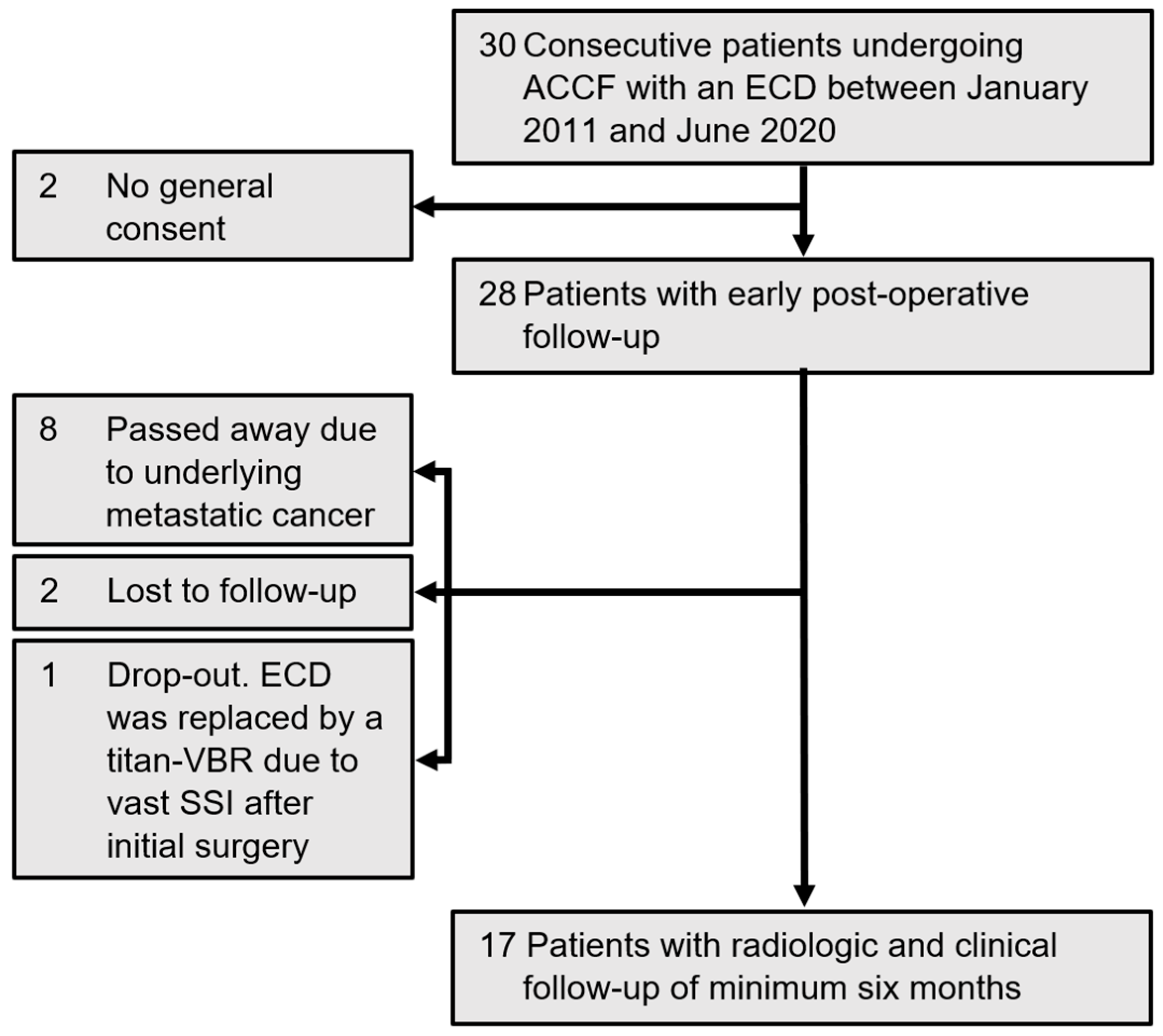
2.1. Study Population

In total, 28 patients (mean age 60.7 ± 13 (range: 20–84) years; 64.3% female) were analyzed. Nineteen (68%) suffered from tumorous lesions, five (17.9%) from degenerative disease, and four (14.3%) from a traumatic fracture. Demographic data, indications, and presenting symptoms are summarized in Table 1. The only primary tumor was multiple myeloma (n = 3; 15.8%), whereas spinal metastases were most commonly derived from lung cancers (n = 5; 26.3%). Mean pre-operative Spinal Instability Neoplastic Score (SINS) in tumor patients was 11.9 ± 2.3 (range: 8–16); 17 (89.5%) of those patients had a pathologic fracture [20]. Radiological and clinical parameters were assessed pre-operatively, early post-operatively, six weeks, and six months after surgery, respectively, and every six months in the clinical routine.

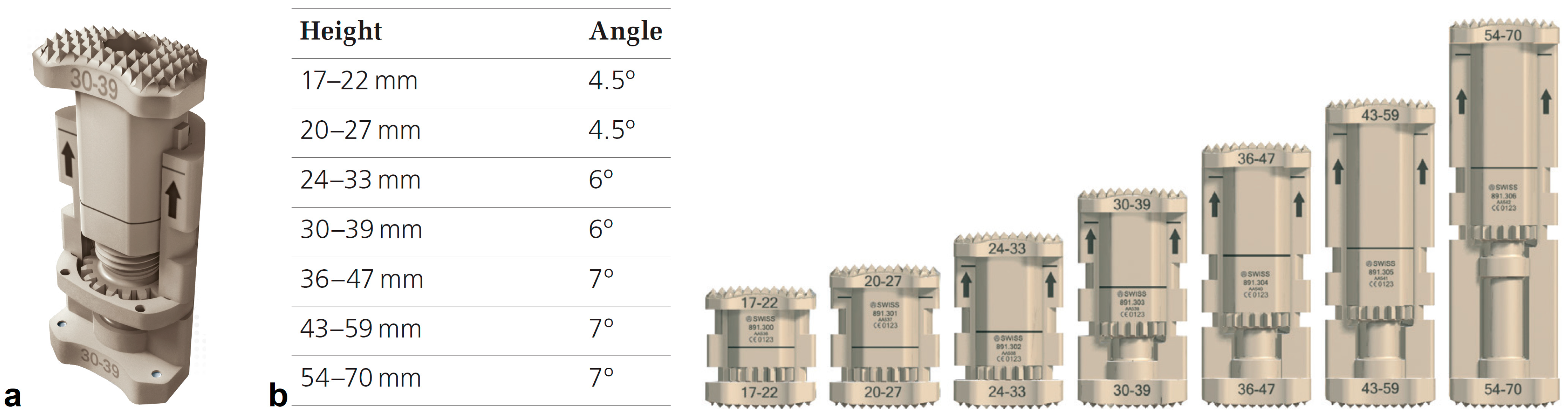
2.2. Surgical Technique

The ECD (Figure 2) was implanted in all cases in combination with a titanium (n = 14) or carbon (n = 14) anterior plate–screw osteosynthesis, using the standard anterior Smith and Robinson approach [2]. After the complete removal of the adjacent intervertebral discs, a corpectomy was performed at the level of interest. We used the sliding caliper to measure the approximate size of the implant. The chosen ECD was attached to the “holding and distraction” instrument to align it in situ in the frontal and sagittal planes. The ECD was expanded to the desired physiologic height followed by installing the locking clip of the expansion mechanism. Allograft chips were placed anterior and lateral to the cage. No chips were placed between the cage and the spinal cord. Additionally, a posterior spondylodesis was mandatory in 12 (42.9%) cases due to multi-level corpectomy or affection of the cervico-thoracic junction. Post-operative care comprised a soft neck collar for six weeks and pain-adapted analgesia.

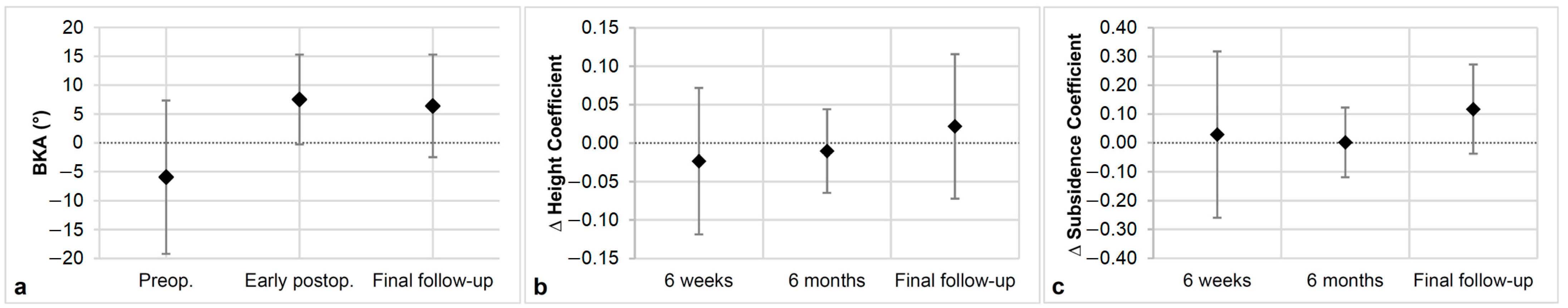
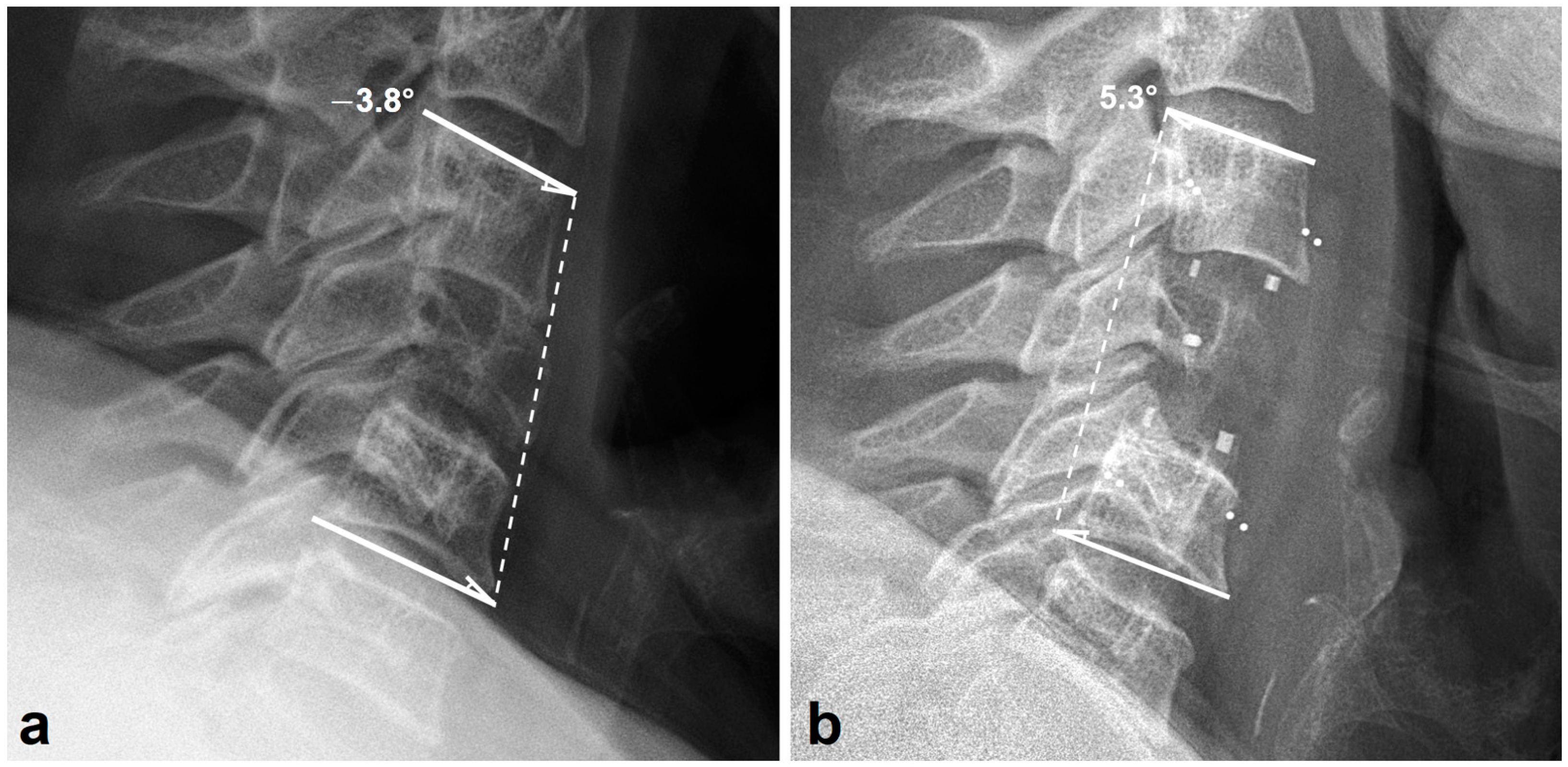
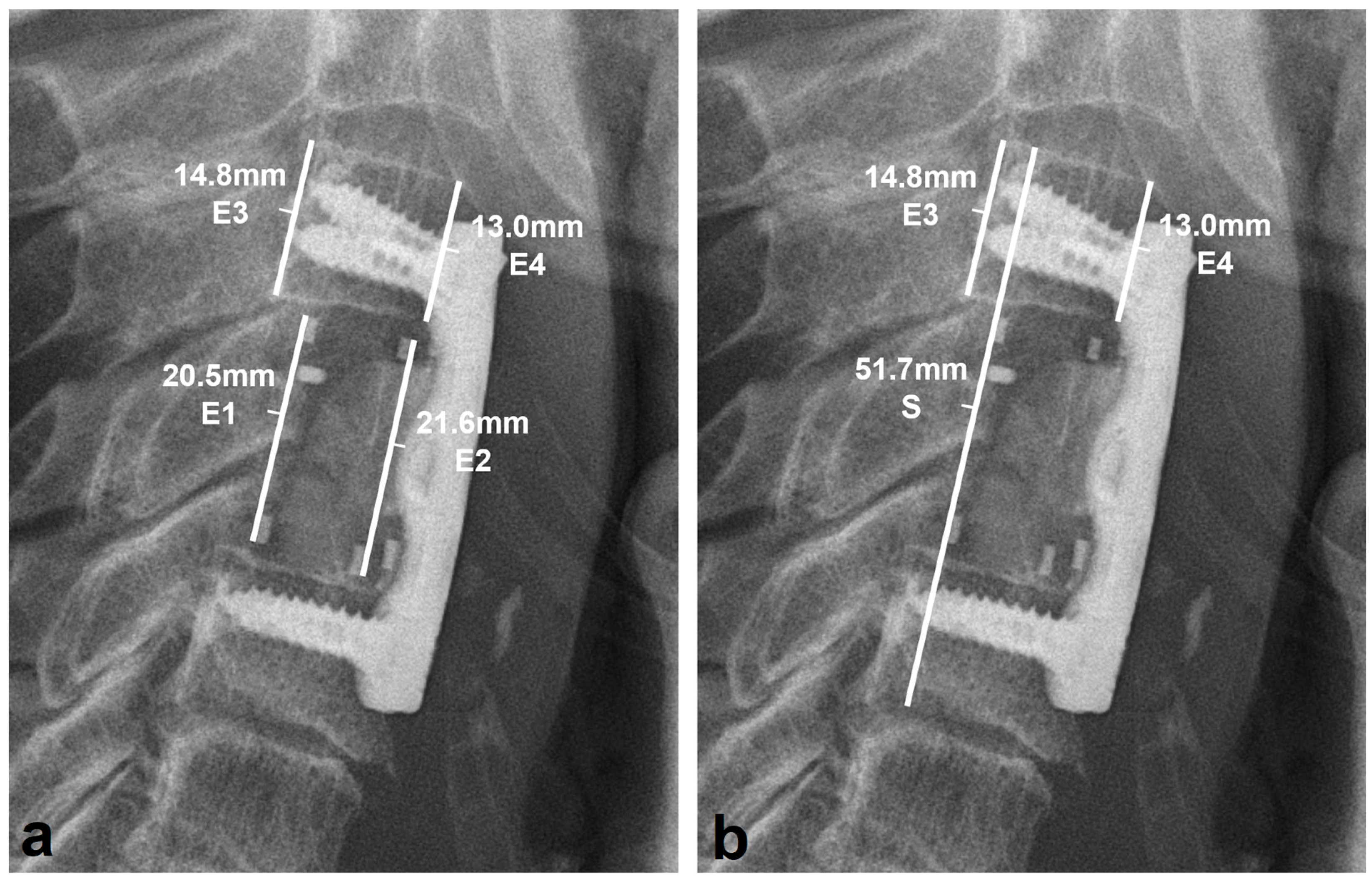
2.3. Radiologic Outcome

The Sectra Workstation IDS7 software (Version 23.2, Sectra AB, Linköping, Sweden) was used to conduct radiologic assessment and measurements on standing lateral cervico-thoracic spine radiographs. If the conventional radiograph was not suited for exact measurements, CT or MRI images were used to perform measurements if available.
As the primary outcome, the adjustment of local kyphosis after surgery was assessed using the “bisegmental kyphotic angle (BKA)” [21]. The BKA is the angle between the caudal endplate of the caudal adjacent vertebral body and the cranial endplate of the supra-adjacent vertebral body (Figure 3). Global lordosis of the cervical spine was determined by measuring the “sagittal alignment of the cervical spine (SACS)” defined by the angle between the lower endplate of C2 and the lower endplate of C7 on a lateral radiograph [22]. If the ECD was in position C7 or below, the caudal endplate of the caudal adjacent healthy vertebra was used.
The “height coefficient (HC)”, as published before [21], was used to assess the stability of the ECD’s expansion mechanism (Figure 4a). The height coefficient difference (∆HC) was calculated by subtracting the height coefficient at the given follow-up from the height coefficient early post-operative. Subsidence of the ECD into the adjacent vertebral endplates was evaluated using the subsidence coefficient (SC), as proposed by Schnake et al. (Figure 4b) [23]. The subsidence coefficient difference (∆SC) was calculated analogously to ∆HC. In addition, correct cage placement (>90% of the ECD has contact with the adjacent vertebral endplates) was evaluated early post-operatively.
Bony fusion evaluation was performed at six months and the final follow-up, respectively, using the grading system by Eck et al. [24]. Visible trabeculae and remodeling on a lateral radiograph between the ECD and adjoining vertebral bodies is defined as grade I (definite fusion), whereas a not completely incorporated vertebral body replacement device without visible lucency > 1 mm is defined as grade II (probable fusion). With lucency at the bottom and/or top of the VBR, the fusion was defined as grade III (probably not). Therefore, grade I and grade II were considered successful fusion.

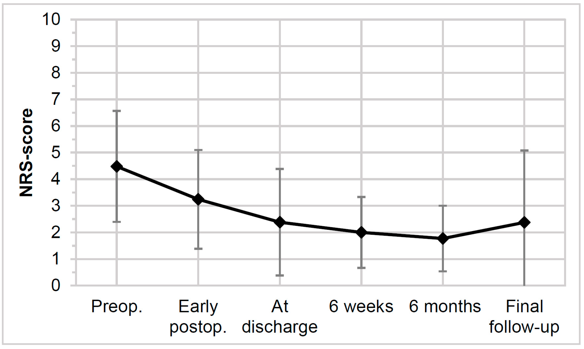
2.4. Clinical Outcome

The primary clinical outcome was quantified by evaluating the patient’s symptoms pre- and post-operation using Odom’s criteria [25]. The absence of all pre-operative symptoms characterizes an “excellent” outcome; a “good” outcome is defined by minimal persistence of pre-operative symptoms; and a “fair” outcome signifies relief from some pre-operative symptoms. Conversely, unchanged or worsened symptoms represent a “poor” outcome. A Numerical Rating Scale (NRS) ranging from zero to ten assessed experienced level of cervical pain.
The American Spinal Injury Association Impairment Scale (AIS) was used to characterize neurologic deterioration [26].
The Karnofsky Performance Status Scale (KPSS) was determined to assess the quality of life and the level of mobility in patients with underlying carcinosis [27]. In addition, before surgery, the SINS was determined in those patients [20].

2.5. Complication Assessment

Intraoperative complications were distinguished as complications associated with the ECD (e.g., a faulty device, no expansion possible, expansion mechanism not lockable) and general complications (e.g., difficulty with intubation, high blood loss (≥1000 mL for single-stage; ≥1500 mL two- or three-stage procedures), with severe circulatory problems resulting in prolonged surgical time or abortion of the procedure).
Post-operative complications were subdivided into general post-operative complications (SSI, wound healing disorder, post-operative instability) and approach-specific complications (odynophagia, dysphagia, cerebrospinal fluid (CSF) leak, hoarseness, esophagus perforation).

2.6. Statistical Analysis

Statistical analysis was performed with GraphPad PRISM Version 8.0.1 (GraphPad Software, San Diego, CA, USA). Shapiro–Wilk test was used to test normal Gaussian distribution. Descriptive statistics are shown as mean ± standard deviation (range minimum-maximum) if normally distributed. If non-normally distributed, descriptive statistics are shown with median [Interquartile range (IQR) 25% percentile; 75% percentile] (range minimum–maximum). Single-group comparisons were performed using paired (unpaired) t-tests for normally distributed data and Wilcoxon signed-rank test (Mann–Whitney U-Test) for non-normally distributed data. One-way ANOVA was conducted for paired multiple comparisons with complete data and a mixed-effects analysis in case of missing data points. Statistical significance was set at α ≤ 0.05.

3. Results

3.1. Surgical Details

A summary of surgical details is given in Table 2. C7 was replaced most frequently (n = 10, 35.7%). All patients (n = 28) received an anterior plate–screw osteosynthesis, covering two segments in most cases (50%). Twelve (42.9%) patients received an additional posterior spondylodesis during the initial surgery. During follow-up, a secondary stabilization was necessary in four (14.3%) other patients (median 27 (range: 21–303) days after the initial surgery).

3.2. Radiologic Outcomes

The average pre-operative kyphotic BKA of mean −5.9 ± 13.3 (range: −39.3–7.0) degrees improved significantly to a lordotic posture with a mean BKA of 7.5 ± 7.8 (range: −7.8–23.0) degrees directly after surgery, resulting in a mean correction of 13.5 ± 9.7 (range: 3.8–40.8) degrees (p < 0.001). At the last follow-up, the BKA remained stable at 6.4 ± 8.9 (range: −13–23.0) degrees, resulting in a significant mean correction of 12.3 ± 9.6 (range: 3.8–40.3) degrees compared to pre-operatively (p < 0.001) (Figure 5a). BKA at the final follow-up showed a mean loss of correction of 1.2 ± 2.5 (range: 0–10) degrees (p = 0.17) compared to early post-operative. The sagittal alignment of the cervical spine also substantially improved from a mean of 8.9 ± 15.8 (range: −23.9–35.4) degrees to a mean SACS of 13.8 ± 14.6 (range: −14.7–40.1) degrees early post-operatively (p = 0.27). SACS remained stable throughout the study, showing almost no considerable reduction in correction (p = 0.08), with an average SACS of 18.7 ± 14.7 degrees (range: −9.0–44.0) at the final follow-up.
∆HC (Figure 5b) showed no statistically relevant differences at the last follow-up (p = 0.96). The mean value of 0.02 ± 0.9 (range: −0.09–0.23) at the final follow-up indicates no failure of the expansion system or the stability of the ECD. At the last follow-up, the ∆SC (Figure 5c) showed no differences in subsidence (0.12 ± 0.16 (range: −0.19–0.48); p = 0.99).
Seventeen patients were evaluated for radiographic fusion at the final follow-up on average 1.0 [0.6; 1.8] (range 0.5–6.0) years post-operatively. Grade I “definite fusion” was noted in nine patients (52.9%) and grade II “probable fusion” in eight patients (47.1%) [24]. There was no grade III or grade IV present. A correct ECD placement, assessed on early post-operative radiographs, was found in all patients (n = 28, 100%) and did not differ over time.

3.3. Clinical Outcomes

Table 3 demonstrates the main clinical outcomes. Directly after surgery, the majority of patients reported an “excellent” (n = 3; 10.7%) or “good” (n = 16; 57.1%) outcome. At the final follow-up, most patients showed an “excellent” (n = 8; 50%) or “good” (n = 6; 37.5%) and no “poor” outcome. Experienced pain decreased significantly from an average of 4.5 ± 2.3 (range: 0–9) pre-operatively to 2.4 ± 2.0 (range: 0–6) at discharge (p = 0.009) and 1.8 ± 1.2 (range: 0–4; p = 0.004) at six months post-operatively (Figure 6). At the final follow-up, patients experienced a mean NRS of 2.34 ± 2.8 (range: 0–10), showing a stable clinical situation compared to the post-operative state (p = 0.066) and a significantly improved pain relief compared to the pre-operative status (p = 0.021). Furthermore, 20 patients (71.4%) showed an AIS grade of E post-operatively compared to 16 (59.3%) pre-operatively. Before surgery, two patients (7.4%) showed a grade of C, one improved to D early post-operatively, and one improved to a grade of E until the final follow-up. One patient’s pre-operative AIS grade of D remained constant during follow-up. One trauma patient (tetraplegia after a fall from a tree) showed a grade of A pre-operatively, which remained unchanged after surgery; the patient was lost to follow-up due to further care in a paraplegic center. Five (17.9%) of all the patients improved in at least one AIS grade directly after surgery; the grade did not worsen over time. A total of 93.8% (n = 15) of the examined patients at the final follow-up showed an AIS grade of E, except one (6.3%) with a grade of D.
An average KPSS of 75.8 ± 13.9 (range: 50–100) pre-operatively (n = 19) improved significantly (p < 0.001) to 62.2 ± 10 (range: 40–80) at discharge. The mean KPSS at the final follow-up was 74.5 ± 19.2 (range: 30–100), resulting in a stable score compared to that post-operatively (p = 0.28). Except for two patients, all tumor patients (n = 17; 89.5%) were treated with either post-operative irradiation or adjuvant chemo- or systemic therapies. Throughout this study, 14 (50.0%) patients passed away on average 1.0 ± 1.1 years (range: 0.05–3.7) after initial surgery with a mean age of 65.6 ± 10.6 years (range: 46–86); all suffered from carcinosis.

3.4. Complications

No intraoperative complications related to the ECD occurred. The mean blood loss was 451 ± 420 (40–1700) milliliters. Eight (28.6%) patients experienced approach-specific post-operative complications; 91% of those were minor complications, with odynophagia being the most common (n = 4), followed by dysphagia (n = 3) and hoarseness (n = 3). The minor complications resolved without reoperation and recovered on average after 6.8 ± 6.9 (2–23) days. In one case, an esophageal perforation occurred, requiring multiple revision surgeries. Due to bacterial colonization, the ECD had to be replaced with a titanium-VBR; a posterior spondylodesis was added. After ECD explantation, the patient dropped out of our series.
Five (17.9%) patients required reoperation. Three of those patients suffered from SSI. Two of the infections occurred with the additional posterior spondylodesis and one with the anterior approach. In none of those cases was it necessary to remove the implants. In two cases, wound revision was indicated due to wound healing disorder with the posterior approach.
In no case did an ECD have to be replaced due to insufficient stability caused by the ECD or the malfunctioning of the expansion system.

4. Discussion

An ACCF has been established as a favorable and safe surgical procedure to stabilize the anterior cervical spinal column. Several stabilization systems for an ACCF have been developed. Recently, the ECD cage was introduced, providing personalized individual adaptation, better stress distribution, and radiolucency compared to previous systems. This study aimed to assess the general safety and efficacy of the ECD by analyzing (1) the radiographic outcome, (2) the clinical outcome, and (3) the complication rates in patients suffering from metastatic diseases, traumatic cervical fractures, or progressive degenerative disease.
Our study indicates that the ECD is a valuable tool to realign and maintain local cervico-thoracic alignment and anterior stability in patients who have undergone cervico-a thoracic anterior corpectomy. The BKA improved from a pre-operative mean of 5.9 ± 13.3 degrees kyphosis to a mean of 7.5 ± 7.8 degrees lordosis directly post-operatively (p < 0.001). There was a deniable loss of correction of 1.2 ± 2.5 degrees at the last follow-up compared to that directly post-operatively (p = 0.17). The same applies to the SACS, demonstrating recovery and conserving a physiological lordotic posture [28]. Furthermore, all patients with a follow-up of at least six months showed a successful bony fusion, therefore achieving the optimal purpose of a VBR [29].
Furthermore, we proved the safety of the expansion and locking mechanism of the ECD. A collapse of the VBR was not recorded, and ∆HC showed no substantial differences at the final follow-up (p = 0.96). The same applies to the subsidence. At the final follow-up, the mean ∆SC only showed subsidence of 0.12 ± 0.16 (p = 0.99). As proven before and according to the corresponding literature, minor and even statistically significant subsidence has no clinical relevance in those cases [30], an assertion supported by our predominantly good and excellent clinical results according to Odom’s criteria. One patient showed a ∆SC of 0.48 at the final follow-up while being the only patient to have a one-level decrease in Odom’s criteria at the last follow-up compared to the previous evaluation (“Excellent” 16 months, “Good” six years post-operative). Several studies report on VBR or cage subsidence risk factors, such as age, female sex, multi-level ACCF, and a C6 corpectomy [30,31,32,33]. Our patient (71 years, female) had multiple risk factors for VBR subsidence. Besides being female and elderly, she suffered from rheumatoid arthritis and received a bi-level ACCF (C4–C7). Consequently, we can conclude that the subsidence was caused by an accumulation of risk factors rather than the ECD or its design [30,31,32,33]. A failure of the expansion mechanism was excluded in this case.
The clinical outcomes showed that an ACCF with an ECD leads to cervical pain relief and neurological improvement compared to the pre-operative state. Directly after surgery, 92.9% (n = 26) of patients reported at least a “relief of some pre-operative symptoms”; only two patients (7.1%) reported a “poor” outcome according to Odom’s criteria [25]. Both were still experiencing unchanged cervical pain; one patient had already suffered from chronic cervicobrachial pain before surgery. Experienced pain measured with the NRS was greatly relieved at discharge from the hospital (p = 0.009) compared to the pre-operative baseline. It remained substantially lowered until the last follow-up (p = 0.066) and remained significantly improved compared to the pre-operative state (p = 0.021). Functional neurologic impairments, measured by the AIS grade, improved in 17.9% of all patients early post-operatively (mean 6.3 ± 2.9 (range: 2–12) days after surgery). Fifty-nine percent of all patients reported no neurological impairment (AIS E) pre-operatively. In no patient neurological did impairment worsen after surgery compared to the pre-operative status. The promising clinical results are comparable to other studies using allo- or autograft titanium mesh cages, or titanium VBRs for ACCF [3,4,34,35].
Surgical tumor debulking, coupled with post-operative irradiation and chemo- or systemic therapy, is crucial in tumor control of spinal metastases [36,37]. Our data, measured with the KPSS, show an improvement in the general well-being of tumor patients who survived at least six months after surgery. In our study population, the KPSS improved by a mean of 8.2 points on the scale to 84 ± 8.4 (range: 70–100) at six months post-operatively compared to the pre-operative score. The natural course of carcinosis explains a decline in the KPSS at the final follow-up [36]. In contrast to titanium mesh cages or titanium VBRs, the ECD allows for optimal pre-irradiation CT planning and monitoring of tumor progression by MRI or CT owing to the radiolucency of PEEK, producing no significant artifacts while providing similar biomechanical characteristics to those of titanium implants [38,39]. An MRI example is given in Figure 7.
No device-related intraoperative complications occurred. One patient experienced a secondarily diagnosed esophageal perforation. The early vast infection was solved by a series of debridement and suture of the lesion. Finally, a replacement of the ECD cage was necessary.
Patients (n = 7) with approach-specific complications (odynophagia, dysphagia, hoarseness) had the symptoms recovered at discharge. Without aftermath, general post-operative complications were resolved by revision surgeries or blood transfusion. Due to progressive instability, four patients received a posterior spondylodesis in a revision procedure (median 27 [22.5; 99] (range: 21–303) days after the initial surgery). Three of those were suffering from cervical metastases in which a sole ACCF did not provide sufficient stability due to poor bone quality of the adjacent vertebral bodies. The remaining patient experienced an esophageal perforation requiring multiple revision surgeries including posterior stabilization as described above. In summary, the surgical approach or the underlying disease rather than the implant caused post-operative complications. According to a meta-analysis by Wang et al., the incidence of complications in our study population is within the usual range for an ACCF. Wang et al. described an incidence of dysphagia after an ACCF of 9.9% (4.8–15.9%) and that of infection of 14.2% (−1.1–30.3%). In our population, the incidence of dysphagia was 10.7% (n = 3) and 10.7% for infection (n = 3), respectively [40].

5. Limitations

The main limitation of this study is the retrospective design and a missing control group. Additionally, the small size of the study population constrained the extent of statistical analysis and the examination of risk factors. Additionally, it was unfeasible to form a study cohort of sufficient size that exclusively comprised patients who had undergone an anterior cervical corpectomy and fusion (ACCF) with the ECD and without posterior spondylodesis. This limitation rendered the analysis of isolated cases of ACCF with the ECD unachievable. Furthermore, most patients requiring cervical corpectomy suffer from severe metastatic disease. Therefore, post-operative follow-up was limited due to the restricted life expectancy of the underlying tumorous disease. As follow-up CT scans were not accessible in most cases, bony fusion assessment was performed on conventional radiographs. According to Fogel et al., fusion assessment on plain radiographs features a similar accuracy as that on CT scans if a radiolucent implant is used; our results support this assertion [41]. Two patients had a CT scan at a comparable time as a that of a radiograph, showing the same bony fusion grade on both imaging procedures (Figure 8). Despite these limitations, reporting outcomes of new techniques for the surgical therapy of severe but rare cervical pathologies is essential.

6. Conclusions

This is the first study analyzing the radiologic and clinical outcome of the ECD PEEK cage in the cervico-thoracic spine. The study revealed that the ECD restored and maintained the anterior vertebral column in patients with metastatic, traumatic, and degenerative diseases. Furthermore, the rate and degree of complications did not exceed those in other surgical treatment modalities for these pathologies. Thus, the ECD PEEK cage is a safe and efficient therapy option for anterior vertebral column reconstruction, even though studies with greater patient cohorts or a prospective design are necessary to support this thesis.

Author Contributions

Conceptualization and study design, M.C.D.; data collection and analysis, M.Š., A.K.L. and K.A.C.O.; writing—original draft preparation, M.Š.; writing—review and editing, M.C.D., K.A.C.O., M.Š., C.E.A. and L.M.B.; visualization, M.Š. All authors have read and agreed to the published version of the manuscript.

Funding

DePuy Synthes Inc., Raynham, MA, USA, granted a research grant to the institution of the authors, Department of Orthopaedic Surgery and Traumatology, Inselspital, Bern University Hospital, University of Bern, Bern, Switzerland.

Institutional Review Board Statement

The study was conducted in accordance with the Declaration of Helsinki and approved by the Ethics Committee in Bern (KEK 2020-01127, approval date: 2 June 2020).

Informed Consent Statement

Informed consent was obtained from all subjects involved in the study.

Data Availability Statement

The data presented in this study are available on request from the corresponding author. The data are not publicly available due to concerns related to the privacy of the patients involved.

Conflicts of Interest

The funders had no role in the design of the study; in the collection, analyses, or interpretation of data; in the writing of the manuscript; or in the decision to publish the results.

References

  1. Denis, F. The Three Column Spine and Its Significance in the Classification of Acute Thoracolumbar Spinal Injuries. Spine 1983, 8, 817–831. [Google Scholar] [CrossRef] [PubMed]
  2. Smith, G.W.; Robinson, R.A. The treatment of certain cervical-spine disorders by anterior removal of the intervertebral disc and interbody fusion. J. Bone Jt. Surg. Am. 1958, 40, 607–624. [Google Scholar] [CrossRef] [PubMed]
  3. Yang, Y.; Fei, Q.; Li, J.; Su, N.; Wang, B.; Li, D.; Meng, H.; Wang, Q.; Lin, J.; Ma, Z. Comparison between anterior cervical discectomy with fusion and anterior cervical corpectomy with fusion for the treatment of cervical spondylotic myelopathy: A meta-analysis. Ther. Clin. Risk Manag. 2015, 11, 1707–1718. [Google Scholar] [CrossRef] [PubMed]
  4. Schulz, C.; Mauer, U.M.; Mathieu, R. Implantatassoziierte Komplikationen sowie klinischer und radiologischer Verlauf nach anteriorer zervikaler Korpektomie und Cage-Fusion—Retrospektiver Vergleich von PEEK-gegen Titan-Cages. Z. Orthop. Unfall. 2017, 155, 201–208. [Google Scholar] [CrossRef]
  5. Lu, D.C.; Wang, V.; Chou, D. The use of allograft or autograft and expandable titanium cages for the treatment of vertebral osteomyelitis. Neurosurgery 2009, 64, 122–129, discussion 129–130. [Google Scholar] [CrossRef] [PubMed]
  6. Elder, B.D.; Lo, S.-F.; Kosztowski, T.A.; Goodwin, C.R.; Lina, I.A.; Locke, J.E.; Witham, T.F. A systematic review of the use of expandable cages in the cervical spine. Neurosurg. Rev. 2016, 39, 1–11. [Google Scholar] [CrossRef] [PubMed]
  7. Dorai, Z.; Morgan, H.; Coimbra, C. Titanium cage reconstruction after cervical corpectomy. J. Neurosurg. Spine 2003, 99, 3–7. [Google Scholar] [CrossRef] [PubMed]
  8. Sénégas, J. Mechanical supplementation by non-rigid fixation in degenerative intervertebral lumbar segments: The Wallis system. Eur. Spine J. 2002, 11 (Suppl. S2), S164–S169. [Google Scholar] [CrossRef] [PubMed]
  9. Kurtz, S.M.; Devine, J.N. PEEK biomaterials in trauma, orthopedic, and spinal implants. Biomaterials 2007, 28, 4845–4869. [Google Scholar] [CrossRef] [PubMed]
  10. Morrison, C.; Macnair, R.; MacDonald, C.; Wykman, A.; Goldie, I.; Grant, M. In vitro biocompatibility testing of polymers for orthopaedic implants using cultured fibroblasts and osteoblasts. Biomaterials 1995, 16, 987–992. [Google Scholar] [CrossRef] [PubMed]
  11. Wenz, L.M.; Merritt, K.; Brown, S.A.; Moet, A.; Steffee, A.D. In vitro biocompatibility of polyetheretherketone and polysulfone composites. J. Biomed. Mater. Res. 1990, 24, 207–215. [Google Scholar] [CrossRef]
  12. Brandao, R.C.S.; Martins, W.d.S.; Arantes, A.; Gusmão, S.S.; Perrin, G.; Barrey, C. Titanium versus polyetheretherketone implants for vertebral body replacement in the treatment of 77 thoracolumbar spinal fractures. Surg. Neurol. Int. 2017, 8, 191. [Google Scholar] [CrossRef] [PubMed]
  13. Vadapalli, S.; Sairyo, K.; Goel, V.K.; Robon, M.; Biyani, A.; Khandha, A.; Ebraheim, N.A. Biomechanical Rationale for Using Polyetheretherketone (PEEK) Spacers for Lumbar Interbody Fusion—A Finite Element Study. Spine 2006, 31, E992–E998. [Google Scholar] [CrossRef] [PubMed]
  14. Shen, F.H.; Gasbarrini, A.M.; Lui, D.F.; Reynolds, J.; Capua, J.; Boriani, S.M. Integrated Custom Composite Polyetheretherketone/Carbon fiber (PEEK/CF) Vertebral Body Replacement (VBR) in the Treatment of Bone Tumors of the Spine. Spine 2022, 47, 252–260. [Google Scholar] [CrossRef] [PubMed]
  15. Hee, H.T.; Kundnani, V. Rationale for use of polyetheretherketone polymer interbody cage device in cervical spine surgery. Spine J. 2010, 10, 66–69. [Google Scholar] [CrossRef] [PubMed]
  16. Raslan, F.; Koehler, S.; Berg, F.; Rueckriegel, S.; Ernestus, R.-I.; Meinhardt, M.; Westermaier, T. Vertebral body replacement with PEEK-cages after anterior corpectomy in multilevel cervical spinal stenosis: A clinical and radiological evaluation. Arch. Orthop. Trauma Surg. 2014, 134, 611–618. [Google Scholar] [CrossRef] [PubMed]
  17. Synthes GmbH. ECD—Expandable Corpectomy Device: Surgical Technique. 2017. Available online: http://synthes.vo.llnwd.net/o16/LLNWMB8/INT%20Mobile/Synthes%20International/Product%20Support%20Material/legacy_Synthes_PDF/DSEM-SPN-1215-0389-1_LR.pdf (accessed on 15 October 2021).
  18. Das, A.K.; Purohit, D.K.; Gupta, A.; Kataria, R. Comparison of Radiological and Clinical Outcomes between Expandable and Non-expandable Cages Following Cervical Corpectomy: A Systematic Review and Meta-Analysis. Asian Spine J. 2023, 17, 567–581. [Google Scholar] [CrossRef] [PubMed]
  19. Brenke, C.; Fischer, S.; Carolus, A.; Schmieder, K.; Ening, G. Complications associated with cervical vertebral body replacement with expandable titanium cages. J. Clin. Neurosci. 2016, 32, 35–40. [Google Scholar] [CrossRef]
  20. Fisher, C.G.; DiPaola, C.P.; Ryken, T.C.; Bilsky, M.H.; Shaffrey, C.I.; Berven, S.H.; Harrop, J.S.; Fehlings, M.G.; Boriani, S.; Chou, D.; et al. A novel classification system for spinal instability in neoplastic disease: An evidence-based approach and expert consensus from the Spine Oncology Study Group. Spine 2010, 35, E1221–E1229. [Google Scholar] [CrossRef] [PubMed]
  21. Deml, M.C.; Sepulveda, C.A.M.; Albers, C.E.; Hoppe, S.; Bigdon, S.F.; Häckel, S.; Milavec, H.; Benneker, L.M. Anterior column reconstruction of the thoracolumbar spine with a new modular PEEK vertebral body replacement device: Retrospective clinical and radiologic cohort analysis of 48 cases with 1.7-years follow-up. Eur. Spine J. 2020, 29, 3194–3202. [Google Scholar] [CrossRef] [PubMed]
  22. Harrison, D.E.; Harrison, D.D.; Cailliet, R.; Troyanovich, S.J.; Janik, T.J.; Holland, B. Cobb method or harrison posterior tangent method: Which to choose for lateral cervical radiographic analysis. Spine 2000, 25, 2072–2078. [Google Scholar] [CrossRef] [PubMed]
  23. Schnake, K.J.; Stavridis, S.I.; Kandziora, F. Five-year clinical and radiological results of combined anteroposterior stabilization of thoracolumbar fractures. J. Neurosurg. Spine 2014, 20, 497–504. [Google Scholar] [CrossRef] [PubMed]
  24. Eck, K.R.; Lenke, L.G.; Bridwell, K.H.; Gilula, L.A.; Lashgari, C.J.; Riew, K.D. Radiographic Assessment of Anterior Titanium Mesh Cages. J. Spinal Disord. 2000, 13, 501–509. [Google Scholar] [CrossRef] [PubMed]
  25. Odom, G.L.; Finney, W.; Woodhall, B. Cervical Disk Lesions. J. Am. Med. Assoc. 1958, 166, 23–28. [Google Scholar] [CrossRef] [PubMed]
  26. Kirshblum, S.; Snider, B.; Rupp, R.; Read, M.S. Updates of the International Standards for Neurologic Classification of Spinal Cord Injury: 2015 and 2019. Phys. Med. Rehabil. Clin. N. Am. 2020, 31, 319–330. [Google Scholar] [CrossRef] [PubMed]
  27. Karnofsky, D.A.; Abelmann, W.H.; Craver, L.F.; Burchenal, J.H. The use of the nitrogen mustards in the palliative treatment of carcinoma. With particular reference to bronchogenic carcinoma. Cancer 1948, 1, 634–656. [Google Scholar] [CrossRef]
  28. Guo, G.-M.; Li, J.; Diao, Q.-X.; Zhu, T.-H.; Song, Z.-X.; Guo, Y.-Y.; Gao, Y.-Z. Cervical lordosis in asymptomatic individuals: A meta-analysis. J. Orthop. Surg. Res. 2018, 13, 147. [Google Scholar] [CrossRef] [PubMed]
  29. Zhang, Y.; Deng, X.; Jiang, D.; Luo, X.; Tang, K.; Zhao, Z.; Zhong, W.; Lei, T.; Quan, Z. Long-term results of anterior cervical corpectomy and fusion with nano-hydroxyapatite/polyamide 66 strut for cervical spondylotic myelopathy. Sci. Rep. 2016, 6, 26751. [Google Scholar] [CrossRef] [PubMed]
  30. Ji, C.; Yu, S.; Yan, N.; Wang, J.; Hou, F.; Hou, T.; Cai, W. Risk factors for subsidence of titanium mesh cage following single-level anterior cervical corpectomy and fusion. BMC Musculoskelet. Disord. 2020, 21, 32. [Google Scholar] [CrossRef] [PubMed]
  31. Tatter, C.; Persson, O.; Burström, G.; Edström, E.; Elmi-Terander, A. Anterior Cervical Corpectomy and Fusion for Degenerative and Traumatic Spine Disorders, Single-Center Experience of a Case Series of 119 Patients. Oper. Neurosurg. 2020, 20, 8–17. [Google Scholar] [CrossRef] [PubMed]
  32. Wu, J.; Luo, D.; Ye, X.; Luo, X.; Yan, L.; Qian, H. Anatomy-related risk factors for the subsidence of titanium mesh cage in cervical reconstruction after one-level corpectomy. Int. J. Clin. Exp. Med. 2015, 8, 7405–7411. [Google Scholar] [PubMed]
  33. Hasegawa, K.; Abe, M.; Washio, T.; Hara, T. An experimental study on the interface strength between titanium mesh cage and vertebra in reference to vertebral bone mineral density. Spine 2001, 26, 957–963. [Google Scholar] [CrossRef] [PubMed]
  34. Macdonald, R.L.; Fehlings, M.G.; Tator, C.H.; Lozano, A.; Fleming, J.R.; Gentili, F.; Bernstein, M.; Wallace, M.C.; Tasker, R.R. Multilevel anterior cervical corpectomy and fibular allograft fusion for cervical myelopathy. J. Neurosurg. 1997, 86, 990–997. [Google Scholar] [CrossRef] [PubMed]
  35. Rieger, A.; Holz, C.; Marx, T.; Sanchin, L.; Menzel, M. Vertebral autograft used as bone transplant for anterior cervical corpectomy: Technical note. Neurosurgery 2003, 52, 449–454, discussion 453–454. [Google Scholar] [CrossRef] [PubMed]
  36. Rothrock, R.J.; Barzilai, O.; Reiner, A.S.; Lis, E.; Schmitt, A.M.; Higginson, D.S.; Yamada, Y.; Bilsky, M.H.; Laufer, I. Survival Trends After Surgery for Spinal Metastatic Tumors: 20-Year Cancer Center Experience. Neurosurgery 2020, 88, 402–412. [Google Scholar] [CrossRef] [PubMed]
  37. Fehlings, M.G.; Nater, A.; Tetreault, L.; Kopjar, B.; Arnold, P.; Dekutoski, M.; Finkelstein, J.; Fisher, C.; France, J.; Gokaslan, Z.; et al. Survival and Clinical Outcomes in Surgically Treated Patients With Metastatic Epidural Spinal Cord Compression: Results of the Prospective Multicenter AOSpine Study. J. Clin. Oncol. 2016, 34, 268–276. [Google Scholar] [CrossRef] [PubMed]
  38. Krätzig, T.; Mende, K.C.; Mohme, M.; Kniep, H.; Dreimann, M.; Stangenberg, M.; Westphal, M.; Gauer, T.; Eicker, S.O. Carbon fiber–reinforced PEEK versus titanium implants: An in vitro comparison of susceptibility artifacts in CT and MR imaging. Neurosurg. Rev. 2021, 44, 2163–2170. [Google Scholar] [CrossRef] [PubMed]
  39. Kandziora, F.; Pflugmacher, R.; Schaefer, J.; Scholz, M.; Ludwig, K.; Schleicher, P.; Haas, N.P. Biomechanical comparison of expandable cages for vertebral body replacement in the cervical spine. J. Neurosurg. Spine 2003, 99, 91–97. [Google Scholar] [CrossRef] [PubMed]
  40. Wang, T.; Tian, X.-M.; Liu, S.-K.; Wang, H.; Zhang, Y.-Z.; Ding, W.-Y. Prevalence of complications after surgery in treatment for cervical compressive myelopathy. Medicine 2017, 96, e6421. [Google Scholar] [CrossRef] [PubMed]
  41. Fogel, G.R.; Toohey, J.S.; Neidre, A.; Brantigan, J.W. Fusion assessment of posterior lumbar interbody fusion using radiolucent cages: X-ray films and helical computed tomography scans compared with surgical exploration of fusion. Spine J. 2008, 8, 570–577. [Google Scholar] [CrossRef] [PubMed]
Figure 1. Patient selection process.
Std 13 00008 g001
Figure 2. The Expandable Corpectomy Device (DePuy Synthes Spine Inc., Raynham, MA, USA) with (a) an exemplary 3D rendering and (b) different available sizes (17–22 mm–50–70 mm) with corresponding endplate angulations (4.5°–7°). The device is entirely made of PEEK. The body contains a continuous expansion mechanism, which a locking clip secures. The arrows are pointing cranially. Each of the spiked anatomically shaped endplates has three radiopaque markers to scrutinize the device’s position in situ. (Printed with permission from DePuy Synthes).
Std 13 00008 g002
Figure 3. Measurement of the bisegmental kyphotic angle (BKA) on a lateral projection of the cervical spine in a patient suffering from multiple myeloma in C4. (a) Pre-operative BKA: −3.8°. (b) Early post-operative BKA: +5.3°, showing adjustment of local kyphosis after ACCF. ECD in C4, visible rectangular radiopaque markers at the cranial and caudal ECD endplates, anterior plate–screw fixation (carbon) covering C3–C5 with carbon screws (Ico-Tec AG, Altstätten, Switzerland). Kyphotic angles are indicated as a negative value (−) and lordotic ones as a positive value (+), respectively.
Std 13 00008 g003
Figure 4. Measurements to calculate the height coefficient (HC, (a)) and subsidence coefficient (SC, (b)) of the VBR as published previously [21,23]. ECD replacing C4 and titan anterior plate–screw fixation with titan screws (CSLP, DePuy Synthes Spine Inc., Raynham, MA, USA), ranging from C3-C5. E1: posterior ECD height; E2: anterior ECD height; E3: posterior height of the adjacent cranial vertebra; E4: anterior height of the adjacent cranial vertebra; S: measured along the longitudinal axis of the ECD, the distance between the superior endplate of the ECD-adjacent cranial vertebral body and the inferior endplate of the caudal ECD-adjacent vertebral body. (a) HC is determined by dividing the mean ECD height “c” (c = (E1 + E2)/2) by the mean vertebral height “v” (v = (E3 + E4)/2) ⇒ HC = c/v. (b) SC is calculated by dividing “S” by “v” ⇒ SC = S/v.
Std 13 00008 g004
Figure 5. (a) Progression of the bisegmental kyphotic angle (BKA) over time. Negative values imply kyphosis, and positive values equal lordosis. (b) The height coefficient over time, pictured as height coefficient difference (∆HC), is calculated by subtracting the height coefficient at the given follow-up from the height coefficient early post-operative. Negative values imply a compression of the upper adjacent vertebral body, and positive values imply a loss of height of the ECD. (c) Subsidence coefficient difference (∆SC) at the consecutive follow-up intervals. A negative value implies compression of the upper adjacent vertebral body, and a positive value implies subsidence into the cranial or caudal adjacent vertebral body. Preop.—pre-operative, postop.—post-operative, ∆—difference.
Std 13 00008 g005
Figure 6. Progression of experienced cervical pain across subsequent follow-up intervals quantified using a Numerical Rating Scale from 0 to 10 (0—no pain at all; 10—worst imaginable pain). NRS—Numerical Rating Scale, preop.—pre-operative, postop.—post-operative.
Std 13 00008 g006
Figure 7. Cervical T2-weighted MRI scans of a 49-year-old patient after ACCF due to multiple myeloma in C5, with an ECD in position C5 and anterior plate–screw osteosynthesis covering segments C4/C5 and C5/C6 (carbon plating, carbon screws). The radiolucent PEEK produces no visible imaging artifacts. (a) Sagittal plane, (b) frontal plane, (c) axial plane.
Std 13 00008 g007
Figure 8. Bony fusion assessment on a conventional radiograph ((a) lateral view, (b) a.p. view) 12 months after surgery and CT scan ((c) sagittal plane, (d) frontal plane) of the same patient. The CT scan was acquired one month after the conventional radiograph. ECD is in position C5, with anterior plate–screw osteosynthesis (carbon plating, carbon screws) covering segments C4/C5 and C5/C6. The radiolucency of the ECD allows for an accurate assessment of bony fusion on a conventional radiograph. Both images show a bony fusion grade I (definitive fusion).
Std 13 00008 g008aStd 13 00008 g008b
Table 1. Demographic data of the study population.
Parametern% of n = 28
PopulationFemale1864.3
Male1035.7
Age60.7 ± 13 [20–84] *
BMI26.3 ± 5.4 [17.1–40.3] **
Indication for surgeryNeoplasm1967.9
Degenerative517.9
Traumatic414.3
Presenting symptomsNeck pain28100
Cervical radiculopathy1864.3
Somatosensory disorder1139.3
Reduced motor function1139.3
Myelopathy517.9
Radiculopathy and myelopathy414.3
Radiculopathy with somatosensory disorder and motor deficits932.1
* Mean, SD, [range] (years); ** Mean, SD, [range] (kg/m2).
Table 2. General surgical information.
Parametern, or Unit (x)% of n = 28
General surgery informationMean duration, SD, [range] (min)136 ± 54 [50–270]-
Mean blood loss, SD, [range] (mL)451 ± 420 [40–1700]-
Single-stage procedures1657.1
Two-stage procedures1139.3
Three-stage procedures13.6
Number of vertebral bodies replacedOne2071.4
Two adjacent725.0
Three adjacent13.1
Anterior fusion osteosynthesisTwo segments1450
Three segments828.6
Four segments517.9
Six segments13.6
Posterior spondylodesis at initial surgeryNumber of patients1242.9
Two segments13.6
Three segments13.6
Four and more segments1035.7
Table 3. Detailed clinical outcome comparing pre-operative status to final follow-up. ASIA Impairment Scale grades and clinical outcome were classified using Odom’s criteria across consecutive follow-up intervals. The percentage of patients at a follow-up interval is shown.
Preop.Early Postop.6 MonthsFinal Follow-Up
ASIA Impairment Scale (in %)Grade E59.371.478.693.8
Grade D29.621.421.46.3
Grade C7.43.600
Grade B0000
Grade A3.73.600
Odom’s criteria (in %)Excellent-10.728.650.0
Good-57.150.037.5
Fair-25.021.412.5
Poor-7.100
Disclaimer/Publisher’s Note: The statements, opinions and data contained in all publications are solely those of the individual author(s) and contributor(s) and not of MDPI and/or the editor(s). MDPI and/or the editor(s) disclaim responsibility for any injury to people or property resulting from any ideas, methods, instructions or products referred to in the content.

