Article

Changing Levels of Myokines after Aerobic Training and Resistance Training in Post-Menopausal Obese Females: A Randomized Controlled Trial

1
Laboratory of Exercise Physiology, Department of Sport Science, College of Art and Culture, Kangwon National University, Gangwon-do 24341, Korea
2
Department of Sport Industry, Busan University of Foreign Studies, Busan 46234, Korea
3
Institute of Sport Science, Kangwon National University, Gangwon-do 24341, Korea
4
Waseda Institute for Sport Sciences, Waseda University, Saitama 341-0018, Japan
5
Nasaret International Hospital, Incheon 21972, Korea
*
Author to whom correspondence should be addressed.
Sustainability 2020, 12(20), 8413; https://doi.org/10.3390/su12208413
Submission received: 11 September 2020 / Revised: 7 October 2020 / Accepted: 12 October 2020 / Published: 13 October 2020
(This article belongs to the Special Issue Sustainable Physical Activity, Sport and Active Recreation)

Abstract

:
The purpose of this study was to investigate changes in the levels of myokines in post- menopausal obese females (PMOF) after regular aerobic and resistance training. A community-based, randomized controlled trial study of 41 PMOF from Buk-gu Community Center in Chuncheon, Gangwon-do, Republic of Korea, was conducted from November 2017 through October 2018. These participants were randomly assigned to an aerobic exercise group (n = 21) or a resistance exercise group (n = 20). The 12-week exercise program was conducted three days a week (Monday, Wednesday, and Friday). Body composition, physical fitness, and myokines were measured at baseline, 6 weeks, and 12 weeks. The two-way within-factor ANOVA revealed group × time interaction for body mass index (BMI, p < 0.05). In the resistance exercise group, muscle endurance (p < 0.001), power (p < 0.01), and agility (p < 0.001) improved significantly at 12 weeks compared to baseline and 6 weeks. In the aerobic exercise group, muscle strength (p < 0.05), flexibility (p < 0.05), muscle endurance (p < 0.001), and agility (p < 0.001) were greater at 12 weeks compared to baseline and 6 weeks. The levels of IL-6 (p < 0.001), IL-15 (p < 0.001), and BDNF (p < 0.001) were greater at 12 weeks compared to baseline and 6 weeks in both exercise groups. Aerobic exercise training and resistance exercise training changed the levels of myokines and improved body composition and physical fitness in PMOF. These findings provide preliminary evidence that PMOF need to exercise or perform physical activity to improve or maintain their levels of myokines and physical fitness.

1. Introduction

The human body consists of about 600 muscles that account for about 40–50% of the total body weight of non-obese adults [1]. Skeletal muscles can adapt to mechanical, nerve, and humoral stimulation. They play an important role in physical activity, energy consumption, and glucose elimination in all humans [2]. The concept that skeletal muscles can secrete humoral elements to actively communicate with other organs was proposed several years ago [3,4]. Henningsen et al. [5] and Pedersen et al. [6] used the term “myokines” to describe cytokines and other peptides that are expressed and released by muscle cells.
Myokines, cytokines, and other peptides are secreted by muscle tissue, endocrine glands, and other cells and tissues, targeting distant organs during exercise or physical activity [7]. In response to muscle contractions, skeletal muscles can circulate to express and release myokines. This myokine-producing organ has revealed an entirely new paradigm, showing that skeletal muscle is an endocrine organ that might affect the metabolism of tissues and organs by secreting factors such as hormones [8]. More than one hundred myokines have been identified, including interleukin-6 (IL-6), interleukin-15 (IL-15), brain-derived neurotrophic factor (BDNF), insulin-like growth factor (IGF1), fibroblast growth factor 2 (FGF2), fibroblast growth factor 21 (FGF21), follistatin-related protein 1 (FSTL1), and irisin [9].
Previous studies have reported that physical activity could significantly increase the circulating levels of skeletal muscle-derived myokines (i.e., IL-6) that trigger beneficial changes in the circulating levels of several other inflammatory mediators [10]. The levels of IL-15 mRNA were upregulated in human skeletal muscles following a bout of strength training in young males [11]. BDNF mRNA and protein expression levels were also increased in the skeletal muscles of untrained males after acute aerobic exercise [12].
Regular exercise or physical activity has many benefits for postmenopausal females. Javadivala et al. [13] reported that regular physical activity was effective in decreasing menopausal symptoms as well as healthy aging in postmenopausal females. Cebula et al. [14] showed that six weeks of treadmill walking at an intensity corresponding to maximum lipid metabolism may improvement the composition of postmenopausal females. Females, particularly postmenopausal females, have decreased muscle fibers [15]. During menopause, females undergo dynamic changes in sex hormones that may increase body fat, leading to obesity, type 2 diabetes, cardiovascular disease, and other chronic diseases [16].
In the above studies, exercise caused changes in myokines, and the benefits of exercise were reported in postmenopausal females. Moreover, it is well-known that exercise or physical activity can decrease body weight in the obese population. However, the previous studies were insufficient due to the review of myokines and they were short-term interventional studies. In addition, the association between myokines and obese adults with regular exercise or physical activity has not been previously reported. Therefore, the purpose of this study was to investigate the changes in the levels of myokines after regular aerobic and resistance training in post-menopausal obese females (PMOF).

2. Material and Methods

2.1. Participation

A total of 41 PMOF volunteered to participate in this study and perform regular physical exercise (aerobic exercise and resistance exercise). The participants were randomly assigned to an aerobic exercise group (n = 21) or a resistance exercise group (n = 20). To be included in the present study, the participants had to be (1) postmenopausal (absence of a menstrual cycle for at least three months) on the date of the assessment, (2) not receiving hormone replacement treatment, and 3) not using drugs such as beta-blockers, and statins. The sample size of the subjects was calculated using ANOVA with a large effect size of 0.90, a significance level of 0.05, and a power of 0.80 (G power 3.1.2). The calculated sample size per group was 15.
The participants did not participate in a regular structured resistance or aerobic exercise program for at least four months before the study. The purpose and method of the study were explained to all participants who agreed to participate. This study was performed following the ethical standards of the Declaration of Helsinki. The participants provided written informed consent before participation. The study was approved by the Kangwon National University Review Board for Human Subjects (KWNUIRB-2016-04-009-002).
The physical characteristics of the participants are shown in Table 1.

2.2. Measurement of Body Composition

The physical and anthropometric variables were measured in both groups. Body mass and height were measured to the nearest 0.1 kg and 0.1 cm, respectively, using a body composition analyzer (Inbody 720, Body Composition Analyzer; Biospace, Seoul, Korea). Body mass index (BMI) was calculated as the weight in kilograms divided by the square of the height in meters.

2.3. Physical Fitness Test

Physical fitness variables were measured at baseline, 6 weeks, and 12 weeks in both exercise groups. Muscle strength, muscle endurance, flexibility, power, and agility were utilized in the evaluation of physical fitness.
Grip strength was used to assess muscle strength, which was measured with a digital grip dynamometer (GRIP-D 5101; TAKEI, Co., Tokyo, Japan). The legs were slightly spread and the arms opened in a natural way. After straightening the arms at about 15° and keeping the torso upright, the dynamometer was grabbed.
Sit-ups (BS-SU Inbody, Seoul, Korea) were used to assess muscle endurance. Sit-ups were performed with both hands crossed over the head and the abdominal strength used to raise the upper body for 30 s was measured.
Sit and reach (TKK-5403; TAKEI, Co., Tokyo, Japan) were used to assess flexibility, which was measured using an instrument with the participants in a posture of 90° at the waist with the knees bent forward without bending. The distance (cm) the fingertips were pushed forward was measured.
Side-step (BS-SS Inbody, Seoul, Korea) was used to assess agility, which was measured with a side-step meter with both feet spread out around the center line. We moved to the step with the start at both sides at a 100-cm intervals, and one foot returned to the first posture beyond the right (or left) line. The number of times both feet moved around the center line for 20 s was measured.
Vertical jump (BS-FS Inbody, Seoul, Korea) was used to assess power using a vertical jump-measuring instrument on a mat, taking a ready position and jumping with the start signal. The basic vertical jump has the subject stand facing a smooth wall with both feet flat on the floor and toes touching the wall. At that time, the knee was not lifted into the air, and the highest value from two trials was recorded. All tests were performed twice and the best score was retained.

2.4. Exercise Program

The 12-week exercise program intervention consisted of three days of aerobic exercise or resistance exercise per week (Monday, Wednesday, and Friday) in each group. The resistance exercise group had 10 min of warm-up activities, 40 min of the main resistance exercise program, and 10 min of cool-down. The intensity was set at 55–65% of the one-repetition maximum (1-RM) with a 1-min rest period between all sets and a 1-min rest between the different exercises. The main resistance exercises were the squat and lunge for the lower body, the chest press and vertical fly for the chest, lat pull-downs and long pulls for the back, and crunches for the abdomen. The 1-RM was calculated by: 1-RM = lifted weight (lb)/(1.0278 − repetitions × 0.0278). All exercises were performed by remeasuring the 1-RM every two weeks.
The aerobic exercise group had 10 min of warm-up activities, 30 min of the main aerobic exercise program, and 10 min of cool-down. When the aerobic exercise was performed on a treadmill, the intensity was set at 50~60% of the heart rate reserve (HRR) via continuous monitoring the participants’ heart rates (Polar M400; Polar H7 WearLink; Polar Electro Oy, Kempele, Finland). Experienced exercise physiologists were responsible for the training sessions together with the principal investigator.
A familiarization session took place during the first week. The participants were taught how to perform each resistance exercise safely. They were familiarized with the study procedures during the week before the implementation of each exercise program.
Exercise program is shown in Table 2.

2.5. One-Repetition Maximum Test

1-RM testing was performed to measure maximum strength exerted in the lower body and upper body exercises. A series of sub-maximal warm-up trials for each exercise at 50% of the participant’s perceived maximal effort was performed before the actual 1-RM testing. The weight on the Cybex isotonic weight machines was incrementally increased until the participant reached the maximum weight that could be successfully lifted in one repetition. One-minute rest periods were provided between each attempt. The 1-RM was measured in four to six trials.

2.6. Blood Collection and Myokine Analysis

Fasting venous blood samples were collected from all participants at baseline, 6 weeks, and 12 weeks. Fasting was maintained for 12 h. Blood samples were collected on the following day. The day before the blood collection, the participants were asked to get enough sleep and refrain from radical movements as much as possible. All samples were taken at 08:30 a.m. from the antecubital vein. The samples were immediately centrifuged at 3500× g at 4 °C for 10 min, and the serum was stored at −80 °C until further analysis. The serum myokine (IL-6, IL-15, and BDNF) levels were measured using enzyme-linked immunosorbent assay Dueset kits (R&D Systems, Minneapolis, MN, USA) according to the manufacturer’s instructions.

2.7. Statistical Analysis

All results are reported as the mean ± standard deviation. All data were analyzed using SPSS version 25.0 (SPSS Inc., Chicago, IL, USA). First, an independent samples t-test was performed to assess the group differences in the baseline variables. For the two groups (resistance exercise vs. aerobic exercise) by three stages (baseline, 6 weeks, and 12 weeks), two-way within-subject factor ANOVA was used to examine whether the exercise type and time influenced physical fitness and myokines. A Bonferroni test was used for post-hoc analysis. Statistical significance was accepted at p < 0.05.

3. Results

3.1. Changes in Body Composition after Training

Changes in the body composition of the participants in each group are shown in Table 3. Two-way within-factor ANOVA revealed significant (p < 0.05) group × time interaction for body mass index (BMI). Post-hoc analysis using the Bonferroni test indicated that weight, % fat, BMI, WHR, and SBP decreased significantly at 12 weeks from the values at baseline and 6 weeks in the resistance exercise group. In addition, weight, BMI, and % fat were greater at 12 weeks compared to the baseline and 6 week values in the aerobic exercise group.

3.2. Changes in Physical Fitness after Training

The changes in physical fitness in each group are shown in Table 4. There was no significant group × time interaction. Post-hoc analysis using the Bonferroni test indicated that muscle endurance, power, and agility in the resistance exercise group increased significantly at 12 weeks compared to those at baseline and 6 weeks. In the aerobic exercise group, muscle strength, flexibility, muscle endurance, and agility were also greater at 12 weeks compared to those at baseline and 6 weeks.

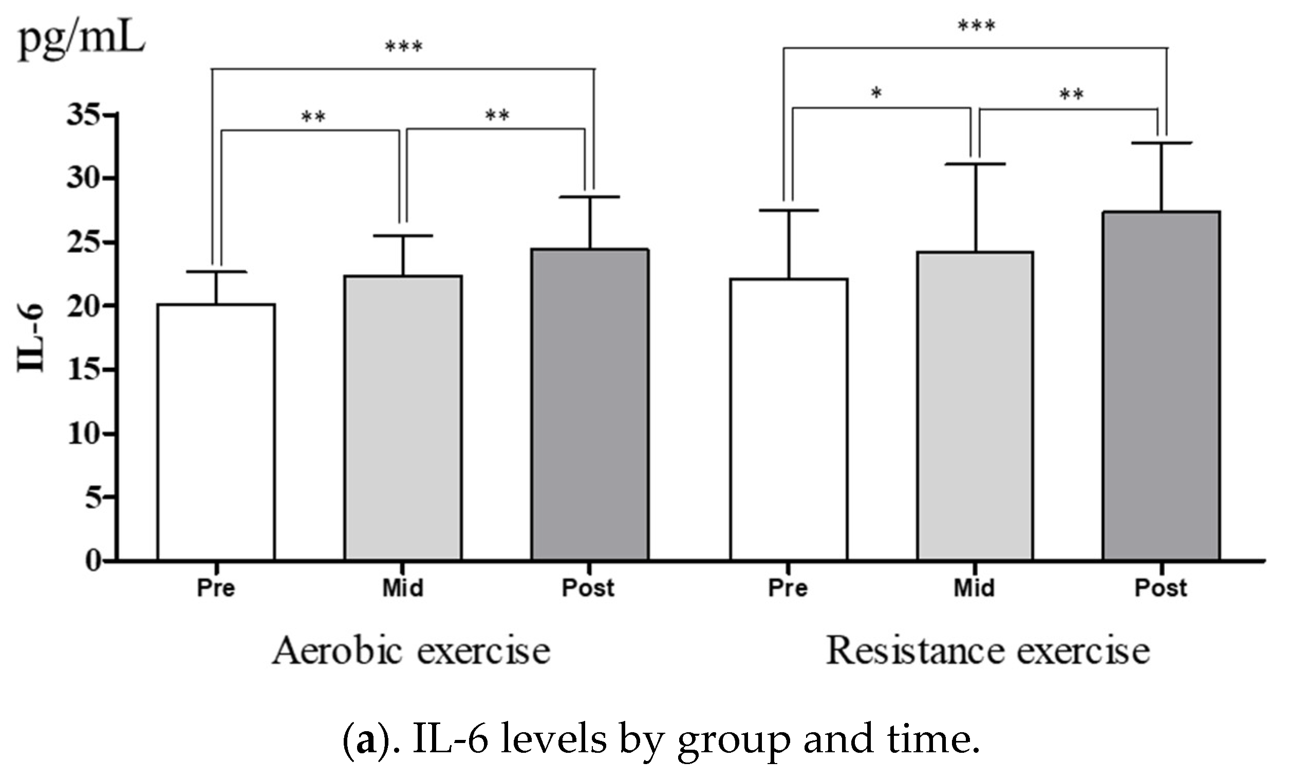
3.3. Change in Myokine Levels after Training

Figure 1 shows the changes in the levels of myokines after training. There was no significant group × time interaction. Post-hoc analysis using the Bonferroni test indicated that levels of the IL-6 (Figure 1a), IL-15 (Figure 1b), and BDNF (Figure 1c) levels were greater at 12 weeks than those at baseline and 6 weeks in both exercise groups.

4. Discussion

In this study, changes in physical fitness and myokine levels after aerobic exercise training and resistance exercise training in PMOF were investigated. The main finding of this study was that the myokine levels increased significantly after aerobic and resistance exercise training in both groups. In addition, weight, BMI, and % fat decreased significantly after exercise training in both groups. Muscle mass increased significantly only in the resistance exercise training group. Regarding physical fitness, muscle endurance and agility increased significantly after exercise training in both groups.
The circulating levels of IL-6 are affected by the duration and intensity of human muscle contractions [17]. IL-6 increases glucose absorption and fatty acid oxidation in vitro through the AMP-active protein kinase and PI3K-Akt signal pathways [18]. In addition, muscle-derived IL-6 can inhibit low TNF-α (tumor-necrosis factor-alpha) production, thereby inhibiting TNF-α-induced insulin resistance. Therefore, it may be important to mediate the beneficial health effects of exercise [19]. As a kind of myokine, interleukin-15 (IL-15) was shown to inhibit adipose tissue deposition in both laboratory animals and human subjects [20]. The complex regulation of IL-15 expression and secretion has been summarized [20]. IL-15 also exhibits pro-inflammatory and anti-inflammatory action in various tissues. It has both a positive anticancer effect by stimulating NK cells and a deleterious effect associated with inflammatory bowel disease [21]. BDNF is known to be released mainly in the hypothalamus. It is a key factor that regulates nerve development, plasticity, and energy homeostasis [22]. Interestingly, the gene and protein expression levels of BDNF are upregulated in human skeletal muscle after exercise (aerobic and resistance) [23]. Previous studies have reported that 12 weeks of resistance training significantly increased the levels of BDNF, but not the levels of IL-15 [24]. Eaton et al. [25] reported that IL-6 levels increased significantly after high-intensity interval exercise. Banitalebi et al. [26] showed that combined exercise conducted for 10 weeks significantly decreased the levels of IL-6 and IL-15 in overweight women with type II diabetes. We observed that the levels of IL-6, IL-15, and BDNF increased significantly after exercise training in both groups (aerobic exercise and resistance exercise). Myokines contribute to the autocrine regulation of metabolism in the muscle itself and the paracrine/endocrine regulation of other adjacent/remote organs [27]. BDNF and IL-6 are involved in AMPK-mediated fat oxidation. IL-6 and IL-15 also stimulate the lipolysis of visceral fat [28]. It is known that exercise training requires more muscle contraction.
Paradoxical associations of BMI, overweight status, and obesity were found in postmenopausal females [29]. In this study, the participants were obese (more than 30% body fat) and postmenopausal. Regular exercise (physical activity) induces metabolic and mitochondrial adaptation, improving energy metabolism and the function of many organs [30]. The skeletal muscle is the largest organ in the human body. Several years ago, a volumetric factor (i.e., cytokines) produced and released from muscle cell contractions was identified [31]. Although adipose tissue is considered a major source of cytokines (i.e., adipokines), muscles can also produce and release myokines. This suggests that, in addition to adipose tissue, skeletal muscle can also be a major source of secreted molecules [32]. PMOF have abundant adipose tissues that can secrete adipokines known to induce a pathogenic and pro-inflammatory environment [33]. Thus, obesity increases the secretion of cytokines and PMOF are exposed to various diseases. Therefore, it is important for PMOF to reduce fat and increase muscle mass. In this study, we observed changes in the levels of myokines in PMOF after 6 weeks and 12 weeks of aerobic exercise training or resistance exercise training. The results revealed that the levels of myokines, such as IL-6, IL-15, and BDNF, were significantly increased after exercise training in both groups. In general, resistance exercises increase myofibrillar protein [34] and aerobic exercises promote improved endurance and fatigue resistance [35]. These exercise-specific changes seem to be in contrast to each other. However, the muscle fiber type or myosin heavy chain composition transformation occurs in the same direction regardless of the type of exercise [36]. The concentration of myokines secreted by muscle contraction was changed not only in the resistance exercise group but also in the aerobic exercise group. These results suggest that the positive changes in the myokine levels of PMOF did not depend upon the type of exercise they performed.
Body-weight, BMI, and % body fat were significantly decreased after exercise compared to those at baseline. The levels of myokine levels were also changed after exercise. There was a significant increase in muscle mass in the resistance training group, but not in the aerobic training group. Changes in muscle mass might be due to differences in the characteristics of aerobic and anaerobic exercise. Moreover, changes in body composition might have affected changes in the physical fitness of obese women. The resistance training group showed significant improvements in muscle endurance, power, and agility, while the aerobic training group showed significant improvements in muscle strength, muscle endurance, and agility. Physical fitness and physical activity or exercise training have well-known health benefits for the obese population [37]. Many recent studies have shown that resistance exercise training had a similar effect to aerobic exercise training, including a positive effect on obesity [38,39]. Most previous studies support regular exercise because it can increase muscle mass and decrease weight loss and the % body fat. The increase in myokines due to contractions of the large muscles and the loss of body weight might have improved physical fitness.
The present study had some limitations. The age of the participants in this study varied widely. Although the age difference between the two groups was not significant, subsequent studies with smaller age difference are needed. Another limitation was that the sample size was small, which limited our ability to determine the significance of the results. Therefore, additional studies with larger sample sizes and a control group are required to determine the effectiveness of regular aerobic and resistance exercise on changes in myokines in PMOF.

5. Conclusions

In conclusion, this study indicated that aerobic exercise training and resistance exercise training changed the levels of myokines and improved body composition and physical fitness in PMOF. Additionally, aerobic exercise training and resistance exercise training are traditionally effective non-pharmaceutical treatment interventions for improving and maintaining physical functions. The increase and maintenance of myokines in PMOF helps to promote health. These findings provide preliminary evidence that PMOF need to perform aerobic and resistance exercise or physical activity to improve or maintain their levels of myokines and physical fitness.

Author Contributions

Conceptualization, S.K. and S.-T.L.; data curation, S.K. and S.-T.L.; formal analysis, S.K. and S.-T.L.; investigation, S.K., I.B.P. and S.-T.L.; methodology, S.K., I.B.P. and S.-T.L.; writing—original draft, S.K. and S.-T.L. All authors have read and agreed to the published version of the manuscript.

Funding

This research received no external funding.

Conflicts of Interest

The authors declare no conflict of interest.

Abbreviations

IL-6interleukin-6
IL-15interleukin-15
BDNFbrain-derived neurotrophic factor
IGF-1insulin-like growth factor
FGF2fibroblast growth factor 2
FGF21fibroblast growth factor 21
FSTL1follistatin-related protein 1
BMIbody mass index
WHRwaist-hip ratio
RMrepetition maximum
HRheart rate
SPSSstatistical package for social science
ANOVA analysis of variance
AMPadenosine monophosphate
TNF-αtumor-necrosis factor-alpha
AMPKactivated protein kinase
PMOFpost- menopausal obese females

References

  1. Schnyder, S.; Handschin, C. Skeletal muscle as an endocrine organ: PGC-1α, myokines and exercise. Bone 2015, 80, 115–125. [Google Scholar] [CrossRef] [Green Version]
  2. Turner, N.; Cooney, G.J.; Kraegen, E.W.; Bruce, C.R. Fatty acid metabolism, energy expenditure and insulin resistance in muscle. J. Endocrinol. 2014, 220, 61–79. [Google Scholar] [CrossRef] [Green Version]
  3. Hawley, J.A.; Hargreaves, M.; Joyner, M.J.; Zierath, J.R. Integrative biology of exercise. Cell 2014, 159, 738–749. [Google Scholar] [CrossRef] [Green Version]
  4. Giudice, J.; Taylor, J.M. Muscle as a paracrine and endocrine organ. Curr. Opin. Pharmacol. 2017, 34, 49–55. [Google Scholar] [CrossRef]
  5. Henningsen, J.; Rigbolt, K.T.; Blagoev, B.; Pedersen, B.K.; Kratchmarova, I. Dynamics of the skeletal muscle secretome during myoblast differentiation. Mol. Cell. Proteom. 2010, 9, 2482–2496. [Google Scholar] [CrossRef] [Green Version]
  6. Pedersen, B.K.; Febbraio, M.A. Muscles, exercise and obesity: Skeletal muscle as a secretory organ. Nat. Rev. Endocrinol. 2012, 8, 457–465. [Google Scholar] [CrossRef]
  7. Pedersen, L.; Hojman, P. Muscle-to-organ cross talk mediated by myokines. Adipocyte 2012, 1, 164–167. [Google Scholar] [CrossRef] [Green Version]
  8. Pedersen, B.K. Muscles and their myokines. J. Exp. Biol. 2011, 214, 337–346. [Google Scholar] [CrossRef] [PubMed] [Green Version]
  9. Díaz, B.B.; González, D.A.; Gannar, F.; Pérez, M.C.R.; de León, A.C. Myokines, physical activity, insulin resistance and autoimmune diseases. Immunol. Lett. 2018, 203, 1–5. [Google Scholar] [CrossRef]
  10. Nimmo, M.A.; Leggate, M.; Viana, J.L.; King, J.A. The effect of physical activity on mediators of inflammation. Diabetes Obes. Metab. 2013, 3, 51–60. [Google Scholar] [CrossRef]
  11. Nielsen, A.R.; Mounier, R.; Plomgaard, P.; Mortensen, O.H.; Penkowa, M.; Speerschneider, T.; Pilegaard, H.; Pedersen, B.K. Expression of interleukin-15 in human skeletal muscle effect of exercise and muscle fibre type composition. J. Physiol. 2007, 584, 305–312. [Google Scholar] [CrossRef]
  12. Matthews, V.B.; Aström, M.B.; Chan, M.H.; Bruce, C.R.; Krabbe, K.S.; Prelovsek, O.; Akerström, T.; Yfanti, C.; Broholm, C.; Mortensen, O.H.; et al. Brain-derived neurotrophic factor is produced by skeletal muscle cells in response to contraction and enhances fat oxidation via activation of AMP-activated protein kinase. Diabetologia 2009, 52, 1409–1418. [Google Scholar] [CrossRef] [Green Version]
  13. Javadivala, Z.; Kousha, A.; Allahverdipour, H.; Jafarabadi, M.A.; Tallebian, H. Modeling the relationship between physical activity and quality of life in menopausal-aged women: A cross-sectional study. J. Res. Health Sci. 2013, 13, 168–175. [Google Scholar]
  14. Cebula, A.; Tyka, A.K.; Tyka, A.; Pałka, T.; Pilch, W.; Luty, L.; Mucha, D. Physiological response and cardiorespiratory adaptation after a 6-week Nordic Walking training targeted at lipid oxidation in a group of post-menopausal women. PLoS ONE 2020, 15, e0230917. [Google Scholar] [CrossRef] [Green Version]
  15. Aubertin-Leheudre, M.; Lord, C.; Goulet, E.D.; Khalil, A.; Dionne, I.J. Effect of sarcopenia on cardiovascular disease risk factors in obese postmenopausal women. Obesity 2006, 14, 2277–2283. [Google Scholar] [CrossRef]
  16. Cauley, J.A.; Gutai, J.P.; Kuller, L.H.; le Donne, D.; Powell, J.G. The epidemiology of serum sex hormones in postmenopausal women. Am. J. Epidemiol. 1989, 129, 1120–1131. [Google Scholar] [CrossRef]
  17. Helge, J.W.; Stallknecht, B.; Pedersen, B.K.; Galbo, H.; Kiens, B.; Richter, E.A. The effect of graded exercise on IL-6 release and glucose uptake in human skeletal muscle. J. Physiol. 2003, 546, 299–305. [Google Scholar] [CrossRef]
  18. Coles, C.A. Adipokines in healthy skeletal muscle and metabolic disease. Adv. Exp. Med. Biol. 2016, 900, 133–160. [Google Scholar]
  19. Pedersen, B.K.; Fischer, C.P. Beneficial health effects of exercise-the role of IL-6 as a myokine. Trends Pharmacol. Sci. 2007, 28, 152–156. [Google Scholar] [CrossRef]
  20. Quinn, L.S.; Anderson, B.G. Interleukin-15, IL-15 receptor-α, and obesity: Concordance of laboratory animal and human genetic studies. J. Obes. 2011, 2011, 456347. [Google Scholar] [CrossRef] [Green Version]
  21. Dozio, E.; Malavazos, A.E.; Vianello, E.; Briganti, S.; Dogliotti, G.; Bandera, F. Interleukin-15 and soluble interleukin-15 receptor α in coronary artery disease patients: Association with epicardial fat and indices of adipose tissue distribution. PLoS ONE 2014, 9, e90960. [Google Scholar] [CrossRef] [PubMed]
  22. Shibata, A.; Hanatani, A.; Izumi, Y.; Kitada, R.; Iwata, S.; Yoshiyama, M. Serum brain-derived neurotrophic factor level and exercise tolerance complement each other in predicting the prognosis of patients with heart failure. Heart Vessels 2018, 33, 1325–1333. [Google Scholar] [CrossRef] [PubMed]
  23. Pedersen, B.K.; Pedersen, M.; Krabbe, K.S.; Bruunsgaard, H.; Matthews, V.B.; Febbraio, M.A. Role of exercise-induced brain-derived neurotrophic factor production in the regulation of energy homeostasis in mammals. Exp. Physiol. 2009, 94, 1153–1160. [Google Scholar] [CrossRef] [PubMed]
  24. Urzi, F.; Marusic, U.; Ličen, S.; Buzan, E. Effects of elastic resistance training on functional performance and myokines in older women-a randomized controlled trial. J. Am. Med. Dir. Assoc. 2019, 20, 830–834. [Google Scholar] [CrossRef]
  25. Eaton, M.; Granata, C.; Barry, J.; Safdar, A.; Bishop, D.; Little, J.P. Impact of a single bout of high-intensity interval exercise and short-term interval training on interleukin-6, FNDC5, and METRNL mRNA expression in human skeletal muscle. J. Sport Health Sci. 2018, 7, 191–196. [Google Scholar] [CrossRef]
  26. Banitalebi, E.; Kazemi, A.; Faramarzi, M.; Nasiri, S.; Haghighi, M.M. Effects of sprint interval or combined aerobic and resistance training on myokines in overweight women with type 2 diabetes: A randomized controlled trial. Life Sci. 2019, 217, 101–109. [Google Scholar] [CrossRef]
  27. Kim, S.; Choi, J.Y.; Moon, S.; Park, D.H.; Kwak, H.B.; Kang, J.H. Roles of myokines in exercise-induced improvement of neuropsychiatric function. Pflug. Arch. 2019, 471, 491–505. [Google Scholar] [CrossRef]
  28. Pedersen, B.K. Anti-inflammatory effects of exercise: Role in diabetes and cardiovascular disease. Eur. J. Clin. Investig. 2017, 47, 600–611. [Google Scholar] [CrossRef] [Green Version]
  29. Tanaka, S.; Kuroda, T.; Saito, M.; Shiraki, M. Overweight/obesity and underweight are both risk factors for osteoporotic fractures at different sites in Japanese postmenopausal women. Osteoporos Int. 2013, 24, 69–76. [Google Scholar] [CrossRef]
  30. Drake, J.C.; Wilson, R.J.; Yan, Z. Molecular mechanisms for mitochondrial adaptation to exercise training in skeletal muscle. FASEB J. 2016, 30, 13–22. [Google Scholar] [CrossRef]
  31. Peake, J.M.; Gatta, P.D.; Suzuki, K.; Nieman, D.C. Cytokine expression and secretion by skeletal muscle cells: Regulatory mechanisms and exercise effects. Exerc. Immunol. Rev. 2015, 21, 8–25. [Google Scholar] [PubMed]
  32. Bleau, C.; Karelis, A.D.; St-Pierre, D.H.; Lamontagne, L. Crosstalk between intestinal microbiota, adipose tissue and skeletal muscle as an early event in systemic low-grade inflammation and the development of obesity and diabetes. Diabetes Metab. Res. Rev. 2015, 31, 545–561. [Google Scholar] [CrossRef] [PubMed]
  33. Palmer, B.F.; Clegg, D.J. The sexual dimorphism of obesity. Mol. Cell. Endocrinol. 2015, 402, 113–119. [Google Scholar] [CrossRef] [PubMed] [Green Version]
  34. Tesch, P.A. Skeletal muscle adaptations consequent to long-term heavy resistance exercise. Med. Sci. Sports Exerc. 1988, 20, 132–134. [Google Scholar] [CrossRef]
  35. Holloszy, J.P.; Booth, F.W. Biochemical adaptations to endurance exercise in muscle. Annu. Rev. Physiol. 1976, 38, 273–291. [Google Scholar] [CrossRef]
  36. Kraemer, W.J.; Patton, J.F.; Gordon, S.E.; Harman, E.A.; Deschenes, M.R.; Reynolds, K.; Newton, R.U.; Triplett, N.T.; Dziados, J.E. Compatibility of high-intensity strength and endurance training on hormonal and skeletal muscle adaptations. J. Appl. Physiol. 1995, 78, 976–989. [Google Scholar] [CrossRef]
  37. Mora-Gonzalez, J.; Esteban-Cornejo, I.; Cadenas-Sanchez, C.; Migueles, J.H.; Rodriguez-Ayllon, M.; Molina-García, P.; Hillman, C.H.; Catena, A.; Pontifex, M.B.; Ortega, F.B.; et al. Fitness, physical activity, working memory, and neuroelectric activity in children with overweight/obesity. Scand. J. Med. Sci. Sports 2018, 29, 1352–1363. [Google Scholar] [CrossRef]
  38. Francois, M.E.; Durrer, C.; Pistawka, K.J.; Halperin, F.A.; Little, J.P. Resistance-based interval exercise acutely improves endothelial function in type 2 diabetes. Am. J. Physiol. Heart Circ. Physiol. 2016, 311, 1258–1267. [Google Scholar] [CrossRef]
  39. Talebi-Garakani, E.; Safarzade, A. Resistance training decreases serum inflammatory markers in diabetic rats. Endocrine 2013, 43, 564–570. [Google Scholar] [CrossRef]
Figure 1. Myokine levels by group and time. * p < 0.05, ** p < 0.01, *** p < 0.001.
Sustainability 12 08413 g001aSustainability 12 08413 g001b
Table 1. Characteristics of the subjects.
VariableResistance Exercise (n = 20)Aerobic Exercise (n = 21)p-Value
Age (years)52.50 ± 7.6556.67 ± 5.430.051
Height (cm)157.89 ± 4.30155.67 ± 5.960.180
Weight (kg)62.82 ± 10.0962.06 ± 9.190.801
BMI (kg/m2)25.16 ± 3.6725.67 ± 3.670.655
% fat (%)36.02 ± 5.8736.53 ± 5.890.781
Muscle mass (kg)21.60 ± 3.0021.26 ± 2.980.719
SBP (mmHg)129.60 ± 18.29129.24 ± 16.380.947
DBP (mmHg)81.00 ± 8.6982.00 ± 9.950.734
Values are the mean ± SD. BMI: body mass index; SBP: systolic blood pressure; DBP: diastolic blood pressure.
Table 2. Exercise program.
ExerciseTypeTime (min)Intensity
Resistance ExerciseWarm-up103 times per week, 12 weeks
3 sets
55~65% RM
1. Squat40
2. Chest press
3. Lat pull-down
4. Lunge
5. Vertical fly
6. Long pull
7. Crunch
Cool-down10
Aerobic ExerciseWarm-up103 times per week, 12 weeks
50~60% HRR
Treadmill running30
Cool-down10
RM: repetition maximum: HRR: heart rate reserve.
Table 3. Measurement of Body Composition parameters by group and time.
VariableTimeType of Exercisep-Value (Interaction)
Resistance Exercise (n = 20)Aerobic Exercise (n = 21)
Weight (kg)Pre62.82 ± 10.09 a*62.06 ± 9.19 a**0.050
Mid62.57 ± 9.7060.90 ± 9.00 b**
Post61.58 ± 8.92 c*60.69 ± 9.24
BMI (kg/m2)Pre25.16 ± 3.67 a*25.67 ± 3.67 a**0.024
Mid25.12 ± 3.68 25.15 ± 3.58 b***
Post24.69 ± 3.29 c* 25.07 ± 3.70
% fat (%)Pre36.02 ± 5.87 a**36.53 ± 5.89 a***0.983
Mid34.52 ± 6.09 b*34.90 ± 5.55 b***
Post33.88 ± 5.1734.30 ± 5.28 c*
Muscle mass (kg)Pre21.60 ± 3.00 a*21.26 ± 2.980.295
Mid22.10 ± 3.1021.34 ± 3.01
Post22.02 ± 3.1721.47 ± 3.15
WHRPre0.91 ± 0.05 a*0.91 ± 0.05 a**0.519
Mid0.89 ± 0.050.89 ± 0.04
Post0.89 ± 0.04 c*0.89 ± 0.04
SBP (mmHg)Pre129.6 ± 18.3 a*129.2 ± 16.40.987
Mid130.8 ± 16.1130.3 ± 15.9
Post122.6 ± 19.3 c**122.7 ± 11.7 c*
DBP (mmHg)Pre81.00 ± 8.6982.00 ± 9.950.636
Mid84.40 ± 10.8085.90 ± 9.43
Post80.00 ± 11.01 c**79.14 ± 10.22 c*
Values are expressed as the mean ± SD. BMI: body mass index; WHR: waist–hip ratio; SBP: systolic blood pressure; DBP: diastolic blood pressure; Pre: baseline; Mid: 6 weeks; Post: 12 weeks. a: pre vs. post; b: pre vs. mid; c: mid vs. post. * p < 0.05, ** p < 0.01, *** p < 0.001.
Table 4. Measurement of physical fitness parameters by group and time.
VariableTimeType of Exercisep-Value (Interaction)
Resistance Exercise (n = 20)Aerobic Exercise (n = 21)
Muscle strength (kg)Pre21.18 ± 5.1019.72 ± 6.53 a*0.389
Mid22.60 ± 5.3022.67 ± 5.17 b*
Post21.64 ± 4.6321.81 ± 4.51
Flexibility (cm)Pre14.72 ± 8.0217.68 ± 6.17 a*0.293
Mid18.20 ± 6.01 b*19.78 ± 6.41 b**
Post17.24 ± 6.7119.97 ± 6.82
Muscle endurance (rep/30 s)Pre14.05 ± 11.88 a***10.48 ± 8.35 a***0.489
Mid19.35 ± 11.86 b**13.52 ± 9.51 b**
Post20.80 ± 13.0416.26 ± 9.10 c*
Power (cm)Pre121.85 ± 24.27 a**115.26 ± 22.930.309
Mid129.75 ± 22.64 b**121.52 ± 18.39
Post131.95 ± 24.47115.42 ± 32.31
Agility (rep/20 s)Pre28.50 ± 3.68 a***28.04 ± 5.13 a***0.710
Mid32.45 ± 4.11 b***31.83 ± 3.10 b***
Post34.10 ± 4.32 c**32.96 ± 3.84
Values are expressed as the mean ± SD. Pre: baseline; Mid: 6 weeks; Post: 12 weeks. a: pre vs. post; b: pre vs. mid; c: mid vs. post. * p < 0.05, ** p < 0.01, *** p < 0.001.

