Article

Slow-Paced Breathing: Influence of Inhalation/Exhalation Ratio and of Respiratory Pauses on Cardiac Vagal Activity

1
Department of Performance Psychology, Institute of Psychology, German Sport University Cologne, Cologne 50933, Germany
2
UFR STAPS, EA 4260 CESAMS, Normandie Université, 14000 Caen, France
3
Department of Health & Social Psychology, Institute of Psychology, German Sport University Cologne, 50933 Cologne, Germany
4
UFR Psychologie, EA3918 CERREV, Normandie Université, 14000 Caen, France
5
INSERM, Unité Mixte de Recherche (UMR) S1158 Neurophysiologie Respiratoire Expérimentale et Clinique, Sorbonne Université, 75000 Paris, France
6
UMR-S 1075 COMETE, Normandie Université, 14000 Caen, France
7
INSERM, UMR-S 1075 COMETE, 14000 Caen, France
*
Author to whom correspondence should be addressed.
Sustainability 2021, 13(14), 7775; https://doi.org/10.3390/su13147775
Submission received: 25 May 2021 / Revised: 2 July 2021 / Accepted: 9 July 2021 / Published: 12 July 2021

Abstract

:
Slow-paced breathing has been shown to enhance the self-regulation abilities of athletes via its influence on cardiac vagal activity. However, the role of certain respiratory parameters (i.e., inhalation/exhalation ratio and presence of a respiratory pause between respiratory phases) still needs to be clarified. The aim of this experiment was to investigate the influence of these respiratory parameters on the effects of slow-paced breathing on cardiac vagal activity. A total of 64 athletes (27 female; Mage = 22, age range = 18–30 years old) participated in a within-subject experimental design. Participants performed six breathing conditions within one session, with a 5 min washout period between each condition. Each condition lasted 5 min, with 30 respiratory cycles, and each respiratory cycle lasted 10 s (six cycles per minute), with inhalation/exhalation ratios of 0.8, 1.0, 1.2; and with or without respiratory pauses (0.4 s) between respiratory phases. Results indicated that the root mean square of successive differences (RMSSD), a marker of cardiac vagal activity, was higher when exhalation was longer than inhalation. The presence of a brief (0.4 s) post-inhalation and post-exhalation respiratory pause did not further influence RMSSD. Athletes practicing slow-paced breathing are recommended to use an inhalation/exhalation ratio in which the exhalation phase is longer than the inhalation phase.

1. Introduction

During sport competitions, athletes are required to effectively regulate their emotions [1,2,3] and cope with stressors [4,5]. Among the strategies addressing athletes’ emotional regulation, slow-paced breathing (SPB), the voluntarily slowing down of breathing frequency, has been increasingly used (e.g., [6,7,8,9]). However, the effectiveness of varying certain parameters of SPB, such as the inhalation/exhalation ratio and the presence of a respiratory pause (i.e., brief cessation of air flow) between respiratory phases, still needs to be understood. Consequently, the current study aims to further understand the role of varying these two parameters on the effectiveness of SPB, as measured by cardiac vagal activity (CVA), an indicator for self-regulation mechanisms [10,11,12,13,14,15,16].
SPB is a technique used to decrease overall activation and trigger relaxation [17]. It involves timed inhalation and exhalation periods (“paced”), at a rate of around six cycles per minute (cpm), which is at least half as slow than the spontaneous breathing rate, normally ranging between 12 and 20 cpm [18,19]. The exact mechanisms by which SPB influences emotion regulation are still debated, but they are likely to involve the strengthening of the baroreflex, the action on pulmonary afferents, as well as specific oscillations in brain networks involved in emotion regulation [20,21,22,23,24,25]. Overall SPB is suggested to trigger the activation of the vagus nerve [21], the main nerve of the parasympathetic nervous system which is responsible for rest and digest functions, as well as self-regulation within the body [11,26,27].
Heart rate variability (HRV), the variation in the time intervals between adjacent heart beats [10,28,29], is a non-invasive indicator of CVA, as explained by the neurovisceral integration model [11,27]. Among the time domain measures of the HRV parameters that index CVA, the root mean square of successive differences (RMSSD) is commonly used. The interpretation of the physiological underpinnings of HRV frequency parameters requires taking into account the respiratory frequency. When breathing frequency is higher than 9 cpm, CVA is reflected in the high-frequency (HF) band [10,28,29]. However, when breathing frequency is lower than 9 cpm, CVA is then shifted to low-frequency (LF) HRV [30]. Given that RMSSD is relatively free from respiratory influences, and more statistically reliable than frequency-domain indicators [31,32], it is the main outcome of interest in this study.
CVA, whether considering its resting, reactivity, or recovery levels [12], has been found to be related to different cognitive and physical aspects of sport performance. For example, morning resting measurements have been used to monitor the effects of training loads, to adjust training loads, and to predict subsequent performance [33,34,35]. Additionally, CVA has been linked to executive cognitive performance in athletes [36,37,38,39,40], to their coping effectiveness with stress and pressure [37,41,42,43,44,45,46], and to sport performance, such as shooting [47,48] and dart throwing performance [49]. Consequently, increasing CVA can be considered an appropriate aim for athletes.
Among the methods used to increase CVA [50,51,52], SPB has been found to be effective in athletes, either combined with biofeedback [53,54] or without biofeedback (e.g., [6,7,8,9]). Biofeedback includes displaying physiological variables of interest (e.g., heart rate, heart rate variability, respiratory frequency) to the person being monitored, through real-time measures realized with a dedicated device, smartphone, or a computer [55]. The use of biofeedback may contribute to additional motivation through providing positive reinforcement to the client [25,55,56,57]. Additionally, biofeedback can assist in identifying the breathing pattern that leads to the highest CVA increase [24,25,58,59]. Nonetheless, so far, no clear evidence has emerged of the physiological benefits of adding biofeedback to SPB [60]. Consequently, the current study focuses on SPB without biofeedback, given that from an applied perspective, it requires athletes to use less technology.
SPB without biofeedback has been associated with positive cognitive and physiological outcomes in athletes [8,9,61,62,63]. More specifically, positive effects were found on executive functions such as inhibition in resting conditions [61,62], working memory, and cognitive flexibility [62], and after physical exertion on inhibition [9]. A robust increase in CVA during SPB has systematically been found, even if the effects tend to cease immediately upon stopping SPB [8,62]. Importantly, long-term SPB interventions (15 min per day for 30 days) seem to trigger increases in resting CVA [63].
Regarding the characteristics of SPB, it is usually performed with a longer exhalation than inhalation phase [24,25]. Adapting a longer exhalation phase is suggested to create a larger increase in CVA, as can be explained through the properties of respiratory sinus arrhythmia (RSA) [64,65]. RSA reflects the influence of respiration on heart rate—more specifically, the fact that heart rate increases with inhalation, and decreases with exhalation [66,67]. Inhalation is driven by sympathetic nervous activity, and exhalation by parasympathetic nervous activity. Theoretically, a longer exhalation would activate the parasympathetic nervous system more strongly. This has been confirmed experimentally in previous research [64,65], where a longer exhalation phase in comparison to inhalation was found to provoke larger increases in RSA, as calculated by the difference between maximum and minimum cardiac interbeat intervals per breath. Although this way of calculating RSA is suggested to reflect CVA, it does not belong to the classic HRV parameters recommended to index CVA [10,28,29]. In addition, Strauss-Blasche, Moser, Voica, McLeod, Klammer and Marktl [65] did not focus on SPB (10 cpm), SPB being characterized by breathing frequencies lower than 10 cpm [22]. Additionally, the sample sizes used in those previous studies were rather small, with N = 12 [65] and N = 23 [64]. The inhalation/exhalation ratio was also investigated by Lin, et al. [68]. Contradictory to previous studies, they concluded that an equal inhalation/exhalation ratio was the most beneficial regarding HRV. However, this experiment presents several drawbacks: RMSSD was not reported, and among the variables reported, only LF may have indexed CVA. However, the findings are difficult to interpret, due to the use of a between-subject design involving N = 11 or N = 12 in each of the four respiratory patterns (6 cpm 5:5; 6 cpm 4:6; 5.5 cpm 5:5; and 5.5 cpm 4:6). Finally, Edmonds, Kennedy, Hughes and Calzada [58] investigated the influence of different breathing patterns around 6 cpm: 1:1 breathing ratio with post-inhalation and post-exhalation respiratory pauses, 1:1 ratio with no respiratory pauses, 1:2 breathing ratio with no respiratory pauses, 1:2 with post-inhalation and post-exhalation respiratory pauses, and finally, a condition requiring the participants to breathe in sync with their heart rate. The focus was not on between-subject analysis, but on within-subject analysis, and the authors found that for each participant, a specific breathing pattern produced the highest increase in LF. Moreover, all breathing patterns were found to produce a descriptive increase in LF; however, no inferential statistics were run at the group level to investigate differences between breathing patterns. To sum up, given the large interindividual variability of HRV frequency-domain variables [10,69] and the lower reliability of frequency parameters when breathing characteristics are modified [31,32], we wanted to address in this study the shortcomings of previous experiments by using a larger sample size, measuring RMSSD as a robust indicator of CVA, which is relatively free from respiratory influences, and by using a within-subject design.
The second parameter of interest in this study is the presence of a post-inhalation and post-exhalation respiratory pause during SPB, an aspect that has received very little attention so far (for an exception, see [70]). In [70], the authors tested the influence of a post-exhalation respiratory pause on HRV (4 s). In comparison to a condition without post-exhalation respiratory pause, a post-exhalation respiratory pause showed a higher HF-HRV, while no differences were found regarding RMSSD and LF-HRV. The authors concluded that a post-exhalation respiratory pause improves CVA and hence self-regulatory control. However, this interpretation of the results is likely inaccurate, given that HF-HRV does not reflect CVA when breathing frequency is lower than 9 cpm [10,28,29]. The pattern of results based on the increase in both RMSSD and LF from baseline in both conditions, with and without post-exhalation respiratory pause, suggests that they both trigger an increase in CVA. Finally, the authors did not provide a physiological rationale for having a post-exhalation respiratory pause besides triggering a higher HF-HRV. Other authors suggested that a post-inhalation respiratory pause may trigger bradycardia via a rapid activation of vagal activity, based on the effects of breath-holding [71]. It may be speculated that SPB provokes a certain strain on respiratory muscles due to the forced and prolonged inhalation and exhalation phases, and would potentially also result in hyperventilation with increased tidal volume [70]. Consequently, the current study aimed to test the influence of a brief post-inhalation and post-exhalation respiratory pause on CVA.
To sum up, research points to positive effects of SPB without biofeedback on CVA [8,9,62,63]. However, the influence of certain characteristics of SPB still needs to be clarified, such as the influence of the inhalation/exhalation ratio, as well as the presence of a respiratory pause between the respiratory phases. The current study therefore aims to address these issues. Regarding the inhalation/exhalation ratio, we hypothesize that a longer exhalation phase in comparison to inhalation would trigger larger increases in CVA [64,65]. Regarding the respiratory pause, we hypothesized that a brief post-inhalation and post-exhalation respiratory pause may potentially produce a bradycardia and reduce the demands on respiratory muscles, therefore resulting in a greater increase in CVA [70,71].

2. Materials and Methods

2.1. Participants

Regarding sample size, previous research on this topic using a within-subject design had a rather low sample size, N = 12 for Strauss-Blasche, Moser, Voica, McLeod, Klammer and Marktl [65], and N = 23 for Van Diest, Verstappen, Aubert, Widjaja, Vansteenwegen and Vlemincx [64]. Following recommendations for HRV research [10,69], we recruited 66 athletes to take part in this research project. Athletes were here defined as individuals regularly engaging in sport training. Recruiting a homogenous athletic sample helps to limit inter-individual differences in HRV that can be found in the general population, enabling a better interpretation of the findings [69]. Exclusion criteria were self-reported cardiovascular diseases and other chronic diseases that might influence breathing or HRV patterns, such as asthma, diabetes, psychiatric, and neurological diseases [10]. Due to technical issues, the data of 2 participants had to be excluded, and the final sample comprised 64 athletes (27 female; MAge = 22, age range = 18–30 years old; BMI: M = 23.10, SD = 2.16; waist-to-hips ratio: M = 0.80, SD = 0.08; number of sport hours per week: M = 7.5 h; SD = 3.2). The experimental protocol was approved by the Ethics Committee of the local university (Project Identification Code 06/11/2014).

2.2. Material and Measures

2.2.1. Heart Rate Variability

HRV was measured with an ECG device (Faros 180°, Bittium, Kuopio, Finland) at a sampling rate of 500 Hz. Two disposable ECG pre-gelled electrodes (Ambu L-00-S/25, Ambu GmbH, Bad Nauheim, Germany) were used. The negative electrode was placed in the right infraclavicular fossa (just below the right clavicle) while the positive electrode was placed on the left side of the chest, below the pectoral muscle in the left anterior axillary line. From ECG recordings, we extracted RMSSD with Kubios (University of Eastern Finland, Kuopio, Finland). The ECG signal was visually inspected for artefacts and these were corrected manually in the cases required (<0.001% of the heartbeats), as recommended by Laborde, Mosley and Thayer [10]. In order to provide an overview of the different HRV parameters, following Laborde, Mosley and Thayer [10], we also extracted the R-R interval, the heart rate, the standard deviation of the NN interval (SDNN) for the time-domain and the frequency domain with Fast Fourier Transform LF (0.04 to 0.15 Hz), HF (0.15 to 0.40 Hz), and the LF/HF ratio. Respiratory frequency was computed via the ECG-derived respiration algorithm of Kubios [72].

2.2.2. Slow-Paced Breathing

Similar to previous research (e.g., [8,73]), SPB was conducted with a video showing a ball moving up and down at the rate of 6 cpm, based on the EZ-Air software (Thought Technology Ltd., Montreal, Canada). Participants were instructed to inhale continuously through the nose while the ball was going up, and exhale continuously with pursed lips when the ball was going down. The video displayed a 5 min SPB exercise, with six conditions, varying in the inhalation/exhalation ratio (inhalation = exhalation; inhalation > exhalation; inhalation < exhalation) and the presence/absence of a respiratory pause (0.4 s) after both inhalation and exhalation phases (see Figure 1). The inhalation/exhalation ratio was 4.5 s/5.5 s and 5.5 s/4.5 s, based on Allen and Friedman [74]. The six conditions were the following: 4.6 s/0.4 s/4.6 s/0.4 s (inhalation/exhalation ratio = 1.0); 4.1 s/0.4 s/5.1 s/0.4 s (inhalation/exhalation ratio = 0.8); 5.1 s/0.4 s/4.1 s/0.4 s (inhalation/exhalation ratio = 1.2); 5 s/5 s (inhalation/exhalation ratio = 1); 4.5 s/5.5 s (inhalation/exhalation ratio = 0.8); 5.5 s/4.5 s (inhalation/exhalation ratio = 1.2).

2.2.3. Procedure

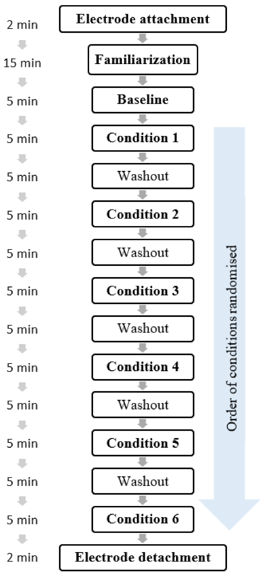
Participants were recruited via flyers on the campus of the local university and via posts on social network groups linked to the local university. In line with recommendations for psychophysiological experiments involving HRV measurements [10], participants were instructed to follow their usual sleep routine the night prior to the experiment, not to consume alcohol or engage in strenuous physical activity in the previous 24 h, nor drink or eat 2 h before taking part in the experiment. All participants gave written informed consent before participation, and were informed that they could withdraw from the study at any time without explanation and without any consequences. The participants attended the lab once in accordance with the within-subject design. The whole session lasted 90 min (the protocol is described in Figure 2). After being welcomed to the lab, they were asked to fill out an informed consent form and a demographic questionnaire regarding variables potentially influencing HRV [10,51,52]. The ECG device was attached, and participants watched a 15 min familiarization video to get acquainted with SPB. The participants started with a 5 min rest period (baseline), where they were breathing spontaneously, with open eyes. After the 5 min rest period, they performed the six breathing conditions in a randomized order, with a 5 min washout period between each respiratory condition, similarly to Russell, Scott, Boggero and Carlson [70]. The washout period characteristics were similar to those of the baseline. At the end of the experiment, the ECG device was detached, and participants were thanked and debriefed.

2.3. Data Analysis

HRV variables were exported from the Kubios output. Data were checked for normality and outliers. Regarding outliers, 0.002% of the cases were found to be univariate outliers (>2 SD, z-scores higher than 2.58; none were found being >3 SD, with z-scores higher than 3.29). Running the analyses without them did not change the pattern of results; therefore, they were kept in the analysis. As the RMSSD data were non-normally distributed, a log-transformation was applied, as is usually recommended for HRV research [10].
As a manipulation check, we first checked whether the participants were breathing at 6 cpm during the different conditions, and whether the breathing frequencies differed among conditions, by conducting a repeated-measures ANOVA. Further, we conducted a series of t-tests (two-tailed) to show whether the breathing conditions differed from baseline, with log RMSSD as the dependent variable, with Bonferroni correction (0.05/6 = 0.008). We conducted a repeated-measures ANOVA, with inhalation/exhalation ratio (inhalation = exhalation; inhalation > exhalation; inhalation < exhalation) and respiratory pause (with/without) as independent variables, and log RMSSD as the dependent variable.

3. Results

Descriptive statistics are presented in Table 1 for all study variables. The first manipulation check revealed that participants followed the 6 cpm breathing frequency, ranging from 6.48 (SD = 0.20) to 6.55 (SD = 0.26). A repeated-measures ANOVA with the Greenhouse–Geisser correction was conducted, and showed no significant effect of condition on breathing frequency, with F (4.401, 277.232) = 0.696, p = 0.608, and partial η2 = 0.01.
The first analysis showed that log RMSSD was significantly higher in all the breathing conditions than in the baseline, for inhalation > exhalation (respiratory pause) with t (63) = 8.693, p < 0.001, Cohen’s d = 1.09; for inhalation < exhalation (respiratory pause) with t (63) = 10.853, p < 0.001, Cohen’s d = 1.36; for inhalation = exhalation (respiratory pause) with t (63) = 9.925, p < 0.001, Cohen’s d = 1.24; for inhalation > exhalation (no respiratory pause) with t (63) = 8.393, p < 0.001, Cohen’s d = 1.05; for inhalation > exhalation (no respiratory pause) with t (63) = 9.428, p < 0.001, Cohen’s d = 1.05; and for inhalation > exhalation (no respiratory pause) with t (63) = 8.944, p < 0.001, Cohen’s d = 1.12.
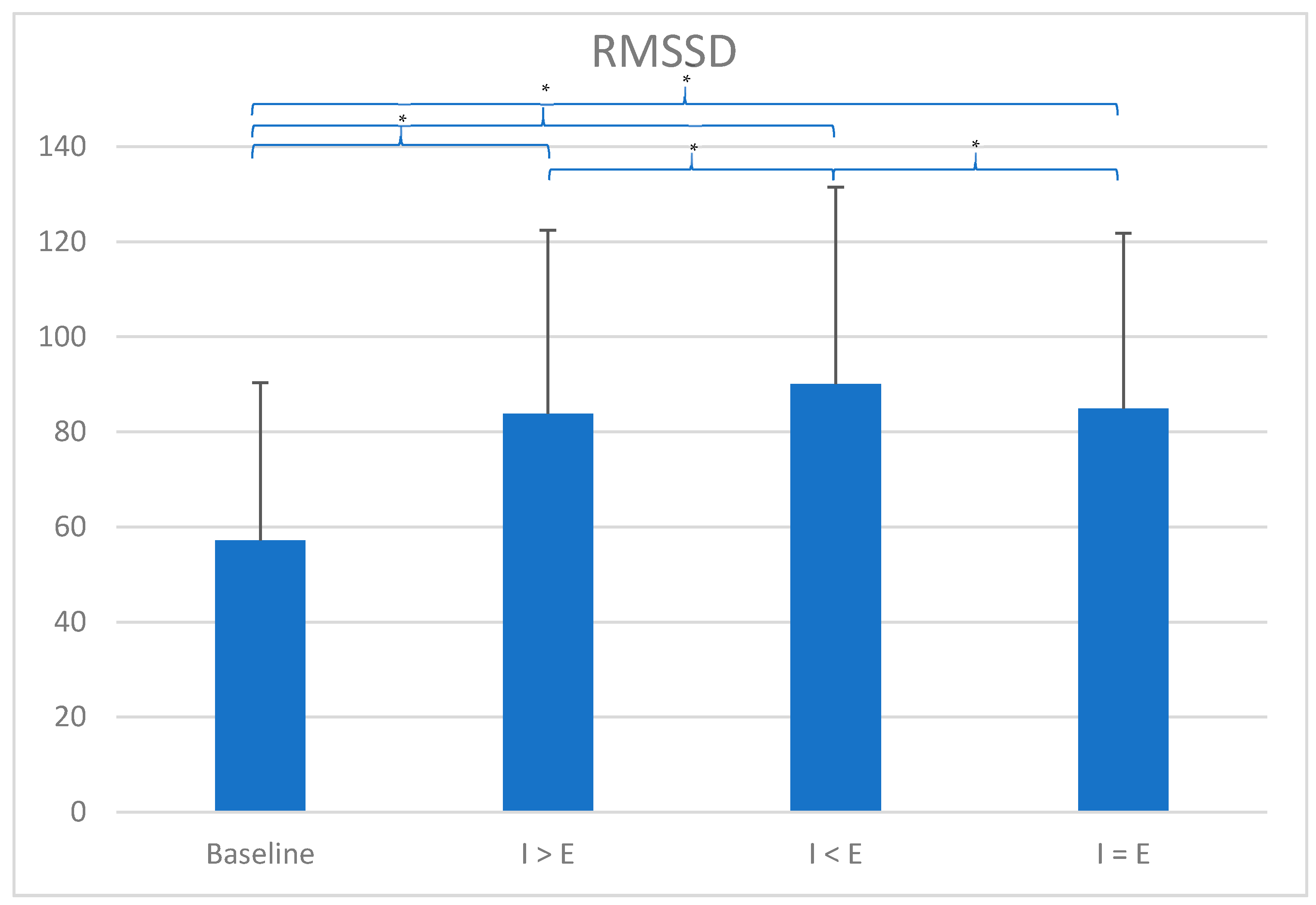
A repeated-measures ANOVA with the Greenhouse–Geisser correction was conducted and showed a significant main effect of inhalation/exhalation ratio, with F (1.996, 125.753) = 8.778, p < 0.001, and partial η2 = 0.12; no main effect of respiratory pause, with F (1, 63) = 2.495, p = 0.119, and partial η2 = 0.04; and no interaction effect between inhalation/exhalation ratio and respiratory pause, with F (1.676, 105.567) = 0.141, p = 0.832, and partial η2 = 0. Regarding the main effect of inhalation/exhalation ratio, further post-hoc t-tests were conducted, applying Bonferroni’s correction with the alpha level adjusted to p = 0.016 (0.05/3). Log RMSSD was found to be significantly higher in the condition with inhalation < exhalation in comparison to inhalation > exhalation, with t (63) = 4.059, Cohen’s d = 0.51, and p < 0.001; and in comparison to inhalation = exhalation, with t (63) = 2.928, Cohen’s d = 0.37, and p = 0.012. No differences were found between the condition inhalation > exhalation and inhalation = exhalation, with t (63) = 1.155, Cohen’s d = 0.14, and p = 0.758. The results of this analysis are presented in Figure 3.

4. Discussion

The aim of this study was to investigate the influence of the inhalation/exhalation ratio and of a respiratory pause on CVA during SPB. Regarding the inhalation/exhalation ratio, findings showed that CVA is higher when the exhalation phase lasts longer than the inhalation phase, confirming our hypothesis. Regarding the presence of a respiratory pause, contrary to our hypothesis, there was no evidence for CVA to be influenced by the presence of a respiratory pause after the inhalation or exhalation phase.
Findings concerning the inhalation/exhalation ratio are in line with previous research [64,65], with the exception of Lin, Tai and Fan [68]; however, their study suffered from a number of methodological issues, regarding sample size, design, and HRV variables reported, as noted in the introduction section. Based on the characteristics of RSA [66,67], heart rate increases with inhalation and decreases with exhalation. Inhalation is driven by sympathetic nervous activity and inhibits parasympathetic nervous activity, while exhalation reactivates parasympathetic nervous activity. Consequently, we can assume that a longer exhalation provokes a longer activation of the parasympathetic nervous system, which is reflected in CVA. It is expected that at slow breathing frequencies, more acetylcholine is released during exhalation, due to its longer duration [75]. Due to the time constants of acetylcholine hydrolysis around 1.5 s to 2.0 s [76,77], longer exhalation is suggested to provoke a summation of sinoatrial responses, and hence maximally inhibit sinoatrial node firing. This longer exhalation in comparison to inhalation was also found to trigger more benefits, for example in terms of pain perception [78]. However, we have to note that the inhalation/exhalation ratio range was reduced in this study (0.8–1.2) in comparison to other studies, such as 0.5–1 [58]; 0.4–2.3 [64], or 1–3.4 [65]. Our rationale was to investigate whether slight variations in the ratio would already have an effect on CVA, but larger ranges may reveal different physiological effects.
With regard to the respiratory pause during SPB, the findings did not show any changes in CVA with the presence or absence of a respiratory pause. As we mentioned in the introduction, Russell, Scott, Boggero and Carlson [70] concluded inaccurately that a post-exhalation respiratory pause (4 s) increased CVA, drawing conclusions on HF-HRV during SPB, which in this case does not reflect CVA [30]. Based on previous research [70,71], we argued that a post-inhalation and post-exhalation respiratory pause may be less demanding for the participant, by not requiring such a prolonged inhalation and exhalation, and that it would additionally trigger a bradycardia. Given that no changes were observed in CVA, it may be that the respiratory pause we chose in this design (0.4 s) may have been too short to elicit these effects. Other research used a respiratory pause of 4 s but had an exhalation phase of 2 s [70], which we deemed too short given that the inhalation phase lasted 4 s, and regarding previous research, a longer exhalation triggers higher increases in CVA [64,65].
Our study had several strengths, such as a larger sample size than previous inhalation/exhalation ratio-related research [64,65] and the investigation of RMSSD to index CVA, as it is suggested to be relatively free of respiratory influences [31]. Limitations include first the sample chosen, comprising only athletes. This limits the generalization of our findings, given that athletes have higher resting HRV than the general population [79], and a ceiling effect may appear with SPB. Future research must investigate this research question in different samples. Additionally, demographics related to the sport practiced were collected. Second, the 5 min washout period between the conditions might not have been sufficiently long to cancel the effects of previous breathing techniques. Third, our equipment did not allow us to control precisely the exact duration of the inhalation, exhalation, and respiratory pauses, so it is not possible for us to evaluate how accurately the participants performed the breathing techniques. Still, we were able to control for respiratory frequency, to assure that our participants followed the 6 cpm rhythm, by using the respiration algorithm of Kubios [72]. Additionally, the experimenter paid close attention that the participants were following the breathing pacer. Finally, we also checked the visual display of R-R values with Kubios, given that during slow-paced breathing, oscillations matching the respiratory frequency can be observed [24]. Nonetheless, future research should use a respiratory belt to offer an online measurement of respiratory frequency. The fourth limitation is the design of the respiratory pause—future research should endeavor to disentangle the effects of a post-inhalation and post-exhalation respiratory pauses by investigating them separately, and should also consider respiratory pauses of different durations. We originally chose a 0.4 s respiratory pause duration to enable our participants to avoid switching abruptly from inhalation to exhalation, and from exhalation to inhalation. However, we acknowledge that this duration is much shorter than those used in previous studies, such as 4 s in Russell, Scott, Boggero and Carlson [70], and consequently, future research should investigate whether longer respiratory pauses trigger different physiological effects. Additionally, the inhalation/exhalation ratio range was reduced (0.8–1.2), and future research should consider investigating larger ranges [58,64,65].
Furthermore, the investigation of a respiratory pause should also include gas exchange measurement and consider, in particular, end-tidal CO2 values for enhanced understanding of its consequences on CVA, as well as consider its impact on tidal volume, given the effects of respiratory pauses on RSA [67,70,71]. Additionally, other variables related to HRV may be considered, such as the RSA, calculated as the difference between the maximum and minimum cardiac interbeat interval per breath. The RSA has also been suggested to index CVA [80,81,82] and has been used in previous research investigating a similar research question [64]. Finally, future research should also consider investigating the inhalation/exhalation ratio at different breathing frequencies [64,68,83] and take into account the effects on other cardiovascular parameters, such as baroreflex sensitivity [84] and blood pressure [85].

5. Conclusions

In conclusion, the aim of this study was to investigate the role of two characteristics of SPB on CVA, namely the inhalation/exhalation ratio and the presence of a respiratory pause. Findings showed that adopting a respiratory pattern with a longer exhalation phase triggered higher CVA in comparison to respiratory patterns with longer inhalation than exhalation, or with equal duration of both phases. No changes in CVA were provoked by a respiratory pause, but methodological limitations in our design guarantee further scrutiny of this parameter in the future.
Manipulating the autonomic nervous system is often the target of sport psychological techniques [86,87], with either activating or relaxing purposes, through methods such as power posing [88] and hypnosis [13]. Within the autonomic nervous system, CVA is a particularly desirable target for athletes, given its role in self-regulation [10,11,12,14,15,16]. In contrast to other methods that exist to stimulate the vagus nerve, such as transcutaneous vagus nerve stimulation [89,90,91], SPB without biofeedback does not require external devices besides a respiratory pacer, and can be easily implemented as an acute [8,9,61,62] or long-term intervention [63] in athletes. SPB with respiratory patterns involving a longer exhalation phase compared to inhalation may therefore show potential as a performance habit [92] in order to trigger the highest possible changes in CVA.

Author Contributions

Conceptualization, S.L. and F.D.; methodology, S.L. and F.D.; formal analysis, S.L. and M.Y.; investigation, S.L. and M.Y.; resources, S.L.; data curation, S.L.; writing—original draft preparation, S.L., M.I., N.Z., U.B., M.Y., C.S.-C., F.D.; writing—review and editing, S.L., M.I., N.Z., U.B., M.Y., C.S.-C., F.D.; visualization, M.I. and N.Z.; supervision, S.L. and F.D.; project administration, S.L. and F.D. All authors have read and agreed to the published version of the manuscript.

Funding

This research received no external funding.

Institutional Review Board Statement

The experimental protocol was approved by the Ethics Committee of the German University Cologne (Project Identification Code 06/11/2014).

Informed Consent Statement

Informed consent was obtained from all subjects involved in the study.

Data Availability Statement

Data can be shared by the corresponding author upon reasonable request.

Conflicts of Interest

The authors declare no conflict of interest.

Correction Statement

Due to an error in article production, several article preparation template sentences were included in the initial version of this publication. This information has been removed and the manuscript updated. This change does not affect the scientific content of the article.

References

  1. Ruiz, M.; Robazza, C. Feelings in Sport—Theory, Research, and Practical Implications for Performance and Well-Being; Routledge: London, UK, 2021. [Google Scholar]
  2. Jones, M.V. Controlling emotions in sport. Sport Psychol. 2003, 17, 471–486. [Google Scholar] [CrossRef]
  3. Lazarus, R.S. How emotions influence performance in competitive sports. Sport Psychol. 2000, 14, 229–252. [Google Scholar] [CrossRef]
  4. Nicholls, A.R.; Polman, R.C. Coping in sport: A systematic review. J. Sports Sci. 2007, 25, 11–31. [Google Scholar] [CrossRef] [PubMed]
  5. Nicholls, A.R.; Taylor, N.J.; Carroll, S.; Perry, J.L. The Development of a New Sport-Specific Classification of Coping and a Meta-Analysis of the Relationship between Different Coping Strategies and Moderators on Sporting Outcomes. Front. Psychol. 2016, 7, 1674. [Google Scholar] [CrossRef]
  6. Gross, M.J.; Shearer, D.A.; Bringer, J.D.; Hall, R.; Cook, C.J.; Kilduff, L.P. Abbreviated Resonant Frequency Training to Augment Heart Rate Variability and Enhance On-Demand Emotional Regulation in Elite Sport Support Staff. Appl. Psychophysiol. Biofeedback 2016, 41, 263–274. [Google Scholar] [CrossRef] [PubMed]
  7. Gross, M.J.; Hall, R.; Bringer, J.D.; Cook, C.J.; Kilduff, L.P.; Shearer, D.A. Resonant frequency training in elite sport: A case study example. J. Sport Psychol. Action 2017, 8, 173–183. [Google Scholar] [CrossRef]
  8. You, M.; Laborde, S.; Salvotti, C.; Zammit, N.; Mosley, E.; Dosseville, F. Influence of a Single Slow-Paced Breathing Session on Cardiac Vagal Activity in Athletes. Int. J. Ment. Health Addict. 2021. [Google Scholar] [CrossRef]
  9. Laborde, S.; Lentes, T.; Hosang, T.J.; Borges, U.; Mosley, E.; Dosseville, F. Influence of slow-paced breathing on inhibition after physical exertion. Front. Psychol. 2019, 10. [Google Scholar] [CrossRef]
  10. Laborde, S.; Mosley, E.; Thayer, J.F. Heart Rate Variability and Cardiac Vagal Tone in Psychophysiological Research—Recommendations for Experiment Planning, Data Analysis, and Data Reporting. Front. Physiol. 2017, 8, 213. [Google Scholar] [CrossRef]
  11. Thayer, J.F.; Hansen, A.L.; Saus-Rose, E.; Johnsen, B.H. Heart rate variability, prefrontal neural function, and cognitive performance: The neurovisceral integration perspective on self-regulation, adaptation, and health. Ann. Behav. Med. 2009, 37, 141–153. [Google Scholar] [CrossRef]
  12. Laborde, S.; Mosley, E.; Mertgen, A. Vagal Tank Theory: The Three Rs of Cardiac Vagal Control Functioning—Resting, Reactivity, and Recovery. Front. Neurosci. 2018, 12. [Google Scholar] [CrossRef] [PubMed]
  13. Laborde, S.; Heuer, S.; Mosley, E. Effects of a Brief Hypnosis Relaxation Induction on Subjective Psychological States, Cardiac Vagal Activity, and Breathing Frequency. Int. J. Clin. Exp. Hypn. 2018, 66, 386–403. [Google Scholar] [CrossRef]
  14. Laborde, S.; Mosley, E. Commentary: Heart rate variability and self-control–A meta-analysis. Front. Psychol. 2016, 7. [Google Scholar] [CrossRef]
  15. Zahn, D.; Wenzel, M.; Kubiak, T. Response: Commentary: Heart rate variability and self-control–A meta-analysis. Front. Psychol. 2016, 7. [Google Scholar] [CrossRef]
  16. Zahn, D.; Adams, J.; Krohn, J.; Wenzel, M.; Mann, C.G.; Gomille, L.K.; Jacobi-Scherbening, V.; Kubiak, T. Heart rate variability and self-control—A meta-analysis. Biol. Psychol. 2016, 115, 9–26. [Google Scholar] [CrossRef]
  17. Russo, M.A.; Santarelli, D.M.; O’Rourke, D. The physiological effects of slow breathing in the healthy human. Breathe 2017, 13, 298–309. [Google Scholar] [CrossRef] [PubMed]
  18. Sherwood, L. Fundamentals of Physiology: A Human Perspective, 3rd ed.; Brooks/Cole: Belmont, CA, USA, 2006. [Google Scholar]
  19. Tortora, G.J.; Derrickson, B.H. Principles of Anatomy and Physiology; John Wiley & Sons, Inc.: Hoboken, NJ, USA, 2014. [Google Scholar]
  20. Noble, D.J.; Hochman, S. Hypothesis: Pulmonary afferent activity patterns during slow, deep breathing contribute to the neural induction of physiological relaxation. Front. Physiol. 2019, 10, 1176. [Google Scholar] [CrossRef]
  21. Gerritsen, R.J.S.; Band, G.P.H. Breath of Life: The Respiratory Vagal Stimulation Model of Contemplative Activity. Front. Hum. Neurosci. 2018, 12, 397. [Google Scholar] [CrossRef] [PubMed]
  22. Zaccaro, A.; Piarulli, A.; Laurino, M.; Garbella, E.; Menicucci, D.; Neri, B.; Gemignani, A. How Breath-Control Can Change Your Life: A Systematic Review on Psycho-Physiological Correlates of Slow Breathing. Front. Hum. Neurosci. 2018, 12, 353. [Google Scholar] [CrossRef]
  23. Mather, M.; Thayer, J.F. How heart rate variability affects emotion regulation brain networks. Curr. Opin. Behav. Sci. 2018, 19, 98–104. [Google Scholar] [CrossRef]
  24. Lehrer, P.M.; Gevirtz, R. Heart rate variability biofeedback: How and why does it work? Front. Psychol. 2014, 5. [Google Scholar] [CrossRef]
  25. Shaffer, F.; Meehan, Z.M. A practical guide to resonance frequency assessment for heart rate variability biofeedback. Front. Neurosci. 2020, 14. [Google Scholar] [CrossRef] [PubMed]
  26. Thayer, J.F.; Lane, R.D. Claude Bernard and the heart-brain connection: Further elaboration of a model of neurovisceral integration. Neurosci. Biobehav. Rev. 2009, 33, 81–88. [Google Scholar] [CrossRef] [PubMed]
  27. Smith, R.; Thayer, J.F.; Khalsa, S.S.; Lane, R.D. The hierarchical basis of neurovisceral integration. Neurosci. Biobehav. Rev. 2017, 75, 274–296. [Google Scholar] [CrossRef]
  28. Berntson, G.G.; Bigger, J.T.; Eckberg, D.L.; Grossman, P.; Kaufmann, P.G.; Malik, M.; Nagaraja, H.N.; Porges, S.W.; Saul, J.P.; Stone, P.H.; et al. Heart rate variability: Origins, methods, and interpretive caveats. Psychophysiology 1997, 34, 623–648. [Google Scholar] [CrossRef]
  29. Malik, M. Heart rate variability. Standards of measurement, physiological interpretation, and clinical use. Task Force of the European Society of Cardiology and the North American Society of Pacing and Electrophysiology. Eur. Heart J. 1996, 17, 354–381. [Google Scholar] [CrossRef]
  30. Kromenacker, B.W.; Sanova, A.A.; Marcus, F.I.; Allen, J.J.B.; Lane, R.D. Vagal Mediation of Low-Frequency Heart Rate Variability During Slow Yogic Breathing. Psychosom. Med. 2018, 80, 581–587. [Google Scholar] [CrossRef]
  31. Penttila, J.; Helminen, A.; Jartti, T.; Kuusela, T.; Huikuri, H.V.; Tulppo, M.P.; Coffeng, R.; Scheinin, H. Time domain, geometrical and frequency domain analysis of cardiac vagal outflow: Effects of various respiratory patterns. Clin. Physiol. 2001, 21, 365–376. [Google Scholar] [CrossRef] [PubMed]
  32. Kuss, O.; Schumann, B.; Kluttig, A.; Greiser, K.H.; Haerting, J. Time domain parameters can be estimated with less statistical error than frequency domain parameters in the analysis of heart rate variability. J. Electrocardiol. 2008, 41, 287–291. [Google Scholar] [CrossRef]
  33. Bellenger, C.R.; Fuller, J.T.; Thomson, R.L.; Davison, K.; Robertson, E.Y.; Buckley, J.D. Monitoring Athletic Training Status Through Autonomic Heart Rate Regulation: A Systematic Review and Meta-Analysis. Sports Med. 2016, 46, 1461–1486. [Google Scholar] [CrossRef] [PubMed]
  34. Buchheit, M. Monitoring training status with HR measures: Do all roads lead to Rome? Front. Physiol. 2014, 5, 73. [Google Scholar] [CrossRef]
  35. Stanley, J.; Peake, J.M.; Buchheit, M. Cardiac parasympathetic reactivation following exercise: Implications for training prescription. Sports Med. 2013, 43, 1259–1277. [Google Scholar] [CrossRef]
  36. Laborde, S.; Raab, M. The tale of hearts and reason: The influence of mood on decision making. J. Sport Exerc. Psychol. 2013, 35, 339–357. [Google Scholar] [CrossRef] [PubMed]
  37. Laborde, S.; Raab, M.; Kinrade, N.P. Is the ability to keep your mind sharp under pressure reflected in your heart? Evidence for the neurophysiological bases of decision reinvestment. Biol. Psychol. 2014, 100C, 34–42. [Google Scholar] [CrossRef]
  38. Laborde, S.; Furley, P.; Schempp, C. The relationship between working memory, reinvestment, and heart rate variability. Physiol. Behav. 2015, 139, 430–436. [Google Scholar] [CrossRef]
  39. Klatt, S.; Noel, B.; Musculus, L.; Werner, K.; Laborde, S.; Lopes, M.C.; Greco, P.J.; Memmert, D.; Raab, M. Creative and Intuitive Decision-Making Processes: A Comparison of Brazilian and German Soccer Coaches and Players. Res. Quaterly Exerc. Sport 2019, 1–15. [Google Scholar] [CrossRef]
  40. Mosley, E.; Laborde, S.; Kavanagh, E. Coping related variables, cardiac vagal activity and working memory performance under pressure. Acta Psychol. 2018, 191, 179–189. [Google Scholar] [CrossRef]
  41. Laborde, S.; Lautenbach, F.; Allen, M.S. The contribution of coping-related variables and heart rate variability to visual search performance under pressure. Physiol. Behav. 2015, 139, 532–540. [Google Scholar] [CrossRef] [PubMed]
  42. Laborde, S.; Brüll, A.; Weber, J.; Anders, L.S. Trait emotional intelligence in sports: A protective role against stress through heart rate variability? Personal. Individ. Differ. 2011, 51, 23–27. [Google Scholar] [CrossRef]
  43. Frenkel, M.O.; Laborde, S.; Rummel, J.; Giessing, L.; Kasperk, C.; Plessner, H.; Heck, R.-B.; Strahler, J. Heidelberg Risk Sport-Specific Stress Test: A Paradigm to Investigate the Risk Sport-Specific Psycho-Physiological Arousal. Front. Psychol. 2019, 10. [Google Scholar] [CrossRef] [PubMed]
  44. Minns, D.; Mosley, E.; Laborde, S.; Wimshurst, Z.L. The contribution of cardiac vagal activity on peripheral perception under pressure. Prog. Brain Res. 2018, 240, 93–107. [Google Scholar] [CrossRef]
  45. Vacher, P.; Filaire, E.; Mourot, L.; Nicolas, M. Stress and recovery in sports: Effects on heart rate variability, cortisol, and subjective experience. Int. J. Psychophysiol. 2019, 143, 25–35. [Google Scholar] [CrossRef]
  46. Fortes, L.S.; da Costa, B.D.V.; Paes, P.P.; do Nascimento Junior, J.R.A.; Fiorese, L.; Ferreira, M.E.C. Influence of Competitive-Anxiety on Heart Rate Variability in Swimmers. J. Sports Sci. Med. 2017, 16, 498–504. [Google Scholar] [PubMed]
  47. Mosley, E.; Laborde, S.; Kavanagh, E. The contribution of coping-related variables and cardiac vagal activity on prone rifle shooting performance under pressure. J. Psychophysiol. 2018, 1–17. [Google Scholar] [CrossRef]
  48. Ortega, E.; Wang, C.J.K. Pre-performance Physiological State: Heart Rate Variability as a Predictor of Shooting Performance. Appl. Psychophysiol. Biofeedback 2018, 43, 75–85. [Google Scholar] [CrossRef]
  49. Mosley, E.; Laborde, S.; Kavanagh, E. The contribution of coping related variables and cardiac vagal activity on the performance of a dart throwing task under pressure. Physiol. Behav. 2017, 179, 116–125. [Google Scholar] [CrossRef]
  50. Laborde, S.; Mosley, E.; Ueberholz, L. Enhancing cardiac vagal activity: Factors of interest for sport psychology. Prog. Brain Res. 2018, 240, 71–92. [Google Scholar] [CrossRef]
  51. Fatisson, J.; Oswald, V.; Lalonde, F. Influence diagram of physiological and environmental factors affecting heart rate variability: An extended literature overview. Heart Int. 2016, 11, e32–e40. [Google Scholar] [CrossRef]
  52. Laborde, S.; Mosley, E.; Mertgen, A. A unifying conceptual framework of factors associated to cardiac vagal control. Heliyon 2018, 4, e01002. [Google Scholar] [CrossRef] [PubMed]
  53. Jimenez Morgan, S.; Molina Mora, J.A. Effect of Heart Rate Variability Biofeedback on Sport Performance, a Systematic Review. Appl. Psychophysiol. Biofeedback 2017. [Google Scholar] [CrossRef]
  54. Pagaduan, J.; Chen, Y.-S.; Fell, J.W.; Xuan Wu, S.S. Can Heart Rate Variability Biofeedback Improve Athletic Performance? A Systematic Review. J. Hum. Kinet. 2020, 73, 103–114. [Google Scholar] [CrossRef]
  55. Schwartz, M.; Andrasik, F. Biofeedback: A Practitioner’s Guide, 4th ed.; The Guilford Press: New York, NY, USA, 2017. [Google Scholar]
  56. Wheat, A.L.; Larkin, K.T. Biofeedback of heart rate variability and related physiology: A critical review. Appl. Psychophysiol. Biofeedback 2010, 35, 229–242. [Google Scholar] [CrossRef]
  57. Lehrer, P.M.; Kaur, K.; Sharma, A.; Shah, K.; Huseby, R.; Bhavsar, J.; Zhang, Y. Heart Rate Variability Biofeedback Improves Emotional and Physical Health and Performance: A Systematic Review and Meta Analysis. Appl. Psychophysiol. Biofeedback 2020. [Google Scholar] [CrossRef]
  58. Edmonds, W.A.; Kennedy, T.D.; Hughes, P.A.; Calzada, P.J. A Single-Participants Investigation of the Effects of Various Biofeedback-Assisted Breathing Patterns on Heart Rate Variability: A Practitioner's Approach. Biofeedback 2009, 37, 141–146. [Google Scholar] [CrossRef]
  59. Lehrer, P.M.; Vaschillo, E.; Vaschillo, B. Resonant frequency biofeedback training to increase cardiac variability: Rationale and manual for training. Appl. Psychophysiol. Biofeedback 2000, 25, 177–191. [Google Scholar] [CrossRef] [PubMed]
  60. Wells, R.; Outhred, T.; Heathers, J.A.; Quintana, D.S.; Kemp, A.H. Matter over mind: A randomised-controlled trial of single-session biofeedback training on performance anxiety and heart rate variability in musicians. PLoS ONE 2012, 7, e46597. [Google Scholar] [CrossRef] [PubMed]
  61. Hoffmann, S.; Jendreizik, L.; Ettinger, U.; Laborde, S. Keeping the pace: The effect of slow-paced breathing on error monitoring. Int. J. Psychophysiol. 2019. [Google Scholar] [CrossRef]
  62. Laborde, S.; Allen, M.; Borges, U.; Hosang, T.; Furley, P.; Mosley, E.; Dosseville, F. The influence of slow-paced breathing on executive function. J. Psychophysiol. 2021. [Google Scholar] [CrossRef]
  63. Laborde, S.; Hosang, T.; Mosley, E.; Dosseville, F. Influence of a 30-day slow paced breathing intervention compared to social media use on subjective sleep quality and cardiac vagal activity. J. Clin. Med. 2019, 8, 193. [Google Scholar] [CrossRef]
  64. Van Diest, I.; Verstappen, K.; Aubert, A.E.; Widjaja, D.; Vansteenwegen, D.; Vlemincx, E. Inhalation/Exhalation ratio modulates the effect of slow breathing on heart rate variability and relaxation. Appl. Psychophysiol. Biofeedback 2014, 39, 171–180. [Google Scholar] [CrossRef]
  65. Strauss-Blasche, G.; Moser, M.; Voica, M.; McLeod, D.R.; Klammer, N.; Marktl, W. Relative timing of inspiration and expiration affects respiratory sinus arrhythmia. Clin. Exp. Pharmacol. Physiol. 2000, 27, 601–606. [Google Scholar] [CrossRef] [PubMed]
  66. Eckberg, D.L. Human sinus arrhythmia as an index of vagal cardiac outflow. J. Appl. Physiol. 1983, 54, 961–966. [Google Scholar] [CrossRef]
  67. Berntson, G.G.; Cacioppo, J.T.; Quigley, K.S. Respiratory sinus arrhythmia: Autonomic origins, physiological mechanisms, and psychophysiological implications. Psychophysiology 1993, 30, 183–196. [Google Scholar] [CrossRef]
  68. Lin, I.M.; Tai, L.Y.; Fan, S.Y. Breathing at a rate of 5.5 breaths per minute with equal inhalation-to-exhalation ratio increases heart rate variability. Int. J. Psychophysiol. 2014, 91, 206–211. [Google Scholar] [CrossRef]
  69. Quintana, D.S.; Heathers, J.A. Considerations in the assessment of heart rate variability in biobehavioral research. Front. Physiol. 2014, 5, 805. [Google Scholar] [CrossRef]
  70. Russell, M.E.B.; Scott, A.B.; Boggero, I.A.; Carlson, C.R. Inclusion of a rest period in diaphragmatic breathing increases high frequency heart rate variability: Implications for behavioral therapy. Psychophysiology 2017, 54, 358–365. [Google Scholar] [CrossRef] [PubMed]
  71. Grossman, P. Respiration, stress, and cardiovascular function. Psychophysiology 1983, 20, 284–300. [Google Scholar] [CrossRef]
  72. Tarvainen, M.P.; Niskanen, J.P.; Lipponen, J.A.; Ranta-Aho, P.O.; Karjalainen, P.A. Kubios HRV--heart rate variability analysis software. Comput. Methods Programs Biomed. 2014, 113, 210–220. [Google Scholar] [CrossRef] [PubMed]
  73. Laborde, S.; Allen, M.S.; Gohring, N.; Dosseville, F. The effect of slow-paced breathing on stress management in adolescents with intellectual disability. J. Intellect. Disabil. Res. 2017, 61, 560–567. [Google Scholar] [CrossRef] [PubMed]
  74. Allen, B.; Friedman, B.H. Positive emotion reduces dyspnea during slow paced breathing. Psychophysiology 2012, 49, 690–696. [Google Scholar] [CrossRef]
  75. Taylor, J.A.; Myers, C.W.; Halliwill, J.R.; Seidel, H.; Eckberg, D.L. Sympathetic restraint of respiratory sinus arrhythmia: Implications for vagal-cardiac tone assessment in humans. Am. J. Physiol. Heart Circ. Physiol. 2001, 280, H2804–H2814. [Google Scholar] [CrossRef] [PubMed]
  76. Eckberg, D.L.; Eckberg, M.J. Human sinus node responses to repetitive, ramped carotid baroreceptor stimuli. Am. J. Physiol. 1982, 242, H638–H644. [Google Scholar] [CrossRef] [PubMed]
  77. Baskerville, A.L.; Eckberg, D.L.; Thompson, M.A. Arterial pressure and pulse interval responses to repetitive carotid baroreceptor stimuli in man. J. Physiol. 1979, 297, 61–71. [Google Scholar] [CrossRef]
  78. Jafari, H.; Gholamrezaei, A.; Franssen, M.; Van Oudenhove, L.; Aziz, Q.; Van den Bergh, O.; Vlaeyen, J.W.S.; Van Diest, I. Can slow deep breathing reduce pain? An experimental study exploring mechanisms. J. Pain 2020, 21, 1018–1030. [Google Scholar] [CrossRef]
  79. Aubert, A.E.; Seps, B.; Beckers, F. Heart rate variability in athletes. Sports Med. 2003, 33, 889–919. [Google Scholar] [CrossRef]
  80. Wheeler, T.; Watkins, P.J. Cardiac denervation in diabetes. Br. Med. J. 1973, 4, 584–586. [Google Scholar] [CrossRef]
  81. Ewing, D.J. Cardiovascular reflexes and autonomic neuropathy. Clin. Sci. Mol. Med. 1978, 55, 321–327. [Google Scholar] [CrossRef]
  82. Ewing, D.J.; Clarke, B.F. Diagnosis and management of diabetic autonomic neuropathy. Br. Med. J. Clin. Res. Ed. 1982, 285, 916–918. [Google Scholar] [CrossRef] [PubMed]
  83. Vaschillo, E.G.; Vaschillo, B.; Lehrer, P.M. Characteristics of resonance in heart rate variability stimulated by biofeedback. Appl. Psychophysiol. Biofeedback 2006, 31, 129–142. [Google Scholar] [CrossRef]
  84. Mason, H.; Vandoni, M.; Debarbieri, G.; Codrons, E.; Ugargol, V.; Bernardi, L. Cardiovascular and respiratory effect of yogic slow breathing in the yoga beginner: What is the best approach? Evid. Based Complement Altern. Med 2013, 2013, 743504. [Google Scholar] [CrossRef]
  85. Herakova, N.; Nwobodo, N.H.N.; Wang, Y.; Chen, F.; Zheng, D. Effect of respiratory pattern on automated clinical blood pressure measurement: An observational study with normotensive subjects. Clin. Hypertens. 2017, 23, 15. [Google Scholar] [CrossRef] [PubMed]
  86. Weinberg, R.S.; Gould, D. Foundations of Sport and Exercise Psychology, 7th ed.; Human Kinetics: Champaign, IL, USA, 2019. [Google Scholar]
  87. Hoffmann, S.; Borges, U.; Broker, L.; Laborde, S.; Liepelt, R.; Lobinger, B.H.; Loffler, J.; Musculus, L.; Raab, M. The Psychophysiology of Action: A Multidisciplinary Endeavor for Integrating Action and Cognition. Front. Psychol. 2018, 9, 1423. [Google Scholar] [CrossRef] [PubMed]
  88. Laborde, S.; Strack, N.; Mosley, E. The influence of power posing on cardiac vagal activity. Acta Psychol. 2019, 199, 102899. [Google Scholar] [CrossRef] [PubMed]
  89. Borges, U.; Laborde, S.; Raab, M. Influence of transcutaneous vagus nerve stimulation on cardiac vagal activity: Not different from sham stimulation and no effect of stimulation intensity. PLoS ONE 2019, 14, e0223848. [Google Scholar] [CrossRef]
  90. Borges, U.; Knops, L.; Laborde, S.; Klatt, S.; Raab, M. Transcutaneous Vagus Nerve Stimulation May Enhance Only Specific Aspects of the Core Executive Functions. A Randomized Crossover Trial. Front. Neurosci. 2020, 14. [Google Scholar] [CrossRef]
  91. Borges, U.; Pfannenstiel, M.; Tsukahara, J.; Laborde, S.; Klatt, S.; Raab, M. Transcutaneous vagus nerve stimulation via tragus or cymba conchae: Are its psychophysiological effects dependent on the stimulation area? Int. J. Psychophysiol. 2021. [Google Scholar] [CrossRef] [PubMed]
  92. Laborde, S.; Kauschke, D.; Hosang, T.J.; Javelle, F.; Mosley, E. Performance Habits: A Framework Proposal. Front. Psychol. 2020, 11. [Google Scholar] [CrossRef]
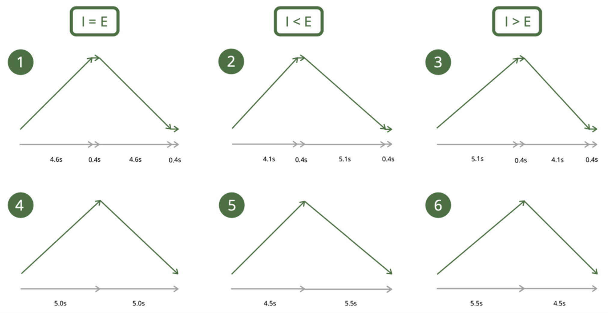
Figure 1. The six breathing conditions, realized in a randomized order during the experiment. Note: ascending arrows depict the inhalation phase and descending arrows depict the exhalation phase; I: inhalation, E: exhalation; time indicated in seconds.
Sustainability 13 07775 g001
Figure 2. Experimental protocol.
Sustainability 13 07775 g002
Figure 3. Root mean square of successive differences (RMSSD) and inhalation/exhalation ratio. Note: We display here the raw RMSSD values given that they are more informative for the reader, as opposed to the log transformed values on which the analyses were based. The main effect of the inhalation/exhalation ratio is shown here, meaning that the conditions with respiratory pauses and without respiratory pauses are pooled for each ratio. I: inhalation; E: exhalation.
Sustainability 13 07775 g003
Table 1. Descriptive statistics.
ConditionsHRSDNNRMSSDLog RMSSDLFHFLF/HFBreathing Frequency
MSDMSDMSDMMSDSDMSDMSDMSD
Baseline67.187.9792.2237.3757.1433.171.704.093.910.223735.746586.001266.251450.3511.452.03
I > E RP68.236.68138.1154.9485.2938.951.8917.5214.530.2014,031.9310,768.081427.021473.486.550.26
I < E RP68.236.53133.8945.0490.2741.381.9114.3012.380.2014,587.3811,754.301954.861918.536.490.30
I = E RP68.376.57132.0642.7486.8239.351.8916.5212.950.2114,352.5711,242.161664.051702.176.510.22
I > E no RP68.917.06127.9439.0282.4438.091.8716.9212.220.2012,761.979874.211415.161621.836.520.23
I < E no RP68.596.86161.1042.8089.9341.391.9116.1313.150.2014,580.1611,277.361602.471403.406.540.32
I = E no RP68.786.30128.2738.7282.9434.541.8815.9111.810.1813,597.239134.121465.511506.676.480.20
Note: I: inhalation; E: exhalation; Rest: with post-inhalation and post-exhalation respiratory pauses; SDNN = standard deviation of all RR intervals; RMSSD: root mean square of the successive differences; LF = low-frequency; HF = high-frequency; RP: respiratory pause.
Publisher’s Note: MDPI stays neutral with regard to jurisdictional claims in published maps and institutional affiliations.