Article

Evaluation of Heavy Metal Tolerance Level of the Antarctic Bacterial Community in Biodegradation of Waste Canola Oil

by
Khadijah Nabilah Mohd Zahri
1,
Claudio Gomez-Fuentes
2,3,
Suriana Sabri
4,
Azham Zulkharnain
5,
Khalilah Abdul Khalil
6,
Sooa Lim
7 and
Siti Aqlima Ahmad
1,3,*
1
Department of Biochemistry, Faculty of Biotechnology and Biomolecular Sciences, Universiti Putra Malaysia, Serdang 43400, Selangor, Malaysia
2
Department of Chemical Engineering, Universidad de Magallanes, Avenida Bulnes, Punta Arenas 01855, Chile
3
Center for Research and Antarctic Environmental Monitoring (CIMAA), Universidad de Magallanes, Avenida Bulnes, Punta Arenas 01855, Chile
4
Department of Microbiology, Faculty of Biotechnology and Biomolecular Sciences, Universiti Putra Malaysia, Serdang 43400, Selangor, Malaysia
5
Department of Bioscience and Engineering, College of Systems Engineering and Science, Shibaura Institute of Technology, 307 Fukasaku, Minuma-ku, Saitama 337-8570, Japan
6
School of Biology, Faculty of Applied Sciences, Universiti Teknologi MARA, Section 2, Shah Alam 45000, Selangor, Malaysia
7
Department of Pharmaceutical Engineering, Hoseo University, Asan-si 31499, Chungnam, Korea
*
Author to whom correspondence should be addressed.
Sustainability 2021, 13(19), 10749; https://doi.org/10.3390/su131910749
Submission received: 23 July 2021 / Revised: 3 September 2021 / Accepted: 3 September 2021 / Published: 28 September 2021
(This article belongs to the Collection Waste Utilization and Resource Recovery)

Abstract

:
Heavy metal contamination is accidentally becoming prevalent in Antarctica, one of the world’s most pristine regions. Anthropogenic as well as natural causes can result in heavy metal contamination. Each heavy metal has a different toxic effect on various microorganisms and species, which can interfere with other pollutant bioremediation processes. This study focused on the effect of co-contaminant heavy metals on waste canola oil (WCO) biodegradation by the BS14 bacterial community collected from Antarctic soil. The toxicity of different heavy metals in 1 ppm of concentration to the WCO-degrading bacteria was evaluated and further analyzed using half maximal inhibition concentration (IC50) and effective concentration (EC50) tests. The results obtained indicated that Ag and Hg significantly impeded bacterial growth and degradation of WCO, while interestingly, Cr, As, and Pb had the opposite effect. Meanwhile, Cd, Al, Zn, Ni, Co, and Cu only slightly inhibited the bacterial community in WCO biodegradation. The IC50 values of Ag and Hg for WCO degradation were found to be 0.47 and 0.54 ppm, respectively. Meanwhile, Cr, As, and Pb were well-tolerated and induced bacterial growth and WCO degradation, resulting in the EC50 values of 3.00, 23.80, and 28.98 ppm, respectively. The ability of the BS14 community to tolerate heavy metals while biodegrading WCO in low-temperature conditions was successfully confirmed, which is a crucial aspect in biodegrading oil due to the co-contamination of oil and heavy metals that can occur simultaneously, and at the same time it can be applied in heavy metal-contaminated areas.

1. Introduction

Metals are defined by their elemental state physical properties, which include metallic lustre, and ability to lose electrons to produce positive ions, as well as to conduct heat and electricity. Metallic elements include transition metals, metalloids, lanthanides, and actinides. They are classified as heavy metals owing to their high atomic weight or density. Each heavy metal has a specific gravity ranging from 3.5 to 6 [1]. Metals have a broad array of uses and are vital to human society, which is dominated by industry. They can be found in fertilizers, pesticides, biosolids, manures in agriculture, as well as in industrial wastewater, including from mining and milling of metal ores [2].
Several heavy metals are essential cofactors for various enzymes. These essential elements are classified as trace elements, which include arsenic (As), cobalt (Co), chromium (Cr), copper (Cu), iron (Fe), vanadium (V), manganese (Mn), nickel (Ni), selenium (Se), molybdenum (Mo), and zinc (Zn) [3]. Metals perform critical biochemical and physiological roles in systems biology; hence, their deficiency or overabundance will cause metabolic disruptions and diseases. However, heavy metal toxicity is primarily concerned with metal bioavailability or the number of organisms that are eventually absorbed into the body through absorption, migration, and transformation. High heavy metal concentrations can disrupt cell membranes, alter enzyme specificity, impair the function of cellular metabolic pathways, and alter the structure of DNA [4]. In addition, heavy metal accumulation in organisms can have various potential health consequences. Heavy metals have been shown in studies to be carcinogenic, teratogenic, and/or mutagenic to some microorganisms, based on the amount of exposure and the length of time [5].
In Antarctica, excessive amounts of heavy metal have been found in the aquatic habitats, atmosphere, and soil, as well as in terrestrial organisms [6]. Generally, the two main causes of heavy metals in the Antarctic environment are from natural geogenic or lithogenic sources and anthropogenic sources. It has been revealed that various metals like Cu, Fe, Ni, As, V, Zn, Mn, Co, aluminium (Al), barium (Ba), beryllium (Be), uranium (U), lead (Pb), silver (Ag), cadmium (Cd), bismuth (Bi), mercury (Hg), and methylmercury (MeHg) have contributed to local contamination in Antarctica over the past five decades. For example, the natural causes of atmospheric heavy metals around Antarctica were glacial erosion of local granodioritic rocks, sea-salt spray, wind-blown crustal dust and volcanic emissions [7,8,9,10,11,12]. Nonetheless, the major local causes of heavy metal pollutants in the Antarctic are human activities through accidental oil spills, abandoned dump sites, waste outfalls, and exhaust emissions. A study conducted at King George Island showed that heavy metals could cause soil contamination from fossil fuel consumption, including Cd and Cu [13]. Additionally, high amount of As, Cd, Cu, Cr, Ni, and Zn have been found in Admiralty Bay, Antarctica [14]. According to Romaniuk et al. [15], high concentration of As, Cd, Co Cr, Cu, Hg, Ni, and Zn were detected in soil collected from King George Island during 2012. Similar heavy metals also could be found in the Artigas base (including Mn and Pb) and Prydz Bay, Antarctica during soil sample collection in 2015 to 2016 [16,17].
Heavy metals in the Antarctic environment and their potential impacts on living organisms could affect the biodegradation process of other pollutants, especially hydrocarbons. The existence of heavy metals in hydrocarbon pollutants could disrupt and damage many species, including microorganisms and animals, but also suppress and kill useful microorganisms and eventually hinder the bioremediation activity. Moreover, it has been proved from various past research that the occurrence of heavy metals in polluted areas can be a main challenge for the removal of other contaminants [18,19,20].
The effects of co-contaminating heavy metals on the microbiota community for the bioremediation of oil have been reported in past research, since they are often found together in polluted sites. Cd, Pb, Mn, Cu, Co, Mo, Cr, Zn, Ni, Al, Hg, and Ni were among the metals tested on the biodegradation of diesel, black oil, crude oil, and total petroleum hydrocarbon (TPH) [21,22,23,24]. Hence, this current work aimed to assess the influences of heavy metals on the bioremediation of cooking oil by the Antarctic bacterial community. This included Al, As, Ag, Zn, Co, Cr, Pb, Ni, Cd, Co, and Hg. Although Al is not categorizedd as a heavy metal, Al contamination in the Antarctic has been reported in past research, which is a reason for this study to include Al as one of the metals that can affect bacterial activities in Antarctica. In the meantime, the relationship between the metals in different concentrations and the biodegradation process was also investigated in this study, by identifying the minimal and maximal metal concentrations that influence the microorganisms in degrading cooking oil. The data on the effects of heavy metals on biodegradation of WCO can be obtained and used for future bioremediation processes such as wastewater treatment in Antarctica.

2. Materials and Methods

2.1. Collection and Activation of the Microbial Sample

A soil sample was collected from northwest Antarctic Peninsula at Base General Bernardo O’Higgins Riquelme (63°19′20.6″ S 57°53′53.6″ W) and labeled as BS14. Approximately 500 mg of the soil sample was transferred into a nutrient broth (NB) (Friedmann Schmidt, Germany) for the bacteria activation process. The sample was incubated on a 150 rpm orbital shaker at 10 °C for 3 to 4 days prior to transfer three times into new media of NB to remove solid soil particles from the media. Then, several copies of 25% glycerol stock were prepared and stored in the −80 °C chiller. The waste canola oil (WCO) was acquired directly from the station. The sampling method is described in more detail in Zahri et al. [25].

2.2. Bacterial Culture Preparation

Bacterial cells were obtained by reviving the BS14 bacterial culture from glycerol stock into NB and incubated at 10 °C on a 150 rpm orbital shaker for 4 days. The culture was then spun down for 10 min under 4 °C at 7000 g. The cell at the bottom of the tube (pellet) was resuspended and washed 2 times with phosphate buffered saline (pH 7.4). Suspension cells were obtained, and the bacterial sample’s inoculum size was standardized to 1.0 ± 0.01 of bacteria turbidity at 600 nm.

2.3. Cell Culture Media

Minimal salt media (MSM) was used in this study, which consisted of 9.52 g/L of Na2HPO4, 2.00 g/L of NaH2PO4, 0.13% w/v of NaCl, 1.00 g/L of (NH4)2SO4 1.13 g/L of yeast extract and 1% v/v of WCO, while the pH was adjusted to 7.3 using HCl. The amount of sterilized WCO (using 0.45 nm filter) added in MSM was at 1% v/v. The incubation conditions were at 13 °C and the sample was shaken in a 150 rpm orbital shaker for 6 days. These conditions were obtained based on the past study on the optimization of biodegradation of WCO using same bacterial community sample (BS14) [25].

2.4. Effects of Heavy Metals on WCO Biodegradation and Growth of Bacterial Community

One milliliter of the bacterial community was incubated in 250 mL conical flasks containing 50 mL of MSM. Each flask was separately supplemented with 1 ppm of a standard solution of heavy metals, Zn2+, Co2+, As3+, Ag+, Cr2+, Pb2+, Ni2+, Al3+, Cd2+, Co2+, or Hg2+ (Sigma-Aldrich, Taufkirchen, Germany) to observe their overall effects on bacterial growth and WCO biodegradation. The control media test was carried out without heavy metals in the flask. Experiments to determine the half-maximal efficiency (EC50) and inhibition concentration (IC50) were carried out on the heavy metals that either allowed a high percentage of WCO biodegradation or significantly inhibited the biodegradation of WCO. The IC50 was determined by adding 0–1 ppm of the selected heavy metals separately into the MSM medium cultures. The EC50 was determined by adding more than 1 ppm of the selected heavy metals into the MSM medium cultures.
The growth of the bacterial community was evaluated using a UV–vis spectrophotometer at 600 nm of optical density (Jenway, Germany) after six days of incubation. One milliliter of bacteria grown from MSM was spun down for 10 min at 10,000 rpm. After that, the supernatant was removed, and the bacterial pellet was resuspended with sterile distilled water before we measured the turbidity of cells. Meanwhile, gravimetric analysis was used to examine the degradation of WCO [26] by adding n-hexane in a ratio of 1:1 (medium to solvent). The funnels were vigorously shaken and left for 15 min to allow the organic phase to separate [27]. Then, under a fume hood, the upper layer of media was transferred into a glass Petri dish and concentrated. The residual WCO was gravimetrically determined, and the degradation was expressed as Equation (1) relative to the abiotic loss of WCO in experimental controls [28]:
WCO   Biodegradation   ( % ) = ( Weight   of   residual   WCO   ( abiotic   control ) weight   of   residual   WCO   ( sample ) Original   weight   of   WCO   introduced ) × 100

2.5. Dose-Response Curve

The effects of different concentrations of heavy metals were determined through the efficiency concentration (EC50) and inhibition concentration (IC50) using inhibition dose response (inhibitor vs. response, four parameters) and stimulation dose response (agonist vs. response, four parameter), respectively. Graphpad Prism software (version 5.0) with the mean and standard error mean (SEM) values was used (n = 3).

3. Results

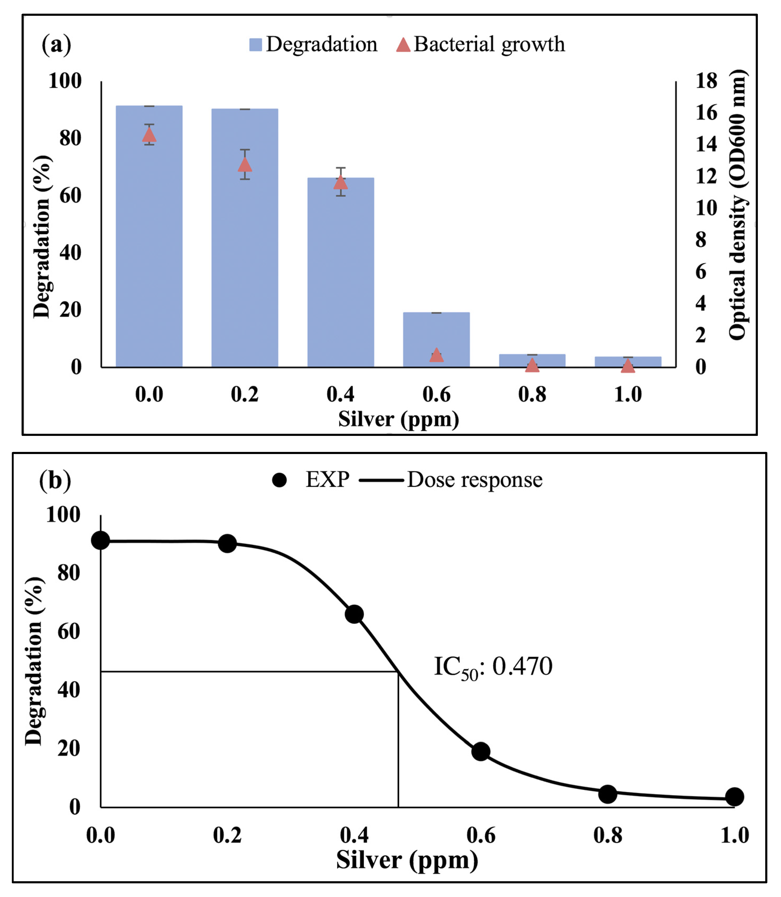
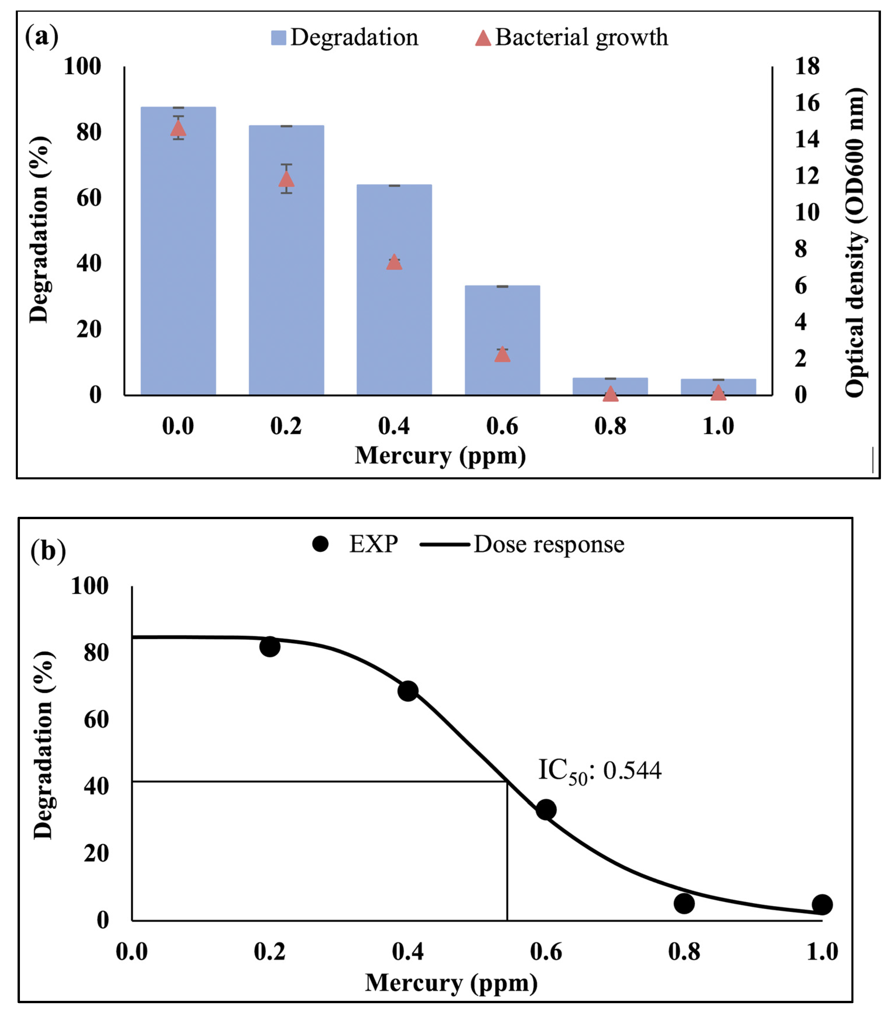
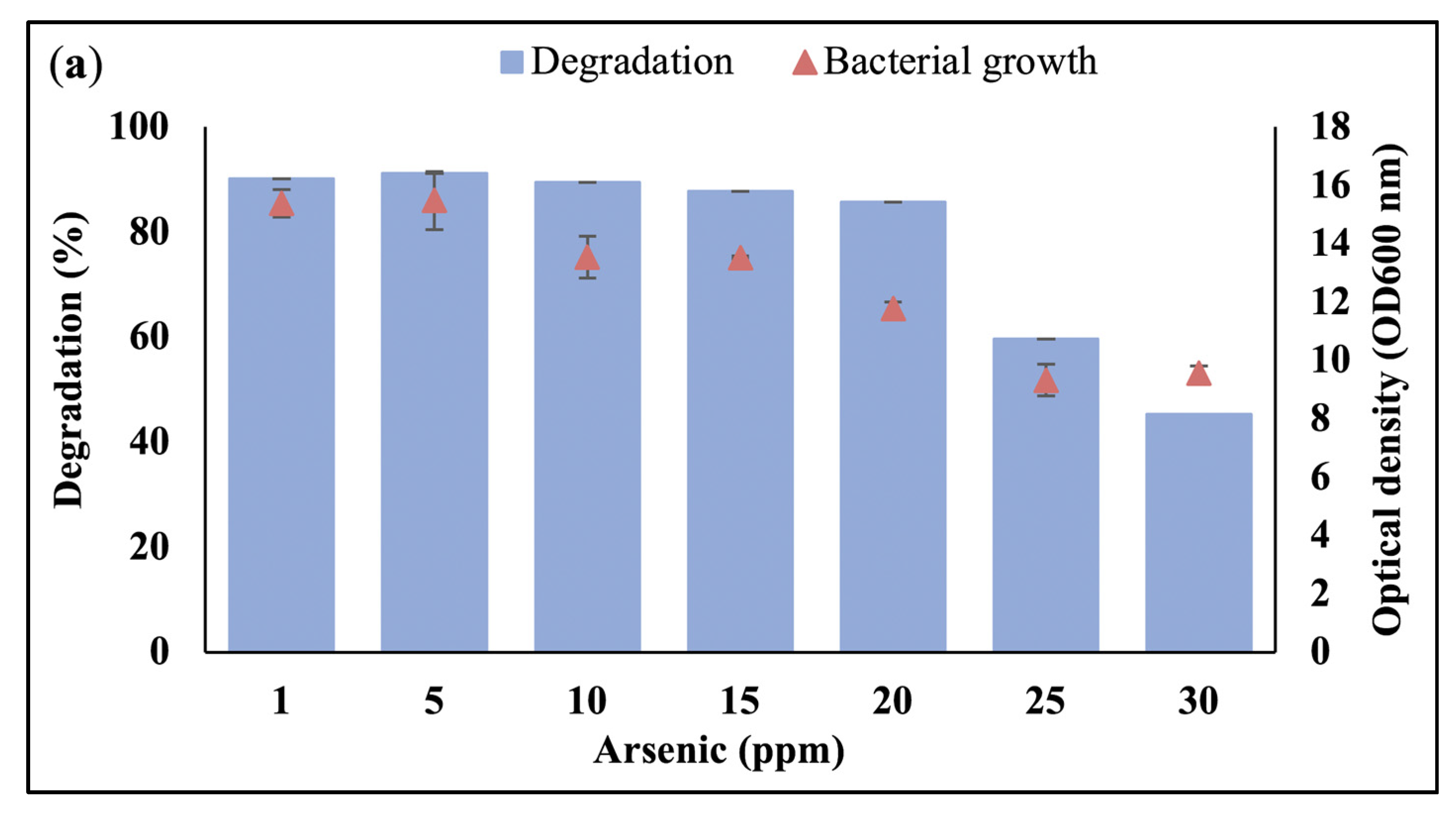
3.1. Effect of Heavy Metals on Bacterial Growth and Waste Canola Oil Biodegradation

Different types of heavy metals exhibited different effects on bacterial growth and WCO biodegradation. Interestingly, Figure 1 shows that the presence of Cr, Pb, and As substantially enhanced the percentage biodegradation of WCO (p < 0.05) up to 91.24%, 89.69%, and 93.46%, respectively, compared to the control (no heavy metals) which obtained 88.78%. All bacterial cultures with these three heavy metals showed high readings on their turbidity except for As. Although the OD600 for the BS14 bacterial community in As medium was low at 13, it was still capable of biodegrading WCO at a high percentage. Other media that were supplemented with Al, Cu, Zn, Ni, Co, and Cd exhibited slightly lower degradation of WCO (~62–85%) and (~13–17) of absorbance value for the cell OD600. In contrast, the presence of Ag and Hg significantly inhibited the growth of bacterial cells, leading to very low degradation percentages. To summarize, the WCO degradation was hampered in an increasing order by Ag > Hg > Cd > Co > Ni > Zn > Cu > Al > C > Pb > Cr > As.

3.2. Dose-Response Analysis

Dose-response analysis was carried out based on the level of statistical significance, where Ag and Hg were identified as the most significantly inhibiting (p < 0.001) heavy metals in WCO biodegradation. Thus, the half-maximal inhibition concentration (IC50) analysis was applied for these two heavy metals in this study. Meanwhile, Cr, Pb, and As were chosen for further analysis in the dose-response study for half-maximal effectiveness concentration (EC50) since these heavy metals can induce the WCO biodegradation process.

3.2.1. Half-Maximal Inhibition Concentration (IC50)

The range of Ag and Hg concentrations used in IC50 in this study was 0 to 1 ppm. It can be seen in Figure 2a,b that low concentrations of Ag still allowed the BS14 community to biodegrade more than 50% of the WCO. At 0.2 and 0.4 ppm of Ag, the bacterial community showed OD600 of 12.78 and 11.68 and degraded WCO by 90.19% and 66.05%, respectively. Nevertheless, there was no major difference in either bacterial growth or degradation at 0 and 0.2 ppm of silver (p > 0.05). However, at 0.6 ppm of Ag, the bacterial community suffered a massive drop in its ability to degrade WCO (19.06%) and showed a very low OD600 value (0.81).
The inhibition dose effect (IC50) of Ag exposure for WCO degradation was 0.470 ppm with a 95% confidence interval of 0.463 to 0.476 ppm. The model was validated through the statistical parameters provided during data analysis. The value coefficient determination (R2) and standard error estimate (sy.x) were 0.999 and 1.083, respectively. In general, the BS14 Antarctic bacterial community used in this study was able to survive and perform biodegradation activity up to ~0.5 ppm of Ag. Concurrently, the slope value obtained from the software in Ag dose-response analysis was 5.92, where a lower slope value indicates a slow decrease of toxic effect.
Figure 3a shows similar outcomes were also seen for the effect of Hg, where a high concentration of Hg reduced both bacterial community turbidity and WCO biodegradation activity. Hg also followed the same pattern in the Ag media, where concentrations below 0.4 ppm resulted in high degradation percentages of WCO. Almost 65% degradation could be achieved in the presence of 0.4 ppm of Hg with an OD600 of 7.32 for the growth, as well as 81.93% and 11.86 in 0.2 ppm of Ag media. There was no significant difference between all parameters in WCO degradation except for at 0.8 and 1.0 ppm of Hg. At these concentrations, the OD600 values obtained for the bacterial community were low (0.11 to 0.16), and the WCO could only be degraded by 4.76 to 5.12%, respectively.
The IC50 and R2 values for Hg were revealed to be 0.544 ppm and 0.994, respectively (Figure 3b). The confidence interval range was between 0.520 to 0.570 ppm, which indicates the ability of this BS14 Antarctic bacterial community to tolerate Hg while biodegrading WCO. At the same time, the hill slope and sy.x value of the model were low, at 5.02 and 2.96, respectively. A low value of error indicates that the actual values are close to the true regression line and provide accurate analysis predictions. This signifies the distance between the experimental and fitted values was small.

3.2.2. Half-Maximal Effective Concentration (EC50)

To investigate the level of the toxicity of selected heavy metals on the BS14 bacterial growth and WCO biodegradation activity, increasing concentrations of selected heavy metals were used. The exposure of the bacterial community to Cr at high concentrations (more than 1 ppm) lowered their growth and capability to degrade WCO. Nonetheless, BS14 still managed to degrade 62% of WCO at 3 ppm of Cr. Bacterial cells grew better in the medium that was supplemented with 1 ppm of Cr, where the OD600 value was 21.04, with 93.01% of WCO being degraded (Figure 4a). Consequently, when comparing the control media (absence of heavy metal) (Figure 1) to the media containing 1 ppm Cr (Figure 4a), an increase in WCO degradation (4.29%) was observed. This suggests that the presence of up to 1 ppm of Cr induced the biodegradation of WCO, as illustrated in Figure 4a. However, there was a substantial drop in growth of bacteria and WCO degradation at 2 ppm of Cr, which indicates that Cr concentrations higher than 1.0 ppm were toxic for cell growth.
The present study demonstrated that metals might affect bacterial growth at a concentration below EC50 values. The estimated EC50 value for Cr was 3.00 ppm, with the 95% confidence interval at 2.907 to 3.115 ppm (Figure 4b). The range of the obtained EC50 value was wide, showing a higher sensitivity of the bacterial growth and degradation process to Cr. A substantial correlation between the concentration of Cr and the percentage of degradation (response) can be observed with the R2 value at 0.994 (above 0.90). The errors for sy.x and hill slope of the dose-response of Cr were 2.109 and −6.38, respectively.
High concentrations were also tested for As and Pb media on the bacterial growth and WCO biodegradation in this study. Significant differences were observed between each concentration at 1 to 20 ppm of As, exhibiting high degradation percentages (Figure 5a). The range of the WCO degradation obtained for this experiment was between 85.71% and 90.20%. The WCO degradation increased slightly by ~2% as the As concentration increased to 5 ppm. However, subsequently higher As concentrations reduced the bacterial growth as well as the degradation activities. As shown in Figure 5a, the lowest degradation was at 30 ppm with 45.35%, and an OD600 value of 9.57.
The toxicity value for EC50 in the dose-response model for As was found to be 23.80 ppm. The estimated confidence interval ranged between 23.60 and 24.18 ppm, which showed that the bacterial community’s relative degradation was at the lowest upon exposure to the high concentrations of As. From the equation of the EC50 model, the R2, sy.x, and slope of fitted lines were 0.997, 1.022, and −13.11, respectively.
The effects of Pb can be observed in Figure 6a. The addition of up to 5 ppm of Pb resulted in a 5% to 13% increase of WCO degradation compared to the control medium (Figure 1). However, at higher concentrations, the growth and WCO degradation decreased, with 30 ppm of Pb reducing the WCO degradation to only 38% and an OD600 of 6171.
The EC50 value together with ± 95% confidence interval of the Pb medium were the highest results obtained in the dose-response study at 28.98 ppm (24.85 to 28.84 ppm). This indicates that the BS14 community used in this study is less sensitive to Pb than all other types of heavy metals tested. The R2 and sy.x values acquired were 0.977 and 2.841, respectively. The exposure to Pb displayed a slow toxicity effect as the hill slope in this media was negative in value, which was −5.78. The toxicity ranking (highest to lowest) based on all heavy metals tested on the EC50 was Cr > As > Pb.

4. Discussion

In this study, the growth of the bacterial community was evaluated using optical density measurement. Despite general concerns that other materials may affect turbidity values, samples were centrifuged for bacterial cell collection before measurement to minimize any interference. This approach was also chosen as it is accurate for indicating the bacterial growth in a community compared to other methods, namely colony-forming unit (CFU) counts, and protein or DNA concentrations, due to high concentration of bacterial cells produced during this study. CFU counts particularly are not reliable for counting cells in a bacterial community as not all strains are colony-forming species and agar media may not be favorable for all species. Gravimetric analysis was carried out in this study to determine the biodegradation percentage of WCO. This method was sufficient to achieve the main objective of this study, which was to evaluate WCO reduction with heavy metals’ influence in the selected Antarctic bacterial community. Detailed insights on the biodegradation efficiencies and dynamics can only be investigated by utilizing sophisticated analytical equipment, however, gravimetric analysis allows data to be collected with minimal requirements, which is ideal for studies in the field and remote locations like Antarctica.
The level of metal toxicity is determined by the contact period and the amount of heavy metals absorbed in the microorganisms. Most microorganisms, including bacteria, are greatly affected by heavy metals as their normal biological activities are severely hindered. In general, high metal toxicity damages cell structures and inhibits cytoplasmic enzymes due to oxidative stress [29,30]. Each heavy metal can cause different toxicity effects via different mechanisms to bacteria. Heavy metals cannot be degraded in the bioremediation process but can only be transformed, absorbed, precipitated, tolerated, or resisted. In the present study, Cd, Al, Co, Zn, Ni, and Cu somewhat reduced the WCO degradation by the BS14 Antarctic bacterial community. The presence of Cd can disrupt protein and nucleic acid synthesis, causing DNA damage and impeding cell repair mechanisms [31]. In past research, Micrococcus sp. and Pseudomonas aeruginosa showed reduced growth rates in kerosene with the addition of Cd at 50 to 200 ppm [32]. Meanwhile, Al can be a competitor to other essential elements such as iron and magnesium in bacteria. It can also bind to the DNA and the cell membrane, causing cell disruption [33]. Moreover, Al becomes soluble and toxic to the environment when the pH is low [34]. Similarly, although Co is an essential heavy metal, it could disrupt other important metals’ self-regulating biological processes, causing oxidative stress from reactive oxygen species (ROS) production [35,36]. Regardless of whether it is an essential or non-essential metal, a high enough metal concentration can turn it into a toxic compound. For example, 1 ppm of Al and Co suppressed the growth and diesel biodegradation by a marine bacterial consortium collected from the Antarctic Peninsula region [37]. Another essential metal found in organisms is Zn, which can be toxic at excessive levels. Although Zn is required for protein utilization mechanisms, it may also inhibit essential processes in cells that involve enzyme reactions [38]. Like other metals, Ni has similar toxicity effects; it can damage proteins and DNA by binding to various biomolecules and changing their properties. It has been reported that the total petroleum hydrocarbon (TPH) biodegradation time was reduced from 0 to 35 days in the existence of Cd, Ni, and Zn by a bacterial and fungal consortium [39]. Cu is another metal classified as an essential cofactor in bacterial metabolism but is extremely toxic to microorganisms at high concentrations. Several routes of Cu toxicity in bacteria have been discussed by Giachino and Waldron [40], such as (1) inhibition of lipoprotein maturation and causing accumulation in the inner membrane, (2) formation of non-native disulfide bonds that can cause protein misfolding, (3) weakening the cells’ peptidoglycan, (4) displacing other metals from protein binding sites, and 4) disrupting the cellular redox potential. In previous research, diesel degradation by Pseudomonas extremaustralis was lowered by 7% in the presence of Cu, although the degradation was increased after glucose was added in the media [41]. This is because glucose has a survival benefit on the cellular envelopes, reducing the effects of stressors such as Cu and diesel.
In the present study, Ag and Hg exhibited the strongest toxic effects on the BS14 bacterial community. High concentrations of both metals severely suppressed the bacterial growth in the media. The use of the Ag ion as an antibacterial agent in many medical applications indicates the effectiveness of this metal in killing bacteria [42,43]. The antimicrobial mechanism of the Ag ion is strongly correlated to its interaction with sulfhydryl groups (amino acids with disulfide bonds, amino acids without sulphur bonds, and sulfur-containing compounds). Ag was also suggested to bind to vital functional groups of enzymes on the bacterial plasma/cytoplasmic membrane. This can lead to the deposition of Ag in the bacteria’s vacuole and cell wall, which inhibits cell division [44]. A past study confirmed that the Ag ion, mostly in silver nanoparticle form, can inhibit bacterial growth and activity, including pathogenic microbes such as P. aeruginosa, S. aureus, and E. coli [45]. Ibrahim et al. [19] found that the IC50 of Ag for Rhodococcus sp. strain AQ5-07 when degrading WCO was 0.322 ppm. In comparison, the present study obtained an IC50 value for Ag of 0.470 ppm, almost 0.15 ppm higher.
Hg is known as a toxic and rare element in nature that exists as a metal as well as inorganic and organic compounds. Hg is a carcinogenic and neurotoxic metal that can have acute health effects, including causing nervous system diseases, and cell and system damage [46]. Hg is also known to impede critical bacterial processes, such as degradation of organic compounds in aerobic and anaerobic conditions. The modification and/or displacement of metal nutrients from cellular sites, as well as the blocking of functional groups such as enzymes, polynucleotides, and essential nutrient transport systems, are among its lethal consequences. This may result in changes in the active conformation of biological molecules and disruptions in the integrity of cellular and organelle membranes [47]. The absence of a metal resistance mechanism in bacterial metabolism can cause bacterial death even at low metal concentrations. A study on hydrocarbon degradation in the presence of metals including Hg by an unknown bacterial community showed that the bacteria did not have any resistance mechanism, although they originated from soil that was highly polluted by metals [48].
Nonetheless, in some cases, some bacteria belonging to the Pseudomonas, Bacillus, and Rhodococcus genera can tolerate and resist Hg due to long-term exposure to the heavy metal, which allows the bacteria to develop resistance mechanisms and metal-ion homeostasis [49]. The level of metal toxicity in microbes can be measured through IC50 dose-response analysis. In the present study, the IC50 of Hg showed a high concentration (0.544 ppm) compared to past studies using Arthrobacter sp. (0.211 to 0.335 ppm) and Rhodococcus sp. (0.454 ppm) isolated from the Antarctic in degrading diesel and canola oil, respectively [18,19]. A low IC50 value means that the metal is more potent at a low concentration and will thus show higher systemic toxicity to the bacteria. The higher value observed for IC50 of Hg in the current study may have been caused by the diversity of bacterial species in the BS14 community, which allowed a better WCO degradation in the presence of Hg at higher concentrations. This condition is known as bacterial synergism and especially occurs when the bacterial growth reaches a significantly higher density.
The EC50 values of the heavy metals could indicate their effect on the bacteria, whether there is an antagonistic, additive, or synergistic effect. The BS14 bacterial community has a high tolerance for Cr, As, and Pb, as reflected in the EC50 values obtained. A previous study investigated the EC50 toxicity threshold of heavy metals against Pseudomonas fluorescens for dehydrogenase activity assessment in single and mixed heavy metals [50]. They found that the EC50 values of Ni(II), Co(II), and Zn(II) were 0.356, 0.123, and 0.180 mM, respectively. Meanwhile, another study on heavy metals’ toxicity on algae growth obtained EC50 values that showed the algae were tolerant to Pb, Cr, Ni, and Cd [51].
Several studies have examined the ability of microbes to persist in high concentrations of heavy metals. General mechanisms of heavy metal resistance in microbes include: (1) toxic metals are sequestered by structural proteins or intracellular metal-binding proteins and peptides, (2) biochemical pathways are modified to avoid uptake of heavy metals, (3) metals are converted to innocuous forms by enzymes, and (4) metal intracellular concentrations are reduced by precise efflux systems. Heavy metals and trace elements can be used by some microbes as a final electron acceptor, allowing them to obtain the energy required to remove metals through their systems (non-enzymatic and enzymatic) [30]. Other microbes immobilize metals and act as metal sinks through various mechanisms such as bioconversion, bioaccumulation, biosorption, and precipitation. Some bacterial species have evolved mechanisms for metal ion resistance and remediation to survive. Bacterial biomass in living and nonliving cells is responsible for the interaction of heavy metals’ removal. The viable cells work intracellularly on or within the cell wall and extracellularly for dead biomass cells [31].
Cr compounds can induce DNA damage, chromosomal aberrations, gene mutation, and alter chromatid exchange [52]. In general, the hexavalent chromium, Cr(VI), is extremely toxic contrasted to Cr(III) [53]. However, the ability of several bacteria, particularly in a bacterial community, to increase the competence of genetic elements through gene transfer and the availability of active resistance genes, allows them to survive in harsh conditions. Reduced uptake and regulated efflux mechanisms are the genetic basis of Cr(VI) resistance, which is plasmid-mediated [54]. Various chromate reductases, including ChrR, NemA, LpDH, and YieF, catalyze the reduction of Cr(VI) to Cr(III) in chromium-resistant microbes, facilitating electron transfer from the electron acceptor (Cr(VI)) to electron donors (NAD(P)H), and simultaneously generating reactive oxygen species (ROS). Those certain enzymes can be found in the cytoplasm or attached to the microbial membrane. [55]. Ramirez-Diaz et al. [56] summarized that Pseudomonas, Shewanella, E. coli, Caulobacter, and Cupriavidus possess a bacterial system related to chromate tolerance.
It is widely known that As is both an essential micronutrient and a toxic heavy metal for most living cells. Arsenite, As(III), is mainly responsible for the biological effect, while arsenate, As(V), can be a toxic analogue for inorganic phosphate in the phosphorylation metabolism. Owing to its affinity for closely spaced cysteine thiolates, As(III) is toxic. By binding to catalytic site cysteines, it disables enzymes and receptors, forms disulfide bonds, and creates reactive oxygen species to lower glutathione, causing membrane deterioration and death of cells [57]. Certain As-resistant microbes obtain their vitality via redox reaction, detoxification, methylation of As(V) and As(III), which use phosphate transporters to speed up the uptake of As(V), and at the same time, As(III) goes through the aqua glycophorin (GLpF) through the membrane. Adsorption, organic ligand production, compartmentalization, biosorption, and mineral weathering can all be used by microbial isolates to solubilize As [58]. Bacterial operons encoding an analogous resistance gene of arsenic known as ars have been discovered on both the transmissible plasmids and chromosomes of microorganisms. These operons are typically made of three or five ars genes formed into a specific transcriptional unit. The plasmid-encoded ars operon was originally isolated from a conjugative R-factor R773 and pI258 plasmids. The R773 plasmid contains five genes; arsA, arsB, arsC, arsD, and arsR, whereas pI258 contains three genes; arsR, arsB, and arsC [59,60]. According to past papers, the bacterial genera that can metabolize arsenic and oil are Pseudomonas, Rhodococcus, Acinetobacter, Arthrobacter, Serratia, Alcaligenes, Bacillus, and Corynebacterium [61,62,63,64].
Pb is toxic and one of the most pervasive metal pollutants that can inhibit enzyme activity, damage DNA, and disrupt cell membrane permeability [65]. Although Pb can cause toxicity effects at very low concentrations to many organisms, including humans, animals, plants, and microbes, the BS14 bacterial community used in this study was able to survive and perform the WCO degradation process at high Pb concentrations. Previous studies suggest that Pb resistance genes present in bacteria could have allowed them to survive in those conditions. Initial studies showed that pbrTRABCD is the Pb-specific resistance determinant. The main components of the resistance system, an efflux P-type ATPase (PbrA) and gene transcriptional regulator C55-PP (PbrB), are required for full Pb(II) resistance. Similar gene clusters (PbrR, PbrA, PbrB) were found in numerous bacterial species, such as Cupriavidus metallidurans strain CH34, Acidovorax sp. strain JS42, Shewanella frigidimarina strain NCIMB400, Klebsiella pneumonia strain CG43, and Ralstonia picletti strains 12D and 12J [66]. Additionally, the ability of the hydrocarbonoclastic bacterial community (Bacillus, Ochrobactrum, Pseudomonas, Sphingomonas, Xanthomonas) to tolerate extremely high concentrations of Pb(II) at 1000 ppm in oil-containing media showed the possibility of the existence of active Pb-resistance genes in these bacteria [5]. Interestingly, Baciilus circulans, Cellulosimicrobium funkei, and Sinorhizobium meliloti could not tolerate a high concentration of Pb(II) in oil-free media compared to oil-containing media.
High bacterial growth and degradation activity often indicate the capability of the bacteria to eliminate the heavy metals at the same time. For instance, the presence of Pb and Cd produced the highest crude oil degradation (90% to 100%) and bacterial growth (6 to 7.5 cfu/mL) at day 8 using a consortium culture as reported by Wong et al. [67]. Furthermore, the consortium culture removed almost 100% of both metals during the oil degradation. Meanwhile, Rhodococcus pyridinvoran strain GM3 was also shown to tolerate 150 ppm of As(V) and Pb(II) during phenol degradation [68].

5. Conclusions

The current research disclosed the impact of heavy metals on WCO degradation by the BS14 Antarctic bacterial community. The IC50 and EC50 dose-response analyses suggest that certain concentrations of Cr, As, and Pb promoted higher bacterial growth and WCO degradation, while Ag and Hg showed strong inhibitory effects. Based on our findings, the outcome of biodegradation of WCO in the presence of heavy metals can be predicted to guide field applications of WCO bioremediation in Antarctica or other cold environments in the future. The ability of a bacterial community to survive in high metal concentrations indicates that it can be used for bioremediation of soil contaminated with WCO, metals, or both. Further analysis on the effects of heavy metals during WCO biodegradation might also yield useful data for the bioremediation of heavy metals. Thus, subsequent analysis on the determination of heavy metal removal during biodegradation of WCO will be carried out in the future.

Author Contributions

Conceptualization, S.A.A., C.G.-F., and A.Z.; methodology, S.A.A., K.N.M.Z., and S.S.; software, K.N.M.Z. and S.A.A.; validation, S.A.A., S.S., C.G.-F., A.Z., K.A.K., and S.L.; formal analysis, K.N.M.Z., S.A.A., and K.A.K.; investigation, K.N.M.Z.; resources, S.A.A. and C.G.-F.; writing—original draft preparation, K.N.M.Z.; writing—review and editing S.A.A., C.G.-F., S.S., A.Z., K.A.K., and S.L.; supervision, S.A.A., C.G.-F., S.S., K.A.K., and A.Z.; project administration, S.A.A. and C.G.-F.; funding acquisition, S.A.A., C.G.-F., and S.L. All authors have read and agreed to the published version of the manuscript.

Funding

This project was financially supported by the Putra‒IPM fund under the research grant attached to S.A. Ahmad (GPM‒2018/9660000, GPM‒2019/9678900) disbursed by Universiti Putra Malaysia (UPM) and YPASM Smart Partnership Initiative (6300247) by the Sultan Mizan Antarctic Research Foundation (YPASM). S. Lim is supported by the National Research Foundation of Korea (NRF) grant (No. 2021R1G1A1010154) funded by the Korea government (MSIT). C.G. Fuentes is supported by the Centro de Investigacion y Monitoreo Ambiental Antàrctico (CIMAA).

Institutional Review Board Statement

Not applicable.

Informed Consent Statement

Not applicable.

Data Availability Statement

Not applicable.

Acknowledgments

The authors would like to thank Universiti Putra Malaysia, Centro de Investigacion y Monitoreo Ambiental Antàrctico (CIMAA), Centro de Asuntos Antárticos del Ejército, the Antarctic General Bernardo O’Higgins Station staff 2018 especially the Comandante de la Base O’Higgins; Teniente Coronel Jose Ignacio Alvarado Camps, the Comandante de la sección de exploracion y rescate O’higgins; Capitan René Salgado Rebolledo and the staff; especially the Chef; Suboficial Juan David Sandoval Navarrete and Sargento Juan Eduardo Cortínez Padovani and Sultan Mizan Antarctic Research Foundation (YPASM).

Conflicts of Interest

The authors declare no conflict of interest.

References

  1. Duffus, J.H. Heavy metals—A meaningless term? Pure Appl. Chem. 2002, 74, 793–807. [Google Scholar] [CrossRef] [Green Version]
  2. Wuana, R.A.; Okiemen, F.E. Heavy metals in contaminated soils: A review of sources, chemistry, risks and best available strategies for remediation. Int. Sch. Res. Net. 2011, 2011, 402647. [Google Scholar] [CrossRef] [Green Version]
  3. Briffa, J.; Sinagra, E.; Blundell, R. Heavy metal pollution in the environment and their toxicological effects on humans. Heliyon 2020, 6, e04691. [Google Scholar] [CrossRef] [PubMed]
  4. Igiri, B.E.; Okuduwa, S.I.R.; Idoko, G.O.; Akabuogu, E.P.; Odeyi, A.O.; Ejiogu, I.K. Toxicity and bioremediation of heavy metals contaminated ecosystem from tannery wastewater: A review. J. Toxicol. 2018, 2018, 2568038. [Google Scholar] [CrossRef] [PubMed]
  5. Ali, N.; Khanafer, M.; Al-Awadhi, H.; Radwan, S. Self-cleaning of very heavily oil-polluted sites proceeds even under heavy-metal stress while involved bacteria exhibit bizarre pleomorphism. Ecotoxicol. Environ. Saf. 2020, 200, 110717. [Google Scholar] [CrossRef] [PubMed]
  6. Szopinska, M.; Namiesnik, J.; Polkowska, Z. How important is research on pollution levels in Antarctica? Historical approach, difficulties and current trends. Rev. Environ. Contam. Toxicol. 2017, 239, 79–156. [Google Scholar] [CrossRef] [PubMed]
  7. Chu, W.; Dang, N.; Kok, Y.; Yap, K.I.; Phang, S. Heavy metal pollution in Antarctica and its potential impacts on algae. Polar Sci. 2019, 20, 75–83. [Google Scholar] [CrossRef]
  8. Udisti, R.; Dayan, U.; Becagli, S.; Busetto, M.; Frosini, D.; Legrand, M.; Lucarelli, F.; Preunkert, S.; Severi, M.; Traversi, R.; et al. Sea spray aerosol in central Antarctica. Present atmospheric behaviour and implications for paleoclimatic reconstructions. Atmos. Environ. 2012, 52, 109–120. [Google Scholar] [CrossRef]
  9. Vallelonga, P.; Barbante, C.; Cozzi, G.; Gaspari, V.; Candelone, J.; de Velde, K.V.; Morgan, V.I.; Rosman, K.J.R.; Boutron, C.F.; Cescon, P. Elemental indicators of natural and anthropogenic aerosol inputs to Law Dome, Antarctica. Ann. Glaciol. 2004, 39, 169–174. [Google Scholar] [CrossRef] [Green Version]
  10. Cool Antarctica. Human Impacts on Antarctica and Threats to the Environment—Mining and Oil. Available online: https://www.coolantarctica.com (accessed on 17 June 2021).
  11. Kerminen. V.; Teinila, K.; Hillamo, R. Chemistry of sea-salt particles in the summer Antarctic atmosphere. Atmos. Environ. 2000, 34, 2817–2825. [Google Scholar] [CrossRef]
  12. Dick, A.L.; Peel, D.A. Trace elements in Antarctic air and snowfall. International Glaciological Society. Ann. Glaciol. 1985, 7, 12–19. [Google Scholar] [CrossRef] [Green Version]
  13. Polyakov, V.; Abakumov, E.; Mavlyudov, B. Black carbon as a source of trace elements and nutrients in ice sheet of King George Island, Antarctica. Geosciences 2020, 10, 465. [Google Scholar] [CrossRef]
  14. Ribeiro, A.P.; Figueira, R.C.L.; Martins, C.C.; Silva, C.R.A.; Franca, E.J.; Bicego, M.C.; Mahiques, M.M.; Montone, R.C. Arsenic and trace element contents in sediment profiles from the Admiralty Bay, King George Island, Antarctica. Mar. Pollut. Bull. 2011, 62, 192–196. [Google Scholar] [CrossRef]
  15. Romaniuk, K.; Ciok, A.; Decewicz, P.; Witold, U.; Karol, B.; Marta, N.; Julia, P.; Marek, K.Z.; Dariusz, B.; Lukasz, D. Insight into heavy metal resistome of soil psychrotolerant bacteria originating from King George Island (Antarctica). Polar Biol. 2018, 41, 1319–1333. [Google Scholar] [CrossRef] [Green Version]
  16. Bueno, C.; Kandratavicius, N.; Venturini, N.; Figueira, R.C.L.; Perez, L.; Iglesias, K.; Brugnoli, E. An Evaluation of Trace Metal Concentration in Terrestrial and Aquatic Environments near Artigas Antarctic Scientific Base (King George Island, Maritime Antarctica). Water Air Soil Pollut. 2018, 229, 398. [Google Scholar] [CrossRef]
  17. Xu, Q.; Chu, Z.; Gao, Y.; Mei, Y.; Yang, Z.; Huang, Y.; Yang, L.; Xie, Z.; Sun, L. Levels, sources and influence mechanisms of heavy metal contamination in topsoils in Mirror Peninsula, East Antarctica. Environ. Pollut. 2020, 257, 113552. [Google Scholar] [CrossRef] [PubMed]
  18. Abdulrasheed, M.; Roslee, A.F.; Zakaria, N.N.; Lee, G.L.Y.; Convey, P.; Napis, S.; Ahmad, S.A. Effects of heavy metals on diesel metabolism of psychrotolerant strains of Arthrobacter sp. from Antarctica. J. Environ. Biol. 2020, 41, 966–972. [Google Scholar] [CrossRef]
  19. Ibrahim, S.; Zulkharnain, A.; Zahri, K.N.M.; Lee, G.L.Y.; Convey, P.; Gomez-Fuentes, C.; Sabri, S.; Khalil, K.; Alias, S.; Gonzalez-Rocha, G.; et al. Effect of heavy metals and other xenobiotics on biodegradation of waste canola oil by cold-adapted Rhodococcus sp. strain AQ5-07. Rev. Mex. Ing. Quím. 2020, 19, 1041–1052. [Google Scholar] [CrossRef] [Green Version]
  20. Abioye, O.P.; Aina, P.F.; Ijah, J.U.J.; Aransiola, A.S. Effects of cadmium and lead on the biodegradation of diesel-contaminated soil. J. Taibah Univ. Sci. 2019, 13, 628–638. [Google Scholar] [CrossRef] [Green Version]
  21. Khudur, L.S.; Gleeson, D.B.; Ryan, M.H.; Shahsavari, E.; Haleyur, N.; Nugegoda, D.; Ball, A.S. Implications of co-contamination with aged heavy metals and total petroleum hydrocarbons on natural attenuation and ecotoxicity in Australian soils. Environ. Pollut. 2018, 243, 94–102. [Google Scholar] [CrossRef]
  22. Zukauskaite, A.; Jakubauskaite, V.; Belous, O.; Ambrazaitiene, D.; Stasiskiene, Z. Impact of heavy metals on the oil products biodegradation process. Waste Manag. Res. 2008, 26, 500–507. [Google Scholar] [CrossRef]
  23. Mustafa, S.; Al-Douseri, A.; Majki, K.; Al-Saleh, E. Potential of crude oil-degrading bacteria to co-resist heavy metals in soil. WIT Trans. Ecol. Environ. 2013, 173, 697–705. [Google Scholar] [CrossRef] [Green Version]
  24. Li, Q.; You, P.; Hu, Q.; Leng, B.; Wang, J.; Chen, J.; Wan, S.; Wang, B.; Yuan, C.; Zhou, R.; et al. Effects of co-contamination of heavy metals and total petroleum hydrocarbons on soil bacterial community and function network reconstitution. Ecotoxicol. Environ. Saf. 2020, 111083. [Google Scholar] [CrossRef]
  25. Zahri, K.N.M.; Zulkharnain, A.; Gomez-Fuentes, C.; Sabri, S.; Khalil, K.A.K.; Convey, P.; Ahmad, S.A. The use of response surface methodology as a statistical tool for the optimsation of waste and pure canola oil biodegradation by Antarctic soil bacteria. Life 2021, 11, 456. [Google Scholar] [CrossRef]
  26. Shahaby, A.F.; Alharthi, A.A.; El Tarras, A.E. Bioremediation of petroleum oil by potential biosurfactant-producing bacteria using gravimetric assay. Inter. J. Microbiol. Appl. Sci. 2015, 4, 390–403. [Google Scholar]
  27. Brooksbank, A.M.; Latchford, J.W.; Mudge, S.M. Degradation and modification of fats, oils and grease by commercial microbial supplements. World J. Microbiol. Biotechnol. 2014, 23, 977–985. [Google Scholar] [CrossRef]
  28. Patowary, K.; Patowary, R.; Kalita, M.C.; Deka, S. Development of an efficient bacterial consortium for the potential remediation of hydrocarbons from contaminated sites. Front. Microbiol. 2016, 14. [Google Scholar] [CrossRef] [PubMed]
  29. Jan, A.T.; Azam, M.; Siddiqui, K.; Ali, A.; Choi, I.; Haq, Q.M.R. Heavy metals and human health: Mechanistic insight into toxicity and counter defense system of antioxidants. Int. J. Mol. Sci. 2015, 16, 29592–29630. [Google Scholar] [CrossRef] [Green Version]
  30. Ojuderie, O.B.; Babalola, O.O. Microbial and plant-assisted bioremediation of heavy metal polluted environments: A review. Int. J. Environ. Res. Public Health 2017, 14, 1504. [Google Scholar] [CrossRef] [Green Version]
  31. Kapahi, M.; Sachdeva, S. Bioremediation options for heavy metal pollution. J. Health Pollut. 2018, 9, 191203. [Google Scholar] [CrossRef] [PubMed] [Green Version]
  32. Jude, O.U.; Eze, C.N.; Kalu, A.U. Impacts of some heavy metals on bacterial utilization of kerosene in liquid medium. Sci. Res. Essays 2020, 15, 26–32. [Google Scholar] [CrossRef]
  33. Pina, R.G.; Cervantes, C. Microbial interactions with aluminium. BioMetals 1996, 9, 311–316. [Google Scholar] [CrossRef] [PubMed]
  34. Purwanti, I.F.; Kurniawan, S.B.; Simanjuntak, D.Y. Removal of Aluminium in contaminated soil using locally isolated Vibrio Alginolyticus J. Ecol. Eng. 2019, 20, 135–140. [Google Scholar] [CrossRef]
  35. Barras, F.; Fontecave, M. Cobalt stress in Escherichia coli and Salmonella enterica: Molecular bases for toxicity and resistance. Metallomics 2011, 3, 1130–1134. [Google Scholar] [CrossRef] [PubMed]
  36. Majtan, T.; Frerman, F.E.; Kraus, J.P. Effect of cobalt on Escherichia coli metabolism and metalloporphyrin formation. BioMetals 2011, 24, 335–347. [Google Scholar] [CrossRef] [PubMed] [Green Version]
  37. Zakaria, N.N.; Roslee, A.F.A.; Gomez-Fuentes, C.; Zulkharnain, A.; Abdulrasheed, M.; Sabri, S.; Ramirez-Moreno, N.; Ahmad, S.A. Kinetic studies of marine psychrotolerant microorganisms capable of degrading diesel in the presence of heavy metal. Rev. Mexi. Ing. Quim. 2020, 10, 1375–1388. [Google Scholar] [CrossRef]
  38. Bong, C.W.; Obayashi, Y.; Suzuki, S. Effect of exposure of zinc at low concentration to bacterial production in seawater. Mem. Fac. Agr. Ehime Univ. 2011, 56, 42–45. [Google Scholar]
  39. Aggary, S.E.; Latinwo, G.K.; Dada, E.O.; Owabor, C.N. Bioremediation of crude oil- Contaminated soil in the presence of nickel, zinc and cadmium heavy metals using bacterial and fungal consortia—bioaugmentation strategy. J. Environ. Treat. Tech. 2019, 7, 179–195. [Google Scholar]
  40. Giachino, A.; Waldron, K.J. Copper tolerance in bacteria requires the activation of multiple accessory pathways. Mol. Microbiol. 2020, 2020, 377–390. [Google Scholar] [CrossRef]
  41. Colonella, M.A.; Lizarraga, L.; Rossi, L.; Pena, R.D.; Egoburo, D.; Lopez, N.I.; Lustman, L.J.R. Effect of copper on diesel degradation in Pseudomonas extremaustralis. Extremophiles 2019, 23, 91–99. [Google Scholar] [CrossRef]
  42. Kedziora, A.; Speruda, M.; Krzyzewska, E.; Rybka, J.; Lukowiak, A.; Bugla-Ploskonsa, G. Similarities and differences between silver ions and silver in nanoforms as antibacterial agents. In. J. Mol. Sci. 2018, 19, 444. [Google Scholar] [CrossRef] [PubMed] [Green Version]
  43. Prabhu, S.; Poulse, E.K. Silver nanoparticles: Mechanism of antimicrobial action, synthesis, medical applications, and toxicity effects. Int. Nano Lett. 2012, 2, 32. [Google Scholar] [CrossRef] [Green Version]
  44. Jung, W.K.; Koo, H.C.; Kim, K.W.; Shin, S.; Kim, S.H.; Park, Y.H. Antibacterial activity and mechanism of action of the silver ion in Staphylococcus aureus and Escherichia coli. App. Environ. Microbiol. 2008, 74, 2171–2178. [Google Scholar] [CrossRef] [Green Version]
  45. Mohamed, D.S.; El-Baky, R.M.A.; Sandle, T.; Mandour, S.A.; Ahmed, E.F. Antimicrobial activity of silver-treated bacteria against other multi-drug resistant pathogens in their environment. Antibiotic 2020, 9, 181. [Google Scholar] [CrossRef] [PubMed] [Green Version]
  46. Vishnivetskaya, T.A.; Mosher, J.J.; Palumbo, A.V.; Yang, Z.K.; Podar, M.; Broen, S.D.; Brooks, S.C.; Gu, B.; Southworth, G.R.; Drake, M.M.; et al. Mercury and other heavy metals influence bacterial community structure in contaminated Tennessee streams. Appl. Environ. Microbiol. 2010, 77, 302–311. [Google Scholar] [CrossRef] [PubMed] [Green Version]
  47. Zhang, L.; Lin, X.; Wang, J.; Jiang, F.; Wei, L.; Chen, G.; Hao, X. Effects of lead and mercury on sulfate-reducing bacterial activity in a biological process for flue gas desulfurization wastewater treatment. Sci. Rep. 2018, 6, 30455. [Google Scholar] [CrossRef] [PubMed] [Green Version]
  48. Riis, V.; Babel, W.; Pucci, O.H. Influence of heavy metals on the microbial degradation of diesel fuel. Chemosphere 2020, 49, 559–568. [Google Scholar] [CrossRef]
  49. Irawati, W.; Soraya, Y.; Baskoro, A.H. A study on mercury resistant bacteria isolated from a gold mine in Pongkor Village, Bogor, Indonesia. Hayati J. Biosci. 2012, 19, 197–200. [Google Scholar] [CrossRef] [Green Version]
  50. Nweke, C.O.; Umah, S.I.; Ohale, V.K. Toxicity of four metals and their mixtures to Pseudomonas fluorescens: An assessment using fixed ration ray design. Ecotoxicol. Environ. Contam. 2018, 13, 1–14. [Google Scholar] [CrossRef]
  51. Magdaleno, A.; De Cabo, L.; Arreghini, S.; Salinas, S. Assessment of heavy metal contamination and water quality in an urban river from Argentina. Braz. J. Aquat. Sci. Technol. 2014, 18, 113–120. [Google Scholar] [CrossRef] [Green Version]
  52. Agency for Toxic Substance and Disease Registry. 2008; Chromium Toxicity. Available online: https://www.atsdr.cdc.gov (accessed on 10 June 2021).
  53. Oliveira, H. Chromium as an Environmental Pollutant: Insight on induced plant toxicity. J. Bot. 2012, 2012, 375843. [Google Scholar] [CrossRef]
  54. Kanmani, P.; Aravind, J.; Preston, D. Remediation of chromium contaminants using bacteria. Int. J. Environ. Sci. Technol. 2012, 9, 183–193. [Google Scholar] [CrossRef] [Green Version]
  55. Baldiris, R.; Acosta-Tapia, N.; Montes, A.; Hernandez, J.; Viva-Reyes, R. Reduction of hexavalent chromium and detection of chromate reductase (ChrR) in Stenotrophomonas maltophilia. Molecules 2018, 23, 406. [Google Scholar] [CrossRef] [Green Version]
  56. Ramirez-Diaz, M.I.; Diaz-Perez, C.; Vargas, E.; Riveros-Rosas, H.; Campos-Garcia, J.; Cervantes, C. Mechanisms of bacterial resistance to chromium compound. BioMetals 2008, 21, 321–332. [Google Scholar] [CrossRef]
  57. Dhuldhaj, U.P.; Sharma, N.K.; Singh, S. Microbial Removal of Arsenic: An overview. In Bioremediation of Pollutants; Maheshwari, D.K., Dubey, R.C., Eds.; IK International Publishing House Pvt. Ltd.: New Delhi, India, 2012. [Google Scholar]
  58. Alka, S.; Shahir, S.; Ibrahim, N.; Chai, T.; Bahari, Z.M.; Manan, F.A. The role of plant growth promoting bacteria on arsenic removal: A review of existing perspectives. Environ. Technol. Innov. 2020, 17, 100602. [Google Scholar] [CrossRef]
  59. Ordonez, E.; Letek, M.; Valbuena, N.; Gil, J.A.; Mateos, L.M. Analysis of genes involved in arsenic resistance in Corynebacterium glutamicum ATCC 13032. Appl. Environ. Microbiol. 2005, 71, 6206–6215. [Google Scholar] [CrossRef] [Green Version]
  60. Kaur, S.; Kamli, M.R.; Ali, A. Role of arsenic and its resistance in nature. Can. J. Microbiol. 2011, 57, 79–774. [Google Scholar] [CrossRef]
  61. Titah, N.S.; Abdullah, S.T.S.; Idris, M.; Anuar, N.; Basri, H.; Mukhlisin, M.; Tangahu, B.V.; Purwanti, I.F.; Kurniawan, S.B. Arsenic resistance and biosorption by isolated Rhizobacteria from the roots of Ludwigia octovalvis. Int. J. Microbiol. 2018, 2018, 3101498. [Google Scholar] [CrossRef] [Green Version]
  62. Brown, E.; Mengmeng, Z.; Taotao, F.; Juanli, W.; Junbo, N. Mechanisms of bacterial degradation of arsenic. Indian J. Microbiol. Res. 2018, 5, 436–441. [Google Scholar] [CrossRef]
  63. Abatenh, E.; Gizaw, B.; Tsegaye, Z.; Wassie, M. Application of microorganisms in bioremediation—Review. J. Environ. Microbiol. 2017, 1, 2–9. [Google Scholar]
  64. Das, N.; Chandran, P. Microbial degradation of petroleum hydrocarbon contaminant: A overview. Biotechnol. Res. Int. 2011, 2011, 941810. [Google Scholar] [CrossRef] [Green Version]
  65. Tiquia-Arashiro, S.M. Lead absorption mechanisms in bacteria as strategies for lead bioremediation. App. Microbiol. Biotechnol. 2018, 102, 5437–5444. [Google Scholar] [CrossRef]
  66. Hynninen, A. Zinc, Cadmium and Lead Resistance Mechanisms in Bacteria and Their Contribution to Biosensing. Ph.D. Thesis, University of Helsinki, Helsinki, Findland, 2010. [Google Scholar]
  67. Wong, K.; Quilty, B.; Surif, S. Degradation of crude oil in the presence of Lead (Pb) and cadmium (Cd) by a metal-adapted consortium culture. Adv. Environ. Biol. 2013, 7, 577–585. [Google Scholar]
  68. Defiery, M.E.J.; Reddy, G. Lag phase and biomass determination of Rhodococcus pyridinivorans GM3 for degradation of phenol. J. Phys. 2014, 1003, 012007. [Google Scholar] [CrossRef]
Figure 1. Effect of 1 ppm of heavy metals on the biodegradation of WCO by Antarctic BS14 bacterial community. Control: no heavy metals present.
Sustainability 13 10749 g001
Figure 2. Effect of (a) Ag concentration and (b) IC50 of Ag on WCO biodegradation and growth of BS14 Antarctic bacterial community.
Sustainability 13 10749 g002
Figure 3. Effect of (a) Hg concentration and (b) IC50 of Hg on WCO biodegradation and growth of BS14 Antarctic bacterial community.
Sustainability 13 10749 g003
Figure 4. Effect of (a) Cr concentration and (b) EC50 of Cr on WCO biodegradation and growth of BS14 Antarctic bacterial community.
Sustainability 13 10749 g004aSustainability 13 10749 g004b
Figure 5. Effect of (a) As concentration and (b) EC50 of As on WCO biodegradation and growth of BS14 Antarctic bacterial community.
Sustainability 13 10749 g005aSustainability 13 10749 g005b
Figure 6. Effect of (a) Pb concentration and (b) EC50 of Pb on WCO biodegradation and growth of BS14 Antarctic bacterial community.
Sustainability 13 10749 g006aSustainability 13 10749 g006b
Publisher’s Note: MDPI stays neutral with regard to jurisdictional claims in published maps and institutional affiliations.

