Article

Lipid Extracts from Caulerpa lentillifera Waste: An Alternative Product in a Circular Economy

by
Thanyarat Srinorasing
1,
Nattayaporn Chirasuwan
1,
Boosya Bunnag
1,2 and
Ratana Chaiklahan
1,*
1
Pilot Plant Development and Training Institute, King Mongkut’s University of Technology Thonburi, Bang Khun Thain, Bangkok 10150, Thailand
2
School of Bioresources and Technology, King Mongkut’s University of Technology Thonburi, Bang Khun Thain, Bangkok 10150, Thailand
*
Author to whom correspondence should be addressed.
Sustainability 2021, 13(8), 4491; https://doi.org/10.3390/su13084491
Submission received: 16 February 2021 / Revised: 25 March 2021 / Accepted: 14 April 2021 / Published: 17 April 2021

Abstract

:
Nowadays, a circular economy is one of the main strategies for developing a sustainable economy. The objective of this work was to increase the value of Caulerpa lentillifera waste and use it efficiently as a resource for lipid extraction. A crude lipid yield of approximately 27.69% of the dry weight was obtained under the following optimized conditions that consisted of a three-stage extraction with 15 min/stage using a ratio of 1:10 (w/v) at room temperature. The crude extracts contained a total fatty acids (TFA) content of approximately 58.60% of the weight of the extracts, which consisted of 10 types of fatty acids. The proportion of palmitic acid (16:0) as the main component was above 70% of the TFA content. Moreover, it also contained chlorophylls a and b and carotenoids at approximately 1.77, 0.91, and 0.70 µg/mg of the extract, respectively. The total phenolic and flavonoids content, including 2,2′-azino-bis(3-ethylbenzothiazoline-6-sulfonic acid) and 1,1′- diphenyl-2-picrahydrazyl (ABTS and DPPH, respectively) scavenging activities, showed that the lipid extracts had high potential for antioxidant activity. Moreover, the extracts also demonstrated α-glucosidase inhibitory activity with an IC50 value of 8.97 mg/mL. Thus, this extract could be used as an alternative product in a circular economy for preventing diabetes disease.

1. Introduction

Caulerpa lentillifera is consumed as food in Asia–Pacific regions, such as Vietnam, the Philippines, Malaysia, Indonesia, and Japan. It has become a famous ingredient for food during the last five years in Thailand. Many farms in the southern region along the coastlines of the Gulf of Thailand produce this food. C. lentillifera is a marine macroalgae known as sea grapes or green caviar. It is rich in minerals, essential amino acids, and fibers and includes several bioactive compounds, such as polysaccharides (20–64% of dry weight) and lipids (0.86–1.57% of dry weight) [1,2,3]. Even though the lipid content is low, many studies have reported that lipids from C. lentillifera have potential for a wide variety of biological activities, such as antioxidant, antibacterial, antifungal, and antidiabetic activities [4,5,6,7].
Production of Caulerpa and its qualities, such as nutritional value, the erect fronds, and ramuli and stolon fragments depend on salinity, light (irradiance and daylength), and temperature, all of which are parameters that fluctuate seasonally [8,9,10] and can affect agricultural income because the price of food-grade C. lentillifera product depends upon the physical qualities of the fronds, such as their size, length, number of branches, density, and color and/or appearance. Therefore, farmers will select the high-quality fronds for the food market (Figure 1). Normally, around 60–70% of C. lentillifera biomass that is selected is low quality with about 20–25% that are reused as propagules for the next crop, and then the remaining (useless) biomass becomes landfill on their farms. Moreover, a limitation of C. lentillifera is its short shelf life (only 5–6 days), after which it is discarded as waste. Due to the fact that C. lentillifera is a salt-containing marine alga, the management of waste by landfill may cause high saline levels in the soil. According to a study by Chaiklahan et al. [11], it was found that the nutritional values of C. lentillifera waste were not different from food-grade products although the color was yellow–brown. Therefore, the waste could be used as a material for extraction of bioactive compounds, such as polysaccharides and lipids, in order to increase the value of C. lentillifera waste and transform poor-quality material to new products with high value-added.
Bio-, circular, and green economies (BCG) models play an important role in the economic development of many countries in order to add value to agricultural products, efficiently use resources, and reduce waste to zero. These efforts are a key strategy for sustainable economic development because significant losses and waste in the production processes are becoming a serious nutritional, economical, and environmental problem [12,13]. Large amounts of agro-industrial waste are removed from the production process as undesirable material since these compounds have a great potential for being transformed into energy, bio-based products, or functional compounds, such as antioxidants that provide health benefits because they are cheap sources of these antioxidants [14,15,16]. Many scientists are researching this concept. For example, Munir et al. [17] extracted bioactive compounds from waste onion skin, and Ferri et al. [16] validated possible routes of using winery waste to produce bioactive substances via two procedures: (1) solvent-based extraction and (2) pressurized liquid extraction, which yielded products with strong antioxidant properties. In addition, our previous study [11] found that the polysaccharide extracts from C. lentillifera waste showed potential for preventing diabetes through α-glucosidase inhibitory activity.
Natural sources for α-glucosidase inhibitors are plants and macroalgae, and many candidates for this inhibitor are secondary metabolites, including alkaloids, flavonoids, phenols, and terpenoids [18,19,20]. Moreover, nojirimycin and fagomine were the first natural iminosugars to be discovered from a microbe (Streptomyces) and plant (Fagopyrum esculentum) [18]. Diabetes mellitus is a very serious, chronic metabolic disorder that is characterized by high blood glucose levels [21]. Type 2 diabetes mellitus (T2DM) is more prevalent than type 1 and affects around 90% of diabetic patients. This disease is caused by an imbalance between blood sugar absorption and insulin secretion. Carbohydrates are normally converted to glucose by α-glucosidase enzymes for later intestinal absorption, which leads to an increase in blood glucose levels. The capability of a drug or diet to delay the production of glucose by inhibiting carbohydrate hydrolyzing enzymes is one of the therapeutic approaches for decreasing hyperglycemia [22,23]. However, Yen et al. [24] reported that α-glucosidase inhibitor (AGI) use was associated with higher risks of all-cause mortality and non-cardiovascular deaths in insulin-treated patients with T2DM. Therefore, giving AGIs to insulin-treated patients may not be appropriate.
According to Bhatia et al. [25], methanolic extracts from plant (Cornus capitata) showed potential for the treatment of hyperglycemia. Likewise, Xiancui et al. [19] reported that lipid extracts from macroalgae, such as Rhodomela confervoides (Huds.) Silva, Gracilaria textorii (Suringar) DeToni, Plocamium telfairiae Harv., and Enteromorpha intestinal (L.) Link, showed strong inhibitory activity toward α-glucosidase. To increase the value of C. lentillifera waste, lipid extraction is an alternative route for new products with high value-added. The aim of this work was to optimize the extraction process by investigating the effects of the sample-to-solvent ratio, extraction time, and repetition number of extractions on the yield of lipid. The characteristics of the lipid extracts and their antioxidant properties were also studied to develop a guideline for applications.

2. Materials and Methods
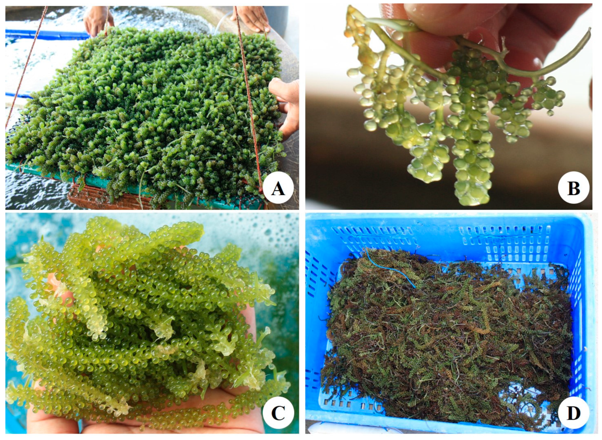
2.1. Materials

C. lentillifera was cultivated and collected at Phetchaburi Coastal Aquaculture Research and Development Center in Phetchaburi Province, Thailand. The cultivation, collection, and preparation for C. lentillifera material were described in our previous study [11]. Dried C. lentillifera powder was obtained after drying in a hot-air oven at 65 °C for 24 h and milling the product with a blender.

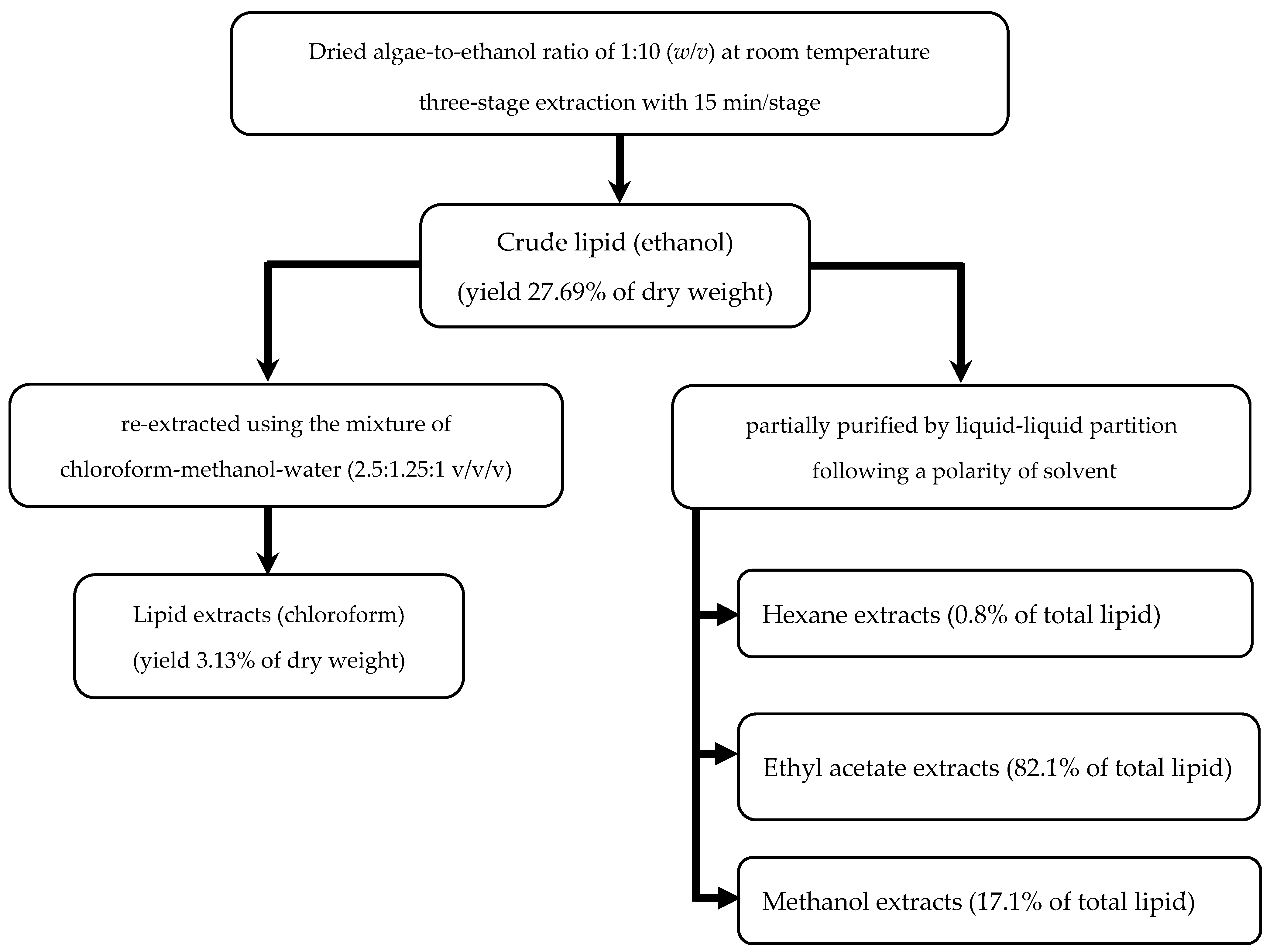
2.2. Lipid Extraction Conditions

Ethanol was used as a solvent for lipid extraction because it is safe for applications. To determine the effect of the sample-to-solvent ratio on the lipid yield, dried C. lentillifera powder weighing 30, 15, 10, 7.5, and 5 g was added to 150 mL of ethanol in order to prepare mixtures with solid-to-ethanol ratios of 1:5, 1:10, 1:15, 1:20, and 1:30 (w/v), respectively. The extraction process was performed in a single stage at room temperature for 30 min under continuous mixing using a stirrer at 300 rpm.
To determine the optimum extraction time, a ratio of 1:10 (80 g of dried biomass in 800 mL of ethanol) was used at room temperature, and extraction times ranged from 15 to 180 min. For investigation of the repetition number of extractions, the extraction was performed using a biomass/ethanol ratio of 1:10 (w/v) for 15 and 30 min in the first stage. The residue was separated by filtration and then re-extracted with fresh ethanol at each extraction stage (up to six stages).
The lipid solution was separated from the algae residue using filter papers (Whatman no. 4). The solvent in the lipid solution was then removed with a vacuum rotary evaporation to obtain crude lipids, which contained fatty acids and pigments. The yield of crude lipid extracts was measured as the percentage of dry weight using Equation (1):
Yield (% dry weight) = (weight of lipids/dried sample weight) × 100

2.3. Analyses

Lipid content was determined using a mixture of chloroform, methanol, and water (2.5:1.25:1 v/v/v) following the procedures described by Sato and Murata [26] with slight modifications. For partial purification, the crude lipid sample was dissolved in 30% methanol/water and then partitioned with hexane. The hexane fraction was collected and evaporated. The aqueous methanol phase was then sequentially partitioned with ethyl acetate and methanol, and each fraction was collected and evaporated.
Total fatty acid (TFA) in the samples was determined using the transesterification protocol proposed by Lapage and Roy [27] with modifications. Lipid extract (50–100 mg) was directly trans-methylated in 2.5% HCl in methanol at 85 °C for 90 min using heneicosanoic acid (C21:0; Sigma-Aldrich) as an internal standard. Fatty acid methyl esters were analyzed with gas chromatography (GC 17-A; Shimadzu, Japan) using capillary columns of fused silica glass (60 m × 0.25 mm, BPX70, SGE, USA) and a film thickness of 0.25 μm. The split ratio was 1:50, and the column temperature was initially set at 170 °C, which was increased to 205 °C at a rate of 1 °C per minute. The injector and flame ionization detector temperatures were 250 and 260 °C, respectively. Identification was carried out by co-chromatography with authentic standards (Sigma-Aldrich).
Pigments were determined using methanol and measured at 470, 666, and 648 nm for carotenoids and chlorophylls a and b, respectively [28]. Total phenolic content was estimated using the Folin-Ciocalteu reagent, and the absorbance was measured at 765 nm [29]. The content was calculated based on the standard curve of gallic acid, and the results were expressed as mg gallic acid equivalents (mg GAE) per gram of sample.
Total flavonoids content was determined using the aluminum chloride colorimetric method described by Pallab et al. [30] with slight modifications. The lipid solution was mixed with 1.5 mL of methanol, 0.1 mL of 2% aluminum chloride, and 0.1 mL of 1 M potassium acetate solution. Distilled water (2.8 mL) was added, and the solution was mixed well. After a 30 min incubation period, the absorbance of the reaction mixture was measured at 415 nm. The total flavonoid content was calculated based on the standard quercetin curve, and the results were expressed as mg quercetin equivalents (mg QE) per gram of extract.
DPPH radical scavenging activity was measured using DPPH (Sigma Chemical Co., St. Louis, MO, USA) with a modification of the method reported by Li et al. [31]. Briefly, 0.1 mM DPPH solution in ethanol was added to the lipid solution. After incubation in the dark for 30 min, the absorbance was measured at 517 nm. In the same way, ABTS decolorization scavenging activity was measured using ABTS (Sigma Chemical Co., St. Louis, MO, USA) following the procedure reported by Zheleva-Dimitrova et al. [32] with some modifications. ABTS+ radicals were prepared by mixing 7 mM ABTS in water and 2.45 mM potassium persulfate (1:1, v/v) and kept in the dark at room temperature for 16 h before use. The ABTS·solution was then diluted with methanol to obtain an absorbance of 0.700 at 734 nm. Next, 2 mL of ABTS·solution was added to the lipid solution. After incubation in the dark for 7 min, the absorbance was measured at 734 nm. Trolox was used as a standard for both DPPH and ABTS.
The α-glucosidase inhibitory activity of the lipid extracts was determined using the procedures described by Alam et al. [33] with some modifications. The formation of p-nitrophenol that resulted from the cleavage of p-nitrophenyl-α-D-glucopyranose was estimated for the evaluation of α-glucosidase inhibitory activity. The enzyme, α-glucosidase (Sigma-Aldrich, Steinheim, Germany), and the substrate, p-nitrophenyl-α-D-glucopyranose (Sigma-Aldrich, Buchs, Switzerland), were dissolved in 50 mM phosphate buffer (pH 6.5) and incubated at 26.8 °C for 15 min. The optical densities were read at 415 nm in a multi-detection micro-plate reader, and acarbose was used as a standard.

2.4. Statistics

The results are expressed as the mean ± standard deviation (SD) of triplicate datasets from independent experiments. Statistical analysis was performed through an analysis of variance (ANOVA) with a confidence level of 95% (p < 0.05).

3. Results and Discussion

3.1. Optimum Conditions for Lipid Extraction

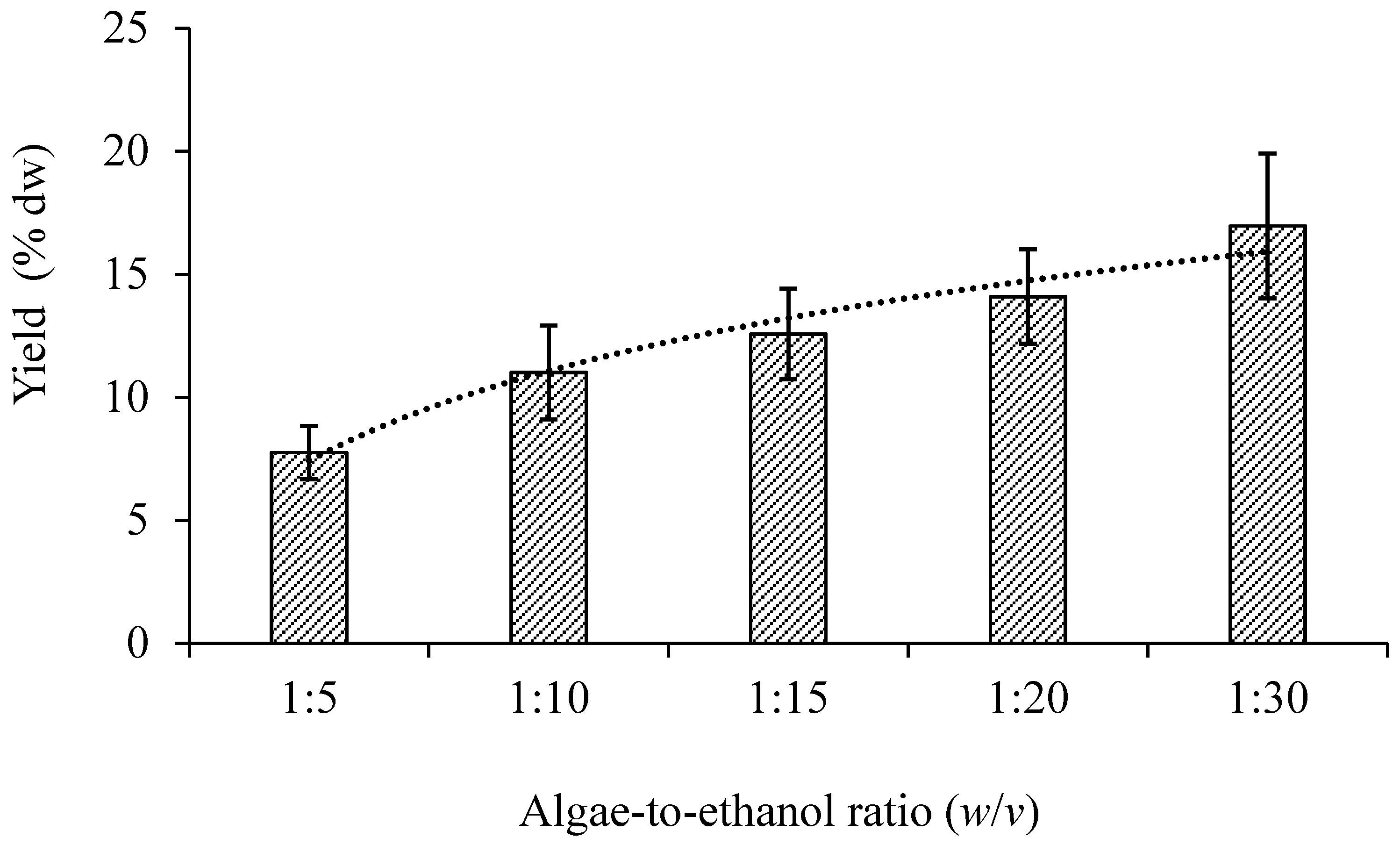
3.1.1. Sample-to-Solvent Ratio

The results showed that an increase in volume of ethanol used for extraction led to an increase in the lipid yield from 7.7% to 16.9% of dry weight (Figure 2). Although the lipid yield from a ratio of 1:30 (w/v) was highest, a high volume of ethanol may not be economically feasible based on the price and energy input needed for evaporation. The lipid yield from a ratio of 1:10 (w/v) was 42.8% higher than that at 1:5 (w/v), but no significant differences between ratios of 1:10 (w/v) and 1:15 (w/v) or ratios of 1:15 (w/v) and 1:20 (w/v) were observed. The lipid yield from a ratio of 1:20 (w/v) was only 28% higher than that of a ratio of 1:10 (w/v), but twice the volume of ethanol was needed for extraction. Hence, a ratio of 1:10 (w/v) was considered a suitable extraction condition based on the lipid yield and the volume of solvent.

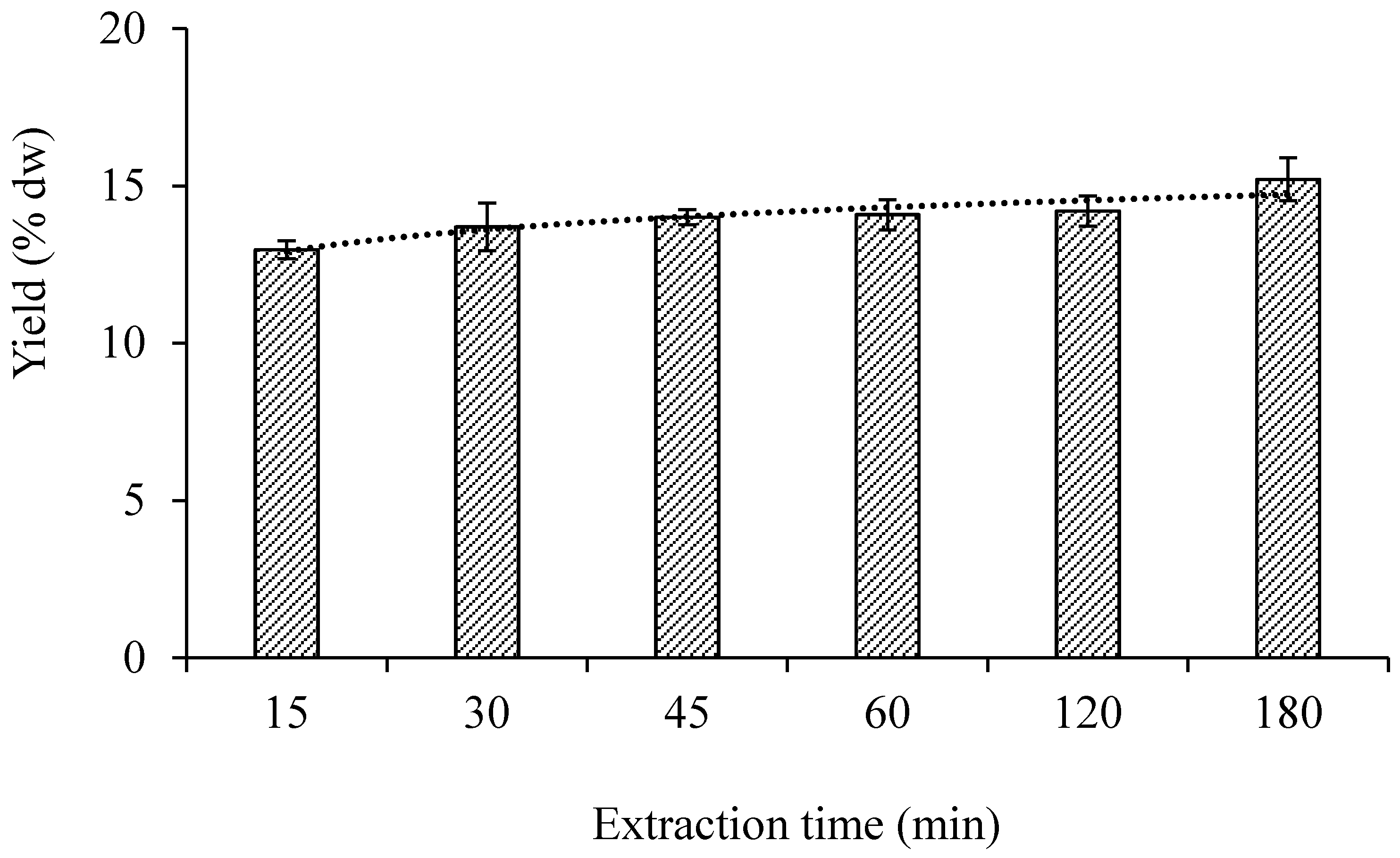
3.1.2. Extraction Time

The lipid yield was approximately 13–15% of the dry weight when the extraction was performed with a ratio of 1:10 (w/v) at room temperature for 15 to 180 min (Figure 3). Although the extraction times of 30 to 120 min showed no differences in the lipid yield, the lipid yield at 15 min (13.0% of dry weight) was significantly lower than at 45 min (14.0% of dry weight). Consequently, extraction times of 15 and 30 min were selected for further investigations of the extraction process.

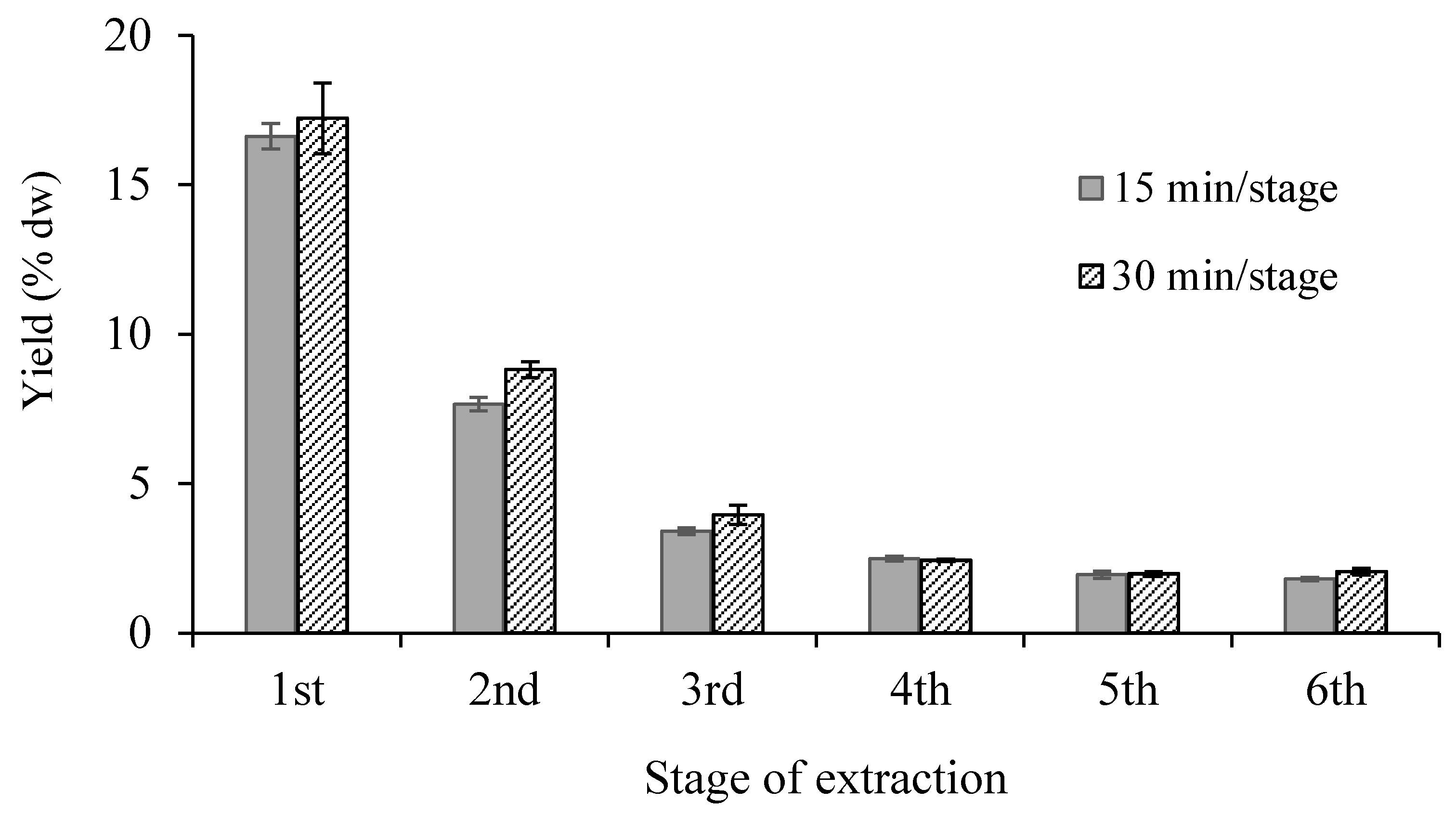
3.1.3. Repetition Number of Extractions

The results showed that the lipid yields at 15 and 30 min for the first extraction (a single stage) were approximately 16.6% and 17.2% of the dry weight. These values decreased by 50% and 75% in the second and third extractions, and after that, the lipid yield was around 2% of the dry weight (Figure 4). The lipid extract resembled a gummy solid rather than dried power, which may have resulted in slightly higher lipid yields in this experiment. When total time of extraction, total volume of ethanol, and total lipid yields were considered, it was found that the lipid yield of two-stage and three-stage extractions increased by around 48% and 70%, respectively, compared to a single-stage extraction (Table 1).
When a three-stage extraction and equal volumes of ethanol (450 mL) were used, the lipid yield at the 15 min/stage (27.69% of dry weight) was slightly lower (10% less) than that extracted at the 30 min/stage (30.00% of dry weight), but the total extraction time was reduced by 50% (from 90 to 45 min). Therefore, the three-stage extraction with the 15 min/stage was considered a suitable condition based on the short time for extraction.

3.2. Compositions of Lipid Extracts

The crude lipid was extracted using ethanol as a solvent, and therefore, the lipid yields were higher than the lipid content in the raw material (C. lentillifera: around 2% of dry weight) [11]. The lipid content in the algae material was determined using a mixture of chloroform, methanol, and water, and after homogenization, more chloroform and water were added to obtain two separate phases: (1) a methanol/water phase (upper phase) and (2) a chloroform phase (lower phase). The total lipids remained in the chloroform, and the polar molecules (proteins, salt, and impurities) partitioned into the methanol/water phase. Moreover, the intermediate fluff layer was withdrawn and discarded, resulting in low impurities in the lipid extracts.
To demonstrate the reason, the crude lipid was re-extracted following the method of Sato and Murata [26] using the same mixture of solvents. The result showed that the lipid yield decreased from 27.69% to 3.13% of the dry weight (Figure 5). Furthermore, the crude lipid was also partially purified by liquid-liquid partition using solvents of polarity (hexane 0.1, ethyl acetate 4.4, and methanol 5.1) to obtain the hexane-, ethyl acetate-, and methanol-soluble fractions, respectively, as shown in Figure 5. Most of the lipid was dissolved in the ethyl acetate fraction, which accounted for 82.1% of the total lipid, whereas the proportions of lipid in hexane and methanol were 0.8% and 17.1%, respectively.
Li et al. [34] reported that the ethyl acetate-soluble fraction of crude ethanol extract from C. recemosa exhibited the strongest antioxidant activity because the phenolic compounds of medium polarity and antioxidant capacity were distributed in this fraction. Similarly, Chirasuwan et al. [35] also found that sulphoquinovosyl diacylglycerol (SQDG), an active antiviral agent against herpes simplex virus type 1, was distributed in the ethyl acetate-soluble fraction of the crude lipid from Spirulina platensis. The polarity of ethanol is 4.3, which is almost the same as that of ethyl acetate and chloroform (4.4 and 4.1, respectively). Therefore, it was expected that lipid extract from C. lentillifera waste would contain bioactive compounds and present an antioxidant capacity.
The lipid extracts contained 58.60% TFA by weight, which consisted of 10 types of fatty acids. Palmitic acid (16:0) was a major component that accounted for around 74.48% of the TFA and resulted in a high proportion of 82.56% saturated fatty acids (SFA). The proportions of monounsaturated and polyunsaturated fatty acids (MFA and PFA, respectively) were approximately 12.68% and 3.09%, respectively (Table 2). This result corresponded to those of Ratana-arporn and Chirapart [1], who reported that C. lentillifera harvested from Phetchaburi Province, Thailand, contained palmitic acid (67.83%) as a main component.
Stearic acid (18:0) and palmitoleic acid (16:1) accounted for 11.1% and 6.08% of the TFA, respectively. However, the content of fatty acid and distribution of SFA, MFA, and PFA depend on seasonal changes [36]. In addition, the crude lipid also contained chlorophylls a and b and carotenoids, accounting for 1.77, 0.91, and 0.70 µg/mg of the extracts, respectively.

3.3. Antioxidant Properties and Applications

Antioxidant activity can be determined both quantitatively and qualitatively. Total phenolic and flavonoid contents were tested in the qualitative analysis using a colorimetric assay. The crude lipid extracts from C. lentillifera waste contained total phenolic and flavonoid contents of around 2.07 mg GAE/g sample and 5.40 mg QE/g sample, respectively. These values increased to 17.46 mg GAE/g sample and 55.48 mg QE/g sample, respectively, when the sample was purified lipid (ethyl acetate fraction; Table 3).
Nguyen et al. [3] reported that the ethanolic extract from C. lentillifera contained a total phenolic content of approximately 1.3–2.04 mg GAE/g sample. Similarly, Yap et al. [37] found that methanolic extracts from C. lentillifera contained a total phenolic content of 4.52 mg GAE/g sample and a total flavonoid content of 4.93 mg QE/g sample, whereas extracts from C. racemosa contained total phenolic and total flavonoid contents of 10.33 mg GAE/g sample and 24.52 mg QE/g sample, respectively. Osuna-Ruiz et al. [38] found that methanol extracts of C. sertularioides contained total phenolic and total flavonoid contents of 0.68 mg GAE/100 mg sample and 14.6 mg QE/g sample, respectively. Tanna et al. [39] reported that aqueous-methanol extracts of Caulerpa spp. had total phenolic contents of around 23.8 to 29.2 mg GAE/g sample and total flavonoid contents of approximately 523 to 769 mg QE/g sample.
ABTS and DPPH are tested in the quantitative analysis based on the capability of redox molecules in natural products to scavenge free radicals, which is expressed as the concentration with 50% ABTS and DPPH radical scavenging effect (IC50). The IC50 values of ABTS and DPPH were approximately 1.67 and 3.55 mg/mL for the crude lipid and 0.21 and 0.43 mg/mL for the purified lipid (ethyl acetate fraction), respectively. Crude lipid extracts also exhibited α-glucosidase inhibitory activity with an IC50 value of 8.97 mg/mL, which could potentially be used in food supplements for preventing diabetes. This result corresponds to those of Sharma and Rhyu [5] and Sharma et al. [6] who found that ethanolic extract from C. lentillifera has anti-diabetic potential by causing significant decreases in dipeptidyl peptidase-IV and α-glucosidase enzyme activities. Furthermore, this extract has the potential to cause an increase in insulin secretion from rat insulinoma cells and stimulate the glucose uptake in 3T3-L1 adipocytes.
Sabirin et al. [7] reported that the methanolic extract from C. lentillifera possessed antibacterial activity against common causative agents for oral infections, Staphylococcus aureus and Streptococcus mutans. Sharma et al. [40] found that the ethanolic extract of C. okamurae was effective in preventing body weight gain and fat accumulation and reduced plasma and hepatic lipid profiles. Likewise, Rahman et al. [41] reported that the ethanolic extract of C. recemosa could potentially be used as a hypolipemic agent since it exhibited a significant hypolipemic effect at a dose of 200 mg/kg body weight. Based on this information, lipid extracts from Caulerpa have the potential as a food supplement or agent for good health that might be beneficial for aging societies and people with diabetes or hyperlipidemia.
Results from this work demonstrate a possible route for sustainable development, and the value of C. lentillifera waste can be increased by transformation to a new product, such as a bioactive substance with market value. In addition, the process of lipid extraction from C. lentillifera waste is possible for scaling-up in commercial production for three reasons: (1) the extraction uses a simple technique; (2) the crude lipid has potential as an α-glucosidase inhibitor; thus, it does not need future purification; and (3) ethanol as a solvent for extraction is safe for human consumption and can be recycled. For the production of C. lentillifera biomass in Thailand, the capacity of fresh biomass in Phetchaburi Province is approximately 25 tons/month based on information from personal communication with the Caulerpa entrepreneur. In case 70% of biomass is a useless (waste) product, and only 25% can be reused as propagule, useless biomass is around 11.25 tons/month. Of note, fresh Caulerpa biomass contains approximately 95% moisture; thus, 562.50 kg of dried material/month allowed for lipid extraction. Based on these amounts and a 27% lipid yield, crude lipids of around 150 kg dried extract/month were obtained. Selling crude lipid as a food supplement could result in more income for farmers.

4. Conclusions

The optimum conditions for lipid extraction from C. lentillifera waste consisted of a three-stage extraction (15 min/stage) using a dried algae-to-ethanol ratio of 1:10 (w/v) at room temperature. Under these conditions, a crude lipid yield of around 27.69% of dry weight was obtained. Since the crude lipid shows the potential for preventing diabetes, it could increase the value of C. lentillifera waste and the utilization of algae resources, which could lead to sustainable development.

Author Contributions

Conceptualization, R.C.; investigation, T.S. and N.C.; data curation, R.C.; writing materials and methods, T.S.; writing-review and editing, R.C.; project administration, R.C.; supervisor, B.B. All authors have read and agreed to the published version of the manuscript.

Funding

This work was supported by the Biodiversity-Based Economy Development Office (Public Or-ganization; BEDO) and the National Research Council of Thailand (NRTC) (grant number: BEDO NRTC 25/2019).

Acknowledgments

We would like to thank Khun Montakan Tamtin and Khun Prapat Kosawatpat, Phetchaburi Coastal Aquaculture Research and Development Center, for providing C. lentillifera biomass. We also thank the Algal Biotechnology Research Center, King Mongkut’s University of Technology Thonburi, for providing facilities.

Conflicts of Interest

The authors declare no conflict of interest.

References

  1. Ratana-arporn, P.; Chirapart, A. Nutritional evaluation of tropical green seaweeds Caulerpa lentillifera and Ulva reticulate. Nat. Sci. 2006, 40, 75–83. [Google Scholar]
  2. Matanjun, P.; Mohamed, S.; Mustapha, M.N.; Muhammad, K. Nutrient content of tropical edible seaweeds, Eucheuma cottonii, Caulerpa lentillifera and Sargassum polycystum. J. Appl. Phycol. 2009, 21, 75–80. [Google Scholar] [CrossRef]
  3. Nguyen, V.T.; Ueng, J.P.; Tsai, G.J. Proximate composition, total phenolic content, and antioxidant activity of seagrape (Caulerpa lentillifera). J. Food Sci. 2011, 76, C950–C958. [Google Scholar] [CrossRef]
  4. Sarini, A.W.; Nor’Aishah, H.; Mohd Zaini, N. Determination of antioxidant activity for seven types of macroalgae. In Proceedings of the 5th International Conference on Food Engineering and Biotechnology, IPCBEE, Penang, Malaysia, 12–14 March 2014; Volume 65, pp. 51–55. [Google Scholar]
  5. Sharma, B.R.; Rhyu, D.Y. Anti-diabetic effects of Caulerpa lentillifera: Stimulation of insulin secretion in pancreatic β-cells and enhancement of glucose uptake in adipocytes. Asian Pac. J. Trop. Biomed. 2014, 4, 575–580. [Google Scholar] [CrossRef] [Green Version]
  6. Sharma, B.R.; Kim, H.J.; Rhyu, D.Y. Caulerpa lentillifera extract ameliorates insulin resistance and regulates glucose metabolism in C57BL/KsJ-db/db mice via PI3K/AKT signaling pathway in myocytes. J. Transl. Med. 2015, 13, 1–10. [Google Scholar] [CrossRef] [Green Version]
  7. Sabirin, F.; Kazi, J.A.; Ibrahim, I.; Rashit, M.M.A. Screening of seaweeds potential against oral infections. J. Appl. Sci. Res. 2015, 11, 1–6. [Google Scholar]
  8. West, E.J.; West, R.J. Growth and survival of the invasive alga, Caulerpa taxifolia, in different salinities and temperatures: Implications for coastal lake management. Hydrobiologia 2007, 577, 87–94. [Google Scholar] [CrossRef]
  9. Ukabi, S.; Dubinsky, Z.; Steinberger, Y.; Israel, A. Temperature and irradiance effects on growth and photosynthesis of Caulerpa (Chlorophyta) species from the eastern Mediterranean. Aquat. Bot. 2013, 104, 106–110. [Google Scholar] [CrossRef]
  10. Gao, X.; Choi, H.G.; Park, S.K.; Sun, Z.M.; Nam, K.W. Assessment of optimal growth conditions for cultivation of the edible Caulerpa okamurae (Caulerpales, Chlorophyta) from Korea. J Appl. Phycol. 2019, 31, 1855–1862. [Google Scholar] [CrossRef]
  11. Chaiklahan, R.; Srinorasing, T.; Chirasuwan, N.; Tamtin, M.; Bunnag, B. The potential of polysaccharide extracts from Caulerpa lentillifera waste. Int. J. Biol. Macromol. 2020, 161, 1021–1028. [Google Scholar] [CrossRef] [PubMed]
  12. Sagar, N.A.; Pareek, S.; Sharma, S.; Yahia, E.M.; Lobo, M.G. Fruit and vegetable waste: Bioactive compounds, their extraction, and possible utilization. Compr. Rev. Food Sci. Food Saf. 2018, 17, 512–531. [Google Scholar] [CrossRef] [Green Version]
  13. Freitas, L.C.; Barbosa, J.R.; da Costa, A.L.C.; Bezerra, F.W.F.; Pinto, R.H.H.; de Carvalho Junior, R.N. From waste to sustainable industry: How can agro-industrial wastes help in the development of new products? Resour. Conserv. Recycl. 2021, 169, 105466. [Google Scholar] [CrossRef]
  14. Galanakis, C.M. Recovery of high added-value components from food wastes: Conventional, emerging technologies and commercialized applications. Trends Food Sci. Technol. 2012, 26, 68–87. [Google Scholar] [CrossRef]
  15. Galanakis, C.M. Emerging technologies for the production of nutraceuticals from agricultural by-products: A viewpoint of opportunities and challenges. Food Bioprod. Process. 2013, 91, 575–579. [Google Scholar] [CrossRef]
  16. Ferri, M.; Vannini, M.; Ehrnell, M.; Eliasson, L.; Xanthakis, E.; Monari, S.; Tassoni, A. From winery waste to bioactive compounds and new polymeric biocomposites: A contribution to the circular economy concept. J. Adv. Res. 2020, 24, 1–11. [Google Scholar] [CrossRef] [PubMed]
  17. Munir, M.; Kheirkhah, H.; Baroutian, S.; Quek, S.Y.; Young, B.R. Subcritical water extraction of bioactive compounds from waste onion skin. J. Clean Prod. 2018, 183, 487–494. [Google Scholar] [CrossRef]
  18. Assefa, S.T.; Yang, E.Y.; Chae, S.Y.; Song, M.; Lee, J.; Cho, M.C.; Jang, S. Alpha glucosidase inhibitory activities of plants with focus on common vegetables. Plants 2020, 9, 2. [Google Scholar] [CrossRef] [PubMed] [Green Version]
  19. Xiancui, L.; Rongli, N.; Xiao, F.; Lijun, H.; Lixin, Z. Macroalgae as a source of alpha-glucosidase inhibitors. Chin. J. Oceanol. Limnol. 2005, 23, 354–356. [Google Scholar] [CrossRef]
  20. Yin, Z.; Zhang, W.; Feng, F.; Zhang, Y.; Kang, W. α-Glucosidase inhibitors isolated from medicinal plants. Food Sci. Hum. Wellness 2014, 3, 136–174. [Google Scholar] [CrossRef] [Green Version]
  21. Kumar, S.; Narwal, S.; Kumar, V.; Prakash, O. α-glucosidase inhibitors from plants: A natural approach to treat diabetes. Pharmacogn. Rev. 2011, 5, 19–29. [Google Scholar] [CrossRef] [Green Version]
  22. Nguyen, T.T.M.; Nguyen, T.N.; Nguyen, X.H.; Huynh, N.N.T.; Min, S.B. Screening of α-glucosidase inhibitory activity of Vietnamese medicinal plants: Isolation of active principles from Oroxylum indicum. Nat. Prod. Sci. 2012, 18, 47–51. [Google Scholar]
  23. Telagari, M.; Hullatti, K. In-vitro α-amylase and α-glucosidase inhibitory activity of Adiantumcaudatum Linn. and Celosia argentea Linn. extracts and fractions. Indian J. Pharmacol. 2015, 47, 425–429. [Google Scholar] [PubMed] [Green Version]
  24. Yen, F.S.; Wei, J.C.C.; Lin, M.C.; Hsu, C.C.; Hwu, C.M. Long-term outcomes of adding alpha-glucosidase inhibitors in insulin-treated patients with type 2 diabetes. BMC Endocr. Disord. 2021, 21, 25. [Google Scholar] [CrossRef] [PubMed]
  25. Bhatia, A.; Singh, B.; Arora, R.; Arora, S. In vitro evaluation of the α-glucosidase inhibitory potential of methanolic extracts of traditionally used antidiabetic plants. BMC Complement. Altern. Med. 2019, 19, 74. [Google Scholar] [CrossRef] [Green Version]
  26. Sato, N.; Murata, N. Membrane lipid. Meth. Enzymol. 1988, 167, 251–259. [Google Scholar]
  27. Lapage, G.; Roy, C.C. Improved recovery of fatty acid through direct transesterification without prior extraction of purification. J. Lipid Res. 1984, 25, 1391–1396. [Google Scholar] [CrossRef]
  28. Othman, R.; Amin, N.A.; Sani, M.S.A.; Fadzillah, N.A.; Jamaludin, M.A. Carotenoid and chlorophyll profiles in five species of Malaysian seaweed as potential halal active pharmaceutical ingredient (API). Int. J. Adv. Sci. Eng. Inf. Technol. 2018, 8, 1610–1616. [Google Scholar]
  29. Wu, X.J.; Hansen, C. Antioxidant capacity, phenolic content, and polysaccharide content of Lentinus edodes grown in whey permeate-based submerged culture. J. Food Sci. 2008, 73, M1–M8. [Google Scholar] [CrossRef] [PubMed]
  30. Pallab, K.; Tapan, K.B.; Tapas, K.P.; Ramen, K. Estimation of total flavonoids content (TFC) and antioxidant activities of methanolic whole plant extract of Biophytum sensitivum linn. J. Drug Deliv. Ther. 2013, 3, 33–37. [Google Scholar]
  31. Li, X.; Zhou, A.; Han, Y. Anti-oxidation and anti-microorganism activities of purification polysaccharide from Lygodium japonicum in vitro. Carbohydr. Polym. 2006, 66, 34–42. [Google Scholar] [CrossRef]
  32. Zheleva-Dimitrova, D.; Nedialkov, P.; Kitanov, G. Radical scavenging and antioxidant activities of methanolic extracts from Hypericum species growing in Bulgaria. Pharmacogn. Mag. 2010, 6, 74–78. [Google Scholar] [CrossRef] [PubMed] [Green Version]
  33. Alam, M.A.; Zaidul, I.S.M.; Ghafoor, K.; Sahena, F.; Hakim, M.A.; Rafii, M.Y.; Abir, H.M.; Bostanudin, M.F.; Perumal, V.; Khatib, A. In vitro antioxidant and, α-glucosidase inhibitory activities and comprehensive metabolite profiling of methanol extract and its fractions from Clinacanthus nutans. BMC Complement. Altern. Med. 2017, 17, 181–191. [Google Scholar] [CrossRef] [Green Version]
  34. Li, Z.; Wang, B.; Zhang, Q.; Qu, Y.; Xu, H.; Li, G. Preparation and antioxidant property of extract and semipurified fractions of Caulerpa racemose. J. Appl. Phycol. 2012, 24, 1527–1536. [Google Scholar] [CrossRef]
  35. Chirasuwan, N.; Chaiklahan, R.; Kittakoop, P.; Chanasattru, W.; Ruengjitchatchawalya, M.; Tanticharoen, M.; Bunnag, B. Anti HSV–1 activity of sulphoquinovosyl diacylglycerol isolated from Spirulina platensis. Sci. Asia 2009, 35, 137–141. [Google Scholar] [CrossRef]
  36. Blažina, M.; Iveša, L.; Najdek, M. Caulerpa racemosa: Adaptive varieties studied by fatty acid composition (Northern Adriatic Sea, Vrsar, Croatia). Eur. J. Phycol. 2009, 44, 183–189. [Google Scholar] [CrossRef]
  37. Yap, W.F.; Tay, V.; Tan, S.H.; Yow, Y.Y.; Chew, J. Decoding antioxidant and antibacterial potentials of Malaysian green seaweeds: Caulerpa racemosa and Caulerpa lentillifera. Antibiotics 2019, 8, 152. [Google Scholar] [CrossRef] [Green Version]
  38. Osuna-Ruiz, I.; López-Saiz, C.M.; Burgos-Hernández, A.; Velázquez, C.; Nieves-Soto, M.; Hurtado-Oliva, M.A. Antioxidant, antimutagenic and antiproliferative activities in selected seaweed species from Sinaloa, Mexico. Pharm. Biol. 2016, 54, 2196–2210. [Google Scholar] [CrossRef]
  39. Tanna, B.; Choudhary, B.; Mishra, A. Metabolite profiling, antioxidant, scavenging and anti-proliferative activities of selected tropical green seaweeds reveal the nutraceutical potential of Caulerpa spp. Algal Res. 2018, 36, 96–105. [Google Scholar] [CrossRef]
  40. Sharma, B.R.; Kim, H.J.; Kim, M.S.; Park, C.M.; Rhyu, D.Y. Caulerpa okmurae extract inhibits adipogenesis in 3T3–L1 adipocytes and prevents high–fat diet–induced obesity in C57BL/6 mice. Nutr. Res. 2017, 47, 44–52. [Google Scholar] [CrossRef]
  41. Rahman, S.M.; Neaz, S.; Alam, M.M.; Nur, J. Hypolipidemic activity of ethanolic extract of Caulerpa recemosa. BIRDEM Med. J. 2019, 9, 197–201. [Google Scholar] [CrossRef]
Figure 1. Caulerpa lentillifera products (A,B), food grade (C), and waste grade (D).
Sustainability 13 04491 g001
Figure 2. Effect of sample-to-solvent ratio on lipid yield.
Sustainability 13 04491 g002
Figure 3. Effect of extraction time on lipid yield.
Sustainability 13 04491 g003
Figure 4. Effect of number of extractions on lipid yield.
Sustainability 13 04491 g004
Figure 5. Diagram for purification of lipid extracts.
Sustainability 13 04491 g005
Table 1. Lipid yields under different conditions.
Number
of Stage
Total Time of Extraction (min)Total Volume of Ethanol (mL) *Total Yield (% dw)
153015301530
1153015015016.6217.23
2306030030024.2826.04
3459045045027.6930.00
460 600 30.18
* Each stage used 150 mL of ethanol.
Table 2. Composition of lipid extracts.
CompositionsValue
Chlorophyll a (µg/mg of extract)1.77 ± 0.25
Chlorophyll b (µg/mg of extract)0.91 ± 0.09
Carotenoids (µg/mg of extract)0.70 ± 0.09
Total fatty acid (TFA; % weight of lipid extract)58.60 ± 4.95
Fatty acid compositions (% mol.)
Myristic acid (14:0)4.89 ± 0.36
Palmitic acid (16:0)74.48 ± 3.24
Palmitoleic acid (16:1n7)6.82 ± 0.38
Stearic acid (18:0)2.50 ± 0.28
Vaccenic acid (18:1n7)3.03 ± 0.28
Oleic acid (18:1n9)2.92 ± 0.39
Linoleic acid (18:2)2.69 ± 0.38
Linoleic acid (18:3)0.93 ± 0.13
Arachidonic acid (20:4)0.95 ± 0.09
Eicosapentaenoic acid (20:5)1.01 ± 0.34
Saturated fatty acids (SFA)82.56 ± 2.37
Monounsaturated fatty acids (MFA)12.68 ± 0.44
Polyunsaturated fatty acids (PFA)3.09 ± 0.69
Table 3. Antioxidant properties of lipid extracts.
CharacteristicCrude Lipid Purified Lipid
Total phenolic (mg GAE/g sample)2.07 ± 0.3417.46 ± 2.24
Total flavonoids (mg QE/g sample)5.40 ± 0.7655.48 ± 7.78
IC50 ABTS (mg/mL)1.67 ± 0.090.21 ± 0.06
IC50 DPPH (mg/mL)3.55 ± 0.530.43 ± 0.11
IC50 α-glucosidase (mg/mL)8.97 ± 0.19Not determined
IC50 ABTS and DPPH of trolox were 0.036 and 0.515 mg/mL, respectively. IC50 α-glucosidase of acarbose was 0.28 mg/mL.
Publisher’s Note: MDPI stays neutral with regard to jurisdictional claims in published maps and institutional affiliations.

