Article

The Influence of Yarrowia lipolytica Glycosylation on the Biochemical Properties and Oligomerization of Heterologous Invertase

1
Department of Biotechnology and Food Microbiology, Wroclaw University of Environmental and Life Sciences, Chełmońskiego 37, 50-375 Wroclaw, Poland
2
Department of Bioengineering and Imperial College Centre for Synthetic Biology, Imperial College London, London SW7 2AZ, UK
*
Author to whom correspondence should be addressed.
Sustainability 2022, 14(13), 7926; https://doi.org/10.3390/su14137926
Submission received: 13 May 2022 / Revised: 19 June 2022 / Accepted: 24 June 2022 / Published: 29 June 2022
(This article belongs to the Section Sustainable Food)

Abstract

:
Invertases are important enzymes used in the food industry. Despite many studies on the invertase-encoding SUC2 gene expression in the industrial yeast Yarrowia lipolytica, no biochemical characteristics of this enzyme expressed as heterologous protein have been provided. Here, two isoforms of extracellular invertase produced by Y. lipolytica were detected using ion-exchange chromatography. Specific activities of 226.45 and 432.66 U/mg for the first and second isoform, respectively, were determined. Basic characteristics of this enzyme were similar to the one isolated from Saccharomyces cerevisiae (optimum pH and temperature, metal ions inhibition, substrate specificity and fructooligosaccharides (FOS) biosynthesis). The apparent differences were higher KM for sucrose (67 mM) and lower molecular mass (66 kDa) resulting from lower N-glycosylation level (9.1% of mass). The N-glycan structures determined by MALDI-TOF and HPLC represented high mannose structures, though with much shorter chains than hypermannosylated glycans from S. cerevisiae. Furthermore, galactose was detected as the modifying sugar in the glycan structures of invertase expressed in Y. lipolytica. N-glycans did not affect invertase activity but were important for its oligomerization. The expressed enzyme aggregated into dimers, tetramers, hexamers, and octamers, as well as structures of higher molecular mass, which might be decamers, which have not been described so far in the literature.

1. Introduction

Invertase, β-fructofuranosidase (EC 3.2.1.26), catalyzes hydrolysis of various β-D-fructofuranoside substrates, such as sucrose, raffinose, or stachyose, releasing β-fructose from their nonreducing termini [1,2,3]. The catalytic mechanism implies two conserved aspartic and glutamic residues, which act as a nucleophile and an acid/base catalyst, respectively [4,5]. Hydrolysis of the glycosidic bond proceeds via double displacement mechanism. During this process, transient covalent sugar-enzyme intermediates are formed. Invertases have been widely studied in Saccharomyces cerevisiae [6,7,8], Schizosaccharomyces pombe [9,10,11], Rhodotorula glutinis [12,13], Candida utilis [14,15], Schwanniomyces occidentalis [4,16], and Fusarium solani [17]. In the wide range of microorganisms, invertases possess different biochemical properties, such as enzymatic activity at different pH or temperature. Most of the fungal invertases show optimal pH in the range of 4.4–6.0 [7,14,18,19] with the exception of the invertase synthetized by F. solani with maximum activity at pH 2.6.
In contrast to the pH, the range of the optimal temperatures for the highest enzymatic activity of invertase is wide. Enzymes expressed by Leucosporidium antarcticum and Aspergillus flavus show the maximum activity at 30 °C, whereas C. utilis, Cladosporium cladosporioides, and Xanthophyllomyces dendrorhous express invertases acting at 70 °C [14,18,19,20,21,22]. Additionally, the affinity of invertases for sucrose is one of the most variable parameters. There are high affinity enzymes, defined by very low Michaelis–Menten constants (KM) of 0.64 mM for invertase of A. flavus or 1.54–2.0 mM for C. utilis [14,22]. On the other hand, the enzymes expressed by R. glutinis and C. cladosporioides present very low affinities, with KM of 227 mM or 447 mM, respectively [12,18,23]. Furthermore, regarding their molecular mass, fungal invertases are very heterogenous. The monomers of invertases from C. utilis, F. solani, and A. flavus have molecular weight of 62–67 kDa while X. dendrorhous, R. dairenensis, and Sch. pombe produce invertase monomers of 160–205 kDa [1,9,14,17,20,22,23].
Invertase from S. cerevisiae is the best-known example of fungal β-fructofuranosidases. It shows a maximum activity at pH 4.5 and 60 °C with KM of 25 mM [6,7]. The genome of S. cerevisiae contains several cumulative polymeric genes, SUC1-10 [24,25]. In this gene family, SUC2 encodes two different forms of invertase, extracellular, or glycosylated, and intracellular—non-glycosylated [24]. These two isoforms are translated from two distinct and differentially regulated mRNA molecules. Additionally, a sequence encoding the extracellular form possesses a signal sequence that directs the protein via the secretory pathway to the periplasmic space or outside the cell.
The SUC2 gene was previously used as a selective marker in transformations of many microorganisms, including industrially relevant yeast Yarrowia lipolytica [26,27,28]. This particular species has numerous applications, including expression of heterologous proteins of industrial and pharmaceutical relevance [29,30]. Hence, there is a need to understand how the hosts’ posttranslational machinery affects the non-native protein properties.
Despite many studies on the invertase expression in Y. lipolytica, no biochemical characterization of this enzyme produced as a heterologous protein is available. Here we address this gap and show that the choice of host organism and its posttranslational modifications can influence the oligomerization and the stability of the oligomeric forms of invertase, without changing most of its biochemical properties.

2. Materials and Methods

2.1. Microorganism

The invertase-expressing Y. lipolytica A-101-B56-5 (suc+) strain was described previously [27]. The strain was routinely cultivated on YM agar plates at 28 °C and stored at 4 °C.

2.2. Culture Conditions

Cultures of Y. lipolytica A-101-B56-5 strain for invertase production were carried out for 72 h in 5 L stirred-tank reactors BIO-STAT B-PLUS (Sartorius, Frankfurt, Germany) with working volume of 2 L at 28 °C, 800 rpm and aeration rate of 0.8 vvm (vessel volume per minute). Production media contained in 1 L of tap water: carbon source 100 g, NH4Cl 1.5 g, KH2PO4 0.7 g, MgSO4×7H2O 1.0 g, YE 0.3 g, thiamine 3 × 10⁻⁶ g. pH was automatically maintained at pH 6.8 by addition of 40% (w/v) NaOH solution. Inoculum consisted of 10% of the total working volume. The inoculation medium contained in 1 L of tap water: carbon source 50 g, NH4Cl 1.5 g, YE 1.0 g, peptone 1.0 g. The cultures were grown in 0.5 L flasks containing 0.1 L of medium on a rotary shaker (Elpan, Poznań, Poland), 170 rpm, 28 °C for 48 h. Sucrose, glucose, a mixture of glucose and fructose (1:1), or glycerol were used as carbon sources. Inoculation and production media were prepared with the same substrate. Glucose, fructose, and sucrose used in this study were commercially available in the grocer’s shop, whereas glycerol was of technical grade (Chempur, Piekary Śląskie, Poland).
Osmolality of media was measured using Marcel OS3000 osmometer (Marcel Ltd., Zielonka, Poland) and reached in mOsmol/kg the value of 680 for glucose-, 686 for glucose + fructose-, 410 for sucrose-, and 1351 for glycerol-based medium.

2.3. Purification of Extracellular Invertase

2.3.1. Ultrafiltration—Concentrate Preparation

After 72 h of the invertase biosynthesis, the cultures were centrifuged (30 min, 5000 rpm at 4 °C) using 3–16 K centrifuge (Sigma, St. Louis, MO, USA). Supernatants were filtered on 0.2 µm pore-size membranes. Post-culture media containing invertase were concentrated using LabscaleTM TFF Ultrafiltration System (Millipore, Billerica, MA, USA) with Pellicon XL Biomax 50 cassette (MWCO 50 kDa). The whole procedure was carried out at 4 °C, with inlet pressure of 3 bar and outlet pressure of 1 bar to the final volume of approximately 50 mL (limited by samples viscosity). Next, the samples were dialyzed against 0.1 M acetate buffer (pH 5.0) for 24 h at 4 °C, using cellulose membrane dialysis tubing (MWCO 12 kDa), and were additionally concentrated (conditions—as presented above).

2.3.2. Ion Exchange Chromatography

Extracellular invertase obtained from the concentrate of sucrose-based medium was purified by ion exchange chromatography using DEAE-Sephadex A50 (Pharmacia, Uppsala, Sweden). The column (2 × 30 cm) was equilibrated with 0.1 M acetate buffer, pH 5.0. The obtained concentrate (28 mL, 72 mg of protein) was loaded onto the column. Non-bound proteins were washed out with 0.1 M acetate buffer and the invertase was eluted with 0.1–0.3 M KCl step gradient with a flow rate of 0.3 mL/min at 4 °C. Fractions of 2 mL were collected. Protein concentration was monitored by measuring the absorbance at 280 nm and the invertase activity was measured as described below. The enzyme-containing fractions were pooled and lyophilized at −35 °C using Alpha 2-4 LSC freeze-dryer (Christ, Osterode am Harz, Germany). Samples were stored at −20 °C.

2.4. Polyacrylamide Gel Electrophoresis (PAGE, Non-Denaturing Conditions)

PAGE was performed using Mini-PROTEAN® Tetra Cell (Bio-Rad, Richmond, VA, USA) in 7% separating gel, pH 7.3. The separation was done in the Tris-Glycine running buffer, pH 8.3 for 3 h with constant current 10 mA. Molecular weight was estimated in comparison with marker proteins (bovine serum albumin—66 kDa, urease from Jack Bean—monomer 90 kDa, dimer 180 kDa), using BioGene software (BioGene Ltd., Kimbolton, UK). After the electrophoresis, gels were divided into two parts. One part of the gel (with protein leader) was stained with 0.1% Coomassie Brilliant Blue R-250. The second part of the gel (containing invertase samples) was incubated for 10 min at 37 °C in 0.1 M sucrose prepared in 0.1 M acetate buffer, pH 5.0, followed by staining with 2,3,5-triphenylotetrazolium chloride (1 mg/mL) in 0.5 M NaOH [31].

2.5. Enzyme Activity, Protein, and Carbon Sources Determination

Invertase activity and protein concentration were measured in post-culture media as described previously [32]. The samples were dialyzed against 0.1 M acetate buffer (pH 5.0) during 24 h at 4 °C, before the measurement of the activity.
For the transfructosylating activity measurements, invertase was incubated in sucrose solution (500 g/L) in 0.1 M acetate buffer (pH 5.0). The total reaction volume was 3 mL and contained 1 U/mL of invertase. Samples were incubated for 48 h at 40 °C, 400 rpm (Thermomixer comfort, Eppendorf, Hamburg, Germany). For the analysis, 0.1 mL samples were being taken every 2 h, incubated for 10 min at 100 °C, and analyzed by HPLC. The concentrations of glucose, fructose, sucrose, and 1-kestose were determined by HPLC method using Bio-Rad Aminex HPX-87H column (Bio-Rad Laboratories, Hercules, CA, USA) coupled to RI-101 detector (Shodex, Ogimachi, Japan). The column was eluted with 0.01 N H2SO4 at room temperature and flow rate 0.6 mL/min. Sugars were identified and quantified with reference to the existent standards.

2.6. Optimum of pH and Temperature, Substrate Specificity and Affinity, Thermostability and Ions Inactivation

Invertase activity was examined in a pH range of 3.0–9.0. The following buffers were used: 0.1 M acetate buffer (pH 3.0–6.0), 0.1 M phosphate buffer (pH 6.5–8.0), and 0.1 M borate buffer (pH 8.–9.0). The temperature was tested in the range of 25–75 °C at pH 5.0. To investigate the temperature stability of invertase the enzyme solutions were pre-incubated at 20 °C, 40 °C, 60 °C, and 80 °C for 0.5–3.0 h. Afterwards, the enzyme activity was tested under the standard assay’s conditions (37 °C, pH 5.0, 10 min; [32]).
Invertase activity was investigated in the presence of the following 1 mM ions solution: CaCl2, MgCl2, CoCl2, MnCl2, CuCl2, ZnCl2, FeCl3, CuSO4, ZnSO4, and EDTA in the reaction mixture under standard assay conditions.
The Michaelis–Menten constant (KM) and maximum reaction rate (Vmax) were calculated from Lineweaver–Burk plot, as a function of the sucrose concentration (0.005–0.1 M) at pH 5.0.
Substrate specificity was determined to 0.1 M of sucrose, raffinose, maltose, lactose, trehalose, and 3.5% inulin under standard assay conditions.

2.7. Deglycosylation Analysis

Digestion of N-glycans attached to heterologous invertase was performed under non-denaturing conditions at 37 °C for 3 h in 50 mM citrate buffer (pH 5.5) with 500 U of endoglycosidase H (EndoH, NEB, Ipswich, MA, USA). Digestion mixture total volume of 20 µL contained 1 U of invertase. Enzyme activity assay and polyacrylamide gel electrophoresis (PAGE) were performed as described above.

2.8. N-Glycan Purification, Hydrolysis, and Analysis with MALDI-TOF and HPLC

N-Glycans were released from invertase with 500 units of PNGase F (NEB; 500 units/μL) overnight at 37 °C. The obtained supernatant and two consecutive washes of the pellet were pooled, cleaned up using C18- (Sep-Pak Cartridges C18; Waters) and graphitized carbon columns (Supelclean ENVI-Carb SPE bulk packing; Supelco), and 2-AB labeled (2-AB: 2-aminobenzamide; Sigma-Aldrich) as described by Buser and colleagues [33].
Furthermore, glycans were resuspended in 50 mM sodium citrate (pH 5) for α-mannosidase treatment (Jack Bean, Sigma M7257; ~20 units/mg), in 50 mM sodium citrate (pH 4.5) for β-HexNAc’ase treatment (Jack Bean, Sigma; ~50 units/mg), in 50 mM sodium citrate (pH 5) for double digest with α-mannosidase and β-HexNAc’ase, and in 50 mM sodium acetate (pH 5.2) for α1–2 mannosidase treatment (Trichoderma reesei); 1 μL of enzyme was used in a total volume of 20 μL. The reaction was incubated overnight at 37 °C. Glycans were desalted and purified as described above. The eluates were dried and resuspended in 0.1% trifluoroacetic acid (TFA), spotted on a MALDI plate, and covered with matrix (10 mg/mL 2,5-dihydroxybenzoic acid (Sigma), 70% acetonitrile, and 0.1% trifluoroacetic acid). MALDI-TOF-MS and MS/MS were performed with ABI 4800 MALDI TOF/TOFTM (Applied Biosystems Inc., Waltham, MA, USA).
To analyze the monosaccharide content, N-glycans were released from invertase using PNGase F and hydrolyzed with 2M TFA. The sugars were determined using Dionex ICS-3000 (Dionex, Sunnyvale, CA, USA) ion chromatography system with PAD detector equipped with CarboPac PA20 column (Thermo Fisher Scientific, Waltham, MA, USA).

3. Results and Discussion

The suc+ transformant of Y. lipolytica A-101-B56-5 produces high amounts of extra- and intracellular invertase [32,34]. The measured enzyme activity reached 34.23 U/mg of protein (Table 1). To purify the extracellular invertase produced by this strain grown on sucrose, the supernatant was ultrafiltered and subject to the ion exchange chromatography.
The ultrafiltration process was performed twice, due to the sample’s viscosity before the ion exchange chromatography. The enzyme sample was concentrated 60 times and the final specific activity reached 89.71 U/mg of protein. The enzyme was eluted from the ion exchanger in two fractions. Specific activity of the first fraction eluted with 0.1 M KCl was 226.45 U/mg of protein. The next one, eluted with 0.2 M KCl, had activity of 432.66 U/mg of protein. A similar invertase purification process (2 peaks) was presented by Rashad and Nooman [35]; however, Andjelković et al. [6] reported more heterogenous preparation of external invertase from S. cerevisiae (4 peaks). Purification fold of Y. lipolytica invertase (12.6) was lower than that obtained by Rodriguez et al. [36], Rashad and Nooman [35], and Shaheen et al. [2] who used a two-step chromatography. However, it was evidently higher than the one obtained by Rubio et al. [12] and Uma et al. [22], who used either one- or two-step chromatography.
The purified enzyme of Y. lipolytica had maximum activity at 60 °C and pH 4.5, while at 65 °C nearly 90% loss of activity was observed (Figure 1A,B). This corresponds to the maximum of invertase activity isolated from the industrial strains of baker’s yeast S. cerevisiae [6]. Additionally, another S. cerevisiae strain, NRRL Y-12632, produces invertase showing maximum activity at pH 6.0 and at 50 °C [35]. Depending on the origin of invertase, a broad range of optimal pH from 2.6 for F. solani to 6.0 for S. cerevisiae and C. cladosporioides was described [17,18,19,23,35]. The same situation was observed for the optimal temperature of invertase activity ranging from 30 °C for L. antarcticum and A. flavus to 70 °C for C. utilis, X. dendrorhous, and C. cladosporioides [14,18,19,20,21,22,23].
Extracellular invertase produced by Y. lipolytica was stable at 20 °C and 40 °C (Figure 1C). Interestingly, a noticeable increase in its activity was observed at 20 °C. The most probable reason for that was the ability of the invertase subunits to aggregate. This issue is discussed below. Y. lipolytica invertase lost more than 60% of its activity after 30 min incubation at 60 °C (Figure 1C). Similar thermostability characterized the invertase from S. cerevisiae [33,35]. A more stable invertase isoenzyme was described for X. dendrorhous [20]. This enzyme lost less than 10% of its activity during 4 days of incubation at 40–50 °C and 50% after 20 min of incubation at 70°. The extremely unstable invertase expressed by L. antarcticum which loses its activity after 24 h incubation at 4 °C was also described [21].
Heterologous invertase from Y. lipolytica showed hydrolytic activity for sugars with β-linked D-fructose at their non-reducing termini, such as sucrose, raffinose, and inulin (Figure 2A). In agreement with Álvaro-Benito et al. [4], Gascón et al. [7], and Linde et al. [20], this invertase showed no activity for other glycosidic bonds, e.g., found in maltose, lactose and trehalose. Nonetheless, there are examples of invertases which are able to hydrolyze palatinose, maltose, turanose, or lactose [20,37,38,39].
The effect of metal ions on the activity of a purified extracellular invertase is shown in Figure 2B,C. The enzyme was strongly inhibited by Zn2+, Cu2+, and Fe3+ in the reaction mixture. The inhibitory effect of these metal ions confirms that acidic amino acids, as well as cysteine, play an important role in the invertase active site [19,35,40]. However, there are also examples of invertases which are not affected by metal ions, or even show an increase in their activity in the presence of Zn2+ and/or Cu2+ [12,17]. The effect of the other tested metal ions and EDTA on the activity extracellular invertase was negligible (Figure 2).
The KM value of the purified Y. lipolytica A-101-B56-5 invertase reached 63 mM (Figure 3). A similar value was obtained for the invertase isolated from S. cerevisiae—60 mM [35]. However, most of S. cerevisiae invertases present higher affinity for sucrose—25 mM [6,7].
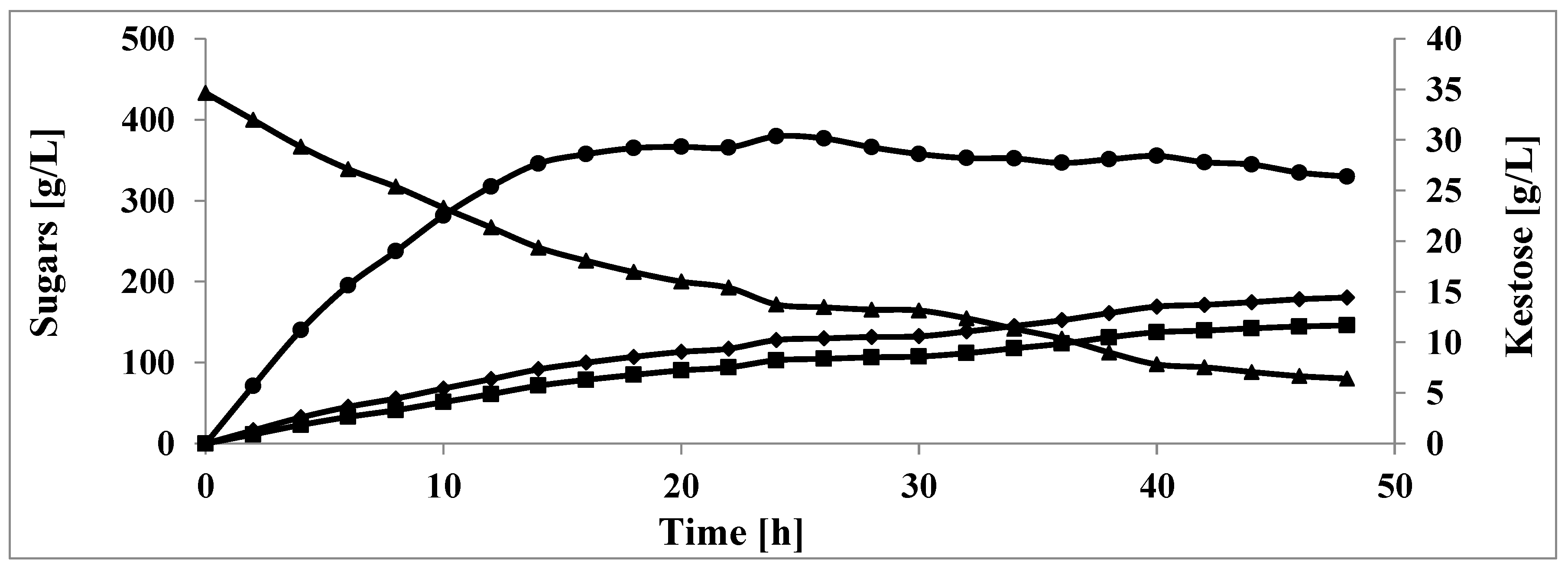
Next, the transfructosidase activity of the analyzed invertase in high sucrose concentration (500 g/L) was investigated. Fructooligosaccharides (FOS) are oligosaccharides with the number of fructose units ranging from 2 to 60, terminated by glucose unit. They are not hydrolyzed by small intestinal glycosidases and are considered as soluble dietary fiber. Furthermore, FOS have important beneficial physiological effects such as low carcinogenicity and prebiotic effect, improving mineral absorption, and decreasing levels of serum cholesterol, triacylglycerols, and phospholipids [41]. The heterologous invertase expressed by Y. lipolytica was able to synthesize up to 30 g/L of 1-kestose within 24 h (Figure 4). After this period, the concentration of kestose slowly decreased. The critical point of invertase transfructosylating and hydrolytic activity was at 17% of sucrose concentration in the reaction mixture. The 1-kestose was the unique FOS produced by Y. lipolytica invertase. The study of FOS expressed by S. cerevisiae invertase showed that this enzyme is able to generate five different molecules: 1-kestose, 6-β-fructofuranosylglucose, inulobiose, 6-kestose, and neokestose in aqueous and anhydrous organic media [42,43]. Similar products were obtained during FOS synthesis by invertase from R. dairenensis [1]. The maximum concentration of FOS (87.9 g/L) was reached at 71 h at 13% of sucrose concentration in the reaction mixture and contained 6-kestose (68.9 g/L), neokestose (10.6 g/L), 1-kestose (2.6 g/L), and tetrasaccharides (12.7 g/L). It is possible to obtain 101 g/L of FOS by the invertase from Sch. occidentalis, where 6-kestose and 1-kestose concentration reached the level of 76 and 25 g/L, respectively [4,44].
The molecular mass and oligomerization pattern of native invertase were determined by PAGE in non-denaturing conditions. The purified enzyme yielded five bands corresponding to dimer, tetramer, hexamer, octamer, and previously undescribed putative decamer (Figure 5).
The aggregation into oligomers is a known phenomenon for described invertases, including the enzyme from S. cerevisiae [5,8,45,46,47]. These authors showed that the cytoplasmic enzyme aggregates into dimers; however, its periplasmic form can exist as dimers, tetramers, hexamers, and octamers. The latter form is predominant in the periplasm and prevents enzyme secretion into the medium [5,47]. The enzyme produced in S. cerevisiae is subject to hypermannosylation and the high degree of glycosylation helps to stabilize the octamers [48]. Similarly, retention of invertase in the periplasm was already proved for heterologous invertase produced by Y. lipolytica [28,32]. The maximum amount of secreted invertase reached 25% of the total enzyme produced, when native signal peptide was used [28]. In the current work we confirmed oligomers formation by heterologous invertase produced by Y. lipolytica; however, clear bands representing higher oligomers, so far undescribed in the literature, were observed (Figure 5). Only one piece of information about higher molecular mass aggregates than octamers observed during invertase purification was found in the scientific literature [5]. No further characterizations of any larger structures were found. Yeast invertases from other species form oligomers such as dimers—C. utilis, R. glutinis, and X. dendrorhous; pentamers—Sch. pombe; and hexamers—Sch. pombe and Blastobotrys adeninivorans [9,12,14,20,40]. Y. lipolytica invertase monomer reported in the present study (66 kDa) had similar molecular mass to the enzymes expressed by F. solani and A. flavus—65–67 kDa [17,22]. The molecular mass of the heterologous Y. lipolytica invertase dimer (132 kDa), was similar to that of S. cerevisiae secreted invertase monomer, 135 kDa [8]; however, a smaller monomeric form of S. cerevisiae invertase (95.5 kDa) was also described [35]. The molecular mass of protein part of S. cerevisiae invertase is 60 kDa [6]. Therefore, the invertase produced by Y. lipolytica as a product of S. cerevisiae SUC2 gene expression contained only 9.1% of carbohydrates in the molecule. In contrast, N-glycans of the native form of this enzyme expressed by S. cerevisiae constitute around 50% of its molecular mass [47]. Similarly, to our findings, S. cerevisiae invertase expressed in P. pastoris contained a lower amount of the sugar component [48]; however, its expression in Sch. pombe resulted in N-glycans constituting up to 65% of the molecular mass of heterologous invertase [49].
The surprising finding of oligomerization pattern of heterologous invertase expressed in Y. lipolytica made it intriguing to determine the structure of N-glycans attached to this protein. The typical structure analysis, involving glycan release using PNGase F and their trimming using different mannosidases, showed the occurrence of modification of the high-mannose structure with another sugar not trimmed by the used enzymes (Figure 6). N-glycan structures with up to 14 mannoses were observed. After different mannosidase treatment, structures with up to nine hexoses were still observed in the spectrum (Figure 6B). Acid hydrolysis of the oligosacsaccharide components and analysis of the composition of individual monosaccharides proved that galactose is the N-glycan modifying sugar in Y. lipolytica yeast. So far, galactose has been found only in the mannans of Y. lipolytica forming galactomannans [50,51]; however, to our knowledge, no galactose modification was observed in the N-glycans present on secreted proteins in this yeast.
The different N-glycan structures present on heterologous invertase expressed by Y. lipolytica compared to hypermannosylated invertase produced by S. cerevisiae did not change the basic biochemical characteristics of this enzyme (described above). The apparent differences caused by a different glycosylation profile were only the KM of heterologous invertase for sucrose as well as invertase oligomerization. We further analyzed the oligomers formation and their stability (Figure 7A). The release of invertase glycans using EndoH resulted in dissociation of oligomers into monomers, which was more pronounced for the heterologous invertase (Figure 7, lane 2 vs. lane 5). Increased content of oligosaccharides in S. cerevisiae invertase resulted in higher thermostability of its oligomers in comparison to Y. lipolytica invertase (Figure 7A, lane 3 vs. lane 6). This result confirms the finding that high glycosylation stabilizes the oligomeric forms of invertase [5,46]. The most stable form of S. cerevisiae invertase is the dimer, which further associates into octamers, which dissociates only under denaturing conditions [8,46]. Moreover, formation of oligomers is also enhanced by low pH, high ionic strength or osmolality, freezing, and high protein concentration [46]. Therefore, the next step in our study was to investigate the influence of the osmolality on the invertase subunits aggregation. Consequently, strain Y. lipolytica A-101-B56-5 was grown in the media with different carbon sources, such as glycerol, sucrose, glucose, or a mixture of glucose and fructose (for details see Materials and Methods section), which resulted in different invertase oligomerization patterns (Figure 7B). In the medium with glycerol, with the highest osmolality, the enzyme appeared mostly in the form of tetramers and the putative decamers. Sucrose-based medium, with the lowest osmolality, contained the highest concentration of the enzyme, showing the whole range of oligomers. Interestingly, in the case of glucose and mixture of glucose and fructose as carbon sources, the osmolality was similar. In glucose-based medium only dimers and tetramers were formed, whereas in a medium with a mixture of glucose and fructose the whole range of oligomers was observed. Thus, it is difficult to explain the differences in the invertase oligomerization considering only the influence of osmolality, especially, that the analyzed invertase in every sample was concentrated and dialyzed before the zymogram analysis.

4. Conclusions

The present study provides an interesting insight into the biochemical properties of invertase produced as heterologous protein by Y. lipolytica. We described two isoforms of Y. lipolytica extracellular invertase detected by the ion-exchange chromatography, which were purified 6.6 and 12.6 times, respectively. The basic enzymatic properties were similar to the enzyme isolated from S. cerevisiae (optimum pH and temperature, metal ions inhibition, substrate specificity, synthesis of 1-kestose). The enzyme expressed by Y. lipolytica presented higher KM (67 mM) and lower molecular mass of monomer (66 kDa) than the enzyme produced as native protein by S. cerevisiae. The N-glycans constituted only 9.1% of the total invertase molecular mass when produced by Y. lipolytica cells. In agreement with the other studies on invertase, the heterologous enzyme synthesized by Y. lipolytica aggregates into dimers, tetramers, hexamers, octamers, and additionally, previously undescribed putative decamers. Moreover, the glycan part of invertase does not affect its activity; however, it plays an important role in the oligomerization. Further research is required to understand the unusual pattern of invertase oligomerization and the N-glycans influence on this phenomenon.
To ensure efficient production of heterologous proteins in Y. lipolytica, it is especially important to understand the influence of culture conditions, including substrate use, on the production of these macromolecules. In this study, varying invertase aggregation profiles were obtained depending on the substrate used. This brings about further possibilities of developing industrial processes with these microorganisms, when the biosynthesis of heterologous proteins will take place during the production of polyhydroxy alcohols, which Y. lipolytica produce in large amounts under high osmotic pressure. For this reason, the development of a sustainable economy based on the management of waste materials for the production of more than one valuable metabolite seems to be of great industrial importance.

Author Contributions

K.S. performed the bioreactor cultures and wrote the manuscript; P.H. purified invertase from different cultures, analyzed the glycan structures, and corrected the manuscript; P.M. performed the biochemical characterization of invertase; B.W. performed PAGE and zymograms; M.R. conceptualized the study, helped to interpret data, and corrected the manuscript; Z.L. supervised the research at every stage, performed analysis and interpretation of the results, and read and corrected the manuscript. All authors have read and agreed to the published version of the manuscript.

Funding

The article publication fee is financed by Wroclaw University of Environmental and Life Sciences.

Informed Consent Statement

Not applicable.

Conflicts of Interest

The authors declare no conflict of interest.

References

  1. Gutiérrez-Alonso, P.; Fernández-Arrojo, L.; Plou, F.J.; Fernández-Lobato, M. Biochemical characterization of a β-fructofuranosidase from Rhodotorula dairenensis with transfructosylating activity. FEMS Yeast Res. 2009, 9, 768–773. [Google Scholar] [CrossRef] [PubMed] [Green Version]
  2. Shaheen, I.; Bhatti, H.N.; Ashraf, T. Production, purification and thermal characterization of invertase from newly isolated Fusarium sp. under solid-state fermentation. Int. J. Food Sci. Technol. 2008, 43, 1152–1158. [Google Scholar] [CrossRef]
  3. Zhou, J.; He, L.; Gao, Y.; Han, N.; Zhang, R.; Wu, Q.; Li, J.; Tang, X.; Xu, B.; Ding, J.; et al. Characterization of a novel low-temperature-active, alkaline and sucrose-tolerant invertase. Sci. Rep. 2016, 6, 32081. [Google Scholar] [CrossRef] [PubMed] [Green Version]
  4. Álvaro-Benito, M.; Polo, A.; González, B.; Fernández-Lobato, M.; Sanz-Aparicio, J. Structural and kinetic analysis of Schwanniomyces occidentalis invertase reveals a new oligomerization pattern and the role of its supplementary domain in substrate binding. J. Biol. Chem. 2010, 285, 13930–13941. [Google Scholar] [CrossRef] [PubMed] [Green Version]
  5. Sainz-Polo, M.A.; Ramirez-Escudero, M.; Lafraya, A.; Gonzalez, B.; Marin-Navarro, J.; Polaina, J.; Sainz-Aparicio, J. Three-dimensional Structure of Saccharomyces Invertase. J. Biol. Chem. 2013, 288, 9755–9766. [Google Scholar] [CrossRef] [PubMed] [Green Version]
  6. Andjelković, U.; Pićurić, S.; Vujčić, Z. Purification and characterization of Saccharomyces cerevisiae external invertase isoforms. Food Chem. 2010, 120, 799–804. [Google Scholar] [CrossRef]
  7. Gascón, S.; Neumann, N.P.; Lampen, J.O. Comparative study of the properties of the purified internal and external invertase from yeast. J. Biol. Chem. 1968, 243, 1573–1577. [Google Scholar] [CrossRef]
  8. Neuman, N.P.; Lampen, J.O. Purification and properties of yeast invertase. Biochemistry 1967, 6, 468–475. [Google Scholar] [CrossRef]
  9. Moreno, S.; Sanchez, Y.; Rodriguez, L. Purification and characterization of the invertase from Schizosaccharomyces pombe. Biochem. J. 1990, 267, 697–702. [Google Scholar] [CrossRef] [Green Version]
  10. Aburigal, A.A.A.; Elkhalifa, E.A.; Sulieman EA, M.; Elamin, H.B. Extraction and partial kinetic properties of invertase from Schizosaccharomyces pombe. Int. J. Food Sci. Nutr. Eng. 2014, 4, 80–85. [Google Scholar] [CrossRef]
  11. Domizio, P.; Liu, Y.; Bisson, L.F.; Barile, D. Cell wall polysaccharides released during the alcoholic fermentation by Schizosaccharomyces pombe and S. japonicus: Quantification and characterization. Food Microb. 2017, 61, 136–149. [Google Scholar] [CrossRef] [PubMed] [Green Version]
  12. Rubio, M.C.; Runco, R.; Navarro, A.R. Invertase from a strain of Rhodotorula glutinis. Phytochemistry 2002, 61, 605–609. [Google Scholar] [CrossRef]
  13. Canli, O.; Erdal, S.; Taskin, M.; Kurbanoglu, E.B. Effects of extremely low magnetic field on the production of invertase by Rhodotorula glutinis. TIH 2011, 27, 35–39. [Google Scholar] [CrossRef] [PubMed]
  14. Belcarz, A.; Ginalska, G.; Lobarzewski, J.; Pencel, C. The novel non-glycosylated invertase from Candida utilis (the properties and the conditions of production and purification). Acta Biochim. Biophys. 2002, 1594, 40–53. [Google Scholar] [CrossRef]
  15. Alekseeva, O.V.; Sabirzianova, T.A.; Celiakh, I.O.; Kalebina, T.S.; Kulaev, I.S. Export of an invertase by yeast cells (Candida utilis). Prikl. Biokhim. Mikrobiol. 2014, 50, 156–162. [Google Scholar] [PubMed]
  16. Rodrigo-Frutos, D.; Jiménez-Ortega, E.; Piedrabuena, D.; Ramirez-Escudero, M.; Miguez, N.; Plou, F.J.; Sanz-Aparicio, J.; Fernandez-Lobato, M. New insights into the molecular mechanism behind mannitol and erythritol fructosylation by β-fructofuranosidase from Schwanniomyces occidentalis. Sci. Rep. 2021, 11, 7158. [Google Scholar] [CrossRef]
  17. Bhatti, H.N.; Nawaz, R.; Sheikh, M.A.; Asgher, M.; Abbas, A. Studies on kinetics and thermostability of a novel acid invertase from Fusarium solani. J. Agric. Food Chem. 2006, 54, 4617–4623. [Google Scholar] [CrossRef]
  18. Santana de Almeida, A.C.; Costa de Araújo, L.; Costa, A.M.; Moraes de Abreu, C.A.; Gomes de Andrade Lima, M.A.; de Los Angeles Perez Fernandez Palha, M. Sucrose hydrolysis catalyzed by auto-immobilized invertase into intact cells of Cladosporium cladosporioides. Electr. J. Biotechnol. 2005, 8, 54–62. [Google Scholar] [CrossRef]
  19. Manoochehri, H.; Hosseini, N.F.; Saidijam, M.; Taheri, M.; Rezaee, H.; Nouri, F. A review on invertase: Its potentials and applications. Biocatal. Agric. Biotechnol. 2020, 25, 101599. [Google Scholar] [CrossRef]
  20. Linde, D.; Macias, I.; Fernández-Arrojo, L.; Plou, F.J.; Jiménez, A.; Fernández-Lobato, M. Molecular and biochemical characterization of a β-fructofuranosidase from Xanthophyllomyces dendrorhous. Appl. Environ. Microbiol. 2009, 75, 1065–1073. [Google Scholar] [CrossRef] [Green Version]
  21. Turkiewicz, M.; Pazgier, M.; Donachie, S.P.; Kalinowska, H. Invertase and α-glucosidase production by the endemic Antarctic Marine yeast Leucosporidium antarcticum. Pol. Polar Res. 2005, 26, 125–136. [Google Scholar]
  22. Uma, C.; Gomathi, D.; Muthulakshmi, C.; Gopalakrishnan, V.K. Production, purification and characterization of invertase by Aspergillus flavus using fruit peel waste as substrate. Adv. Biol. Res. 2010, 4, 31–36. [Google Scholar]
  23. Nadeem, H.; Rashid, M.H.; Siddique, M.H.; Azeem, F.; Muzammil, S.; Javed MRAli, M.A.; Rasul, I.; Riaz, M. Microbial invertases: A review on kinetics, thermodynamics, physiochemical properties. Process Biochem. 2015, 50, 1202–1210. [Google Scholar] [CrossRef]
  24. Carlson, M.; Botstein, D. Two differentially regulated mRNAs with different 5‘ ends encode secreted with intracellular forms of yeast invertase. Cell 1982, 28, 145–154. [Google Scholar] [CrossRef]
  25. Naumov, G.I.; Naumova, E.S. Polygenic control for fermentation of β-fructosides in the yeast Saccharomyces cerevisiae: New genes SUC9 and SUC10. Microbiology 2010, 79, 160–166. [Google Scholar] [CrossRef]
  26. Förster, A.; Aurich, A.; Mauersberger, S.; Barth, G. Citric acid production from sucrose using a recombinant strain of the yeast Yarrowia lipolytica. Appl. Microbiol. Biotechnol. 2007, 75, 1409–1417. [Google Scholar] [CrossRef]
  27. Walczak, E.; Robak, M. Growth on sucrose of Yarrowia lipolytica yeats clones with invertase gene from Saccharomyces cerevisiae. Acta Sci. Pol. Biotechnol. 2009, 8, 25–36. [Google Scholar]
  28. Lazar, Z.; Rossignol, T.; Verbeke, J.; Crutz-Le Coq, A.M.; Nicaud, J.M.; Robak, M. Optimized invertase expression and secretion cassette for improving Yarrowia lipolytica growth on sucrose for industrial applications. J. Ind. Microbiol. Biotechnol. 2013, 40, 1273–1283. [Google Scholar] [CrossRef] [Green Version]
  29. Madzak, C.; Beckerich, J.M. Heterologous protein expression and secretion in Yarrowia lipolytica. In Yarrowia lipolytica; Springer: Berlin/Heidelberg, Germany, 2013; pp. 1–76. [Google Scholar]
  30. Vandermies, M.; Fickers, P. Bioreactor-scale strategies for the production of recombinant protein in the yeast Yarrowia lipolytica. Microorganisms 2019, 7, 40. [Google Scholar] [CrossRef] [Green Version]
  31. Ballou, C.E. Isolation, characterization, and properties of Saccharomyces cerevisiae mnn mutants with nonconditional protein glycosylation defects. Methods Enzymol. 1990, 185, 440–470. [Google Scholar] [CrossRef]
  32. Lazar, Z.; Walczak, E.; Robak, M. Simultaneous production of citric acid and invertase by Yarrowia lipolytica SUC+ transformants. Bioresour. Technol. 2011, 102, 6982–6989. [Google Scholar] [CrossRef] [PubMed]
  33. Buser, R.; Lazar, Z.; Käser, S.; Künzler, M.; Aebi, M. Identification, Characterization, and Biosynthesis of a Novel N-Glycan Modification in the Fruiting Body of the Basidiomycete Coprinopsis cinerea. J. Biol. Chem. 2010, 285, 10715–10723. [Google Scholar] [CrossRef] [PubMed] [Green Version]
  34. Lazar, Z.; Żubrowski, D.; Korzun-Chłopicka, U.; Robak, M. Recombinant Strain of Yarrowia lipolytica in Simultaneous Biosynthesis of Citrate and Invertase from Sucrose. Acta Sci. Pol. Biotechnol. 2016, 15, 25–35. [Google Scholar]
  35. Rashad, M.M.; Nooman, M.U. Production, purification and characterization of extracellular invertase from Saccharomyces cerevisiae NRRL Y-12632 by solid-state fermentation of red carrot residue. Aust. J. Bas. Sci. 2009, 3, 1910–1919. [Google Scholar]
  36. Rodriguez, J.; Perez, J.A.; Ruiz, T.; Rodriguez, L. Characterization of the invertase from Pichia anomala. Biochem. J. 1995, 306, 235–239. [Google Scholar] [CrossRef] [PubMed] [Green Version]
  37. Barbosa, P.M.G.; de Morais, T.P.; de Andrade Silva, C.A.; da Silva Santos, F.R.; Garcia, N.F.L.; Fonseca, G.G.; Leite, R.S.R.; da Paz, M.F. Biochemical characterization and evaluation of invertases produced from Saccharomyces cerevisiae CAT-1 and Rhodotorula mucilaginosa for the production of fructooligosaccharides. Prep. Biochem. Biotechnol. 2018, 48, 506–513. [Google Scholar] [CrossRef] [PubMed]
  38. Boer, E.; Wartmann, T.; Luther, B.; Mantueffel, R.; Bode, R.; Gellissen, G.; Kunze, G. Characterization of the AINV gene and the encoded invertase from the dimorphic yeast Arxula adeninivorans. Antoine Van Leeuwenhoek 2004, 86, 121–134. [Google Scholar] [CrossRef]
  39. De Godoy, V.R.; Muller, G.; Stambuk, B. Efficient maltotriose fermentation through hydrolysis mediated by the intracellular invertase of Saccharomyces cerevisiae. BMC Proc. 2014, 8, 181. [Google Scholar] [CrossRef] [Green Version]
  40. Belcastro, M.; Marino, T.; Russo, N.; Toscano, M. Interaction of cysteine with Cu2+ and group IIb (Zn2+, Cd2+, Hg2+) metal cations: A theoretical study. J. Mass Spectrom. 2005, 40, 300–306. [Google Scholar] [CrossRef]
  41. Sabater-Molina, M.; Larqué, E.; Torrella, F.; Zamora, S. Dietary fructooligosaccharides and potential benefits on health. J. Physiol. Biochem. 2009, 65, 315–328. [Google Scholar] [CrossRef]
  42. Farine, S.; Versluis, C.; Bonnici, P.J.; Heck, A.; L’homme, C.; Puigserver, A.; Biagini, A. Application of high performance anion exchange chromatography to study invertase-catalysed hydrolysis of sucrose and formation of intermediate fructan products. Appl. Microbiol. Biotechnol. 2001, 55, 55–60. [Google Scholar] [CrossRef] [PubMed]
  43. Khandekar, D.C.; Palai, T.; Agarwal, A.; Bhattacharya, P.K. Kinetics of sucrose conversion to fructo-oligosaccharides using enzyme (invertase) under free condition. Bioprocess Biosyst. Eng. 2014, 37, 2529–2537. [Google Scholar] [CrossRef] [PubMed]
  44. Mano, M.C.R.; Neri-Numa, I.A.; da Silva, J.B.; Paulino, B.N.; Pessoa, M.G.; Pastore, G.M. Oligosaccharide biotechnology: An approach of prebiotic revolution on the industry. Appl. Microbiol. Biotechnol. 2018, 102, 17–37. [Google Scholar] [CrossRef] [PubMed]
  45. Esmon, P.C.; Esmon, B.E.; Schauer, I.E.; Taylor, A.; Scheckman, R. Structure, assembly, and secretion of octameric invertase. J. Biol. Chem. 1987, 262, 4387–4394. [Google Scholar] [CrossRef]
  46. Tammi, M.; Ballou, L.; Taylor, A.; Ballou, C.E. Effect of glycosylation on yeast invertase oligomer stability. J. Biol. Chem. 1987, 262, 4395–4401. [Google Scholar] [CrossRef]
  47. Reddy, V.A.; Johnson, R.S.; Biemann, K.; Williams, R.S.; Ziegler, F.D.; Trimble, R.B.; Maley, F. Characterization of the glycosylation sites in yeast external invertase. I. N-linked oligosaccharide content of the individual sequins. J. Biol. Chem. 1988, 263, 6978–6985. [Google Scholar] [CrossRef]
  48. Trimble, R.B.; Atkinson, P.H.; Tschopp, J.F.; Townsend, R.R.; Maley, F. Structure of oligosaccharides on Saccharomyces SUC2 invertase secreted by methylotrophic yeast Pichia pastoris. J. Biol. Chem. 1991, 266, 22807–22817. [Google Scholar] [CrossRef]
  49. Zárate, V.; Belda, F. Characterization of the heterologous invertase produced Schizosaccharomyces pombe from the SUC2 gene of Saccharomyces cerevisiae. J. Appl. Bacteriol. 1996, 80, 45–52. [Google Scholar] [CrossRef]
  50. Schweigkofler, W.; Lopandic, K.; Molnár, O.; Prillinger, H. Analysis of phylogenetic relationships among Ascomycota with yeast phases using ribosomal DNA sequences and cell wall sugars. Org. Divers. Evol. 2002, 2, 1–17. [Google Scholar] [CrossRef] [Green Version]
  51. Teparić, R.; Lozančić, M.; Mrša, V. Evolutionary Overview of Molecular Interactions and Enzymatic Activities in the Yeast Cell Walls. Int. J. Mol. Sci. 2020, 21, 8996. [Google Scholar] [CrossRef]
Figure 1. Temperature (A), pH dependence (B), and thermostability profiles (C) of extracellular invertase from Y. lipolytica A-101-B56-5 (20 °C ♦; 40 °C ■; 60 °C ▲; 80 °C ●).
Sustainability 14 07926 g001
Figure 2. Substrate specificity (A) and effect of metal ions (B,C) on Y. lipolytica A-101-B56-5 invertase activity.
Sustainability 14 07926 g002
Figure 3. Lineweaver–Burk plot of invertase activity.
Sustainability 14 07926 g003
Figure 4. Time course of 1-kestose production catalyzed by Y. lipolytica A-101-B56-5 extracellular invertase (sucrose ▲, glucose ♦, fructose ■, 1-kestose ●).
Sustainability 14 07926 g004
Figure 5. Zymogram of PAGE Y.lipolytica invertase. Molecular mass determination: lane (1)-BSA; (2)-urease from Jack bean, (3)-invertase from Y. lipolytca A-101-B56-5; II—X invertase polymers position (dimer to decamer, respectively).
Sustainability 14 07926 g005
Figure 6. Mass spectrum of undigested N-glycans released from heterologous invertase expressed in Y. lipolytica (A) and α-mannosidase digested N-glycans from membrane proteins (B). M7, M8, and M9 indicate the number of mannose molecules. ●—mannose, ■—N-acetylglucosamine, ○—hexose.
Sustainability 14 07926 g006
Figure 7. Zymogram of PAGE Y.lipolytica invertase. (A) N-glycan influence on external invertase polymerization; S. cerevisiae invertase lanes (1–3), Y.lipolytica A-101-B56-5 invertase lanes (4–6); lanes (1, 4) not incubated; lanes (2, 5) incubated 3 h at 37 °C with EndoH; lanes (3, 6) incubated 3 h at 37 °C without EndoH. (B) Effect of carbon source on Y. lipolytca A-101-B56-5 invertase polymerization: lane: (1)-glucose, (2)-glucose+fructose, (3)-sucrose, and (4)-glycerol. I—X invertase polymers position (monomer to decamer, respectively).
Sustainability 14 07926 g007
Table 1. Purification scheme of the heterologous invertase from Y. lipolytica A-101-B56-5.
Purification StepTotal Protein (mg)Total Activity (U)Specific Activity (U/mg of Protein)Recovery Yield [%]Purification (Fold)
Crude enzyme208712034.23100.01.00
Ultrafiltration I141486434.5068.31.01
Dialysis86.1499858.0570.21.70
Ultrafiltration II42.0376889.7152.92.62
Ion exchange chromatography
I1.55351226.454.96.62
II2.481073432.6615.112.64
Publisher’s Note: MDPI stays neutral with regard to jurisdictional claims in published maps and institutional affiliations.