Article

Peruvian Native Bacterial Strains as Potential Bioremediation Agents in Hg-Polluted Soils by Artisanal Mining Activities in Southern Peru

by
Patricia López-Casaperalta
1,
Camilo Febres-Molina
2,
Jorge Alberto Aguilar-Pineda
1,
Julio Cesar Bernabe-Ortiz
1 and
Fernando Fernandez-F
1,*
1
Vicerrectorado de Investigación, Universidad Católica de Santa María, Urb. San José s/n, Umacollo, Arequipa 04013, Peru
2
Facultad de Ciencias Exactas, Universidad Andres Bello, Santiago 8370134, Chile
*
Author to whom correspondence should be addressed.
Sustainability 2022, 14(16), 10272; https://doi.org/10.3390/su141610272
Submission received: 7 June 2022 / Revised: 1 August 2022 / Accepted: 13 August 2022 / Published: 18 August 2022
(This article belongs to the Special Issue Sustainability of Arid Lands in Southern Peru)

Abstract

:
Bioremediation of soils and waters due to mercury (Hg) contamination represents one of the most critical environmental challenges. In addition, this challenge is even greater in arid soils due to the low economic interest in these regions. Such is the case of the Secocha Annex, located in the Arequipa province of Camaná in Southern Peru. In this region, the excessive use of Hg in artisanal and small-scale gold mining (ASGM) activities has seriously endangered the health of its inhabitants. Unfortunately, there are few projects aimed at improving the environmental and sanitary conditions of this locality. An alternative to conventional cleaning technology is the use of native microorganisms that allow the recovery of ecological environments with low-cost and low-tech techniques. This work aims to present two new Hg-resistant bacterial strains obtained from Hg-polluted soils of the Secocha Annex as potential bioremediation agents. Both strains showed growth capacity on Hg substrates and their adsorption behaviors and Hg removal capacities were evaluated. By deoxyribonucleic acid (DNA) analysis, both Gram-positive strains were identified as Kocuria sp. (99.35% similarity) and Zhihengliuella sp. (99.78% similarity). Spectrometry results showed elimination capacities with values close to 28.4 and 33.3 % in an incubation time period of 45 days, with the maximum elimination efficiency in the first 24 h. These results are encouraging and offer new possibilities for the use of native strains in the bioremediation of arid soils contaminated with Hg in the Secocha Annex. Furthermore, due to the low cost and minimization of negative impacts, this technique and our bacterial strains could be used in other regions of Peru.

1. Introduction

In the last decade, mining activity has played an essential role in the economy of Peru, currently being the fourth most significant economic activity in the country [1]. Worldwide, Peru ranked tenth in gold production, with 2.9% of total extraction, in 2021. In terms of reserves, it is ninth, with almost 2000 fine metric tons (FMT). At the national level, gold extraction represents 0.84% of the gross domestic product and it is the precious metal with the third-highest extraction volume (97.3 FMT). However, it is estimated that around 15% of the gold extracted comes from illegal and informal mining [2]. Moreover, there are data indicating that these activities grew by 17.6% in the first two months of 2022 compared to the last two months of 2021 [3]. Both illegal and informal gold extraction are related to artisanal small-scale gold mining (ASGM) [2].
ASGM is a common gold mining practice widespread worldwide, especially in developing countries [4,5]. According to the PureEarth report, 10 to 25% of total gold production comes from ASGM activities [6]. Inexpensive, easy-to-learn, accessible technology and an uncomplicated mining procedure have enabled the growth of this small-scale mining industry [4,7]. An example is Peru, where it is estimated that there may be around 500,000 people engaged in this mining type [8]. Unfortunately, the lack of economic opportunities, the high price and demand for gold and the scarce legislation regarding its extraction are only some of the main reasons for the rise of ASGM [5,9,10]. These mining activities represent the only source of income for many families, especially in regions where alternatives to agriculture or livestock farming are limited [4].
Despite being a source of employment for thousands of families, ASGM negatively impacts the environment and human health [4,7,11]. This issue is because miners use the amalgamation technique to extract gold from ores, which uses large amounts of Hg. It is estimated that 20 g of Hg is used and wasted for every 1 g of gold obtained [12]. The technique consists of adding elemental mercury (Hg 0 ), which will form an amalgam with the gold contained in crushed rocks. Once the amalgam is formed, it will be washed and then burned to evaporate the Hg and obtain the isolated gold. However, water and rock debris contaminated with Hg are discharged into the tailing during the washing process. In addition, in the evaporation stage, the recovery of Hg is practically nil, so the vapors are released into the atmosphere [4,13]. In both stages, the environment is affected as a receiver of this contaminating Hg, resulting in a high Hg concentration in soils and aquifers. This pollution has devastating environmental consequences and is one of the primary causes of Hg soil contamination in many developing countries [14,15,16].
In terms of human health concerns, the effects of Hg toxicity are devastating, although its effects depend on the chemical form, dose, and time that the body is exposed to this metal [17,18,19]. Chemically, Hg 0 , inorganic compounds (Hg 2 + ), and organic compounds (alkyl compounds, MeHg, EtHg, etc.) are the three primary forms with the most significant toxicity found in the environment [20]. Hg is considered a neurotoxin, so one of its first adverse effects is on the central and peripheral nervous system. Inhalation of Hg vapor can also damage the immune, digestive, respiratory, and kidney systems [21]. Inorganic compounds are corrosive, so the skin, eyes, digestive system, and kidneys are the most affected. However, alkyl compounds are considered very dangerous as they tend to be accepted by the body and accumulate in the tissues [19]. Among these compounds, MeHg (methylmercury) stands out, since small exposures can be highly harmful and cause death [22,23,24]. In addition, this compound can cross the placenta and the blood–brain barrier. The problem is that once Hg reaches environmental sources, it can undergo a natural methylation process, transforming into the deathly methylmercury [25,26].
Dependence on Hg for gold extraction from ore has led this practice to become the largest source of this metal’s release on the planet [13,27,28]. According to the global mercury assessment report, around 2000–2500 tonnes of Hg are released into the environment, of which 60% correspond to ASGM activities [29]. Peru is no exception; in 2010, it was calculated that Hg emissions from this activity reached 70 tons [30]. The lack of recent data only reflects the limited control of this activity by the authorities.
Several studies are underway to remediate contaminated soils and aquifers [31,32,33]. However, bioremediation techniques are increasingly used as they are considered low-cost and environmentally friendly due to using microorganisms or plants, often native, to remove contaminants [34,35,36,37]. Depending on the characteristics of the pollutant and the contaminated source, the microorganisms that can be used are usually bacteria, yeasts, or fungi. Therefore, the use of bioremediation to treat Hg-contaminated soils is becoming more and more widespread, and its effectiveness is remarkable [35,36,38,39].
In this sense, in previous work, we exposed the environmental problems suffered by the population in the Secocha Annex with the rise of ASGM activities [40]. The Secocha Annex is located in the northwest of the province of Camaná and its soils are considered arid of the xeric type. Unfortunately, economic opportunities are scarce and a large part of the population is engaged in ASGM, suffering the consequences of gold overexploitation and environmental Hg pollution [41]. This locality is considered the area of Arequipa that registers the highest annual loss of Hg because of ASGM activities (27.82 tons) [42]. Current Peruvian legislation considers the maximum Hg concentration for this type of soil to be 24 mg·kg 1 (dry weight). In this area, the results of the analyses reached values of up to 49 mg·kg 1 . In 2018, this town was summoned by residents and local authorities to be declared a state of health emergency because of the alarming level of Hg contamination in soils and bodies of water [41,43]. In the mentioned work, through DNA analysis and atomic absorption spectrometry, we identified a Gram-positive bacterium Zhihengliuella alba sp. with a high Hg removal capacity. In this work, we aim to continue with the search for new bacterial strains, and the results of our assays showed two promising bacteria of Kocuria Rosea and another from the Zhihengliuella alba genus with Hg removal capacity that could be used in the remediation of native soils contaminated with Hg. These findings are encouraging and demonstrate that this native strain may be the key to the bioremediation of Hg-polluted soils and water in arid environments.

2. Materials and Methods

2.1. Study Area

Located in the Arequipa province of Camaná, in Southern Peru, the Secocha Annex is a locality with a population of only 5119 inhabitants (Figure 1). According to the study carried out by Huerta et al., the type of soil in this region is xeric or highly arid [44].
Poor legislation regarding land use has allowed the rise of informal mining in the region. According to the study carried out by Aranda et al. [42], this has caused miners with little experience to carry out ASGM activities, resulting in Hg losses of over 5%. Hg losses vary between 1 and 3% in other regions of Arequipa. The Department of Energy and Mines of Arequipa has reported that around 13 pounds of gold are mined in this region per day [45].

2.2. Sampling and Isolation of Native Bacterial Strains

As in our previous work [40], this study references the “Analysis of soils in points of interest” report, produced by the Environmental Regulatory Authority (ARMA) of the Peruvian Government. This report was carried out according to the regulation D.S. N◦ 011-2017 MINAM—Environmental Quality Standards (ECA; from Spanish, Estándares de Calidad Ambiental) for soils [46]. All different sample soil points were chosen based on the highest values of Hg concentration listed in this report. The parameters used were the Hg level of the soils sampled (Table 1). Current Peruvian legislation considers the maximum Hg concentration for this type of soil to be 24 mg·kg 1 (dry weight). In this area, the results of the analyses reached values of up to 49 mg·kg 1 .
For all soil samples, the sampling protocol followed was to remove the top layer of soil (3 cm). Then, only the subsequent 15 cm of soil was collected. Next, the samples were sieved using a 1 mm aperture mesh to remove the unwanted material. The sieved material was placed in duly identified sterile containers. Each container was transferred into thermal boxes and stored at 4 °C for posterior laboratory analysis.
The stock solution of HgCl 2 was prepared by dissolving 1 g of HgCl 2 in 100 mL of distilled, deionized, and sterilized water. Two culture media were prepared for the growth of the bacterial strains: nutrient agar and blood agar. For the first one, 4.6 g of nutrient agar powder (DifcoTM) was used, while, for the second, we used 8 g of base blood agar (MerckTM). For both agars, 200 mL of distilled water was used as a solvent. Next, 100 μ L of HgCl 2 stock solution was added to both mixtures to obtain culture media at 5 mg·L 1 HgCl 2 . The pH of the solutions was 6.8 and 7.3, respectively. The culture media were autoclaved at 121 °C for 15 min, then cooled to 50 °C and transferred to Petri dishes. In the case of blood agar, 10 mL of sterile defibrillated sheep blood was added once cooled.
For the bacteria strain isolation, the serial plate depletion method was used. First, the soil samples obtained were mixed in a UV-sterilized bag. From this mixture, we weighed 10 g of soil and dissolved it in 10 mL of sterile, deionized, distilled water in a sterile Pyrex bottle. Then, they were seeded in two plates of nutritive agar to cultivate in an incubator for 72 h at 35 °C in an aerobic environment. As a result, two Hg-resistant native bacterial strains were obtained, which were identified as T1.1 and T3. Both strains were selected for their high Hg removal capacity in the tests performed.

2.3. Purification

Purified strains were obtained by repeating the plate depletion method described above ten times. This purification stage is essential to ensure the absence of microorganisms contaminating the assays or erroneous readings in the molecular sequences. Therefore, the methodologies described by Stanchi et al. were followed in the isolation and purification procedures [47].

2.4. DNA Extraction, Sequencing, and Molecular Identification of the Resistant Bacteria

Pure bacterial colonies were sent to the Uchumayo DNA Biotechnology Institute in Arequipa, Peru, for DNA extraction. In their labs, the extraction was carried out using the bead-beating technique described by Fujimoto et al. [48]. The 16S rRNA gene sequencing analysis was carried out in the Molecular Biology Lab at Harvard University, Cambridge, MA, USA. Once identified, using the BLASTn server [49], the sequences were aligned and compared with those stored in the NCBI database to determine their degree of homology. Finally, the phylogenetic trees of bacterial strains were constructed by Molecular Evolutionary Genetics Analysis (MEGA) software using the top ten most similar sequences [50].

2.5. Morphological Characterization and Antibiograms

Morphology analyses of the two bacterial colonies were carried out to characterize them. The parameters used were: colony shape, size, border type, colony elevation, color, pH, and optimal growth manifestation temperature. Moreover, Gram stain tests were performed to identify them.
Antibiogram studies were performed to determine the antibiotic sensitivity and resistance of T1.1 and T3 strains following the Bauer–Kirby disk diffusion procedure [51]. This technique involves placing disks of selected antibiotics and distributing them symmetrically on the surface of a medium inoculated with bacterial strains. After incubation for 48 h at 37 °C, the inhibition halos produced by the antibiotic disks were measured. In this work, the antibiotic disks used were: ciprofloxacin (CIP), lincomycin (MY15), azithromycin (AZM), amikacin (AN30), amoxicillin (AML10), levofloxacin (LEV), enrofloxacin (ENR5), and amoxicillin + clavulanic acid (AMC30). According to the diameter of inhibition, the strains were classified as sensitive, intermediate, and resistant. The inhibition diameter of each antibiotic is shown in Table 2.

2.6. Hg Removal Capacity of T1.1 and T3 Strains

Hg determination in vitro tests were carried out to evaluate the degree of Hg removal of the T1.1 and T3 strains. The stock solution was prepared by the following procedure. First, 200 g of each contaminated soil sample was weighed and diluted in 1 L of distilled water. The samples were left to settle for 24 h. The suspension was then filtered through Whatman 41 filter paper and sterilized at 120 °C and 1 bar. Next, 500 mL of this filtrate was poured into Pyrex bottles, adding 162 mg·L 1 of Hg. The bacterial strains were inoculated in this stock solution. Samples were incubated for 12, 24, 48, 720, and 1080 h. At the same time, the Hg removed was measured. The Hg removal experimental results were obtained through the atomic absorption spectrometry analysis method with the cold vapor technique. The experimental graphical models were obtained using the adsorption capacity, q i , as a function of incubating time. The Hg removal methodology and the procedure to obtain the graphs were the same as those used in our previous work [40].

2.7. Kinetic Modeling

Two kinetic models were used to explain and predict the changes in the removal capacity of strains T1.1 and T3. In addition, these models allowed measurement of the concentration of the contaminant at different times (interpolation) and determintation of the saturation times of the strains (extrapolation). The chosen kinetic models have been widely used in various studies on bioremediation by bacterial strains [52,53,54]. The Weber and Morris (W–M) model is a pseudo-second-order kinetic model [55]. The W–M adsorption mechanism is based on the uniform porous property of the adsorbent and the diffusion capacity of the adsorbate. Thus, the initial rate is directly related to the adsorbate concentration. The W–M equation is:
q W M = K d * t 0.5
where q W M is the adsorption capacity of this model; K d is the intraparticle diffusion rate constant (mg·g 1 · time 0.5 ); and t is the time that the process takes.
The second model used is based on an allometric function [56], obtained by iteration using the OriginPro 2020 program [57]. This model has been used in investigations of the nonlinear growth of biological systems [58,59,60]. The equation is:
q A = A * t B
where q A is the adsorption capacity of the allometric model; A and B in Equation (2) are parameters obtained by iterative fits.

2.8. Statistical Analysis

Experimental bacterial growth and removal capacity data were analyzed using the SPSS v.25.0 software [61]. Next, analyses of variance (ANOVA) were performed to test differences between the obtained results. Finally, Tukey’s tests were used to detect the significant differences.

3. Results and Discussion

In this research, we have evaluated the Hg removal capacity of two bacterial strains obtained from Hg-polluted soils in the Secocha Annex region. Here, the soils are xeric arid and Hg contamination reaches hazardous levels for human health. It should be noted that despite the diversity of ecosystems in Peru, almost 50% of the population live in arid and semi-arid zones [62]. Nonetheless, the quality standards for Hg concentration allowed in soils are high, even compared to countries with similar characteristics in terms of soil type. An example is Australia, which is known for its large extent of arid and semi-arid soils [63]. In this country, the legislation allows a high Hg content in its soils up to a maximum of 4000 mg·kg 1 [64]. In contrast, in Canada, with soils rich in organic matter, the Hg concentration in soils is more stringent (Table 1) [65]. In this sense, the Hg content allowed by Peruvian environmental legislation [46] is similar to Canadian laws.
As can be seen, Hg concentration values for Peruvian and Canadian standards are lower than those of the Australian values. The main difference is in the definition of the receiving organisms of potential exposure. While the Australian standard is based solely on risk to human health, the Peruvian and Canadian standards take a broader focus, also considering ecological receptors that are often more sensitive to exposure than the human body itself. Thus, Peruvian legislation protects individuals against the risk of intoxication or poisoning by Hg. However, many ASGM activities have not been controlled, leading to situations where contamination unfortunately exceeds environmental limits. In this sense, the Hg content observed in the soil samples of the Secocha Annex exceeds the maximum standards for each soil type analyzed in this region. It should be noted that in the case of commercial, industrial, and extractive soils, the values are more than double the allowed level (49 mg·kg 1 of dry weight).

3.1. Culture, Isolation, and Identification of Bacterial Strains

Pure strains with Hg removal capacity were obtained from cultures grown on nutrient agar and blood agar at 5 mg·L 1 HgCl 2 . In the process, strains T1.1 and T3 were identified by their growth ability under these conditions. Both strains were identified based on morphologic and phenotypic properties, and through 16S rRNA sequencing. The partial DNA sequences (929 and 907 query lengths, respectively) were submitted to the BLASTn server.

3.1.1. T1.1 Strain

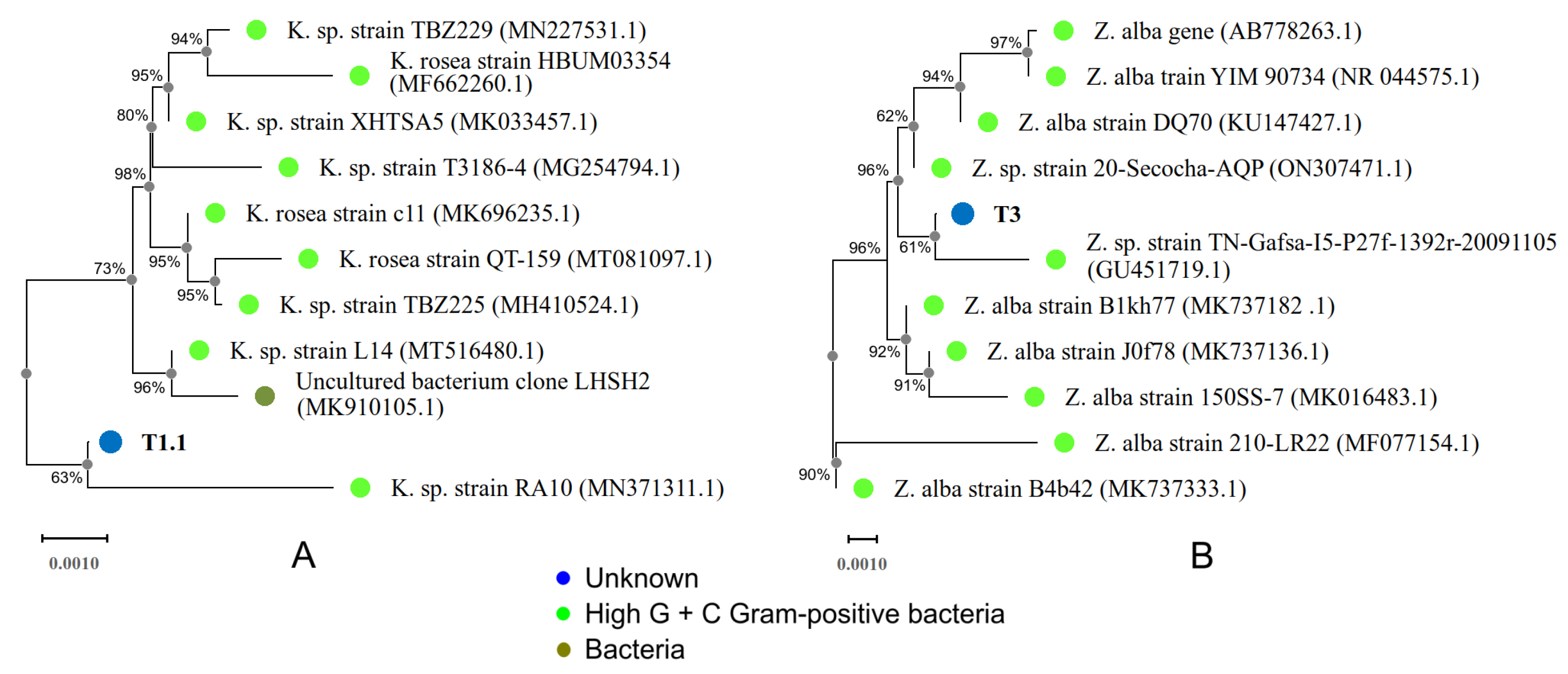
The alignment of the pure T1.1 strain revealed high sequence similarity with different bacterial strains (99.35%). The E values for all alignments were 0. The comparison was made with the first 100 strains analyzed, and 87% belonged to the Kocuria genus. Table 3 shows the top 10 bacterial strains ordered according to the BLASTn server results. Phylogenetically, the T1.1 strain showed its position in a distinct branch joined to the Kocuria sp. strain RA10 (MN371311.1) [66] (Figure 2A). However, the confidence probability was the lowest, at only 63%. Uncultured bacterium clone LHSH2, obtained from volcanic soils, was the only strain that did not coincide with this genus [67]. Taxonomically, Kocuria spp. belong to the phylum Actinobacteria, class Actinobacteria, order Micrococcales, family Micrococcaceae, and genus Kocuria. [68]. These bacteria are Gram-positive, anaerobic, non-motile, catalase-positive, coagulase-negative, and nitrite-reduction-negative, and produce pale, non-hemolytic colonies on the blood agar [69]. Kocuria spp. have been widely used in different treatments for the bioremediation of heavy-metal-contaminated wastewater [70,71] and Hg-polluted soils [72,73]. Furthermore, this bacterial strain has shown great resistance and growth capacity under extreme conditions [74].

3.1.2. T3 Strain

In the case of the isolated T3 strain, the search results showed that of the 100 sequences analyzed, 49% belonged to the Zhihengliuella genus. Furthermore, the top ten bacterial strains with the highest score were from this genus. The highest percentage of similarity was obtained by three bacterial strains (99.78%): Zhihengliuella alba (Z. a.) strain DQ70 (KU147427.1 [75]), Z. a. gene (AB778263.1 [76]), and Z. a. strain YIM 90734 (NR_044575.1 [77]) (Table 3). Taxonomically, this genus belongs to the phylum Actinobacteria, class Actinobacteria, order Micrococcales, and family Micrococcaceae [68]. Zhang et al. describe the Zhihengliuella genus as a Gram-positive, non-motile, short-rod actinobacterium [78]. This genus has a potential capacity to remove heavy metals and other pollutants [79,80,81]; however, few studies have mentioned its Hg biodegradation capacity specifically in soils.
Noteworthy to mention is that the T3 strain shares an identity of 99.67% with the strain Zhihengliuella sp. 20-Secocha-AQP. This last bacterial strain was presented by our research group and was obtained in the same region of the Secocha Annex. Both Zhihengliuella spp. showed a higher Hg removal capacity than the T1.1 strain of the Kocuria genus (40 and 33% against 28%, respectively) [40]. This removal capacity could suggest that these microorganisms may be developing adaptive mechanisms to unfavorable environments to counteract the toxic effects of these pollutants [82].

3.2. Morphological Characterization and Antibiograms of T1.1 and T3 Strains

In order to characterize the bacterial strains morphologically and biochemically, bacterial colonies were seeded in nutrient agar plates with 5 mg·L 1 of HgCl 2 . These cultures were incubated for 72 h at 35 °C in an aerobic environment. Then, the strains were isolated and purified by performing subcultures in blood agar plates with 5 mg·L 1 of HgCl 2 . The use of blood agar allowed the rapid growth of bacteria and, being a non-selective medium, facilitated the isolation and purification of the colonies. In addition, the HgCl 2 concentration chosen in the culture media was used to discriminate whether bacteria were Hg-resistant or not [83].
The isolation and identification of the etiological agent and its sensitivity to the antimicrobial ones allows for the more reliable and safer management of the bacterial strains, as this will prevent indiscriminate use and the possibility of selecting resistant bacterial strains that are harmful to health [84]. In order to assess the antibiotic sensitivity test results, the Kirby–Bauer qualitative technique was used to obtain the antibiograms. Both T1.1 and T3 bacteria were identified as strains of the actinobacteria class. Therefore, the antibiotics used for antibiogram analyses were those recommended for this class of bacteria. The quantities per disc (QPD) and diameters of the inhibition halos are shown in Table 2 [68,85].

3.2.1. T1.1 Strain

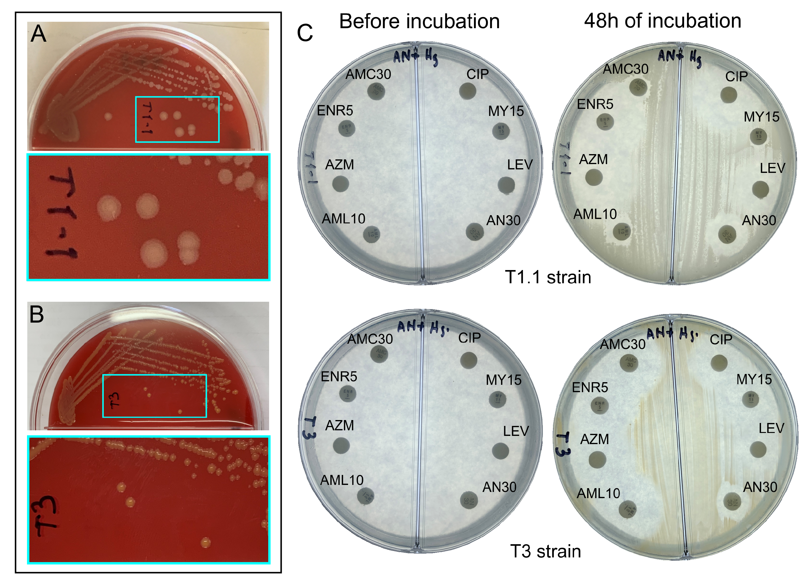
Based on the morphological characteristics and biochemical tests, identifying the T1.1 strain as Kocuria sp. was corroborated. After 72 h of incubation, this strain showed a circular form with a diameter of 3.5 mm. The color of the colony was slightly pink, characteristic of the Kocuria rosea spp. In addition, it was observed that the edges of the colony were irregular, having a convex rise. The favorable pH for its growth was 7 at 37 °C (Figure 3A). The biochemical analysis showed that it was a Gram-positive, oxidase-negative, catalase-positive bacterium, as expected. Among the main characteristics of this bacterium are the growth temperature range (25–37 °C) and its tolerance to saline environments (7.5% NaCl). These qualities make it ideal for the bioremediation of soils with arid or semi-arid characteristics. However, it stops growing at low temperatures (around 5 °C) and high percentages of salinity (10–15%) [69,74].
The analysis of the antibiogram indicates that this bacterium is resistant to four antibiotics (MY15, AN30, AML10, and AMC30), being intermediate to AZM (Figure 3C). This multi-resistance to antibiotics is expected in Hg-resistant bacteria. Several studies suggest that plasmids and transposons contain the genes that make some bacteria resistant to heavy metals [86,87,88]. In this sense, these same mobile genetic elements mediate antibiotic resistance, making them drug-resistant. On the other hand, the bacteria were sensitive to CIP, LEV, and ENR5. In the case of LEV, it was the antibiotic with the most significant inhibition halo diameter (22 mm), exceeding the sensitivity standard by 5 mm. This test is in fact very relevant since, in the case of bacteria of the genus Kocuria, there are reports that they cause infections in humans that could lead to peritonitis, endocarditis, brain abscess, and meningitis [89]. Thus, their identification may allow their clinical management in order to establish health control measures [89,90,91].

3.2.2. T3 Strain

Morphological analysis showed that the T3 strains formed circular and yellow-colored colonies, glistening, convex raised, with a 2.4 mm diameter after incubation for 72 h. The edges of the colonies were regular, with a favorable pH between 6 and 7 at 35 °C (Figure 3B). Biochemical tests showed that the strains were Gram-positive, oxidase-negative, and catalase-positive, as expected. Bacteria of the Zhihengliuella genus have interesting culture characteristics that make them suitable for use in bioremediation. They have a wide range of growth temperatures, 4–45 °C (optimum 28–37 °C). They also grow at acidic and basic pHs (5.0–9.0) and high salinity percentages (0–15% w/v) [77,78]. In the particular case of the Secocha region, the average temperature ranges between 21 and 24 °C. The pH would be expected to be alkaline, with higher percentages of salinity indicating an arid soil with scarce precipitation. These characteristics create environmental conditions that are favorable for their growth and replication. Moreover, this could partly explain why two strains of the Zhihengliuella species were found in this region.
The T3 strain was sensitive to AML10, LEV, and AMC30 (Figure 3C and Table 2, lower-row values). In the case of AML10, this antibiotic exhibited the highest inhibition halo, exceeding the standard value by 4 mm. At once, the antibiotics MY15 and ENR5 had intermediate action or increased exposure; both inhibition halos reached 16 mm. Regarding drug resistance, this strain showed small inhibition halos to CIP, AZM, and AN30 antibiotics. This susceptibility to antibiotics in the Zhihengliuella spp. has been tested in other works [92,93]; however, the information is scarce. In addition to this, their pathogenicity has not yet been determined [68].
If we compare these results with those obtained in our previous work with the Zhihengliuella sp. 20-Secocha-AQP strain (Z-20SAQP), several differences can be observed. The Z-20SAQP strain was resistant to amoxicillin compounds, inhibiting the growth of the bacteria completely. In addition, its sensitivity to antibiotics was remarkably higher than that of the T3 strain. In some cases, the inhibition halos exceeded 110% of the upper limit values (AZM and LEV). In contrast, the inhibition diameter values were close to the lower and upper values in the T3 strain for all antibiotics tested. These results reinforce the hypothesis that they are two species of the Zhihengliuella genus. However, more robust studies are suggested to confirm this.

3.3. Hg Removal by the T1.1 and T3 Strains

The removal capacity of the two isolated bacterial strains was carried out by in vitro evaluation of resistance to Hg. Cold vapor atomic absorption spectrometry tests were used to obtain the experimental values at the times mentioned above. The results are shown in Table 4 in the columns of the experimental values. Different Hg removal behaviors were observed in the bacterial strains. While, for strain T1.1 in the first 12 h, the highest removal capacity for Hg (9 mg·g 1 ) was detected at a rate of 0.75 mg·g 1 · h 1 , it rapidly declined in the following 12 h (3 mg·g 1 Hg removed). After 48 h of the test, the removal rate reached a constant value of 0.04 mg·g 1 · h 1 , which was practically maintained until the end of the test (0.03 mg·g 1 · h 1 ). On the other hand, for the T3 bacterium, the removal rate at the beginning of the test was slow (0.25 mg·g 1 · h 1 ). However, its removal capacity reached its highest performance within 12–48 h, with removal rates of 0.5 mg·g 1 · h 1 being observed. Although a decrease in Hg removal was recorded, it is observed that in both strains, at the end of the test (1080 h), the removal capacity remained latent; however, it dropped to minimum values of 0.02 and 0.01 mg·g 1 · h 1 , respectively.
If we compare these results with those obtained in our previous work [40], it can be seen that both strains of the Zhihengliuella genus have the highest Hg removal capacity. This is maintained during the first 48 h and then decreases in the last few hours of the test. These results demonstrate that the strains of this genus of bacteria are capable of removing Hg in arid conditions with high performance and that this capacity can be maintained over time. On the other hand, although the T1.1 Kokuria strain showed a more limited removal capacity, at the end of 1080 h, it was able to remove 28.4% of the Hg in the contaminated soil samples. Previous works on the removal of Hg from contaminated soils in Peru have been carried out; however, the samples have been extracted from non-arid soils [94,95]. These investigations have shown high removal rates of Hg, reaching up to 100% in concentrations of up to 100 mg·L 1 of Hg. However, it is known that arid soil compositions and high salt levels can influence the Hg cycle. Moreover, in this work, high concentrations of Hg were used (162 mg·L 1 ), which can serve to inhibit the bioremediation of bacterial strains. These reasons would explain the decrease in Hg removal in strains T1.1 and T3.

3.4. Kinetics Studies of Hg Removal

Adsorption kinetic models play a key role to determine the mechanisms involved in the sorption of solid–liquid systems [96]. In this work, the experimental data were tested against the Weber–Morris and the allometric models (Equations (1) and (2)). All experimental points were analyzed from a stock solution of Hg with a concentration of 162 mg·L 1 , a value from which, according to the measurement at different times, the Hg removal capacity of the T1.1 and T3 bacterial strains could be recorded (Table 4).
Figure 4A,B show the experimental and kinetic adsorption capacities (q) at different times of the assays (experimental data are shown in red line). In the Weber–Morris model, the removal curves were obtained using the intraparticle diffusion constants: K d , T 1.1 = 1.41 and K d , T 3 = 1.76 mg·g 1 · h 0.5 . For the allometric curves, the A and B parameters were 3.32 and 0.37 for T1.1, and 3.02 and 0.42 for T3, respectively. For the two kinetic models, regression analyses were performed to establish the coefficient of determination, R 2 . The results show that the allometric model performed the best fit to the observed values, with R 2 of 0.99 (T1.1) and 0.96 (T3), compared to the Weber–Morris model (0.96 for both strains). The two strains had a high removal ratio at the beginning of the assays and the allometric model was able to predict these behaviors, although it underestimated the zone of slow removal in T1.1 and overestimated it in T3. In the other case, the Weber–Morris model had a minor slope at the beginning of the assays, underestimating the values in the high removal region and overestimating those in the slow removal region. In the region of slow absorption, the Weber–Morris model was more adjusted to the final concentration of the test with strain T1.1, with a relative error of 0.80% (3.70% in the allometric model). However, for the T3 strain, the smallest relative error was obtained with the allometric model (4.28% against 7.11% in Weber–Morris model).
The rates and acceleration curves of the removal capacities ( ν r e m , a r e m ) were calculated to analyze the saturation behavior of both strains (Figure 4C,D). The curves obtained with the allometric functions show the high rate of Hg adsorption of the T1.1 and T3 strains in the first four hours after starting the assays. Results show that both strains have similar behavior in their removal speed and acceleration. These allometric functions have made it possible to determine the saturation times of both strains. For the T1.1 strain, the saturation point occurs after 65 days (1560 h), while, for the T3 strain, it occurs at 85 days (2040 h). The constant removal rate for both strains was 0.014 mg·g 1 · h 1 . Several studies carried out using allometric models have successfully explained the biodegradation processes using bacterial strains [97,98,99].

3.5. Study Limitations

Several study limitations may have affected our results. Firstly, 16S rRNA gene sequences are widely used to establish phylogenetic relationships between organisms. However, the taxonomic identification of new organisms requires more advanced and in-depth studies such as complete genome sequencing. Unfortunately, in this research, such studies are beyond our scope. Secondly, in situ experimentation is necessary to demonstrate the removal capacity of the proposed bacterial strains. In addition, since Hg is known to be an inhibitor of plant growth, studies such as sowing and germination of plants could indicate remnants of Hg in treated soils. Therefore, further studies are proposed to address this problem. Thirdly, it is necessary to carry out a complete sampling of the Hg removal capacity of the strains studied. This would allow us a better description of the adsorption kinetics in order to elucidate the Hg elimination mechanism.

4. Conclusions

The bioremediation of arid mercury-polluted soils is a significant environmental challenge. This research aims to present two new Peruvian native bacterial strains obtained from contaminated soils in the region of the Secocha Annex, Southern Peru. Both strains of the Kocuria and Zhihengliuella genera were able to remove mercury at a high-level concentration of this pollutant. Moreover, their long-term removal capacities are encouraging for their use as bioremediation agents. These characteristics can be helpful in the bioremediation process in polluted soils under extreme conditions. Therefore, it is recommended to carry out advanced experimental work to demonstrate the mechanism of mercury removal. In this way, it will be possible to determine how this adsorption occurs. In addition, both strains could present phytostimulant activity on native plants, as is the case of the bacterial strain Kocuria, which, according to several studies, allows the growth of plants in soils contaminated with heavy metals. This, in turn, would also help in the bioremediation process.

Author Contributions

Conceptualization, F.F.-F., P.L.-C. and J.A.A.-P.; methodology, P.L.-C. and J.C.B.-O.; software, C.F.-M.; validation, F.F.-F. and P.L.-C.; formal analysis, F.F.-F. and C.F.-M.; investigation, F.F.-F. and J.A.A.-P.; resources, C.F.-M. and J.C.B.-O.; data curation, F.F.-F. and P.L.-C.; writing—original draft preparation, J.A.A.-P. and C.F.-M.; writing—review and editing, J.A.A.-P., C.F.-M., F.F.-F. and P.L.-C.; visualization, J.C.B.-O.; supervision, F.F.-F., P.L.-C. and J.A.A.-P.; project administration, P.L.-C.; funding acquisition, P.L.-C. All authors have read and agreed to the published version of the manuscript.

Funding

This research received no external funding.

Institutional Review Board Statement

Not applicable.

Informed Consent Statement

Not applicable.

Data Availability Statement

Not applicable.

Acknowledgments

The authors appreciate the support of the UCSM.

Conflicts of Interest

The authors declare no conflict of interest.

References

  1. MINAM. Anuario Minero 2021; Ministerio del Ambiente (Ministry of Environment): Lima, Peru, 2021.
  2. Aranoglu, F.; Flamand, T.; Duzgun, S. Analysis of Artisanal and Small-Scale Gold Mining in Peru under Climate Impacts Using System Dynamics Modeling. Sustainability 2022, 14, 7390. [Google Scholar] [CrossRef]
  3. La Minería Informal se Extiende por la Amazonía sin Control ni Cifras Oficiales. Available online: https://ojo-publico.com/3529/la-mineria-informal-se-extiende-por-la-amazonia-sin-control (accessed on 11 July 2022).
  4. Veiga, M.M.; Maxson, P.A.; Hylander, L.D. Origin and consumption of mercury in small-scale gold mining. J. Clean. Prod. 2006, 14, 436–447. [Google Scholar] [CrossRef]
  5. Tabelin, C.B.; Silwamba, M.; Paglinawan, F.C.; Mondejar, A.J.S.; Duc, H.G.; Resabal, V.J.; Opiso, E.M.; Igarashi, T.; Tomiyama, S.; Ito, M.; et al. Solid-phase partitioning and release-retention mechanisms of copper, lead, zinc and arsenic in soils impacted by artisanal and small-scale gold mining (ASGM) activities. Chemosphere 2020, 260, 127574. [Google Scholar] [CrossRef] [PubMed]
  6. La Minería Informal se Extiende por la Amazonía sin Control ni Cifras Oficiales. Available online: https://www.pureearth.org/the-toxic-toll-of-mercury-facts-figures-and-the-future-of-dancing-cat-fever-disease/ (accessed on 18 July 2022).
  7. Zolnikov, T.R.; Ortiz, D.R. A systematic review on the management and treatment of mercury in artisanal gold mining. Sci. Total Environ. 2018, 633, 816–824. [Google Scholar] [CrossRef]
  8. Cuzcano, V.T. Minería ilegal e informal en el Perú: Impacto socioeconómico; Sonimágenes del Perú SCRL: Lima, Peru, 2015; Cuadernos 21 de CooperAcción 2; Available online: https://cooperaccion.org.pe/publicaciones/mineria-ilegal-e-informal-en-el-peru-impacto-socioeconomico-2/ (accessed on 20 April 2022).
  9. Wiener, L. La gobernanza de la minería en pequeña escala en el Perú; Sonimágenes del Perú SCRL: Lima, Peru, 2019; Available online: https://cooperaccion.org.pe/wp-content/uploads/2019/06/La-Gobernanza-de-la-Minería.pdf (accessed on 2 June 2022).
  10. Adamek, K.; Lupa, M.; Zawadzki, M. for tracking changes caused by illegal gold mining in Madre de Dios, Peru. Misc. Geogr. 2021, 25, 205–212. [Google Scholar]
  11. Smith, N.M. “Our gold is dirty, but we want to improve”: Challenges to addressing mercury use in artisanal and small-scale gold mining in Peru. J. Clean. Prod. 2019, 222, 646–654. [Google Scholar] [CrossRef]
  12. Gunson, A.J.; Veiga, M.M. Mercury and artisanal mining in China. Environ. Pract. 2004, 6, 109–120. [Google Scholar] [CrossRef]
  13. Esdaile, L.J.; Chalker, J.M. The mercury problem in artisanal and small-scale gold mining. Chem.—A Eur. J. 2018, 24, 6905–6916. [Google Scholar] [CrossRef]
  14. Hilson, G.; Hilson, C.J.; Pardie, S. Improving awareness of mercury pollution in small-scale gold mining communities: Challenges and ways forward in rural Ghana. Environ. Res. 2007, 103, 275–287. [Google Scholar] [CrossRef]
  15. Davies, G.R. A toxic free future: Is there a role for alternatives to mercury in small-scale gold mining? Futures 2014, 62, 113–119. [Google Scholar] [CrossRef]
  16. Budnik, L.T.; Casteleyn, L. Mercury pollution in modern times and its socio-medical consequences. Sci. Total Environ. 2019, 654, 720–734. [Google Scholar] [CrossRef] [PubMed]
  17. Bernhoft, R.A. Mercury toxicity and treatment: A review of the literature. J. Environ. Public Health 2012, 2012, 460508. [Google Scholar] [CrossRef] [PubMed]
  18. Efectos en la Salud por la Exposición al Mercurio. Available online: https://espanol.epa.gov/espanol/efectos-en-la-salud-por-la-exposicion-al-mercurio (accessed on 10 July 2022).
  19. El Mercurio y la Salud. Available online: https://www.who.int/es/news-room/fact-sheets/detail/mercury-and-health#:~:text=El%20mercurio%20elemental%20y%20el,con%20consecuencias%20a%20veces%20fatales (accessed on 2 July 2022).
  20. Bhardwaj, V.; Nurchi, V.M.; Sahoo, S.K. Mercury toxicity and detection using chromo-fluorogenic chemosensors. Pharmaceuticals 2021, 14, 123. [Google Scholar] [CrossRef] [PubMed]
  21. Rustagi, N.; Singh, R. Mercury and health care. Indian J. Occup. Environ. Med. 2010, 14, 45. [Google Scholar]
  22. Methylmercury. Available online: https://www.greenfacts.org/es/mercurio/n-3/mercurio-2.htm#1p2 (accessed on 5 July 2022).
  23. Jackson, A.C. Chronic neurological disease due to methylmercury poisoning. Can. J. Neurol. Sci. 2018, 45, 620–623. [Google Scholar] [CrossRef] [PubMed]
  24. Manceau, A.; Bourdineaud, J.P.; Oliveira, R.B.; Sarrazin, S.L.; Krabbenhoft, D.P.; Eagles-Smith, C.A.; Ackerman, J.T.; Stewart, A.R.; Ward-Deitrich, C.; del Castillo Busto, M.E.; et al. Demethylation of methylmercury in bird, fish, and earthworm. Environ. Sci. Technol. 2021, 55, 1527–1534. [Google Scholar] [CrossRef] [PubMed]
  25. Ma, M.; Du, H.; Wang, D. Mercury methylation by anaerobic microorganisms: A review. Crit. Rev. Environ. Sci. Technol. 2019, 49, 1893–1936. [Google Scholar] [CrossRef]
  26. Lei, P.; Tang, C.; Wang, Y.; Wu, M.; Kwong, R.W.; Jiang, T.; Zhong, H. Understanding the effects of sulfur input on mercury methylation in rice paddy soils. Sci. Total Environ. 2021, 778, 146325. [Google Scholar] [CrossRef]
  27. Telmer, K.H.; Veiga, M.M. World emissions of mercury from artisanal and small scale gold mining. In Mercury Fate and Transport in the Global Atmosphere; Springer: Boston, MA, USA, 2009; pp. 131–172. [Google Scholar]
  28. Moody, K.H.; Hasan, K.M.; Aljic, S.; Blakeman, V.M.; Hicks, L.P.; Loving, D.C.; Moore, M.E.; Hammett, B.S.; Silva-González, M.; Seney, C.S.; et al. Mercury emissions from Peruvian gold shops: Potential ramifications for Minamata compliance in artisanal and small-scale gold mining communities. Environ. Res. 2020, 182, 109042. [Google Scholar] [CrossRef]
  29. UN Environment. Global Mercury Assessment 2018. UN Environment Programme, Chemicals and Health Branch. 2019. Available online: https://www.unep.org/resources/publication/global-mercury-assessment-2018 (accessed on 5 June 2022).
  30. Mercury-Free Mining: UNDP, GEF and the Government of Peru Work Together to Reduce the Use of Mercury in Artisanal and Small-Scale Gold Mining. Available online: https://www.thegef.org/newsroom/news/mercury-free-mining-undp-gef-and-government-peru-work-together-reduce-use-mercury (accessed on 19 July 2022).
  31. Wang, W.; Han, C.; Xie, F. Efficient mercury recovery from mercuric-thiosulfate solutions by ultraviolet photolysis. Environ. Chem. Lett. 2018, 16, 1049–1054. [Google Scholar] [CrossRef]
  32. Guney, M.; Akimzhanova, Z.; Kumisbek, A.; Beisova, K.; Kismelyeva, S.; Satayeva, A.; Inglezakis, V.; Karaca, F. Mercury (HG) contaminated sites in kazakhstan: Review of current cases and site remediation responses. Int. J. Environ. Res. Public Health 2020, 17, 8936. [Google Scholar] [CrossRef] [PubMed]
  33. Liu, Y.; Zhang, R.; Sun, Z.; Shen, Q.; Li, Y.; Wang, Y.; Xia, S.; Zhao, J.; Wang, X. Remediation of artificially contaminated soil and groundwater with copper using hydroxyapatite/calcium silicate hydrate recovered from phosphorus-rich wastewater. Environ. Pollut. 2021, 272, 115978. [Google Scholar] [CrossRef] [PubMed]
  34. Chibuike, G.U.; Obiora, S.C. Heavy metal polluted soils: Effect on plants and bioremediation methods. Appl. Environ. Soil Sci. 2014, 2014, 752708. [Google Scholar] [CrossRef]
  35. Chang, J.; Duan, Y.; Dong, J.; Shen, S.; Si, G.; He, F.; Yang, Q.; Chen, J. Bioremediation of Hg-contaminated soil by combining a novel Hg-volatilizing Lecythophora sp. fungus, DC-F1, with biochar: Performance and the response of soil fungal community. Sci. Total Environ. 2019, 671, 676–684. [Google Scholar] [CrossRef]
  36. Zhao, M.M.; Kou, J.B.; Chen, Y.P.; Xue, L.G.; Fan, T.T.; Wang, S.M. Bioremediation of wastewater containing mercury using three newly isolated bacterial strains. J. Clean. Prod. 2021, 299, 126869. [Google Scholar] [CrossRef]
  37. Chang, J.; Yang, Q.; Dong, J.; Ji, B.; Si, G.; He, F.; Li, B.; Chen, J. Reduction in Hg phytoavailability in soil using Hg-volatilizing bacteria and biochar and the response of the native bacterial community. Microb. Biotechnol. 2019, 12, 1014–1023. [Google Scholar] [CrossRef]
  38. Mahbub, K.R.; Bahar, M.M.; Labbate, M.; Krishnan, K.; Andrews, S.; Naidu, R.; Megharaj, M. Bioremediation of mercury: Not properly exploited in contaminated soils! Appl. Microbiol. Biotechnol. 2017, 101, 963–976. [Google Scholar] [CrossRef]
  39. Chen, S.; Lin, W.; Chien, C.; Tsang, D.C.; Kao, C. Development of a two-stage biotransformation system for mercury-contaminated soil remediation. Chemosphere 2018, 200, 266–273. [Google Scholar] [CrossRef]
  40. Fernandez-F, F.; Lopez-C, P.; Febres-Molina, C.; Gamero-Begazo, P.L.; Gómez, B.; Bernabe-Ortiz, J.C.; Cáceres-Huambo, A.; Aguilar-Pineda, J.A. Identification and Characterization of Peruvian Native Bacterial Strains as Bioremediation of Hg-Polluted Water and Soils Due to Artisanal and Small-Scale Gold Mining in the Secocha Annex, Arequipa. Sustainability 2022, 14, 2669. [Google Scholar] [CrossRef]
  41. Available online: https://cdn.www.gob.pe/uploads/document/file/2429209/msdoc.pdf (accessed on 6 May 2022).
  42. Arana, M.; Montoya, P. Reporte de Inventario: Estimaciones de Referencia del Uso y Consumo de Mercurio en la Minería de oro Artesanal y de Pequeña Escala en Perú. 2017. Available online: https://www.artisanalgold.org/wordpress/wp-content/uploads/2020/09/Reporte-de-inventario-MAPE_Peru.pdf (accessed on 11 June 2022).
  43. Arequipa: Piden Estado de Emergencia Para Secocha por Contaminación. Available online: https://elbuho.pe/2018/05/arequipa-piden-estado-de-emergencia-para-secocha-por-contaminacion/ (accessed on 2 May 2022).
  44. Huerta, A.; Lavado, W.; Atlas de Zonas Áridas del Perú: Una evaluación presente y futura. Servicio Nacional de Meteorología e Hidrología del Perú. 2021. Available online: https://repositorio.senamhi.gob.pe/handle/20.500.12542/1206 (accessed on 3 June 2022).
  45. Secocha, el Desolador Nuevo Paraje Arequipeño de la Minería Ilegal. Available online: https://energiminas.com/secocha-el-desolador-nuevo-paraje-arequipeno-de-la-mineria-ilegal/ (accessed on 3 May 2022).
  46. Decreto Supremo N° 011-2017-MINAM.-. Available online: https://www.minam.gob.pe/disposiciones/decreto-supremo-n-011-2017-minam/ (accessed on 23 August 2021).
  47. Stanchi, N.O.; Martino, P.E.; Gentilini, E.; Reinoso, E.; Echeverría, M.; Leardini, N.; Copes, J.A. Microbiología Veterinaria; Buenos Aires: Inter-Médica, Argentina, 2007. [Google Scholar]
  48. Fujimoto, S.; Nakagami, Y.; Kojima, F. Optimal bacterial DNA isolation method using bead-beating technique. Mem. Kyushu Univ Health Scis Med. Sch 2004, 3, 33–38. [Google Scholar]
  49. Zhang, Z.; Schwartz, S.; Wagner, L.; Miller, W. A greedy algorithm for aligning DNA sequences. J. Comput. Biol. 2000, 7, 203–214. [Google Scholar] [CrossRef] [PubMed]
  50. Tamura, K.; Stecher, G.; Kumar, S. MEGA11: Molecular evolutionary genetics analysis version 11. Mol. Biol. Evol. 2021, 38, 3022–3027. [Google Scholar] [CrossRef] [PubMed]
  51. Bauer, A. Antibiotic susceptibility testing by a standardized single disc method. Am. J. Clin. Pathol. 1966, 45, 149–158. [Google Scholar] [CrossRef]
  52. Igwe, J.; Abia, A. Adsorption kinetics and intraparticulate diffusivities for bioremediation of Co (II), Fe (II) and Cu (II) ions from waste water using modified and unmodified maize cob. Int. J. Phys. Sci. 2007, 2, 119–127. [Google Scholar]
  53. Chwastowski, J.; Staroń, P.; Kołoczek, H.; Banach, M. Adsorption of hexavalent chromium from aqueous solutions using Canadian peat and coconut fiber. J. Mol. Liq. 2017, 248, 981–989. [Google Scholar] [CrossRef]
  54. Kalpana, R.; Maheshwaran, M.; Vimali, E.; Soosai, M.R.; Shivamathi, C.S.; Moorthy, I.G.; Ashokkumar, B.; Varalakshmi, P. Decolorization of textile dye by halophilic Exiguobacterium sp. VK1: Biomass and exopolysaccharide (EPS) enhancement for bioremediation of Malachite Green. ChemistrySelect 2020, 5, 8787–8797. [Google Scholar] [CrossRef]
  55. Weber, W.J., Jr.; Morris, J.C. Kinetics of adsorption on carbon from solution. J. Sanit. Eng. Div. 1963, 89, 31–59. [Google Scholar] [CrossRef]
  56. Moré, J.J. The Levenberg-Marquardt algorithm: Implementation and theory. In Numerical Analysis; Springer: Berlin/Heidelberg, Germany, 1978; pp. 105–116. [Google Scholar]
  57. OriginLab Corporation. Origin(Pro), Version 2020*. Available online: https://www.originlab.com/2020 (accessed on 5 September 2021).
  58. Greenman, J.; Ieropoulos, I.A. Allometric scaling of microbial fuel cells and stacks: The lifeform case for scale-up. J. Power Sources 2017, 356, 365–370. [Google Scholar] [CrossRef]
  59. Rimskaya-Korsakova, N.; Fontaneto, D.; Galkin, S.; Malakhov, V.; Martínez, A. Geochemistry drives the allometric growth of the hydrothermal vent tubeworm Riftia pachyptila (Annelida: Siboglinidae). Zool. J. Linn. Soc. 2021, 193, 281–294. [Google Scholar] [CrossRef]
  60. Shadrack, R.S.; Manabu, I.; Yokoyama, S. Efficacy of single and mix probiotic bacteria strain on growth indices, physiological condition and bio-chemical composition of juvenile amberjack (Seriola dumerili). Aquac. Rep. 2021, 20, 100753. [Google Scholar] [CrossRef]
  61. IBM Corp. IBM SPSS Statistics for Windows. Available online: https://www.ibm.com/pe-es/products/spss-statistics?lnk=ushpv18ct7 (accessed on 10 August 2021).
  62. Rau, P.; Bourrel, L.; Labat, D.; Frappart, F.; Ruelland, D.; Lavado, W.; Dewitte, B.; Felipe, O. Hydroclimatic change disparity of Peruvian Pacific drainage catchments. Theor. Appl. Climatol. 2018, 134, 139–153. [Google Scholar] [CrossRef]
  63. Yukie, H.; Otto, S. Desertification: A Visual Synthesis; United Nations Convention to Combat Desertification: Paris, France, 2011; Volume 102, Available online: https://library.unccd.int/Details/fullCatalogue/10 (accessed on 5 May 2022).
  64. National Environment Protection Council. Schedule B(1) Guideline on the Investigation Levels for Soil and Groundwater, National Environment Protection (Assessment of Site Contamination) Measure. 2011. Available online: https://www.legislation.gov.au/Details/F2011L02093 (accessed on 6 May 2022).
  65. Cepa, C. Canadian Soil Quality Guidelines for the Protection of Environmental and Human Health; National Guidelines and Standards Office: Quebec, QC, Canada, 2007. [Google Scholar]
  66. Kocuria, sp. Strain RA10 16S ribosomal RNA Gene, Partial Sequence. Available online: https://www.ncbi.nlm.nih.gov/nucleotide/MN371311.1?report=genbank&log\protect\T1\textdollar=nucltop&blast_rank=4&RID=E8YY4ZB0016 (accessed on 4 May 2022).
  67. Uncultured Bacterium Clone LHSH2 16S ribosomal RNA Gene, Partial Sequence. Available online: https://www.ncbi.nlm.nih.gov/nuccore/MK910105.1/ (accessed on 4 May 2022).
  68. Sorescu, I.; Stoica, C. Online Advanced Bacterial Identification Software, an Original Tool for Phenotypic Bacterial Identification. Rom. Biotechnol. Lett. 2021, 26, 3047–3053. [Google Scholar] [CrossRef]
  69. Stackebrandt, E.; Koch, C.; Gvozdiak, O.; Schumann, P. Taxonomic Dissection of the Genus Micrococcus: Kocuria gen. nov., Nesterenkonia gen. nov., Kytococcus gen. nov., Dermacoccus gen. nov., and Micrococcus Cohn 1872 gen. emend. Int. J. Syst. Evol. Microbiol. 1995, 45, 682–692. [Google Scholar] [CrossRef] [PubMed]
  70. Karnwal, A.; Bhardwaj, V.; Dohroo, A.; Upadhyay, A.K.; Bala, R. Effect of microbial surfactants on heavy metal polluted wastewater. Pollut. Res. 2018, 37, 39–46. [Google Scholar]
  71. Karkouri, A.E.; Kaoutar, H.; Arroud, H.; Hassouni, M.E. Hexavalent chromium removal from a tannery effluent by a bacterial consortium. Moroc. J. Chem. 2019, 7, 4–7. [Google Scholar]
  72. François, F.; Lombard, C.; Guigner, J.M.; Soreau, P.; Brian-Jaisson, F.; Martino, G.; Vandervennet, M.; Garcia, D.; Molinier, A.L.; Pignol, D.; et al. Isolation and characterization of environmental bacteria capable of extracellular biosorption of mercury. Appl. Environ. Microbiol. 2012, 78, 1097–1106. [Google Scholar] [CrossRef]
  73. Velásquez-Riaño, M.; Benavides-Otaya, H.D. Bioremediation techniques applied to aqueous media contaminated with mercury. Crit. Rev. Biotechnol. 2016, 36, 1124–1130. [Google Scholar] [CrossRef]
  74. Gholami, M.; Etemadifar, Z.; Bouzari, M. Isolation a new strain of Kocuria rosea capable of tolerating extreme conditions. J. Environ. Radioact. 2015, 144, 113–119. [Google Scholar] [CrossRef]
  75. Zhihengliuella alba Strain DQ70 16S ribosomal RNA Gene, Partial Sequence. Available online: https://www.ncbi.nlm.nih.gov/nucleotide/KU147427.1?report=genbank&log\protect\T1\textdollar=nucltop&blast_rank=1&RID=98K6KHS4016 (accessed on 1 April 2022).
  76. Hamada, M.; Shibata, C.; Tamura, T.; Suzuki, K.i. Zhihengliuella flava sp. nov., an actinobacterium isolated from sea sediment, and emended description of the genus Zhihengliuella. Int. J. Syst. Evol. Microbiol. 2013, 63, 4760–4764. [Google Scholar] [CrossRef]
  77. Tang, S.K.; Wang, Y.; Chen, Y.; Lou, K.; Cao, L.L.; Xu, L.H.; Li, W.J. Zhihengliuella alba sp. nov., and emended description of the genus Zhihengliuella. Int. J. Syst. Evol. Microbiol. 2009, 59, 2025–2032. [Google Scholar] [CrossRef]
  78. Zhang, Y.Q.; Schumann, P.; Yu, L.Y.; Liu, H.Y.; Zhang, Y.Q.; Xu, L.H.; Stackebrandt, E.; Jiang, C.L.; Li, W.J. Zhihengliuella halotolerans gen. nov., sp. nov., a novel member of the family Micrococcaceae. Int. J. Syst. Evol. Microbiol. 2007, 57, 1018–1023. [Google Scholar] [CrossRef] [PubMed]
  79. Mishra, A.; Gupta, B.; Kumar, N.; Singh, R.; Varma, A.; Thakur, I.S. Synthesis of calcite-based bio-composite biochar for enhanced biosorption and detoxification of chromium Cr (VI) by Zhihengliuella sp. ISTPL4. Bioresour. Technol. 2020, 307, 123262. [Google Scholar] [CrossRef] [PubMed]
  80. Gupta, B.; Mishra, A.; Singh, R.; Thakur, I.S. Fabrication of calcite based biocomposites for catalytic removal of heavy metals from electroplating industrial effluent. Environ. Technol. Innov. 2021, 21, 101278. [Google Scholar] [CrossRef]
  81. Mishra, A.; Rathour, R.; Singh, R.; Kumari, T.; Thakur, I.S. Degradation and detoxification of phenanthrene by actinobacterium Zhihengliuella sp. ISTPL4. Environ. Sci. Pollut. Res. 2020, 27, 27256–27267. [Google Scholar] [CrossRef] [PubMed]
  82. Igiri, B.E.; Okoduwa, S.I.; Idoko, G.O.; Akabuogu, E.P.; Adeyi, A.O.; Ejiogu, I.K. Toxicity and bioremediation of heavy metals contaminated ecosystem from tannery wastewater: A review. J. Toxicol. 2018, 2018, 2568038. [Google Scholar] [CrossRef] [PubMed]
  83. Von Canstein, H.; Li, Y.; Leonhäuser, J.; Haase, E.; Felske, A.; Deckwer, W.D.; Wagner-Döbler, I. Spatially oscillating activity and microbial succession of mercury-reducing biofilms in a technical-scale bioremediation system. Appl. Environ. Microbiol. 2002, 68, 1938–1946. [Google Scholar] [CrossRef] [PubMed]
  84. Restrepo, J.F.P. Guía para el uso de antibióticos en cirugia; Fundación OFA para el Avance de las Ciencias Biomédicas: Bogotá, Colombia, 1986. [Google Scholar]
  85. Antimicrobial Wild Type Distributions of Microorganisms. Available online: https://mic.eucast.org/search/?search%5Bmethod%5D=diff&search%5Bantibiotic%5D=2&search%5Bspecies%5D=-1&search%5Bdisk_content%5D=-1&search%5Blimit%5D=50 (accessed on 9 May 2022).
  86. Saranya, K.; Sundaramanickam, A.; Shekhar, S.; Swaminathan, S.; Balasubramanian, T. Bioremediation of mercury by Vibrio fluvialis screened from industrial effluents. BioMed Res. Int. 2017, 2017, 6509648. [Google Scholar] [CrossRef]
  87. Abbas, S.; Rafatullah, M.; Hossain, K.; Ismail, N.; Tajarudin, H.; Abdul Khalil, H. A review on mechanism and future perspectives of cadmium-resistant bacteria. Int. J. Environ. Sci. Technol. 2018, 15, 243–262. [Google Scholar] [CrossRef]
  88. Naguib, M.M.; Khairalla, A.S.; El-Gendy, A.O.; Elkhatib, W.F. Isolation and characterization of mercury-resistant bacteria from wastewater sources in Egypt. Can. J. Microbiol. 2019, 65, 308–321. [Google Scholar] [CrossRef]
  89. Kandi, V.; Palange, P.; Vaish, R.; Bhatti, A.B.; Kale, V.; Kandi, M.R.; Bhoomagiri, M.R. Emerging bacterial infection: Identification and clinical significance of Kocuria species. Cureus 2016, 8, e731. [Google Scholar] [CrossRef]
  90. Valencia, V.C.; de La Cruz, C.O.; Bixano, O.A.; Ruíz, M.K.H.; Estrada, E.E.S. Infecciones asociadas a Kocuria kristinae: Presentación de dos casos y revisión de la literatura. Gaceta Médica de México 2014, 150, 183–185. [Google Scholar]
  91. Agudelo, G.A.C.; Arango, I.V.; Villamil, G.E.R.; Rincón, C.I.G.; Ríos, J.G.T.; Correa, J.C.C.; Vega, F.R.; Vélez, V.P. Infección diseminada por Kocuria spp.: Reporte de caso y revisión de la literatura. Enfermedades Infecc. Microbiol. 2021, 41, 39–44. [Google Scholar]
  92. Jha, B.; Singh, V.K.; Weiss, A.; Hartmann, A.; Schmid, M. Zhihengliuella somnathii sp. nov., a halotolerant actinobacterium from the rhizosphere of a halophyte Salicornia brachiata. Int. J. Syst. Evol. Microbiol. 2015, 65, 3137–3142. [Google Scholar] [CrossRef] [PubMed]
  93. Baik, K.S.; Lim, C.H.; Park, S.C.; Choe, H.N.; Kim, H.J.; Kim, D.; Lee, K.H.; Seong, C.N. Zhihengliuella aestuarii sp. nov., isolated from tidal flat sediment. Int. J. Syst. Evol. Microbiol. 2011, 61, 1671–1676. [Google Scholar] [CrossRef]
  94. Puicon, Y.; Hurtado, J.E. Bioremediacion de Suelos Contaminados con Mercurio Utilizando Pseudomonas sp. Aisladas de Zonas de Mineria Informal; Universidad Peruana Cayetano Heredia: Lima, Perú, 2019; pp. 1–7. [Google Scholar]
  95. Sánchez Dávila, J.; Hurtado Custodio, J. Mercury reduction by bacteria isolated from informal mining zones. Adv. Mater. Res. Trans. Tech. Publ. 2009, 71, 637–640. [Google Scholar] [CrossRef]
  96. Ismadji, S.; Soetaredjo, F.E.; Ayucitra, A. The kinetic studies in the adsorption of hazardous substances using clay minerals. In Clay Materials for Environmental Remediation; Springer: Cham, Switzerland, 2015; pp. 113–120. [Google Scholar]
  97. Olson, P.E.; Wong, T.; Leigh, M.B.; Fletcher, J.S. Allometric modeling of plant root growth and its application in rhizosphere remediation of soil contaminants. Environ. Sci. Technol. 2003, 37, 638–643. [Google Scholar] [CrossRef]
  98. Ciadamidaro, L.; Girardclos, O.; Bert, V.; Zappelini, C.; Yung, L.; Foulon, J.; Papin, A.; Roy, S.; Blaudez, D.; Chalot, M. Poplar biomass production at phytomanagement sites is significantly enhanced by mycorrhizal inoculation. Environ. Exp. Bot. 2017, 139, 48–56. [Google Scholar] [CrossRef]
  99. Liu, J.; Jia, X.; Wen, J.; Zhou, Z. Substrate interactions and kinetics study of phenolic compounds biodegradation by Pseudomonas sp. cbp1-3. Biochem. Eng. J. 2012, 67, 156–166. [Google Scholar] [CrossRef]
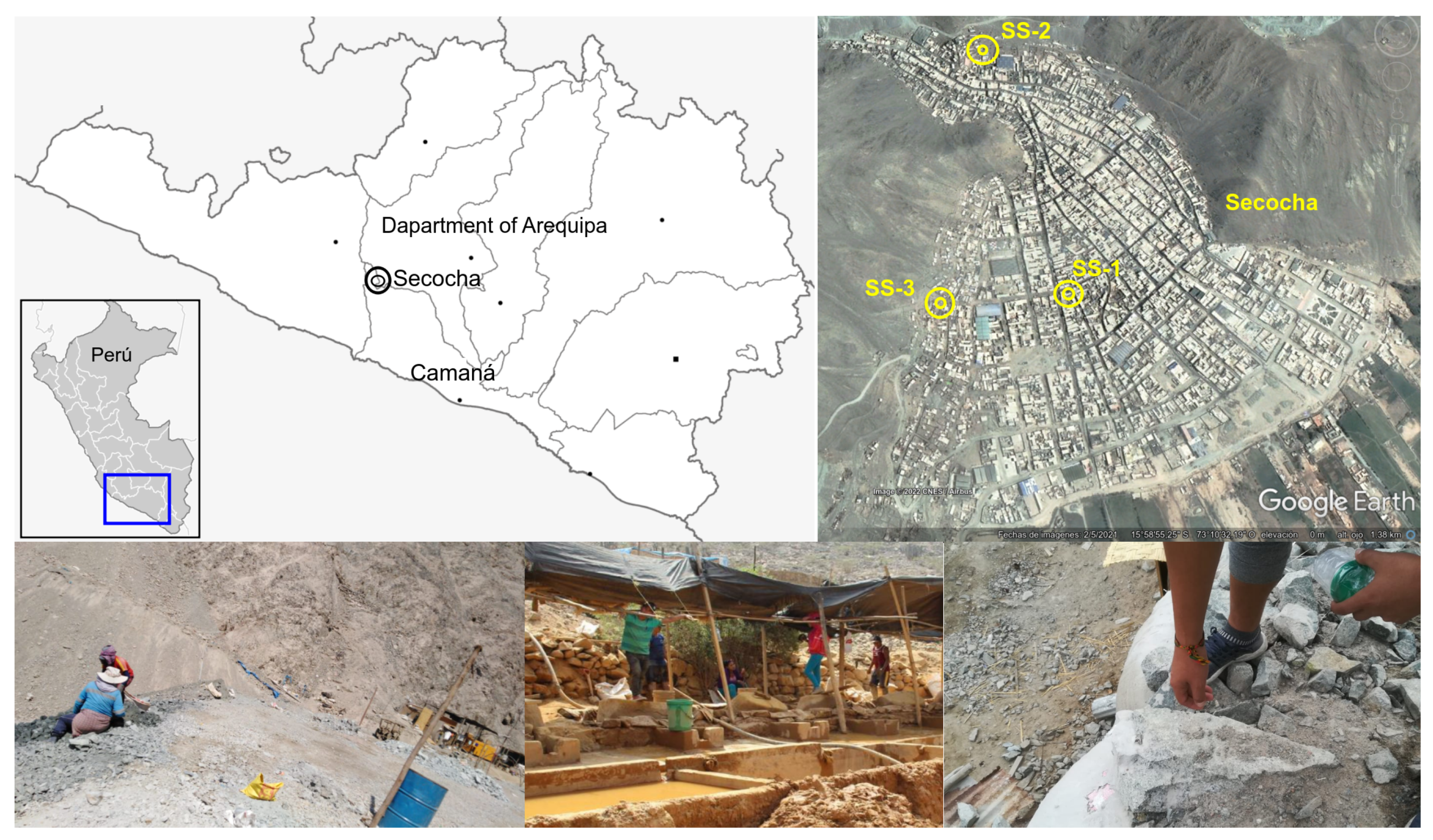
Figure 1. Location of the Secocha Annex town in Southern Peru. The yellow dots marked as SS show the areas from which the samples were extracted. In this region, the soils are extremely arid (xeric type), characterized by the scarcity of water and vegetation.
Sustainability 14 10272 g001
Figure 2. Evolutionary relationships of the isolated T1.1 (A) and T3 (B) strains. The phylogenetic trees were drawn to scale and involved the top 10 nucleotide sequences for each strain and using the neighbor-joining method. The sums of branch lengths of the optimal trees were 0.01526934 and 0.02350536, respectively. The evolutionary distances were computed using the Maximum Composite Likelihood method. There were a total of 1473 and 1496 positions in the final datasets. The percentage before branch points represents the confidence probability estimated using the bootstrap test (100 replicates).
Sustainability 14 10272 g002
Figure 3. Morphological characterization and antibiograms of the studied bacterial strains. (A,B) Growth and formation of bacterial colonies. (C) Petri dishes with the antibiotic discs distributed symmetrically in the medium inoculated with the T1.1 and T3 strains. Systems were incubated for 48 h at 37 °C.
Sustainability 14 10272 g003
Figure 4. Experimental and kinetic model curves of T1.1 and T3 strains. (A,B) Hg removal capacity curves of the experimental and kinetic models (q t ) throughout the removal test. (C,D) Rate and acceleration curves of the removal capacity were obtained using the allometric functions.
Sustainability 14 10272 g004
Table 1. Maximum values of Hg content in soils allowed by the environmental legislation of Peru, Canada, and Australia and those obtained in Secocha Annex soils.
Soil TypeECASQG, CanadaHIL, AustraliaSecocha
PeruCCME 2002NEPM 2013Annex
Agricultural6.06.6 12.0
Residential, Park6.06.6400.07.7
Commercial, Industrial, Extractive24.024.0 (50.0)4000.049.0
All values are in mg·kg−1 (dry weight).
Table 2. Antibiograms of several antibiotics applied to the T1.1 and T3 strains.
Antibiotic a QPD b Inhibition Zone b Measure c Category
RSDiameter
Ciprofloxacin (CIP)5 µg15.021.023.0Sensitive (2)
13.0Resistant
Lincomycin (MY15)2 µg16.021.00.0Resistant
18.0Intermediate
Azithromycin (AZM)15 µg13.018.016.0Intermediate
11.0Resistant
Amikacin (AN30)30 µg14.017.012.0Resistant
12.0Resistant
Amoxicillin (AML10)25 µg11.014.00.0Resistant
18.0Sensitive (4)
Levofloxacin (LEV)5 µg13.017.022.0Sensitive (5)
17.0Sensitive (0)
Enrofloxacin (ENR5)5 µg16.023.025.0Sensitive (2)
18.0Intermediate
Amoxicillin +30 µg13.018.00.0Resistant
Clavulanic ac. (AMC30) 20.0Sensitive (2)
a Quantity μg per disk. b Inhibition zone and measure diameter are in mm. R = resistant, S = sensitive. c Values in parentheses represent exceedance of the sensitivity inhibition diameter in millimeters. For the last two columns, upper-row values correspond to the T1.1 strain and those of the lower one to the T3 strain.
Table 3. Sequence similarity percentages of the top 10 bacterial strains for the T1.1 and T3 strains.
DescriptionTotalQueryPercentAccessionAccession
ScoreCoverIdentityLength
T1.1 strain
Kocuria sp. strain L14169410099.351408MT516480.1
Kocuria rosea strain QT-159169410099.351437MT081097.1
Kocuria sp. strain XHTSA5169410099.351399MK033457.1
Kocuria sp. strain RA10169410099.351078MN371311.1
Kocuria sp. strain TBZ229169410099.351398MN227531.1
Uncultured bacterium clone LHSH2169410099.351435MK910105.1
Kocuria rosea strain c11169410099.351401MK696235.1
Kocuria sp. strain T3186-4169410099.351185MG254794.1
Kocuria rosea strain HBUM03354169410099.351450MF662260.1
Kocuria sp. strain TBZ225169410099.351414MH410524.1
T3 strain
Z. a. strain DQ7016649999.781409KU147427.1
Z. a. gene16649999.781484AB778263.1
Z. a. strain YIM 9073416649999.781454NR_044575.1
Z. a. strain B1kh77166310099.671379MK737182.1
Z. a. strain J0f78166110099.671366MK737136.1
Z. sp. strain 20-Secocha-AQP166110099.67930ON307471.1
Z. sp. strain TN-Gafsa-I5-P27f-1392r-20091105166110099.671289GU451719.1
Z. a. strain 150SS-7163310098.901443MK016483.1
Z. a. strain 210-LR2216319999.121437MF077154.1
Z. a. strain B4b4216289799.771344MK737333.1
Z. a.: Zhihengliuella alba.
Table 4. Hg adsorption capacities (q) and removal rates of the studied strains.
Time (h)Experimental Values a Weber–Morris Model b Allometric Model
q exp % ads.Rateq W M % ads.Rateq A % ads.Rate
T1.1 strain
00.000.00-0.000.00-0.000.00-
129.005.560.754.893.020.418.355.150.70
2412.007.410.256.914.270.1710.796.660.20
4813.008.020.049.786.040.1213.968.620.13
72036.0022.220.0337.8623.370.0438.1123.530.04
108046.0028.400.0346.3728.620.0244.3027.350.02
T3 strain
00.000.00-0.000.00-0.000.00-
123.001.850.256.103.770.518.555.280.71
249.005.560.508.625.320.2111.447.060.24
4821.0012.960.5012.197.520.1515.299.440.16
72050.0030.660.0447.2329.150.0547.5229.330.05
108054.0033.330.0157.8435.700.0356.3134.760.02
q values are in mg·g−1 and rates in mg·g−1·h−1. a The Weber–Morris model values were obtained according to the intraparticular diffusion equation. b The allometric values were obtained using the Levenberg–Marquardt algorithm.
Publisher’s Note: MDPI stays neutral with regard to jurisdictional claims in published maps and institutional affiliations.