Article

Interaction of Microplastic Presence and Oxidative Stress in Freshwater Fish: A Regional Scale Research, East Anatolia of Türkiye (Erzurum & Erzincan & Bingöl)

1
Department of Aquaculture, Faculty of Fisheries, Ataturk University, Erzurum 25240, Türkiye
2
Department of Organic Agriculture Management, Faculty of Applied Sciences, Igdir University, Igdir 76002, Türkiye
3
Department of Chemistry, Faculty of Arts and Science, Bingöl University, Bingöl 12000, Türkiye
4
Department of Veterinary Health, Food Agriculture and Livestock Vocational School, Bingöl University, Bingöl 12000, Türkiye
5
Department of Basic Sciences, Faculty of Fisheries, Ataturk University, Erzurum 25240, Türkiye
6
Department of Plant and Animal Production, Finike Vocational School, Akdeniz University, Antalya 07058, Türkiye
7
Department of Seafood Processing Technology, Faculty of Fisheries, Ataturk University, Erzurum 25240, Türkiye
*
Author to whom correspondence should be addressed.
Sustainability 2022, 14(19), 12009; https://doi.org/10.3390/su141912009
Submission received: 25 August 2022 / Revised: 14 September 2022 / Accepted: 16 September 2022 / Published: 22 September 2022
(This article belongs to the Special Issue Pollution and Toxicology of Aquatic Ecosystems)

Abstract

:
The presence of microplastic (MP) in different fish species taken from stations in Erzurum, Erzincan and Bingöl was examined. The obtained data were classified and shared with the scientific world as the first record made in this region. In the obtained results, the most dominant color was black (39–58%) and the most prevalent forms were fragment and fiber. The sizes (0–50, 50–100 µm) of microplastics differed according to the region and species. When the number of MPs in the gastrointestinal systems of different fish species in the Bingöl, Erzurum and Erzincan provinces was evaluated, the most microplastics were found in Squalius squalus (20.7%) and Blicca bjoerkna (18.2%) in Bingöl province from among six different species. In Erzincan province, four fish species were sampled, and the rates were (29.7%) in Capoeta umbla and (26.6%) in Blicca bjoerkna. The highest abundance in Erzurum province was determined in Cyprinus carpio (53.0%). In the analyses performed on liver tissues, the highest ROS, which is the indicator of oxidative damage, was listed as Bingöl > Erzincan > Erzurum, while MDA levels were recorded as Bingöl > Erzurum > Erzincan, from high to low. When the differences between species were examined, the highest SOD and CAT activity was determined in the Mugil cephalus species. Considering the total MP numbers in fish samples, 47 MP was determined in this species. On the other hand, in the Squalius squalus species, where the highest total MP was determined, SOD and CAT activities were found to be low in Bingöl province. Therewithal, the high levels of ROS and MDA in this species can be said to induce oxidative stress due to the presence of microplastics on the one hand and to reduce antioxidant levels on the other hand. When the findings were evaluated, it was concluded that MPs in freshwater are a potential stressor, and freshwater environments may represent a critical target habitat for future MP removal and remediation strategies.

1. Introduction

Pollution of marine/freshwater mediums, especially with microplastics (MP), is a worldwide ecological issue that raises academic concerns. This situation has spurred a considerable number of studies that focus on the formation of MP, its interference with chemical contaminants, its uptake by fish, and the adverse effects [1,2]. While research on MP contamination has been carried out especially on marine ecosystems, research on freshwater mediums is scarce [2,3].
Determination of MP contamination in freshwater is very important because of; (i) high biodiversity of freshwater ecosystems threatened by human-induced stressors [4] (ii) people’s dependence on fresh water for a range of ecosystem services, including food and drinking water, which poses a human health risk (iii) river transport to the world’s oceans [5] being recognized as the main source of plastics in the seas [6,7].
It is assumed that some (if not all) of the microplastics entering rivers will be floating and will be easily transported downstream. Because sources of MPs are anthropogenic, populated or industrial areas with high flow input are likely to contain more MPs than areas subject to little anthropogenic input [8,9,10]. It had been reported that MP pollution may have more negative consequences in lake and river ecosystems as closed river basins act as a source [11]. MPs found in fish guts can be considered a representation of MP contamination in rivers, as some of the MPs in the environment are likely to be contained in the biota [12,13]. There is a significant discrepancy between reports on plastics in freshwater and marine systems, with only about 4% of studies on microplastics reported for freshwater [3].
It is known that ingestion of MPs by marine organisms can cause obstruction of the gastrointestinal tract or inflammatory responses resulting in a range of adverse effects such as lower energy reserves, reduced reproduction/growth, oxidative damage, metabolism disruption, and cellular lesions. Among the limited information on freshwater fish, Rochman et al. [14] reported that exposure to polyethylene-MPs (PE-MPs) induces hepatic stress, including glycogen depletion, fatty vacuolization, and cell necrosis in Japanese medaka (Oryzias latipes). It had also been reported that bioaccumulation of PS-MPs can disrupt lipid and energy metabolism and can also induce oxidative stress in zebrafish (D. rerio) liver [15]. These studies showed that the bioaccumulation of MPs can induce toxic effects in different freshwater fish species, and this should be carefully evaluated.
Since fish are representative groups of aquatic ecosystems, they are considered to be the common biological model used to determine the toxicity of MPs [16]. As very fine-sized microplastics pass through the gastrointestinal tract, they can be absorbed from the gut, displaced, and threaten survival as they enter the bloodstream [17]. Although MPs generally accumulate in the gastrointestinal tract of fish, they can also be transported to the liver and other organs. In order to detect organs with high MP toxicity and evaluate the mechanisms involved, it is important to identify the main MPs accumulated in fish organs following MP exposure [18]. Fish MP absorption can occur orally, through the gills and skin, and can accumulate rapidly in the gills and intestines [19]. Long-term MP exposure in fish can induce an inflammatory response in the gut, which can lead to metabolic disorders and diseases due to intestinal imbalance and damage to the microbial composition [20].
Rivers are dynamic mediums and can be subject to the accumulation of external inputs such as wastewater inputs and litter, and agricultural runoff, which contributes to the MP load [10,21]. However, research so far has not fully elucidated the extent to which freshwater fish ingest MPs, the variety of complex influences that can affect ingestion, and the potential impact on ecosystems. This study was designed to elucidate the action mechanism, stress response, damage and metabolic disorders caused by MP and MP uptake in freshwater fish in Turkey (Erzurum, Erzincan, Bingöl) as preliminary data for future studies. The study will use the following method:
  • Determination of MP abundance and characteristics in dominant fish species at the determined stations;
  • Evaluation and characterization of MPs found in gastrointestinal system (GIS);
  • Determination of the effect of MP presence on oxidative damage in liver tissues by evaluation of oxidative stress marker levels.

2. Materials and Methods

2.1. Fish Sampling

Fish samplings were made with Smith Root LR-24 Electrofisher at the determined stations in 3 different cities’ (Erzurum, Erzincan and Bingöl) streams (Figure 1). They were classified according to their species and weight and brought to the laboratories with laptap coolers supported by ice cassettes. Inland Fish Species Identification Key was used for the determination of the caught fish species (at the necessary count to ensure reliability in the statistical analysis).

2.2. Laboratory Studies

Contamination control Care was taken to use non-plastic glass/aluminum/chrome and steel materials to prevent contamination of the equipment used at all stages, from hunting to sampling in field studies. Sampled fish species from the stations were brought to Atatürk University Faculty of Fisheries Laboratories in coolers and kept in a deep freezer (−20 °C) until analysis. After determining the morphological features of the fish samples (such as length, weight, species determination, and intestinal weights), samples were taken from all species for the incidence of MP intake level, the gastrointestinal tract was removed in these groups, and they were weighed and transferred to beakers [22].
In order to prevent MP contamination from a different source during the samples’ preparation, the researchers used cotton lab coats and nitrile gloves at all stages of the process. All work surfaces and dissection materials were cleaned with 70% ethanol and used, and after the dissection process, the fish were washed 2 times with ultrapure water [23]. In all procedures, 6 clean glass Petri dishes were placed next to the work area and analyzed as procedure blank controls. In intestinal samples, 6 operational cavities containing ultrapure water (to evaluate possible laboratory contamination that may have occurred during the digestive procedures despite all care) were analyzed in place of the fish sample, without tissues, in parallel [24].

2.3. Fish Sampling Analyses

Preparation of gastrointestinal tissues for microplastic analysis: The gastrointestinal tissues dissected from the sampled fish were weighed and placed in 800 mL glass bottles, 30 mL of KOH (10%, V/V) was added to 1 g of tissue for each fish digestive system, and the mouths of the glass bottles were covered with aluminum foil. Digestive samples were kept in a shaking incubator at 60 °C for 48 h to digest organic material [23,24,25]. At the end of the incubation period, the entire homogenate was vacuum filtered through glass-microfiber filter membranes (pore size 1.2 µm). The filters were placed in glass Petri dishes and dried for 24 h (40 °C) [26].
MP characterization and identification: A visual evaluation of the items photographed in the stereomicroscope, including maximum length, color and shape, was made. At this stage, the longest or widest dimensions of each particle were also included in the closest group [22]. The MP morphotypes identified in the species as a result of the sampling were color (blue, black, gray/whitish, yellow, red/pink, green), shape (fragments- irregular pieces; lumps- spherical and oval markets; fibers- thin and long pieces) and size (˂200 µm, 201–300 µm, 301–500 µm, 501–1000 µm, 1001–2000 µm, 2001–3000 µm and 3001–5000 µm) using a software program (ImageJ). Polymer identification from the gastrointestinal tract was made using the Fourier Transform Infrared Spectroscopy (Fourier-transform infrared spectroscopy, FT-IR), which is widely used due to its economy of particles per individual [24,27].
Homogenate preparation: The livers were homogenized at 3 times their weight with phosphate buffer and centrifuged at 13,000 rpm and biochemical analyses were made in the supernatant. Superoxide dismutase (SOD) activity was determined after the inhibition of cytochrome C reduction rate by superoxide radical at λ = 560 nm [28]. Catalase (CAT) activity was mixed with the enzyme extract by adding H2O2-phosphate buffer, and the activity was measured at 240 nm using a spectrophotometer and expressed as µmol/min/mg protein [29]. Glutathione peroxidase (GPx) activity was determined by oxidation of nicotinamide adenine dinucleotide phosphate (NADPH) at λ = 340 nm in the presence of t-butyl [30]. Glutathione reductase (GR) activity was measured using Beutler’s method. One enzyme unit is defined as the oxidation of 1 mmol NADPH per min under the assay condition (25 °C, pH 8.0) [31]. Glucose 6-phosphate dehydrogenase (G6PD) activity was determined by monitoring NADPH production at 340 nm and 25 °C [31]. Malondialdehit (MDA) level is based on the spectrophotometric measurement of the pink-red color formed as a result of the reaction of MDA with thiobarbituric acid [32]. Reactive Oxygen Species (ROS) in fish was determined with some modifications of Gupta et al [33]. Tissues from each fish were homogenized separately in Tris-HCl. Homogenate samples in a volume of 100 µL were mixed with 1 mL of the same buffer and 5 µL of 10 µM 2′,7′-dichlorofluorescent diacetate (DCFDA). The resulting mixtures were incubated at 37 °C for 30 min (Lab. Companion SI-600 incubator shaker, Jelio Tech., Gangnam, Korea). After incubation, fluorescence intensity was measured in samples using a fluorescent spectrophotometer LS55 (PerkinElmer, Waltham, MA, USA) with λ = 485 nm excitation and λ = 525 nm emission.
Statistical analyses and results’ interpretation: The statistical analysis of the obtained data as a result of the analyses aimed at interpreting the data of MPs, the interaction of fish species and size with the MP presence, and the interaction of MP presence and oxidative stress findings, was determined at the 0.05 level by using the Statistical Analysis SPSS package program (version 20.0). Differences in oxidative stress response of fish between species in each of the provinces were subjected to Duncan’s multiple comparison test by performing general linear analysis (GLA).

3. Results

3.1. Average MP, Total MP and Physical Characteristics in Fish Samples

The morphological characteristics and average MP, total MP, mean length, body weight, and GIT weight of 80 fish samples at different sites are shown in Table 1.

3.2. Abundance of Microplastics in Fish

In the total MP amount in fish species caught from different provinces, six fish species (n = 7, total 42 fish) had a total of 512 MPs in Bingöl, two fish species (n = 7, total 14 fish) had a total of 100 MPs in Erzurum, and four fish species (n = 7, total 28 fish) had a total of 222 MPs in Erzincan.
When examined on a species basis in the Bingöl, Erzurum and Erzincan provinces; among the six different species caught in Bingöl, the maximum MP count was determined in S. squalus (20.7%) and B. bjoerkna (18.2%), in Erzincan province the maximum MP count was found in C. umbla (29.7%) and B. bjoerkna (26.6%). In Erzurum province, C. carpio (53.0%)’s amounts were found to be higher than the other species (Figure 2).
No significant difference was found when the MP presence of the same species in different provinces was compared (p ˃ 0.05) (Figure 3).

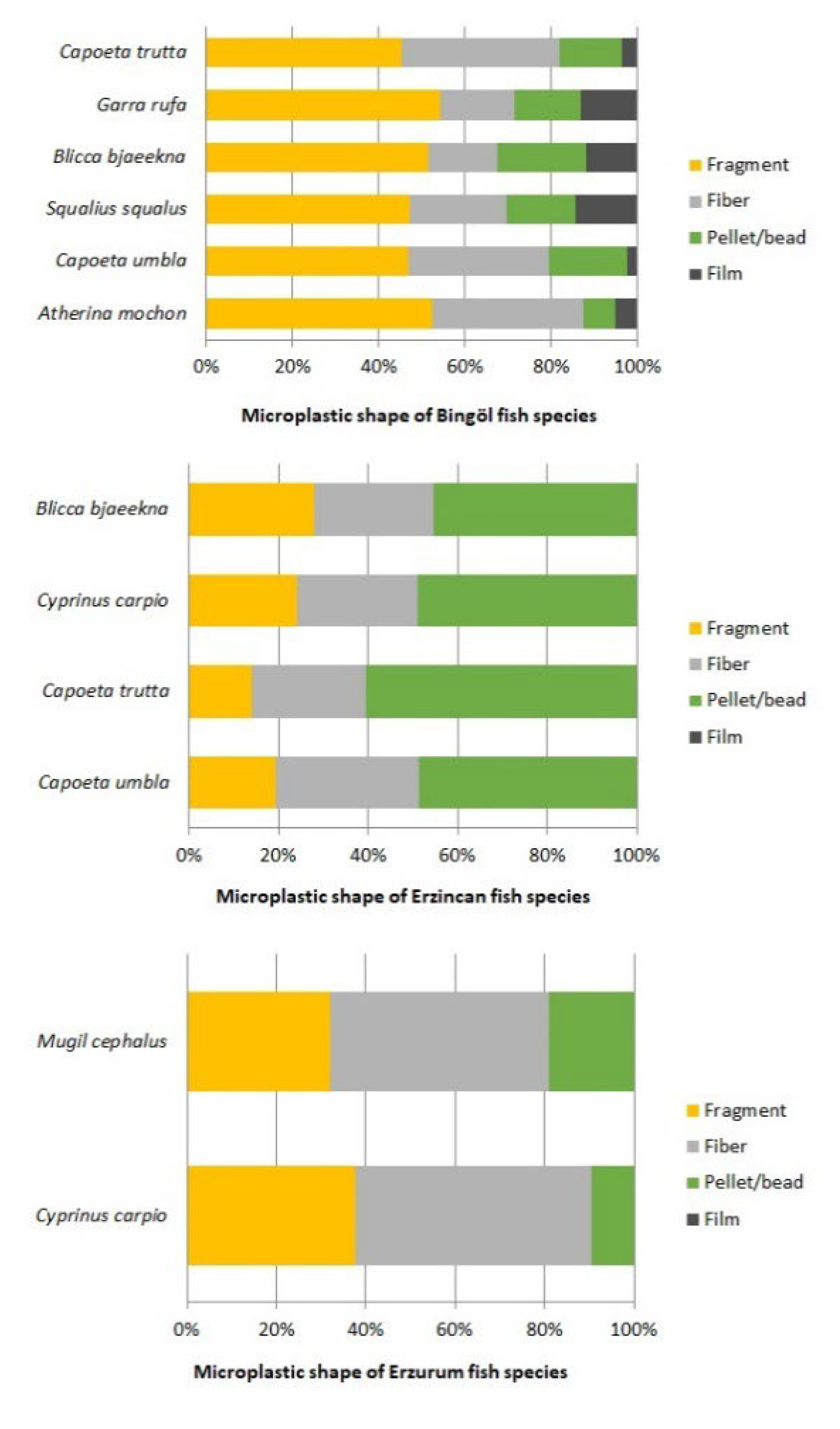
3.3. Physical Characterization of Microplastics

Microplastics in different shapes (fragment, fiber, pellet/bead, and film) were determined in the digestive systems of the examined fish species (Figure 4). Looking at the shape rates by province; in Bingöl, fragment was found in A. mochon (52.4%), C. umbla (46.9%), S. squalus (47.2%), B. bjoerkna (51.6%), G. rufa (54.3%), and C. trutta (45.6%). Fiber was dominant in Erzincan at C. umbla (56.1%), C. trutta (57.1%), C. carpio (49.1%) Blicca bjoerkna (44.1%). The same situation was also valid in Eruzurm where fiber MP was found in C. carpio (52.8%) and M. cephalus (48.9%) (Figure 4). There was no difference between B. bjoerkna (Bingöl and Erzincan, fragment) and C. carpio (Erzincan and Erzurum, fiber) species taken from different provinces, and a similar MP shape was dominant. However, the predominant microplastic form was fragment in Bingöl and fiber in Erzincan, in the C. trutta species (Figure 5).
The color of MPs (black, blue, grey/whitish, red/pink, yellow, green) in the digestive systems of fish species in the Bingöl, Erzurum and Erzincan provinces were determined as follows: the dominant color in Bingöl was black and the percentages according to the species were: A. mochon (37.8%), C. umbla (42.9%), S. squalus (37.7%), B. bjoerkna (26.9%), G. rufa (34.8%), C. trutta (57.8%) (Figure 6). In Erzincan, the dominant color was determined as black in all species and the ratios were: C. umbla (63.6%), C. trutta (83.3%), C. carpio (69.1%), B. bjoerkna (57.6%) (Figure 5). The dominant MP color was determined as black in Erzurum’s fish species in C. carpio (66.0%) and in M. cephalus (83.0%) (Figure 6). The second most dominant microplastic color was determined as blue in all three provinces (Figure 6). The MP sizes (0–50, 50–100, 100–200, 200–500, 500–1000, 1000–2000, 2000–3000 µm) differed according to the province and fish species. In Bingöl province, the most dominant MP size in the digestive systems of fish was in the range of 50–100 µm with percentages of: A. mochon (30.5%), C. umbla (40.8%) and C. trutta (44.4%). However, in the same province, in the species of S. squalus, B. bjaeekna and G. rufa, the dominant range was 0–50 µm with percentages of 35.8%, 39.8%, and 38.0%, respectively (Figure 5). The dominant microplastic size was determined as 50–100 µm in Erzincan provinces’ fish species of C. umbla (25.8%), C. trutta (45.2), C. carpio (45.5%), B. bjoerkna (44.1%) (Figure 6). In Erzurum, in the C. carpio (39.6%) species the dominant size was in the 50–100 µm range. Another fish species, Mugil cephalus (42.6%), showed the dominant microplastic size of 0–50 µm (Figure 6).

3.4. Characterization of Microplastics

More than 10% (85 MP) of the total determined MP amount (834 MP) and those larger than 40 µm were evaluated using FTIR spectra. Common polymer types of 85 selected MPs are Polypropylene (PP), Polyester (PET), Polysulfide rubber (PCR), Polychloroprene chlorine (PCP), Polyvinyl chloride (PVC), Polyisobutylene rubber (PIB), Ethylene propylene (EPR), Ethyl-acrylate (EA), Polyacrylate (PA) and their percentages are given in Table 2 and Figure 7.
Table 2. Polymer composition of ATR-FTIR verified particles.
Polymer Type of Bingöl, Erzincan and ErzurumMicroplastic Percentage (%)
Polypropylene (PP)31.8
Polyester (PET)20.0
Polysulfide rubber (PCR)7.1
Polychloroprene chlorine (PCP)8.2
Polyvinyl chloride (PVC)8.2
Polyisobutylene rubber (PIB)8.2
Ethylene propylene (EPR)4.7
Ethyl-acrylate (EA)5.9
Polyacrylate (PA)5.9
Figure 7. Examples of polymer spectra with ATR-FTIR analyses of observed MPs in fish gastrointestinal tract identified among the 85 microplastics.
Sustainability 14 12009 g007

3.5. Enzyme Results

In order to determine the level of oxidative stress caused by MPs in the sampling areas, the activities of a number of enzymes in the obtained species were evaluated.
In the analyses made between provinces, the highest SOD and CAT activity was determined in Erzurum. When the GPx, GR and G6PD enzymes were evaluated, the highest activity was detected in the species caught in Bingöl province. ROS, which is the indicator of oxidative damage in the fish species from the sampling areas, was ranked as the highest in Bingöl > Erzincan > Erzurum, while MDA levels were recorded as Bingöl > Erzurum > Erzincan from high to low. When evaluated in general terms, it can be said that oxidative damage is high in the species sampled in the Bingöl province. The lowest SOD-CAT activities were also obtained in this province (Figure 8).
When the enzyme activations are evaluated in terms of the sampled species, SOD-CAT activity is the highest. The species with the lowest ROS activity was determined as M. cephalus. The species with the lowest SOD activity was determined as Gobius niger, where GPx and G6PD enzymes were detected in higher amounts compared to other species. Squalis, the species with the lowest CAT activity, had a high MDA level (Figure 9, Figure 10 and Figure 11).

4. Discussion

Environmental monitoring agencies around the world still do not have standardized suggestions for sampling methods of MPs in water bodies, making it difficult to compare our results with other studies. In the presented study, the highest total MP was determined in the Bingöl S. squalis species. Besides, fragment-shaped microplastics were numerically predominant in all sampling areas [(Bingöl G. rufa (54.3%), Erzincan B. bjoerkna (44.1%)]. These data are also coherent with data in the literature, where fragment is the most common morphological type [34,35,36,37]. Although fibers were also known as a prevailing morphology [38,39,40,41], they were recorded as the second most widespread morphology in Turkey. Our findings suggest that the fragments are from secondary origins, possibly as a result of the degradation of freshwater mesoplastics. MPs get into freshwater ecosystems primarily from secondary MPs produced by the impairment of bigger pieces of plastic [5,10,42,43]. Therefore, MP presence in water can be attributed to many sources, including discharge of untreated wastewater, road runoff, infiltration of landfill leachate, effluent from wastewater treatment plants, and inadequate waste disposal. This explanation is provided due to the absence of industries around the sampled streams that could be primary sources of microplastics. Extensive connections can be made regarding the observed MP morphologies and potential sources. The point in question earlier, that fragments were the most prevalent morphology, suggests that fragmentation of bigger fragments of plastic and garbage could be an important source of MPs from the sample area. Yonkos et al. [44] and Mani et al. [45] reported that higher amounts of MPs can be found closer to urban areas, while particle numbers may be regularly higher near urban areas. Horton et al. [46] reported that, in addition to finding large numbers of particles downstream of urban discharge points, they also found particles in rural areas where human inputs are expected to be low.
Recent research has revealed that fibers mainly arise from the use and washing of clothing, reaching aquatic mediums through the drainage of domestic wastewater [47,48]. Observation of a high fiber content in the sampling areas [(Erzincan C. trutta (57.1%), Erzurum C. carpio (52.8%))] suggests that wastewater treatment may be poor in these provinces and domestic pollution may be an important factor. Also, one explanation for this investigation is that the fibers can become entangled and form agglomerates, stoppering the organs and thereby preventing them from being excreted from the organism along with the feces [49]. While MP shape and color distributions vary between habitats and trophic groups, the dominant color determined in the three different provinces in the present study was black. In the provinces sampled, the highest values for the dominant color black were determined as: Erzurum M. cephalus (83.0%), Bingöl C. trutta (57.8%), Erzincan C. trutta (83.3%). This data is coherent with the findings of Atamanalp et al. [2]. This may be due to the natural wear of vehicle tires, which are easily washed off the road in rainy weather and can enter the aquatic body through the city’s streams and drainage systems [50].
In the natural environment, the large surface area and hydrophobicity of MPs allow them to accumulate hazardous chemicals (for example, heavy metals and hydrophobic organic pollutants) at a significantly higher concentration than in the ambient matrix [26,51]. This concern has sparked increased research interest in the consolidated effects of MPs and related xenobiotics on aquatic living. Plastic contaminants in aquatic mediums decompose into smaller pieces through sunlight, waves, and physical and chemical reactions with aquatic organisms. Substances that decompose to MP level can be mistakenly taken as food by aquatic organisms [52].
Potential MP entry points through direct uptake into aquatic food webs are found at nearly every trophic level. Phytoplankton can directly adsorb nanoplastics through their cell walls, and zooplankton such as Daphnia and fish can take up nanoplastics and MPs directly from the water column [53]. MPs occur at particularly high concentrations in marine and estuarine environments, with the accumulation of various MPs in freshwater and marine fish species [54]. After ingestion, MPs can accumulate in the gastrointestinal tract of fish, causing blockages throughout the digestive tract and reducing feeding by creating a feeling of satiety [55]. MP particles accumulated in the body pass through the cells and enter the circulatory or lymphatic system and are distributed throughout the body. MP intake may cause problems with fish nutrition and growth due to structural and functional disruption of the gastrointestinal tract [56].
The difference in MP accretion by fish is primarily related to ingestion style (feeding strategy including ingestion, sucking and filter feeding [57,58]), intestinal structure, and plastic contamination of habitat [40,59,60]. According to one theoretical model, fish tend to ingest MPs that are more like their prey. Fish food includes aquatic plants, algae, plankton and small organisms, humus and organic/inorganic wastes; since some of the MPs have a similar morphology to these, they create a visual illusion and are effectively taken up by fish [26,51]. In freshwater ecosystems, the roles of habitat and habitat-related feeding choice are less clear and are fairly diversified according to various research [61,62]. In general, benthic organisms with chemosensory control of feeding near the bottom of freshwater habitats are thought to be more exposed to MPs because concentrations in the sediment are conceived to be greater than in the water column [9,10]. Besides, the existing findings suggest that an enhanced sense of taste limits the unintentional ingestion of MP particles. The fact that C. trutta has a large amount of mud in its stomach during the summer months when digestion is fast and that phytoplanktonic organisms that are abundant in the environment are among the nutritional organisms that participate in their gastrointestinal system may be due to the fact that the mouth of the fish is ventral, and it does not have a particular food preference [63]. G. rufa feeds on aquatic grasses and plankton and algae on the surfaces of rocks and stones in water [64]. S. squalus feeds on a variety of plant and animal materials. Larger individuals are solitary, and the largest feed mainly on fish. Considering the diet of the examined species, the fact that MPs enter via the trophic chain is confirmed. In the area, other uptake pathways, such as transfer via the food chain, may have a more extensive influence on the MP load of benthic fish species [65,66]. In this research, it was determined that the lowest total MP in C. trutta species caught from the Erzincan sampling area was 42 per/MP and 57.1% was fiber, the dominant color was black, and the size was 50–100 µm. The highest total MP was recorded in the S. squalus species, which was caught from the Bingöl sample area, as 106 units/MP and fragments at the rate of 47.2%, the dominant color was black, and the size was 0–50 µm.
Ingested MPs can lead to a variety of harmful effects on fish. Pure plastics can induce acute inactivity, expenditure of energy reservoirs [67], and liver inflammation, while MPs connected with consistent organic contaminants have been exhibited to provoke chronic or acute toxic influences [53]. The fish liver is the tissue most used to evaluate metabolic disorders caused by MPs in fish species because this organ is primarily responsible for the detoxification and inactivation of exogenous compounds [68].
Considering that oxidative stress arises from the imbalance between the formation of oxidative combination and the performance of antioxidant defense systems, and this defense is caused by the presence of chemical pollutants, it is expected that the enzymatic response will undergo changes as a result of the presence of microplastics.
The exact pattern of oxidative stress induced by MP exposure is difficult to define, but it does affect oxidative homeostasis [69]. In addition, MPs can also adhere to fish skin or migrate to other tissues such as muscle gill and liver [70,71]. It has also been documented that very fine plastic particles can pass from living cells to the circulatory or lymphatic system, causing microplastics to disperse throughout the body [72]. Unfortunately, data on the presence of microplastics in tissues outside the digestive tract of fish are currently very limited.
The production of free radicals and reactive oxygen species (ROS) has a defense system based on the activity of antioxidant enzymes [73]. The antioxidant defense system provides potential biomarkers of stress conditions [74]. Antioxidant enzymes such as catalase (CAT) and superoxide dismutase (SOD) provide the first line of defense to combat free radical damage. The superoxide radical is typically the first ROS to form, eventually leading to the formation of hydrogen peroxide (H2O2) and hydroxyl radicals [75]. When SOD activities were analyzed statistically, the highest activity was determined in C. trutta, M. cephalus and C. carpio species, while the lowest was determined in G. niger species. Similar values were found in C. umbla/B. bjoerkna and G. rufa/S. squalus species. The highest CAT activity was found in M. cephalus and C. carpio species, while the lowest was found in G. rufa, A. machon and S. squalus species. Statistically similar/close results were obtained in C. trutta, C. umbla, G. niger and B. bjoerkna species. The highest GPx activity was determined in G. niger species, while the lowest value was determined in G. ruffa species. Similar/closer results were obtained in C. umbla, A. machon, M. cephalus, C. carpio species. The species with the highest GR activity was S. squalus, and the other species examined gave statistically similar/close results in terms of GR activity. The species with the highest G6PD activity was G niger, except for C. trutta and B. bjoerkna, other species were evaluated as statistically similar. ROS was evaluated as the highest in the G. niger species and statistically similar results were obtained in other species except S. squalis and B. bjoerkna. Although changes were determined in all species examined at the MDA level, statistically similar results emerged.
Although the balance between ROS generation and antioxidant activity is crucial to protect fish from environmental contamination [76], it is difficult to directly see ROS generation in fish tissues [77]. Besides, the observation of certain phenomena, including lipid peroxidation following ROS production, may confirm the generation of oxidative stress by pollutions [78]. This situation may be related to the importance of maintaining the oxidative balance of the examined tissues, since detoxification processes are usually triggered when multiple oxidative reactions and high free radical formation occur in the liver [79]. In the study findings, high MDA levels were noted with depletion of antioxidant enzymes in the liver. Among the antioxidant enzymes used by organisms to balance oxidative damage as a result of ROS-induced cellular damage are: superoxide dismutase (SOD), which converts O2 to H2O2; catalase (CAT), which reduces H2O2 to water; and glutathione peroxidase (GPx), which detoxifies H2O2 or organic hydroperoxides into water [80].
The liver has been used to evaluate metabolic disorders caused by MPs as it is the main organ in charge of the inactivation and detoxification of exogenous molecules [81,82]. In this study, the province with the highest CAT and SOD activities was determined as Erzurum > Erzincan > Bingöl. On the other hand, ROS level was recorded as Bingöl > Erzincan > Erzurum, and MDA level as Bingöl > Erzurum > Erzincan. Particularly, exposure of aquatic organisms to MPs can stimulate the generation of ROS, which leads to oxidative injury to the macromolecules of tissues [49,83,84]. Deterioration of redox homeostasis in fish by toxic substances takes place as a result of a quick rise in the original concentration of ROS as energy is directed to synthetize molecules and antioxidant enzymes that increase antioxidant capacity [85].
Fish are defined by a complex antioxidant system that involves mechanisms such as CAT and SOD as well as GPX and G6PD [86]. When the interactions between species were examined, the highest SOD and CAT activity was determined in the Mugil cephalus species. Considering the total MP numbers in fish samples, 47 MP was determined in the M. cephalus species. In contrast, CAT and SOD activities were found to be low in the S. squalus species, where the highest total MP was determined. At the same time, it can be said that the high levels of ROS and MDA in this species induce oxidative stress depending on the presence and density of microplastics, and on the contrary, this reduces antioxidant levels.
In the presented study, the highest MP amount was obtained in the Squalis species in Bingöl. When the ROS and MDA results were examined, the highest values were determined in Bingöl province. Exposure of aquatic organisms to MPs induces ROS generation, which stimulates or inhibits antioxidant response. Furthermore, exposure to MPs disrupts (or affects) glutathione and dependent response cycles in fish, which contributes to antioxidant reactions. Thus, exposure of fish to various MPs causes oxidative damage by disrupting the antioxidant balance between antioxidant capacity and ROS generation. Similar to other toxicants, MPs induced ROS generation in fish or inhibited their antioxidant talent, based on their sensitivity to oxidative stress. Aquatic organisms, especially fish, have evolved antioxidant enzyme functions to inhibit oxidative detriment from ROS production caused by exposure to a variety of toxic materials, including MP exposure [87]. This inhibition was likely related to the oxidative stress reply stimulated by MP exposure. MDA level increased with the presence of microplastics, which supports that microplastics induce oxidative stress. The data show that MPs induce lipid peroxidation in a concentration-dependent manner.

5. Conclusions

This study provides evidence of microplastic contamination of the dominant fish species caught in streams in the Erzurum, Erzincan and Bingöl provinces. Besides the MP abundance, lipid oxidative damage and antioxidant enzymes in MP-containing fish were evaluated. These findings point to oxidative damage and lipid peroxidation damage and suggest a relationship between these changes and the contamination of fish with microplastics. Given the crucial role of fish as a source of protein for humans, continued surveys are strongly recommended to indicate the ecotoxicological effects of MPs on fish from an individual to population level and to scientifically evaluate the threats to these species. Although the majority (>90%) of MPs taken up by organisms are fecal, enabling the discovery of diagnostic biomarkers to overthrow the introduction of MPs into aquatic mediums to protect the aquatic medium can also help encourage the progress and practice of suitable regulations or policies. Consequently, the use of more comprehensive study advances to screen for the effects of environmental conditions may even increase the likelihood of defining the extent of damaging influences.

Author Contributions

Conceptualization, M.A. and G.A; methodology, formal analysis, M.K (Muammer Kırıcı), M.K. (Mahinur Kırıcı) and M.K. (Mine Kokturk).; investigation, A.U., V.P. and A.A.; writing—review and editing, A.U., V.P., G.A. and M.A.; writing—original draft preparation, A.U. and M.A.; writing—review and editing. All authors have read and agreed to the published version of the manuscript.

Funding

This research received no external funding.

Institutional Review Board Statement

Not applicable.

Informed Consent Statement

Not applicable.

Data Availability Statement

The data that support the findings of this study are available from the corresponding author upon reasonable request.

Conflicts of Interest

The authors declare no conflict of interest.

References

  1. Ivleva, N.P.; Wiesheu, A.C.; Niessner, R. Microplastic in aquatic ecosystems. Angew. Chem. Int. Ed. 2017, 56, 1720–1739. [Google Scholar] [CrossRef] [PubMed]
  2. Atamanalp, M.; Köktürk, M.; Uçar, A.; Duyar, H.A.; Özdemir, S.; Parlak, V.; Esenbuğa, N.; Alak, G. Microplastics in tissues (brain, gill, muscle and gastrointestinal) of Mullus barbatus and Alosa immaculata. Arch. Environ. Contam. Toxicol. 2021, 81, 460–469. [Google Scholar] [CrossRef] [PubMed]
  3. Lambert, S.; Wagner, M. Microplastics Are Contaminants of Emerging Concern in Freshwater Environments: An Overview; Springer Science and Business Media LLC: Berlin/Heidelberg, Germany, 2018; pp. 1–23. [Google Scholar]
  4. Dudgeon, D. Multiple threats imperil freshwater biodiversity in the Anthropocene. Curr. Biol. 2019, 29, R960–R967. [Google Scholar] [CrossRef] [PubMed]
  5. Wong, J.K.H.; Lee, K.K.; Tang, K.H.D.; Yap, P.S. Microplastics in the freshwater and terrestrial environments: Prevalence, fates, impacts and sustainable solutions. Sci. Total Environ. 2020, 719, 137512. [Google Scholar] [CrossRef]
  6. Roebroek, C.T.; Harrigan, S.; Van Emmerik, T.H.; Baugh, C.; Eilander, D.; Prudhomme, C.; Pappenberger, F. Plastic in global rivers: Are floods making it worse? Environ. Res. Lett. 2021, 16, 025003. [Google Scholar] [CrossRef]
  7. Simon-Sánchez, L.; Grelaud, M.; Garcia-Orellana, J.; Ziveri, P. River Deltas as hotspots of microplastic accumulation: The case study of the Ebro River (NW Mediterranean). Sci. Total Environ. 2019, 687, 1186–1196. [Google Scholar] [CrossRef]
  8. McCormick, A.; Hoellein, T.J.; Mason, S.A.; Schluep, J.; Kelly, J.J. Microplastic is an abundant and distinct microbial habitat in an urban river. Environ. Sci. Technol. 2014, 48, 11863–11871. [Google Scholar] [CrossRef]
  9. Dris, R.; Imhof, H.; Sanchez, W.; Gasperi, J.; Galgani, F.; Tassin, B.; Laforsch, C. Beyond the ocean: Contamination of freshwater ecosystems with (micro-)plastic particles. Environ. Chem. 2015, 12, 539. [Google Scholar] [CrossRef]
  10. Horton, A.A.; Svendsen, C.; Williams, R.J.; Spurgeon, D.J.; Lahive, E. Large microplastic particles in sediments of tributaries of the River Thames, UK—Abundance, sources and methods for effective quantification. Mar. Pollut. Bull. 2017, 114, 218–226. [Google Scholar] [CrossRef]
  11. Erdoğan, Ş. Microplastic pollution in freshwater ecosystems: A case study from Turkey. J. Fish. 2020, 37, 213–221. [Google Scholar]
  12. Van Sebille, E.; Wilcox, C.; Lebreton, L.; Maximenko, N.; Hardesty, B.D.; A Van Franeker, J.; Eriksen, M.; Siegel, D.; Galgani, F.; Law, K. A global inventory of small floating plastic debris. Environ. Res. Lett. 2015, 10, 124006. [Google Scholar] [CrossRef]
  13. Horton, A.A.; Jürgens, M.D.; Lahive, E.; van Bodegom, P.M.; Vijver, M.G. The influence of exposure and physiology on microplastic ingestion by the freshwater fish Rutilus rutilus (roach) in the River Thames, UK. Environ. Pollut. 2018, 236, 188–194. [Google Scholar] [CrossRef] [PubMed]
  14. Rochman, C.M.; Hoh, E.; Hentschel, B.T.; Kaye, S. Long-term field measurement of sorption of organic contaminants to five types of plasticpellets: Implications for plastic marine debris. Environ. Sci. Technol. 2013, 47, 1646–1654. [Google Scholar] [PubMed]
  15. Lu, Y.; Zhang, Y.; Deng, Y.; Jiang, W.; Zhao, Y.; Geng, J.; Ding, L.; Ren, H. Uptake and accumulation of polystyrene microplastics in zebrafish (Danio rerio) and toxic effects in liver. Environ. Sci. Technol. 2016, 50, 4054–4060. [Google Scholar] [CrossRef]
  16. Jambeck, J.R.; Geyer, R.; Wilcox, C.; Siegler, T.R.; Perryman, M.; Andrady, A.; Narayan, R.; Law, K.L. Plastic waste inputs from land into the ocean. Science 2015, 347, 768–771. [Google Scholar] [CrossRef]
  17. Mallik, A.; Xavier, K.M.; Naidu, B.C.; Nayak, B.B. Ecotoxicological and physiological risks of microplastics on fish and their possible mitigation measures. Sci. Total Environ. 2021, 779, 146433. [Google Scholar] [CrossRef]
  18. Jovanović, B. Ingestion of microplastics by fish and its potential consequences from a physical perspective. Integr. Environ. Assess. Manag. 2017, 13, 510–515. [Google Scholar] [CrossRef]
  19. Bhagat, J.; Zang, L.; Nishimura, N.; Shimada, Y. Zebrafish: An emerging model to study microplastic and nanoplastic toxicity. Sci. Total Environ. 2020, 728, 138707. [Google Scholar] [CrossRef]
  20. Kang, P.; Ji, B.; Zhao, Y.; Wei, T. How can we trace microplastics in wastewater treatment plants: A review of the current knowledge on their analysis approaches. Sci. Total Environ. 2020, 745, 140943. [Google Scholar] [CrossRef]
  21. Nizzetto, L.; Bussi, G.; Futter, M.N.; Butterfield, D.; Whitehead, P.G. A theoretical assessment of microplastic transport in river catchments and their retention by soils and river sediments. Environ. Sci. Processes Impacts 2016, 18, 1050–1059. [Google Scholar] [CrossRef]
  22. Hossain, M.S.; Sobhan, F.; Uddin, M.N.; Sharifuzzaman, S.M.; Chowdhury, S.R.; Sarker, S.; Chowdhury, M.S.N. Microplastics in fishes from the Northern Bay of Bengal. Sci. Total Environ. 2019, 690, 821–830. [Google Scholar] [CrossRef]
  23. Karami, A.; Golieskardi, A.; Choo, C.K.; Romano, N.; Ho, Y.B.; Salamatinia, B. A high-performance protocol for extraction of microplastics in fish. Sci. Total Environ. 2017, 578, 485–494. [Google Scholar] [CrossRef] [PubMed]
  24. Barboza, L.G.A.; Lopes, C.; Oliveira, P.; Bessa, F.; Otero, V.; Henriques, B.; Raimundo, J.; Caetano, M.; Vale, C.; Guilhermino, L. Microplastics in wild fish from North East Atlantic Ocean and its potential for causing neurotoxic effects, lipid oxidative damage, and human health risks associated with ingestion exposure. Sci. Total Environ. 2020, 717, 134625. [Google Scholar] [CrossRef] [PubMed]
  25. Dehaut, A.; Cassone, A.-L.; Frère, L.; Hermabessiere, L.; Himber, C.; Rinnert, E.; Rivière, G.; Lambert, C.; Soudant, P.; Huvet, A.; et al. Microplastics in seafood: Benchmark protocol for their extraction and characterization. Environ. Pollut. 2016, 215, 223–233. [Google Scholar] [CrossRef] [PubMed]
  26. Alak, G.; Köktürk, M.; Atamanalp, M. Evaluation of different packaging methods and storage temperature on MPs abundance and fillet quality of rainbow trout. J. Hazard. Mater. 2021, 420, 126573. [Google Scholar] [CrossRef]
  27. Al-Salem, S.M.; Uddin, S.; Lyons, B. Evidence of microplastics (MP) in gut content of major consumed marine fish species in the State of Kuwait (of the Arabian/Persian Gulf). Mar. Pollut. Bull. 2020, 154, 111052. [Google Scholar] [CrossRef]
  28. Sun, Y.I.; Oberley, L.W.; Li, Y. A simple method for clinical assay of superoxide dismutase. Clin. Chem. 1988, 34, 497–500. [Google Scholar] [CrossRef]
  29. Aebi, H. Catalase. In Methods of Enzymatic Analysis; Academic Press: Cambridge, MA, USA, 1974; pp. 673–684. [Google Scholar]
  30. Beutler, H.O. A new enzymatic method for determination of sulphite in food. Food Chem. 1984, 15, 157–164. [Google Scholar] [CrossRef]
  31. Beutler, E. Red Cell Metabolism; A Manual of Biochemical Methods 12; Academic Press: London, UK, 1971; pp. 68–70. [Google Scholar]
  32. Luo, P.; Ren, Z.; Wu, X.; Zhang, H.; Zhang, H.; Feng, J. Structural and biochemical mechanism responsible for the stay-green phenotype in common wheat. Chin. Sci. Bull. 2006, 51, 2595–2603. [Google Scholar] [CrossRef]
  33. Gupta, R.; Dubey, D.K.; Kannan, G.M.; Flora, S.J.S. Concomitant administration of Moringa oleifera seed powder in the remediation of arsenic-induced oxidative stress in mouse. Cell Biol. Int. 2007, 31, 44–56. [Google Scholar] [CrossRef]
  34. Zhang, W.; Zhang, S.; Wang, J.; Wang, Y.; Mu, J.; Wang, P.; Lin, X.; Ma, D. Microplastic pollution in the surface waters of the Bohai Sea, China. Environ. Pollut. 2017, 231, 541–548. [Google Scholar] [CrossRef] [PubMed]
  35. Koelmans, A.A.; Nor, N.H.M.; Hermsen, E.; Kooi, M.; Mintenig, S.M.; De France, J. Microplastics in freshwaters and drinking water: Critical review and assessment of data quality. Water Res. 2019, 155, 410–422. [Google Scholar] [CrossRef] [PubMed]
  36. Pan, Z.; Liu, Q.; Sun, Y.; Sun, X.; Lin, H. Environmental implications of microplastic pollution in the Northwestern Pacific Ocean. Mar. Pollut. Bull. 2019, 146, 215–224. [Google Scholar] [CrossRef]
  37. Bertoldi, C.; Lara, L.Z.; Fernanda, A.D.L.; Martins, F.C.; Battisti, M.A.; Hinrichs, R.; Fernandes, A.N. First evidence of microplastic contamination in the freshwater of Lake Guaíba, Porto Alegre, Brazil. Sci. Total Environ. 2021, 759, 143503. [Google Scholar] [CrossRef]
  38. Alfonso, M.B.; Scordo, F.; Seitz, C.; Manstretta, G.M.M.; Ronda, A.C.; Arias, A.H.; Tomba, J.P.; Silva, L.I.; Perillo, G.M.E.; Piccolo, M.C. First evidence of microplastics in nine lakes across Patagonia (South America). Sci. Total Environ. 2020, 733, 139385. [Google Scholar] [CrossRef]
  39. Chen, Y.; Leng, Y.; Liu, X.; Wang, J. Microplastic pollution in vegetable farmlands of suburb Wuhan, central China. Environ. Pollut. 2020, 257, 113449. [Google Scholar] [CrossRef] [PubMed]
  40. Feng, S.; Lu, H.; Tian, P.; Xue, Y.; Lu, J.; Tang, M.; Feng, W. Analysis of microplastics in a remote region of the Tibetan Plateau: Implications for natural environmental response to human activities. Sci. Total Environ. 2020, 739, 140087. [Google Scholar] [CrossRef]
  41. Hu, D.; Zhang, Y.; Shen, M. Investigation on microplastic pollution of Dongting Lake and its affiliated rivers. Mar. Pollut. Bull. 2020, 160, 111555. [Google Scholar] [CrossRef]
  42. Xiong, X.; Zhang, K.; Chen, X.; Shi, H.; Luo, Z.; Wu, C. Sources and distribution of microplastics in China’s largest inland lake–Qinghai Lake. Environ. Pollut. 2018, 235, 899–906. [Google Scholar] [CrossRef]
  43. Vaughan, R.; Turner, S.D.; Rose, N.L. Microplastics in the sediments of a UK urban lake. Environ. Pollut. 2017, 229, 10–18. [Google Scholar] [CrossRef]
  44. Yonkos, L.T.; Friedel, E.A.; Perez-Reyes, A.C.; Ghosal, S.; Arthur, C.D. Microplastics in four estuarine rivers in the Chesapeake Bay, USA. Environ. Sci. Technol. 2014, 48, 14195–14202. [Google Scholar] [CrossRef] [PubMed]
  45. Mani, T.; Hauk, A.; Walter, U.; Burkhardt-Holm, P. Microplastics profile along the Rhine River. Sci. Rep. 2015, 5, 17988. [Google Scholar] [CrossRef]
  46. Horton, A.A.; Barnes, D.K. Microplastic pollution in a rapidly changing world: Implications for remote and vulnerable marine ecosystems. Sci. Total Environ. 2020, 738, 140349. [Google Scholar] [CrossRef] [PubMed]
  47. De Jesus Piñon-Colin, T.; Rodriguez-Jimenez, R.; Rogel-Hernandez, E.; Alvarez-Andrade, A.; Wakida, F.T. Microplastics in stormwater runoff in a semiarid region, Tijuana, Mexico. Sci. Total Environ. 2020, 704, 135411. [Google Scholar] [CrossRef] [PubMed]
  48. Talbot, R.; Granek, E.; Chang, H.; Wood, R.; Brander, S. Spatial and temporal variations of microplastic concentrations in Portland’s freshwater ecosystems. Sci. Total Environ. 2022, 833, 155143. [Google Scholar] [CrossRef]
  49. Solomando, A.; Capó, X.; Alomar, C.; Compa, M.; Valencia, J.M.; Sureda, A.; Deudero, S. Assessment of the effect of long-term exposure to microplastics and depuration period in Sparus aurata Linnaeus, 1758: Liver and blood biomarkers. Sci. Total Environ. 2021, 786, 147479. [Google Scholar] [CrossRef]
  50. Ballent, A.; Corcoran, P.L.; Madden, O.; Helm, P.A.; Longstaffe, F.J. Sources and sinks of microplastics in Canadian Lake Ontario nearshore, tributary and beach sediments. Mar. Pollut. Bull. 2016, 110, 383–395. [Google Scholar] [CrossRef]
  51. Atamanalp, M.; Köktürk, M.; Parlak, V.; Ucar, A.; Arslan, G.; Alak, G. A new record for the presence of microplastics in dominant fish species of the Karasu River Erzurum, Turkey. Environ. Sci. Pollut. Res. 2022, 29, 7866–7876. [Google Scholar] [CrossRef]
  52. Mattsson, K.; Björkroth, F.; Karlsson, T.; Hassellöv, M. Nanofragmentation of expanded polystyrene under simulated environmental weathering (thermooxidative degradation and hydrodynamic turbulence). Front. Mar. Sci. 2021, 7, 1252. [Google Scholar] [CrossRef]
  53. Krause, S.; Baranov, V.; Nel, H.A.; Drummond, J.D.; Kukkola, A.; Hoellein, T.; Smith, G.H.S.; Lewandowski, J.; Bonet, B.; Packman, A.I.; et al. Gathering at the top? Environmental controls of microplastic uptake and biomagnification in freshwater food webs. Environ. Pollut. 2021, 268, 115750. [Google Scholar] [CrossRef]
  54. Assas, M.; Qiu, X.; Chen, K.; Ogawa, H.; Xu, H.; Shimasaki, Y.; Oshima, Y. Bioaccumulation and reproductive effects of fluorescent microplastics in medaka fish. Mar. Pollut. Bull. 2020, 158, 111446. [Google Scholar] [CrossRef] [PubMed]
  55. Lusher, A.L.; Welden, N.A.; Sobral, P.; Cole, M. Sampling, isolating and identifying microplastics ingested by fish and invertebrates. In Analysis of Nanoplastics and Microplastics in Food; CRC Press: Boca Raton, FL, USA, 2020; pp. 119–148. [Google Scholar]
  56. Wang, J.; Huang, M.; Wang, Q.; Sun, Y.; Zhao, Y.; Huang, Y. LDPE microplastics significantly alter the temporal turnover of soil microbial communities. Sci. Total Environ. 2020, 726, 138682. [Google Scholar] [CrossRef] [PubMed]
  57. Wu, X.; Pan, J.; Li, M.; Li, Y.; Bartlam, M.; Wang, Y. Selective enrichment of bacterial pathogens by microplastic biofilm. Water Res. 2019, 165, 114979. [Google Scholar] [CrossRef] [PubMed]
  58. Li, S.; Wang, P.; Zhang, C.; Zhou, X.; Yin, Z.; Hu, T.; Hu, D.; Liu, C.; Zhu, L. Influence of polystyrene microplastics on the growth, photosynthetic efficiency and aggregation of freshwater microalgae Chlamydomonas reinhardtii. Sci. Total Environ. 2020, 714, 136767. [Google Scholar] [CrossRef]
  59. Jabeen, K.; Su, L.; Li, J.; Yang, D.; Tong, C.; Mu, J.; Shi, H. Microplastics and mesoplastics in fish from coastal and fresh waters of China. Environ. Pollut. 2017, 221, 141–149. [Google Scholar] [CrossRef]
  60. Yuan, F.; Zhao, H.; Sun, H.; Zhao, J.; Sun, Y. Abundance, morphology, and removal efficiency of microplastics in two wastewater treatment plants in Nanjing, China. Environ. Sci. Pollut. Res. 2021, 28, 9327–9337. [Google Scholar] [CrossRef]
  61. Phillips, M.B.; Bonner, T.H. Occurrence and amount of microplastic ingested by fishes in watersheds of the Gulf of Mexico. Mar. Pollut. Bull. 2015, 100, 264–269. [Google Scholar] [CrossRef]
  62. Roch, S.; Walter, T.; Ittner, L.D.; Friedrich, C.; Brinker, A. A systematic study of the microplastic burden in freshwater fishes of south-western Germany—Are we searching at the right scale? Sci. Total Environ. 2019, 689, 1001–1011. [Google Scholar] [CrossRef]
  63. Yılmaz, F.; Solak, K. Feeding Organisms Living in Capoeta trutta (Heckel;1843) in the Tigris River and Changes in these Organisms According to Month and Age. Turk. J. Zool. 1999, 23, 973–978. [Google Scholar]
  64. Koyun, M. Biogeographical Distribution of Garra rufa (HECKEL, 1843) in freshwater Sources of Turkey. Sci. J Bingöl. Univ. 2011, 1, 5–8. [Google Scholar]
  65. Welden, N.A.; Abylkhani, B.; Howarth, L.M. The effects of trophic transfer and environmental factors on microplastic uptake by plaice, Pleuronectes plastessa, and spider crab, Maja squinado. Environ. Pollut. 2018, 239, 351–358. [Google Scholar] [CrossRef] [PubMed]
  66. Roch, S.; Friedrich, C.; Brinker, A. Uptake routes of microplastics in fishes: Practical and theoretical approaches to test existing theories. Sci. Rep. 2020, 10, 3896. [Google Scholar] [CrossRef] [PubMed]
  67. Wright, S.L.; Kelly, F.J. Plastic and human health: A micro issue? Environ. Sci. Technol. 2017, 51, 6634–6647. [Google Scholar] [CrossRef]
  68. Uçar, A.; Parlak, V.; Özgeriş, F.B.; Yeltekin, A.; Alak, G.; Atamanalp, M. Determination of Fipronil toxicity by different biomarkers in gill and liver tissue of rainbow trout (Oncorhynchus mykiss). In Vitro Cell. Dev. Biol. Anim. 2020, 56, 543–549. [Google Scholar] [CrossRef] [PubMed]
  69. Zheng, Y.; Li, J.; Cao, W.; Liu, X.; Jiang, F.; Ding, J.; Yin, X.; Sun, C. Distribution characteristics of microplastics in the seawater and sediment: A case study in Jiaozhou Bay, China. Sci. Total Environ. 2019, 674, 27–35. [Google Scholar] [CrossRef] [PubMed]
  70. Abbasi, S.; Soltani, N.; Keshavarzi, B.; Moore, F.; Turner, A.; Hassanaghaei, M. Microplastics in different tissues of fish and prawn from the Musa Estuary, Persian Gulf. Chemosphere 2018, 205, 80–87. [Google Scholar] [CrossRef]
  71. Su, L.; Cai, H.; Kolandhasamy, P.; Wu, C.; Rochman, C.M.; Shi, H. Using the Asian clam as an indicator of microplastic pollution in freshwater ecosystems. Environ. Pollut. 2018, 234, 347–355. [Google Scholar] [CrossRef]
  72. Smith, M.; Love, D.C.; Rochman, C.M.; Neff, R.A. Microplastics in Seafood and the Implications for Human Health. Curr. Environ. Health Rep. 2018, 5, 375–386. [Google Scholar] [CrossRef]
  73. Birben, E.; Sahiner, U.M.; Sackesen, C.; Erzurum, S.; Kalayci, O. Oxidative stress and antioxidant defense. World Allergy Organ. J. 2012, 5, 9–19. [Google Scholar] [CrossRef]
  74. Hodkovicova, N.; Chmelova, L.; Sehonova, P.; Blahova, J.; Doubkova, V.; Plhalova, L.; Fiorino, E.; Vojtek, L.; Vicenova, M.; Siroka, Z.; et al. The effects of a therapeutic formalin bath on selected immunological and oxidative stress parameters in common carp (Cyprinus carpio). Sci. Total Environ. 2019, 653, 1120–1127. [Google Scholar] [CrossRef]
  75. Halliwell, B.; Gutteridge, J.M. Free Radicals in Biology and Medicine; Oxford University Press: New York, NY, USA, 2015. [Google Scholar]
  76. Wilhelm Filho, D.; Althoff, S.L.; Dafré, A.L.; Boveris, A. Antioxidant defenses, longevity and ecophysiology of South American bats. Comp. Biochem. Physiol. Part C Toxicol. Pharmacol. 2007, 146, 214–220. [Google Scholar] [CrossRef] [PubMed]
  77. Mugoni, V.; Camporeale, A.; Santoro, M.M. Analysis of oxidative stress in zebrafish embryos. J. Vis. Exp. 2014, 89, e51328. [Google Scholar] [CrossRef] [PubMed]
  78. Sreejai, R.; Jaya, D.S. Studies on the changes in lipid peroxidation and antioxidants in fishes exposed to hydrogen sulfide. Toxicol. Int. 2010, 17, 71–77. [Google Scholar]
  79. De Oliveira Ramos, C.; Campos, K.K.D.; de Paula Costa, G.; Cangussu, S.D.; Talvani, A.; Bezerra, F.S. Taurine treatment decreases inflammation and oxidative stress in lungs of adult mice exposed to cigarette smoke. Regul. Toxicol. Pharmacol. 2018, 98, 50–57. [Google Scholar] [CrossRef]
  80. Moniruzzaman, M.; Mukherjee, M.; Das, D.; Chakraborty, S.B. Effectiveness of melatonin to restore fish brain activity in face of permethrin induced toxicity. Environ. Pollut. 2020, 266, 115230. [Google Scholar] [CrossRef] [PubMed]
  81. Alomar, C.; Sureda, A.; Capó, X.; Guijarro, B.; Tejada, S.; Deudero, S. Microplastic ingestion by Mullus surmuletus Linnaeus, 1758 fish and its potential for causing oxidative stress. Environ. Res. 2017, 159, 135–142. [Google Scholar] [CrossRef]
  82. Ding, J.; Zhang, S.; Razanajatovo, R.M.; Zou, H.; Zhu, W. Accumulation, tissue distribution, and biochemical effects of polystyrene microplastics in the freshwater fish red tilapia (Oreochromis niloticus). Environ. Pollut. 2018, 238, 1–9. [Google Scholar] [CrossRef]
  83. Trestrail, C.; Nugegoda, D.; Shimeta, J. Invertebrate responses to microplastic ingestion: Reviewing the role of the antioxidant system. Sci. Total Environ. 2020, 734, 138559. [Google Scholar] [CrossRef]
  84. Capo, X.; Rubio, M.; Solomando, A.; Alomar, C.; Compa, M.; Sureda, A.; Deudero, S. Microplastic intake and enzymatic responses in Mytilus galloprovincialis reared at the vicinities of an aquaculture station. Chemosphere 2021, 280, 130575. [Google Scholar] [CrossRef]
  85. Kim, J.H.; Yu, Y.B.; Choi, J.H. Toxic effects on bioaccumulation, hematological parameters, oxidative stress, immune responses and neurotoxicity in fish exposed to microplastics: A review. J. Hazard. Mater. 2021, 413, 125423. [Google Scholar] [CrossRef]
  86. Sureda, A.; Box, A.; Enseñat, M.; Alou, E.; Tauler, P.; Deudero, S.; Pons, A. Enzymatic antioxidant response of a labrid fish (Coris julis) liver to environmental caulerpenyne. Comp. Biochem. Physiol. Part C Toxicol. Pharmacol. 2006, 144, 191–196. [Google Scholar] [CrossRef] [PubMed]
  87. Espinosa-Diez, C.; Miguel, V.; Mennerich, D.; Kietzmann, T.; Sánchez-Pérez, P.; Cadenas, S.; Lamas, S. Antioxidant responses and cellular adjustments to oxidative stress. Redox Biol. 2015, 6, 183–197. [Google Scholar] [CrossRef] [PubMed] [Green Version]
Figure 1. Research area, sampling stations in Erzurum, Erzincan and Bayburt provinces.
Sustainability 14 12009 g001
Figure 2. Total microplastic rates in fish species of different regions.
Sustainability 14 12009 g002
Figure 3. Variation of microplastics in similar fish species in different regions. Data are expressed as means ± S.D. (n = 7; ANOVA with Tukey’s post hoc test).
Sustainability 14 12009 g003
Figure 4. Images of MP found in the gastrointestinal tract of fish (red arrow pointed): fragments (AF), films (G,H), pellets/beads (I,J), and fibers (KR). Scale bars represent 0.3 mm.
Sustainability 14 12009 g004
Figure 5. Proportional distributions of MP shape in fish species sampled from Erzurum, Erzincan and Bayburt provinces.
Sustainability 14 12009 g005
Figure 6. Proportional distributions of MPs based on color and size in fish species sampled from Erzurum, Erzincan and Bayburt provinces.
Sustainability 14 12009 g006
Figure 8. Antioxidant capacity and ROS and MDA content in fish species sampled from Erzurum, Erzincan and Bayburt provinces. Data are presented as mean ± SE. Superscripts (a,b,c) show statistically significant results compared to control (p < 0.05).
Sustainability 14 12009 g008
Figure 9. SOD-CAT variation in fish species sampled from Erzurum, Erzincan and Bayburt provinces. Data are presented as mean ± SE. Superscripts (a,b,c) show statistically significant results compared to control (p < 0.05).
Sustainability 14 12009 g009
Figure 10. GPx-GR-G6PD variation in fish species sampled from Erzurum, Erzincan and Bayburt provinces. Data are presented as mean ± SE. Superscripts (a,b,c) show statistically significant results compared to control (p < 0.05).
Sustainability 14 12009 g010
Figure 11. ROS-MDA content variation in fish species sampled from Erzurum, Erzincan and Bayburt provinces. Data are presented as mean ± SE. Superscripts (a,b,c) show statistically significant results compared to control (p < 0.05).
Sustainability 14 12009 g011
Table 1. A summary of the physical properties, gastrointestinal tract weight, total MP and average MP of different fish species in each city.
CitySpecies (n = 7)Average Fish Weight (g)Average Fish Length (cm)Average Gastrointestinal Tract Weight (g)Average Number of MPTotal MP
BingölAtherina mochon51.60 ± 17.6918.97 ± 2.621.83 ± 1.2711.71 ± 4.9982
Capoeta umbla124.76 ± 38.7422.24 ± 2.3212.45 ± 2.897.00 ± 5.2949
Squalius squalus38.51 ± 8.6014.71 ± 1.092.11 ± 0.3215.14 ± 5.70106
Blicca bjoerkna39.88 ± 21.2814.46 ± 2.971.08 ± 0.5413.29 ± 9.8893
Garra rufa19.46 ± 3.3011.29 ± 1.001.30 ± 0.5013.14 ± 2.4192
C. trutta44.49 ± 13.3814.81 ± 1.705.36 ± 2.8612.86 ± 1.2190
ErzincanC. umbla36.02 ± 35.6114.24 ± 4.243.34 ± 3.689.43 ± 2.2366
C. trutta63.86 ± 14.0716.63 ± 0.4511.01 ± 4.196.00 ± 1.6342
Cyprinus carpio229.45 ± 180.1924.90 ± 7.4226.50 ± 21.317.86 ± 2.9755
B. bjoerkna63.06 ± 29.1117.74 ± 2.966.21 ± 1.908.43 ± 2.6459
ErzurumCyprinus carpio228.82 ± 163.1424.99 ± 5.1129.67 ± 24.727.57 ± 3.5153
Mugil cephalus213.34 ± 65.6224.13 ± 2.6118.42 ± 6.246.71 ± 2.6947
Publisher’s Note: MDPI stays neutral with regard to jurisdictional claims in published maps and institutional affiliations.

