
Microfluidic Devices and Microfluidics-Integrated Electrochemical and Optical (Bio)Sensors for Pollution Analysis: A Review

 ,
,  and
and
Abstract
:1. Introduction
2. Environmental Pollution: Pollution Types and Potential Solutions for Their Reduction/Sustainable Management
3. Design and Fabrication of Microfluidic Devices
3.1. Component Materials for Microfluidic Devices
3.2. Microchip Fabrication
4. Microfluidic Detection Systems and Microfluidics-Integrated (Bio)Sensors for Pollution Analysis
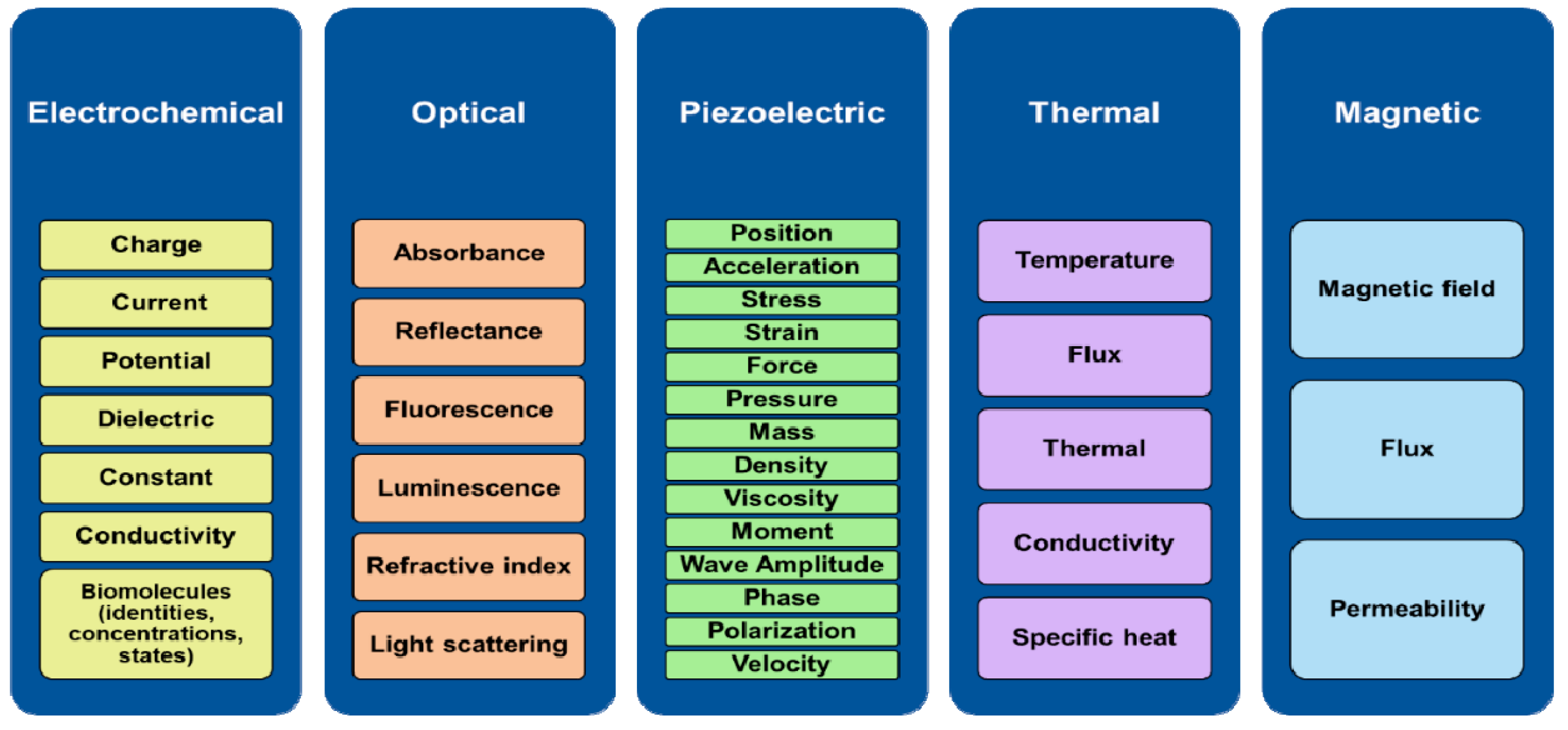
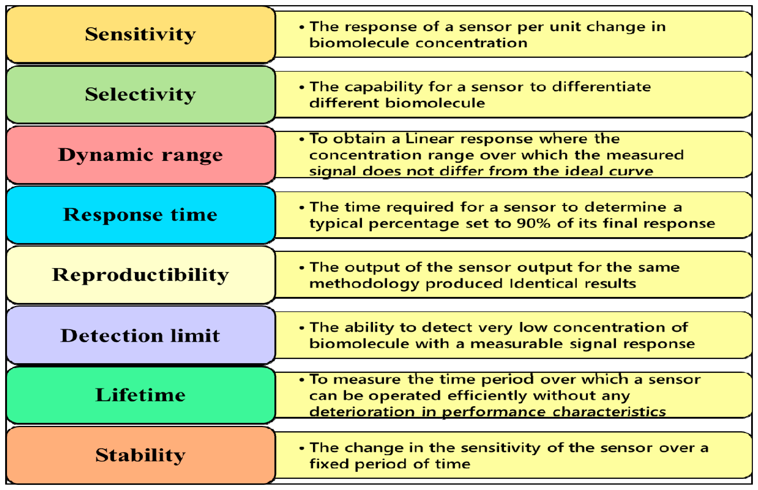
4.1. Sensor Types and Their Required Characteristics for the Detection and Monitoring of Environmental Contaminants
4.2. Miniaturization and Integration of Electrochemical Sensors in Microfluidic Systems
4.2.1. Microelectrode Materials Used in Electrochemical Device Sensors
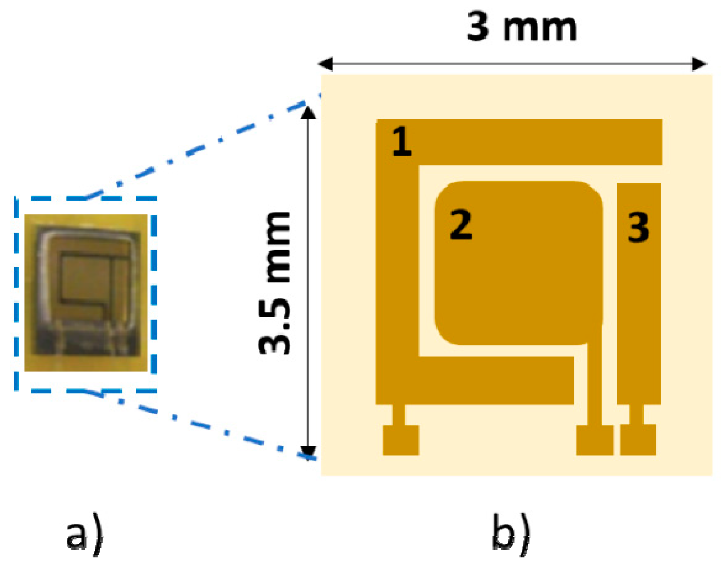
4.2.2. Microelectrodes Fabricated for Use in Microfluidic Detection Systems and Microfluidics-Integrated (Bio)Sensors
4.3. Miniaturization and Integration of Optical Sensors in Microfluidic Systems
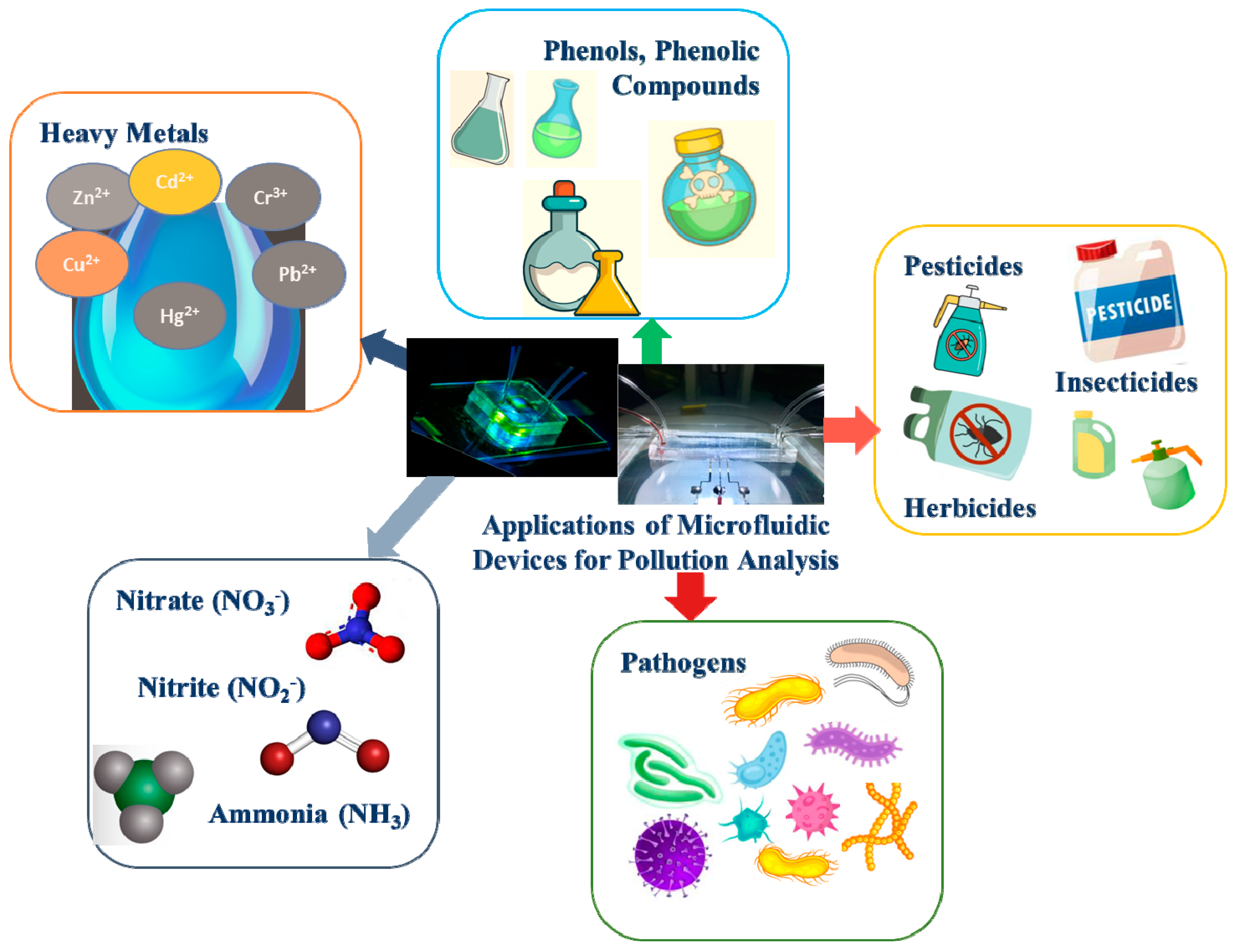
4.4. Microfluidic Detection Systems for Pollution Analysis
4.4.1. Microfluidic Detection Systems for Heavy Metals
4.4.2. Microfluidic Detection Systems for Phenols or Phenolic Compounds
4.4.3. Microfluidic Detection Systems for Nitrites, Nitrates, and Ammonia
4.4.4. Microfluidic Detection Systems for Pathogens
5. Conclusions and Future Perspectives
Author Contributions
Funding
Informed Consent Statement
Conflicts of Interest
References
- Zaharia, C. Evaluation of environmental impact produced by different economic activities with the global pollution index. Environ. Sci. Pollut. Res. 2012, 19, 2448–2455. [Google Scholar] [CrossRef] [PubMed]
- Vallero, D. Chapter 29—Air Pollutant Emissions. In Fundamentals of Air Pollution, 5th ed.; Vallero, D., Ed.; Academic Press: Boston, MA, USA, 2014; pp. 787–827. [Google Scholar] [CrossRef]
- Vidu, R.; Matei, E.; Predescu, A.M.; Alhalaili, B.; Pantilimon, C.; Tarcea, C.; Predescu, C. Removal of Heavy Metals from Wastewaters: A Challenge from Current Treatment Methods to Nanotechnology Applications. Toxics 2020, 8, 101. [Google Scholar] [CrossRef] [PubMed]
- Rusănescu, C.; Jinescu, C.; Rusanescu, M.; Begea, M.; Ghermec, O. Evaluation of air pollution by NO2, SO2, PM10 in Bucharest. Rev. Chim. 2018, 69, 105–111. [Google Scholar] [CrossRef]
- Rada, E.C.; Schiavon, M.; Torretta, V. A regulatory strategy for the emission control of hexavalent chromium from waste-to-energy plants. J. Clean. Prod. 2021, 278, 123415. [Google Scholar] [CrossRef]
- Bringezu, S.; Potočnik, J.; Schandl, H.; Lu, Y.; Ramaswami, A.; Swilling, M.; Suh, S. Multi-Scale Governance of Sustainable Natural Resource Use—Challenges and Opportunities for Monitoring and Institutional Development at the National and Global Level. Sustainability 2016, 8, 778. [Google Scholar] [CrossRef] [Green Version]
- Nakagawa, M. Trust in sustainable natural resource development. Nat. Hum. Behav. 2019, 3, 542. [Google Scholar] [CrossRef] [Green Version]
- Lee, T.C.; Anser, M.K.; Nassani, A.A.; Haffar, M.; Zaman, K.; Abro, M.M.Q. Managing Natural Resources through Sustainable Environmental Actions: A Cross-Sectional Study of 138 Countries. Sustainability 2021, 13, 12475. [Google Scholar] [CrossRef]
- Kumar, S.; Kumar, S.; Ali, M.A.; Anand, P.; Agrawal, V.V.; John, R.; Maji, S.; Malhotra, B.D. Microfluidic-integrated biosensors: Prospects for point-of-care diagnostics. Biotechnol. J. 2013, 8, 1267–1279. [Google Scholar] [CrossRef]
- Raipure, S.; Mehetre, D. Wireless sensor network based pollution monitoring system in metropolitan cities. In Proceedings of the 2015 International Conference on Communications and Signal Processing (ICCSP), Melmaruvathur, India, 2–4 April 2015; pp. 1835–1838. [Google Scholar] [CrossRef]
- Ullah, S.; Branquinho, R.; Mateus, T.; Martins, R.; Fortunato, E.; Rasheed, T.; Sher, F. Solution Combustion Synthesis of Transparent Conducting Thin Films for Sustainable Photovoltaic Applications. Sustainability 2020, 12, 10423. [Google Scholar] [CrossRef]
- Fleischer, M.; Meixner, H. Characterization and crystallite growth of semiconducting high-temperature-stable Ga2O3 thin films. J. Mater. Sci. Lett. 1992, 11, 1728–1731. [Google Scholar] [CrossRef]
- Bavasso, I.; Vilardi, G.; Stoller, M.; Chianese, A.; Di Palma, L. Perspectives in Nanotechnology Based Innovative Applications For The Environment. Chem. Eng. Trans. 2016, 47, 55–60. [Google Scholar] [CrossRef]
- Alhalaili, B.; Vidu, R.; Popescu, I.N.; Samyamanthula, D.R.; Islam, M.S. Novel Approach to Synthesize Nanostructured Gallium Oxide for Devices Operating in Harsh Environmental Conditions. Sustainability 2021, 13, 10197. [Google Scholar] [CrossRef]
- Ardeleanu, M.N.; Popescu, I.N.; Udroiu, I.N.; Diaconu, E.M.; Mihai, S.; Lungu, E.; Alhalaili, B.; Vidu, R. Novel PDMS-Based Sensor System for MPWM Measurements of Picoliter Volumes in Microfluidic Devices. Sensors 2019, 19, 4886. [Google Scholar] [CrossRef] [PubMed] [Green Version]
- Whitesides, G.M. The origins and the future of microfluidics. Nature 2006, 442, 368–373. [Google Scholar] [CrossRef] [PubMed]
- Wang, T.; Yu, C.; Xie, X. Microfluidics for Environmental Applications; Springer: Berlin/Heidelberg, Germany, 2022; pp. 1–24. [Google Scholar] [CrossRef]
- Florian, H.; Mocanu, A.; Vlasin, C.; Machado, J.; Carvalho, V.; Soares, F.; Astilean, A.; Avram, C. Deaf people feeling music rhythm by using a sensing and actuating device. Sens. Actuators A-Phys. 2017, 267, 431–442. [Google Scholar] [CrossRef]
- Bezerra, K.; Carvalho, V.; Matos, D.; Machado, J.; Soares, F.; Ferraz, A. A Faster and More Secure Human Blood Type Determining Product-Concept Design. J. Med. Devices-Trans. Asme 2016, 10, 044510. [Google Scholar] [CrossRef]
- Dincer, C.; Bruch, R.; Costa-Rama, E.; Fernandez-Abedu, M.T.; Merkoci, A.; Manz, A.; Urban, G.A.; Guder, F. Disposable Sensors in Diagnostics, Food, and Environmental Monitoring. Adv. Mater. 2019, 31, 1806739. [Google Scholar] [CrossRef]
- Rackus, D.G.; Shamsi, M.H.; Wheeler, A.R. Electrochemistry, biosensors and microfluidics: A convergence of fields. Chem. Soc. Rev. 2015, 44, 5320–5340. [Google Scholar] [CrossRef]
- Pereira, F.; Carvalho, V.; Soares, F.; Machado, J.; Bezerra, K.; Silva, R.; Matos, D. Development of a Medical Care Terminal for Efficient Monitoring of Bedridden Subjects. J. Eng. 2016, 2016, 3591059. [Google Scholar] [CrossRef] [Green Version]
- Menon, S.; Mathew, M.R.; Sam, S.; Keerthi, K.; Kumar, K.G. Recent advances and challenges in electrochemical biosensors for emerging and re-emerging infectious diseases. J. Electroanal. Chem. 2020, 878, 114596. [Google Scholar] [CrossRef]
- Mehrotra, P. Biosensors and their applications—A review. J. Oral Biol. Craniofacial Res. 2016, 6, 153–159. [Google Scholar] [CrossRef] [PubMed] [Green Version]
- Wei, P.; Ning, Z.; Ye, S.; Sun, L.; Yang, F.; Wong, K.C.; Westerdahl, D.; Louie, P.K.K. Impact Analysis of Temperature and Humidity Conditions on Electrochemical Sensor Response in Ambient Air Quality Monitoring. Sensors 2018, 18, 59. [Google Scholar] [CrossRef] [PubMed] [Green Version]
- Trivedi, D.; Crosse, J.; Tanti, J.; Cass, A.J.; Toghill, K.E. The electrochemical determination of formaldehyde in aqueous media using nickel modified electrodes. Sens. Actuators B Chem. 2018, 270, 298–303. [Google Scholar] [CrossRef] [Green Version]
- Muralikrishna, I.; Manickam, V. Environmental Management: Science and Engineering for Industry; Elsevier: Amsterdam, The Netherlands, 2017; pp. 1–639. Available online: https://www.elsevier.com/books/environmental-management/krishna/978-0-12-811989-1 (accessed on 1 August 2022).
- Santangelo, M.F.; Shtepliuk, I.; Filippini, D.; Ivanov, I.G.; Yakimova, R.; Eriksson, J. Real-time sensing of lead with epitaxial graphene-integrated microfluidic devices. Sens. Actuators B Chem. 2019, 288, 425–431. [Google Scholar] [CrossRef]
- Ukaogo, P.O.; Ewuzie, U.; Onwuka, C.V. 21—Environmental pollution: Causes, effects, and the remedies. In Microorganisms for Sustainable Environment and Health; Chowdhary, P., Raj, A., Verma, D., Akhter, Y., Eds.; Elsevier: Amsterdam, The Netherlands, 2020; pp. 419–429. [Google Scholar] [CrossRef]
- Wongkaew, N.; Simsek, M.; Griesche, C.; Baeumner, A.J. Functional nanomaterials and nanostructures enhancing electrochemical biosensors and lab-on-a-chip performances: Recent progress, applications, and future perspective. Chem. Rev. 2018, 119, 120–194. [Google Scholar] [CrossRef]
- Arduini, F.; Cinti, S.; Scognamiglio, V.; Moscone, D. Paper-Based Electrochemical Devices in Biomedical Field: Recent Advances and Perspectives. In Past, Present and Future Challenges of Biosensors and Bioanalytical Tools in Analytical Chemistry: A Tribute to Professor Marco Mascini; Palchetti, I., Hansen, P.D., Eds.; Elsevier Science Bv: Amsterdam, The Netherlands, 2017; Volume 77, pp. 385–413. [Google Scholar] [CrossRef]
- Ardeleanu, M.N.; Mihai, S.; Vidu, R.; Diaconu, E.M.; Popescu, I.N. Design of Microfluidic Device and Measurements of MPWM for Single Cell/Particle Manipulation. Sci. Bull. Valahia Univ. Mater. Mech. 2019, 17, 5. [Google Scholar] [CrossRef] [Green Version]
- Ren, K.; Zhou, J.; Wu, H. Materials for Microfluidic Chip Fabrication. Acc. Chem. Res. 2013, 46, 2396–2406. [Google Scholar] [CrossRef]
- Guijt, R.M.; Baltussen, E.; van der Steen, G.; Frank, H.; Billiet, H.; Schalkhammer, T.; Laugere, F.; Vellekoop, M.; Berthold, A.; Sarro, L.; et al. Capillary electrophoresis with on-chip four-electrode capacitively coupled conductivity detection for application in bioanalysis. Electrophoresis 2001, 22, 2537–2541. [Google Scholar] [CrossRef]
- Nge, P.N.; Rogers, C.I.; Woolley, A.T. Advances in Microfluidic Materials, Functions, Integration, and Applications. Chem. Rev. 2013, 113, 2550–2583. [Google Scholar] [CrossRef] [Green Version]
- McDonald, J.C.; Duffy, D.C.; Anderson, J.R.; Chiu, D.T.; Wu, H.K.; Schueller, O.J.A.; Whitesides, G.M. Fabrication of microfluidic systems in poly(dimethylsiloxane). Electrophoresis 2000, 21, 27–40. [Google Scholar] [CrossRef]
- Zhang, W.H.; Lin, S.C.; Wang, C.M.; Hu, J.; Li, C.; Zhuang, Z.X.; Zhou, Y.L.; Mathies, R.A.; Yang, C.Y.J. PMMA/PDMS valves and pumps for disposable microfluidics. Lab Chip 2009, 9, 3088–3094. [Google Scholar] [CrossRef] [PubMed] [Green Version]
- Roberts, M.A.; Rossier, J.S.; Bercier, P.; Girault, H. UV laser machined polymer substrates for the development of microdiagnostic systems. Anal. Chem. 1997, 69, 2035–2042. [Google Scholar] [CrossRef] [PubMed]
- Pipper, J.; Inoue, M.; Ng, L.F.P.; Neuzil, P.; Zhang, Y.; Novak, L. Catching bird flu in a droplet. Nat. Med. 2007, 13, 1259–1263. [Google Scholar] [CrossRef] [PubMed]
- Pumera, M.; Merkoci, A.; Alegret, S. New materials for electrochemical sensing VII. Microfluidic chip platforms. Trends Anal. Chem. 2006, 25, 219–235. [Google Scholar] [CrossRef]
- Woolley, A.T.; Lao, K.; Glazer, A.N.; Mathies, R.A. Capillary Electrophoresis Chips with Integrated Electrochemical Detection. Anal. Chem. 1998, 70, 684–688. [Google Scholar] [CrossRef]
- Iliescu, C.; Taylor, H.; Avram, M.; Miao, J.; Franssila, S. A practical guide for the fabrication of microfluidic devices using glass and silicon. Biomicrofluidics 2012, 6, 016505. [Google Scholar] [CrossRef] [Green Version]
- Goral, V.N.; Zaytseva, N.V.; Baeumner, A.J. Electrochemical microfluidic biosensor for the detection of nucleic acid sequences. Lab Chip 2006, 6, 414–421. [Google Scholar] [CrossRef]
- Pemg, B.-Y.; Wu, C.-W.; Shen, Y.-K.; Lin, Y. Microfluidic chip fabrication using hot embossing and thermal bonding of COP. Polym. Adv. Technol. 2010, 21, 457–466. [Google Scholar] [CrossRef]
- Lee, M.J.; Yeom, J.; Choi, J.-H.; Shin, J.H.; Kim, T.H.; Jeon, J.-W.; Na, J.-G.; Shin, K.; Oh, B.-K. Pump-Free Glass-Based Capillary Microfluidic Immuno-Assay Chip for Electrochemical Detection of Prostate-Specific Antigen. J. Nanosci. Nanotechnol. 2020, 20, 4629–4633. [Google Scholar] [CrossRef]
- Jackson, D.J.; Naber, J.F.; Roussel, T.J.; Crain, M.M.; Walsh, K.M.; Keynton, R.S.; Baldwin, R.P. Portable High-Voltage Power Supply and Electrochemical Detection Circuits for Microchip Capillary Electrophoresis. Anal. Chem. 2003, 75, 3643–3649. [Google Scholar] [CrossRef]
- Wang, T.; Chen, J.; Zhou, T.; Song, L. Fabricating Microstructures on Glass for Microfluidic Chips by Glass Molding Process. Micromachines 2018, 9, 269. [Google Scholar] [CrossRef] [PubMed] [Green Version]
- Fakunle, E.S.; Fritsch, I. Low-temperature co-fired ceramic microchannels with individually addressable screen-printed gold electrodes on four walls for self-contained electrochemical immunoassays. Anal. Bioanal. Chem. 2010, 398, 2605–2615. [Google Scholar] [CrossRef] [PubMed]
- Li, Y.; Peng, Z.; Holl, N.J.; Hassan, M.R.; Pappas, J.M.; Wei, C.; Izadi, O.H.; Wang, Y.; Dong, X.; Wang, C.; et al. MXene–Graphene Field-Effect Transistor Sensing of Influenza Virus and SARS-CoV-2. ACS Omega 2021, 6, 6643–6653. [Google Scholar] [CrossRef] [PubMed]
- Khan, R.; Andreescu, S. MXenes-Based Bioanalytical Sensors: Design, Characterization, and Applications. Sensors 2020, 20, 5434. [Google Scholar] [CrossRef]
- Kim, S.J.; Koh, H.-J.; Ren, C.E.; Kwon, O.; Maleski, K.; Cho, S.-Y.; Anasori, B.; Kim, C.-K.; Choi, Y.-K.; Kim, J.; et al. Metallic Ti3C2Tx MXene Gas Sensors with Ultrahigh Signal-to-Noise Ratio. ACS Nano 2018, 12, 986–993. [Google Scholar] [CrossRef]
- Alhabeb, M.; Maleski, K.; Anasori, B.; Lelyukh, P.; Clark, L.; Sin, S.; Gogotsi, Y. Guidelines for Synthesis and Processing of Two-Dimensional Titanium Carbide (Ti3C2Tx MXene). Chem. Mater. 2017, 29, 7633–7644. [Google Scholar] [CrossRef]
- Déctor, A.; Esquivel, J.P.; González, M.J.; Guerra-Balcázar, M.; Ledesma-García, J.; Sabaté, N.; Arriaga, L.G. Formic acid microfluidic fuel cell evaluation in different oxidant conditions. Electrochim. Acta 2013, 92, 31–35. [Google Scholar] [CrossRef]
- Duffy, D.C.; McDonald, J.C.; Schueller, O.J.A.; Whitesides, G.M. Rapid Prototyping of Microfluidic Systems in Poly(dimethylsiloxane). Anal. Chem. 1998, 70, 4974–4984. [Google Scholar] [CrossRef]
- Kim, J.-y.; deMello, A.J.; Chang, S.-I.; Hong, J.; O'Hare, D. Thermoset polyester droplet-based microfluidic devices for high frequency generation. Lab Chip 2011, 11, 4108–4112. [Google Scholar] [CrossRef]
- Roy, E.; Galas, J.-C.; Veres, T. Thermoplastic elastomers for microfluidics: Towards a high-throughput fabrication method of multilayered microfluidic devices. Lab Chip 2011, 11, 3193–3196. [Google Scholar] [CrossRef]
- Pan, T.; Fiorini, G.S.; Chiu, D.T.; Woolley, A.T. In-channel atom-transfer radical polymerization of thermoset polyester microfluidic devices for bioanalytical applications. Electrophoresis 2007, 28, 2904–2911. [Google Scholar] [CrossRef] [PubMed] [Green Version]
- Becker, H.; Gärtner, C. Polymer microfabrication technologies for microfluidic systems. Anal. Bioanal. Chem. 2008, 390, 89–111. [Google Scholar] [CrossRef] [PubMed]
- Fiorini, G.S.; Yim, M.; Jeffries, G.D.M.; Schiro, P.G.; Mutch, S.A.; Lorenz, R.M.; Chiu, D.T. Fabrication improvements for thermoset polyester (TPE) microfluidic devices. Lab Chip 2007, 7, 923–926. [Google Scholar] [CrossRef]
- Shadpour, H.; Musyimi, H.; Chen, J.; Soper, S.A. Physiochemical properties of various polymer substrates and their effects on microchip electrophoresis performance. J. Chromatogr. A 2006, 1111, 238–251. [Google Scholar] [CrossRef]
- Young, E.W.K.; Berthier, E.; Guckenberger, D.J.; Sackmann, E.; Lamers, C.; Meyvantsson, I.; Huttenlocher, A.; Beebe, D.J. Rapid Prototyping of Arrayed Microfluidic Systems in Polystyrene for Cell-Based Assays. Anal. Chem. 2011, 83, 1408–1417. [Google Scholar] [CrossRef] [PubMed]
- Attia, U.M.; Marson, S.; Alcock, J.R. Micro-injection moulding of polymer microfluidic devices. Microfluid. Nanofluidics 2009, 7, 1–28. [Google Scholar] [CrossRef] [Green Version]
- Wang, J.; Pumera, M.; Chatrathi, M.P.; Escarpa, A.; Konrad, R.; Griebel, A.; Dörner, W.; Löwe, H. Towards disposable lab-on-a-chip: Poly(methylmethacrylate) microchip electrophoresis device with electrochemical detection. Electrophoresis 2002, 23, 596–601. [Google Scholar] [CrossRef]
- Muck, A.; Wang, J.; Jacobs, M.; Chen, G.; Chatrathi, M.P.; Jurka, V.; Výborný, Z.; Spillman, S.D.; Sridharan, G.; Schöning, M.J. Fabrication of Poly(methyl methacrylate) Microfluidic Chips by Atmospheric Molding. Anal. Chem. 2004, 76, 2290–2297. [Google Scholar] [CrossRef] [Green Version]
- Soper, S.A.; Ford, S.M.; Qi, S.; McCarley, R.L.; Kelly, K.; Murphy, M.C. Peer Reviewed: Polymeric Microelectromechanical Systems. Anal. Chem. 2000, 72, 642A–651A. [Google Scholar] [CrossRef]
- Hong, T.-F.; Ju, W.-J.; Wu, M.-C.; Tai, C.-H.; Tsai, C.-H.; Fu, L.-M. Rapid prototyping of PMMA microfluidic chips utilizing a CO2 laser. Microfluid. Nanofluidics 2010, 9, 1125–1133. [Google Scholar] [CrossRef]
- Viswanathan, S.; Narayanan, T.N.; Aran, K.; Fink, K.D.; Paredes, J.; Ajayan, P.M.; Filipek, S.; Miszta, P.; Tekin, H.C.; Inci, F.; et al. Graphene–protein field effect biosensors: Glucose sensing. Mater. Today 2015, 18, 513–522. [Google Scholar] [CrossRef]
- Liu, Y.; Rauch, C.B.; Stevens, R.L.; Lenigk, R.; Yang, J.; Rhine, D.B.; Grodzinski, P. DNA Amplification and Hybridization Assays in Integrated Plastic Monolithic Devices. Anal. Chem. 2002, 74, 3063–3070. [Google Scholar] [CrossRef] [PubMed]
- Bai, X.; Wu, Z.; Josserand, J.; Jensen, H.; Schafer, H.; Girault, H.H. Passive Conductivity Detection for Capillary Electrophoresis. Anal. Chem. 2004, 76, 3126–3131. [Google Scholar] [CrossRef] [PubMed]
- Niculescu, A.-G.; Chircov, C.; Bîrcă, A.C.; Grumezescu, A.M. Fabrication and Applications of Microfluidic Devices: A Review. Int. J. Mol. Sci. 2021, 22, 2011. [Google Scholar] [CrossRef] [PubMed]
- Gómez-de Pedro, S.; Lopes, D.; Miltsov, S.; Izquierdo, D.; Alonso-Chamarro, J.; Puyol, M. Optical microfluidic system based on ionophore modified gold nanoparticles for the continuous monitoring of mercuric ion. Sens. Actuators B Chem. 2014, 194, 19–26. [Google Scholar] [CrossRef]
- Zou, Z.; Jang, A.; MacKnight, E.; Wu, P.-M.; Do, J.; Bishop, P.L.; Ahn, C.H. Environmentally friendly disposable sensors with microfabricated on-chip planar bismuth electrode for in situ heavy metal ions measurement. Sens. Actuators B Chem. 2008, 134, 18–24. [Google Scholar] [CrossRef]
- Jung, W.; Jang, A.; Bishop, P.L.; Ahn, C.H. A polymer lab chip sensor with microfabricated planar silver electrode for continuous and on-site heavy metal measurement. Sens. Actuators B Chem. 2011, 155, 145–153. [Google Scholar] [CrossRef]
- Nie, J.; Fu, J.; He, Y. Hydrogels: The Next Generation Body Materials for Microfluidic Chips? Small 2020, 16, 2003797. [Google Scholar] [CrossRef]
- Lee, S.-H.; Moon, J.J.; West, J.L. Three-dimensional micropatterning of bioactive hydrogels via two-photon laser scanning photolithography for guided 3D cell migration. Biomaterials 2008, 29, 2962–2968. [Google Scholar] [CrossRef] [Green Version]
- Asthana, A.; Lee, K.H.; Kim, K.-O.; Kim, D.-M.; Kim, D.-P. Rapid and cost-effective fabrication of selectively permeable calcium-alginate microfluidic device using “modified” embedded template method. Biomicrofluidics 2012, 6, 012821. [Google Scholar] [CrossRef] [Green Version]
- Gao, G.; Park, J.Y.; Kim, B.S.; Jang, J.; Cho, D.-W. Coaxial Cell Printing of Freestanding, Perfusable, and Functional In Vitro Vascular Models for Recapitulation of Native Vascular Endothelium Pathophysiology. Adv. Healthc. Mater. 2018, 7, 1801102. [Google Scholar] [CrossRef]
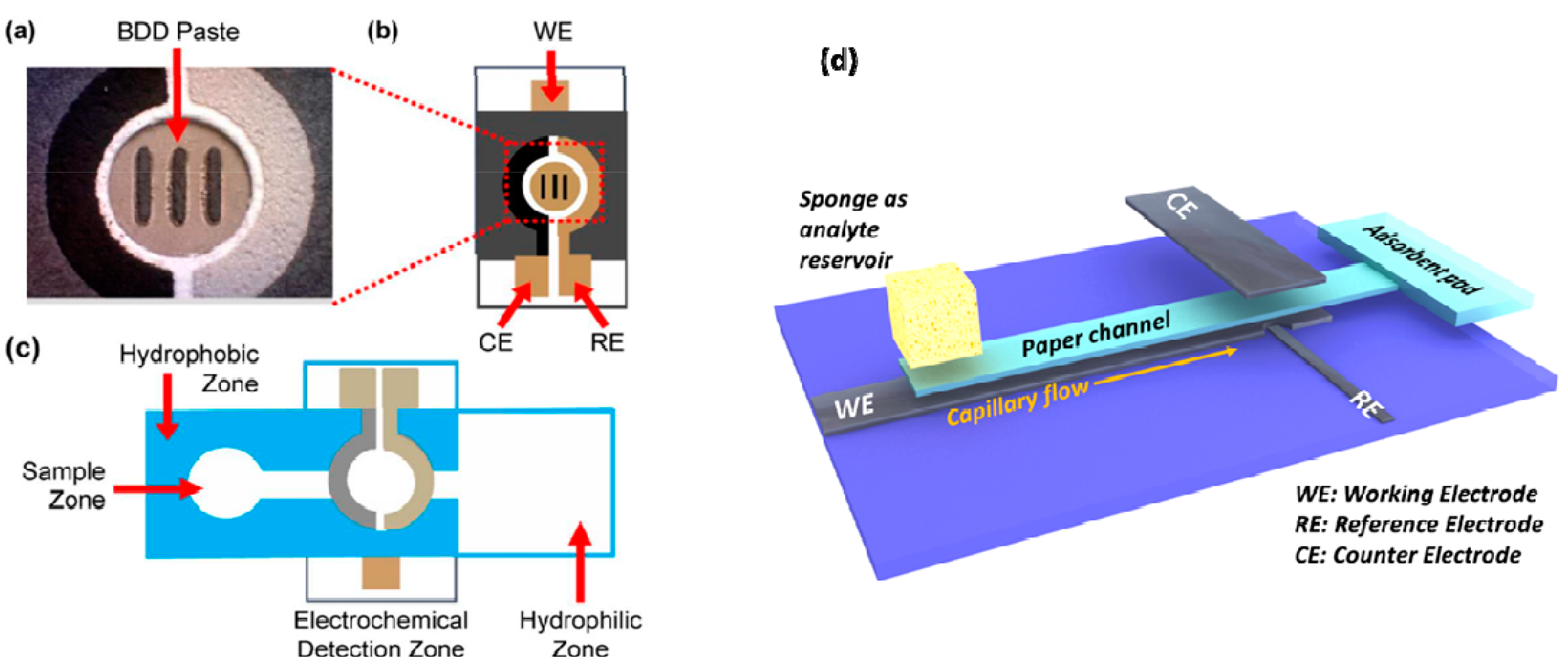
- Dungchai, W.; Chailapakul, O.; Henry, C.S. Electrochemical Detection for Paper-Based Microfluidics. Anal. Chem. 2009, 81, 5821–5826. [Google Scholar] [CrossRef] [PubMed]
- Dungchai, W.; Chailapakul, O.; Henry, C.S. A low-cost, simple, and rapid fabrication method for paper-based microfluidics using wax screen-printing. Analyst 2011, 136, 77–82. [Google Scholar] [CrossRef] [PubMed]
- Xu, Y.; Liu, M.; Kong, N.; Liu, J. Lab-on-paper micro- and nano-analytical devices: Fabrication, modification, detection and emerging applications. Microchim. Acta 2016, 183, 1521–1542. [Google Scholar] [CrossRef]
- Mao, K.; Min, X.; Zhang, H.; Zhang, K.; Cao, H.; Guo, Y.; Yang, Z. Paper-based microfluidics for rapid diagnostics and drug delivery. J. Control. Release Off. J. Control. Release Soc. 2020, 322, 187–199. [Google Scholar] [CrossRef] [PubMed]
- Magro, L.; Jacquelin, B.; Escadafal, C.; Garneret, P.; Kwasiborski, A.; Manuguerra, J.-C.; Monti, F.; Sakuntabhai, A.; Vanhomwegen, J.; Lafaye, P.; et al. Paper-based RNA detection and multiplexed analysis for Ebola virus diagnostics. Sci. Rep. 2017, 7, 1347. [Google Scholar] [CrossRef] [PubMed] [Green Version]
- Li, Z.; Li, F.; Hu, J.; Wee, W.H.; Han, Y.L.; Pingguan-Murphy, B.; Lu, T.J.; Xu, F. Direct writing electrodes using a ball pen for paper-based point-of-care testing. Analyst 2015, 140, 5526–5535. [Google Scholar] [CrossRef]
- Tang, T.; Yuan, Y.; Yalikun, Y.; Hosokawa, Y.; Li, M.; Tanaka, Y. Glass based micro total analysis systems: Materials, fabrication methods, and applications. Sens. Actuators B Chem. 2021, 339, 129859. [Google Scholar] [CrossRef]
- Pinti, M.; Kambham, T.; Wang, B.; Prakash, S. Fabrication of Centimeter Long, Ultra-Low Aspect Ratio Nanochannel Networks in Borosilicate Glass Substrates. J. Nanotechnol. Eng. Med. 2013, 4, 020905. [Google Scholar] [CrossRef]
- Suzuki, H. Advances in the Microfabrication of Electrochemical Sensors and Systems. Electroanalysis 2000, 12, 703–715. [Google Scholar] [CrossRef]
- Deng, J.; Wei, W.; Chen, Z.; Lin, B.; Zhao, W.; Luo, Y.; Zhang, X. Engineered Liver-On-A-Chip Platform to Mimic Liver Functions and Its Biomedical Applications: A Review. Micromachines 2019, 10, 676. [Google Scholar] [CrossRef] [PubMed] [Green Version]
- Sassa, F.; Morimoto, K.; Satoh, W.; Suzuki, H. Electrochemical techniques for microfluidic applications. Electrophoresis 2008, 29, 1787–1800. [Google Scholar] [CrossRef] [PubMed]
- Janata, J. Potentiometric Microsensors. Chem. Rev. 1990, 90, 691–703. [Google Scholar] [CrossRef]
- Nie, Z.; Nijhuis, C.A.; Gong, J.; Chen, X.; Kumachev, A.; Martinez, A.W.; Narovlyansky, M.; Whitesides, G.M. Electrochemical sensing in paper-based microfluidic devices. Lab Chip 2010, 10, 477–483. [Google Scholar] [CrossRef] [PubMed] [Green Version]
- Mappes, T.; Achenbach, S.; Mohr, J. Process conditions in X-ray lithography for the fabrication of devices with sub-micron feature sizes. Microsyst. Technol. 2007, 13, 355–360. [Google Scholar] [CrossRef]
- Cao, H.; Tegenfeldt, J.O.; Austin, R.H.; Chou, S.Y. Gradient nanostructures for interfacing microfluidics and nanofluidics. Appl. Phys. Lett. 2002, 81, 3058–3060. [Google Scholar] [CrossRef]
- Zhou, W.; Min, G.; Zhang, J.; Liu, Y.; Wang, J.; Zhang, Y.; Sun, F. Nanoimprint Lithography: A Processing Technique for Nanofabrication Advancement. Nano-Micro Lett. 2011, 3, 135–140. [Google Scholar] [CrossRef] [Green Version]
- Qi, S.; Liu, X.; Ford, S.; Barrows, J.; Thomas, G.; Kelly, K.; McCandless, A.; Lian, K.; Goettert, J.; Soper, S.A. Microfluidic devices fabricated in poly(methyl methacrylate) using hot-embossing with integrated sampling capillary and fiber optics for fluorescence detection. Lab Chip 2002, 2, 88–95. [Google Scholar] [CrossRef]
- Feltis, B.N.; Sexton, B.A.; Glenn, F.L.; Best, M.J.; Wilkins, M.; Davis, T.J. A hand-held surface plasmon resonance biosensor for the detection of ricin and other biological agents. Biosens. Bioelectron. 2008, 23, 1131–1136. [Google Scholar] [CrossRef]
- Koller, D.M.; Hohenau, A.; Ditlbacher, H.; Galler, N.; Baudrion, A.-L.; Reil, F.; Schausberger, S.; Aussenegg, F.R.; Leitner, A.; Krenn, J.R. Three-dimensional SU-8 sub-micrometer structuring by electron beam lithography. Microelectron. Eng. 2008, 85, 1639–1641. [Google Scholar] [CrossRef]
- Szekely, L.; Freitag, R. Module for real time non-invasive control of the electroosmotic flow in microfluidic systems. Anal. Chim. Acta 2005, 539, 165–171. [Google Scholar] [CrossRef]
- Alahmad, W.; Uraisin, K.; Nacapricha, D.; Kaneta, T. A miniaturized chemiluminescence detection system for a microfluidic paper-based analytical device and its application to the determination of chromium(iii). Anal. Methods 2016, 8, 5414–5420. [Google Scholar] [CrossRef]
- Lu, Y.; Shi, W.; Qin, J.; Lin, B. Fabrication and Characterization of Paper-Based Microfluidics Prepared in Nitrocellulose Membrane By Wax Printing. Anal. Chem. 2010, 82, 329–335. [Google Scholar] [CrossRef] [PubMed]
- Nantaphol, S.; Channon, R.B.; Kondo, T.; Siangproh, W.; Chailapakul, O.; Henry, C.S. Boron Doped Diamond Paste Electrodes for Microfluidic Paper-Based Analytical Devices. Anal. Chem. 2017, 89, 4100–4107. [Google Scholar] [CrossRef] [PubMed]
- Abe, K.; Kotera, K.; Suzuki, K.; Citterio, D. Inkjet-printed paperfluidic immuno-chemical sensing device. Anal. Bioanal. Chem. 2010, 398, 885–893. [Google Scholar] [CrossRef]
- Alkasir, R.S.J.; Ornatska, M.; Andreescu, S. Colorimetric Paper Bioassay for the Detection of Phenolic Compounds. Anal. Chem. 2012, 84, 9729–9737. [Google Scholar] [CrossRef]
- Jing, G.; Polaczyk, A.; Oerther, D.B.; Papautsky, I. Development of a microfluidic biosensor for detection of environmental mycobacteria. Sens. Actuators B Chem. 2007, 123, 614–621. [Google Scholar] [CrossRef]
- Patru, M.; Isac, L.; Cunha, L.; Martins, P.; Lanceros-Mendez, S.; Oncioiu, G.; Cristea, D.; Munteanu, D. Structural, mechanical and piezoelectric properties of polycrystalline AlN films sputtered on titanium bottom electrodes. Appl. Surf. Sci. 2015, 354, 267–278. [Google Scholar] [CrossRef]
- Xie, B.; Ramanathan, K.; Danielsson, B. Mini/micro thermal biosensors and other related devices for biochemical/clinical analysis and monitoring. Trends Anal. Chem. 2000, 19, 340–349. [Google Scholar] [CrossRef]
- Llandro, J.; Palfreyman, J.J.; Ionescu, A.; Barnes, C.H.W. Magnetic biosensor technologies for medical applications: A review. Med. Biol. Eng. Comput. 2010, 48, 977–998. [Google Scholar] [CrossRef]
- Rajendran, S.T.; Scarano, E.; Bergkamp, M.H.; Capria, A.M.; Cheng, C.H.; Sanger, K.; Ferrari, G.; Nielsen, L.H.; Hwu, E.T.; Zor, K.; et al. Modular, Lightweight, Wireless Potentiostat-on-a-Disc for Electrochemical Detection in Centrifugal Microfluidics. Anal. Chem. 2019, 91, 11620–11628. [Google Scholar] [CrossRef] [PubMed]
- Krejcova, L.; Michalek, P.; Merlos Rodrigo, M.; Heger, Z.; Krizkova, S.; Vaculovicova, M.; Hynek, D.; Adam, V.; Kizek, R. Nanoscale virus biosensors: State of the art. Nanobiosens. Dis. Diagn. 2015, 4, 47–66. [Google Scholar] [CrossRef] [Green Version]
- Srivastava, A.K.; Dev, A.; Karmakar, S. Nanosensors and nanobiosensors in food and agriculture. Environ. Chem. Lett. 2018, 16, 161–182. [Google Scholar] [CrossRef]
- Bani-Yaseen, A.D. Fabrication of Electrochemically Deposited Microelectrodes for Microfluidic MEMS Applications. Int. J. Electrochem. Sci 2010, 5, 1837–1846. Available online: http://www.electrochemsci.org/papers/vol5/5121837.pdf (accessed on 1 August 2022).
- Hanrahan, G.; Patil, D.G.; Wang, J. Electrochemical sensors for environmental monitoring: Design, development and applications. J. Environ. Monit. 2004, 6, 657–664. [Google Scholar] [CrossRef]
- Abdel-Karim, R.; Reda, Y.; Abdel-Fattah, A. Review—Nanostructured Materials-Based Nanosensors. J. Electrochem. Soc. 2020, 167, 037554. [Google Scholar] [CrossRef]
- Vargas-Bernal, R.; Rodríguez-Miranda, E.; Herrera-Pérez, G. Evolution and Expectations of Enzymatic Biosensors for Pesticides. In Pesticides—Advances in Chemical and Botanical Pesticides; IntechOpen: London, UK, 2012; pp. 329–356. [Google Scholar] [CrossRef] [Green Version]
- Malik, P.; Katyal, V.; Malik, V.; Asatkar, A.; Inwati, G.; Mukherjee, T.K. Nanobiosensors: Concepts and Variations. ISRN Nanomater. 2013, 2013, 327435. [Google Scholar] [CrossRef]
- Satoh, W.; Hosono, H.; Yokomaku, H.; Morimoto, K.; Upadhyay, S.; Suzuki, H. Integrated electrochemical analysis system with microfluidic and sensing functions. Sensors 2008, 8, 1111–1127. [Google Scholar] [CrossRef] [Green Version]
- Kasturi, S.; Torati, S.R.; Eom, Y.; Kim, C. Microvalve-controlled miniaturized electrochemical lab-on-a-chip based biosensor for the detection of β-amyloid biomarker. J. Ind. Eng. Chem. 2021, 97, 349–355. [Google Scholar] [CrossRef]
- Bhalla, N.; Jolly, P.; Formisano, N.; Estrela, P. Introduction to biosensors. Essays Biochem. 2016, 60, 1–8. [Google Scholar] [CrossRef] [Green Version]
- Dejous, C.; Hallil, H.; Raimbault, V.; Rukkumani, R.; Yakhmi, J.V. Using microsensors to promote the development of innovative therapeutic nanostructures. In Nanostructures for Novel Therapy; Elsevier: Amsterdam, The Netherlands, 2017. [Google Scholar] [CrossRef]
- Polk, B.J.; Stelzenmuller, A.; Mijares, G.; MacCrehan, W.; Gaitan, M. Ag/AgCl microelectrodes with improved stability for microfluidics. Sens. Actuators B-Chem. 2006, 114, 239–247. [Google Scholar] [CrossRef]
- Kovarik, M.L.; Li, M.W.; Martin, R.S. Integration of a carbon microelectrode with a microfabricated palladium decoupler for use in microchip capillary electrophoresis/electrochemistry. Electrophoresis 2005, 26, 202–210. [Google Scholar] [CrossRef] [PubMed]
- Kong, Y.; Chen, H.W.; Wang, Y.R.; Soper, S.A. Fabrication of a gold microelectrode for amperometric detection on a polycarbonate ellectrophoresis chip by photodirected electroless plating. Electrophoresis 2006, 27, 2940–2950. [Google Scholar] [CrossRef] [PubMed]
- Wang, J.; Chatrathi, M.P.; Tian, B.M.; Polsky, R. Capillary electrophoresis chips with thick-film amperometric detectors: Separation and detection of hydrazine compounds. Electroanalysis 2000, 12, 691–694. [Google Scholar] [CrossRef]
- Vazquez, M.; Frankenfeld, C.; Coltro, W.K.T.; Carrilho, E.; Diamond, D.; Lunte, S.M. Dual contactless conductivity and amperometric detection on hybrid PDMS/glass electrophoresis microchips. Analyst 2010, 135, 96–103. [Google Scholar] [CrossRef] [PubMed]
- Wang, J.; Chen, G.; Chatrathi, M.P. Nickel amperometric detector prepared by electroless deposition for microchip electrophoretic measurement of alcohols and sugars. Electroanalysis 2004, 16, 1603–1608. [Google Scholar] [CrossRef]
- Wang, J.; Pumera, M.; Chatrathi, M.P.; Escarpa, A.; Musameh, M.; Collins, G.; Mulchandani, A.; Lin, Y.H.; Olsen, K. Single-channel microchip for fast screening and detailed identification of nitroaromatic explosives or organophosphate nerve agents. Anal. Chem. 2002, 74, 1187–1191. [Google Scholar] [CrossRef] [PubMed]
- Wallis, G.; Pomerant, D.I. Field Assisted Glass-Metal Sealing. J. Appl. Phys. 1969, 40, 3946. [Google Scholar] [CrossRef]
- Bicelli, L.P.; Bozzini, B.; Mele, C.; D'Urzo, L. A review of nanostructural aspects of metal electrodeposition. Int. J. Electrochem. Sci. 2008, 3, 356–408. Available online: http://www.electrochemsci.org/papers/vol3/3040356.pdf (accessed on 1 August 2022).
- Wang, Y.; He, Q.H.; Dong, Y.Y.; Chen, H.W. In-channel modification of biosensor electrodes integrated on a polycarbonate microfluidic chip for micro flow-injection amperometric determination of glucose. Sens. Actuators B-Chem. 2010, 145, 553–560. [Google Scholar] [CrossRef]
- Wang, J.; Lu, J.M.; Luo, D.B.; Wang, J.Y.; Jiang, M.; Tian, B.M.; Olsen, K. Renewable-reagent electrochemical sensor for monitoring trace metal contaminants. Anal. Chem. 1997, 69, 2640–2645. [Google Scholar] [CrossRef]
- Umapathi, R.; Park, B.; Sonwal, S.; Rani, G.M.; Cho, Y.; Huh, Y.S. Advances in optical-sensing strategies for the on-site detection of pesticides in agricultural foods. Trends Food Sci. Technol. 2022, 119, 69–89. [Google Scholar] [CrossRef]
- Yu, J.Q.; Huang, W.; Chin, L.K.; Lei, L.; Lin, Z.P.; Ser, W.; Chen, H.; Ayi, T.C.; Yap, P.H.; Chen, C.H.; et al. Droplet optofluidic imaging for λ-bacteriophage detection via co-culture with host cell Escherichia coli. Lab Chip 2014, 14, 3519–3524. [Google Scholar] [CrossRef] [PubMed] [Green Version]
- Long, F.; Zhu, A.; Wang, H. Optofluidics-based DNA structure-competitive aptasensor for rapid on-site detection of lead(II) in an aquatic environment. Anal. Chim. Acta 2014, 849, 43–49. [Google Scholar] [CrossRef] [PubMed]
- Chauhan, S.; Upadhyay, L.S.B. An efficient protocol to use iron oxide nanoparticles in microfluidic paper device for arsenic detection. MethodsX 2018, 5, 1528–1533. [Google Scholar] [CrossRef]
- Milani, A.; Statham, P.J.; Mowlem, M.C.; Connelly, D.P. Development and application of a microfluidic in-situ analyzer for dissolved Fe and Mn in natural waters. Talanta 2015, 136, 15–22. [Google Scholar] [CrossRef] [PubMed]
- Takabayashi, Y.; Uemoto, M.; Aoki, K.; Odake, T.; Korenaga, T. Development and optimization of a lab-on-a-chip device for the measurement of trace nitrogen dioxide gas in the atmosphere. Analyst 2006, 131, 573–578. [Google Scholar] [CrossRef]
- Gauri, S.; Abidin, Z.Z.; Kamuri, M.F.; Mahdi, M.A.; Md Yunus, N.A. Detection of Aeromonas hydrophila Using Fiber Optic Microchannel Sensor. J. Sens. 2017, 2017, 8365189. [Google Scholar] [CrossRef] [Green Version]
- Lv, J.; Zhang, Z. A Microchip with Air Sampling and Chemiluminescence Detection for Analyzing Iron in Nature Water and in Whole Blood. Anal. Lett. 2004, 37, 1401–1410. [Google Scholar] [CrossRef]
- Denisov, I.; Lukyanenko, K.; Yakimov, A.; Kukhtevich, I.; Esimbekova, E.; Belobrov, P. Disposable luciferase-based microfluidic chip for rapid assay of water pollution. Luminescence 2018, 33, 1054–1061. [Google Scholar] [CrossRef]
- Anand Mohan, S.; Banshi, D.G. Ion-imprinted nanoparticles for the concurrent estimation of Pb(II) and Cu(II) ions over a two channel surface plasmon resonance-based fiber optic platform. J. Biomed. Opt. 2018, 23, 017001. [Google Scholar] [CrossRef]
- Fujii, S.-I.; Tokuyama, T.; Abo, M.; Okubo, A. Fluorometric Determination of Sulfite and Nitrite in Aqueous Samples Using a Novel Detection Unit of a Microfluidic Device. Anal. Sci. 2004, 20, 209–212. [Google Scholar] [CrossRef] [PubMed] [Green Version]
- Lafleur, J.P.; Senkbeil, S.; Jensen, T.G.; Kutter, J.P. Gold nanoparticle-based optical microfluidic sensors for analysis of environmental pollutants. Lab Chip 2012, 12, 4651–4656. [Google Scholar] [CrossRef] [PubMed]
- Zhao, L.; Wu, T.; Lefèvre, J.-P.; Leray, I.; Delaire, J.A. Fluorimetric lead detection in a microfluidic device. Lab Chip 2009, 9, 2818–2823. [Google Scholar] [CrossRef] [PubMed]
- Shin, D.; Tryk, D.A.; Fujishima, A.; Muck, A., Jr.; Chen, G.; Wang, J. Microchip capillary electrophoresis with a boron-doped diamond electrochemical detector for analysis of aromatic amines. Electrophoresis 2004, 25, 3017–3023. [Google Scholar] [CrossRef]
- Hong, Y.; Wu, M.; Chen, G.; Dai, Z.; Zhang, Y.; Chen, G.; Dong, X. 3D Printed Microfluidic Device with Microporous Mn2O3-Modified Screen Printed Electrode for Real-Time Determination of Heavy Metal Ions. ACS Appl. Mater. Interfaces 2016, 8, 32940–32947. [Google Scholar] [CrossRef]
- Zhou, J.; Ren, K.; Zheng, Y.; Su, J.; Zhao, Y.; Ryan, D.; Wu, H. Fabrication of a microfluidic Ag/AgCl reference electrode and its application for portable and disposable electrochemical microchips. Electrophoresis 2010, 31, 3083–3089. [Google Scholar] [CrossRef]
- Freitas, C.B.; Moreira, R.C.; de Oliveira Tavares, M.G.; Coltro, W.K.T. Monitoring of nitrite, nitrate, chloride and sulfate in environmental samples using electrophoresis microchips coupled with contactless conductivity detection. Talanta 2016, 147, 335–341. [Google Scholar] [CrossRef]
- Kudr, J.; Zitka, O.; Klimanek, M.; Vrba, R.; Adam, V. Microfluidic electrochemical devices for pollution analysis—A review. Sens. Actuators B Chem. 2017, 246, 578–590. [Google Scholar] [CrossRef]
- Mayorga-Martinez, C.C.; Hlavata, L.; Miserere, S.; López-Marzo, A.; Labuda, J.; Pons, J.; Merkoçi, A. An integrated phenol ‘sensoremoval’ microfluidic nanostructured platform. Biosens. Bioelectron. 2014, 55, 355–359. [Google Scholar] [CrossRef]
- Liu, B.; Zhang, Y.; Mayer, D.; Krause, H.-J.; Jin, Q.; Zhao, J.; Offenhäusser, A. A simplified poly(dimethylsiloxane) capillary electrophoresis microchip integrated with a low-noise contactless conductivity detector. Electrophoresis 2011, 32, 699–704. [Google Scholar] [CrossRef] [PubMed]
- Kubáň, P.; Hauser, P.C. Contactless conductivity detection for analytical techniques—Developments from 2012 to 2014. Electrophoresis 2015, 36, 195–211. [Google Scholar] [CrossRef] [PubMed] [Green Version]
- Lace, A.; Ryan, D.; Bowkett, M.; Cleary, J. Arsenic Monitoring in Water by Colorimetry Using an Optimized Leucomalachite Green Method. Molecules 2019, 24, 339. [Google Scholar] [CrossRef] [Green Version]
- Li, M.; Cao, R.; Nilghaz, A.; Guan, L.; Zhang, X.; Shen, W. “Periodic-Table-Style” Paper Device for Monitoring Heavy Metals in Water. Anal. Chem. 2015, 87, 2555–2559. [Google Scholar] [CrossRef] [PubMed]
- Chen, W.; Fang, X.; Li, H.; Cao, H.; Kong, J. A Simple Paper-Based Colorimetric Device for Rapid Mercury(II) Assay. Sci. Rep. 2016, 6, 31948. [Google Scholar] [CrossRef]
- Cai, L.; Fang, Y.; Mo, Y.; Huang, Y.; Xu, C.; Zhang, Z.; Wang, M. Visual quantification of Hg on a microfluidic paper-based analytical device using distance-based detection technique. AIP Adv. 2017, 7, 085214. [Google Scholar] [CrossRef] [Green Version]
- Bell, J.; Climent, E.; Hecht, M.; Buurman, M.; Rurack, K. Combining a Droplet-Based Microfluidic Tubing System with Gated Indicator Releasing Nanoparticles for Mercury Trace Detection. ACS Sens. 2016, 1, 334–338. [Google Scholar] [CrossRef]
- Du, W.-B.; Fang, Q.; He, Q.-H.; Fang, Z.-L. High-Throughput Nanoliter Sample Introduction Microfluidic Chip-Based Flow Injection Analysis System with Gravity-Driven Flows. Anal. Chem. 2005, 77, 1330–1337. [Google Scholar] [CrossRef]
- Shen, L.-L.; Zhang, G.-R.; Li, W.; Biesalski, M.; Etzold, B.J.M. Modifier-Free Microfluidic Electrochemical Sensor for Heavy-Metal Detection. ACS Omega 2017, 2, 4593–4603. [Google Scholar] [CrossRef] [Green Version]
- Shi, J.; Tang, F.; Xing, H.; Zheng, H.; Lianhua, B.; Wei, W. Electrochemical Detection of Pb and Cd in Paper-Based Microfluidic Devices. J. Braz. Chem. Soc. 2012, 23, 1124–1130. [Google Scholar] [CrossRef]
- Pungjunun, K.; Chaiyo, S.; Jantrahong, I.; Nantaphol, S.; Siangproh, W.; Chailapakul, O. Anodic stripping voltammetric determination of total arsenic using a gold nanoparticle-modified boron-doped diamond electrode on a paper-based device. Microchim. Acta 2018, 185, 324. [Google Scholar] [CrossRef] [PubMed]
- Le, T.S.; Da Costa, P.; Huguet, P.; Sistat, P.; Pichot, F.; Silva, F.; Renaud, L.; Cretin, M. Upstream microelectrodialysis for heavy metals detection on boron doped diamond. J. Electroanal. Chem. 2012, 670, 50–55. [Google Scholar] [CrossRef]
- Gutiérrez-Capitán, M.; Ipatov, A.; Merlos, Á.; Jiménez-Jorquera, C.; Fernández-Sánchez, C. Compact Electrochemical Flow System for the Analysis of Environmental Pollutants. Electroanalysis 2014, 26, 497–506. [Google Scholar] [CrossRef]
- Medina-Sánchez, M.; Cadevall, M.; Ros, J.; Merkoçi, A. Eco-friendly electrochemical lab-on-paper for heavy metal detection. Anal. Bioanal. Chem. 2015, 407, 8445–8449. [Google Scholar] [CrossRef] [PubMed]
- Nguyen, H.L.; Cao, H.H.; Nguyen, D.T.; Nguyen, V.-A. Sodium Dodecyl Sulfate Doped Polyaniline for Enhancing the Electrochemical Sensitivity of Mercury Ions. Electroanalysis 2017, 29, 595–601. [Google Scholar] [CrossRef]
- Zhang, M.; Ge, L.; Ge, S.; Yan, M.; Yu, J.; Huang, J.; Liu, S. Three-dimensional paper-based electrochemiluminescence device for simultaneous detection of Pb2+ and Hg2+ based on potential-control technique. Biosens. Bioelectron. 2013, 41, 544–550. [Google Scholar] [CrossRef] [PubMed]
- Huang, J.; Zhang, X.; Zhou, L.; You, T. Simultaneous electrochemical determination of dihydroxybenzene isomers using electrospun nitrogen-doped carbon nanofiber film electrode. Sens. Actuators B Chem. 2016, 224, 568–576. [Google Scholar] [CrossRef]
- Gan, T.; Wang, Z.; Wang, Y.; Li, X.; Sun, J.; Liu, Y. Flexible graphene oxide−wrapped SnO2 hollow spheres with high electrochemical sensing performance in simultaneous determination of 4−aminophenol and 4−chlorophenol. Electrochim. Acta 2017, 250, 1–9. [Google Scholar] [CrossRef]
- Ho, W.F.; Nguyen, L.T.; Yang, K.-L. A microfluidic sensor for detecting chlorophenols using cross-linked enzyme aggregates (CLEAs). Lab Chip 2019, 19, 634–640. [Google Scholar] [CrossRef]
- Wang, J.; Chatrathi, M.P.; Tian, B. Capillary electrophoresis microchips with thick-film amperometric detectors: Separation and detection of phenolic compounds. Anal. Chim. Acta 2000, 416, 9–14. [Google Scholar] [CrossRef]
- Sanz, J.; de Marcos, S.; Galbán, J. Autoindicating optical properties of laccase as the base of an optical biosensor film for phenol determination. Anal. Bioanal. Chem. 2012, 404, 351–359. [Google Scholar] [CrossRef] [PubMed]
- Karami, C.; Taher, M.A. A catechol biosensor based on immobilizing laccase to Fe3O4@Au core-shell nanoparticles. Int. J. Biol. Macromol. 2019, 129, 84–90. [Google Scholar] [CrossRef] [PubMed]
- Silva, L.I.B.; Ferreira, F.D.P.; Freitas, A.C.; Rocha-Santos, T.A.P.; Duarte, A.C. Optical fiber biosensor coupled to chromatographic separation for screening of dopamine, norepinephrine and epinephrine in human urine and plasma. Talanta 2009, 80, 853–857. [Google Scholar] [CrossRef] [PubMed]
- Abdullah, J.; Ahmad, M.; Heng, L.Y.; Karuppiah, N.; Sidek, H. An Optical Biosensor based on Immobilization of Laccase and MBTH in Stacked Films for the Detection of Catechol. Sensors 2007, 7, 2238. [Google Scholar] [CrossRef] [PubMed] [Green Version]
- Mayorga-Martinez, C.C.; Hlavata, L.; Miserere, S.; López-Marzo, A.; Labuda, J.; Pons, J.; Merkoçi, A. Nanostructured CaCO3-poly(ethyleneimine) microparticles for phenol sensing in fluidic microsystem. Electrophoresis 2013, 34, 2011–2016. [Google Scholar] [CrossRef]
- Shiddiky, M.J.A.; Park, H.; Shim, Y.-B. Direct Analysis of Trace Phenolics with a Microchip: In-Channel Sample Preconcentration, Separation, and Electrochemical Detection. Anal. Chem. 2006, 78, 6809–6817. [Google Scholar] [CrossRef]
- Ding, Y.; Ayon, A.; García, C.D. Electrochemical detection of phenolic compounds using cylindrical carbon-ink electrodes and microchip capillary electrophoresis. Anal. Chim. Acta 2007, 584, 244–251. [Google Scholar] [CrossRef]
- Ferreira, F.D.; Silva, L.I.; Freitas, A.C.; Rocha-Santos, T.A.; Duarte, A.C. High performance liquid chromatography coupled to an optical fiber detector coated with laccase for screening catecholamines in plasma and urine. J. Chromatogr. A 2009, 1216, 7049–7054. [Google Scholar] [CrossRef]
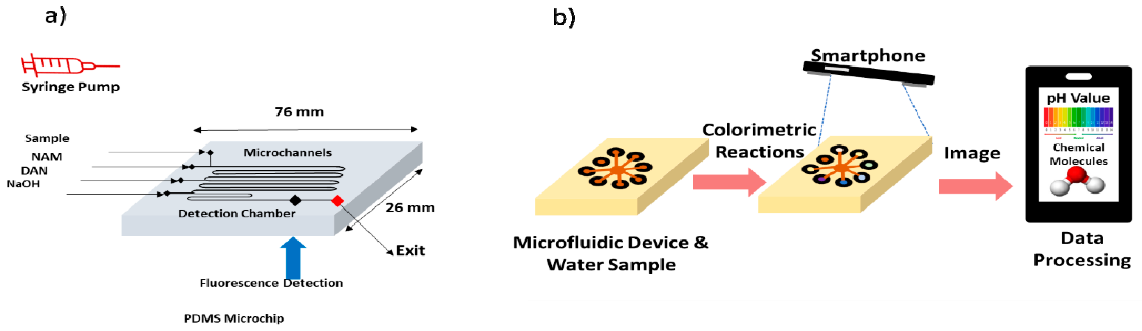
- Lopez-Ruiz, N.; Curto, V.F.; Erenas, M.M.; Benito-Lopez, F.; Diamond, D.; Palma, A.J.; Capitan-Vallvey, L.F. Smartphone-Based Simultaneous pH and Nitrite Colorimetric Determination for Paper Microfluidic Devices. Anal. Chem. 2014, 86, 9554–9562. [Google Scholar] [CrossRef]
- Sieben, V.J.; Floquet, C.F.A.; Ogilvie, I.R.G.; Mowlem, M.C.; Morgan, H. Microfluidic colourimetric chemical analysis system: Application to nitrite detection. Anal. Methods 2010, 2, 484–491. [Google Scholar] [CrossRef]
- Jayawardane, B.M.; Wei, S.; McKelvie, I.D.; Kolev, S.D. Microfluidic Paper-Based Analytical Device for the Determination of Nitrite and Nitrate. Anal. Chem. 2014, 86, 7274–7279. [Google Scholar] [CrossRef] [PubMed]
- Gallardo-Gonzalez, J.; Baraket, A.; Boudjaoui, S.; Metzner, T.; Hauser, F.; Rößler, T.; Krause, S.; Zine, N.; Streklas, A.; Alcácer, A.; et al. A fully integrated passive microfluidic Lab-on-a-Chip for real-time electrochemical detection of ammonium: Sewage applications. Sci. Total Environ. 2019, 653, 1223–1230. [Google Scholar] [CrossRef] [PubMed]
- Aravamudhan, S.; Bhansali, S. Development of micro-fluidic nitrate-selective sensor based on doped-polypyrrole nanowires. Sens. Actuators B Chem. 2008, 132, 623–630. [Google Scholar] [CrossRef]
- Kim, D.; Goldberg, I.B.; Judy, J.W. Microfabricated electrochemical nitrate sensor using double-potential-step chronocoulometry. Sens. Actuators B Chem. 2009, 135, 618–624. [Google Scholar] [CrossRef]
- Nie, D.; Li, P.; Zhang, D.; Zhou, T.; Liang, Y.; Shi, G. Simultaneous determination of nitroaromatic compounds in water using capillary electrophoresis with amperometric detection on an electrode modified with a mesoporous nano-structured carbon material. Electrophoresis 2010, 31, 2981–2988. [Google Scholar] [CrossRef]
- Bodor, R.; Madajová, V.; Kaniansky, D.; Masár, M.; Jöhnck, M.; Stanislawski, B. Isotachophoresis and isotachophoresis—Zone electrophoresis separations of inorganic anions present in water samples on a planar chip with column-coupling separation channels and conductivity detection. J. Chromatogr. A 2001, 916, 155–165. [Google Scholar] [CrossRef]
- Kamuri, M.F.; Abidin, Z.Z.; Jun, L.H.; Yaacob, M.H.; Hamidon, M.N.B.; Yunus, N.A.M.; Kamaruddin, S. Performance Evaluation of Free-Space Fibre Optic Detection in a Lab-on-Chip for Microorganism. J. Sens. 2019, 2019, 1026905. [Google Scholar] [CrossRef]
- Delehanty, J.B.; Ligler, F.S. A Microarray Immunoassay for Simultaneous Detection of Proteins and Bacteria. Anal. Chem. 2002, 74, 5681–5687. [Google Scholar] [CrossRef]
- King, K.D.; Anderson, G.P.; Bullock, K.E.; Regina, M.J.; Saaski, E.W.; Ligler, F.S. Detecting staphylococcal enterotoxin B using an automated fiber optic biosensor. Biosens. Bioelectron. 1999, 14, 163–170. [Google Scholar] [CrossRef]
- Azinheiro, S.; Kant, K.; Shahbazi, M.-A.; Garrido-Maestu, A.; Prado, M.; Dieguez, L. A smart microfluidic platform for rapid multiplexed detection of foodborne pathogens. Food Control 2020, 114, 107242. [Google Scholar] [CrossRef]
- Schwartz, O.; Bercovici, M. Microfluidic Assay for Continuous Bacteria Detection Using Antimicrobial Peptides and Isotachophoresis. Anal. Chem. 2014, 86, 10106–10113. [Google Scholar] [CrossRef] [PubMed]
- Choi, J.R.; Hu, J.; Tang, R.; Gong, Y.; Feng, S.; Ren, H.; Wen, T.; Li, X.; Wan Abas, W.A.B.; Pingguan-Murphy, B.; et al. An integrated paper-based sample-to-answer biosensor for nucleic acid testing at the point of care. Lab Chip 2016, 16, 611–621. [Google Scholar] [CrossRef] [PubMed]
- Stokes, D.L.; Griffin, G.D.; Vo-Dinh, T. Detection of E. coli using a microfluidics-based antibody biochip detection system. Fresenius’ J. Anal. Chem. 2001, 369, 295–301. [Google Scholar] [CrossRef]
- Hou, Y.-H.; Wang, J.-J.; Jiang, Y.-Z.; Lv, C.; Xia, L.; Hong, S.-L.; Lin, M.; Lin, Y.; Zhang, Z.-L.; Pang, D.-W. A colorimetric and electrochemical immunosensor for point-of-care detection of enterovirus 71. Biosens. Bioelectron. 2018, 99, 186–192. [Google Scholar] [CrossRef] [PubMed]
- Jijie, R.; Kahlouche, K.; Barras, A.; Yamakawa, N.; Bouckaert, J.; Gharbi, T.; Szunerits, S.; Boukherroub, R. Reduced graphene oxide/polyethylenimine based immunosensor for the selective and sensitive electrochemical detection of uropathogenic Escherichia coli. Sens. Actuators B Chem. 2018, 260, 255–263. [Google Scholar] [CrossRef]
- Liu, J.; Jasim, I.; Shen, Z.; Zhao, L.; Dweik, M.; Zhang, S.; Almasri, M. A microfluidic based biosensor for rapid detection of Salmonella in food products. PLoS ONE 2019, 14, e0216873. [Google Scholar] [CrossRef] [Green Version]
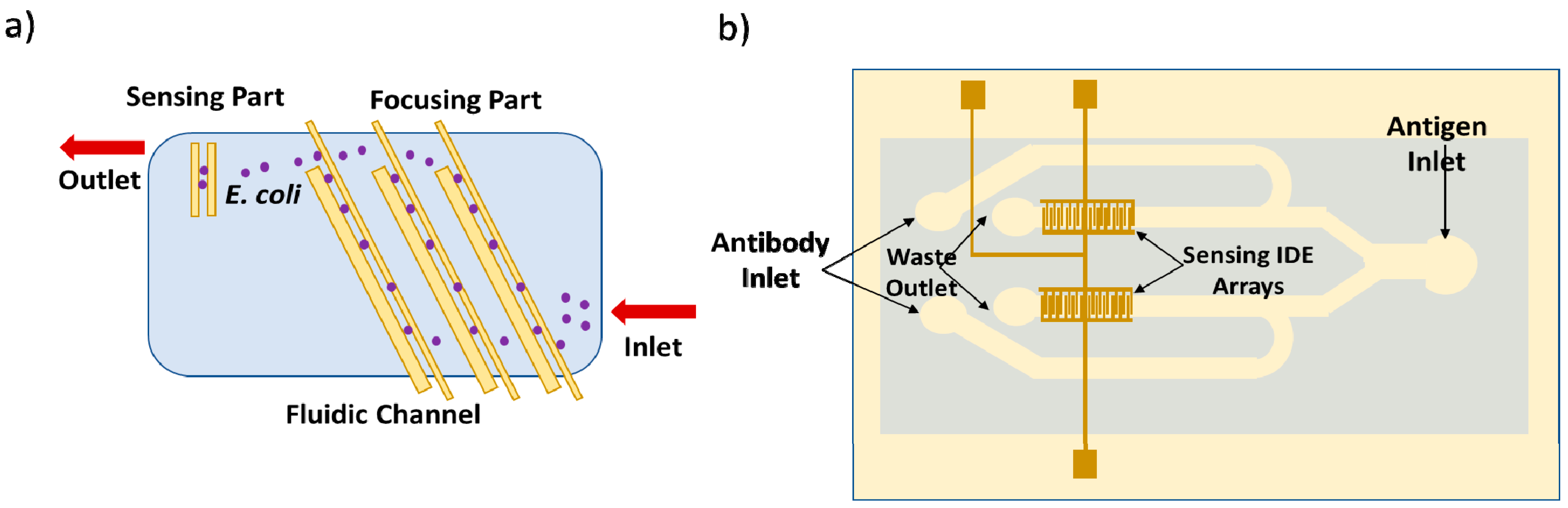
- Kim, M.; Jung, T.; Kim, Y.; Lee, C.; Woo, K.; Seol, J.H.; Yang, S. A microfluidic device for label-free detection of Escherichia coli in drinking water using positive dielectrophoretic focusing, capturing, and impedance measurement. Biosens. Bioelectron. 2015, 74, 1011–1015. [Google Scholar] [CrossRef]
- Tian, F.; Lyu, J.; Shi, J.; Tan, F.; Yang, M. A polymeric microfluidic device integrated with nanoporous alumina membranes for simultaneous detection of multiple foodborne pathogens. Sens. Actuators B Chem. 2016, 225, 312–318. [Google Scholar] [CrossRef]
- Safavieh, M.; Ahmed, M.U.; Tolba, M.; Zourob, M. Microfluidic electrochemical assay for rapid detection and quantification of Escherichia coli. Biosens. Bioelectron. 2012, 31, 523–528. [Google Scholar] [CrossRef]
- Altintas, Z.; Akgun, M.; Kokturk, G.; Uludag, Y. A fully automated microfluidic-based electrochemical sensor for real-time bacteria detection. Biosens. Bioelectron. 2018, 100, 541–548. [Google Scholar] [CrossRef]
- Jasim, I.; Shen, Z.; Mlaji, Z.; Yuksek, N.S.; Abdullah, A.; Liu, J.; Dastider, S.G.; El-Dweik, M.; Zhang, S.; Almasri, M. An impedance biosensor for simultaneous detection of low concentration of Salmonella serogroups in poultry and fresh produce samples. Biosens. Bioelectron. 2019, 126, 292–300. [Google Scholar] [CrossRef] [PubMed]
- Yao, L.; Wang, L.; Huang, F.; Cai, G.; Xi, X.; Lin, J. A microfluidic impedance biosensor based on immunomagnetic separation and urease catalysis for continuous-flow detection of E. coli O157:H7. Sens. Actuators B Chem. 2018, 259, 1013–1021. [Google Scholar] [CrossRef]
- Thiha, A.; Ibrahim, F.; Muniandy, S.; Dinshaw, I.J.; Teh, S.J.; Thong, K.L.; Leo, B.F.; Madou, M. All-carbon suspended nanowire sensors as a rapid highly-sensitive label-free chemiresistive biosensing platform. Biosens. Bioelectron. 2018, 107, 145–152. [Google Scholar] [CrossRef] [PubMed]
- Singh, C.; Ali, M.A.; Reddy, V.; Singh, D.; Kim, C.G.; Sumana, G.; Malhotra, B.D. Biofunctionalized graphene oxide wrapped carbon nanotubes enabled microfluidic immunochip for bacterial cells detection. Sens. Actuators B Chem. 2018, 255, 2495–2503. [Google Scholar] [CrossRef]
- Chen, Q.; Wang, D.; Cai, G.; Xiong, Y.; Li, Y.; Wang, M.; Huo, H.; Lin, J. Fast and sensitive detection of foodborne pathogen using electrochemical impedance analysis, urease catalysis and microfluidics. Biosens. Bioelectron. 2016, 86, 770–776. [Google Scholar] [CrossRef]
- Elsayed, A.A.; Erfan, M.; Sabry, Y.M.; Dris, R.; Gaspéri, J.; Barbier, J.-S.; Marty, F.; Bouanis, F.; Luo, S.; Nguyen, B.T.T.; et al. A microfluidic chip enables fast analysis of water microplastics by optical spectroscopy. Sci. Rep. 2021, 11, 10533. [Google Scholar] [CrossRef]


| Material Types | Characteristics | Fabrication Methods |
|---|---|---|
| Silicon (or silicon-based substrates) | (i) Resistant to organic solvents; (ii) Ease in depositing metals; (iii) High thermal conductivity [39]; (iv) Stable electroosmotic mobility; (v) High elastic modulus (130 to 180 GPa); (vi) The precise definition of nanoscale channels or pores; (vii) Transparent to infrared [35]; (viii) Chemical stability [40]. Drawbacks: (ix) Difficulties in handling them (they are hard), making it difficult to make valves and/or pumps, or active microfluidic components in general; (ix) high costs [34] | (a) Wet (chemical) etching [34,35,40,41,42]; (b) Dry etching [43]; (c) Powder blasting [33]; (d) Micro-hot embossing molding [44]; (e) Photolithography [33] |
| Glass (or glass-based substrates) | (i) Optically transparent; (ii) Electrically insulating (amorphous); (iii) Compatible with biological samples; (iv) Not permeable to gas; (v) Has a low (relative) non-specific adsorption. Drawbacks: (vi) Vertical walls are more difficult to etch than Si; (vii) Production is time-consuming and expensive [36] | (a) Wet or dry (chemical) etching [35]; (b) Metal or chemical vapor deposition [35]; (c) Patterning and cutting [45]; (d) Photolithographic patterning [46]; (e) Thermal bonding [41]; (f) Molding process [47]; (g) Powder blasting |
| Al-oxide-based materials | (i) Low-temperature co-fired ceramic (LTCC); (ii) LTCC compared to other technologies allows the integration of heaters, sensors, and electronics (control and measurement electronics, and a light-detection system) into a single module; thus, the measurement system can be simplified; (iii) Thick film materials offer the possibility to fabricate not only the networks of conducting paths in a single package, but also other microsystems, electronic components, and sensors [35]. Drawback: No mechanical flexibility | (a) Laminate sheets of Al-oxide-based material are patterned, assembled, and heated at elevated temperatures [48]; (b) Electrodes can be deposited onto LTCC using expansion-matched metal pastes [35] |
| Transition metal carbides and/or nitrides and Mn+1Xn (MXenes) | (i) High intercalation capacity; (ii) High metallic conductivity [49]; (iii) Large surface area; (iv) Good ion-transport properties; (v) Low diffusion barrier; (vi) Biocompatibility; (vii) Hydrophilicity; (viii) Ease of surface functionalization [50]; (ix) Higher signal-to-noise ratio in electrochemical sensing [51] | (a) Wet chemical etching [50]; (b) Selective etching and exfoliation process [49]; (c) Chemical vapor deposition (CVD) growth [52] |
| Polydimethylsiloxane (PDMS) | (i) Optical transparency up to 280 nm; (ii) Ductile (flexible) material; (iii) Elasticity (which can be “adjusted” using crosslinking agents); (iv) Biocompatibility; (v) Sealing capacity of materials such as glass, polystyrene, and PMMA [15]; (vi) Does not require a clean room [15]; (vii) Permeability to gases (is more permeable to CO2 than to O2 or N2) and water vapor; (viii) High thermal stability up to T = 300 °C; (ix) Cost-effective production at micro scale. Drawbacks: (x) Low shear modulus (e.g., cannot be used at for high-frequency droplet generation at high operating pressure [51]; (xi) Swelling in organic solvents; (xiii) Diffusivity [15,32,33] | (a) Device molds made through conventional machining; (b) Device molds made by photolithographic methods [53]; (c) Micromolding–casting process (liquid PDMS prepolymer is thermally cured at mild temperatures of 40–80 °C and can be cast at nanometer resolution from photoresist templates [33,53] or other techniques; (d) “Microwire molding” [15,32]; (e) Rapid prototyping [54] |
| Thermoset polyester (-TPE) | (i) Insoluble; (ii) Highly resistant to creep; (iii) Optically transparent and absorbs UV light [55]; (iv) Inexpensive; (v) Higher elastic modulus (1-100 MPa) than PDMS [56]. Drawbacks: (vi) High stiffness (improper for the fabrication of valves); (vii) High cost; (viii) Hydrophobic [35,57] | (a) Polymerization of polyester and styrene through UV or heat [35]; (b) Photolithography [58]; (c) Replica molding [59] |
| Polystyrene (PS) | (i) Optically transparent; (ii) Biocompatible, (iii) Inert; (iv) Rigid, (v) Relatively hard and brittle; (vi) Good electrical properties; (vii) Surface can be easily functionalized; (viii) Excellent gamma radiation resistance [60]. Drawbacks: (vii) Difficulties encountered in the thermal bonding step [33]; (viii) Hydrophobic (requires chemical modification of styrene PS surface or plasma oxidation to become hydrophilic) [61] | (a) Injection molding [62]; (b) Hot embossing [35]; (c) Prototyping by UV laser photoablation [38] |
| Polymethylmethacrylate (PMMA or PMMA substrate) | (i) Low cost [63]; (ii) Rigid mechanical properties; (iii) Excellent optical transparency; (iv) Compatibility with electrophoresis [37]; (v) Biological compatibility [35]; (vi) Elastic modulus of 3.3 GPa [35]; (vii) Gas impermeability; (viii) Micromachining at 100 °C [35]. Drawback: The cost of PMMA substrate per unit area is high [58] | (a) Hot embossing [63]; (b) Solvent imprinting; (c) Atmospheric pressure molding [64]; and thermal bonding; (d) Injection molding [62]; (e) Laser ablation [65]; (f) CO2 laser micromachining [66]; (g) Plasma etching [37]; (h) Nanoimprinting |
| Polycarbonate (PC) | (i) Good machining properties; (ii) High impact resistance; (iii) Enhanced chemical resistance; (iv) Low water absorptivity (<0.01%); (v) Good electrical insulating properties; (vi) Long-term stability of surface treatments; (vii) Extremely low absorption of impurities; (viii) Low cost; (ix) Durable material; (x) Very high softening temperature (~145 °C) [35]. Drawback: (xi) Low transparency in the visible and near-UV spectra | (a) Prototyping by UV laser photoablation [38]; (b) Hot embossing [67]; (c) CO2 laser machining [68]; (d) Injection molding [62] |
| Polyethylene terephthalate (PET) | (i) Resistant to thermal shock in comparison with silicon-based substrates [40]; (ii) Inexpensive production [40] | Laser ablation [69] |
| Cyclic olefin copolymer (COC) | (i) Optical transparency in the visible and near-UV spectra; enhanced chemical resistance; (ii) Good electrical insulating properties; (iii) Low water absorptivity (<0.01%); (iv) Extremely low level of impurities; (v) Long-term stability of surface treatments [70] | (a) Micromilling method [71]; (b) Photolithography [72,73] |
| Hydrogel | (i) Extremely hydrophilic polymer [74]; (ii) High biocompatibility; (iii) High biodegradability. Drawbacks: (iv) Softness of hydrogels; (v) Silk fibroin, collagen, and gelatin have poor processability; (vi) Complex microfluidic networks cannot be created—only simple or 2D ones; (vii) Channel deformation [74] | (a) Photopatterning [75]; (b) Injection molding [76]; (c) Coaxial extrusion-based 3D printing [77] |
| Paper | (i) Easy to work with; (ii) Can be treated to chemically bind molecules or proteins; (iii) Compatible with biological samples; (iv) Inexpensive material. Drawback: (v) Difficult to distinguish individual channels on the chip [35] | (a) Paper patterning; (b) Photolithography [78]; (c) Screen printing [79]; (d) Inkjet printing [80]; (e) Plasma oxidation; (f) Roll-to roll; (g) Cutting [81,82] and ink-writing [83]; (h) Wax printing [83] |
| Fabrication Methods | Material | Advantages | Disadvantages | Ref. |
|---|---|---|---|---|
| Soft lithography | PDMS | High resolution (down to a few nm); real-time detection; portable; disposable; cost-effective; able to fabricate 3D geometries | Requiring high sample concentration; pattern deformation; vulnerable to defects | [90,93] |
| Hot embossing | PMMA | Cost-effective, precise, and rapid replication of microstructures; mass production | Restricted to thermoplastics; difficult to fabricate complex 3D structures | [94] |
| Injection molding | Thermoplastic polymers | Easy to fabricate complex geometry, fine features, and 3D geometries; low cycle time; mass production; highly automated | Restricted to thermoplastics; high-cost molds; difficult to form large undercut geometries | [62] |
| Laser photoablation | PET | Rapid; large-scale production | Multiple treatment sessions; limited materials | [30,69] |
| Conventional photolithography/opticallithography | Polymers | High wafer throughputs; ideal for microscale features | Usually requires a flat surface to start with; requires chemical post-treatment | [92] |
| Photolithography | PDMS | Portability; cost-effective and high automation; high sensitivity | Low throughput | [95] |
| Electron-beam lithography | SU-8 3010 | Good resolution; can be precisely aligned | Expensive; requires more time to fabricate | [96] |
| X-ray lithography | PMMA | High resolution to fabricate nanopatterns; absorption without spurious scattering; able to produce straight, smooth walls | Difficulties in master fabrication process; time-consuming; high cost | [91] |
| Photolithography and complex pattern | Whatman No.1 chromatography paper, ITW TechniCloth, and Scott hard roll paper towels | Mass production; good stability | Expensive equipment; toxic reagents; fragile when bending | [80] |
| Photolithography or wax printing | SU-8 | Simple; portable; fast; low cost | - | [97,98] |
| Wax printing | Whatman No.1 chromatography, Whatman filter paper, and nitrocellulose (NC) membranes | Simple and fast to fabricate; mass production | Low resolution; not resistant to high temperatures | [99,100] |
| Inkjet printing | Filter paper | Cheap reagents; mass production; compatible with multiple functional inks | Requires an improved ink jet printer; low speed | [101] |
| Inkjet etching | Filter paper | Cheap reagents; prints flexible, foldable channels at 100 cm2 in size | Low resolution; low production; not suitable for complex patterns | [101,102] |
| Screen printing | Whatman No.1 filter paper | Low cost; mass production; multiple functional inks | Low resolution; different patterns need different printing wire | [79] |
| Nanoimprinting | PMMA | Cost-effective; high sensitivity; high resolution; precise control | Expensive; low throughput | [103] |
| Samples | Device Substrate (or Components) | Detection Method (and/or Mechanism) | Fabrication Method | Analyte (Target) | Limit of Detection (LOD) Linear Range (LR) | Ref. |
|---|---|---|---|---|---|---|
| Water sample | Chromatography no. 1 paper | Colorimetry | Patterned paper | Cr(VI) Ni(II) Cu(II) | LOD for Cr(VI): 0.5 mg/L LOD for Ni: 0.5 mg/L LOD for Cu(II): 0.8 mg/L | [152] |
| Sample solution with the addition of nanoparticles (PtNP) | Glass-fiber paper | Colorimetry | Printing technique | Hg(II) | LOD: 0.01 μM | [153] |
| Synthetic samples containing Hg and aqueous NaOH solution (used to extract dithizone from dithizone–CCl4 solution) and then used as a chromogenic reagent | Filter paper | Distance-based colorimetry | Printing technique | Hg(II) | LOD: 0.93 μg/mL | [154] |
| Water sample; sample solution of arsenic prepared in lemon juice | Filter paper | Colorimetric microdetection | Simple pattern-plotting method | As(III) | LOD: 0.01 mg /L | [133] |
| Environmental Samples from (i) Bog Lake; (ii) Killeshin water reservoir; (iii) Laois groundwater; (iv) Barrow Carlow River | -- | Colorimetry (absorbance principle) | -- | As(III) | LOD: 0.19 mg/L LR: 0.07–3 mg/L | [151] |
| Natural water samples at the sub-ppm range | Paper-based device | Miniaturized chemiluminescence | Wax printing of microfluidic paper-based analytical device (μPAD) | Cr(III) | LOD: 0.02 ppm LR: 0.05–1.00 ppm | [98] |
| Seawater | Polymethylmethacrylate (PMMA) | Colorimetry (absorbance principle) | Micromilling in PMMA of microchannels | Fe(II) Mn(II) | LOD for Fe(II): 27 nM LOD for Mn(II): 28 nM LR for Fe(II): 27–200 nM LR for Mn(II): 0.028–6 μM | [134] |
| Lyophilized (prepared with bacterial luciferase and NAD(P)H:FMN-oxidoreductase) and mixed with aqueous starch suspension | Polymethylmethacrylate (PMMA) | Bioluminescence | Micromilling method | Cu(II) | LOD: 3 μM | [138] |
| Environmental water samples | Cyclic olefin copolymer—an amorphous polymer | Surface plasmon resonance | Micromilling method | Hg(II) | LOD: 11 μg/L LR: 11–100 μg/L | [71] |
| Aqueous samples with mixed concentrations of Pb(II) and Cu(II) ions | Plastic-clad silica (PCS) fiber | Fiber optics + surface plasmon resonance | Coating by thermal evaporation of thick copper and silver film over unclad cores of both channels (I and II); dip-coating of non-imprinted (NIP) nanoparticles over the films; | Cu(II) Pb(II) | LOD for Cu(II): 8.18 × 10⁻¹⁰ g/L LOD for Pb(II): 4.06 × 10⁻¹² g/L LR: 4.06–1000 μg/L | [139] |
| Aqueous sample solution and aqueous M1 suspension | Polytetrafluoroethylene (PTFE) /perfluoroalkoxy alkane (PFA) tubes | Fluorescence | -- | Hg(II) | LOD: 0.02 μg/L LR: 0.02–200 μg/L | [155] |
| Aqueous samples, sewage waters | PDMS/glass | Fluorescence | -- | Cd(II) | LOD: 0.45 μg/L LR: 1.12–22.40 μg/L | [137] |
| Natural water | Glass plates | Chemiluminescence + air sampling | Photolithography and wet etching | Fe(II) | LOD: 3 × 10⁻⁷ mol/L LR: 1 × 10⁻⁶ to 5 × 10⁻⁵ mol/L | [137] |
| Diluted stock solution of Fe(II) with demineralized water | Glass | Optical detection (absorbance principle) | Photolithographic and wet-etching techniques; photoresistant coating | Fe(II) | LOD: 1 μM LR: 1–100 μM | [156] |
| Water samples containing certain concentrations of Pb | PDMS substrate | Fluorescence | Molded the channels in PDMS | Pb(II) | LOD: 5 ppb | [142] |
| Samples | Device Substrate | Working Electrode (WE) Type | Detection Method | Fabrication Method | Analyte (Target) | Limit of Detection (LOD) Linear Range (LR) and/or Sensitivity | Ref. |
|---|---|---|---|---|---|---|---|
| Real samples of gas-dissolved salty soda water and groundwater with physical contamination | Whatman filter paper | Carbon | Square-wave anodic stripping voltammetry (SWASV) | Screen-printed carbon electrodes (SPCE) on Whatman filter paper | Pb(II) Cd(II) | LOD for Pb(II): 2 ppb LOD for Cd(II): 2.3 ppb LR for Pb(II) and Cd(II): 2–100 ppb | [158] |
| Rice flourdissolved in methanol–water | Whatman filter paper | Boron-doped diamond (BDD) | Square-wave anodic stripping voltammetry (SWASV) | Electrodeposition of gold nano- particles on boron-doped diamond (AuNP/BDD) electrode | As(III) and As(V) | LOD: 0.02 μg/L LR: 0.1–1.5 μg/L | [159] |
| Aqueous solutions | Whatman grade 1 chromatography paper or polyester–cellulose blend paper | Bismuth plated on carbon | Square-wave anodic stripping voltammetry (SWASV) | Photolithography or wax-printing of microfluidic channels; screen-printed electrodes | Pb(II) | LOD: 1 ppb LR: 5−100 ppb Sensitivity: 0.17 μA (μg/L)−1 | [90] |
| Aqueous samples (heavy metal stock solutions); mud-spiked samples | Whatman filter paper grade 1 | Graphite | Square-wave voltammetry (SWV) | Wax-printing of microfluidic channels; screen-printing of electrodes | Cd(II) and Pb( II) | LOD for Cd(II): 11 ppb; LOD for Pb(II): 7 ppb LR for Cd(II) and Pb(II): 10−100 ppb Sensitivity for Cd(II): 0.015 μA (μg/L)−1 Sensitivity for Pb(II): 0.0025 μA (μg/L)−1 | [162] |
| Standard solutions of Cd(II) and Pb(II) | Whatman grade 1 chromatography paper | Boron-doped diamond paste electrodes (BDDPEs) | Square-wave anodic stripping voltammetry (SWASV) | Print wax patterns on microfluidic paper; stencil printed of an electrode with a mixture of BDD powder and mineral oil | Cd(II) and Pb(II) | LOD for Cd(II): 25 μg/L LR for Cd(II): 25–200 μg/L LOD for Pb(II): 1 μg/L LR for Pb(II): 1–200 μg/L Sensitivity of Cd(II): 0.218 μA μM−1 Sensitivity of Pb(II): 0.305 μA μM−1 | [100] |
| Environmental and biological samples | Cyclic olefin copolymer (COC) | Bismuth | Square-wave anodic stripping voltammetry (SWASV) | Photolithography of COC screen-printed electrode (SPE) | Pb(II) Cd(II) | LOD for Pb(II): 8 ppb; LOD for Cd(II): 9.3 ppb LR for Cd(II): 28−280 ppb LR for Pb( II): 25−400 ppb Sensitivity for Cd(II): 0.065 μA (μg/L)−1 Sensitivity for Pb(II): 0.0022 μA (μg/L)−1 | [72] |
| Deionized (DI) water for experiments; sample solution (with HNO3 and KCl) for electrolyte; silver electroplating solution for Ag electroplating | Cyclic olefin copolymer (COC) | Silver | Square-wave anodic stripping voltammetry (SWASV) | Spin-coated S1818-positive photoresistor patterned on a COC substrate by a photolithographictechnique; microfabricated silver electrodes | Pb(II) | LOD: 0.55 ppb LR: 1−1000 ppb Sensitivity: 0.028 μA (μg/L)−1 | [73] |
| Sample solution containing lead ions | Polymethylmethacrylate (PMMA) | Boron-doped diamond electrode | Square-wave anodic stripping voltammetry (SWASV) | Microelectrodialysercombined with boron-doped diamond electrode | Pb(II) | LOD: 4 μg/L LR: 20–100 μg/L Sensitivity of 15.5 nA L μ/g | [160] |
| Different electroactive pollutants | Polymethylmethacrylate (PMMA) | Gold thin film | Anodic stripping chronoamperometry (AS-CA) | Microfabrication techniques (micromilling in PMMA of microfluidic channels; photolithography of gold thin-film electrodes) | Cu(II) | LOD: <0.3 µM | [161] |
| Water solution containing heavy metal ions | Photosensitive resin | Screen-printed electrode (SPE) modified with Mn2O3 | Differential-pulse anodic stripping voltammetry (DPASV) | Stereolithographyappearance (SLA) for 3D-printed microfluidic device (prototyping); microporous screen-printed electrode modified with Mn2O3 | Cd(II) and Pb(II) | LOD for Cd(II): 0.5 μg/L LR for Cd(II): 0.5 to 8 μg/L LOD for Pb(II): 0.2 μg/L LR for Pb(II): 10 to 100 μg/L | [144] |
| Mixture of heavy metal ions | PDMS/glass | Gold | Capillary electrophoresis with contactless detection (CCD) | Spin-coated PDMS membrane on a glass substrate; patterned electrodes in an antiparallel configuration | Heavy metal ions | LOD: 0.4 μM | [149] |
| Sample solution containing mercury ions | PDMS/glass | Screen-printed electrode coupled with sodium-dodecyl-sulfate-doped polyaniline (PANi–SDS | Cyclic voltammetry (CV) techniques and square-wave voltammetry (SWV) | Replica-molding process for PDMS channel; screen-printed electrode (SPE) | Hg(II) | LOD: 2.4 nM LR: 6 nM to 35 nM | [163] |
| Seawater | PDMS/glass | Platinum | Linear sweep voltammetry (LSV) | Soft lithography of PDMS; patterning of electrodes on glass slides; platinum electrodeposition | Pb(II) Cd(II) | LOD for Pb(II): 150 ppb LOD for Cd(II): 340 ppb | [145] |
| Aqueous analyte | Paper substrate | Modifier-free electrodes; graphite foil | Square-wave voltammetry (SWV) | Cutting, stacking | Cd(II) and Pb(II) | LOD for Cd(II): 1.2 μg/L LR for Cd(II): 5–500 μg/L LOD for Pb(II): 1.8 μg/L LR for Pb(II): 5–100 μg/L Sensitivity for Cd(II) and Pb(II): 0.101 μA (μg/L)−1 | [157] |
| Lake water and human serum samples | 3D paper-based | Gold nanoparticles (NPs) aggregates and C nanocrystals capped silica NPs conjugated with DNA strands | Electrochemiluminescence (ECL) | Wax-printing and screen-printing | Pb(II) and Hg(II) | LOD for Pb(II): 10 pM LOD for Hg(II): 0.2 nM LR for Pb(II): 30 nM–1 μM LR for Hg(II): 0.5 nM–1 μM | [164] |
| Samples | Device Substrate (or Components) | Detection Method (and/or Mechanism) | Fabrication Method | Analyte (Target) | Limit of Detection (LOD) Linear Range (LR) | Ref. |
|---|---|---|---|---|---|---|
| Tap water and river water samples | Fisher brand filter paper (P5; 09−801C) with a diameter of 11 cm and a medium porosity | Colorimetry | Inkjet printing and a layer-by-layer (LbL) assembly approach (formed by alternatively depositing layers of chitosan and alginate polyelectrolytes) onto filter paper | Phenolic compounds (phenol, bisphenol A (BPA), dopamine) | LOD: 0.86 (±0.1) μg/L | [102] |
| Environmental samples | Polyacrylamide film | Florescence (molecular absorption) | --- | Catechol | LR: 9.79 × 10⁻⁶ to 7.50 × 10⁻⁴M | [169] |
| -Standard solutions (mixtures) of catecholamines;-Human urine and plasma samples | Fused silica fiber coated with a polystyrene/divinylbenzene resin (PS/DVB) film | Optical fiber biosensor + chromatographic separation | Mechanically uncladded; enzymatic cladding; dip-coating of single optical fibers (OFs) | Dopamine, norepinephrine, epinephrine | LOD for dopamine: 2.1 pg/mL; LOD for norepinephrine: 2.6 pg/mL; LOD for epinephrine: 3.4 pg/mL | [171] |
| Homogeneous stock sol–gel solution | Hybrid Nafion/sol–gel silicate glass | Optical biosensors (crosslinking immobilization method of laccase and 3-methyl2-benzothiazolinone hydrazone (MBTH) | MBTH mixture was deposited onto a glass slide and coated | Catechol | LOD: 0.33 mM LR: 0.5–8.0 mM | [172] |
| Catechol in water sample | Fe3O4@Au core–shell nanoparticles | Colorimetric detection (absorbance principle) | Laccase-Au-Fe3O4 nanoparticles (NPs) | Catechol | LOD: 2 μM LR: 5–70 μM | [170] |
| Samples | Device Substrate (or Components) | Working Electrode Type | Detection Method | Fabrication Method | Analyte (Target) | Limit of Detection (LOD) Linear Range (LR) | Ref. |
|---|---|---|---|---|---|---|---|
| Domestic water supplies; sample solution: 2,4-dichlorophenol (2,4-DCP) mixed with Folin–Ciocâlteu (FC) reagent | Plastic microfluidic chip with incorporated electrodes | Platinum | Potential difference measurements | Sputtering method of deposition of electrodes on plastic film | 2,4-Dichlorophenol | LOD: 0.1 ppm | [167] |
| Contaminated water sample with phenols | Hybrid PDMS/glass microchip | Graphite | Chronoamperometry. | Soft lithography in PDMS of microchannel; SPE modified with a CO3-poly (ethyleneimine) (PEI) microparticles (MPs) and tyrosinase (Tyr) | Phenols | LOD: 10 nM LR: 0.5 to 5 μM | [173] |
| Contaminated water sample with phenols | Hybrid polydimethylsiloxane (PDMS)/glass chrono-impedimetric microchip; polyester substrate for screen-printed electrode (SPE) | Graphite | Electrochemical impedance spectroscopy (EIS); chrono-impedimetric detection of phenols | Soft lithography in PDMS of channels; sequential deposition of graphite ink and Ag/AgCl ink onto a glass substrate for a screen-printed electrode (SPE) | Phenols | LOD: 4.64 nM LR: 0.01–10 μM | [148] |
| Water samples | Polyethylene -based films | Carbon (screen-printed carbon electrodes) | Micellar electrokinetic chromatography with electrochemical detection (MEKC-EC); amperometric detector | Screen-printed carbon electrodes (SPCEs) | Trace phenolic compounds | LOD: 100 × 10−12–150 × 10−12 M | [174] |
| Samplewaste; mixture of dopamine, epinephrine, catechol, and 4-aminophenol | Poly(dimethylsiloxane) (PDMS)silicon wafer | Cylindrical carbon electrodes | Cyclic voltammetry (CV) | Silicon wafer spin-coated with SU-8 2035-negative photoresistor; micromolding–casting process of liquid PDMS prepolymer | Dopamine, epinephrine, catechol, 4-aminophenol | LOD for dopamine: 140 nM; LR for dopamine: 140–45.00 μm LOD for epinephrine: 105 nM; LR for epinephrine: 0.105–47.90 μm LOD for catechol: 693 nM; LR for catechol: 0.693–188.10 μm LOD for 4-aminophenol: 459 nM LR for 4-aminophenol: 0.459–159.10 μm | [175] |
| Human blood and urine samples | Fiber optics; Teflon plug | Glassy carbon | Chromatography–electrochemical detector (HPLC-ED) | --- | Epinephrine, dopamine, norepinephrine | LOD for epinephrine: 3.5 pg/mL LOD for dopamine: 2.9 pg/mL LOD for norepinephrine: 3.3 pg/mL LR: 5–125 pg/mL | [176] |
| Samples | Device Substrate (or Components) | Detection Method (and/or Mechanism) | Fabrication Method | Analyte (Target) | Limit of Detection (LOD) Linear Range (LR) | Ref. |
|---|---|---|---|---|---|---|
| Aqueous samples (river-, pond-, and rainwater) | PDMS/glass microchip | Laser-induced fluorescence (LIF) | Microchannels made by photolithography and wet-etching methods; microfabricated glass template | Nitrites | LOD: 0.4 × 10⁻⁶ M | [140] |
| Drinking water containing nitrites | PMMA microfluidic chip | Colorimetric chemical analysis (Griess method for nitrite detection on a chip) | Microchip fabrication: micromilling and solvent–vapor bonding procedure | Nitrites | LOD: 14 × 10−6 M | [178] |
| Synthetic and natural water samples; environmental and drinking water | Whatman filter paper grade 1 and 4 | Colorimetry | Inkjet printing method of electrode; patterning grade 1 and 4 filter paper (Whatman) | Nitrites and nitrates | LOD for nitrites: 1 μm LOD for nitrates: 19 μm | [179] |
| Water samples | Standard laboratory Whatman paper grade 1 | Colorimetry | Stamping technique of the paper-based microfluidic devices | nitrites | LOD: 0.52 mg/L | [177] |
| Samples | Device Substrate (or Components) | Working Electrode Type | Detection Method | Fabrication Method | Analyte (Target) | Limit of Detection (LOD) Linear Range (LR) | Ref. |
|---|---|---|---|---|---|---|---|
| Wastewater; ammonium-containing samples | PDMS microfluidic device; silicon substrate wafers | Gold | Cyclic voltammetry (CV) | Microelectrodes made by physical vapor deposition (PVD) followed by photolithography and lift-off; soft lithography and replica molding of PDMS microfluidic systems | Ammonium | LOD: 4 × 10⁻⁵ M | [180] |
| Real-world samples; nitrate samples | Silicon substrate/polyimide protective layer | Silver thin film | Double-potential-step Chronocoulometry (DPSC) | Patterned polyimide insulation layer | NO3 − | LOD: 4–75 μM LR: 500–2000 μM | [182] |
| Seawater | Polypyrrole-covered carbon nanowire | Polypyrrole (PPy)-doped nanowires (NWs) on the interdigitated Pt | Double-potential-step chronocoulometry (DPSC) | Patterned electrochemical reagent chamber of the sensor chip using a thick SU-8 film; assembly of PPy NWs on the Pt lines using dielectrophoresis | NO3 − | LOD: 4.5 μM sensitivity: 1.17–1.65 nA/μM | [181] |
| Wastewater, tap water; river sample | Borosilicate glass tube | Carbon disk electrode modified with mesoporous carbon material (CMK-3) | Capillary electrophoresis with amperometric detection and electrochemical impedance spectroscopy | Carbon disk electrode constructed using a pencil lead | 1,3,5-Trinitro-benzene (TNB), 1,3-dinitrobenzene (DNB),2,4,6-trinitrotoluene (TNT), 2,4-dinitrotoluene (DNT) | LOD for TNB: 4 μg/L LOD for DNB: 4.1 μg/L LOD for TNT: 4.7 μg/L LOD for DNT: 3 μg/L LR for TNB: 10.7–4.7 × 10 3 μg/L LR for DNB: 8.4–3.7 × 10 3 μg/L LR for TNT: 11.4–5.0 × 10 3 μg/L LR for DNT: 9.1–4.0 × 10 3 μg/L | [183] |
| Dirty aquarium water samples (in the absence and presence of fishes) and Meia Ponte River water samples | Commercial glass substrate for device(borosilicate glass microchips with integrated electrodes) | Integrated electrodes | Capacitively coupled contactless conductivity detection (C4D) | ------ | NO3– NO2− | LOD for NO3: 4.4 μM LOD for NO2: 4.9 μM | [146] |
| River water, tap water, mineral water | PMMA microchip, Isotachophoresis (ITP) and column-coupled capillary-zone electrophoresis (CZE) | Thin-film platinum electrodes | Conductivity | Microchip fabrication: substrate hot embossing; metallization of the PMMA covers used as the cover plates; sputtering deposition of thin-film platinum electrodes | Nitrites | LOD: 0.5–0.7 μM | [184] |
| Samples | Device Substrate (or Components) | Detection Method | Fabrication Method | Analyte (Target) | Limit of Detection (LOD) Linear Range (LR) | Ref. |
|---|---|---|---|---|---|---|
| Samples of microorganism-infected water | Glass substrate; dry-film resist (DFR)-basedmicrofluidic chip bonded with multimode fiber pigtails | Absorbance measurements(optical) | Photolithographic fabrication of microchannels | Escherichia coli, Saccharomycescerevisiae, and Aeromonas hydrophila | LOD for A. hydrophila and E. coli: 1.0 × 10⁵ cells/mL LOD for S. cerevisiae: 1.0 × 10⁶ cells/mL | [185] |
| Strains of Aeromonas hydrophila | Glass substrate | Absorbance measurements (optical) | Photolithographic fabrication of microchannels | Aeromonas hydrophila | LOD: 6 μL or 102 cells/mL | [136] |
| Infected water samples | Soda lime glass substrate of microfluidic chip (NS-12A, PerkinElmer, USA) | Fluorescence detection | - | E. coli | LOD: 10 4 CFU/mL | [189] |
| Real samples; biological samples; spiked drinking water | Glass fiber; nitrocellulose membrane; integrated paper-based biosensor; hydrophobic PVC layers; separation of paper | Lateral flow assays (LFA) for bacterial nucleic acid detection; colorimetry | Cell deposition | E. coli | LOD: 10 CFU/mL (Water) | [190] |
| Samples containing mixtures of analytes | PDMS/glass | Fluorescence | - | Cholera toxin; Staphylococcal enterotoxin B; Bacillus globigii | LOD for Cholera toxin: 8 ng/mL; LOD for Staphylococcal enterotoxin B: 4 ng/mL; LOD for Bacillus globigii: 6.2 × 10⁴ cfu/mL | [186] |
| Phosphate-buffered saline samples | Polyethylene channel | Fluorescence | - | Staphylococcal enterotoxin B | LOD: 5 ng/mL | [187] |
| Chicken carcass wash samples | Glass/hybrid | Fluorescence | - | E. coli | LOD: 20 organism | [191] |
| Real samples | 3D PDMS sponge | Fluorescence | The powdered salt particles were rubbed by adding water and then cast into molds (empty syringe) to shape the template for a PDMS sponge | Listeria monocytogenes, Salmonella sp. Salmonella typhimurium | LOD for: 10 3 to 10 4 CFU/mL LOD for: 1.5 × 10 2 CFU/ mL | [188] |
| Samples | Device Substrate (or Components) | Working Electrode Type | Detection Method (and/or Mechanism) | Fabrication Method | Analyte (Target) | Limit of Detection (LOD) Linear Range (LR) | Ref. |
|---|---|---|---|---|---|---|---|
| Bacteria-contaminated drinking water samples; mixture of bacterial suspensions | PDMS microfluidic chip | Gold | Dielectrophoretic impedance measurements | Conventional photolithographic and soft lithographic techniques for a PDMS microfluidic chip; PVD (sputtering) for the electrode material | E. coli | LOD: 300 CFU/mL | [195] |
| Mixed bacterial sample of E. coli O157:H7 and S. aureus | Polyethylene glycol (PEG)-based microfluidic chip integrated with a functionalized nanoporous alumina membrane | Platinum | Linear sweep voltammetry (LSV) | Soft lithography techniques | E. coli and S. aureus | LOD: 100 CFU/mL | [196] |
| Real sample | Poly (dimethylsiloxane) (PDMS) substrate | Carbon | Linear sweep voltammetry (LSV) | Soft lithography techniques for microchannels | E. coli (DNA) | LOD: 24 CFU/mL | [197] |
| E. coli samples | Poly(methyl methacrylate) (PMMA)/silicon dioxide wafer | Gold | Cyclic voltammetry and amperometric measurements | - | E. coli | LOD: 1.99 × 10⁴–3.98 × 10⁹ CFU/mL | [198] |
| Salmonella samples | PDMS/glass | Interdigitated electrode (IDE) | Impedance | Surface micromachining technology for sputtering of Cr and Au on top of glass (SU8 type); PDMS bonding to seal the microchannel | Salmonella serogroups | LOD: 7 cells/mL | [199] |
| Bacterial samples | Glass substrate | Interdigitated array and gold microelectrode | Impedance | 3D printing and PDMS casting of microchannels | Escherichia coli O157:H7 | LOD: 12 CFU/mL | [200] |
| Salmonella-specific aptamer probes | SU-8 substrate and suspended carbon nanowire | Carbon nanowire electrodes | Electrical detection/chemiresistive | Nanowires were deposited by electrospinning; photolithography for SU-8 support structure. | Salmonella typhimurium | LOD: 10 CFU/mL | [201] |
| Real samples of S. typhimurium cells | PDMS/glass for substrate; graphene oxide (GO) nanosheets wrapped in carboxylated multiwalled carbon nanotubes (cMWCNTs) composite | GO-cMWCNTs microelectrode | Electrochemical detection | Soft lithography for PDMS microchannels; wet chemical etching process for fabrication of microelectrodes | Salmonella typhimurium bacterial cells | LOD: 0.376 CFU/mL | [202] |
| Listeria cells, magnetic nanoparticles (MNPs) modified with anti-Listeria monoclonal antibodies, and gold nanoparticles (AuNPs) modified with anti-Listeria polyclonal antibodies and urease | PDMS/glass | Interdigitated microelectrode | Impedance | 3D printing and surface plasma bonding | Listeria monocytogenes | LOD: 10 6 CFU/mL | [203] |
Publisher’s Note: MDPI stays neutral with regard to jurisdictional claims in published maps and institutional affiliations. |
© 2022 by the authors. Licensee MDPI, Basel, Switzerland. This article is an open access article distributed under the terms and conditions of the Creative Commons Attribution (CC BY) license (https://creativecommons.org/licenses/by/4.0/).
Share and Cite
Alhalaili, B.; Popescu, I.N.; Rusanescu, C.O.; Vidu, R. Microfluidic Devices and Microfluidics-Integrated Electrochemical and Optical (Bio)Sensors for Pollution Analysis: A Review. Sustainability 2022, 14, 12844. https://doi.org/10.3390/su141912844
Alhalaili B, Popescu IN, Rusanescu CO, Vidu R. Microfluidic Devices and Microfluidics-Integrated Electrochemical and Optical (Bio)Sensors for Pollution Analysis: A Review. Sustainability. 2022; 14(19):12844. https://doi.org/10.3390/su141912844
Chicago/Turabian StyleAlhalaili, Badriyah, Ileana Nicoleta Popescu, Carmen Otilia Rusanescu, and Ruxandra Vidu. 2022. "Microfluidic Devices and Microfluidics-Integrated Electrochemical and Optical (Bio)Sensors for Pollution Analysis: A Review" Sustainability 14, no. 19: 12844. https://doi.org/10.3390/su141912844
APA StyleAlhalaili, B., Popescu, I. N., Rusanescu, C. O., & Vidu, R. (2022). Microfluidic Devices and Microfluidics-Integrated Electrochemical and Optical (Bio)Sensors for Pollution Analysis: A Review. Sustainability, 14(19), 12844. https://doi.org/10.3390/su141912844