Article

Efficacy of Biological Copper Oxide Nanoparticles on Controlling Damping-Off Disease and Growth Dynamics of Sugar Beet (Beta vulgaris L.) Plants

by
Eman Abou-Salem
1,
Abdulmageed R. Ahmed
1,
Mohssen Elbagory
2,3 and
Alaa El-Dein Omara
3,*
1
Soil and Water Department, Faculty of Agriculture, Kafrelsheikh University, Kafr El-Sheikh 33516, Egypt
2
Department of Biology, Faculty of Science and Arts, King Khalid University, Mohail 61321, Assir, Saudi Arabia
3
Agricultural Research Center, Department of Microbiology, Soils, Water and Environment Research Institute, Giza 12112, Egypt
*
Author to whom correspondence should be addressed.
Sustainability 2022, 14(19), 12871; https://doi.org/10.3390/su141912871
Submission received: 7 September 2022 / Revised: 7 October 2022 / Accepted: 7 October 2022 / Published: 9 October 2022
(This article belongs to the Section Sustainable Agriculture)

Abstract

:
One of the most prevalent diseases affecting sugar beet crops globally is damping-off disease, which is caused by fungi or soil-borne bacteria. The objective of the current study was to assess the antimicrobial activity of various concentrations of CuO-NPs against Fusarium oxysporum, Macrophomina phaseolina, and Pectobacterium carotovorum in a lab setting and how they influenced vegetative growth, physiological traits, antioxidant enzymes, disease incidence percentage, and mineral nutrients of sugar beet plants in a greenhouse experiment. Sugar beet (Beta vulgaris cv. Oscar poly) seeds were soaked in different concentrations (50, 100, and 150 µg L−1) of the tested NPs for two hours pre-sowing. According to in vitro studies, as compared to aqueous copper sulphate and control, CuO-NPs at 25, 35, and 100 µg mL−1 had the greatest inhibitory effect (100%) on the mycelial growth of M. phaseolina, F. oxysporum, and P. carotovorum, respectively. Results from the greenhouse experiment showed that the 150 µg mL−1 concentration produced the greatest reduction in disease incidence %, with efficacy values of 24.53, 13.25, and 23.59% for F. oxysporum, M. phaseolina, and P. carotovorum, respectively. In addition, as compared to untreated control plants, the same concentration of CuO-NPs significantly (p ≤ 0.05) increased the vegetative development, physiological characteristics, antioxidant enzymes, and mineral nutrients of sugar beet plants. Therefore, the antimicrobial activity demonstrated by the biosynthesized CuO NPs indicates that they can resist plant pathogenic microorganisms of sugar beet plants.

1. Introduction

Nanotechnology is a multidisciplinary field that is expanding rapidly as it enters many fields, including medical, engineering, industry, and agriculture [1,2]. Nanoparticles (NPs) are collections of atoms or molecules that are 1 to 100 nm in size [3]. Nanomaterials differ from their bulk counterparts in both their physical and chemical characteristics [4]. In comparison to bulk materials, these characteristics have a higher surface-to-volume ratio, and other characteristics include their size, shape, charge, and zeta potential [5,6]. Several techniques, including chemical, physical, and biological methods, are used to synthesize NPs. Biological processes are favored over chemical and physical methods. Recently, interest in using metabolites produced by many biological entities has increased, such as prokaryotic and eukaryotic organisms, to create metals and their oxide nanoparticles [7].
Among the different metal oxide nanoparticles, copper oxide nanoparticles (CuO-NPs) have been created either extracellularly or intracellularly by a wide range of biological microorganisms, i.e., bacteria, fungi, actinomycetes, algae, and plants [8].
Due to their role in drug transport, antioxidant action, high efficiency against toxic cells and tumors, and antibacterial activity, CuO-NPs exhibit excellent biological activity [9]. Additionally, Cu plays important roles in photosynthesis, mitochondrial respiration, cell wall metabolism, lignification, pollen production, and oxidative stress responses when paired with certain proteins and enzymes [10,11].
Sugar beet (Beta vulgaris L.) is a member of the Chenopodiaceae family. It is the second-largest source of sugar in the world and one of the major sugar crops [12]. The significance of the sugar beet crop is underscored by its ability to prosper as a crop in freshly reclaimed areas and its ability to produce a high sugar yield [13]. The highest root and sugar production was achieved by fertilizing sugar beet plants with micronutrients [14].
According to [15], the application of CuO-NPs to sugar beet plants could be advantageous for growth and development since sugar beet has a high capacity for absorption and reactivity. On the other hand, different bacterial and fungal diseases attack and diminish sugar beet yields and cause economic losses at different plant stages, including Pythium ultimum, Fusarium oxysporum, and Rhizoctonia solani. Consequently, the possibility of using CuO-NPs as antibacterial agents has been brought up in a number of studies. CuO-NPs have demonstrated outstanding efficacy against plant pathogenic fungi such as Phoma destructive, Curvularia lunata, and Alternaria alternate using a well diffusion assay [16]. Additionally, ref. [17] showed strong inhibitory activity against F. oxysporum, F. culmorum, Pseudomonas aeruginosa, and Klebsiella pneumonia.
The objective of this investigation was to determine whether biologically synthesized CuO-NPs were effective at combatting damping-off disease caused by F. oxysporum, Macrophomina phaseolina, and Pectobacterium carotovorum, as well as how they affected sugar beet plants’ vegetative growth, physiological traits, antioxidant enzymes, disease incidence percentage, and mineral nutrients under greenhouse conditions.

2. Materials and Methods

2.1. Bacteria Used and Cultural Conditions

A CuO-NPs solution was prepared at the bacteriology laboratory, Soils, Water and Environment Research Institute (SWERI), Sakha Agricultural Research Station, Agriculture Research Center (ARC), Egypt. According to [18], the preparation of CuO-NPs was carried out biologically using Bacillus circulans NCAIM B. 02324. In this method, cultural growth conditions were employed by using the Nutrient Broth medium at 30 °C and 200 rpm for 48 h. Twenty-five milliliters of the cell-free supernatant were combined with an aqueous solution containing 10 mM copper sulphate (CuSO4. 5H20) and heated at 100 °C for 15 min, whereby the color changed from blue to reddish-brown. Copper oxide nanoparticles (CuO-NPs) were then produced after centrifugation at 10,000 rpm for 15 min. The pellet was centrifuged again for 10 min while being intermittently washed with distilled water to remove the water-soluble molecules from the CuO-NPs suspension. The Cu-NPs were dried at 100 °C and kept for further examination in an airtight container. The diameter and zeta potential of the CuO-NPs that were employed were 87.7 nm and −31.1 mV, respectively.

2.2. Effect of Copper Oxide Nanoparticles on Plant Pathogens Microorganisms (In Vitro)

According to [19], the well diffusion method was used to examine the biosynthesized copper oxide nanoparticles for their antagonistic activity against the two plant pathogen fungi, Fusarium oxysporum SARS11 and Macrophomina phaseolina SARS105, as well as one plant pathogen bacterium, Pectobacterium carotovorum SARS09, in comparison to aqueous copper sulphate in the normal form. For plant pathogen fungi, the concentrations of the nanoparticle suspensions were 0, 5, 15, 30, 60, 70, and, 90 μg mL−1, while for pathogen bacteria, the values were 2, 5, 15, 30, 60, 70, 90, and 100 μg mL−1.
The reduction in pathogen microorganism growth was calculated using the formula RG (%) = C–T / C × 100, where C is the test pathogen’s growth (cm) without the examined substrate and T is the test pathogen’s growth (cm) with the examined substrate present [20].
The pathogenic microorganisms for the study were provided by the Plant Pathology Research Institute, Agricultural Research Center, Egypt.

2.3. Effect of Copper Oxide Nanoparticles on Plant Growth in a Gnotobiotic Clay System of Sugar Beet

Sugar beet seeds (Beta vulgaris cv. Oscar Poly) were surface sterilized with 3% sodium hypochlorite for approximately 2 min, rinsed three times in sterile distilled water with a few drops of hydrogen peroxide, and then soaked in a variety of CuO-NPs suspension concentrations (0, 5, 15, 30, 60, 90, 120, and 150 μg mL−1) for an extended period of time (2 h) at room temperature in the dark. Two seeds were planted in plastic pots with 200 g of sterilized clay soil (which had been autoclaved twice for 4 h at 121 °C and 1.5 bar). Table 1 lists some of the soil’s physical and chemical analyses. One week before planting, the soil was infested with 1 × 10⁵ spores mL−1 of F. oxysporum, M. phaseolina (6 g/200 g soil), and 1 × 10⁹ CFU mL−1 of P. carotovorum (10 mL/200 g soil), followed by irrigation with Hoagland solution [21]. The experiment lasted for 30 days, during which time the parameters of vegetative growth, including root and leaf length (cm plant−1), fresh root and leaf weight (g plant−1), and dry root and leaf weight (g plant−1), were measured.

2.4. Sugar Beet Pot Experiments

A 6 kg polyethylene bag with an internal diameter of 30 cm and a height of 35 cm was used in a greenhouse pot experiment to test the effects of various CuO-NPs suspension concentrations on sugar beet growth dynamics when the plants were artificially infected with plant pathogens. There were six replicates in the entirely randomized experimental design. As previously mentioned, F. oxysporum and M. phaseolina were used to infest the soil, but after 20 days of sowing, P. carotovorum was injected into petioles of the first two true leaves at a rate of 5 mL−1 roots [22].
Soil infected with plant diseases and soil infected by plant pathogens with seeds treated with pesticide (Vitavax, Carboxin 37.5% + Thiram 37.5% DS, 1 g Kg−1, Dhanuka Agritech Ltd.), and soil neither uninfected nor seeds untreated with pesticide were used as controls. Three different concentrations of CuO-NPs suspension (50, 100, and 150 μg mL−1) were also employed. Sugar beet seeds were surface sterilized in accordance with the germination experiment section’s instructions. The surface-sterilized seeds were given the previously indicated amount of time to soak in the various CuO-NPs suspension concentrations. Six seeds were put in each pot, and one plant per pot was kept after the seedlings emerged after one week. Mineral fertilizers were administered in the required amounts and soil moisture was maintained between 70 and 75% of saturation. The sugar beet plants’ vegetative development, physiological characteristics, antioxidant enzymes, disease incidence %, and mineral content parameters were assessed after 90 days.

2.4.1. Plants’ Vegetative Development

Following the harvest of three healthy plants per treatment, the fresh and dry weights of the leaves and roots (g plant−1), as well as their dry weights (g plant−1), were recorded. Using an electronic balance, the fresh and dry weights of leaves and roots were determined (ADAM model PW 214, 500 g, UK).

2.4.2. Physiological Characteristics

A leaf sample from each treatment was obtained and frozen in order to determine the amount of chlorophyll, carotenoids, total soluble sugars, and antioxidant enzyme activity.

Photosynthetic Pigments

According to [23], 0.1 g of leaf samples from each treatment were crushed and extracted in 5 mL of 80% acetone to assay chlorophyll a, b, total, and carotenoids. After being centrifuged at 13,000× g for 10 min, the supernatant was detected at 663, 645, and 470 nm. The quantities of carotenoids and chlorophylls were calculated and reported as µg g−1 FW and mg g−1 FW, respectively.

Total Soluble Sugars (TSS)

We followed the procedure outlined by [24]. A 0.5 g leaf sample from each treatment was placed in a water bath and heated to 80 °C for 30 min after being homogenized in 5 mL of 80% ethanol. The supernatants were collected after being centrifuged at 10,000× g for 10 min in order to measure the concentration of total soluble sugars at 620 nm using a UV spectrophotometer (Bibby Scientific Ltd., Dunmow, Essex, UK, Model 6705) and a glucose standard curve. The outcomes were given as µ g g−1 FW.

2.4.3. Antioxidant Enzymes

Peroxidase Activity

By evaluating the oxidation of pyrogallol to purpurgallin in the presence of H2O2, the peroxidase enzyme activity was evaluated in accordance with the procedures outlined by [25]. The sample cuvette included 3.0 mL of distilled water, 0.5 mL of 0.1M sodium phosphate buffer PH 7, 0.3 mL of enzyme extract, 0.05 pyrogallol, 0.3 mL of enzyme extract, and 0.1 mL of 10% H2O2. Changes in absorbance were used to measure peroxidase activity. Using a UV-visible spectrophotometer (OD600 PG Instruments UK) T80, the absorbance was measured at 425 nm.

Polyphenol Oxidase Assay

According to [26], the reaction mixture contained 1.0 mL of 0.2 M sodium phosphate buffer pH 7, 10.0 mL of 0.001 M catechol, and 1.0 mL of enzyme extract in addition to 3.0 mL of distilled water to dilute the cuvette’s contents to 3.0 mL. The activity of polyphenol oxidase was measured by changes in absorbance. The absorbance was determined at 495 nm using a UV-visible spectrophotometer (OD600 PG Instruments UK) T80.

Phenylalanine Ammonia Layase Assay

According to the procedure outlined by [27], the phenylalanine ammonia layase test enzyme was measured in acetone powder made from leaves using 0.1 g of acetone powder suspended in 10 mL of sodium borate buffer, pH 8.8 borate buffer (1.5 mL, 0.2 M), pH 8.8, 1 mL of 1% phenylalanine, and 2.5 mL of deionized water made up the reaction mixture. One milliliter of deionized water was supplied in place of phenylalanine as a blank. The mixture was incubated at 40 °C for an hour. After the reaction was stopped by adding 0.5 mL of HCL 5N to each tube, the enzyme activity was measured as the optical density at 290 nm using a UV-visible spectrophotometer (OD600 PG Instruments UK) T80.

2.4.4. Disease Incidence Percentage

Plants were uprooted and tested for disease incidence % 90 days after sowing. Based on the formula proposed by [28], percentages of disease incidence were computed.
Disease   incidence = No . of   infected   plants No . of   total   plants   × 100

2.4.5. Chemical Contents of Sugar Beet Leaves

Prior to being processed into a uniform powder in a metal-free mill (IKa-Werke, M 20 Darmstadt, Germany), plant samples were oven-dried at 65 °C for 48 h to ascertain their dry weights. The Micro-Kjeldahl method was used to determine the nitrogen content [29], whereas spectrophotometers (GT 80+, Livingston, UK) and the atomic absorption spectrometry method [30,31] were used to assess phosphorus and potassium, respectively. With the aid of atomic absorption spectrometry (Avanta E; GBC), the amounts of Cu were determined [29]. Three replicates were used to measure each of the above parameters.

2.5. Statistical Analyses

Co Stat’s statistical package software, version 6.303, was used to statistically evaluate the data. A one-way ANOVA was used to compare the various treatments. Tukey’s range tests were used for multiple comparisons at p ≤ 0.05 [32].

3. Results

3.1. Effect of Copper Oxide Nanoparticles on Plant Pathogens Microorganisms (In Vitro)

The antimicrobial activity of the synthesized CuO NPs against the plant pathogens compared to aqueous copper sulfate in the normal form is shown in Figure 1. The zone of inhibition exhibited by the synthesized CuO NPs at different concentrations of 0, 2, 5, 10, 20, 30, and 35 μg mL−1 against F. oxysporum, 0, 2, 5, 10, 20, and 25 μg mL−1 against M. phaseolina, and 0, 2, 5, 10, 20, 30, 40, 50, and 100 μg mL−1 against P. Carotovorum are given in Figure 1A–C and Figure S1a–c (Supplementary Materials), respectively.
In general, the antimicrobial effectiveness of Cu-NPs increased with increasing dosages; for instance, when 70 and 90 µg mL−1 Cu-NPs were applied, the antifungal efficiency of the reaction was 100%. However, when aqueous copper sulfate was applied, the antifungal efficiency of the reaction was 56.25 and 48.88 % for M. phaseolina and F. oxysporum, respectively (Figure 1A,B and Figure S1a,b). On the other hand, P. carotovorum was inhibited by 0.6 and 13 mm when biosynthesized CuO NPs were applied and 0.3 and 0.5 mm by aqueous copper sulfate at 50 and 100 µg mL−1, respectively (Figure 1C and Figure S1c).

3.2. Effect of Copper Oxide Nanoparticles on Plant Growth in a Gnotobiotic Clay System of Sugar Beet

The effect of different concentrations of CuO-NPs (5, 15, 30, 60, 90, 120, and 150 µg mL−1) on some growth parameters of sugar beets infected with plant pathogenic microorganisms (F. oxysporum; M. phaseolina, and P. carotovorum) are shown in Table 2 and Figure S2.
Among all the treatments, seeds soaked in 150 µg mL−1 CuO-NPs showed significantly higher results of 14.15, 13.29 (cm plant−1), 4.06, 5.29 (g plant−1), and 0.40, 0.53 (g plant−1) in soil infected by F. oxysporum, 14.32, 13.59 (cm plant−1), 3.26, 5.76 (g plant−1), and 0.43, 0.58 (g plant−1) in soil infected by M. phaseolina, and 14.44, 13.62 (cm plant−1), 2.47, 5.79 (g plant−1), and 0.38, 0.55 (g plant−1) in soil infected by P. carotovorum for the length of root and leaves, fresh weight of root and leaves, and dry weight of root and leaves, respectively (Table 2 and Figure S2).
However, the lowest results were observed in the control (infected), which attained 7.13, 7.15 (cm plant−1), 0.87, 1.26 (g plant−1), and 0.11, 0.12 (g plant−1) in soil infected by F. oxysporum, 7.39, 6.17 (cm plant−1), 1.02, 1.19 (g plant−1), and 0.14, 0.25 (g plant−1) in soil infected by M. phaseolina, and 6.73, 6.63 (cm plant−1), 0.70, 1.27 (g plant−1), and 0.14, 0.26 (g plant−1) in soil infected by P. carotovorum for the length of root and leaves, fresh weight of root and leaves, and dry weight of root and leaves, respectively (Table 2 and Figure S2).

3.3. Sugar Beet Pot Experiments

3.3.1. Plants’ Vegetative Development

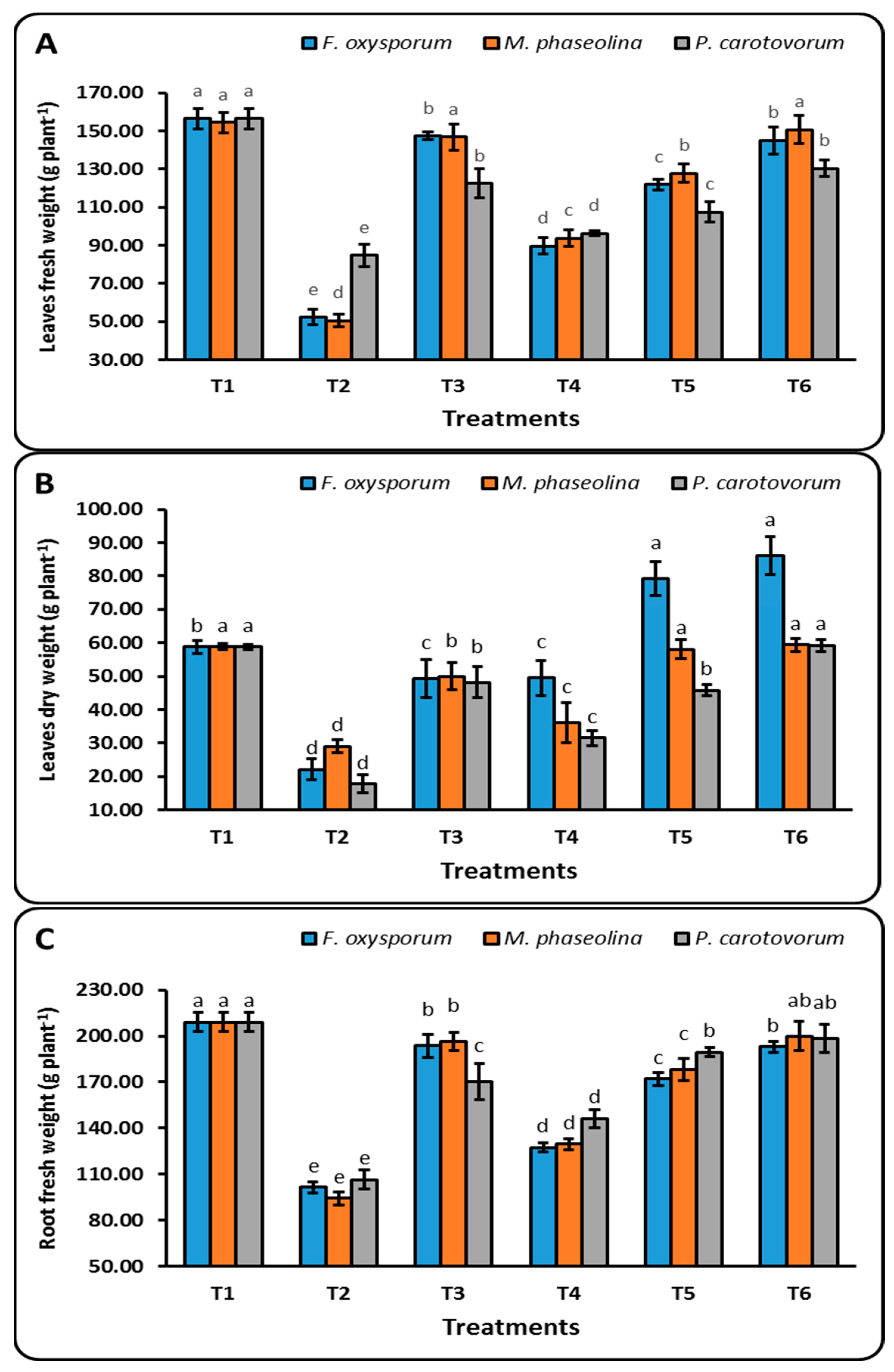
As shown in Figure 2, there are significant differences (P ≤ 0.05) in the vegetative growth of sugar beet plants under different concentrations of CuO-NPs (50, 100, and 150 µg mL−1), as affected by plant pathogenic microorganisms (F. oxysporum, M. phaseolina, and P. carotovorum). These differences can be seen in the leaves’ fresh weight (A), leaves’ dry weight (B), roots’ fresh weight (C), and roots’ dry weight (D). At 90 days after sowing, sugar beet seeds treated with 150 µg mL−1 significantly increased leaves’ fresh weight (g plant−1) from 52.20, 50.57, and 84.70 (T2) to 145.00, 150.70, and 130.27 (T6), whereas the same treatment increased leaves’ dry weight (g plant−1) from 22.10, 29.00, and 17.80 (T2) to 86.03, 59.43, and 59.27 (T6), for F. oxysporum, M. phaseolina, and P. carotovorum, respectively. In the same way, the application of CuO-NPs reduced the negative impact of plant pathogenic microorganisms on roots’ fresh and dry weights (Figure 2). The T6 treatment was more effective than the control and other treatments, with measurements of 193.03 and 100.57 g plant−1 for F. oxysporum, 199.83 and 86.30 g plant−1 for M. phaseolina, and 198.40 and 79.03 g plant−1 for P. carotovorum for roots’ fresh and dry weights, respectively (Figure 2).

3.3.2. Physiological Characteristics

At 90 days after sowing, there were significant differences in the number of chlorophyll (a, b, and total), carotenoids, and total soluble sugar in sugar beet leaves according to the different applications of CuO-NPs (Table 3).
Under soil infected with F. oxysporum, the greatest values of chlorophyll a, b, and total were 3.25, 2.33, and 5.85 mg g−1 FW, followed by 3.05, 2.08, and 5.13 mg g−1 FW for treatments T6 and T5, respectively (Table 3). The same pattern was seen in soil that M. phaseolina and P. carbovorum had treated. On the other hand, T6 treatment was the best treatment under various CuO-NP applications, recording 0.93, 1.27, and 0.79 for carotenoids (µg g−1 FW) and 5.96, 6.08, and 6.05 for TSS (µg g−1 FW) under various plant pathogenic bacteria, namely F. oxysporum; M. phaseolina, and P. carotovorum, respectively (Table 3). According to the aforementioned data, the descending order of T6 > T5 > T4 was established for different applications of CuO-NPs, under plant pathogenic microorganisms (Table 3).

3.3.3. Antioxidant Enzymes

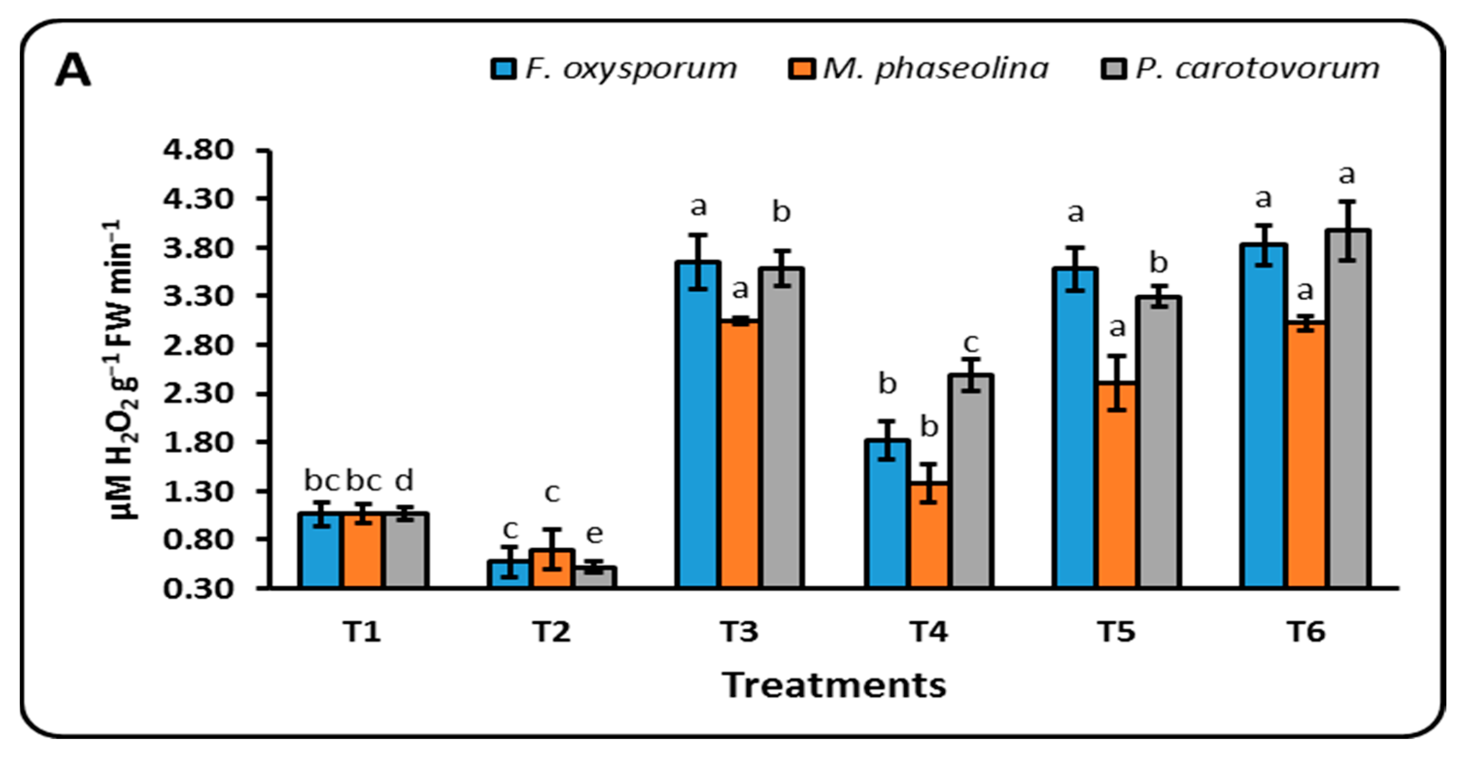
Data shown in Figure 3 demonstrate that various treatments of CuO-NPs and plant pathogenic microorganisms considerably altered the activities of peroxidase (PO, A), polyphenol oxidase (PPO, B), and phenylalanine ammonia layase (PAL, C). Different CuO-NPs concentrations significantly boosted the activity of the antioxidant enzymes PO, PPO, and PAL in sugar beet leaves 90 days after planting compared to the control (Figure 3).
In comparison to other examined treatments, the T6 treatment (seeds soaked in 150 µg mL−1) effectively enhanced the PO and PPO contents for F. oxysporum, M. phaseolina, and P. carotovorum by 3.82, 3.02, and 3.98 µM H2O2 g−1 FW min−1 and 0.29, 0.27, and 0.32 µM tetra guaiacol g−1 FW min−1, respectively.
For F. oxysporum, M. phaseolina, and P. carotovorum, the highest PAL activity levels were seen with the T6 treatment (6.16, 5.74, and 5.49 µmol min−1 g−1 FW), followed by the T5 treatment (3.79, 3.30, and 4.20 µmol min−1 g−1 FW), and the T2 treatment (1.09, 1.15, and 1.60 µmol min−1 g−1 FW), respectively (Figure 3).

3.3.4. Disease Incidence Percent

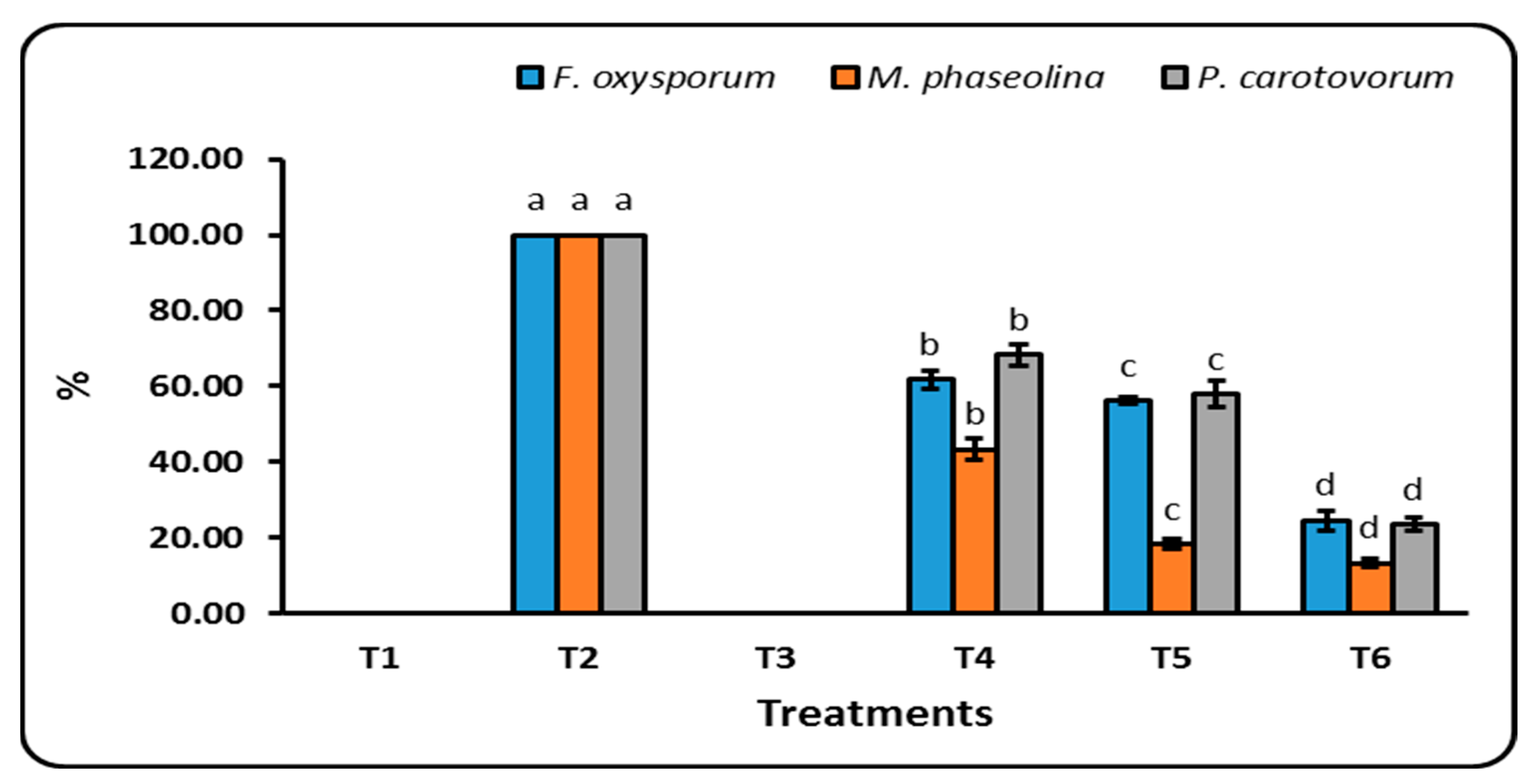
According to the findings shown in Figure 4, seeds treated with CuO-NPs at different doses (50, 100, and 150 µg mL−1) considerably reduced the proportion of soil infection with plant pathogenic microorganisms when compared to the control treatment. In comparison to the control treatment (T2), seeds treated with 150 µg mL−1 (T6) had the greatest effect on lowering infection, with reductions of 24.53, 13.25, and 23.59% followed by 100 µg mL−1 (T5), which recorded 56.18, 18.26, and 58.05% for F. oxysporum, M. phaseolina, and P. carotovorum, respectively (Figure 4).

3.3.5. Some Chemical Contents of Sugar Beet Leaves

There are significant differences (p ≤ 0.05) in the chemical composition of sugar beet leaves (N, P, K, and Cu) under soil infected by plant pathogenic microorganisms (F. oxysporum, M. phaseolina, and P. carotovorum), as influenced by various concentrations of CuO-NPs (Table 4).
Ninety days after sowing, sugar beet seeds treated with 150 µg mL−1 increased N (%) significantly from 0.59, 0.57, and 0.52 (T2) to 1.62, 1.61, and 1.70 (T6), whereas the same treatment increased P (%) from 0.047, 0.068, and 0.052 (T2) to 0.147, 0.153, and 0.114 (T6), for F. oxysporum, M. phaseolina, and P. carotovorum, respectively. Regarding K and Cu (%), the highest values were observed with the T6 treatment, which recorded 3.10 and 0.27% under soil infected by F. oxysporum, 3.46 and 0.025 % for soil infected by M. phaseolina, and 3.10 and 0.029 % for soil infected by P. carotovorum, compared to the control and other treatments, respectively (Table 4).

4. Discussion

Several proteins and metalloenzymes contain copper, a crucial mineral that is important for plant nutrition and health. Due to their distinct features, Cu-NPs perform and behave more effectively than bulk copper particles. Cu-NPs are displaying new uses in healthcare, industry, and agriculture as a result of their antimicrobial properties.

4.1. Effect of Cu-NPs on Plant Pathogens Microorganisms (In Vitro)

Cu-NPs’ antimicrobial potency increases with dosage; for example, when 25 and 35 g/mL of Cu-NPs were used, the reaction’s antifungal potency was 100% (Figure 1). These outcomes are a result of CuO-NPs’ high surface area, which makes them particularly reactive. Due to their high surface-to-volume ratio, Cu-NPs can interact with the cell membrane of bacteria through their surface, killing pathogens [33]. Additionally, the Cu-NPs have a bactericidal effect on bacteria and fungi by interfering with their ability to develop. According to [34], the copper nano sol made from the leaf extract of the Ocimum sanctum plant exhibited antibacterial action (18 mm) against Staphylococcus aureus. Moreover, CuNPs have shown notable action against the bacterial disease Aeromonas hydrophila that affects fish [35]. Additionally, the biosynthesized Cu-NPs demonstrated greater antibacterial activity (zone of inhibition) against a variety of human and fish bacterial pathogens, including Vibrio anguillarum, A. caviae, Bacillus cereus, Proteus mirabilis, and S. aureus [18]. In addition, ref. [36] showed that the antifungal efficacy of Cu-NPs was greatest against F. oxysporum (37.0 mm), Alternaria solani (28.0 mm), and Aspergillus niger (26.5 mm). However, it proved effective as an antibacterial agent against Erwinia amylovora (22.0 mm) and Ralstonia solanacearum (19.0 mm).

4.2. Effect of CuO-NPs on Growth Parameters of Sugar Beet under Soil Infected by Plant Pathogenic Microorganisms

Our results showed that the T9 treatment (seeds soaked in 150 µg mL−1 CuO-NPs) significantly increased the results for the length of roots and leaves, the fresh weight of roots and leaves, and the dry weight of roots and leaves in soil infected with F. oxysporum, M. phaseolina, and P. carotovorum (Table 2 and Figure 2). These encouraging results were likely a result of copper’s function as a crucial micronutrient for plant development. It participates in oxidative stress responses, mitochondrial respiration, photosynthetic electron transport, cell wall metabolism, and hormone signalling [11]. Additionally, copper can result in altered root systems, increased generation of bioactive chemicals, and suppression of plant growth by reactive oxygen species (ROS) [37,38,39].
According to several studies, the application of CuO-NPs increased plant growth criteria [40], soaking wheat plants in Cu-NPs caused them to grow more quickly [41], and cilantro (Coriandrum sativum) plants gained weight when cultivated in soil that had been supplemented with CuO-NPs at an amount of 80 mg kg−1. Hafeez et al. [42] showed that wheat grew more quickly in soils that had been modified with Cu-NPs at concentrations ranging from 10 to 30 ppm. In a different investigation, treatment with a lower CuO-NP concentration (100 mg L−1) caused the root and shoot length of the eggplant seedlings to rise in comparison to control plants [43]. Furthermore, ref. [44] demonstrated that CuO-NPs (50 ppm) induced noticeably higher growth characteristics of wheat plants (shoot and root length, fresh and dry weight of shoot and root, and leaves per plant).

4.3. Physiological Characteristics

Under soil infected with F. oxysporum, M. phaseolina, and P. carbovorum, there were significant differences in the amounts of chlorophyll (a, b, and total), carotenoids, and total soluble sugar in sugar beet leaves according to the different applications of CuO-NPs (Table 3). The enhanced biological and chemical activities of metals at the nanoscale and the corresponding effects of nutrients (Mg, Fe, Zn, and S) on plants may be the causes for the increase in the physiological features of sugar beet plants. Young [45] claimed that increasing carotenoids have a major impact on scavenging ROS and guarding against the anticipated stress on the plant. Additionally, metal NPs can improve the structure of chlorophyll and promote the production of pigments and metabolic processes [46,47]. Because copper is a structural component of regulatory proteins, an enzyme activator, and a component of the photosynthetic electron transport chain, it has been noted that plants require tiny amounts of copper for optimal growth [48]. Our results are consistent with research on wheat plants by [49], tomato plants by [50], green pea plants by [51], and wheat plants by [44].

4.4. Antioxidant Enzymes

The levels of the antioxidant enzymes peroxidase (PO), polyphenol oxidase (PPO), and phenylalanine ammonia lyase (PAL) increased at all tested doses of Cu-NPs (Figure 3). According to [52], copper is involved in a number of physiological and biochemical processes, serves as an activator for antioxidant enzymes, and is incorporated into a number of enzymes, as well as being essential for plant growth and nutrition [53]. Antioxidant enzyme activity increased, showing a protective function against oxidative injury in plants. Our findings are consistent with several studies that have shown the involvement of CuO-NPs in antioxidant enzyme enhancement [54,55]. CuO-NP treatment was observed to increase APX and POD activities in certain earlier experiments [56]. Recent findings from [57] showed that lentil (Lens culinaris) plants treated with various doses of CuO-NPs have elevated APX and SOD activity (0.01, 0.025, and 0.05 mg mL−1). Additionally, 20 days after planting, potato plants treated with CuO-NPs at a dosage of 200 mg L−1 showed an increase in PO and PPO activity [58].

4.5. Disease Incidence Percent

According to our findings, seeds treated with CuO-NPs at different doses (50, 100, and 150 µg mL−1) considerably decrease the proportion of soil infection with plant pathogenic microorganisms (F. oxysporum, M. phaseolina, and P. carotovorum) when compared to control treatment (Figure 4).
These outcomes brought on by CuNPs’ biocidal activity may be explained by the impact of CuNPs or the copper ions released from Cu-NPs. Due to the NPs’ large surface area, they may be aggressively adsorbed to the surface of microorganisms, disrupting cell permeability and releasing essential components [59]. According to [60], Cu interacts with microorganisms in a variety of ways, including by permeating cell membranes, oxidizing membrane lipids, altering proteins, and denaturing nucleic acids, all of which result in cell death. According to [61], field-testing with CuO-NPs at 200 μL L−1 resulted in a 100% decrease in the Rhizoctonia solani-caused black scurf disease. Copper-based NPs showed promise in preventing Phytophthora infestans-caused tomato late blight [62], as well as a number of fungal diseases such as A. alternata and Botrytis cinerea [63], tomato Fusarium wilt [64], and black scurf disease in potato [58].

4.6. N, P, K and Cu of Sugar Beet Leaves

In comparison to control and other treatments, T6 treatment (150 g mL−1) under soil infected by F. oxysporum, M. phaseolina, and P. carotovorum showed the highest values for N, P, K, and Cu % (Table 4). This outcome could be explained in a number of ways, such as by the application of nano-Cu, which reduced the pH of the growing medium and boosted the uptake of nutrients [65]. According to [66], mesoporous aluminosilicates have been used as CuO nanoparticle carriers and may be able to transfer macro- and micronutrients to the soil in the future. The cytochrome oxidase enzyme, which catalyzes electron transport during respiration, contains copper, and it is crucial for lipid and carbohydrate metabolism [67]. Cu nanofertilizers were used at a dose of up to 100 mg L−1 to create the best growth conditions and increase the bioavailability of the various nutrients, including N, P, K, Cu, Fe, Mn, Se, and Zn [68]. Interestingly, CuO-NPs were found to be more effective than the other six metallic oxide nanoparticles (ZnO, TiO, AlO, FeO, NiO, or MnO) at enhancing tomato and eggplant growth and nutrient uptake in soil contaminated with Verticillium dahliae and F. oxysporum f.sp. lycopersici, respectively [64].

5. Conclusions

The goal of this study was to determine the antimicrobial activity of different concentrations of biologically CuO-NPs against F. oxysporum, M. phaseolina, and P. carotovorum and their impact on the growth dynamics of sugar beet plants. According to the results, 150 µg mL−1 produced the greatest reduction in disease incidence %, and increased the vegetative development, physiological characteristics, antioxidant enzymes, and mineral nutrients of sugar beet plants. Therefore, the biosynthesized CuO-NPs antibacterial activity suggests that they can fend off sugar beet plants’ plant pathogenic microbes.

Supplementary Materials

The following supporting information can be downloaded at: https://www.mdpi.com/article/10.3390/su141912871/s1, Figure S1a: Effect of different concentrations of CuSO4.5H2O (A) and CuO-NPs (B) on the antagonism of F. oxysporum. P1: Control; P2: 5 μg mL−1; P3: 15 μg mL−1; P4: 30 μg mL−1; P5: 60 μg mL−1; P6: 70 μg mL−1; and P7: 90 μg mL−1. Figure S1b: Effect of different concentrations of CuO-NPs (A) and CuSO4.5H2O (B) on the antagonism of M. phaseolina. P1: Control; P2: 5 μg mL−1; P3: 15 μg mL−1; P4: 30 μg mL−1; P5: 60 μg mL−1; and P6: 70 μg mL−1. Figure S1c: Effect of different concentrations of CuSO4.5H2O (A) and CuO-NPs (B) on the antagonism of P. Carotovorum. Figure S2. Effect of different concentrations of CuO-NPs on some parameters of seedling of sugar beet under infected with different plant pathogenic microorganisms. C1: Uninfected; C2: Infected; C3: 5 µg mL−1; C4: 15 µg mL−1; C5: 30 µg mL−1; C6: 60 µg mL−1; C7: 90 µg mL−1; C8: 120 µg mL−1; and C9: 150 µg mL−1.

Author Contributions

Conceptualization, E.A.-S., A.R.A., M.E. and A.E.-D.O.; methodology, E.A.-S., A.R.A. and A.E.-D.O.; software, E.A.-S., M.E. and A.E.-D.O.; validation, E.A.-S., M.E. and A.E.-D.O.; formal analysis, E.A.-S. and A.E.-D.O.; investigation, A.R.A. and A.E.-D.O.; resources, E.A.-S., M.E. and A.E.-D.O.; data curation, E.A.-S. and A.E.-D.O.; writing—original draft preparation, A.E.-D.O.; writing—review and editing, A.E.-D.O.; visualization, A.R.A., M.E. and A.E.-D.O.; supervision, A.R.A. and A.E.-D.O.; funding acquisition, M.E. All authors have read and agreed to the published version of the manuscript.

Funding

This research received no external funding.

Institutional Review Board Statement

Not applicable.

Informed Consent Statement

Not applicable.

Data Availability Statement

The study did not report any data.

Acknowledgments

The authors extend their appreciation to the Deanship of Scientific Research at King Khalid University for funding this work through the Research Groups Project under grant number R.G.P. 2/138/43. All of the authors are grateful for the support provided by the Soils, Water, and Environment Research Institute (SWERI), Agriculture Research Center (ARC), Egypt.

Conflicts of Interest

The authors declare no conflict of interest.

References

  1. Mali, S.C.; Dhaka, A.; Githala, C.K.; Trivedi, R. Green synthesis of copper nanoparticles using Celastrus aniculatus Willd. Leaf extract and their photocatalytic and antifungal properties. Biotechnol. Rep. 2020, 27, e00518. [Google Scholar] [CrossRef]
  2. Fouda, A.; Abdel-Maksoud, G.; Abdel-Rahman, M.A.; Salem, S.S.; Hassan, S.E.D.; El-Sadany, M.A.H. Eco-friendly approach utilizing green synthesized nanoparticles for paper conservation against microbes involved in biodeterioration of archaeological manuscript. Int. Biodeterior. Biodegrad. 2019, 142, 160–169. [Google Scholar] [CrossRef]
  3. Pugazhendhi, A.; Prabhu, R.; Muruganantham, K.; Shanmuganathan, R.; Natarajan, S. Anticancer, antimicrobial and photocatalytic activities of green synthesized magnesium oxide nanoparticles (MgONPs) using aqueous extract of Sargassum wightii. J. Photochem. Photobiol. B Biol. 2019, 190, 86–97. [Google Scholar] [CrossRef]
  4. Lin, D.; Xing, B. Root uptake and phytotoxicity of ZnO nanoparticles. Environ. Sci. Technol. 2008, 42, 5580–5585. [Google Scholar] [CrossRef]
  5. Nel, A.; Xia, T.; Mädler, L.; Li, N. Toxic potential of materials at the nanolevel. Science 2006, 311, 622–627. [Google Scholar] [CrossRef] [Green Version]
  6. Shaheen, T.I.; Abd El Aty, A.A. In-situ green myco-synthesis of silver nanoparticles onto cotton fabrics for broad spectrum antimicrobial activity. Int. J. Biol. Macromol. 2018, 118, 2121–2130. [Google Scholar] [CrossRef] [PubMed]
  7. Salem, S.S.; Fouda, A. Green synthesis of metallic nanoparticles and their prospective biotechnological applications: An Overview. Biol. Trace Elem. Res. 2020, 199, 344–370. [Google Scholar] [CrossRef]
  8. Cuevas, R.; Durán, N.; Diez, M.C.; Tortella, G.R.; Rubilar, O. extracellular biosynthesis of copper and copper oxide nanoparticles by Stereum hirsutum, a native white-rot fungus from Chilean forests. J. Nanomater. 2015, 789089. [Google Scholar]
  9. Majumdar, T.D.; Singh, M.; Thapa, M.; Dutta, M.; Mukherjee, A.; Ghosh, C.K. Size-dependent antibacterial activity of copper nanoparticles against Xanthomonas oryzae pv. Oryzae—A synthetic and mechanistic approach. Colloid Interface Sci. Commun. 2019, 32, 100190. [Google Scholar] [CrossRef]
  10. Yruela, I. Copper in plants. Braz. J. Plant. Physiol. 2005, 17, 145–156. [Google Scholar] [CrossRef] [Green Version]
  11. Da Costa, M.V.J.; Sharma, P.K. Effect of copper oxide nanoparticles on growth, morphology, photosynthesis, and antioxidant response in Oryza sativa. Photosynthetica 2016, 54, 110–119. [Google Scholar] [CrossRef]
  12. Watson, L.; Dallwitz, M. The Families of Flowering Plants: Descriptions, Illustrations, Identification, and Information Retrieval. 1992. Available online: http://biodiversityunoedu/delta (accessed on 1 July 2021).
  13. Hassnein, A.M.; Azab, M.A.; El-Hawary, M.A.; Darwish, N.N. Effect of nano fertilization on sugar beet. Al-Azhar J. Agric. Res. 2019, 44, 194–201. [Google Scholar] [CrossRef]
  14. Abbas, M.S.; El-Hassanin, A.S.; Dewdar, M.D.H.; Abd Elaleem, H.A. Impact of nanomicronutrients as foliar fertilization on yield and quality of sugar beet roots. Pak. J. Biol. Sci. 2020, 23, 1416–1423. [Google Scholar] [CrossRef]
  15. Liu, R.; Lal, R. Potentials of engineered nanoparticles as fertilizers for increasing agronomic productions. A review. Sci. Total Environ. 2015, 514, 131–139. [Google Scholar] [CrossRef] [PubMed]
  16. Kanhed, P.; Sonal, B.; Swapnil, G.; Aniket, G.; Amedea, B.; Seabra, O.R.; Nelson, D.; Mahendra, R. In vitro antifungal efficacy of copper nanoparticles against selected crop pathogenic fungi. Mater. Lett. 2014, 115, 13–17. [Google Scholar] [CrossRef]
  17. Shende, S.; Ingle, A.P.; Gade, A.; Rai, M. Green synthesis of copper nanoparticles by Citrus medica Linn. (Idilimbu) juice and its antimicrobial activity. World J. Microbiol. Biotechnol. 2015, 31, 865–873. [Google Scholar] [CrossRef]
  18. Nabila, M.I.; Krishnan, K. Biosynthesis, characterization and antibacterial activity of copper oxide nanoparticles (CuO NPs) from actinomycetes. Biocatal. Agric. Biotechnol. 2018, 15, 56–62. [Google Scholar] [CrossRef]
  19. Ganesan, P.; Reegan, A.D.; David, R.H.A.; Gandhi, M.R.; Paulraj, M.G.; Al-Dhabi, N.A.; Ignacimuthu, S. Antimicrobial activity of some actinomycetes from Western Ghats of Tamil Nadu. India Alex. J. Med. 2017, 53, 101–110. [Google Scholar] [CrossRef] [Green Version]
  20. Whipps, J.M. Effect of media on growth and interactions between a range of soil-borne glasshouse pathogens and antagonistic fungi. New Phytol. 1987, 107, 127–142. [Google Scholar] [CrossRef]
  21. El-Shabrawy, E.; Heba, S. Controlling maize late-wilt and enhancing plant salinity tolerance by some rhizobacterial strains. Egypt. J. Phytopathol. 2018, 46, 235–255. [Google Scholar] [CrossRef]
  22. Abdalla, M.E.; Seadh, S.E.; Hamza, S. Inhibitory effect and morphological changes by organic acids to bacterial strains causing sugar beet soft root rot in Vitro. J. Plant Prot. Pathol. 2019, 10, 187–193. [Google Scholar] [CrossRef]
  23. Lichtenthaler, H.K. Chlorophylls and carotenoids: Pigments of photosynthetic biomembranes. In Methods in Enzymology; Academic Press: San Diego, CA, USA, 1987; Volume 148, pp. 350–382. [Google Scholar]
  24. Hendrix, D.L. Rapid extraction and analysis of nonstructural carbohydrates in plant tissues. Crop. Sci. 1993, 33, 1306–1311. [Google Scholar] [CrossRef]
  25. Srivastava, S.K. Peroxidase and polyphenol-oxidase in Brassica juncea plants infected with Macrophomina phaseolina (Tassi): Goid of and their implication in disease resistance. Phytopathology 1987, 120, 249–254. [Google Scholar] [CrossRef]
  26. Matta, A.I.; Dimond, A.F. Symptoms of Fusarium wilt in relation to quantity of fungus and enzyme activity in tomato stems. Phytopathology 1963, 53, 574–578. [Google Scholar]
  27. Zucker, M. Sequential induction of phenylalanine ammonia layse and lyase in inactivating system in potato tuber disks. Plant Physiol. 1968, 43, 365–374. [Google Scholar] [CrossRef]
  28. Crowe, F.J.; Debons, J.; Darnell, T.; McGrath, D.; Koepsell, P.; Laborde, J.; Redondo, J. Control of Allium white rot with DADS and related products. In Proceedings of the Fifth International Workshop on Allium White Rot, Cordoba, Spain, April 1994; Entwistle, A.R., Melero-Vara, J.M., Eds.; HRI: Norwich, UK; pp. 15–19. [Google Scholar]
  29. Page, A.L.; Miller, R.H.; Keeny, D.R. Methods of Soil Analysis, Part II, Agronomy Monographs ASA and SSSA, 2nd ed.; Madison Book Company: Madison, WI, USA, 1982. [Google Scholar]
  30. Murphy, J.; Riley, J.P. A modified single solution method for the determination of phosphate in natural waters. Anal. Chim. Acta 1962, 27, 31–36. [Google Scholar] [CrossRef]
  31. USDA. Soil Survey Laboratory Methods Manual Soil Survey Investigation Report, No. 42, Version 4; USDA-NRCS: Lincoln, NE, USA, 2004. [Google Scholar]
  32. Gomez, K.A.; Gomez, A. Statistical Procedure for Agricultural Research—Handbook; Wiley: New York, NY, USA, 1984. [Google Scholar]
  33. Usman, M.S.; El-Zowalaty, M.E.; Shameli, K.; Zainuddin, N.; Salama, M.; Ibrahim, N.A. properties of copper nanoparticles. Int. J. Nanomed. 2013, 8, 4467–4479. [Google Scholar]
  34. Patel, B.H.; Channiwala, M.Z.; Chaudhari, S.B.; Mandot, A.A. Biosynthesis of copper nanoparticles; its characterization and efficacy against human pathogenic bacterium. J. Environ. Chem. Eng. 2016, 4, 2163–2169. [Google Scholar] [CrossRef]
  35. Muralisankar, T.; Bhavan, P.S.; Gandimathy, S.; Radhakrishnan, S.; Seenivasan, C.; Aarumugam, P. In-vitro antibacterial activity of Zinc and nanoparticles against Gram-negative bacteria Aeromonas hydrophila. Malaya J. Biosci. 2015, 1, 196–200. [Google Scholar]
  36. El-Batal, A.I.; Gharieb, S.E.; Farag, M.M.; Rasha, M.F. Penicillium chrysogenum-mediated mycogenic synthesis of copper oxide nanoparticles using gamma rays for in vitro antimicrobial activity against some plant pathogens. J. Clust. Sci. 2020, 31, 79–90. [Google Scholar] [CrossRef]
  37. Lequeux, H.; Hermans, C.; Lutts, S.; Verbruggen, N. Response to copper excess in Arabidopsis thaliana: Impact on the root system architecture, hormone distribution, lignin accumulation and mineral profile. Plant. Physiol. Biochem. 2010, 48, 673–682. [Google Scholar] [CrossRef]
  38. Feigl, G.; Kumar, D.; Lehotai, N.; Tugyi, N.; Molnár, Á.; Ördög, A.; Szepesi, Á.; Gémes, K.; Laskay, G.; Erdei, L.; et al. Physiological and morphological responses of the root system of Indian mustard (Brassica juncea L. Czern.) and rapeseed (Brassica napus L.) to copper stress. Ecotoxicol. Environ. Saf. 2013, 94, 179–189. [Google Scholar]
  39. Mourato, M.P.; Moreira, I.N.; Leitão, I.; Pinto, F.R.; Sales, J.R.; Martins, L.L. Effect of heavy metals in plants of the genus Brassica. Int. J. Mol. Sci. 2015, 16, 17975–17998. [Google Scholar] [CrossRef] [PubMed] [Green Version]
  40. Yasmeen, F.; Razzaq, A.; Iqbal, M.N.; Jhanzab, H.M. Effect of silver, copper and iron nanoparticles on wheat germination. Int. J. Biosci. 2015, 6, 112–117. [Google Scholar]
  41. Zuverza-Mena, N.; Medina-Velo, I.A.; Barrios, A.C.; Tan, W.; Peralta-Videa, J.R.; Gardea-Torresdey, J.L. Copper nanoparticles/compounds impact agronomic and physiological parameters in cilantro (Coriandrum sativum). Environ. Sci. Process. Impacts 2015, 17, 1783–1793. [Google Scholar] [CrossRef]
  42. Hafeez, A.; Razzaq, A.; Mahmood, T.; Jhanzab, H.M. Potential of copper nanoparticles to increase growth and yield of wheat. J. Nanosci. Adv. Technol. 2015, 1, 6–11. [Google Scholar]
  43. Baskar, V.; Nayeem, S.; Kuppuraj, S.P.; Muthu, T.; Ramalingam, S. Assessment of the effects of metal oxide nanoparticles on the growth, physiology and metabolic responses in vitro grown eggplant (Solanum melongena). 3 Biotech. 2018, 8, 362. [Google Scholar] [CrossRef] [PubMed]
  44. Badawy, A.A.; Nilly, A.H.A.; Salem, S.S.; Mohamed, F.A.; Amr, F. Efficacy assessment of biosynthesized copper oxide nanoparticles (CuO-NPs) on stored grain insects and their impacts on morphological and physiological traits of wheat (Triticum aestivum L.) plant. Biology 2021, 10, 233. [Google Scholar] [CrossRef] [PubMed]
  45. Young, A.J. The photoprotective role of carotenoids in higher plants. Physiol. Plant. 1991, 83, 702–708. [Google Scholar] [CrossRef]
  46. Amin, M.A.; Badawy, A. Metabolic changes in common bean plants in response to zinc nanoparticles and zinc sulfate. Int. J. Innov. Sci. Eng. Technol. 2017, 4, 321–335. [Google Scholar]
  47. Ewais, E.; Ismail, M.; Badawy, A. Vegetative growth, photosynthetic pigments and yield of Phaseolus vulgaris (L.) plants in response to the application of biologically-synthesized zinc oxide nanoparticles and zinc sulfate. Al Azhar Bull. Sci. 2017, 9, 33–46. [Google Scholar]
  48. Shobha, G.; Moses, V.; Ananda, S. Biological synthesis of copper nanoparticles and its impact. Int. J. Pharm. Sci. Invent. 2014, 3, 6–28. [Google Scholar]
  49. Kasana, R.C.; Panwar, N.R.; Kaul, R.K.; Kumar, P. Biosynthesis and effects of copper nanoparticles on plants. Environ. Chem. Lett. 2017, 15, 233–240. [Google Scholar] [CrossRef]
  50. Singh, A.; Singh, N.B.; Hussain, I.; Singh, H. Effect of biologically synthesized copper oxide nanoparticles on metabolism and antioxidant activity to the crop plants Solanum lycopersicum and Brassica oleracea var. botrytis. J. Biotechnol. 2017, 262, 11–27. [Google Scholar] [CrossRef]
  51. Ochoa, L.; Zuverza-Mena, N.; Medina-Velo, I.A.; Flores-Margez, J.P.; Peralta-Videa, J.R.; Gardea-Torresdey, J.L. Copper oxide nanoparticles and bulk copper oxide, combined with indole-3-acetic acid, alter aluminum, boron, and iron in Pisum sativum seeds. Sci. Total Environ. 2018, 634, 1238–1245. [Google Scholar] [CrossRef] [PubMed]
  52. Bowler, C.; Montagu, M.V.; Inze, D. Superoxide dismutase and stress tolerance. Annu. Rev. Plant Biol. 1992, 43, 83–116. [Google Scholar] [CrossRef]
  53. Kasana, A.; AliNiazee, M.T. A thermal unit summation model for the phenology of Rhagoietis completa (Diptera: Tephritidae). J. Entomol. Soc. Br. Columbia 1997, 94, 13–18. [Google Scholar]
  54. Wang, S.-H.; Yang, Z.-M.; Yang, H.; Lu, B.; Li, S.-Q.; Lu, Y.-P. Copper-induced stress and antioxidative responses in roots of Brassica juncea L. Bot. Bull. Acad. Sin. 2004, 45, 203–212. [Google Scholar]
  55. Ananda, S.; Shobha, G.; Shashidhara, K.; Mahadimane, V. Nano-cuprous oxide enhances seed germination and seedling growth in Lycopersicum esculentum plants. J. Drug Deliv. Ther. 2019, 9, 296–302. [Google Scholar]
  56. Nair, P.M.G.; Chung, I.M. Impact of copper oxide nanoparticles exposure on Arabidopsis thaliana growth, root system development, root lignificaion, and molecular level changes. Environ. Sci. Pollut. Res. 2014, 21, 12709–12722. [Google Scholar] [CrossRef]
  57. Sarkar, J.; Chakraborty, N.; Chatterjee, A.; Bhattacharjee, A.; Dasgupta, D.; Acharya, K. Green synthesized copper oxide nanoparticles ameliorate defence and antioxidant enzymes in Lens culinaris. Nanomaterials 2020, 10, 312. [Google Scholar] [CrossRef] [Green Version]
  58. Ismail Ahmed, M. Efficacy of Copper Oxide and Magnesium Oxide Nanoparticles on Controlling Black Scurf Disease on Potato. Egypt. J. Phytopathol. 2021, 49, 116–130. [Google Scholar] [CrossRef]
  59. Raffi, M.; Mehrwan, S.; Bhatti, T.M.; Akhter, J.I.; Yawar, A.W.; Masood ul Hasan, M. Investigations into the antibacterial behavior of copper nanoparticles against Escherichia coli. Ann. Microbiol. 2010, 60, 75–80. [Google Scholar] [CrossRef]
  60. Oussou-Azo, A.F.; Nakama, T.; Nakamura, M.; Futagami, T.; Vestergaard, M.C.M. Antifungal potential of nanostructured crystalline copper and its oxide forms. Nanomaterials 2020, 10, 1003. [Google Scholar] [CrossRef] [PubMed]
  61. El-Shewy, E.S.A.; Mohemed, F.G.; Abd-latif, F.M.; Hafez, E.M.; Mansour, S.A. The efficacy of copper oxide, tri-calcium phosphate and silicon dioxide nanoparticles in controlling black scurf disease of potato. Ann. Agric. Sci. Moshtohor 2019, 57, 129–138. [Google Scholar] [CrossRef] [Green Version]
  62. Giannousi, K.; Avramidis, I.; Dendrinou-Samara, C. Synthesis, characterization and evaluation of copper-based nanoparticles as agrochemicals against Phytophthora infestans. RSC Adv. 2013, 3, 21743–21752. [Google Scholar] [CrossRef]
  63. Ouda, S.M. Antifungal activity of silver and copper nanoparticles on two plant pathogens, Alternaria alternata and Botrytis cinerea. Res. J. Microbiol. 2014, 9, 34–42. [Google Scholar] [CrossRef] [Green Version]
  64. Elmer, W.H.; White, J.C. The use of metallic oxide nanoparticles to enhance growth of tomatoes and eggplants in disease infested soil or soilless medium. Environ. Sci. Nano 2016, 3, 1072–1079. [Google Scholar] [CrossRef]
  65. Ortas, I.; Rafique, M.; Akpinar, C.; Kacar, Y.A. Growth media and mycorrhizal species effect on acclimatization and nutrient uptake of banana plantlets. Sci. Hortic. 2017, 217, 55–60. [Google Scholar] [CrossRef]
  66. Huo, C.; Ouyang, J.; Yang, H. CuO nanoparticles encapsulated inside Al-MCM-41 mesoporous materials via direct synthetic route. Sci. Rep. 2014, 4, 3682. [Google Scholar] [CrossRef] [Green Version]
  67. Dey, J.K.; Das, S.; MawlongInt, L.G. Nanotechnology and its importance in micronutrient fertilization. J. Curr. Microbiol. Appl. Sci. 2018, 7, 2306–2325. [Google Scholar]
  68. Shalaby, T.A.; Said, M.E.; Mohammed, E.E.; Alaa, E.O.; Hossam, S.E.; Hassan, E. Acclimatization of In Vitro Banana Seedlings Using Root-Applied Bio-Nanofertilizer of Copper and Selenium. Agronomy 2022, 12, 539. [Google Scholar] [CrossRef]
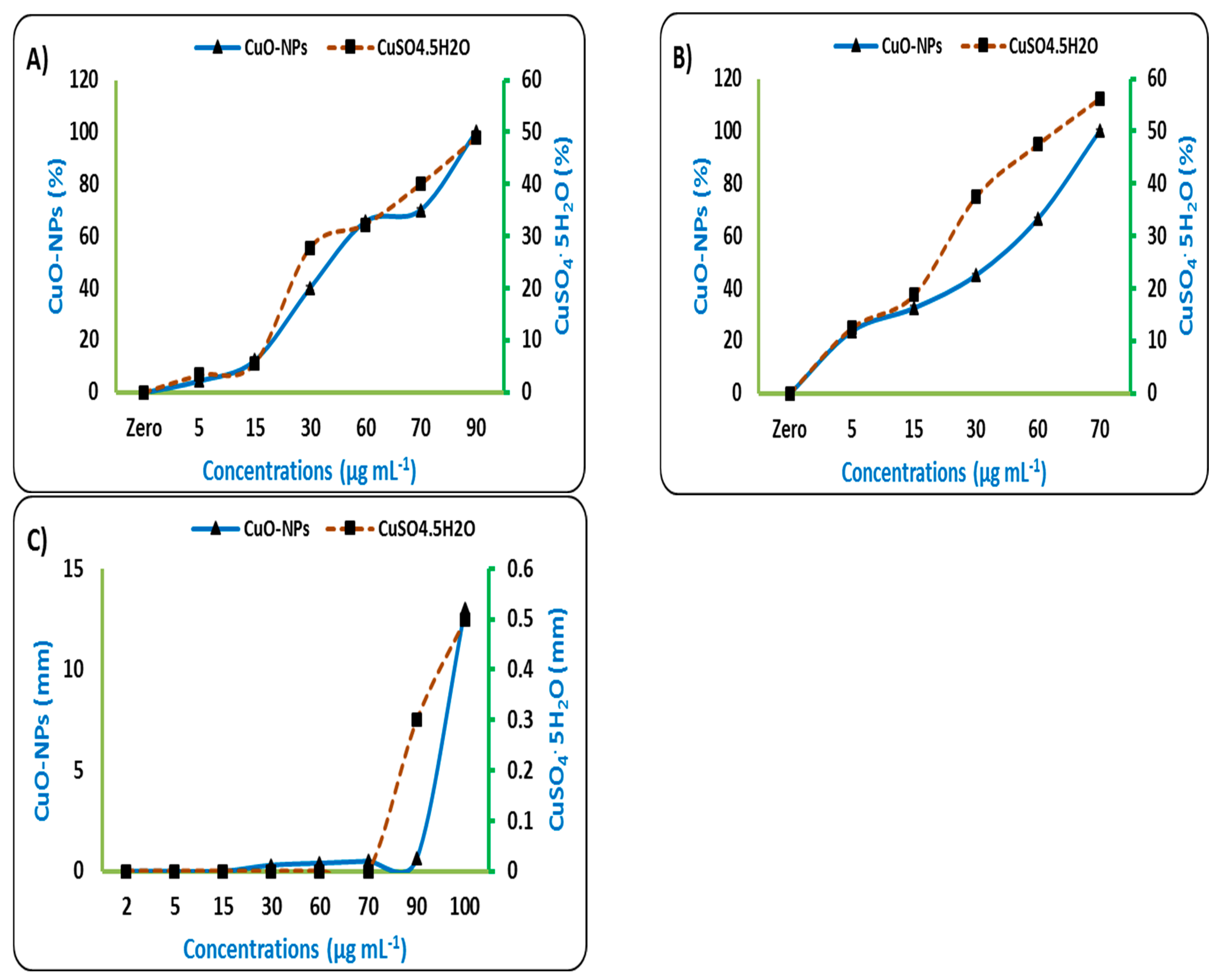
Figure 1. Effect of different concentrations of CuO–NPs and CuSO4.5H2O on the antagonism of plant pathogens microorganisms; F. oxysporum (A); M. phaseolina (B); and P. carotovorum (C).
Sustainability 14 12871 g001
Figure 2. Effect of different concentrations of CuO-NPs on vegetative growth of sugar beet plants (leaves’ fresh weight (A), leaves’ dry weight (B), root fresh weight (C), root dry weight (D)) as affected by plant pathogenic microorganisms after 90 days of sowing. According to Duncan’s test at the 0.05 level, means in the same column that are followed by the same letter are not statistically different. Values are from three replicates and are means and standard deviation (SD). T1: Control; T2: Soil infected; T3: Soil infected and treated seeds with fungicide; T4: Seeds soaked in 50 µg mL−1; T5: Seeds soaked in 100 µg mL−1, T6: Seeds soaked in 150 µg mL−1; a–e: Duncan’s letters.
Sustainability 14 12871 g002aSustainability 14 12871 g002b
Figure 3. Effect of different concentrations of CuO–NPs on activity of antioxidant enzymes of sugar beet plants (POX (A), PPO (B), PAL(C)) after 90 days from sowing. According to Duncan’s test at the 0.05 level, means in the same column that are followed by the same letter are not statistically different. Values are from three replicates and are means standard deviation (SD). T1: Control; T2: Soil infected; T3: Soil infected and treated seeds with fungicide; T4: Seeds soaked in 50 µg mL−1; T5: Seeds soaked in 100 µg mL−1, T6: Seeds soaked in 150 µg mL−1; a–e: Duncan’s letters.
Sustainability 14 12871 g003aSustainability 14 12871 g003b
Figure 4. Effect of different concentrations of CuO–NPs on disease incidence percent of sugar beet plants after 90 days of sowing. According to Duncan’s test at the 0.05 level, means in the same column that are followed by the same letter are not statistically different. Values are from three replicates and are means standard deviation (SD). T1: Control; T2: Soil infected; T3: Soil infected and treated seeds with fungicide; T4: Seeds soaked in 50 µg mL−1; T5: Seeds soaked in 100 µg mL−1, T6: Seeds soaked in 150 µg mL−1; a–d: Duncan’s letters.
Sustainability 14 12871 g004
Table 1. Physical and chemical analyses of soil used in a gnotobiotic clay system and greenhouse experiment.
Mechanical Analysis (%)TexturepH
(1:2.5)
EC
(dSm–1)
OM
(g Kg−1)
Available Elements (mg Kg−1)
SandSiltClayNPKCu
21.6525.1453.21Clayey7.782.6716.988.918.28396.3912.35
EC: Electrical conductivity; OM: Organic matter.
Table 2. Effect of different concentrations of CuO-NPs on some vegetative parameters of sugar beet infected with different plant pathogenic microorganisms after 30 days of sowing.
Concentration
(µg mL−1)
Length (cm)Fresh Weight (g)Dry Weight (g)
RootLeavesRootLeavesRootLeaves
F. oxysporum
C (uninfected)15.94 ± 0.76 a15.33 ± 0.33 a3.38 ± 0.23 bc7.25 ± 0.31 a0.52 ± 0.05 a0.63 ± 0.04 a
C (Infected)7.13 ± 0.12 i7.15 ± 0.17 f0.87 ± 0.07 h1.26 ± 0.22 g0.11 ± 0.03 f0.12 ± 0.02 g
58.46 ± 0.42 h9.40 ± 0.44 e1.27 ± 0.07 g1.99 ± 0.12 f0.19 ± 0.04 e0.24 ± 0.02 f
159.32 ± 0.45 g9.74 ± 0.23 e1.71 ± 0.23 f3.12 ± 0.11 e0.17 ± 0.02 e0.28 ± 0.02 ef
3010.24 ± 0.28 f10.29 ± 0.24 d2.10 ± 0.13 e3.33 ± 0.10 e0.24 ± 0.03 d0.30 ± 0.02 e
6011.19 ± 0.20 e10.47 ± 0.16 d2.69 ± 0.17 d4.13 ± 0.20 d0.25 ± 0.02 d0.37 ± 0.02 d
9012.40 ± 0.44 d11.21 ± 0.18 c3.21 ± 0.18 c4.33 ± 0.08 cd0.28 ± 0.02 cd0.39 ± 0.02 cd
12013.33 ± 0.40 c11.35 ± 0.34 c3.62 ± 0.09 b4.46 ± 0.04 c0.32 ± 0.02 c0.42 ± 0.02 c
15014.15 ± 0.15 b13.29 ± 0.26 b4.06 ± 0.08 a5.29 ± 0.26 b0.40 ± 0.02 b0.53 ± 0.02 b
M. phaseolina
C (uninfected)15.94 ± 0.76 a15.33 ± 0.33 a3.38 ± 0.23 a7.25 ± 0.31 a0.52 ± 0.05 a0.63 ± 0.04 a
C (Infected)7.39 ± 0.43 h6.17 ± 0.17 h1.02 ± 0.09 g1.19 ± 0.08 h0.14 ± 0.03 g0.25 ± 0.03 f
58.35 ± 0.43 g7.24 ± 0.21 g1.45 ± 0.08 f2.06 ± 0.07 g0.18 ± 0.03 fg0.28 ± 0.02 ef
159.39 ± 0.35 f8.12 ± 0.10 f2.13 ± 0.14 e3.09 ± 0.08 f0.17 ± 0.02 fg0.30 ± 0.02 e
3010.37 ± 0.42 e9.37 ± 0.42 e2.65 ± 0.19 d3.37 ± 0.04 e0.21 ± 0.02 ef0.33 ± 0.03 de
6012.38 ± 0.34 d11.37 ± 0.42 d2.89 ± 0.08 c3.68 ± 0.05 d0.25 ± 0.02 de0.36 ± 0.03 d
9013.18 ± 0.16 c12.28 ± 0.25 c3.10 ± 0.13 bc4.11 ± 0.11 c0.29 ± 0.02 d0.37 ± 0.02 d
12013.22 ± 0.25 c12.53 ± 0.23 c3.16 ± 0.09 ab4.35 ± 0.07 c0.35 ± 0.02 c0.42 ± 0.03 c
15014.32 ± 0.34 b13.59 ± 0.27 b3.26 ± 0.07 ab5.76 ± 0.22 b0.43 ± 0.03 b0.58 ± 0.03 b
P. carotovorum
C (uninfected)15.94 ± 0.76 a15.33 ± 0.33 a3.38 ± 0.23 a7.25 ± 0.31 a0.52 ± 0.05 a0.63 ± 0.04 a
C (Infected)6.73 ± 0.09 g6.63 ± 0.17 h0.70 ± 0.04 f1.27 ± 0.05 g0.14 ± 0.03 e0.26 ± 0.04 g
57.28 ± 0.26 g8.10 ± 0.10 g1.15 ± 0.04 e2.51 ± 0.03 f0.13 ± 0.02 e0.28 ± 0.02 fg
158.21 ± 0.20 f9.19 ± 0.18 f1.37 ± 0.06 d3.00 ± 0.08 e0.19 ± 0.02 d0.30 ± 0.02 fg
309.39 ± 0.35 e10.32 ± 0.34 e1.84 ± 0.10 c3.19 ± 0.03 e0.19 ± 0.01 d0.32 ± 0.03 ef
6011.29 ± 0.35 d11.45 ± 0.49 d1.86 ± 0.07 c3.99 ± 0.09 d0.22 ± 0.02 d0.36 ± 0.03 de
9012.20 ± 0.19 c12.23 ± 0.24 c1.85 ± 0.03 c4.07 ± 0.14 d0.30 ± 0.02 c0.37 ± 0.02 d
12012.60 ± 0.38 c12.30 ± 0.28 c1.92 ± 0.04 c4.46 ± 0.05 c0.33 ± 0.03 c0.44 ± 0.03 c
15014.44 ± 0.37 b13.62 ± 0.36 b2.47 ± 0.18 b5.79 ± 0.19 b0.38 ± 0.02 b0.55 ± 0.04 b
According to Duncan’s test at the 0.05 level, means in the same column that are followed by the same letter are not statistically different. Values are from three replicates and are means standard deviation (SD). C: Control; a–g: Duncan’s letters.
Table 3. Effect of different concentrations of CuO-NPs on physiological characteristics of sugar beet plants (Ch a, Ch b, total Ch, carotenoids, and total soluble sugar) after 90 days of sowing.
TreatmentCh a
(mg g−1 FW)
Ch b
(mg g−1 FW)
T. Ch
(mg g−1 FW)
Carotenoids
(µg g−1 FW)
TSS
(µg g−1 FW)
F. oxysporum
T14.21 ± 0.19 a2.45 ± 0.37 a6.67 ± 0.18 a1.08 ± 0.12 a5.67 ± 0.14 bc
T20.90 ± 0.25 d0.70 ± 0.21 b1.61 ± 0.04 e0.20 ± 0.07 e4.42 ± 0.07 e
T33.41 ± 0.27 b2.29 ± 0.41 a5.70 ± 0.15 b0.84 ± 0.13 bc4.75 ± 0.25 d
T41.86 ± 0.33 c1.15 ± 0.26 b3.01 ± 0.34 d0.55 ± 0.06 d5.50 ± 0.13 c
T53.05 ± 0.30 b2.08 ± 0.24 a5.13 ± 0.23 c0.71 ± 0.07 cd5.85 ± 0.04 ab
T63.25 ± 0.36 b2.33 ± 0.19 a5.58 ± 0.20 b0.93 ± 0.13 ab5.96 ± 0.07 a
LSD 0.050.5110.5170.3760.1780.241
M. phaseolina
T14.21 ± 0.19 a2.45 ± 0.37 a6.67 ± 0.18 a1.08 ± 0.12 bc5.67 ± 0.14 c
T21.03 ± 0.31 d0.45 ± 0.15 d1.48 ± 0.45 e0.24 ± 0.12 d4.46 ± 0.07 d
T33.26 ± 0.67 b2.23 ± 0.36 ab5.50 ± 0.59 bc0.91 ± 0.15 c5.96 ± 0.07 ab
T41.97 ± 0.27 c1.07 ± 0.18 c3.04 ± 0.35 d1.00 ± 0.06 c5.54 ± 0.07 c
T52.91 ± 0.37 b1.88 ± 0.09 b4.78 ± 0.46 c1.13 ± 0.08 ab5.88 ± 0.13 b
T63.44 ± 0.16 b2.43 ± 0.19 a5.87 ± 0.25 b1.27 ± 0.01 a6.08 ± 0.07 a
LSD 0.050.6540.4370.7190.1800.173
P. carotovorum
T14.21 ± 0.19 a2.45 ± 0.37 a6.67 ± 0.18 a1.08 ± 0.12 a5.67 ± 0.14 c
T20.99 ± 0.07 e1.21 ± 0.10 d2.20 ± 0.04 e0.51 ± 0.14 d4.42 ± 0.31 d
T33.17 ± 0.13 b2.29 ± 0.31 ab5.47 ± 0.25 b0.98 ± 0.15 ab5.95 ± 0.06 ab
T41.84 ± 0.16 d1.55 ± 0.17 cd3.38 ± 0.32 d0.49 ± 0.03 d5.71 ± 0.07 bc
T52.59 ± 0.11 c1.95 ± 0.12 bc4.54 ± 0.21 c0.59 ± 0.11 cd5.94 ± 0.12 ab
T63.12 ± 0.32 b2.36 ± 0.30 ab5.48 ± 0.18 b0.79 ± 0.15 bc6.05 ± 0.07 a
LSD 0.050.3200.4440.3770.2190.276
According to Duncan’s test at the 0.05 level, means in the same column that are followed by the same letter are not statistically different. Values are from three replicates and are means standard deviation (SD). T1: Control; T2: Soil infected; T3: Soil infected and treated seeds with fungicide; T4: Seeds soaked in 50 µg mL−1; T5: Seeds soaked in 100 µg mL−1, T6: Seeds soaked in 150 µg mL−1; a–d: Duncan’s letters.
Table 4. Effect of different concentrations of CuO-NPs on some chemical contents (N, P, K, and Cu %), of sugar beet leaves 90 days after sowing.
TreatmentNPKCu
F. oxysporum
T11.87 ± 0.12 a0.149 ± 0.005 a3.60 ± 0.13 a0.012 ± 0.001 c
T20.59 ± 0.05 e0.047 ± 0.004 d1.15 ± 0.41 c0.012 ± 0.002 c
T31.56 ± 0.10 b0.144 ± 0.003 a3.29 ± 0.11 ab0.011 ± 0.002 c
T40.80 ± 0.08 d0.082 ± 0.001 c3.16 ± 0.92 ab0.024 ± 0.003 b
T51.33 ± 0.12 c0.118 ± 0.001 b2.53 ± 0.30 b0.026 ± 0.002 a
T61.62 ± 0.05 b0.147 ± 0.002 a3.10 ± 0.65 ab0.027 ± 0.001 a
LSD 0.050.1610.0310.8990.007
M. phaseolina
T11.87 ± 0.12 a0.149 ± 0.005 a3.60 ± 0.13 a0.012 ± 0.002 c
T20.57 ± 0.04 d0.068 ± 0.001 d1.28 ± 0.13 d0.011 ± 0.001 c
T31.58 ± 0.02 b0.147 ±0.003 a3.06 ± 0.20 b0.010 ± 0.001 c
T41.03 ± 0.08 c0.117 ±0.002 c2.35 ± 0.07 c0.022 ± 0.003 b
T51.56 ± 0.03 b0.132 ±0.005 b3.05 ± 0.20 b0.024 ± 0.001 a
T61.61 ± 0.02 b0.153 ±0.007 a3.46 ± 0.04 a0.025 ± 0.003 a
LSD 0.050.1090.007 0.2400.006
P. carotovorum
T11.87 ± 0.12 a0.149 ± 0.005 a3.60 ± 0.13 a0.012 ± 0.002 c
T20.52 ± 0.10 d0.052 ± 0.009 e0.93 ± 0.12 e0.012 ± 0.001 c
T31.46 ± 0.09 b0.136 ± 0.003 b2.95 ± 0.26 bc0.013 ± 0.001 c
T40.88 ± 0.05 c0.087 ± 0.004 d2.06 ± 0.10 d0.025 ± 0.003 b
T51.42 ± 0.12 b0.114 ± 0.006 c2.79 ± 0.12 c0.026 ± 0.001 a
T61.70 ± 0.08 b0.129 ± 0.004 b3.10 ± 0.10 b0.029 ± 0.003 a
LSD 0.050.1710.0120.2430.008
According to Duncan’s test at the 0.05 level, means in the same column that are followed by the same letter are not statistically different. Values are from three replicates and are means standard deviation (SD). T1: Control; T2: Soil infected; T3: Soil infected and treated seeds with fungicide; T4: Seeds soaked in 50 µg mL−1; T5: Seeds soaked in 100 µg mL−1, T6: Seeds soaked in 150 µg mL−1; a–d: Duncan’s letters.
Publisher’s Note: MDPI stays neutral with regard to jurisdictional claims in published maps and institutional affiliations.