Abstract
Punica granatum and Azadirachta indica are plants rich in phytochemicals, which directly contribute to antioxidant activity. The aim of this study was to test A. indica and P. granatum against Fusarium oxysporum f. sp. cumini (Foc), the causal pathogen of Fusarium wilt in cumin plants, in vivo and in vitro. After screening different concentrations of both plants, three concentrations (250, 500, and 1000 µg·mL−1) of P. granatum and A. indica were selected to study their effectiveness against Fusarium wilt in cumin plants. The in vitro study showed that both extracts have the ability to reduce mycelium growth of the pathogen with different degrees of efficacy, but less than the positive control. Under greenhouse conditions, all treatments of cumin plants significantly reduced Fusarium wilt compared to the infected control. The most effective concentration for P. granatum was 1000 µg·mL−1. The use of both extracts significantly increased the fresh and dry weight of cumin plants (g plant−1) compared to infected plants. Total phenols and flavonoids increased in inoculated cumin plants after treatment with both extracts. The results revealed that both extracts are rich in phytochemicals and possess potent in vitro antioxidant activity. Both are rich in carbohydrates, saponins, amino acids, proteins, alkaloids, and terpenoids. In conclusion, the application of methanolic extracts of P. granatum and A. indica can provide an alternative to chemical fungicides to mitigate the Fusarium wilt of cumin and, therefore, future studies should focus on the study of both extracts on different pathogens, as well their ability to reduce disease under field conditions.
1. Introduction
Many vegetables, field crops, flowers, and plantation crops suffer severe losses due to Fusarium wilt [1]. According to previous studies, losses from cumin wilt have been estimated to range from 5 to 60% [1]. Warm winter climes are known to favor Fusarium wilt, especially when conditions are dry. Fusarium wilt disease is one of the most important cumin diseases [2]. The pathogen is seed- and soil-borne, and it is widely prevalent in almost all cumin-growing countries [1]. Chemical fungicides, Vitavax (carboxin)-thiuram or Vitavax-captan, are methods that provide a major deterrence to Fusarium wilt [3], but they are expensive and are known to be hazardous to human health and the environment [4]. Therefore, there is a pressing urgency for less expensive, more environmentally friendly methods of controlling Fusarium wilt.
Certain higher plant products, most notably, phytochemicals, can be a source of antimicrobial resistance, and have attracted attention due to their biodegradability and safety for human health, among other attractive properties [5]. According to Sharma and Bohra [6], Boerhavia diffusa leaf extract effectively inhibited the growth of Fusarium oxysporum f. sp. cumini (Foc) and decreased the severity of cumin wilt. Similar to Datura stramonium, fresh leaf extracts of Datura procera and Calotropis procera demonstrated the highest antifungal efficacy against Foc [7].
Punica granatum is a plant that is rich in phytochemicals, which directly contribute to antioxidant activity. The color of P. granatum is due to the presence of a biochemical pigment called anthocyanin. Anthocyanins are a group of compounds that are rich in ascorbic acid and phenolic acids, and phytochemicals such as gallic acid, flavonoids, amino acids, and saponins. Accumulation of anthocyanins in the vacuole gives P. granatum its red color [8].
The peel fruit of P. granatum is rich in tannins, flavonoids, and phenolic acids, and has various biological functions, including activity against pathogenic microorganisms [9]. Several studies have evidenced the antimicrobial activity of the pomegranate extract against phytopathogenic fungi and bacteria, suggesting a high potential source of natural antifungal and antibacterial agents [10,11,12]. Dahham et al. [13] tested pomegranate extract against certain phytopathogenic fungi, Aspergillus niger, Rhizopus oryzae and Penicillium citrinum, and found that the peel extract can reduce the mycelia growth of all pathogens tested. Additionally, Mangang and Chhetry [14] found that the extract of pomegranate reduced linear growth of Sclerotium rolfsii, Phoma destructiva, Alternaria alternata, R. solani, and F. oxysporum, with different degrees of activity. The metabolites of P. granatum include various types of glucose, fatty acids, vitamins, and polyphenols, and P. granatum also has the ability to prevent diseases such as lung cancer, anemia, myocardial infarction, infertility, and atherosclerosis [15].
Azadirachta indica is a member of the mahogany family, Meliaceae. Recently, several biological activities have been proved for various parts of neem trees, including antibacterial, antifungal, antimalarial, and antioxidant activities [16]. Several researchers have used it as method of control for many plant diseases, including bacterial wilt of tomatoes [16] and soil-borne pathogens such as Fusarium oxysporum, sp. ciceri, Rhizoctonia solani, and Sclerotium rolfsii [17]. There are thousands of different chemical components in neem; however, Azadirachtin is the most well-known. It belongs to a class of compounds called terpenoids that are also found in other parts of the neem tree [18].
To the best of our knowledge, there have been no studies on the effectiveness of A. indica and P. granatum on Fusarium wilt in cumin; therefore, the present work aimed to study the effectiveness of different concentrations of methanol leaf extracts of P. granatum and A. indica against the causal pathogen of cumin wilt, F. oxysporum, under in vitro conditions, and to study their effect on the control of cumin wilt in a greenhouse experiment.
2. Materials and Methods
2.1. Cumin Seeds and Growth of Plants
The cumin seeds (Cuminum cyminum L. cultivar Balady) used in this study were obtained from the Department of Ornamental Plants, Faculty of Agriculture, Egypt. Ten seeds were sown in each plastic pot filled with a pasteurized mixture of soil and sand (4:1 w/w), measuring 30 cm in diameter (2.4 kg). The seedlings were kept in the greenhouse at 50–70% relative humidity (RH) and temperatures between 25 and 30 °C, and they received about 12 h of natural light and 12 h of darkness daily. Each pot of plants was given roughly 50 mL of NPK (12:4:6) fertilizer suspension (1 g L−1) each week, in addition to the necessary amounts of water.
2.2. Source of Pathogen
A highly virulent strain was identified as Fusarium oxysporum f. sp. cumini (FO7) and deposited in the GenBank sequence database under accession number OM918708.1. This isolate was isolated previously by the authors. The isolate was grown on potato dextrose agar (PDA) obtained from HiMedia company, India, on plates (15 mL/plate) incubated at 27 °C for 7 days.
2.3. Preparation of Plant Extracts
Two plant species, Azadirachta indica and Punica granatum, were collected from different places from the Assiut Governorate, Egypt. The Azadirachta indica leaves and the fruit peel of Punica granatum were used. Plant materials were dried at room temperature for 15 days, then ground into fine powders in a grinder and used for the preparation of the methanolic extracts by maceration in 80:20 methanol:water at a 1:10 w/v ratio of sample to solvent, and kept under continuous shaking for three days at room temperature. Whatman No. 1 filter paper was used to filter the macerated materials after they had been filtered through two layers of cheesecloth. Using a rotary evaporator (Heidolph VV2000), the filtrates were mixed, concentrated, and then freeze-dried with a Telstar-LyoQuest plus-55 lyophilizer. The extract’s yield was calculated and kept in opaque glass tubes at −20 °C [19,20].
2.4. Evaluation of Plant Extracts
2.4.1. In Vitro Antifungal Activity
Different volumes of crude methanol extracts were added to the PDA media before being poured into Petri plates in order to achieve extract concentrations of 250, 500, and 1000 µg·mL−1. The plates were then inoculated with 2 mm fungal plugs in the center and cultivated for 10 days at 28 °C [21]. Calculations were used to determine the pathogen’s percentage of inhibited radial growth. Five plates per replication were used to replicate each treatment three times. The commercial fungicide hymexazol [3-hydroxy-5-methylisoxazole 5-methylisoxazol-3(2H)-one hymexazole] was used as the positive control (1000 µg·mL−1). Methanol at 8% was used as the negative control. The percentage of reduction (Mr) of the colony diameter by each extract was computed following the technique used by Nduagu et al. [22]:
where Mr = % reduction in colony diameter, M1 = colony diameter in the untreated medium (negative control), and M2 = colony diameter in the treated medium.
2.4.2. Greenhouse Conditions
The fungal inoculum of F. oxysporum f. sp. cumini (Foc) was prepared as follows: The Foc isolate was grown on a sterilized grain sorghum medium (200 mL + 150 g + 4 g + 4 g + 50 g of water, grain sorghum, sucrose, and clean sand, respectively) and incubated for two weeks at 27 °C [23]. The inoculum was mixed with the soil at 3% (w/w), and then irrigated three times over one week before transplanting to ensure an even distribution and growth of the pathogen isolate. Seven days before planting, sterilized sandy clay soil that had been inoculated with 3% (w/w) fungal inocula was placed in plastic pots (30 cm in diameter) [16]. Disinfected cumin seeds were soaked in the solutions of each peel methanol extract of P. granatum (Pme) and leaf methanol extract of A. indica (Lme) for 12 h before being seeded at a rate of 10 seeds per infested pot. Cumin seeds were also soaked in 8% methanol for the same amount of time and sown at the same rate in pathogen-infested soil as part of the control treatment. Each treatment consisted of five pots used as replicates. After 50 days from planting, the severity of the wilt was noted according to the method described by Ghoneem et al. [24]. Wilt severity was recorded 40 days after sowing. The disease index (%) = {[Σ (number of plants for each disease severity × disease rank)]/(total number of plants × the highest level)} × 100. The severity of Fusarium wilt in cumin was classified into five grades (0–5) according to the criteria of disease grades [24].
2.5. Phytochemical Screening Test
This was determined according to Reddy et al. [25].
2.5.1. Test for Phlobatannins
A total of 1 mL of 1% HCl was added to 1 mL of the extract before it was heated for 10 min. Phlobatannins were present when a precipitate of a red color formed.
2.5.2. Test for Carbohydrates
A total of 1 mL of the extract was mixed with three to five drops of Molisch reagent, and then 1 mL of concentrated sulfuric acid was carefully introduced via the test tube. After standing for two minutes, the liquid was diluted with 5 mL of pure water. Carbohydrates were detected by the formation of a red or dim violet ring at the liquid–liquid interface.
2.5.3. Test for Flavonoids
A total of 1 mL of the extract was added to a test tube, along with a few drops of 1% liquid ammonia, which caused the extract to become yellow, showing the presence of flavonoids.
2.5.4. Test for Alkaloids
A total of 2 mL of the sample and 2 mL of HCl were mixed. Following the addition of six drops of HCN and an additional two drops of picric acid, a pale-yellow cream color suggested the presence of alkaloids.
2.5.5. Test for Terpenoids
A total of 3 mL of conc. H2SO4 and 2 mL of the sample were added, along with 2 mL of chloroform. The resulting red color showed the presence of terpenoids.
2.5.6. Test for Proteins
A small quantity of extract was added after 1 mL of ninhydrin had been dissolved in 1 mL of acetone. Protein was detected by the development of a purple color.
2.5.7. Detection of Saponins
Test for foam: a portion of the extract was rapidly shaken with water and any persisting foam was noted.
2.5.8. Test for Steroids
A total of 1 mL of concentrated sulfuric acid and 1 mL of chloroform were combined with 1 mL of extract. Ten drops of acetic anhydride were also added. The development of dark-red or dark-pink pigments was a sign that steroids were present.
2.6. Non-Enzymatic Assays
For the sample preparation, 1 gram of the root sample of each treatment was collected 35 days after the sowing date and washed with tap water extracted in 10 mL methanol:water (8:2 v/v). The suspension was shaken at 120 rpm for 2 h at 30 °C, then centrifuged at 1013 g for 5 min. The supernatant was collected and used for the determination of the total contents of phenols and flavonoids.
2.6.1. Total Phenol Content (TPC)
The level of TPC was determined in the prepared methanolic extract in accordance with the method reported by Malik and Singh [26]. All chemicals and reagents used in this experiment were purchased from Sigma-Aldrich (Waltham, MA, USA). The reaction mixture contained 50 µL of the sample methanolic extract mixed with distilled H2O (850 µL) and Folin–Ciocalteu reagent (100 µL). An aliquot of 500 µL of sodium carbonate (Na2CO3, 20%) was added to the mixture and left to react for 30 min before measuring the absorbance at 750 nm. A calibration curve of gallic acid was prepared using different concentrations, following the same procedure as for the sample analysis. TPC was expressed as mg gallic acid equivalent/g fresh weight of cumin samples. All treatments were repeated three times.
2.6.2. Total Flavonoid Content (TFC)
TFC was evaluated using a modified method reported by Zhishen et al. [27]. An aliquot of 250 µL of the methanol extract was combined with 75 µL of 5% sodium nitrate (NaNO2) solution and 1.25 µL of sterile distilled water in a 75 µL flask. After 6 min, 150 µL of 10% aluminum chloride (AlCl3), 0.5 µL of 1 M sodium hydroxide (NaOH), and 275 µL of sterile distilled water were added to the solution. A calibration curve was created using known catechin concentrations analyzed the same way as the samples. The absorbance was recorded at 510 nm for the samples and the standard catechin, and the TFC was calculated and displayed as mg catechin equivalent/g fresh weight of orange samples.
2.7. Statistical Analysis
All tests were set up in a completely randomized design, and each experiment consisted of three replicates repeated twice. Using the statistical analysis method, data were subjected to an ANOVA [28]. The LSD test was used to compare the means at p < 0.05 levels.
3. Results and Discussion
Currently, the main method for disease control is through the use of fungicides [29], but chemical control is harmful to the environment and human health, and the control effects decrease gradually due to drug resistance in the pathogens. Therefore, it is necessary to search for natural and safer alternatives to chemicals. Many studies have recommended plant extracts to control fungal, bacterial, and helminthic diseases [16,30].
3.1. In Vitro Antifungal Activity
In this study, the antifungal activity of methanol extracts of two plant extracts, Azadirachta indica and Punica granatum, was tested. All concentrations of both plant extracts were effective in reducing the mycelial growth of Foc (Table 1). In general, P. granatum (Pme) exhibited the highest reduction in mycelium growth of the pathogen at 1000 µg·mL−1, followed by 1000 for A. indica (Lme). The lowest reduction was achieved by A. indica at 250 µg·mL−1.

Table 1.
Antifungal activities of methanol extract of leaf extract Azadirachta indica and peel extract of Punica granatum against Fusarium oxysporum f. sp. cumini.
Tehranifar et al. [31] reported that the phenolic content of pomegranate peel extract was 2.8-fold higher compared to the leaf extract, and the high concentrations probably explained the antifungal activity of the extract. They found a number of biologically active compounds, such as flavonoids (quercetin, rutin, luteolin, pelargonidin, prodelphinidin, apigenin, etc.) and tannins (gallic acid, ellagic acid, punicalagin, and ellagitannin) [32]. Along the same lines, various studies have reported that plant extracts affected the growth of phytopathogenic fungi, as well as reduced disease severity, which may have been due to the secondary metabolites (phenolics, flavonoids, terpenoids, and alkaloids) [33,34,35].
Previous studies [36,37,38] mentioned that peel extracts of P. granatum were effective against many bacteria streptococci strains. Antibacterial activity may be due to the presence of the hydrolysable tannins and polyphenolics in the pomegranate extract, specifically, punicalagin and gallic acid [39,40]. This phenolic compound also has directly antifungal activity against Phytophthora spp. in vitro [41].
Azadirachtin is the main constituent of neem-based products which helps in the protection of crop plants from several pests and diseases [42]. Neem extracts are used to kill soil-borne pathogens, as well as seed-borne pathogens when used for seed treatment [42]. Plant extracts contain a mixture of many components, and their antifungal activity is probably not attributable to a single mechanism.
Values in the same column followed by different letters indicate significant differences among treatments according to the LSD test (p = 0.05).
3.2. Effect of Methanol Extract of Azadirachta indica and Peel Extract of Punica granatum on Disease Reduction under Greenhouse Conditions
The results, presented in Figure 1, show that the tested methanol extract of both plants at different concentrations significantly reduced the disease severity of Fusarium wilt disease in cumin plants under greenhouse conditions. The highest reduction was achieved by P. granatum at a concentration of 1000 µg·mL−1, which showed nearly the same reduction in disease of 75% as hymexazole and A. indica at 1000 µg·mL−1. The other concentrations of both extracts also could reduce the disease severity, but with the highest concentration (1000 µg·mL−1). Pme and Lme reduced the disease severity of Fusarium wilt of cumin (Figure 1). Our findings were in general agreement with those of Hassenein et al. [43], who found that neem leaf extract sprays on tomato plants reduced the severity of Fusarium wilt. In the present study, both extracts reduced the Fusarium wilt of cumin when compared with the untreated control, and the same effect as fungicides was observed in the reduction of disease severity.
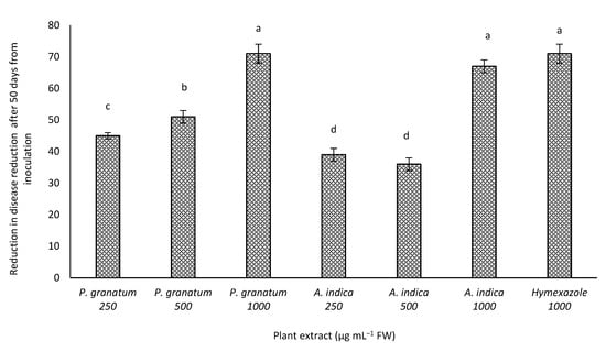
Figure 1.
Effect of different concentrations of methanol extracts of Azadirachta indica and Punica granatum (250, 500, and 1000 µg·mL−1) and fungicide (hymexazole, 1000 µg·mL−1) on Fusarium wilt under greenhouse conditions. Values in columns followed by different letters indicate significant differences among treatments according to a least significant difference test (p = 0.05). Bars indicate the standard error.
3.3. Vegetative Growth
The fresh weight (FW) and dry weight (DW) of the cumin plants increased significantly with both extracts at different concentrations compared to the control group (Figure 2). The height of the cumin plants increased significantly in all treatment groups compared to the control group (Figure 2). The highest increase was found with P. granatum and A. indica treatments at 1000 µg·mL−1, followed by 500 and 250 µg·mL−1 of P. granatum (98.8 and 96.7%, respectively). The same trend was observed for fresh and dry weight, which increased by 88 and 85.6%; this maybe correlated with the reduction in the disease severity. Many studies have noted that plant extracts increase the seed germination, as well as the vigor index, of ginger, garlic, and neem [44,45,46]. Additionally, Abo-Elyousr et al. [4], in their work on Calotropis procera, reported that tomato plants treated with an aqueous extract of C. procera showed increased fresh and dry weights of the tomato seedlings.
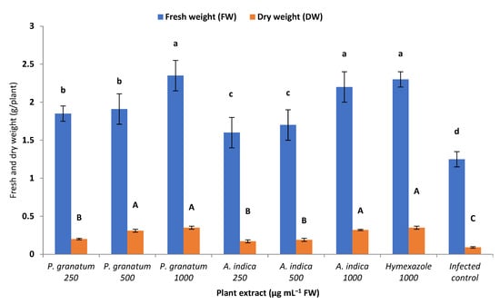
Figure 2.
Effect of different concentrations of methanol extracts of Azadirachta indica and Punica granatum (250, 500, and 1000 µg·mL−1) and fungicide (hymexazole, 1000 µg·mL−1) on fresh and dry weight of whole plants (g plant−1) under greenhouse conditions. Values in columns followed by different letters indicate significant differences among treatments according to a least significant difference test (p = 0.05). Bars indicate the standard error.
3.4. Effect of Different Concentrations of Azadirachta indica and Punica granatum on Phenol and Flavonoid Contents
From the data illustrated in Figure 3, the treatment of cumin plants with Pme and Lme at 1000 µg·mL−1 gave the highest flavonoid and phenol contents in inoculated or un-inoculated plants, followed by 500 µg·mL−1 and the lowest concentration of 250 µg·mL−1, relative to the infected control plants.
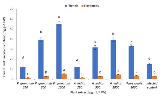
Figure 3.
Effect of different concentrations of methanol extracts of Azadirachta indica and Punica granatum (250, 500, and 1000 µg·mL−1) and fungicide (hymexazole, 1000 µg·mL−1) on phenol content (phenols; mgg−1 FW) and flavonoids (mg g−1 FW) in root extracts of cumin plants after 35 days of treatment with both extracts and inoculation with F. oxysporum. Values in columns followed by different letters indicate significant differences among treatments according to a least significant difference test (p = 0.05).
Obtained results revealed that A. indica and P. granatum (250, 500, and 1000 µg·mL−1) increased the phenol content. In agreement with this, similar results have been reported in the literature. The authors of [4] found that the application of a plant extract (Calotropis procera) to tomato plants significantly reduced wilt severity compared to untreated controls. In this context, phytosterols, a group of steroidal alcohols, and phytochemicals naturally present in plants, have been found to display antifungal activity against Aspergillus, Penicillium, and Fusarium [47]. Phenolic compounds are toxic to phytopathogenic fungi, and the accumulation of these bioactive ingredients at infection sites has been associated with pathogen development restriction [48].
3.5. Phytochemical Screening Test
The results of the qualitative phytochemical analysis (Table 2) of P. granatum peel and Azadirachta indica extracts showed that Pme and Lme are rich in carbohydrates, saponins, amino acids, proteins, alkaloids, and terpenoids. P. granatum peel extract is rich in carbohydrates, amino acids, proteins, alkaloids, and terpenoids. Phytochemical screening results showed that the methanol peel and leaf extract of both plants are rich in amino acids, flavonoids, alkaloids, and terpenoids, and fairly rich in saponins and flavonoids. Peel extract is rich in carbohydrates and saponins, and fairly rich in flavonoids. The secondary metabolites contribute significantly towards the biological activities, such as antioxidant and antidiabetic activities. Phytochemicals are mostly rich in green leaves, where they prevent free-radical formation. Hence, the presence of phytochemicals in the extracts might contribute to their beneficial activities [49].
Table 2.
Qualitative phytochemical analysis of Azadirachta indica and Punica granatum extracts.
Methanol extracts of P. granatum exhibited in vitro antioxidant activity in a concentration-dependent manner. Ascorbic acid was used as the standard chemical for comparing the antioxidant activity. Dietary antioxidants such as ascorbate, carotenoids, and tocopherol from natural compounds could reduce oxidative stress. Medicinal plants are a natural source for antimicrobial substances, which have fewer side effects and an ability to scavenge free radicals.
4. Conclusions
The seeds of cumin plants treated with the methanol extract of Azadirachta indica and Punica granatum at different concentrations reduced F. oxysporum f. sp. cumin radial growth in vitro; thus, the cumin seed pretreatment reduces the disease severity and increases the vegetative growth of cumin seedlings under greenhouse conditions. Seeds treated with both extracts had increased flavonoid and phenol contents in cumin roots at 35 days after the sowing date. Our results show that Azadirachta indica and Punica granatum at different concentrations are promising ecofriendly substances for use against F. oxysporum under greenhouse conditions.
Author Contributions
Conceptualization, M.A.A.M. and K.A.M.A.-E.; methodology, M.S., K.A.M.A.-E. and O.H.M.I.; software, K.A.M.A.-E.; validation, M.A.A.M., K.A.M.A.-E. and A.D.A.-Q.; formal analysis, M.A.A.M. and K.A.M.A.-E.; investigation, M.A.A.M., K.A.M.A.-E. and O.H.M.I.; resources, M.A.A.M. and O.H.M.I.; data curation, M.A.A.M. and A.D.A.-Q.; writing—original draft preparation, O.H.M.I.; writing—review and editing, M.A.A.M. and K.A.M.A.-E.; visualization, O.H.M.I.; supervision, M.A.A.M. and K.A.M.A.-E.; project administration, M.A.A.M.; funding acquisition, M.A.A.M. All authors have read and agreed to the published version of the manuscript.
Funding
This research was funded by the Deputyship for Research and Innovation, Ministry of Education, Saudi Arabia through the project number “IFPRC-156-155-2020” and King Abdulaziz University, DSR, Jeddah, Saudi Arabia.
Institutional Review Board Statement
Not applicable.
Informed Consent Statement
Not applicable.
Data Availability Statement
Publicly available datasets were analyzed in this study. This data can be found here: https://www.ncbi.nlm.nih.gov/ (accessed on 14 April 2022).
Acknowledgments
The authors extend their appreciation to the Deputyship for Research and Innovation, Ministry of Education in Saudi Arabia for funding this research work through the project number “IFPRC-156-155-2020”, and King Abdulaziz University, DSR, Jeddah, Saudi Arabia.
Conflicts of Interest
The authors declare no conflict of interest.
References
- Hilal, A.A.; Soliman, G.L.; El-Shaer, A.H.; El-Zefzaf, H.M. Bio-and chemical controls of soil-borne fungal diseases of the medicinal and aromatic plants: Cumin (Cuminum cyminum L.) and pelargonium (Pelargonium graveolense L.). Egypt. J. Appl. Sci. 2009, 24, 90–112. [Google Scholar]
- Nawade, B.D.; Jadeja, K.B.; Talaviya, J.R.; Vyas, U.M. Comparative analysis of Fusarium oxysporum f. sp. cumini isolates using RAPD marker and cultural characteristics. Trends Biosci. 2014, 7, 2475–2478. [Google Scholar]
- Sharma, O.P.; Pruthi, S.; Mohan, G.; Kaur, M.; Kumari, M. Assessment of fungicides against the early blight of tomato induced by Alternaria solani (Ellis & Martin) under field conditions. Int. J. Chem. Stud. 2020, 8, 693–696. [Google Scholar]
- Abo-Elyousr, K.A.M.; Ali, F.E.; Sallam, M.A. Alternative control of tomato wilt using the aqueous extract of Calotropis procera. Horticulture 2022, 8, 197. [Google Scholar] [CrossRef]
- Kumar, A.; Shukla, R.; Singh, P.; Prasad, C.S.; Dubey, N.K. Assessment of Thymus vulgaris L. essential oil as a safe botanical preservative against post-harvest fungal infestation of food commodities. Innov. Food Sci. Emerg. 2008, 4, 575–580. [Google Scholar] [CrossRef]
- Sharma, S.; Bohra, A. Rhizosphere effect of some important spices plants of Rajasthan. J. Phytol. Res. 2003, 16, 257–258. [Google Scholar]
- Sharma, N.; Trivedi, P.C. Screening of leaf extracts of some medicinal plants for their nematicidal and fungicidal properties against Meloidogyne incognita and Fusarium oxysporum. Asian J. Exp. Sci. 2002, 16, 21–28. [Google Scholar]
- Shaygannia, E.; Mahmoud, B.; Behnam, Z.; Mahmoud, R. A Review Study on Punica granatum L. J. Evid.-Based Complement. Altern. Med. 2016, 21, 221–227. [Google Scholar] [CrossRef]
- Turkyilmaz, M.; Taği, Ş.; Dereli, U.; Ozkan, M. Effects of various pressing programs and yields on the antioxidant activity, antimicrobial activity, phenolic content and colour of of pomegranate juices. Food Chem. 2013, 138, 18101818. [Google Scholar] [CrossRef]
- El Khetabi, A.; Lahlali, R.; Askarne, L.; Ezrari, S.; El Ghadaroui, L.; Tahiri, A.; Hrustić, J.; Amiri, S. Efficacy assessment of pomegranate peel aqueous extract for brown rot (Monilinia spp.) Disease control. Physiol. Mol. Plant Pathol. 2020, 110, 101482. [Google Scholar] [CrossRef]
- Farag, M.A.; Almahdyd, A.; Salah El Dine, R.; Fahmy, S.; Yassin, A.; Porzel, A.; Brandt, W. Structure activity relationships of antimicrobial gallic acid derivatives from pomegranate and acacia fruit extracts against potato bacterial wilt pathogen. Chem. Biodivers. 2015, 12, 955962. [Google Scholar] [CrossRef] [PubMed]
- Khaleel, A.I.; Sijam, K.; Rashid, T.S.; Bin Ahmad, K. Phytochemical determination, and antibacterial activity of Punica granatum peel extracts against plant pathogenic bacteria. Am. J. Plant Sci. 2016, 7, 159166. [Google Scholar] [CrossRef]
- Dahham, S.S.; Ali, M.N.; Tabassum, H.; Khan, M. Studies on antibacterial and antifungal activity of Pomegranate (Puncia granatum L.) American-Eurasian. J. Agric. Environ. Sci. 2010, 9, 273–281. [Google Scholar]
- Mangang, H.C.; Chhetry, G.K.N. Antifungal properties of certain plant extracts against Rhizoctonia solani causing root rot of franch bean in organic soil of Manipur. Int. J. Sci. Res. Publ. 2012, 2, 1–4. [Google Scholar]
- Magni, M.; Postiglione, E.; Marzorati, S.; Verotta, L.; Trasatti, S.P. Green Corrosion Inhibitors from Agri-Food Wastes: The Case of Punica granatum Extract and Its Constituent Ellagic Acid. A Validation Study. Processes 2020, 8, 272. [Google Scholar] [CrossRef]
- Kurimoto, S.-I.; Takaishi, Y.; Ahmed, F.A.; Kashiwada, Y. Triterpenoids from the fruits of Azadirachta indica (Meliaceae). Fitoterapia 2014, 92, 200–205. [Google Scholar] [CrossRef]
- Abo-Elyousr, K.A.M.; Asran, M.R. Antibacterial activity of certain plant extracts against bacterial wilt of tomato. Arch. Phytopathol. Plant Prot. 2009, 42, 573–578. [Google Scholar] [CrossRef]
- Amadioha, A.C.; Uchendu, P.N. Post-harvest Control of tomato fruit rot caused by Fusarium With extracts of Azadirachta indica. Discov. Innov. 2003, 15, 83–86. [Google Scholar] [CrossRef]
- Ibrahim, O.H.M.; Mousa, M.A.A.; Asiry, K.A.; Alhakamy, N.A.; Abo-Elyousr, K.A.M. Azadirachta indica A. Juss Fruit Mesocarp and Epicarp Extracts Induce Antimicrobial and Antiproliferative Effects against Prostate (PC-3), Breast (MCF-7), and Colorectal Adenocarcinoma (Caco-2) Cancer Cell Lines through Upregulation of Proapoptotic Genes. Plants 2022, 11, 1990. [Google Scholar] [CrossRef]
- Somda, I.; Leth, V.; Seeme, P. Evaluation of Lemongrass, Eucalyptus and Neem aqueous extracts for controlling seed-borne fungi of sorghum grown in Burkina Faso. World J. Agric. Sci. 2007, 3, 218–223. [Google Scholar]
- Ibrahim, O.H.M.; Abo-Elyousr, K.A.M.; Asiry, K.A.; Alhakamy, N.A.; Mousa, M.A.A. Phytochemical Characterization, An-timicrobial Activity and In Vitro Antiproliferative Potential of Alchemilla vulgaris Auct Root Extract against Prostate (PC-3), Breast (MCF-7) and Colorectal Adenocarcinoma (Caco-2) Cancer Cell Lines. Plants 2022, 11, 2140. [Google Scholar] [CrossRef] [PubMed]
- Nduagu, C.; Ekefam, E.J.; Nwankiti, A.O. Effect of crude plant extracts on growth of Colletotrichum capsici (Synd) Butler & Bisby, causal agent of pepper anthracnose. J. Appl. Sci. 2008, 6, 184–190. [Google Scholar]
- Abo-Elyousr, K.A.M.; Hashem, M.; Ali, E. Integrated control of cotton root rot disease by mixing fungal biocontrol agents and resistance inducers. Crop Protect. 2009, 28, 295–301. [Google Scholar] [CrossRef]
- Ghoneem, K.M.; Khalil, A.A.; Rashad, E.M.; Ahmed, M.I.M.; Mahmoud, M.S.M. Granular Bioactive Formulation of Trichoderma viride and Arbuscular Mycorrhizal Fungi for Biological Control of Cumin Wilt Disease. Egypt. J. Phytopathol. 2019, 47, 175–195. [Google Scholar] [CrossRef]
- Reddy, C.S.S.; Gayathri, R.; Priya, V.V.; Selvaraj, J.; Kavitha, S. Comparative Evaluation of Anti-diabetic Potential of Aqueous Seed Extracts of Momordica charantia, Seed Kernel Extract of Mangifera indica and Its Herbal Formulation—An in vitro Study. JPRI 2021, 33, 290–299. [Google Scholar] [CrossRef]
- Malik, C.P.; Singh, M.B. Plant Enzymology and Hittoenzymology; Kalyani Publishers: New Delhi, India, 1980; p. 286. [Google Scholar]
- Zhishen, J.; Mengcheng, T.; Jianming, W. The determination of flavonoid contents in mulberry and their scavenging effects on superoxide radicals. Food Chem. 1999, 64, 555–559. [Google Scholar] [CrossRef]
- Gomez, K.A.; Gomez, A.A. Statistical Procedures for Agricultural Research; Wiley: New York, NY, USA, 1984. [Google Scholar]
- Carvalho, F.P. Pesticides, environment, and food safety. Food Energy Secur. 2017, 6, 48–60. [Google Scholar] [CrossRef]
- Enikuomehin, O.A.; Oyedeji, E.O. Fungitoxic effect of some plant extracts against tomato fruit rot pathogens. Arch. Phytopathol. Plant Prot. 2010, 43, 233–240. [Google Scholar] [CrossRef]
- Tehranifar, A.; Selahvarzi, Y.; Kharrazi, M.; Bakhsh, V.J. High potential of agro-industrial by-products of pomegranate (Punica granatum L.) as the powerful antifungal and antioxidant substances. Ind. Crops Produc. 2011, 34, 1523–1527. [Google Scholar] [CrossRef]
- Shafighi, M.; Amjad, L.; MadaniIn, M. In vitro antifungal activity of methanolic extract of various parts of Punica granatum L. Int. J. Sci. Eng. Res. 2012, 3, 1–4. [Google Scholar]
- Mohamed, N.H.; El-Hadidy, A.M. Studies of biologically active constituents of Verbascum eremobium Murb. and its inducing resistance against some diseases of cucumber. Egypt. J. Phytopathol. 2008, 36, 133–150. [Google Scholar]
- Wink, M.; Ashour, M.L.; El-Readi, M.Z. Secondary Metabolites from Plants Inhibiting ABC Transporters and Reversing Resistance of Cancer Cells and Microbes to Cytotoxic and Antimicrobial Agents. Front. Microbiol. 2012, 3, 130. [Google Scholar] [CrossRef] [PubMed]
- Jan, R.; Asaf, S.; Numan, M.; Lubna; Kim, K.M. Plant Secondary Metabolite Biosynthesis and Transcriptional Regulation in Response to Biotic and Abiotic Stress Conditions. Agronomy 2021, 11, 968. [Google Scholar] [CrossRef]
- Singh, R.P.; Chidambara Murthy, K.N.; Jayaprakasha, G.K. Studies on the Antioxidant Activity of Pomegranate (Punica granatum) Peel and Seed Extracts Using in vitro Models. J. Agric. Food Chem. 2002, 50, 81–86. [Google Scholar] [CrossRef]
- Naziri, Z.; Rajaian, H.; Firouzi, R. Antibacterial Effects of Iranian Native Sour and Sweet Pomegranate (Punica granatum) Peel Extracts against Various Pathogenic Bacteria. Iran. J. Veter. Res. 2012, 13, 282–288. [Google Scholar]
- Vasconcelos, L.C.D.S.; Sampaio, M.C.C.; Sampaio, F.C.; Higino, J.S. Use of Punica granatum as an Antifungal Agent against Candidosis Associated with Denture Stomatitis Verwendung von Punica granatum als Antimykotikum gegen Candidose in Verbindung mit Zahnprothesen-Stomatitis. Mycoses 2003, 46, 192–196. [Google Scholar] [CrossRef]
- Reddy, M.K.; Gupta, S.K.; Jacob, M.R.; Khan, S.I.; Ferreira, D. Antioxidant, Antimalarial and Antimicrobial Activities of Tannin-Rich Fractions, Ellagitannins and Phenolic Acids from Punica granatum L. Planta Med. 2007, 73, 461–467. [Google Scholar] [CrossRef]
- Vasconcelos, L.C.D.S.; Sampaio, F.C.; Sampaio, M.C.C.; Pereira, M.D.S.V.; Higino, J.S.; Peixoto, M.H.P. Minimum Inhibitory Concentration of Adherence of Punica granatum Linn (Pomegranate) Gel against S. mutans, S. mitis and C. albicans. Braz. Dent. J. 2006, 17, 223–227. [Google Scholar] [CrossRef]
- Lattanzio, V.; Veronica, M.; Lattenzio, T.; Cardinali, A. Role of phenolic in the resistance mechanisms of plant against fungal pathogens and insects. In Phytochemistry: Advances in Research; Imperator, F., Ed.; Research Signpost: Kerala, India, 2006; pp. 23–67. [Google Scholar]
- Mohanty, S.; Patra, A.; Chhonkar, P. Neem (Azadirachta indica) seed kernel powder retards urease and Nitrification activities in different soils at contrasting Moisture and temperature regimes. Bioresour. Technol. 2008, 99, 894–899. [Google Scholar] [CrossRef]
- Hassenein, N.M.; Ali, M.M.; Youssef, K.A.; Mahmoud, D.A. Control of tomato early blight and wilt using aqueous extract of neem leaves. Phytopathol. Mediter. 2010, 49, 143–151. [Google Scholar]
- Khan, M.I.; Kumar, R. Antifungal activity of leaf extract of Neem on seed mycoflora of wheat. Indian J. Seed Abs. 1992, 15, 299. [Google Scholar]
- Ahmed, M.; Mehbub, H.; Kamrul, H.; Dash, C.K. Efficacy of Different Plant Extract on Reducing Seed Borne Infection and Increasing Germination of Collected Rice Seed Sample Univers. J. Plant Sci. 2013, 1, 66–73. [Google Scholar] [CrossRef]
- Ali, E.F.; Al-Yasi, H.M.; Issa, A.A.; Hessini, K.; Hassan, F.A.S. Ginger extract and fulvic acid foliar applications as novel practical approaches to improve the growth and productivity of Damask Rose. Plants 2022, 11, 412. [Google Scholar] [CrossRef]
- Odhav, M.B.; Mohanlall, V. Antifungal activity of stigmasterol, sitosterol and ergosterol from Bulbine natalensis Baker (Asphodelaceae) B. J. Med. Plants Res. 2012, 6, 5135–5141. [Google Scholar] [CrossRef]
- Abo-Elyousr, K.A.M.; Ibrahim, Y.E.; Balabel, N.M. Induction of disease defensive enzymes in response to treatment with acibenzolar-S-methyl (ASM) and Pseudomonas fluorescens Pf2 and inoculated with Ralstonia solanacearum race 3, biovar2 (phylotype II). J. Phytopathol. 2012, 160, 382–389. [Google Scholar] [CrossRef]
- Moga, M.A.; Dimienescu, O.G.; Bălan, A.; Dima, L.; Toma, S.I.; Bîgiu, N.F.; Blidaru, A. Pharmacological and Therapeutic Properties of Punica granatum Phytochemicals: Possible Roles in Breast Cancer. Molecules 2021, 26, 1054. [Google Scholar] [CrossRef]


Publisher’s Note: MDPI stays neutral with regard to jurisdictional claims in published maps and institutional affiliations. |
© 2022 by the authors. Licensee MDPI, Basel, Switzerland. This article is an open access article distributed under the terms and conditions of the Creative Commons Attribution (CC BY) license (https://creativecommons.org/licenses/by/4.0/).