Article

Synthesis of Urchin-Shaped Gold Nanoparticles Utilizing Green Reducing and Capping Agents at Different Preparation Conditions: An In Vitro Study

1
Mechanical Power Engineering Department, Faculty of Engineering, Mansoura University, Mansoura 35516, Egypt
2
Polymeric Materials Research Department, City of Scientific Research and Technological Applications (SRTA-City), Borg El-Arab City, Alexandria 21934, Egypt
*
Authors to whom correspondence should be addressed.
Sustainability 2022, 14(24), 16838; https://doi.org/10.3390/su142416838
Submission received: 30 September 2022 / Revised: 8 December 2022 / Accepted: 13 December 2022 / Published: 15 December 2022

Abstract

:
Employing environmentally friendly reducing and capping materials to synthesize gold nanoparticles is an exciting research point. However, the used materials usually need a long reduction time that can take days. In this work, the instantaneous production of small-sized (less than 20 nm) gold nanoparticles is investigated using ascorbic acid, gelatin, and a mixture of the two agents at different preparation conditions (at room temperature; 20 ± 3 °C and near boiling temperature; 95 ± 3 °C). Particle size analysis, as well as transmission electron microscopy, were used to assess the produced particles’ physical characteristics. The structural changes and optical characteristics of the nanoparticles were monitored using UV–visible spectroscopy. Fourier Transform Infrared spectroscopy (FTIR) was used to establish the presence of a gelatin coating over the gold nanoparticles. The morphology of the produced nanoparticles at 95 ± 3 °C was spherical with a size ranging from 8–18 nm, whereas urchin-shaped nanoparticles ranging from 24–100 nm were formed at 20 ± 3 °C reaction temperature. The presence of hydroxyl and amine groups associated with the gelatin was confirmed using FTIR. This could be a step for wider usage of green synthesized nanogold particles in several applications.

1. Introduction

Nanotechnology research has recently emerged as a vital key to obtaining novel, desirable properties from well-known conventional materials [1]. Nanomaterials have at least one dimension within the nanometer range [2]. Although almost any material in existence could exist within the nanoscale, noble metals-based nanoparticles hold a special value. They generally have some exceptional characteristics that can be tailored for many useful applications.
Gold is considered one of the most famed noble metals to be prepared in nanoscale forms [3]. Gold nanoparticles drew increasing interest due to their excellent optical, electronic, and magnetic properties [4], as well as their biocompatibility [5] and non-toxicity [6]. Gold-based nanomaterials have been previously prepared in different nanostructures like nanoparticles [7,8], nanorods [9,10], nanowires [11,12], nanostars [13,14], nanospheres [15,16], and nanotriangles [17,18]. Nanogold-based materials were utilized for different purposes, such as catalysis [19], energy storage and conversion [20], biomedical therapeutics [20,21,22], drug delivery [23], photonics [24], and biosensing [25,26].
Composite nanostructures that combine gold with other materials, such as iron [27], silver [28], and graphene [28], were also investigated and prepared. Moreover, researchers managed to prepare fairly complicated composite morphologies. For example, Nghiem et al. [29] managed to prepare a composite comprising a core–shell nanostructure, combining a silica core with a gold shell.
Nanostructure attributes are greatly influenced by their physicochemical characteristics, which include particle volume, form, and composition. Furthermore, for composite nanostructures, the properties are also affected by the molecular distribution within the particle [30]. Tuning and manipulating these characteristics allows control or maximization of the desired attribute to be suitable for a specific application. In the case of catalysis, for example, catalytic activity and selectivity are strongly influenced by the surface area, composition, and shape of the nanostructure [31]. On the other hand, when the object is to enhance the optical attributes of a nanostructure for imaging functions, controlling those characteristics would impact absorption and scattering efficiency [32].
It is evident that the manipulation of these nanostructures during the synthesis process is essential to produce predictable and distinct physical and chemical properties. This is critical not only for optimizing their performances but also for understanding the structure-performance relations. This can be partly achieved through the nanostructure controllable preparation method.
Microemulsion [33], reversed micelles [34], seeding growth [35], sonochemistry [36], photochemistry [37], radiolysis [38], and chemical reduction [39] techniques have all been used for gold nanostructure preparation. Several chemical reductants have been used to reduce the gold precursor (chloroauric acid, HAuCl4), such as 1-amino-2-naphthol-4-sulphonic acid [40], monosodium glutamate [41], sodium borohydride [42], amine [43], etc. Recently, natural materials have been used as green reductants, such as ascorbic acid [44], piper beetle leaf broth [45], a polyphenol extracted from leaf buds of camellia Sinensis tea [46], aspartame [47], Rhizopus oryzae fungal strain [48], and gelatin [3].
One of the earliest and simplest methods for preparing gold nanoparticles is the Turkevich technique [49], which uses sodium citrate to both reduce the gold precursor (HAuCl4) and prevent further particle development and retain the formed gold in the nano range as well as prevent the nanogold from aggregation. The second role of the used sodium citrate is a capping agent that can offset the Van der Waals tractive forces through steric and electrostatic repulsions, hence reducing the aggregation phenomenon. However, the stability and dispersity of the produced gold nanoparticles are often poor [50]. In recent years, another technique was developed to adjust the reaction pH utilizing a buffer rather than a solution to present nanoparticles with high monodispersity [51]. Also, ethylenediaminetetraacetic acid (EDTA) was used to improve the uniformity of the produced nanogold with better size control [52].
Gold nanoparticles have excellent potential for the immobilization of biomolecules because they are biocompatible and bind readily to a range of biomolecules, such as amino acids, proteins, DNA, and enzymes. Nanogold membranes can be used as templates for the immobilization of enzymes [53]. Consequently, the bioconjugate material could be easily separated after reaction by mild centrifugation and easily reused. This membrane can also be used in nanoplasmonic sensors [54].
As can be seen, the search continues for producing proficient gold nanostructures using green reducing and capping agents that combine sustainability and efficiency [55]. In the literature, some authors used surfactants to obtain such small sizes that resulted in chemical contamination. Others claimed to produce nanoparticles instantaneously without quantifying the exact reaction time. On the other hand, most of the published research did not obtain uniform shapes but mostly a mixture of different shapes [56]. In this work, the instantaneous fabrication of gold nanoparticles with two different morphologies using a green, reducing agent (ascorbic acid) and a green capping agent (gelatin) along with distilled water as solvent was investigated and presented. Ascorbic acid (B1), gelatin (B2), as well as a mixture of both (M) were tested as reducing agents of the gold precursor. Two different morphologies were obtained under the sole influence of changing the reaction temperature. Particle size analysis, as well as transmission electron microscopy, were used to assess the produced particles’ physical characteristics. The structural changes and optical characteristics of the nanoparticles were monitored using UV–visible spectroscopy. Moreover, the crystal nature of the prepared gold nanoparticles was determined by X-ray Diffraction (XRD), and Fourier transform infrared spectroscopy (FTIR) was used to establish the presence of a gelatin coating over the gold nanoparticles. Small-sized spherical and urchin-shaped nanoparticles were successfully obtained. Urchin-shaped nanoparticles were formed. This unique shape has special plasmon properties due to the unsmooth surface that causes a greater enhancement of the electromagnetic field at the ends of the urchin-shaped nanoparticle spikes supplemented by a red shift in the peak [57]. For that, disease diagnostics and therapy are strongly suggested as some of the potential applications of the prepared unique urchin-shaped nanogold.

2. Materials and Methods

2.1. Materials

Ascorbic acid (B1) was purchased from HiMedia chemicals. Hydrogen tetrachlorourate (III) (HAuCl4, 30%wt in HCl) and gelatin (B2) from the skin were purchased from Sigma-Aldrich. Ferulic acid (≥99.5%) was obtained from Fluka (Schwerte, Germany). Glycerin (>98%) was purchased from Oxford Laboratory Reagent (Mumbai, India) Ethanol (purity ≥ 98%) was acquired from Sigma-Aldrich. Formic acid (85%) was obtained from Laboratory Chemicals (Cairo, Egypt). For the preparation of the mixture solution, distilled water was used. Figure 1 presents the chemical composition of ascorbic acid and gelatin.

2.2. Methods

Gold Nanoparticles Preparation

Six different sets of experiments were performed to synthesize gold nanoparticles. The nanoparticles were synthesized using only ascorbic acid (B1), only gelatin (B2), and a mixture of them (M) at two different temperatures: near boiling temperature(BT) (95 ± 3 °C) and at room temperature (RT) (20 ± 3 °C). In a beaker, a quantity of 10 mL was taken from tetrachloroauric acid (with 30% Au) solution and diluted with 40 mL of distilled water. It was then heated to near boiling temperature with a continuous stirring at 200 rpm. It formed a clear pale yellowish solution (pH 2 ± 0.07). In another beaker, a gelatin solution (1% by volume; pH 4.3 ± 0.02), an ascorbic acid (1% by volume; pH 3.9 ± 0.04), and a mixture of both (1% by volume; pH 4) were prepared at the same temperature (95 ± 3 °C) along with stirring for 15 min. The boiled solution of the components was immediately poured into the gold precursor solution with continuous mixing (pH 3.3 ± 0.02). The same process was repeated at RT (20 ± 3 °C) without heating. Only a little heating (10 min at 40 °C) was used to dissolve the gelatin, then it was kept until it cooled down to RT before use.

2.3. Characterization

2.3.1. UV–Visible Spectrophotometer Measuring

UV–visible spectra of the instantly-formed gold nanoparticle solutions were recorded over a 200–900 nm wavelength by a Libra S32 Spectrophotometer (Biochrom, UK). The mixing was done immediately inside the used cuvette before measuring.

2.3.2. Transmission Electron Microscopy (TEM) imaging

TEM images were performed by JEM-2100F Transmission Electron Microscope (JEOL, Tokyo, Japan). A drop of gold solution was poured into a water sample over a copper mesh, then dried at room temperature for one day to prepare the sample. The histograms of the particle size distribution were estimated using ImageJ software.

2.3.3. Particle Size Analysis

The samples were recorded by a Tensor 27 Bruker spectrophotometer with 32 scans, 2 cm−1 resolution. The used gold solution was freshly prepared before measuring.

2.3.4. X-ray Diffraction (XRD) Analysis

The morphology of the prepared gold nanoparticles was carried out using a Bruker AXS XRD with CuKα as an X-ray source at 50 kV and 100 mA. The formed nanoparticle was drop-coated onto a glass substrate and dried in a desiccator for 24 h before measuring.

2.3.5. Fourier Transform-Spectroscopy (FT-IR) Analysis

An FT-IR spectrometer (Bruker, Japan) was used to study the chemical structure of the produced gold nanoparticles. FT-IR spectra in the range of 4000 to 400 cm−1 with a resolution of 4 cm−1 were used. The gold nanoparticles were precipitated and washed with ethanol and then dried in the desiccators at RT for a day before measuring.

3. Results and Discussion

Figure 2 presents the different stages of the gold nanoparticle preparation process. It was noticed that in the case of using the mixture and preparing at near boiling temperature, the solution color changed from pale yellowish to red almost immediately with the addition of the first drop of the gold precursor into the reducing and capping agents (Figure 2b). The red color indicates gold nanoparticle formation, which increases with adding more from the mixture, as shown in Figure 2c. On the other hand, in the case of using the mixture and preparation at room temperature, the color change was not noticed by the naked eye for almost 15 min. As for the blank experiments, i.e., ascorbic acid and gelatin at room temperature (B1,RT, B2,RT) and near-boiling temperature (B1,BT, B2,BT), it took up to 30 min before a change in color could be noticed. After adding absolute ethanol, precipitation of the gold nanoparticles could be observed, as seen in Figure 2d.
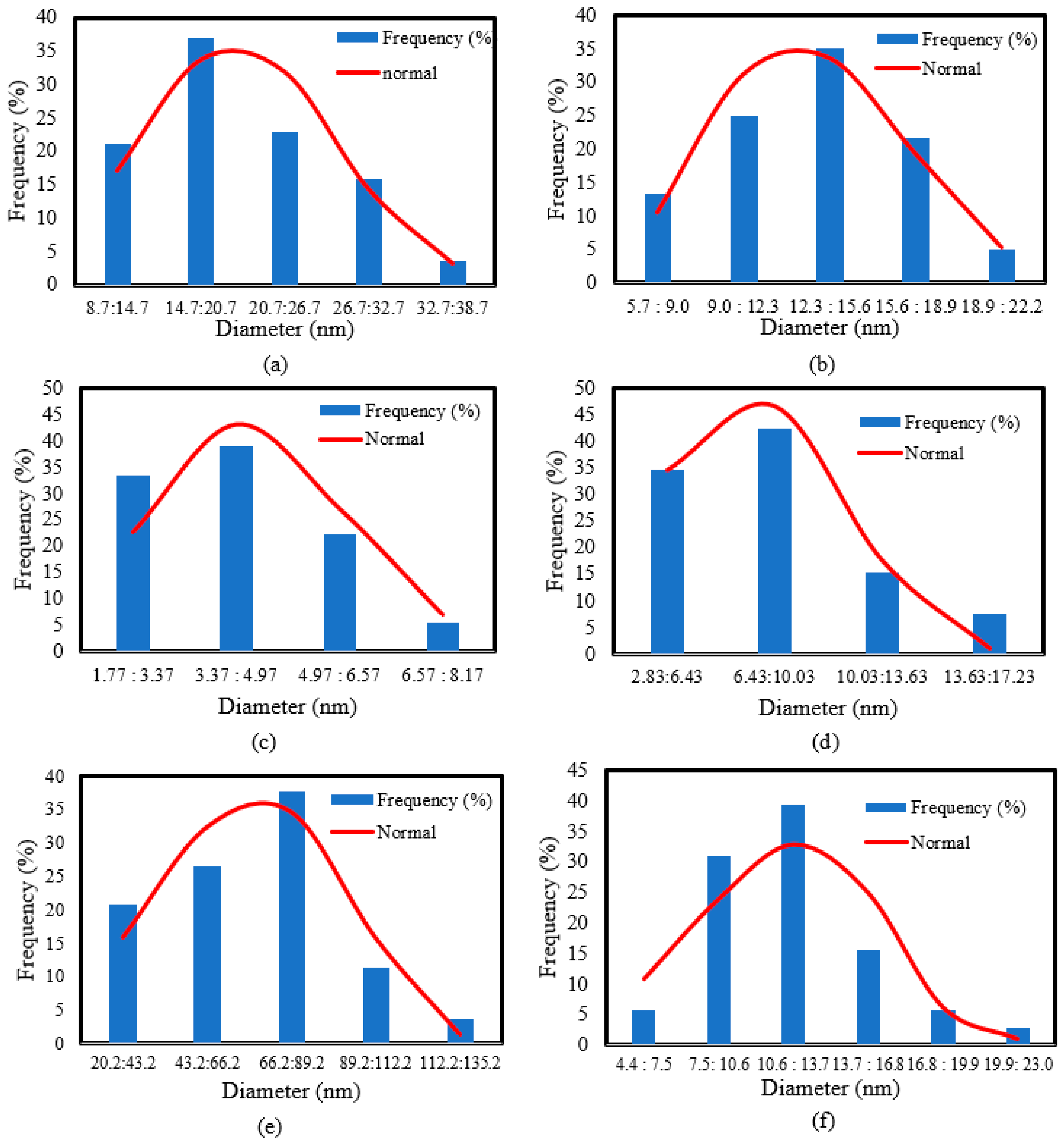
The morphology of the green synthesized gold nanoparticles was observed by TEM. Figure 3 shows samples of TEM images of the gold nanoparticles formed using only ascorbic acid at RT and BT, only gelatin at RT and BT, and a mixture of ascorbic acid and gelatin together at the different preparation temperatures. Figure 4 presents histograms of the particle size distribution estimated using ImageJ software. As can be seen from the figures, most gold nanoparticles formed near boiling temperature had a spherical shape. The average diameter was in the range of 5–20 nm in the case of using the ascorbic acid (B1,BT), and in the range of 3–14 nm in the case of using the gelatin (B2,BT). However, the average diameter of the instantly formed gold particles when using the mixture at (M,BT) was in the range of 5–22 nm. On the other hand, the gold nanoparticles formed at room temperature had different shapes. Irregular shapes with an average diameter in the range of 9–34 nm were formed when using ascorbic acid alone B1,RT. A mixture of spherical and urchin-shaped gold nanoparticles with an average diameter range of 2–8 nm was obtained using gelatin (B2,RT). Surprisingly, using both ascorbic acid and gelatin together at room temperature (M,RT) resulted in the formation of urchin-shaped nanoparticles with particle sizes ranging from 20–120 nm. A larger size of particle than the spherical-shaped particles was obtained at MBT. In general, the reaction temperature affected not only the shape but also the size range of the formed gold nanoparticles. Table 1 summarizes the mean diameter and standard deviation of each case.
The obtained results show that the higher temperature of the reaction resulted in the almost instantaneous creation of gold nanoparticles in a uniform shape. This is in full agreement with previous research [57,58,59], in which the reaction temperature decisively controlled the formation of small gold nanoparticles. Generally, the temperature accelerates the nucleation process and with the aid of the used capping agent, the gold nanoparticles are able to maintain their small size. However, the lower reaction temperature causes a limited nucleation process and provides a chance for particle growth to yield a larger particle size, which the naked eye can notice after longer reaction times (15–30 min). On the other hand, at a relatively low pH medium (pH 3.3 ± 002), the formation of gold nanoparticles is carried out through an intermediate [AuCl3(OH)] which undergoes a LaMer burst nucleation within about 10 s [60,61]. However, at higher pH mediums, a much longer nucleation time (around 60 s) is required, and the particles undergo reduction through [AuCl2(OH)]2 and [AuCl(OH)]3 followed by a much slower growth process. When coupling both effects of high temperature and pH, a uniform low-size range of gold nanoparticles could be produced [58,61].
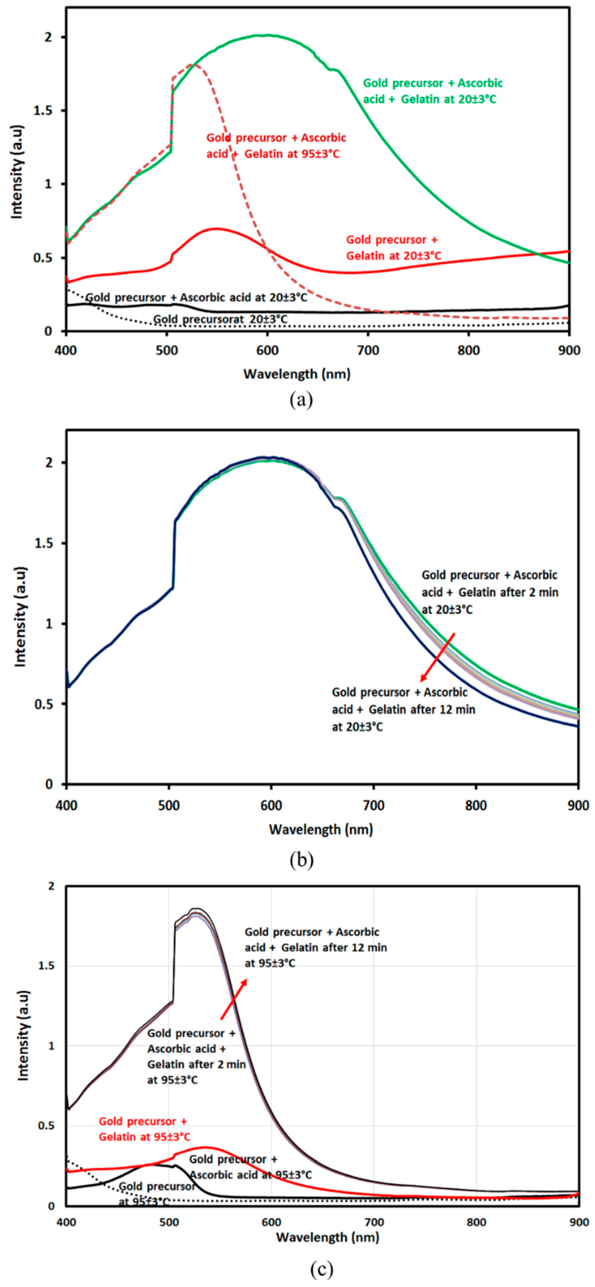
UV–visible spectroscopy can check for morphological changes and optical properties as nanoparticles display surface plasmon resonance (SPR) varying with its form and size at different frequencies [57,61]. The reaction was executed inside the device cuvette for 30 s and the measurement of the mixed solution was done immediately for higher accuracy. Figure 5a shows a UV–visible spectrum of an aqueous solution of gold precursor in water at room temperature (20 ± 3 °C), mixed with only ascorbic acid (B1,RT), and mixed with only gelatin (B2,RT). The figure also presents the spectra of the gold prosecutor mixed with both agents at room temperature (M,RT) as well as near boiling temperature (M,BT). All the measurements took place within 2 min from the reaction beginning (i.e., 30 s for mixing and around 60 s for measuring). As noted, there are no peaks for the used gold precursor solution in water or the solution of gold precursor with the ascorbic acid. However, the presence of gelatin with the gold precursor shows a surface plasmon resonance broad band at around 570–600 nm. In the case of the gold prosecutor with the mixture, a strong absorption band around 500–600 nm is observed that corresponds to the excitation of surface plasmon vibrations in the gold nanoparticles. The presence of both ascorbic acid and gelatin shows a broader SPR beak at around 507–667 nm. It can be seen that the peak was broadened with urchin-shaped gold nanoparticles, as illustrated in TEM images in the previous section, which was prepared at RT (20 ± 3 °C). This broad peak can be attributed to the existence of transversal and longitudinal SPR [62]. The sharp increase in the UV band at a wavelength of about 500 nm may be due to the instantaneous formation of the nanogold particles during the measurement, as the reaction was processed within the measuring cuvette for maximum accuracy. The unsmooth surface causes a greater enhancement of the electromagnetic field at the ends of the urchin-shaped nanoparticle spikes supplemented by a red shift in the peak [57].
Additionally, the uneven distribution of the surface electron layers in the urchin-shaped gold nanoparticles is what causes the difference in absorption qualities between them and the spherical ones [45,63]. On the other hand, the much less broad SPR absorption band of gold nanoparticle that was prepared near boiling temperature (95 ± 3 °C) appeared at 540 nm. Figure 5b shows there was not a noticeable difference in the SPR after 12 min of mixing the gold precursor and both ascorbic acid and gelatin together at room temperature (the measuring was executed with 2-min intervals).
Furthermore, Figure 5c shows the same determined SPR band after 12 min mixing between the gold precursor mixed with both ascorbic acid and gelatin at near boiling temperature (95 ± 3 °C). Regarding using only ascorbic acid with the gold precursor near BT (B1,BT), a small SPR band was formed compared to (B1,RT) in Figure 5a, which can be ascribed to the fast growth of gold nanoparticles with the ascorbic acid with heating.
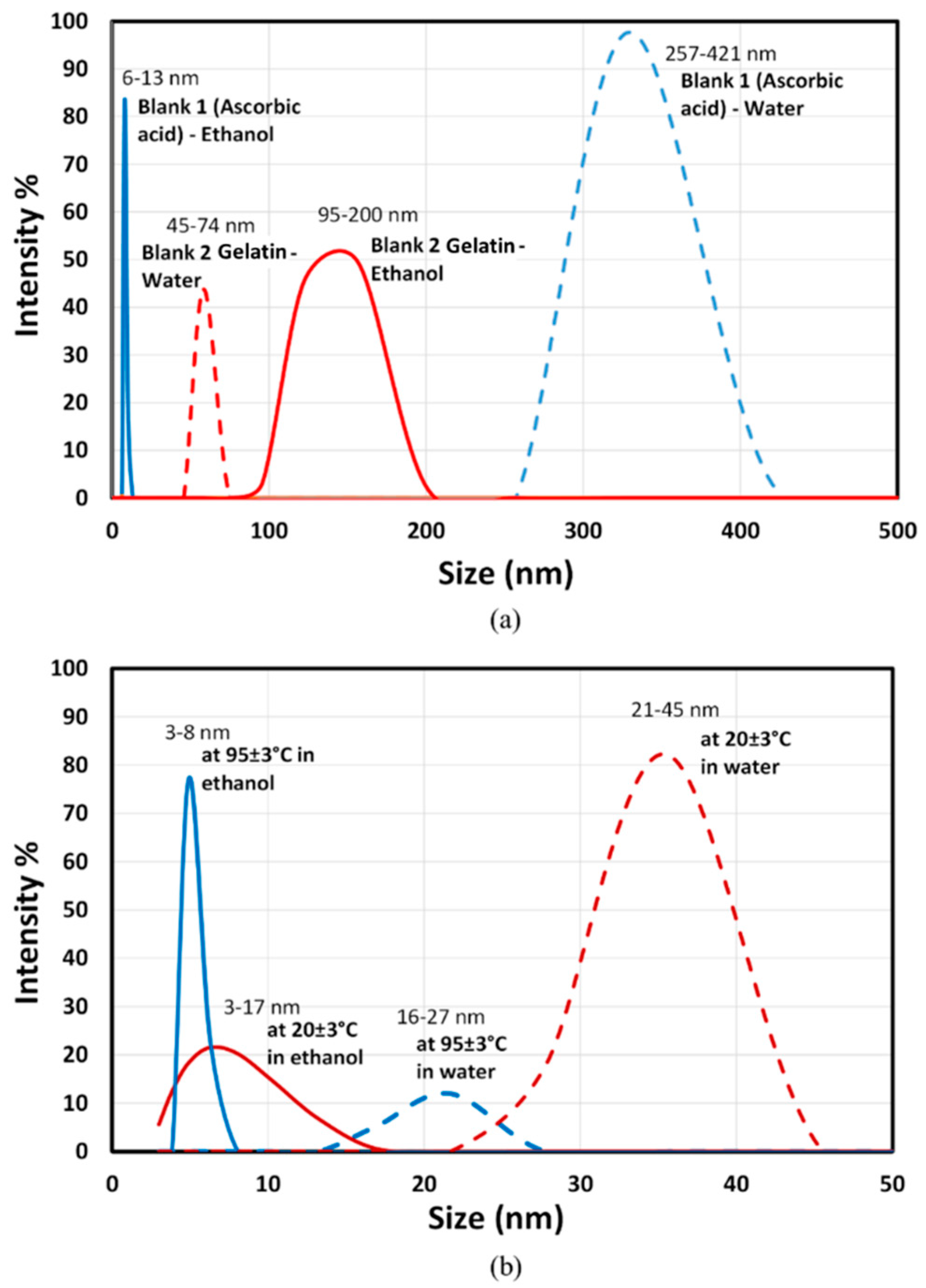
It is considered common knowledge that gold nanoparticles smaller than 2 nm do not demonstrate SPR [57,64]. Such small gold nanoparticles can be determined by using a particle size analyzer as shown in Figure 6. The use of either ascorbic acid or gelatin at near boiling temperature (95 ± 3 °C) can produce gold nanoparticles. To stop the growth of gold particles and/or to reduce the nanoparticle aggregation, the addition of pure ethanol with a ratio of 3:1 ethanol/reaction solution was done after 2 min of mixing at BT and after 15 min mixing at RT. The effect of ethanol is strongly illustrated by a reduction in the particle size of the formed gold nanoparticles produced using only the reducing agent (ascorbic acid) at 95 ± 3 °C from 257–421 nm to a much narrower 6–13 nm size, whereas the addition of ethanol to the solution of gold precursor and gelatin at BT resulted in shifting the particle size range from 45–74 nm to 95–200 nm.
It seems the role of ethanol is different in the two cases; in the case of using ascorbic acid, the used ethanol may reduce the particle size growth, but in the case of using gelatin, the used ethanol reduced the role of gelatin as a capping agent and enhanced the particle aggregation. The degradation effect of the ethanolic solution on the ascorbic acid was previously proven [64] in which the higher ethanol concentration (>50%) promotes lowering the water activity environment that supports the degradation of ascorbic acid into a transitional compound; L-xylosone, and then to other carbon compounds [65]. Therefore, adding the ethanol solution will lead to stopping the reducing reaction and result in a reduction in the particle size of the produced nanogold. Meanwhile, ethanol with a high concentration (>50%) works on the precipitation of gelatin into particles [66]. Therefore, the effectiveness of the gelatin as a capping agent is weakened, and a larger gold particle size is formed. The results of B1,RT, and B2,RT were not within the measuring limit, which may be attributed to the formation of an extremely small particle size (out of the measuring limits of the particle size analyzer; <3 nm). In the case of using both ascorbic acid and gelatin near BT as shown in Figure 6B, the size of produced nanoparticle shifted from 16–27 nm to 3–8 nm. In the case of using both ascorbic acid and gelatin at RT, the size of produced urchin-shaped gold nanoparticles shifted from 21–45 nm to 3–17 nm. From the obtained results, we can assume that the addition of ethanol may be a tool to control the size of the gold nanoparticles according to the targeted final gold particle size.
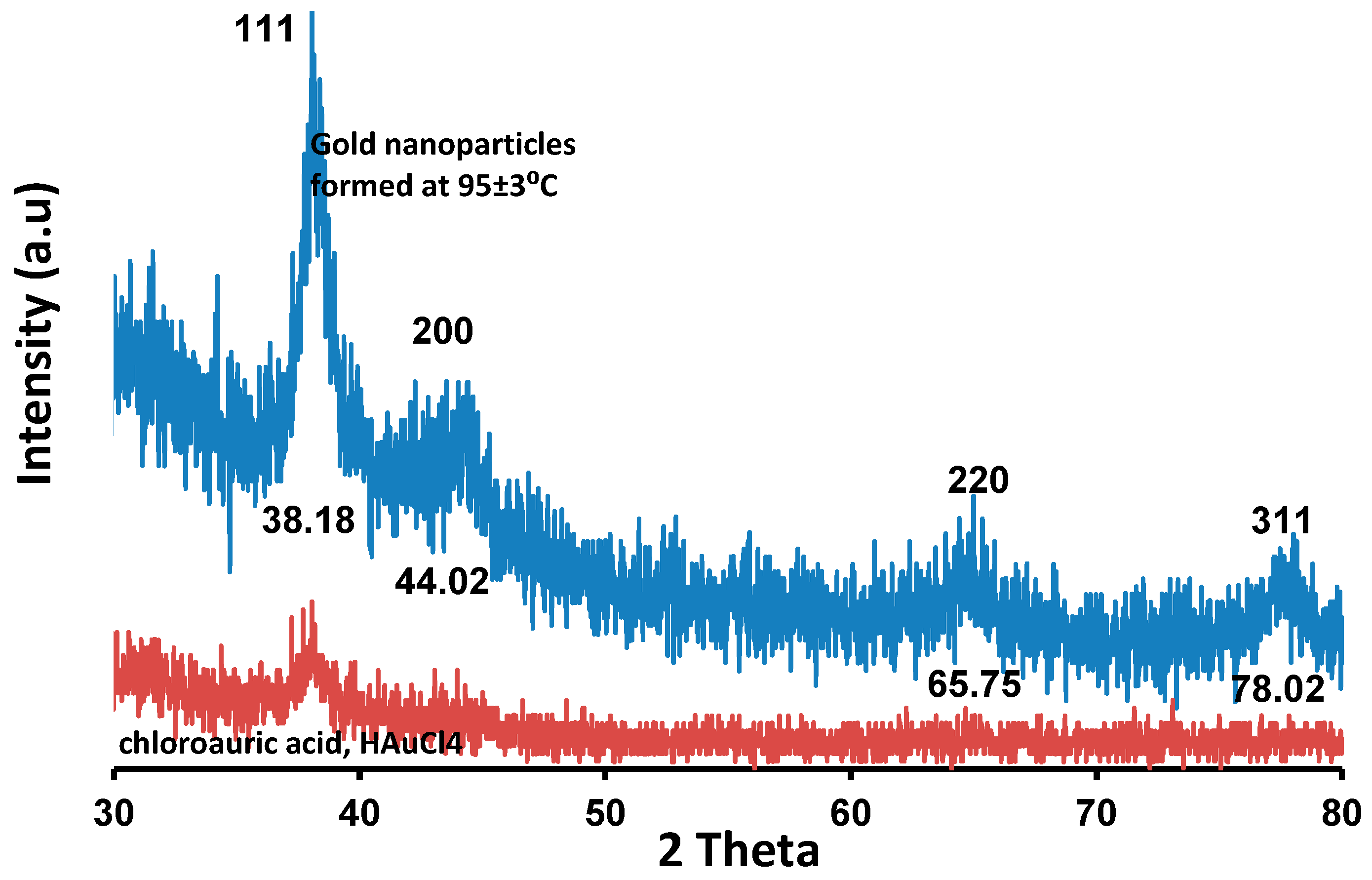
The existence of gold nanoparticles was verified using XRD. Figure 7 shows diffraction peaks for the used gold precursor in distilled water at 95 ± 3 °C reaction temperature and the green synthesized nanoparticles using a mixture of ascorbic acid and gelatin as a reducing agent and a capping agent, respectively at 95 ± 3 °C reaction temperature. The determined four strong Bragg diffraction peaks at 38.18°, 44.02°, 65.75°, and 78.02° can be assigned to the (111), (200), (220), and (311) planes [67,68]. It is possible to interpret these diffraction peaks as a face-centered cubic of gold and a natural crystal structure [58,59].
The FT-IR spectrum shown in Figure 8 illustrates the existence of the following groups: the strong, broad band observed at 3414 cm−1 suggests the existence of the O-H group and primary amine O-H band. The band at 2953 cm−1 may correspond to C-H stretching vibrations of the alkanes group, and the band at 1641 cm−1 indicates the existence of amide I. The narrow peak at 1541 cm−1 can be assigned for the presence of amide II. The peak at 1437 cm−1 may be attributed to –N-H. The peak at 1257 cm−1 can be assigned to –C-O. The narrow peak at 1084 cm−1 can be attributed to the presence of C-N stretching vibrations of aliphatic amines. This FT-IR spectrum indicates the presence of gelatin as a capping agent for gold nanoparticles even after the addition of ethanol to the formed gold nanoparticles. FT-IR proposed that both carboxylic acid groups and amine groups share the conjugation with gold surfaces as well as the possibility of further conjugation with other molecules.

4. Conclusions

Spherical gold nanoparticles and urchin-shaped gold nanoparticles were successfully and instantly produced using a green-natured reducing agent (ascorbic acid), and a green-natured capping agent (gelatin). Gelatin could be used as both a reducing and capping agent as reported in previous research [67], but the formed gold nanoparticles are not urchin-shaped. In in vivo applications, urchin-shaped particles are preferred to spherical ones because they have a lower background and more effectively transmit near-infrared light through biological tissues [58]. For that, the authors strongly propose utilizing the prepared unique urchin-shaped nanogold in disease diagnostics and therapeutical applications.
Table 2 shows that using both ascorbic acid and gelatin together resulted in an instant production of two different morphologies of gold nanoparticles as the effect of the reaction temperature: at RT (20 ± 3 °C) and near BT (95 ± 3 °C). The role of gelatin is mainly as a capping agent, although it can be considered as a second reducing agent. The mixing time in a mixture of ascorbic acid and gelatin was shorter than those of either only ascorbic acid or only gelatin, especially at a room temperature reaction, due to the higher reaction rates indicated by the change in the solution reaction. Using both gelatin and ascorbic acid together resulted in a spontaneous reaction that can be used to shift the formed gold particles to extremely small sizes. The use of ethanol effectively reduced the aggregation of the particles. However, it had the opposite effect in the case of using gelatin alone. Regarding the used analysis techniques, the particle size analyzer was proved unsuitable for irregular (urchin-shaped) nanoparticles. However, it could be used as an indication of the formed, small, and smooth morphology.
From this study, it is clear that the synthesis of biocompatible gold nanoparticles using both ascorbic acid and gelatin together as reducing and capping agents, respectively, is an instant, flexible, and green method. They produced spherical and urchin-shaped gold nanoparticles, which are candidates for use in medical, pharmaceutical, food, and cosmetics applications.

Author Contributions

Conceptualization, N.N.; methodology, N.N.; software, N.N., M.S.S., N.S. and M.R.E.; validation, N.N., M.S.S. and M.R.E.; formal analysis, N.N. and N.S.; investigation, N.N. and M.R.E.; resources, N.N.; data curation, N.N, M.S.S. and N.S.; writing—original draft preparation, N.N. and M.R.E.; writing—review and editing, N.N, M.S.S. and M.R.E.; visualization, M.S.S. and N.S.; supervision, N.N.; project administration, N.N.; funding acquisition, N.N. All authors have read and agreed to the published version of the manuscript.

Funding

This research received no external funding.

Institutional Review Board Statement

Not applicable.

Informed Consent Statement

Not applicable.

Data Availability Statement

All data generated or analyzed during this study are included in this published article.

Acknowledgments

The publication fees of this article have been supported by Mansoura University.

Conflicts of Interest

The authors declare no conflict of interest.

References

  1. Boisselier, E.; Astruc, D. Gold Nanoparticles in Nanomedicine: Preparations, Imaging, Diagnostics, Therapies and Toxicity. Chem. Soc. Rev. 2009, 38, 1759–1782. [Google Scholar] [CrossRef]
  2. Jain, P.K.; Huang, X.; El-Sayed, I.H.; El-Sayed, M.A. Noble Metals on the Nanoscale: Optical and Photothermal Properties and Some Applications in Imaging, Sensing, Biology, and Medicine. Acc. Chem. Res. 2008, 41, 1578–1586. [Google Scholar] [CrossRef]
  3. Zhou, J.; Ralston, J.; Sedev, R.; Beattie, D.A. Functionalized Gold Nanoparticles: Synthesis, Structure and Colloid Stability. J. Colloid Interface Sci. 2009, 331, 251–262. [Google Scholar] [CrossRef] [PubMed]
  4. Sardar, R.; Funston, A.M.; Mulvaney, P.; Murray, R.W. Gold Nanoparticles: Past, Present, and Future. Langmuir 2009, 25, 13840–13851. [Google Scholar] [CrossRef] [PubMed]
  5. Shukla, R.; Bansal, V.; Chaudhary, M.; Basu, A.; Bhonde, R.R.; Sastry, M. Biocompatibility of Gold Nanoparticles and Their Endocytotic Fate inside the Cellular Compartment: A Microscopic Overview. Langmuir 2005, 21, 10644–10654. [Google Scholar] [CrossRef] [PubMed]
  6. Alkilany, A.M.; Murphy, C.J. Toxicity and Cellular Uptake of Gold Nanoparticles: What We Have Learned so Far? J. Nanoparticle Res. 2010, 12, 2313–2333. [Google Scholar] [CrossRef] [PubMed] [Green Version]
  7. Rodríguez-Fernández, J.; Pérez-Juste, J.; García De Abajo, F.J.; Liz-Marzán, L.M. Seeded Growth of Submicron Au Colloids with Quadrupole Plasmon Resonance Modes. Langmuir 2006, 22, 7007–7010. [Google Scholar] [CrossRef]
  8. Hammami, I.; Alabdallah, N.M. Gold Nanoparticles: Synthesis Properties and Applications. J. King Saud Univ. 2021, 33, 101560. [Google Scholar] [CrossRef]
  9. Vigderman, L.; Khanal, B.P.; Zubarev, E.R. Functional Gold Nanorods: Synthesis, Self-Assembly, and Sensing Applications. Adv. Mater. 2012, 24, 4811–4841. [Google Scholar] [CrossRef]
  10. Pellas, V.; Hu, D.; Mazouzi, Y.; Mimoun, Y.; Blanchard, J.; Guibert, C.; Salmain, M.; Boujday, S. Gold Nanorods for LSPR Biosensing: Synthesis, Coating by Silica, and Bioanalytical Applications. Biosensors 2020, 10, 146. [Google Scholar] [CrossRef]
  11. Kim, F.; Sohn, K.; Wu, J.; Huang, J. Chemical Synthesis of Gold Nanowires in Acidic Solutions. J. Am. Chem. Soc. 2008, 130, 14442–14443. [Google Scholar] [CrossRef] [PubMed]
  12. Jiang, X.; Qiu, X.; Fu, G.; Sun, J.; Huang, Z.; Sun, D.; Xu, L.; Zhou, J.; Tang, Y. Highly Simple and Rapid Synthesis of Ultrathin Gold Nanowires with (111)-Dominant Facets and Enhanced Electrocatalytic Properties. J. Mater. Chem. A 2018, 6, 17682–17687. [Google Scholar] [CrossRef]
  13. Senthil Kumar, P.; Pastoriza-Santos, I.; Rodríguez-González, B.; Javier García De Abajo, F.; Liz-Marzán, L.M. High-Yield Synthesis and Optical Response of Gold Nanostars. Nanotechnology 2008, 19, 15606. [Google Scholar] [CrossRef]
  14. Andreiuk, B.; Nicolson, F.; Clark, L.M.; Panikkanvalappil, S.R.; Kenry; Rashidian, M.; Harmsen, S.; Kircher, M.F. Design and Synthesis of Gold Nanostars-Based SERS Nanotags for Bioimaging Applications. Nanotheranostics 2022, 6, 10–30. [Google Scholar] [CrossRef] [PubMed]
  15. Yoon, J.H.; Selbach, F.; Langolf, L.; Schlücker, S. Ideal Dimers of Gold Nanospheres for Precision Plasmonics: Synthesis and Characterization at the Single-Particle Level for Identification of Higher Order Modes. Small 2018, 14, 1702754. [Google Scholar] [CrossRef] [PubMed]
  16. Xuan, Y.; Zhang, R.Y.; Zhao, D.H.; Zhang, X.S.; An, J.; Cheng, K.; Hou, X.L.; Song, X.L.; Zhao, Y.D.; Yang, X.Q. Ultrafast Synthesis of Gold Nanosphere Cluster Coated by Graphene Quantum Dot for Active Targeting PA/CT Imaging and near-Infrared Laser/PH-Triggered Chemo-Photothermal Synergistic Tumor Therapy. Chem. Eng. J. 2019, 369, 87–99. [Google Scholar] [CrossRef]
  17. Scarabelli, L.; Coronado-Puchau, M.; Giner-Casares, J.J.; Langer, J.; Liz-Marzán, L.M. Monodisperse Gold Nanotriangles: Size Control, Large-Scale Self-Assembly, and Performance in Surface-Enhanced Raman Scattering. ACS Nano 2014, 8, 5833–5842. [Google Scholar] [CrossRef]
  18. Koetz, J. The Effect of Surface Modification of Gold Nanotriangles for Surface-Enhanced Raman Scattering Performance. Nanomaterials 2020, 10, 2187. [Google Scholar] [CrossRef]
  19. Carabineiro, S.A.C. Supported Gold Nanoparticles as Catalysts for the Oxidation of Alcohols and Alkanes. Front. Chem. 2019, 7, 702. [Google Scholar] [CrossRef] [Green Version]
  20. Anjana, P.M.; Bindhu, M.R.; Rakhi, R.B. Green Synthesized Gold Nanoparticle Dispersed Porous Carbon Composites for Electrochemical Energy Storage. Mater. Sci. Energy Technol. 2019, 2, 389–395. [Google Scholar] [CrossRef]
  21. Vasam, M.; Punagoti, R.A.; Mourya, R. Biomedical Applications of Gold Nanoparticles. Nanotechnol. Life Sci. 2021, 15, 41–59. [Google Scholar] [CrossRef]
  22. Nawaz, A.; Ali, S.M.; Rana, N.F.; Tanweer, T.; Batool, A.; Webster, T.J.; Menaa, F.; Riaz, S.; Rehman, Z.; Batool, F.; et al. Ciprofloxacin-Loaded Gold Nanoparticles against Antimicrobial Resistance: An In Vivo Assessment. Nanomaterials 2021, 11, 3152. [Google Scholar] [CrossRef] [PubMed]
  23. Riaz, S.; Fatima Rana, N.; Hussain, I.; Tanweer, T.; Nawaz, A.; Menaa, F.; Janjua, H.A.; Alam, T.; Batool, A.; Naeem, A.; et al. Effect of Flavonoid-Coated Gold Nanoparticles on Bacterial Colonization in Mice Organs. Nanomaterials 2020, 10, 1769. [Google Scholar] [CrossRef] [PubMed]
  24. Budaszewski, D.; Chychłowski, M.; Budaszewska, A.; Bartosewicz, B.; Jankiewicz, B.; Woliński, T.R. Enhanced Efficiency of Electric Field Tunability in Photonic Liquid Crystal Fibers Doped with Gold Nanoparticles. Opt. Express 2019, 27, 14260–14269. [Google Scholar] [CrossRef] [PubMed]
  25. Li, Y.; Schluesener, H.J.; Xu, S. Gold Nanoparticle-Based Biosensors. Gold Bull. 2010, 43, 29–41. [Google Scholar] [CrossRef] [Green Version]
  26. Soares, A.L.; Hryniewicz, B.M.; Deller, A.E.; Volpe, J.; Marchesi, L.F.; Souto, D.E.P.; Vidotti, M. Electrodes Based on PEDOT Nanotubes Decorated with Gold Nanoparticles for Biosensing and Energy Storage. ACS Appl. Nano Mater. 2021, 4, 9945–9956. [Google Scholar] [CrossRef]
  27. Teng, Y.; Shi, J.; Pong, P.W.T. Sensitive and Specific Colorimetric Detection of Cancer Cells Based on Folate-Conjugated Gold-Iron-Oxide Composite Nanoparticles. ACS Appl. Nano Mater. 2019, 2, 7421–7431. [Google Scholar] [CrossRef]
  28. Homik, Z.; Kopniczky, J.; Smausz, T.; Berkesi, D.; Hopp, B. Formation of Gold/Silver Composite Nanoparticles by Pulsed Laser Ablation of Gold–Silver Layered Films in Liquid. Appl. Phys. A Mater. Sci. Process. 2022, 128, 797. [Google Scholar] [CrossRef]
  29. Nghiem, T.H.L.; Le, T.N.; Do, T.H.; Vu, T.T.D.; Do, Q.H.; Tran, H.N. Preparation and Characterization of Silica-Gold Core-Shell Nanoparticles. J. Nanoparticle Res. 2013, 15, 2091. [Google Scholar] [CrossRef]
  30. Xia, Y.; Xiong, Y.; Lim, B.; Skrabalak, S.E. Shape-Controlled Synthesis of Metal Nanocrystals: Simple Chemistry Meets Complex Physics? Angew. Chem. Int. Ed. 2009, 48, 60–103. [Google Scholar] [CrossRef] [Green Version]
  31. Zecchina, A.; Groppo, E.; Bordiga, S. Selective Catalysis and Nanoscience: An Inseparable Pair. Chem.-A Eur. J. 2007, 13, 2440–2460. [Google Scholar] [CrossRef] [PubMed]
  32. Nehl, C.L.; Hafner, J.H. Shape-Dependent Plasmon Resonances of Gold Nanoparticles. J. Mater. Chem. 2008, 18, 2415–2419. [Google Scholar] [CrossRef] [Green Version]
  33. Rajapantulu, A.; Bandyopadhyaya, R. Formation of Gold Nanoparticles in Water-in-Oil Microemulsions: Experiment, Mechanism, and Simulation. Langmuir 2021, 37, 6623–6631. [Google Scholar] [CrossRef] [PubMed]
  34. Revina, A.A.; Chernyshova, K.F.; Tabachkova, N.Y.; Parkhomenko, Y.N. Gold Nanoparticles in Reverse Micellar Solutions: Preparation, Optical Properties, and Dimensional Characteristics. Russ. Chem. Bull. 2019, 68, 1164–1170. [Google Scholar] [CrossRef]
  35. Yahaya, M.L.; Zakaria, N.D.; Noordin, R.; Abdul Razak, K. Synthesis of Large and Stable Colloidal Gold Nanoparticles (AuNPs) by Seeding-Growth Method. Mater. Today Proc. 2022, 66, 2943–2947. [Google Scholar] [CrossRef]
  36. Dheyab, M.A.; Aziz, A.A.; Jameel, M.S. Recent Advances in Inorganic Nanomaterials Synthesis Using Sonochemistry: A Comprehensive Review on Iron Oxide, Gold and Iron Oxide Coated Gold Nanoparticles. Molecules 2021, 26, 2453. [Google Scholar] [CrossRef]
  37. Bianchi, P.; Petit, G.; Monbaliu, J.C.M. Scalable and Robust Photochemical Flow Process towards Small Spherical Gold Nanoparticles. React. Chem. Eng. 2020, 5, 1224–1236. [Google Scholar] [CrossRef]
  38. Bondaz, L.; Fontaine, P.; Muller, F.; Pantoustier, N.; Perrin, P.; Morfin, I.; Goldmann, M.; Cousin, F. Controlled Synthesis of Gold Nanoparticles in Copolymers Nanomolds by X-ray Radiolysis. Langmuir 2020, 36, 6132–6144. [Google Scholar] [CrossRef]
  39. Daruich De Souza, C.; Ribeiro Nogueira, B.; Rostelato, M.E.C.M. Review of the Methodologies Used in the Synthesis Gold Nanoparticles by Chemical Reduction. J. Alloy. Compd. 2019, 798, 714–740. [Google Scholar] [CrossRef]
  40. Rawat, P.; Rajput, Y.S.; Bharti, M.K.; Sharma, R. A Method for Synthesis of Gold Nanoparticles Using 1-Amino-2-Naphthol-4-Sulphonic Acid as Reducing Agent. Curr. Sci. 2016, 110, 2297–2300. [Google Scholar] [CrossRef]
  41. Johan, M.R.; Chong, L.C.; Hamizi, N.A. Preparation and Stabilization of Monodisperse Colloidal Gold by Reduction with Monosodium Glutamate and Poly (Methyl Methacrylate). Int. J. Electrochem. Sci. 2012, 7, 4567–4573. [Google Scholar]
  42. Yang, S.K.; Kim, Y. Nanogold Particles Produced by NaBH4 Reduction of Gold Salt in the Presence of Laponite Sol. Bull. Korean Chem. Soc. 2013, 34, 363–364. [Google Scholar] [CrossRef] [Green Version]
  43. Newman, J.D.S.; Blanchard, G.J. Formation of Gold Nanoparticles Using Amine Reducing Agents. Langmuir 2006, 22, 5882–5887. [Google Scholar] [CrossRef] [PubMed]
  44. Khan, Z.; Singh, T.; Hussain, J.I.; Hashmi, A.A. Au(III)-CTAB Reduction by Ascorbic Acid: Preparation and Characterization of Gold Nanoparticles. Colloids Surf. B Biointerfaces 2013, 104, 11–17. [Google Scholar] [CrossRef]
  45. Mallikarjuna, K.; Sushma, N.J.; Deva Prasad Raju, B. Novel, Fast, Bio-Derivatized Sonochemical Synthesis of Gold Nanoparticles by Using Piper Betle Leaf Broth as a Reducing and Capping Agent. In Springer Proceedings in Physics; Springer: Berlin/Heidelberg, Germany, 2013; Volume 143, pp. 41–49. ISBN 9783642342158. [Google Scholar]
  46. Boruah, S.K.; Boruah, P.K.; Sarma, P.; Medhi, C.; Medhi, O.K. A Study on the Electrospinning Behaviour and Nanofibre Morphology of Anionically Charged Lignin. Adv. Mater. Lett. 2012, 3, 481–486. [Google Scholar] [CrossRef]
  47. Wu, S.; Yan, S.; Qi, W.; Huang, R.; Cui, J.; Su, R.; He, Z. Green Synthesis of Gold Nanoparticles Using Aspartame and Their Catalytic Activity for P-Nitrophenol Reduction. Nanoscale Res. Lett. 2015, 10, 213. [Google Scholar] [CrossRef] [PubMed] [Green Version]
  48. Das, S.K.; Das, A.R.; Guha, A.K. Gold Nanoparticles: Microbial Synthesis and Application in Water Hygiene Management. Langmuir 2009, 25, 8192–8199. [Google Scholar] [CrossRef]
  49. Turkevich, J.; Stevenson, P.C.; Hillier, J. A Study of the Nucleation and Growth Processes in the Synthesis of Colloidal Gold. Discuss. Faraday Soc. 1951, 11, 55–75. [Google Scholar] [CrossRef]
  50. Verma, H.N.; Singh, P.; Chavan, R.M. Gold Nanoparticle: Synthesis and Characterization. Vet. World 2014, 7, 72–77. [Google Scholar] [CrossRef] [Green Version]
  51. Schulz, F.; Homolka, T.; Bastús, N.G.; Puntes, V.; Weller, H.; Vossmeyer, T. Little Adjustments Significantly Improve the Turkevich Synthesis of Gold Nanoparticles. Langmuir 2014, 30, 10779–10784. [Google Scholar] [CrossRef]
  52. Dozol, H.; Mériguet, G.; Ancian, B.; Cabuil, V.; Xu, H.; Wang, D.; Abou-Hassan, A. On the Synthesis of Au Nanoparticles Using EDTA as a Reducing Agent. J. Phys. Chem. C 2013, 117, 20958–20966. [Google Scholar] [CrossRef]
  53. Chae, S.Y.; Son, S.; Lee, M.; Jang, M.K.; Nah, J.W. Deoxycholic Acid-Conjugated Chitosan Oligosaccharide Nanoparticles for Efficient Gene Carrier. J. Control. Release 2005, 109, 330–344. [Google Scholar] [CrossRef] [PubMed]
  54. Rodrigues, M.S.; Borges, J.; Lopes, C.; Pereira, R.M.S.; Vasilevskiy, M.I.; Vaz, F. Gas Sensors Based on Localized Surface Plasmon Resonances: Synthesis of Oxide Films with Embedded Metal Nanoparticles, Theory and Simulation, and Sensitivity Enhancement Strategies. Appl. Sci. 2021, 11, 5388. [Google Scholar] [CrossRef]
  55. Uzair, B.; Liaqat, A.; Iqbal, H.; Menaa, B.; Razzaq, A.; Thiripuranathar, G.; Fatima Rana, N.; Menaa, F. Green and Cost-Effective Synthesis of Metallic Nanoparticles by Algae: Safe Methods for Translational Medicine. Bioengineering 2020, 7, 129. [Google Scholar] [CrossRef]
  56. Britto Hurtado, R.; Cortez-Valadez, M.; Ramírez-Rodríguez, L.P.; Larios-Rodriguez, E.; Alvarez, R.A.B.; Rocha-Rocha, O.; Delgado-Beleño, Y.; Martinez-Nuñez, C.E.; Arizpe-Chávez, H.; Hernández-Martínez, A.R.; et al. Instant Synthesis of Gold Nanoparticles at Room Temperature and SERS Applications. Phys. Lett. A 2016, 380, 2658–2663. [Google Scholar] [CrossRef]
  57. Nguyen, D.T.; Kim, D.J.; So, M.G.; Kim, K.S. Experimental Measurements of Gold Nanoparticle Nucleation and Growth by Citrate Reduction of HAuCl4. Adv. Powder Technol. 2010, 21, 111–118. [Google Scholar] [CrossRef]
  58. Neupane, M.P.; Lee, S.J.; Park, I.S.; Lee, M.H.; Bae, T.S.; Kuboki, Y.; Uo, M.; Watari, F. Synthesis of Gelatin-Capped Gold Nanoparticles with Variable Gelatin Concentration. J. Nanoparticle Res. 2011, 13, 491–498. [Google Scholar] [CrossRef]
  59. Nellore, J.; Pauline, P.C.; Amarnath, K. Biogenic Synthesis by Sphearanthus Amaranthoids; towards the Efficient Production of the Biocompatible Gold Nanoparticles. Dig. J. Nanomater. Biostructures 2012, 7, 123–133. [Google Scholar]
  60. Robb, D.T.; Privman, V. Model of Nanocrystal Formation in Solution by Burst Nucleation and Diffusional Growth. Langmuir 2008, 24, 26–35. [Google Scholar] [CrossRef] [Green Version]
  61. Thanh, N.T.K.; Maclean, N.; Mahiddine, S. Mechanisms of Nucleation and Growth of Nanoparticles in Solution. Chem. Rev. 2014, 114, 7610–7630. [Google Scholar] [CrossRef]
  62. Mountrichas, G.; Pispas, S.; Kamitsos, E.I. Effect of Temperature on the Direct Synthesis of Gold Nanoparticles Mediated by Poly(Dimethylaminoethyl Methacrylate) Homopolymer. J. Phys. Chem. C 2014, 118, 22754–22759. [Google Scholar] [CrossRef]
  63. Lim, S.; Gunasekaran, S.; Imm, J.Y. Gelatin-Templated Gold Nanoparticles as Novel Time-Temperature Indicator. J. Food Sci. 2012, 77, N45–N49. [Google Scholar] [CrossRef] [PubMed]
  64. Mock, A.J.; Kang, J. Comment on Degradation of Ascorbic Acid in Ethanolic Solutions. J. Agric. Food Chem. 2013, 61, 2580–2582. [Google Scholar] [CrossRef] [PubMed]
  65. Yuan, J.P.; Chen, F. Degradation of Ascorbic Acid in Aqueous Solution. J. Agric. Food Chem. 1998, 46, 5078–5082. [Google Scholar] [CrossRef]
  66. Mohanty, B.; Bohidar, H.B. Systematic of Alcohol-Induced Simple Coacervation in Aqueous Gelatin Solutions. Biomacromolecules 2003, 4, 1080–1086. [Google Scholar] [CrossRef]
  67. Suarasan, S.; Focsan, M.; Soritau, O.; Maniu, D.; Astilean, S. One-Pot, Green Synthesis of Gold Nanoparticles by Gelatin and Investigation of Their Biological Effects on Osteoblast Cells. Colloids Surf. B Biointerfaces 2015, 132, 122–131. [Google Scholar] [CrossRef]
  68. Allouche, J.; Soulé, S.; Dupin, J.C.; Masse, S.; Coradin, T.; Martinez, H. Design of Gold Nanoshells via a Gelatin-Mediated Self-Assembly of Gold Nanoparticles on Silica Cores. RSC Adv. 2014, 4, 63234–63237. [Google Scholar] [CrossRef]
Figure 1. Structure of ascorbic acid (a) and gelatin (b).
Sustainability 14 16838 g001
Figure 2. Preparing gold nanoparticles using a mixture of ascorbic acid and gelatin at 95 ± 3 °C (a) before mixing, (b) one second after mixing starts, (c) after mixing for one minute, and (d) gold nanoparticles precipitation after adding ethanol.
Sustainability 14 16838 g002
Figure 3. TEM images of the formed gold nanoparticles using only ascorbic acid at; (a) 20 ± 3 °C (B1,RT), (b) 95 ± 3 °C (B1,BT), only gelatin at (c) 20 ± 3 °C (B2,RT), (d) 95 ± 3 °C (B2,BT), and a mixture at (e) 20 ± 3 °C (M,RT), and (f) 95 ± 3 °C (M,BT).
Sustainability 14 16838 g003
Figure 4. Histograms of the particle size distribution via TEM images of the formed gold nanoparticles using only ascorbic acid at (a) 20 ± 3 °C (B1,RT), (b) 95 ± 3 °C (B1,BT); only gelatin at (c) 20 ± 3 °C (B2,RT), (d) 95 ± 3 °C (B2,BT), and a mixture at (e) 20 ± 3 °C (M,RT), and (f) 95 ± 3 °C (M,BT).
Sustainability 14 16838 g004
Figure 5. UV–visible spectra of the formed gold nanoparticles at two different temperatures (RT; 20 ± 3 °C and near BT; 95 ± 3 °C) after 2 min reaction time (a) and after 12 min reaction time ((b); 20 ± 3 °C and (c); 95 ± 3 °C).
Sustainability 14 16838 g005
Figure 6. Particle size analyzer of the formed gold nanoparticles suspended in the reaction solution, and after adding ethanol. The nanoparticles were synthesized using (a) only ascorbic acid (B1,BT) and only gelatin (B2,BT). (b) Using both ascorbic acid and gelatin at both room temperature (M,RT), and near the boiling point (M,BT).
Sustainability 14 16838 g006
Figure 7. XRD diffraction patterns of gold precursor (hydrogen tetrachlorourate) and the formed gold nanoparticles at 95 ± 3 °C after adding ethanol, decantation, and drying in a desiccator.
Sustainability 14 16838 g007
Figure 8. FT-IR spectra of gold nanoparticles formed near BT (red solid line) and at RT (black dashed line).
Sustainability 14 16838 g008
Table 1. Summary of the mean value and standard deviation of the obtained gold nanoparticles at two different reaction temperatures.
SampleMean Value (nm)Standard Deviation (nm)
B1,RT20.36.6
B1,BT14.03.6
B2,RT4.31.5
B2,BT7.23.2
M,RT65.625.1
M,BT11.83.6
Table 2. Summary of the size range and morphology of the obtained gold nanoparticles at two different reaction temperatures.
Preparation TemperaturePropertyAscorbic Acid (B1)
pH 3.6 ± 0.02
Gelatin (B2)
pH 3.1 ± 0.01
Ascorbic Acid and Gelatin (M)
pH 3.3 ± 0.02
RT (20 ± 3 °C)Determined Size (nm)TEM
(water)
Particle sizeTEM
(water)
Particle sizeTEM
(water)
Particle size
WaterEth.waterEth.waterEth.
9–34--2–8--20–12021–453–17
MorphologyIrregularSpherical/Urchin mixUrchin
Mixing time30 min30 min15 min
BT (95 ± 3 °C)Determined Size (nm)TEM
(water)
Particle sizeTEM
(water)
Particle sizeTEM
(water)
Particle size
WaterEth.waterEth.waterEth.
5–20257–4216–133–1445–7495–2005–2216–273–8
MorphologySpherical
Mixing time10 min20 min2 min
Publisher’s Note: MDPI stays neutral with regard to jurisdictional claims in published maps and institutional affiliations.