Article

AAO Template-Assisted Fabrication of Ordered Ag Nanoparticles-Decorated Au Nanotubes Array for Surface-Enhanced Raman Scattering Detection

1
Key Laboratory of Electromagnetic Transformation and Detection of Henan Province, College of Physics and Electronic Information, Luoyang Normal University, Luoyang 471934, China
2
Key Laboratory of Materials Physics and Anhui Key Laboratory of Nanomaterials and Technology, Institute of Solid State Physics, HFIPS, Chinese Academy of Sciences, Hefei 230031, China
3
Institute of Plasma Physics, HFIPS, Chinese Academy of Sciences, Hefei 230031, China
4
University of Science and Technology of China, Hefei 230026, China
*
Author to whom correspondence should be addressed.
Sustainability 2022, 14(3), 1305; https://doi.org/10.3390/su14031305
Submission received: 29 December 2021 / Revised: 21 January 2022 / Accepted: 21 January 2022 / Published: 24 January 2022
(This article belongs to the Special Issue Advanced Semiconductor Materials for Energy, Electronics and Sensors)

Abstract

:
Highly sensitive and reproducible surface-enhanced Raman scattering (SERS) substrates are the main challenge for practical applications. In this work, an ordered and hierarchical Ag nanoparticles (Ag-NPs)-decorated Au nanotubes (Au-NTs) array was achieved based on a funnel-shaped pore anodic aluminum oxide (AAO) template-assisted strategy. First, funnel-pore-AAO templates were fabricated by further oxidation of conical-pore-AAO templates achieved by multistep anodization and etching. Then physical sputtering was used to assemble the Au-NTs and Ag-NPs using the as-prepared funnel-pore-AAO as sacrificial templates. SEM revealed abundant sub-10 nm neighboring gaps and sub-10 nm nanocavities at the bottom of the nanotubes because of the special shape of the AAO template, which resulted in abundant strong “hot spots” contributing to the sensitive SERS detection. The resultant hierarchical substrates manifested a SERS enhancement factor of 1.8 × 10⁷ and reproducible response to 10−11 M rhodamine 6G and 10−8 M methyl parathion, showing potential in SERS-based rapid detection of trace pollutants in the environment.

Graphical Abstract

1. Introduction

Surface-enhanced Raman spectroscopy (SERS) has drawn increasing attention in biological, chemical, and environmental fields for the advantages including ultra-sensitivity, fast response, and fingerprint identification [1,2,3,4]. Based on the electromagnetic enhancement mechanism, sharp tips or edges, rough surfaces, nanogaps, and nanopores can generate huge Raman signal enhancements, which are called plasmonic hot spots [5,6]. In recent decades, much effort has been concentrated on building economically viable SERS sensors. The key challenge is to fabricate structurally uniform and highly sensitive SERS substrates composited with noble metal (Ag or Au) nanostructures. Ag or Au nanoparticles with various morphologies including nanospheres, nanocubes, nanorods, and nanostars synthesized by modified wet chemistry methods have been widely used as SERS substrate [7,8,9,10]. However, the lack of precise control of the arrangement of the nanoparticles, especially the interspace of the neighboring nanoparticles, results in poor uniformity of the SERS signal, and thus the reproducibility remains a challenge. Top-down nano-patterning techniques, such as electron-beam lithography, focused-ion beam lithography, and different photolithography, have also been used to fabricate regularly arranged nanoarchitectures for stable and reproducible SERS substrate [11]. However, these techniques generally are expensive and have low output and poor applicability for specific materials or structures such as three-dimensional (3D) nanostructures that manifest higher SERS detection sensitivity.
Relatively, template-assisted methods provide alternative economical and convenient strategies for fabricating large-scale periodic nanoarchitectures for stable and reproducible SERS substrate [12,13,14,15,16]. For example, many different periodic nanostructured arrays SERS substrates could be obtained using self-assembled polystyrene nanospheres array templates as masks, scaffolds, or molds [17,18]. Especially, porous anodic aluminum oxide (AAO) membranes consisting of naturally formed cylindrical nanopores have been widely used because of the facile fabrication, long-range order of nanopores, equipment accessibility, and controllability of structural parameters including period, pore depth, and diameter [5]. Recently, Ag and Au arrays of various nanostructures such as nanopillars, nanorods, nanoparticles, nanocaps, nanotubes, and nanopores have been achieved by AAO template-assisted electrodeposition or physical ion-sputtering [17,19,20,21,22,23], showing high signal uniformity and reproducibility. Because the nanotube has a hollow cavity that can generate large SERS enhancement and work as a vessel for the analytes, it is interesting and attractive for SERS substrate. However, it is still a challenge to fabricate a short Au or Ag nanotubes array with abundant sub-10 nm neighboring gaps by the AAO template-assisted method [24,25,26], especially with a physical method that is more green, clean, and reproducible.
Herein, we present an effective approach for fabricating large-scale ordered Ag nanoparticles-decorated Au nanotubes arrays (Ag-NPs@Au-NTs) based on physically sputtering metals on AAO templates for SERS detection. The Au-nanotubes (Au-NTs) were achieved by directly sputtering Au on funnel-shaped pore AAO membrane working as the sacrificial template. The as-prepared Ag-NPs@Au-NTs composite structures showed good performance as SERS substrates, including high sensitivity and good signal uniformity. A low concentration of rhodamine 6G (10−11 M) and methyl parathion (10−8 M) was detected, showing potential in rapid and sensitive detection of pollutants.

2. Experimental

2.1. Materials and Chemicals

Oxalic acid, phosphoric acid, methyl parathion, tin tetrachloride, chromium trioxide, rhodamine 6G (R6G), and copper foil were achieved from the Sinopharm Chemical Reagent Limited Corporation. High purity aluminum foil of 99.999% with a thickness of 0.5 mm was ordered from the Beijing Non-ferrous Metals and Rare Earth Research Institute. The tapes were purchased from a local supermarket. Milli-Q deionized (DI) water with a resistivity of 18.2 MΩcm−1 was used for all preparations. All of the chemicals were used without further purification.

2.2. Fabrication of the Funnel-Shaped-Pore-AAO Templates

The AAO templates with highly ordered and hexagonally arranged funnel nanopores were prepared via a modified three-step process based on our previous method [27,28]. First, the Al foil was electrochemically anodized in electrolyte of 0.3 M oxalic acid with a voltage of 40 V (DC) for 6 h at constant temperature of 8 °C. Then the achieved AAO layer was dissolved in acid solution (6 vol% phosphoric and 1.8 wt% chromic acids) for 12 h at constant temperature of 60 °C. This pre-oxidation process could improve the order degree of the nanopores. As the second anodizing step, a 10 times repeated anodizing in oxalic acid electrolyte and pore-widening process in diluted phosphoric acid solution were performed on the resulting Al foil, achieving AAO templates with conical pores and Al2O3 nanotips on the top surface [28]. Here, the anodization duration in oxalic acid for each step was 2 min, while the pore-widening duration in 5 wt% phosphoric acid was 3 min. As the third step, a long oxidation of 10 h was carried out to achieve a robust AAO membrane after the removal of the remaining aluminum at the bottom of the porous AAO in a saturated SnCl4 solution. Then, the achieved AAO membranes were cleaned in the DI water and dried at 50 °C for further use.

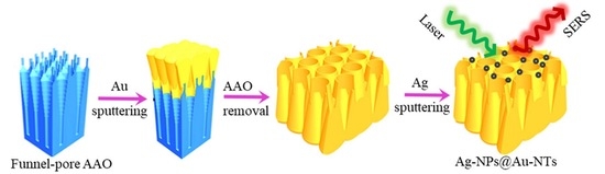
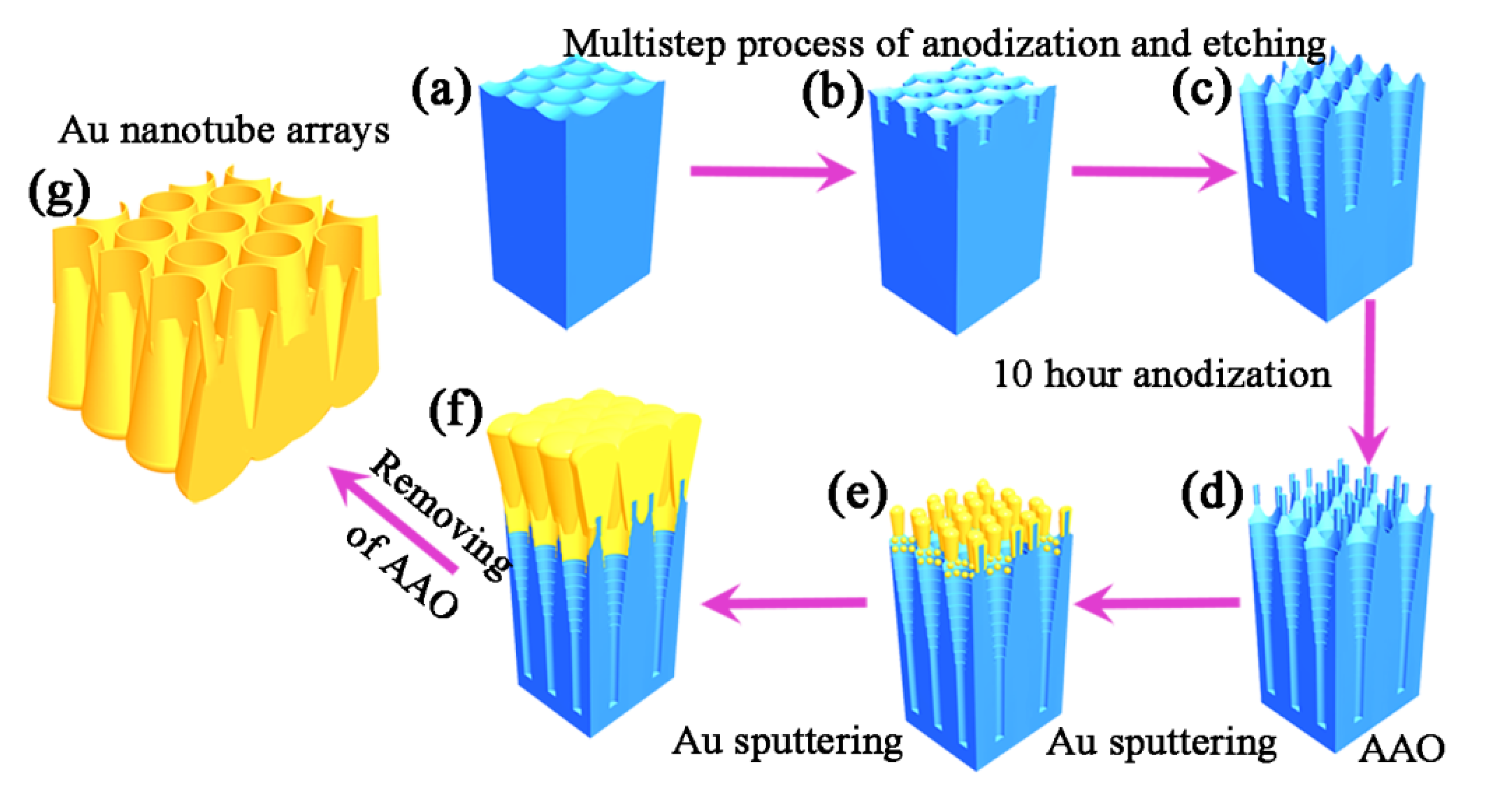
2.3. Preparation of Au-NT Arrays

The Au-NTs arrays were prepared via top-view ion-sputtering of Au on the funnel-shaped-pore-AAO template for 40 min with a current of 20 mA (EMITECH K550). Then, the as-prepared gold membrane was fixed on solid substrate such as glass, plastic, and copper foil with double-sided adhesive tape. Au-NTs array were achieved by etching the AAO template in 6 vol% phosphoric for 1 h at 40 °C, followed by rinsing with DI water and drying with high-purity nitrogen gas. In order to remove the surface amorphous gold particles, the achieved substrates were cleaned in a plasma cleaner. Finally, to further improve the SERS detection sensitivity, small Ag-NPs were sputtered onto the Au-NT array rotated at 2 circles per minute with a current of 20 mA.

2.4. Characterization

Scanning electron microscopy (SEM, sirion 200 FEG) was used for the morphology characterization of the fabricated Au nanostructures. SERS spectra were collected on a Renishaw inVia confocal microprobe Raman system. The excitation wavelength of the incident laser was 532 nm. The laser spot was 1 μm in diameter. For checking the SERS performance, the samples were dipped in 10−6 M R6G aqueous solution for 6 h, followed by naturally drying in ambient environment. For demonstrating the SERS sensitivity, the optimized Ag-NPs@Au-NT arrays were soaked in R6G solutions with different concentration (10−7 to 10−11 M) for 6 h. The point-by-point mapping was collected with scanning steps of 1 μm. Methyl parathion was dissolved in ethanol and then diluted with DI water to 10−6 M, 10−7 M, and 10−8 M for the SERS detection. The effective power of the laser source was 0.5 mW for R6G molecules detection, and 1 mW for methyl parathion detection, respectively.

3. Results and Discussion

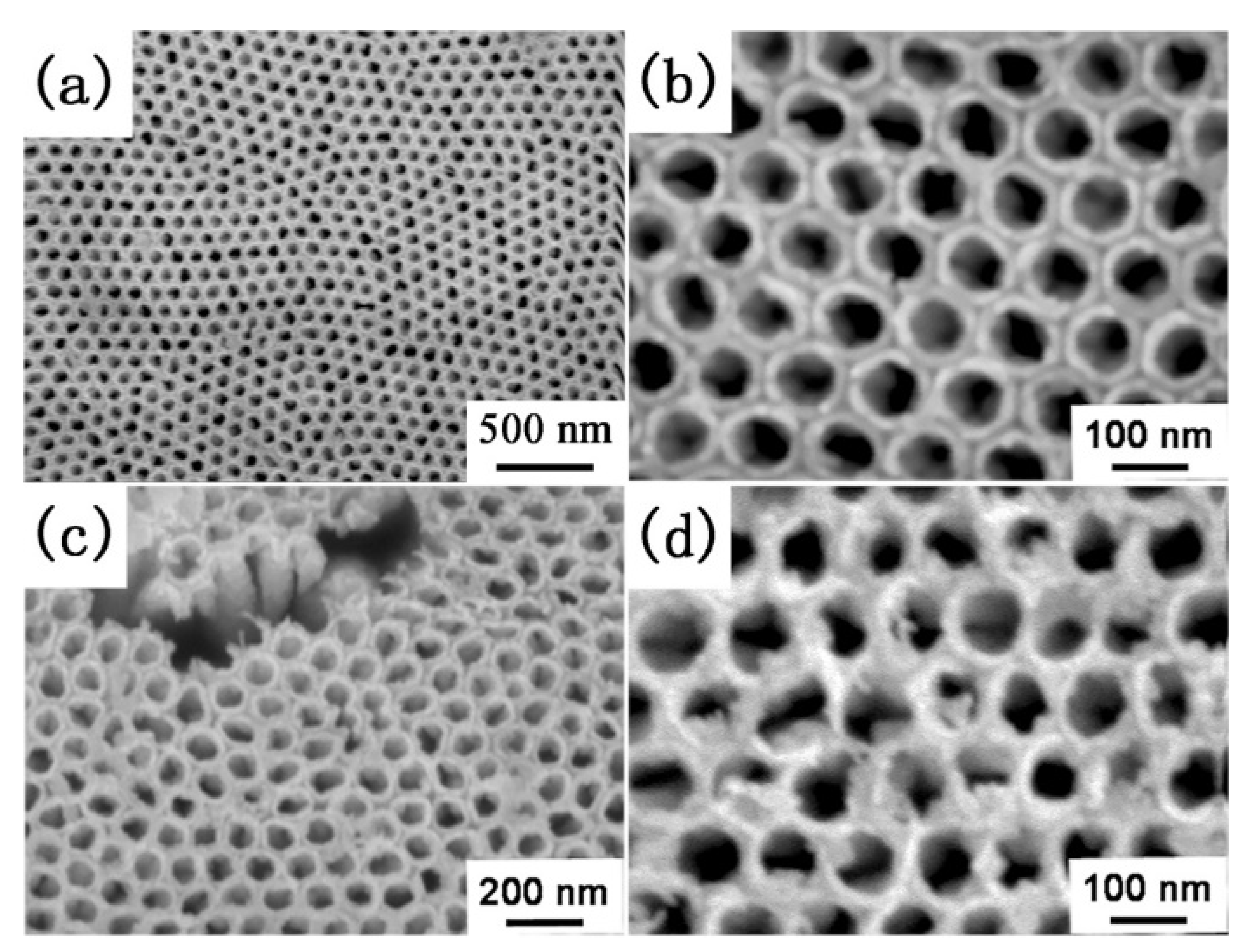
3.1. The Morphology of the Funnel-Shaped-Pore-AAO Templates and Achieved Ag-NPs@Au-NTs Array

The protocol for the fabrication of the Au-NTs array is shown schematically in Scheme 1. At the beginning, the funnel-shaped-pore-AAO templates with Al2O3 nanotips on the top were fabricated through a multistep alternate anodization and etching process. Second, top-view Au-sputtering was applied to achieve 3D Au film on the AAO template. For the larger diameter of the open-end of the funnel-shaped pores, the sputtered Au extended into the pores and finally aggregated to form nanotubes. The achieved Au-NT arrays after removal of the AAO template had abundant sub-10 nm gaps between the Au-NT arrays induced by the thin walls of neighboring pores and nanocavities on the joints of the funnel pores induced by the Al2O3 nanotips. As a result, there were large quantities of efficient “hot spots” for ultrasensitive SERS detection. Importantly, the gaps between the neighboring Au-NTs became narrower from the top to the bottom of the nanotubes because of the cone-shaped pore walls.
The morphology of the achieved funnel-shaped-pore-AAO template is revealed in Figure S1 [29]. The funnel-shaped pores were composed of two sections: conical pores of about 500 nm in length on the top of the AAO, and straight cylindrical pores of 50 μm in length. At the joint of three funnel pores, there was one Al2O3 nanotip (Figure S1), corresponding to our previously work [28]. Additionally, nanotips were arranged hexagonally on the surface of the funnel-shaped-pore-AAO template with a height and diameter of about 130 nm and 12 nm, respectively [28].
By top ion-sputtering of Au, massive Au particles accumulated and consequently coated on the Al2O3 nanotips and the internal face of the conical pores. The achieved Au-nanorods inherited the periodic hexagonal arrangement of the alumina nanotips. As shown in Figure S2a, the diameter of the formed Au-nanorods was about 54 nm after 16 min Au-sputtering. For longer Au-sputtering, the diameter of the Au-nanorods increased gradually, which resulted in a corresponding decrease in the gaps between the Au-nanorods. Meanwhile, the amount of the Au-NPs on the internal surface of the conical pores aggregated to form a continuous film, as illustrated in Scheme 1e. When the Au-sputtering duration reached 24 min (Figure S2b), the Au-nanorods array became very close. For further longer Au-sputtering, for example 40 min, the Au-nanorods stacked and became a continuous Au film (Figure S2c). Meanwhile the Au-NPs on the upper internal surface of the conical pores concatenated and consequently Au-NTs formed.
Since all the Au-NTs were formed inside the nanopores of the AAO templates, the parameters such as the diameter and the geometrical distribution of the nanotube replicated the uniformity of the AAO nanopores, as shown in Figure 1a,b. An enlarged view (Figure 1b) displayed that the Au-NTs were hexagonally arranged. The inner, outer top diameter, and neighboring gaps were about 69, 95, and 8 nm, respectively. The gaps became narrower and narrower from the top to the bottom of the nanotubes with the thinning of the conical pore-wall thickness, as shown in Scheme 1d and Figure 1c. Moreover, the inner diameter of the Au-NTs from the top to the bottom changed from 69 nm to sub-10 nm (Figures S2d and S3), which was also very important for creating strong SERS activity [30,31]. Thus, the as-fabricated large-scale Au-NTs showed potential as highly sensitive and reproducible SERS substrates. To further improve the SERS sensitivity of the substrate, small Ag-NPs were sputtered onto the Au-NTs surface. After top-sputtering of Ag for 4 min, small Ag-NPs of 5–10 nm in size were assembled onto the Au-NTs surface, finally forming uniformly hexagonally arranged Ag-NPs@Au-NTs arrays as shown in Figure 1d. Such hierarchically roughed structures also favored the criteria of an ideal SERS substrate.

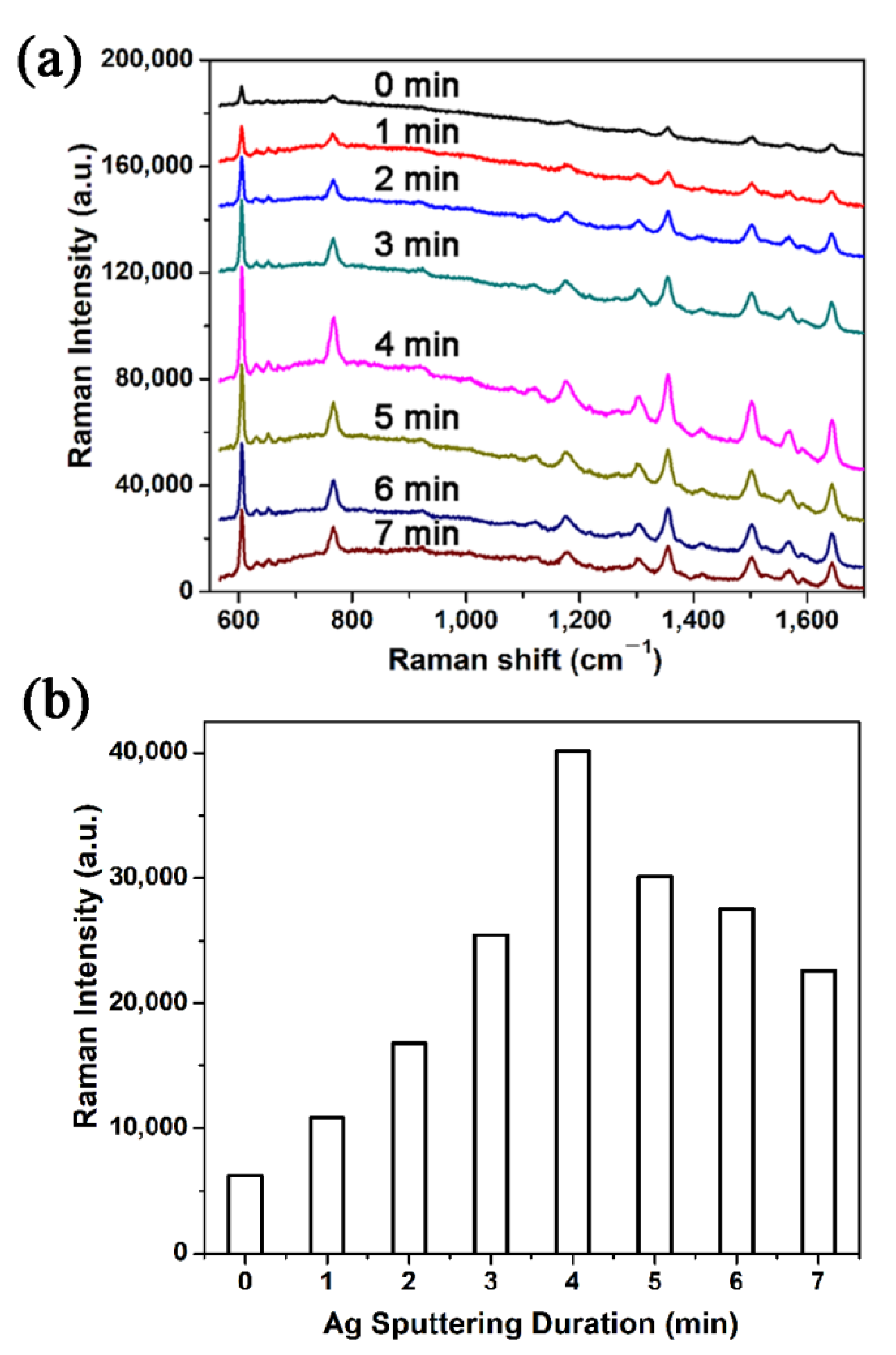
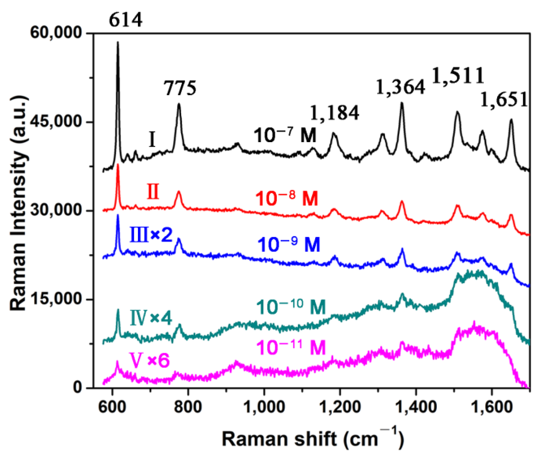
3.2. SERS Sensitivity of the Ag-NPs@Au-NTs Arrays

After the successful fabrication of large-area Au-NTs array, we subsequently adjusted the top spacing of the Au-NTs by sputtering Ag at a slow rate. R6G was also used as a probe molecule to evaluate the SERS activity. As shown in Figure 2a, the SERS spectra of 10−6 M R6G were collected from the as-prepared Ag-NPs@Au-NTs arrays substrates with various Ag-sputtering from 0 to 7 min. Because the SERS enhancement factor of Ag was generally 100 times greater than Au [32], and the Ag-NPs decoration made the Au-NTs surface much rougher and resulted in a consequently higher SERS enhancement based on the electromagnetic enhancement mechanism [5], the SERS detection sensitivity of the as-prepared Ag-NPs@Au-NTs substrates improved after the assembly of Ag-NPs and further increased till Ag-sputtering of 4 min. However, the SERS enhancement was diminished by too much Ag-sputtering (longer than 4 min). Because the interspace of the Au-NTs had been very close, too much assembly of Ag-NPs would induce the disappearance of the sub-10 nm gaps, resulting in the decrease in the SERS enhancement of the substrate.
To demonstrate the SERS sensitivity of the optimized Ag-NPs@Au-NTs array substrate, the SERS spectra of R6G with different concentrations (10−7, 108, 10−9, 10−10, and 10−11 M) on the substrates were measured. As shown in Figure 3, the characteristic Raman peaks of R6G could be clearly recognized. The characteristic peaks at 614, 775, and 1184 cm−1 were attributed to C–C–C ring in-plane vibration mode, C–C–C ring out-of-plane bend mode, and C–H in-plane bend mode, respectively. The characteristic peaks at 1364, 1511, and 1651 cm−1 were assigned to aromatic C–C stretching vibration modes [33]. The intensity of the characteristic peaks decreased with the concentration of the R6G, while such a low concentration as 10−11 M could still be identified, showing the high detection sensitivity of the as-prepared substrate. Additionally, the enhancement factor (EF) of the Ag-NPs@Au-NTs array substrate was estimated to be 1.8 × 10⁷ (Part S4 of the Supplementary Materials), demonstrating the high SERS enhancement capacity of the Ag-NPs@Au-NTs arrays again.

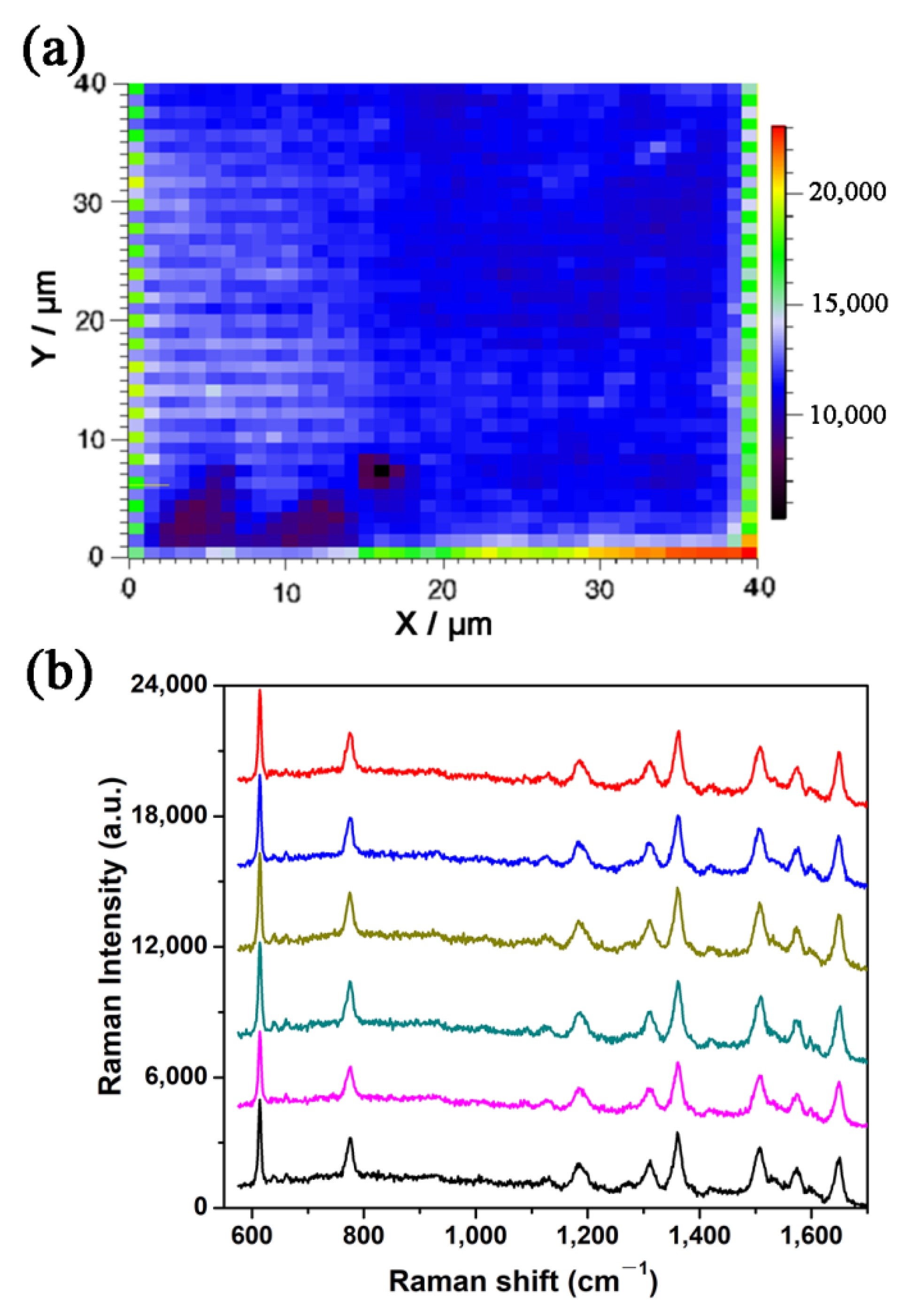
3.3. SERS Signal Homogeneity over the Ag-NPs@Au-NTs Arrays

The homogeneity of the SERS substrate was evaluated first by point-by-point SERS Raman mapping. Figure 4a shows the SERS mapping image of the optimized Ag-NPs@Au-NTs arrays. The image was achieved based on the intensity of the R6G characteristic peak at 614 cm−1 within an area of 40 μm × 40 μm by measuring 1681 points. The relative SERS intensities of the peaks were around 12,000 counts, and the RSD was calculated to be about 8.3%, indicating that the Ag-NPs@Au-NTs arrays had a high uniformity of SERS response. The homogeneity of the SERS signals over the large area was further investigated by comparing the SERS spectra collected randomly on the Ag-NPs@Au-NTs arrays. Six SERS spectra collected at six spots on the substrate were very similar to each other (Figure 4b), intuitively demonstrating the signal uniformity of the substrate. Specifically, two advantages of the as-prepared Ag-NPs@Au-NTs arrays and the fabrication strategy could explain the good Raman signal uniformity and reproducibility. The morphology of the AAO templates could be highly determined by the fabrication parameters and condition. Then, the reproducibility of the specific AAO templates could be realized. Additionally, Au and Ag were physically sputtered onto the AAO templates and the Au-NTs arrays, which had lower deviation than wet chemical methods.
To demonstrate the potential of the Ag-NPs@Au-NTs arrays SERS substrates in pollutants detection, the SERS detection of the toxic organophosphorus pesticide methyl parathion, which has been illegally overused and exposed to the environment [34], was carried out. The normal Raman spectrum of solid methyl parathion powder and SERS spectra of the methyl parathion solution diluted into various concentrations from 10−6 M to 10−8 M are shown in Figure 5. The main characteristic peaks at 858 (O=N=O wagging), 1109 (C–N stretching), 1347 (C–N=O stretching), and 1594 cm−1 all corresponded to the solid film and the previously reported results [35]. A low concentration of methyl parathion down to 10−8 M could still be recognized, showing the great potential of the Ag-NPs@Au-NTs arrays for SERS-based rapid detection of environmental organic pollutants.
It is also worth mentioning that only oxalic and phosphoric acids were used for the fabrication of the conical-pore-AAO templates, while simple physical ion-sputtering was used for the fabrication of the Ag-NPs@Au-NT arrays without involving any organic chemicals. Therefore, this work presented a clean and green strategy for diminishing the potential signal interference caused by the chemical molecules adsorbed on the plasmonic structures during the substrate preparation process.

4. Conclusions

In summary, funnel-shaped-pore-AAO templates were fabricated by long time oxidation of conical-pore-AAO achieved by multistep anodization and etching. Additionally, they were used as sacrificial templates for fabricating uniform hexagonally arranged Au-NTs array with sub-10 nm neighboring gaps and sub-10 nm nanocavities at the bottom of the nanotubes. Furthermore, the SERS activity of the substrates was improved after further sputtering of Ag, forming Ag-NPs@Au-NTs hierarchical arrays. The as-prepared composite substrates demonstrated high SERS detection sensitivity for R6G and methyl parathion with a limit of 10−11 M and 10−8 M, respectively. The hexagonally arranged Ag-NPs@Au-NTs arrays showed potential for the sensitive detection of organic pollutants.

Supplementary Materials

The following supporting information can be downloaded at: https://www.mdpi.com/article/10.3390/su14031305/s1, Figure S1: The SEM images of the funnel-shaped-pore-AAO template; Figure S2: Top-view SEM images of the Au film fabricated by sputtering Au on the funnel-shaped-pore-AAO templates for different durations; Figure S3: SEM images of Au-NTs; Figure S4: The calculation of the enhancement factor (EF) of the Ag-NPs@Au-NTs arrays.

Author Contributions

Conceptualization, K.S. and H.T.; methodology, K.S.; validation, K.S., H.T. and Q.D.; formal analysis, K.S.; investigation, K.S. and H.T.; resources, K.S. and H.T.; data curation, K.S.; writing—original draft preparation, K.S.; writing—review and editing, H.T. and Q.D.; supervision, H.T.; project administration, H.T.; funding acquisition, K.S. and H.T. All authors have read and agreed to the published version of the manuscript.

Funding

This research was funded by [National Natural Science Foundation of China] grant number [51972308, 51801093] and [Natural Science Foundation of Anhui Province] grants number [2008085ME171, 201903a07020021]. And The APC was funded by [51801093].

Data Availability Statement

Not applicable.

Conflicts of Interest

The authors declare no conflict of interest.

References

  1. Yang, L.; Lee, J.-H.; Rathnam, C.; Hou, Y.; Choi, J.-W.; Lee, K.-B. Dual-Enhanced Raman Scattering-Based Characterization of Stem Cell Differentiation Using Graphene-Plasmonic Hybrid Nanoarray. Nano Lett. 2019, 19, 8138–8148. [Google Scholar] [CrossRef] [PubMed]
  2. Shi, Y.; Chen, N.; Su, Y.; Wang, H.; He, Y. Silicon nanohybrid-based SERS chips armed with an internal standard for broad-range, sensitive and reproducible simultaneous quantification of lead(ii) and mercury(ii) in real systems. Nanoscale 2018, 10, 4010–4018. [Google Scholar] [CrossRef]
  3. Liao, P.-H.; Tseng, C.-Y.; Ke, Z.-Y.; Hsieh, C.-L.; Kong, K.V. Operando characterization of chemical reactions in single living cells using SERS. Chem. Commun. 2020, 56, 4852–4855. [Google Scholar] [CrossRef] [PubMed]
  4. Tang, H.; Zhu, C.; Meng, G.; Wu, N. Review—Surface-Enhanced Raman Scattering Sensors for Food Safety and Environmental Monitoring. J. Electrochem. Soc. 2018, 165, B3098–B3118. [Google Scholar] [CrossRef]
  5. Tang, H.; Meng, G.; Li, Z.; Zhu, C.; Huang, Z.; Wang, Z.; Li, F. Hexagonally arranged arrays of urchin-like Ag hemispheres decorated with Ag nanoparticles for surface-enhanced Raman scattering substrates. Nano Res. 2015, 8, 2261–2270. [Google Scholar] [CrossRef]
  6. Li, M.; Cushing, S.K.; Wu, N. Plasmon-enhanced optical sensors: A review. Analyst 2015, 140, 386–406. [Google Scholar] [CrossRef] [PubMed] [Green Version]
  7. Huang, Z.; Meng, G.; Hu, X.; Pan, Q.; Huo, D.; Zhou, H.; Ke, Y.; Wu, N. Plasmon-tunable Au@Ag core-shell spiky nanoparticles for surface-enhanced Raman scattering. Nano Res. 2019, 12, 449–455. [Google Scholar] [CrossRef]
  8. Li, J.; Deng, T.-S.; Liu, X.; Dolan, J.A.; Scherer, N.F.; Nealey, P.F. Hierarchical Assembly of Plasmonic Nanoparticle Heterodimer Arrays with Tunable Sub-5 nm Nanogaps. Nano Lett. 2019, 19, 4314–4320. [Google Scholar] [CrossRef]
  9. Yang, H.; Li, B.Q.; Jiang, X.; Shao, J. Hybrid nanostructure of SiO2@Si with Au-nanoparticles for surface enhanced Raman spectroscopy. Nanoscale 2019, 11, 13484–13493. [Google Scholar] [CrossRef]
  10. Tao, A.; Sinsermsuksakul, P.; Yang, P. Polyhedral Silver Nanocrystals with Distinct Scattering Signatures. Angew. Chem. Int. Ed. 2006, 45, 4597–4601. [Google Scholar] [CrossRef]
  11. Abu Hatab, N.A.; Oran, J.M.; Sepaniak, M.J. Surface-Enhanced Raman Spectroscopy Substrates Created via Electron Beam Lithography and Nanotransfer Printing. ACS Nano 2008, 2, 377–385. [Google Scholar] [CrossRef] [PubMed]
  12. Zhu, C.; Zhao, Q.; Meng, G.; Wang, X.; Hu, X.; Han, F.; Lei, Y. Silver nanoparticle-assembled micro-bowl arrays for sensitive SERS detection of pesticide residue. Nanotechnology 2020, 31, 205303. [Google Scholar] [CrossRef] [PubMed]
  13. Wang, P.; Wu, L.; Lu, Z.; Li, Q.; Yin, W.; Ding, F.; Han, H. Gecko-Inspired Nanotentacle Surface-Enhanced Raman Spectroscopy Substrate for Sampling and Reliable Detection of Pesticide Residues in Fruits and Vegetables. Anal. Chem. 2017, 89, 2424–2431. [Google Scholar] [CrossRef] [PubMed]
  14. Fang, X.; Zheng, C.; Yin, Z.; Wang, Z.; Wang, J.; Liu, J.; Luo, D.; Liu, Y.J. Hierarchically Ordered Silicon Metastructures from Improved Self-Assembly-Based Nanosphere Lithography. ACS Appl. Mater. Interfaces 2020, 12, 12345–12352. [Google Scholar] [CrossRef] [PubMed]
  15. Wang, Y.; Zhao, X.; Chen, L.; Chen, S.; Wei, M.; Gao, M.; Zhao, Y.; Wang, C.; Qu, X.; Zhang, Y.; et al. Ordered Nanocap Array Composed of SiO2-Isolated Ag Islands as SERS Platform. Langmuir 2014, 30, 15285–15291. [Google Scholar] [CrossRef] [PubMed]
  16. Zhang, Y.; Filipczak, P.; He, G.; Nowaczyk, G.; Witczak, L.; Raj, W.; Kozanecki, M.; Matyjaszewski, K.; Pietrasik, J. Synthesis and characterization of Ag NPs templated via polymerization induced self-assembly. Polymer 2017, 129, 144–150. [Google Scholar] [CrossRef]
  17. Zhu, C.; Meng, G.; Zheng, P.; Huang, Q.; Li, Z.; Hu, X.; Wang, X.; Huang, Z.; Li, F.; Wu, N. A Hierarchically Ordered Array of Silver-Nanorod Bundles for Surface-Enhanced Raman Scattering Detection of Phenolic Pollutants. Adv. Mater. 2016, 28, 4871–4876. [Google Scholar] [CrossRef]
  18. Anema, J.R.; Brolo, A.G.; Felten, A.; Bittencourt, C. Surface-enhanced Raman scattering from polystyrene on gold clusters. J. Raman Spectrosc. 2010, 41, 745–751. [Google Scholar] [CrossRef]
  19. Huang, Z.; Meng, G.; Chen, B.; Zhu, C.; Han, F.; Hu, X.; Wang, X.; Zhulin, H.; Guowen, M.; Bin, C.; et al. Surface-Enhanced Raman Scattering from Au-Nanorod Arrays with Sub-5-nm Gaps Stuck Out of an AAO Template. J. Nanosci. Nanotechnol. 2016, 16, 934–938. [Google Scholar] [CrossRef]
  20. Chen, B.; Meng, G.; Huang, Q.; Huang, Z.; Xu, Q.; Zhu, C.; Qian, Y.; Ding, Y. Green Synthesis of Large-Scale Highly Ordered Core@Shell Nanoporous Au@Ag Nanorod Arrays as Sensitive and Reproducible 3D SERS Substrates. ACS Appl. Mater. Interfaces 2014, 6, 15667–15675. [Google Scholar] [CrossRef]
  21. Huang, Z.; Meng, G.; Huang, Q.; Chen, B.; Zhu, C.; Zhang, Z. Large-area Ag nanorod array substrates for SERS: AAO template-assisted fabrication, functionalization, and application in detection PCBs. J. Raman Spectrosc. 2013, 44, 240–246. [Google Scholar] [CrossRef]
  22. Tang, H.; Meng, G.; Huang, Q.; Zhu, C.; Huang, Z.; Li, Z.; Zhang, Z.; Zhang, Y. Urchin-like Au-nanoparticles@Ag-nanohemisphere arrays as active SERS-substrates for recognition of PCBs. RSC Adv. 2014, 4, 19654–19657. [Google Scholar] [CrossRef]
  23. Zhou, Q.; Meng, G.; Huang, Q.; Zhu, C.; Tang, H.; Qian, Y.; Chen, B.; Chen, B. Ag-nanoparticles-decorated NiO-nanoflakes grafted Ni-nanorod arrays stuck out of porous AAO as effective SERS substrates. Phys. Chem. Chem. Phys. 2014, 16, 3686–3692. [Google Scholar] [CrossRef] [PubMed]
  24. Hendren, W.R.; Murphy, A.; Evans, P.; O’Connor, D.; Wurtz, G.A.; Zayats, A.V.; Atkinson, R.; Pollard, R.J. Fabrication and optical properties of gold nanotube arrays. J. Phys. Condens. Matter 2008, 20, 3931–3939. [Google Scholar] [CrossRef]
  25. Lee, W.; Scholz, R.; Nielsch, K.; Gösele, U. A Template-Based Electrochemical Method for the Synthesis of Multisegmented Metallic Nanotubes. Angew. Chem. Int. Ed. 2005, 44, 6050–6054. [Google Scholar] [CrossRef]
  26. Yu, Y.; Kant, K.; Shapter, J.G.; Addai-Mensah, J.; Losic, D. Gold nanotube membranes have catalytic properties. Microporous Mesoporous Mater. 2012, 153, 131–136. [Google Scholar] [CrossRef]
  27. Sun, K.; Huang, Q.; Meng, G.; Lu, Y. Highly Sensitive and Selective Surface-Enhanced Raman Spectroscopy Label-free Detection of 3,3′,4,4′-Tetrachlorobiphenyl Using DNA Aptamer-Modified Ag-Nanorod Arrays. ACS Appl. Mater. Interfaces 2016, 8, 5723–5728. [Google Scholar] [CrossRef]
  28. Sun, K.; Meng, G.; Huang, Q.; Zhao, X.; Zhu, C.; Huang, Z.; Qian, Y.; Wang, X.; Hu, X. Gap-tunable Ag-nanorod arrays on alumina nanotip arrays as effective SERS substrates. J. Mater. Chem. C 2013, 1, 5015–5022. [Google Scholar] [CrossRef]
  29. Yan, B.; Sun, K.; Chao, K.; Alharbi, N.S.; Li, J.; Huang, Q. Fabrication of a Novel Transparent SERS Substrate Comprised of Ag-nanoparticle Arrays and its Application in Rapid Detection of Ractopamine on Meat. Food Anal. Methods 2018, 11, 2329–2335. [Google Scholar] [CrossRef]
  30. Xu, W.; Zhu, X.; Chu, Z.; Wang, Z.; Xiao, Z.; Huang, Z. ScroBiculate sub-10 nm nanocavity arrays as effective sers substrate for the trace determination of 3,3′,4,4′—Polychlorinated biphenyls. Appl. Surf. Sci. 2017, 399, 711–715. [Google Scholar] [CrossRef]
  31. Hou, C.; Meng, G.; Huang, Z.; Chen, B.; Zhu, C.; Li, Z. Ordered arrays of vertically aligned Au-nanotubes grafted with flocky Au/Ag-nanospikes based on electrodeposition and subsequent redox reaction. Electrochem. Commun. 2015, 60, 104–108. [Google Scholar] [CrossRef]
  32. Erol, M.; Han, Y.; Stanley, S.K.; Stafford, C.; Du, H.; Sukhishvili, S. SERS Not to Be Taken for Granted in the Presence of Oxygen. J. Am. Chem. Soc. 2009, 131, 7480–7481. [Google Scholar] [CrossRef] [PubMed]
  33. Li, Z.; Du, Z.; Sun, K.; He, X.; Chen, B. Controllable fabrication of Ag-nanoplate-decorated PAN-nanopillar arrays and their application in surface-enhanced Raman scattering. RSC Adv. 2017, 7, 53157–53163. [Google Scholar] [CrossRef] [Green Version]
  34. Zhou, N.; Meng, G.; Huang, Z.; Ke, Y.; Zhou, Q.; Hu, X. A flexible transparent Ag-NC@PE film as a cut-and-paste SERS substrate for rapid in situ detection of organic pollutants. Analyst 2016, 141, 5864–5869. [Google Scholar] [CrossRef] [PubMed]
  35. Wang, X.; Zhu, C.; Hu, X.; Xu, Q.; Zhao, H.; Meng, G.; Lei, Y. Highly sensitive surface-enhanced Raman scattering detection of organic pesticides based on Ag-nanoplate decorated graphene-sheets. Appl. Surf. Sci. 2019, 486, 405–410. [Google Scholar] [CrossRef]
Scheme 1. The schematic route for the fabrication of the Au nanotube arrays. (a) The aluminum foil after pre-oxidation process; (b) one time anodizing and pore-widening process; (c) 10 times repeated anodizing and pore-widening process; (d) long oxidation of 10 h; (e) Au-sputtering of 16 min; (f) Au-sputtering of 40 min; (g) removal of AAO membrane.
Sustainability 14 01305 sch001
Figure 1. SEM images of Au-NTs achieved by Au-sputtering of 40 min: (a) top view, (b) an enlarged top view and (c) 45 degree oblique view. (d) Au-NTs decorated with Ag-NPs by Ag-sputtering of 4 min.
Sustainability 14 01305 g001
Figure 2. (a) SERS spectra response and (b) intensity comparison of the 614 cm−1 peaks of the R6G molecules adsorbed on the Au-NTs arrays with different Ag-sputtering (0 to 7 min).
Sustainability 14 01305 g002
Figure 3. SERS spectra collected on the optimized Ag-NPs@Au-NT arrays exposed to different concentrations of R6G.
Sustainability 14 01305 g003
Figure 4. (a) SERS mapping of 614 cm−1 band of R6G. (b) Six SERS spectra collected randomly on the SERS substrates.
Sustainability 14 01305 g004
Figure 5. SERS spectra of methyl parathion with different concentrations from 10−6 M to 10−8 M collected on the optimized Ag-NPs@Au-NTs arrays substrate.
Sustainability 14 01305 g005
Publisher’s Note: MDPI stays neutral with regard to jurisdictional claims in published maps and institutional affiliations.

