Article

Optimization of Carboniferous Egyptian Kaolin Treatment for Pharmaceutical Applications

1
Geology Department, Faculty of Science, Ain Shams University, Cairo 11566, Egypt
2
Environmental Science Department, Faculty of Meteorology, Environment and Arid Land Agriculture, King Abdulaziz University (KAU), Jeddah 21589, Saudi Arabia
3
Department of Biological and Geological Sciences, Faculty of Education, Ain Shams University, Cairo 11341, Egypt
*
Author to whom correspondence should be addressed.
Sustainability 2022, 14(4), 2388; https://doi.org/10.3390/su14042388
Submission received: 19 January 2022 / Revised: 13 February 2022 / Accepted: 16 February 2022 / Published: 19 February 2022
(This article belongs to the Section Environmental Sustainability and Applications)

Abstract

:
This paper quantitatively determines the occurrences of potentially toxic elements in Carboniferous kaolin in southwestern Sinai, Egypt. This research describes, in detail, the experimental treatment optimization to be used in pharmaceutical applications. The concentrations of As, Co, Ni, Pb, and V in these kaolin deposits exceed the Permitted Concentrations of Elemental Impurities for oral use in pharmaceutical applications. Herein, six desorbing agents (acetic acid, citric acid, DTPA, EDDS, EDTA, and NTA) were utilized as extracting solutions in batch-wise extractions to select the proper reagents. Parameters such as the pH, the mixing speed and time, and the solid–solution ratio were varied to optimize the extraction conditions. The findings indicate that citric acid and EDTA were effective in the removal of the aforementioned elements. The results reveal that the optimum removal of potentially toxic elements from kaolin can be achieved using citric acid and EDTA concentrations of 0.2 M and 0.1 M, respectively, for the treatment of 5 g of kaolin, under a pH of 4 for citric acid, and a pH of 10 for EDTA. The ideal mixing speed and time are 500 rpm and 6 h, respectively. Using 1:10 S/L of citric acid and EDTA showed removal rates of 100% for all the investigated PTEs. We recommend this treatment for different kinds of kaolin showing various degrees of contamination.

1. Introduction

The usage of clay minerals in cosmetics and for medical purposes has recently increased because of the growing achievements of natural remedies and healthcare research [1]. In pharmaceutical technology, some clay minerals are used as excipients or active substances in the formulation of diverse topical or oral dosage forms [1,2].
In order to be used in pharmaceutical technology, clay minerals should comply with several chemical, physical, and toxicological requirements [1,3]. However, for these requisites, only restricted types of clay minerals are used for these aims. Among them are the kaolin group, talc, some smectites (montmorillonite and saponite), sepiolite, and palygorskite [4]. Kaolinite (Al2Si2O5(OH)4) is the most common mineral of the kaolin group, and it is a planar hydrous 1:1 dioctahedral clay mineral. This mineral exhibits many excellent physical, mechanical, and structural properties, which comply with the pharmaceutical technology requirements and which make them very useful for many pharmaceutical applications [1,5]. The chemical compositions of clay minerals include many potentially toxic elements (PTEs) in significantly different concentrations [5,6,7,8]. Some of these elements have harmful effects, such as As, Cd, Co, Cr, Ni, Pb, and V. Excessive exposure to these PTEs could lead to one disease or another [9,10,11,12,13,14]. Thus, it is necessary to assess the concentrations of PTEs in clay using validated quantitative and qualitative techniques, before their usage in any pharmaceutical applications [3,5].
In Egypt, there are a variety of sedimentary kaolin deposits, which range in age from Carboniferous (in the Sinai Peninsula) to Neogene (in the Sinai Peninsula, Aswan, and the Red Sea Coast) [15,16,17]. One key deposit is located at Abu Zeneima, in southwestern Sinai, and it represents the highest reserves and the purist grade amongst all the Egyptian kaolin deposits. Major Abu Zeneima outcrops are exposed at Wadi Khaboba, Gabal Hazbar, Wadi Abu Natash, Farsh El Ghozlan, Wadi Budra, and Gabal El Dehessa [5].
The Egyptian kaolin are used in many industrial applications, such as ceramics, refractories, whiteware, paints, and paper [15,18]. Previous research has focused on its mineralogy, geochemistry, radioactivity, sedimentological implication, origin, and possible applications [16,17,18,19,20,21]. However, evaluating the kaolin’s suitability for pharmaceutical applications has received minimal attention. Awad et al. [5] were the first to characterize Abu Zeneima kaolin for healthcare uses. Their research shows significant differences in the distributions of the major and trace elements among the samples collected from different locations at Abu Zeneima. In addition, the Wadi Abu Natash exposure provided the highest and purest kaolin ore, and the massive kaolin clay beds, especially, are being considered as promising pharmaceutical excipients [5,22]. However, the authors point out that Abu Natash kaolin contains high concentrations of PTEs, especially As, Cr, Cu, Ni, Pb, and V.
Recently, the significant advances in remediation techniques have decisively shown that desorbing agents, such as acetic acid, citric acid, tartaric acid, diethylenetriaminepentaacetic acid (DTPA), [S,S]-ethylenediaminedisuccinic acid (EDDS), ethylenediaminetetraacetic acid (EDTA), and nitrilotriacetic acid (NTA), can dislodge and dissolve PTEs in clayey soils and contaminated media [23,24,25,26,27,28,29,30,31,32,33]. Therefore, this study aims to quantitatively determine the PTE concentrations in Carboniferous kaolin in Wadi Abu Natash, in southwestern Sinai, Egypt. Furthermore, in order to assess the kaolin for safe use for the pharmaceutical application, several batch experiments were conducted that evaluated the desorption of PTEs with various solubilizing agents. The results reported in this study provide additional data about optimizing kaolinite treatment and, hence, will be valuable in the treatment of different kinds of kaolin deposits.

2. Materials and Methods

2.1. Sample Collection and Preparation

Fifteen kaolin samples were collected from the massive kaolinitic clay beds exposed in Wadi Abu Natash, in Abu Zeneima, southwestern Sinai (28°56′24″–28°56′52″ N, and 33°18′50″–33°19′42″ E) (Figure 1). The studied kaolin deposits are part of the Abu Thora Formation, which belongs to the Carboniferous age. The Paleozoic successions exposed in the study area include the following formations, from the base to the top: Sarabit El Khadim, Abu Hamata, Nasib, Adedia, Um Bogma, and the Abu Thora Formations. The lower four formations are predominately clastic rocks and belong to the Cambro–Ordovician. Um Bogma and the Abu Thora Formations represent a different sedimentation phase (carbonate and clastic rocks), which occurred in the Lower Carboniferous. The entire Paleozoic succession unconformably overlies the Precambrian basement rocks (gneisses and younger granites) and unconformably underlies the Permo–Triassic sandstone and mudstone red beds. The Abu Thora Formation in certain places is capped by basaltic sheets (Mesozoic), and it is also dissected by doleritic and basaltic dykes [34,35].
In order to ensure the representativeness of the sampling, each sample was collected as a composite sample of three subsamples. About 3 kg of each sample was collected and stored in sealed polyethylene bags. The samples were air-dried in a controlled environment to avoid any cross-contaminations. The dried samples were gently disaggregated, sieved through a 2-mm sieve, and were then quartered into various subsets for further analysis.

2.2. Sample Characterizations

The mineralogical compositions of the studied raw kaolin samples were determined by using the X-ray diffraction (XRD) technique using a PANalytical X-Ray Diffraction device model, the X’Pert Pro with a monochromator, with Cu radiation (λ = 1.542 Å), at 45 Kv, 35 mA, and a scanning speed of 0.03°/s. The reflection peaks were measured between 2θ = 5° and 60°. The calculated d-spacings (d Å) and the relative intensities (I/I°) of the diffractograms were determined and were matched up to The International Centre for Diffraction Data (ICDD) standard powder diffraction files [36].
The XRF analysis was carried out for the powder (<74 µm) samples using X-Ray fluorescence equipment, PW 2404, with five analyzing crystals. The concentrations of the analyzed major elements (oxides %) were calculated using the software programs, Super Q and Semi Q, with an accuracy of 99.99%, and a confidence limit of 96.7%.

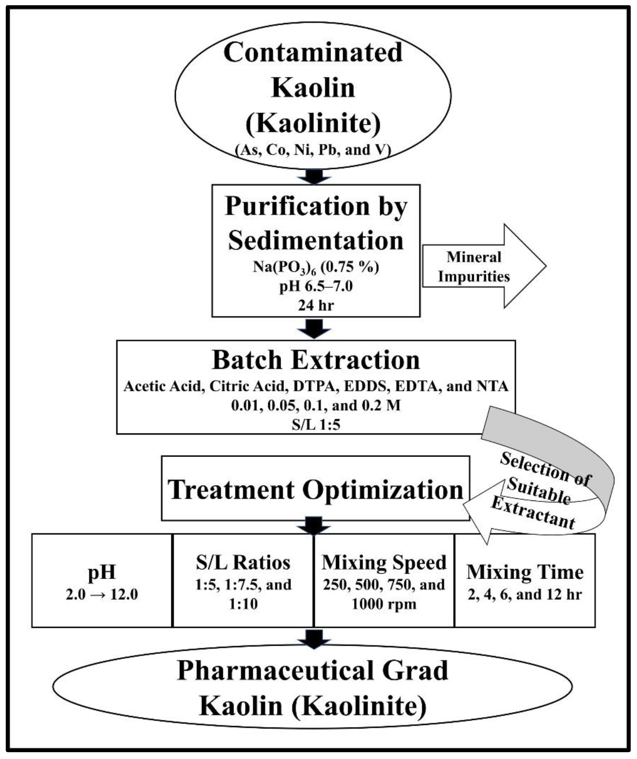
2.3. Purification of Kaolin

Impure kaolin, which may contain other mineral impurities, would complicate the quantitative determination of the chelator/element ratio and the mask chelator’s effectiveness because of the possible occurrence of the competitive binding of the chelator with cations other than PTEs, such as Ca, Mg, Na, and K [23]. Hence, the raw kaolin samples were subjected to purification to eliminate the mineral impurities, following the sedimentation technique adopted by Maynard [37]. Briefly, 50 mL of sodium hexametaphosphate solution (NaPO3)6 (conc. 0.75%) was added to keep the particles flocculated, while the samples were disaggregated by using a mechanical stirrer. The pH was adjusted (6.5 to 7.0) by adding sodium carbonate Na2CO3 (conc. 0.25%). The slurry was allowed to stand for 24 h, after which the supernatant was gently discarded by decantation. The pure white decanted dispersions were centrifuged and washed with distilled water three times.

2.4. Batch Extraction Experiment

A series of batch experiments were conducted to investigate the effectiveness of the desorbing agents in removing the PTEs from the kaolin. Two organic acids, acetic acid (CH3COOH) and citric acid (C6H8O7), and four aminopolycarboxylic acids, DTPA (C14H23N3O10), EDDS (C10H16N2O8), EDTA (C10H16N2O8), and NTA (C6H9NO6), were investigated at concentrations of 0.01, 0.05, 0.1, and 0.2 M for the removal of PTEs from the purified kaolin samples. In addition, one composite sample was prepared from the purified kaolin to be used in these experiments.
The batch extraction experiments were conducted following the procedure adopted by Khodadoust et al. [24] using a solid-to-liquid (S/L) ratio of 1:5. First, 5 g of purified kaolin was placed in a 40-mL glass vial, with 25 mL of the extracting solution. The vial was tightly sealed and then shaken by hand for about one minute to ensure that the sample was thoroughly saturated with the extracting solution. Afterwards, the vials were shaken in a dry shaker at 250 rpm overnight at room temperature. Following the shaking, the solution–solid mixture was centrifuged at 4000 rpm for 28 min. Then, the supernatant was decanted and filtered using a Whatman GF/C glass fiber filter (1.2 µm particle retention). The filtrated supernatant was centrifuged again at 4000 rpm for 28 min to ensure the removal of any suspended fine particles. The batch extractions were performed in triplicate to ensure the reproducibility of the results.
The removal rates of the PTEs were calculated using Equation (1) [24,28]:
Removal   Rate = ( C i C q C i ) × 100
where Ci is the initial concentration of the PTE, and Cq is the concentration in the supernatant solution after treatment with the desorbing agent.

2.5. Treatment Optimization

On the basis of the first batch of experiments results, the treatment parameters expected to affect the removal of PTEs (pH, mixing speed and time, and S/L ratio) were optimized to attain the highest removal of PTEs. Citric acid and EDTA were investigated at concentrations of 0.2 and 0.1 M, respectively, with a specific pH value between 2.0 and 12.0, by adding 0.1 M NaOH or HCl. We also investigated three S/L ratios (1:5, 1:7.5, and 1:10). The experimental conditions were investigated at different rotational speeds (250, 500, 750, and 1000 rpm) and at fixed time intervals (2, 4, 6, and 12 h). All the experiments were performed at room temperature. A schematic diagram of the experimental protocol for the treatment and parameter optimization is presented in Figure 2.

2.6. PTE Determinations

The investigated raw and purified kaolin samples were acid digested using HNO3 (70%) and HF (48%) with silicon dioxide, for 30 min in a closed vessel, using a pressure-controlled microwave (CEM Microwave Digestion). The digestion solutions were measured using optical emission spectroscopy with inductively coupled plasma (ICP-OES) with an ultrasonic nebulizer (Optima 3000, PerkinElmer Inc., Waltham, MA, USA). To determine the PTE concentrations in the solutions at the end of every step of the batch experiments, the filtrated liquid samples were analyzed by a multielement atomic absorption spectrometer (SavantAA, GBC Scientific Equipment, Pulau Pinang, Malaysia).

2.7. Quality Control

All the glassware was cleaned by soaking in a 10% (v/v) HNO3 acid for 15 min, and then by flushing with deionized water about three times, after which it was then dried prior to use. All of the reagents used were of analytical grade, and all the solutions were prepared by using double distilled water. The XRD and XRF analyses were carried out at the accredited Central Laboratories Sector of The Egyptian Mineral Resources Authority (ISO 17025). The PTE determination laboratory analyses of the kaolin samples were carried out at the accredited Central Laboratory for Environmental Quality Monitoring National Water Research Center (ISO/IEC 17025), with the measurement standards provided by the Canadian Association for Laboratory Accreditation Inc. In order to ensure the analytical performance of the ICP-OES, all of the ICP-OES operational parameters were software-controlled. A multielement standard solution (Merck Brand, Darmstadt, Germany) of a known concentration was used to calibrate and quantitate the sample results. This standard was run after each set of 10 samples.

3. Results

3.1. Sample Characterizations

The mineralogical analysis for the bulk kaolin samples of Wadi Abu Natash confirm that they are essentially kaolinite. The X-ray diffraction patterns of the samples indicate that anatase, halite, quartz, and hematite are the main nonclay minerals (Figure 3).
Tables S1 and S2 (in Supplementary Materials) illustrate the major elements and the PTE concentrations for the raw kaolin and the purified samples, respectively. Generally, the concentrations of the major elements showed high silica (SiO2 (average 43.07%)) and alumina (Al2O3 (average 36.22%)) contents. The main Al- and Si-bearing mineral in the studied samples is kaolinite, as was proven by the X-ray diffraction analysis. On the other hand, the TiO2, Na2O, and Fe2O3 contents in most of the samples are relatively low and are linked to the presence of anatase, halite, and hematite minerals, respectively. The Fe2O3 and TiO2 oxides are the main coloring materials present in the studied kaolin deposits. They present as a colloidal phase that is absorbed on the surface of the kaolinite grains and that significantly affects their properties as raw materials for many applications [18]. The purification efficiency, through sedimentation, is clearly evident by the relative increases in the SiO2 and Al2O3 contents, and the decreases in the contents of the other major elements in the purified samples (Table S2 in Supplementary Materials). This is also confirmed by the lack of other mineral impurities in the X-ray diffractogram (Figure 3). Interestingly, the PTE concentrations showed slight decreases after purification. The easily removable amounts of these elements may be surface-attached or may be associated with the colloidal phases of Fe2O3 and TiO2 [38,39,40].

3.2. PTE Concentrations

The PTEs in the kaolin for pharmaceutical purposes could be classified into three classes [41,42] (Table S2 in Supplementary Materials). Class 1 includes Cd, Pb, and As elements, which are known to be human toxicants and should be basically absent. Their average concentrations in the studied purified samples are 0.27, 35.98, and 6.10 ppm, respectively. Cd is present in very low concentrations. The Pb and As concentrations exceed the Permitted Concentrations of Elemental Impurities (PCEI) for oral use (0.5 and 1.5 ppm, respectively) [41,42] in all the studied samples. Class 2 includes Co, V, and Ni, which have less toxicity than Class 1 elements and should be limited in pharmaceuticals. Their average concentrations in the studied purified samples are 5.38, 196.09, and 42.40 ppm, respectively, which exceed the PCEI for oral use (5, 10, and 20 ppm, respectively) [41,42]. Class 3 includes Cr, Cu, and Ba, which have low oral toxicities. Their average concentrations in the studied samples are 279.42, 50.14, and 7.41 ppm, respectively. Because of their low oral toxicities, high values of PCEI were recommended (1100, 300, and 140 ppm, respectively) [41,42], and their concentrations in the studied samples fall below the recommended PCEI limits.

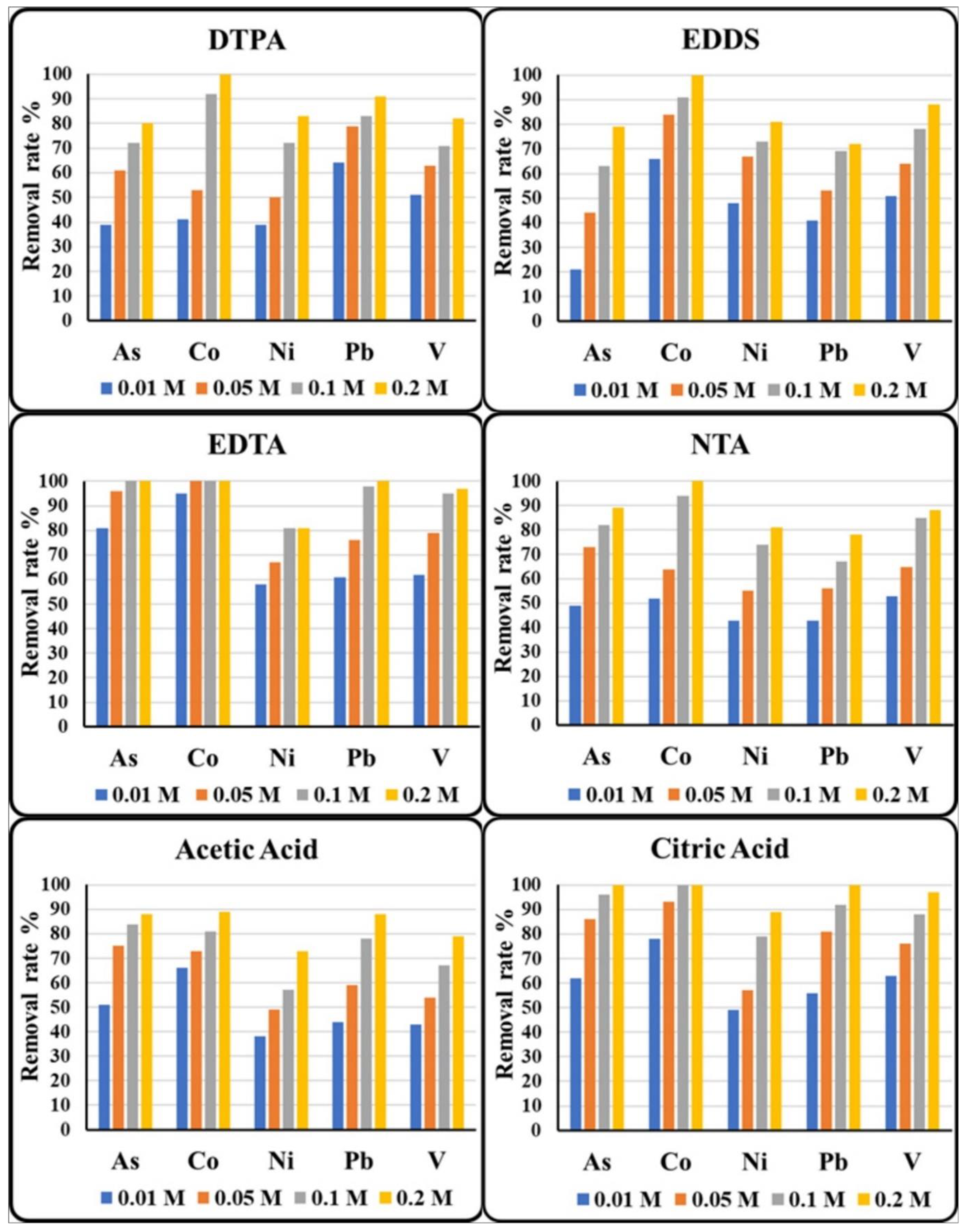
3.3. Selection of Extractant

To achieve the best possible PTE removal efficiency, it is crucial to choose a suitable extractant. The removal rates of the extractable states for the selected PTEs, using six extractants (acetic acid, citric acid, DTPA, EDDS, EDTA, and NTA), under the same extraction conditions, are presented in Figure 4. The results show that each of the six extractants used had significant positive effects on the removal of the selected PTEs from the purified kaolin. The removal efficiency was increased by increasing the extractant concentrations. Similar results were reported by Abu-Zurayk et al. [28], Elliott and Brown [43], Pichtel and Pichtel [44], and Khodadoust et al. [45]. Furthermore, citric acid and EDTA are more effective in the removal of PTEs than acetic acid, DTPA, EDDS, and NTA, which conforms well with the findings of the recent research by Maturi and Reddy [25], Song et al. [27], Amir et al. [29], Lumia et al. [32], and Di Palma and Mecozzi [46]. Citric acid was effective in removing PTEs from contaminated media mainly owing to the better solubilization of the PTEs at low pHs during the extractions [28,45]. EDTA has a high metal–EDTA complexing affinity and forms strong water-soluble metal complexes [44,47].
A citric acid concentration of 0.2 M shows the highest removal efficiency, while EDTA concentrations of 0.1 and 0.2 provide nearly the same removal rate percentages. EDTA was efficient, compared to citric acid, in extracting Ni and V in low concentrations (0.01, 0.05, and 0.1 M). Still, by increasing the citric acid concentration to 0.2 M, the Ni removal rate increased sharply, from 79 to 89%, which was higher than those of EDTA (81% at 0.1 and 0.2 M), while the V removal increased sharply, from 88 to 97%, which was the same for the EDTA (97% at 0.2 M).
The initial elemental variations in samples may influence the removal rates of PTEs [48]. In the present study, As and Co have the lowest initial element concentrations at 6.85 and 5.45 ppm, respectively, and so they show the highest removal rates. On the other hand, although V shows an apparently low removal rate, it is actually the most removed element because of its high initial concentration (204.13 ppm) and its large and hydrated radius.
On the basis of these results, an optimization for the treatment of PTEs using citric acid and EDTA was performed.

3.4. Optimization of the Treatment Parameters Using Citric Acid and EDTA
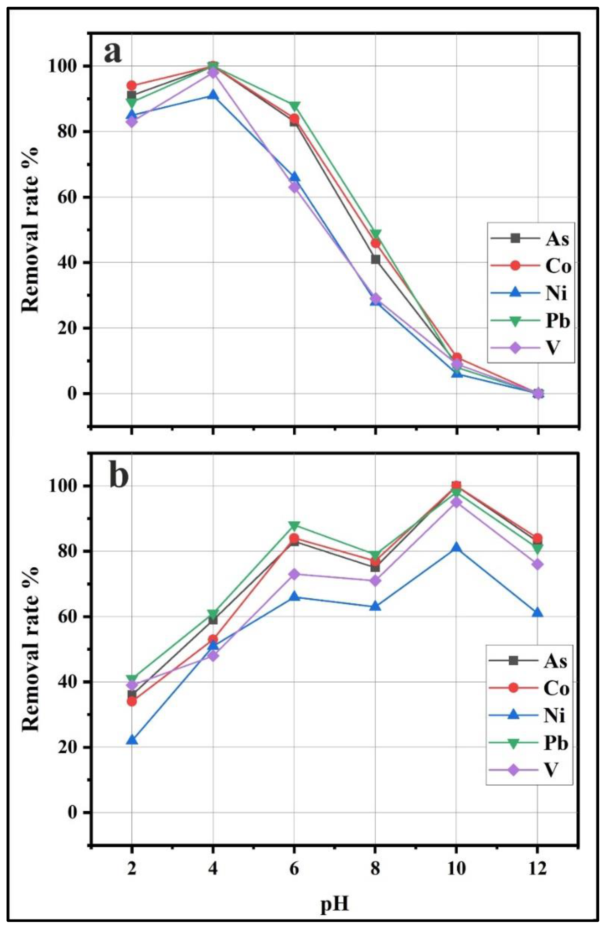
3.4.1. Effect of pH

To evaluate the influence of the pH during the citric acid and EDTA extractions, the pH of the extractant solution was increased from highly acidic (pH of 2) to alkaline (pH of 8). The solution pH value has a decisive effect on the behavior of the citric acid and EDTA as extractants for the selected PTEs (Figure 5). The highest removal rate of PTEs with citric acid was recorded at a pH of 4, and it decreased dramatically with the increasing pH. Kaolinite is a pH-dependent clay mineral, as the pH can influence the desorption of PTEs by changing the surface charge of the kaolinite, or by changing the speciation [49]. At a low pH (less than 5.25), the cation exchange capacity (CEC) decreases and the net charge of the kaolinite changes to positive (H+). Hence, the adsorbed PTE ions are repelled from the charged surface [50,51]. Meanwhile, the removal of PTEs using EDTA is very high under a pH range from 6 to 10. The EDTA attained the best removal efficiency, at a pH of 10, because of its higher thermodynamic stability with metals/metalloids, and because it dissolves the metals/metalloids better in alkaline conditions [52]. Since the maximum removal rate was obtained at a pH of 4 for citric acid, and at a pH of 10 for EDTA, these pH values were selected for all of the subsequent measurements.

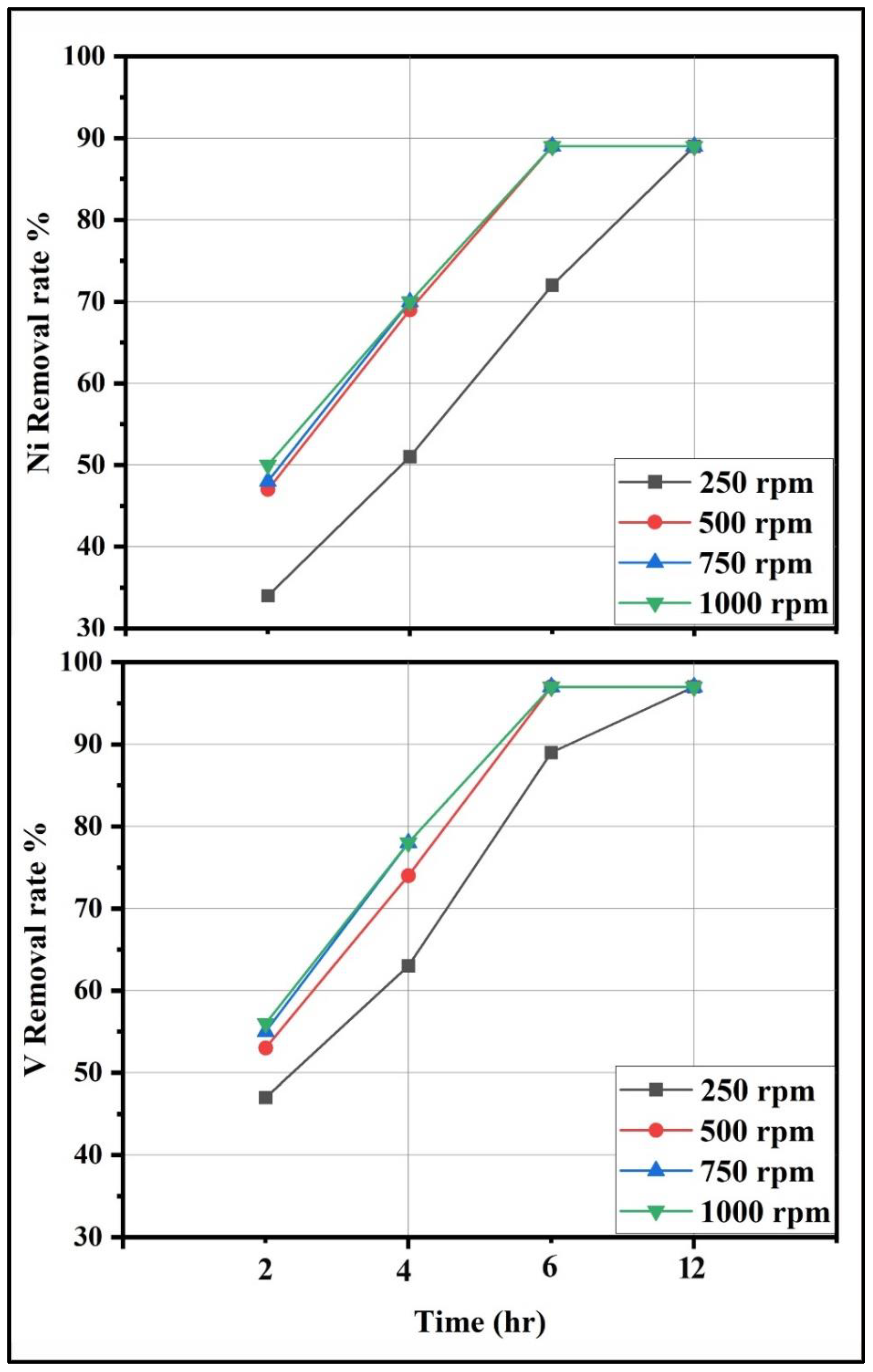
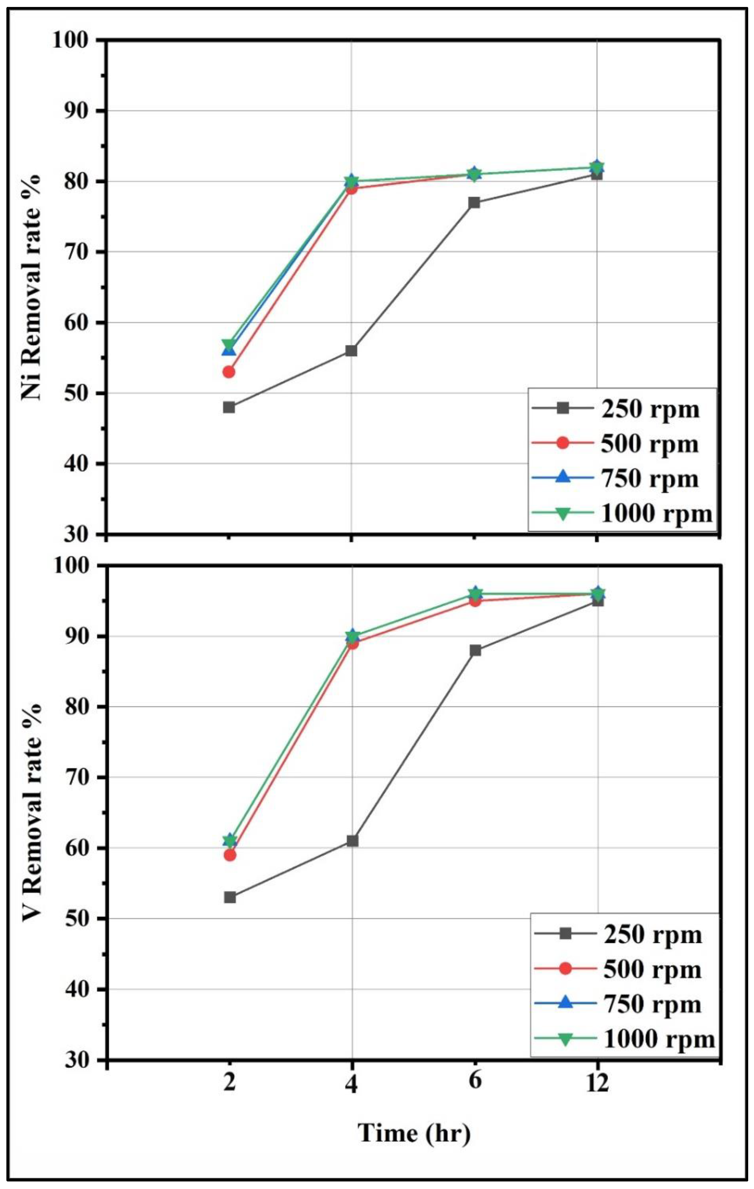
3.4.2. Effect of Mixing Speed and Time

To determine the best mixing speed for the PTE removal, the mixing speed was increased from 250 to 1000 rpm, with increments of 250 rpm, and with time intervals of 2, 4, 6, and 12 h. The removal of Ni and V using citric acid and EDTA were affected by increasing the mixing speed from 250 to 500 rpm (Figure 6 and Figure 7). The increased efficiency is linked to the emergence of intense turbulence, which reduces the thickness of the external mass transfer resistance around the metal particles [28]. Mixing speeds of 500, 750, and 1000 rpm exhibited similar patterns of variation in the Ni and V removal. It is noted that the equilibrium can be reached after 6 h. Thus, it can be concluded that a mixing speed of 500 rpm, with a mixing time of 6 h, are the optimal conditions for the removal of PTEs from kaolinite.

3.4.3. Effect of S/L Ratio

In the treatment of contaminated media, the S/L ratio is an important parameter that affects both the overall removal and the extraction of contaminants, as well as the amount of residual wash water [33,53]. Optimization using different S/L ratios was investigated to ensure this treatment’s suitability for various contaminated kaolin deposits. Citric acid with a 1:7.5 S/L ratio was proven to have better removal efficiencies (95 and 99% for Ni and V, respectively) than those of EDTA (93 and 96% for Ni and V, respectively) (Figure 8). Using 1:10 S/L, both desorbing agents show a removal rate of 100% for Ni and V. Because the extraction efficiency was governed by the molar ratio of the metals to the reagents, the S/L ratio had a significant impact on the washing reactions as more ligands became available by increasing the S/L ratio [54,55]. This treatment is applicable to the treatment of more contaminated kaolin deposits by raising the S/L ratio.

3.5. Potential Applications

3.5.1. Healthcare

Although clay minerals rank as the most adequately studied natural minerals in the healthcare field, they still continue to present a critical challenge in pharmaceutics and biomedicine with regard to their grade and their elemental impurities [1,56]. Undoubtedly, there are several huge clay deposits around the world that are of economic importance.
The results obtained in this study are crucial as they examine the applied concept of clay minerals on the basis of the treatment of natural raw materials in order to increase their economic value. This effective method can be employed in the treatment of diverse kinds of kaolin deposits worldwide, even with different degrees of contamination. In this regard, the present study robustly recommends the further treatment optimization of clay minerals for medicinal use, such as montmorillonite, saponite, vermiculite, halloysite, sepiolite, and palygorskite.

3.5.2. Geophagy

Clay-eating, or kaolin-eating (geophagy), remains a global practice that is commonly observed among peoples, as well as numerous animal species, on all the continents, and especially in Africa [7,8,57,58]. These geophagic kaolin are traditionally sold in open markets and herbalists’ centers as a vital source of mineral nutrients, without any proper health risk assessment or adequate treatment [58]. Many researchers have reported elevated concentrations of PTEs, such as As, Cd, Cu, Pb, Ni, and V in the geophagic kaolin samples in numerous countries [7,8,57,58,59]. The adopted method in this research can be considered a reliable method for the removal of PTEs from geophagic kaolin in order to make such clays safe for human and animal intake and to sustain healthier communities.

3.5.3. Soil Remediation

Some successful applications of kaolinite treatment have already been reported for soil remediation [23,24,25,26,29,45,49,51,60]. Very limited information about the removal of As and V from kaolinite or kaolinitic soil is available [49,60]. The findings of this study can be considered as a guiding contribution to the field of kaolinitic soil remediation that promotes further research in this field.

4. Conclusions

Controlling PTE impurities is one part of a drug product’s overall control strategy to ensure that they do not exceed the PCEI, and they should be considered in the risk assessment of any medicinal product that contains these materials. Five PTEs (As, Co, Ni, Pb, and V) were recorded in the Carboniferous Egyptian kaolin of southwestern Sinai. These PTEs have concentrations that exceed the PCEI standards for oral use in pharmaceutical applications, which limit their healthcare uses. Acetic acid, citric acid, DTPA, EDDS, EDTA, and NTA have significant positive effects on removing these PTEs. Citric acid and EDTA showed higher removal percentages compared to the rest of the extractants. Hence, the treatment of Egyptian kaolin by citric acid and EDTA attained removal efficiencies that could reduce the PTE concentrations to the PCEI limits for oral use in pharmaceutical applications.
With regard to the treatment optimization, the pH had a clear influence on the removal rates of PTEs from the kaolinite under the optimum pH values of 4 and 10 for citric acid and EDTA, respectively. A mixing speed of 500 rpm, with a mixing time of 6 h, are the optimal conditions for the removal of PTEs. Using 1:10 S/L citric acid and EDTA showed a removal rate of 100% for all of the investigated PTEs. We recommend this treatment for different kinds of kaolin showing various degree of contamination. Moreover, this study promotes further research in the healthcare, geophagy, and soil remediation fields.

Supplementary Materials

The following supporting information can be downloaded at: https://www.mdpi.com/article/10.3390/su14042388/s1, Table S1: Chemical composition of the studied Carboniferous Egyptian kaolin raw samples; Table S2: Chemical composition of the studied Carboniferous Egyptian kaolin purified samples.

Author Contributions

Conceptualization, A.G., B.A.A.-M., W.A.A. and S.M.A.E.B.; methodology, A.G. and S.M.A.E.B.; software, A.G., B.A.A.-M., W.A.A. and S.M.A.E.B.; validation, A.G., B.A.A.-M., W.A.A. and S.M.A.E.B.; formal analysis, A.G. and S.M.A.E.B.; investigation, A.G. and S.M.A.E.B.; resources, A.G.; data curation, A.G., B.A.A.-M., W.A.A. and S.M.A.E.B.; writing—original draft preparation, A.G. and W.A.A.; writing—review and editing, A.G., B.A.A.-M., W.A.A. and S.M.A.E.B.; visualization, A.G., B.A.A.-M., W.A.A. and S.M.A.E.B.; supervision, A.G. and B.A.A.-M. All authors have read and agreed to the published version of the manuscript.

Funding

This research received no external funding.

Institutional Review Board Statement

Not applicable.

Informed Consent Statement

Not applicable.

Data Availability Statement

Not applicable.

Acknowledgments

The authors acknowledge with thanks the laboratory facilities offered by the Faculty of Science of Ain Shams University. We especially thank Rizk, S. (Chemistry Department, Faculty of Science, Ain Shams University) for his support and contributions during the batch experiments. Special thanks to Baghdady, A.R. (Geology Department, Faculty of Science, Ain Shams University), Abd El-Sattar, N.E. (Chemistry Department, Faculty of Science, Ain Shams University), and Gouda, M.M. (Hot Laboratories Center, Atomic Energy Authority), for revising this manuscript and providing valuable comments. Appreciative thanks are also extended to Smillie, Z. (University of Stirling, Stirling, UK) for her valuable suggestions and her proofreading of the manuscript.

Conflicts of Interest

The authors declare no conflict of interest.

References

  1. Gomes, C.; Rautureau, M.; Gomes, J.; Silva, E. Minerals in pharmacy and cosmetics. In Minerals Latu Sensu and Human Health Benefits, Toxicity and Pathologies; Gomes, C., Rautureau, M., Eds.; Springe: Wettingen, Switzerland, 2021; pp. 405–441. [Google Scholar] [CrossRef]
  2. Roselli, C.; Desideri, D.; Cantaluppi, C.; Mattioli, M.; Fasson, A.; Meli, M.A. Essential and toxic elements in clays for pharmaceutical and cosmetic use. J. Toxicol. Environ. Health A 2015, 78, 316–324. [Google Scholar] [CrossRef]
  3. López-Galindo, A.; Viseras, C.; Cerezo, P. Compositional, technical and safety specifications of clays to be used as pharmaceutical and cosmetic products. Appl. Clay Sci. 2007, 36, 51–63. [Google Scholar] [CrossRef]
  4. Del Hoyo, C. Layered double hydroxides and human health: An overview. Appl. Clay Sci. 2007, 36, 103–121. [Google Scholar] [CrossRef]
  5. Awad, M.E.; López-Galindo, A.; El-Rahmany, M.M.; El-Desoky, H.M.; Viseras, C. Characterization of Egyptian kaolins for health-care uses. Appl. Clay Sci. 2017, 135, 176–189. [Google Scholar] [CrossRef]
  6. Eker, C.S.; Ari, U.V. Comparison of sandstone and mudstone with different methods for assessing toxic element contamination in the Early–Middle Jurassic sediments of Gümüşhane (NE Turkey). Environ. Earth Sci. 2020, 79, 444. [Google Scholar] [CrossRef]
  7. Oyebanjo, O.; Ekosse, G.; Odiyo, J. Health risk evaluation of trace elements in geophagic kaolinitic clays within Eastern Dahomey and Niger Delta Basins, Nigeria. Int. J. Environ. Res. Public Health 2020, 17, 4813. [Google Scholar] [CrossRef]
  8. Ekosse, G.-I.; Nkeng, G.E.; Bukalo, N.; Oyebanjo, O. Geophagic clays from cameroon: Provenance, metal contamination and health risk assessment. Int. J. Environ. Res. Public Health 2021, 18, 8315. [Google Scholar] [CrossRef]
  9. Baghdady, A.; Awad, S.; Gad, A. Assessment of metal contamination and natural radiation hazards in different soil types near iron ore mines, Bahariya Oasis, Egypt. Arab. J. Geosci. 2018, 11, 506. [Google Scholar] [CrossRef]
  10. Gonzalez-Nahm, S.; Nihlani, K.; House, J.S.; Maguire, R.L.; Skinner, H.G.; Hoyo, C. Associations between maternal cadmium exposure with risk of preterm birth and low birth weight: Effect of Mediterranean diet adherence on affected prenatal outcomes. Toxics 2020, 8, 90. [Google Scholar] [CrossRef]
  11. Gad, A.; Abd El Bakey, S.M.; Sakr, S. Concentrations of heavy metals and associated human health risk in unrefined salts of inland hypersaline lakes, Egypt. Int. J. Environ. Anal. Chem. 2020, 1–15. [Google Scholar] [CrossRef]
  12. Jahromi, M.A.; Jamshidi-Zanjani, A.; Darban, A.K. Heavy metal pollution and human health risk assessment for exposure to surface soil of mining area: A comprehensive study. Environ. Earth Sci. 2020, 79, 365. [Google Scholar] [CrossRef]
  13. Medina-Estévez, F.; Zumbado, M.; Luzardo, O.P.; Rodríguez-Hernández, Á.; Boada, L.D.; Fernández-Fuertes, F.; Santandreu-Jimenez, M.E.; Henríquez-Hernández, L.A. Association between heavy metals and rare earth elements with acute ischemic stroke: A case-control study conducted in the Canary Islands (Spain). Toxics 2020, 8, 66. [Google Scholar] [CrossRef]
  14. Rauf, A.U.; Mallongi, A.; Lee, K.; Daud, A.; Hatta, M.; Al Madhoun, W.; Astuti, R.D.P. Potentially toxic element levels in atmospheric particulates and health risk estimation around industrial areas of Maros, Indonesia. Toxics 2021, 9, 328. [Google Scholar] [CrossRef] [PubMed]
  15. Abdel-Khalek, N.A. The Egyptian kaolin: An outlook in the view of the new climate of investment. Appl. Clay Sci. 1999, 15, 325–336. [Google Scholar] [CrossRef]
  16. Baioumy, H.M. Geochemistry and origin of the Cretaceous sedimentary kaolin deposits, Red Sea, Egypt. Geochemistry 2014, 74, 195–203. [Google Scholar] [CrossRef]
  17. El-Kammar, A.; Abu-Zied, H.T.; Galal, M.; Osman, D. Composition, radioactivity, and possible applications of kaolin deposits of Sinai, Egypt. Arab. J. Geosci. 2017, 10, 463. [Google Scholar] [CrossRef]
  18. Kamel, O.A.; Soliman, F.H.A.; Abdel Maaboud, M.H.M. Sinai Carboniferous kaolins: Their mineralogy, geochemical characteristics and suitability for ceramic industry. Egypt. J. Geol. 1997, 41, 219–238. [Google Scholar]
  19. Baioumy, H.M.; Gilg, H.A.; Taubald, H. Mineralogy and geochemistry of the sedimentary kaolin deposits from Sinai, Egypt: Implications for control by the source rocks. Clays Clay Min. 2012, 60, 633–654. [Google Scholar] [CrossRef]
  20. Awad, M.E.; López-Galindo, A.; Sánchez-Espejo, R.; Sainz-Díaz, C.I.; El-Rahmany, M.M.; Viseras, C. Crystallite size as a function of kaolinite structural order-disorder and kaolin chemical variability: Sedimentological implication. Appl. Clay Sci. 2018, 162, 261–267. [Google Scholar] [CrossRef]
  21. Saber, E.; Ali, M.; El-Sheikh, A. Provenance studies of Kalabsha kaolin deposits, Egypt: A petrographical and geochemical approach. Arab. J. Geosci. 2018, 11, 339. [Google Scholar] [CrossRef]
  22. Awad, M.E.; López-Galindo, A.; Medarević, D.; Đuriš, J.; El-Rahmany, M.M.; Ibrić, S.; Viseras, C. Flow and tableting behaviors of some Egyptian kaolin powders as potential pharmaceutical excipients. Minerals 2020, 10, 23. [Google Scholar] [CrossRef] [Green Version]
  23. Hong, J.; Pintauro, P.N. Desorption-complexation-dissolution characteristics of adsorbed cadmium from kaolin by chelators. Water Air Soil Pollut. 1996, 86, 35–50. [Google Scholar] [CrossRef]
  24. Khodadoust, A.P.; Reddy, K.R.; Maturi, K. Removal of nickel and phenanthrene from kaolin soil using different extractants. Environ. Eng. Sci. 2004, 24, 691–704. [Google Scholar] [CrossRef]
  25. Maturi, K.; Reddy, K.R. Extractants for the removal of mixed contaminants from soils. Soil Sediment Contam. 2008, 17, 586–608. [Google Scholar] [CrossRef]
  26. Niinae, M.; Nishigaki, K.; Aoki, K. Removal of Lead from Contaminated Soils with Chelating Agents. Mater. Trans. 2008, 49, 2377–2382. [Google Scholar] [CrossRef] [Green Version]
  27. Song, S.; Zhu, L.; Zhou, W. Simultaneous removal of phenanthrene and cadmium from contaminated soils by saponin, a plant-derived biosurfactant. Environ. Pollut. 2008, 156, 1368–1370. [Google Scholar] [CrossRef]
  28. Abu-Zurayk, R.A.; Bozeya, A.; Abu-Irmaileh, B.; Abu-Mallouh, S.; Al Bawab, A.; Al-Dujaili, A.H. Assessment of some heavy metals in the Dead Sea Mud and treatment optimization. Soil Sediment Contam. 2017, 26, 364–376. [Google Scholar] [CrossRef]
  29. Amir, M.; Mohsen, S.; Afsaneh, M. Simultaneous desorption and desorption kinetics of phenanthrene, anthracene, and heavy metals from kaolinite with different organic matter content. Soil Sediment Contam. 2018, 27, 200–220. [Google Scholar] [CrossRef]
  30. Zhang, X.; Zeng, S.; Chen, S.; Ma, Y. Change of the Extractability of Cadmium Added to Different Soils: Aging Effect and Modeling. Sustainability 2018, 10, 885. [Google Scholar] [CrossRef] [Green Version]
  31. Liu, L.; Luo, D.; Yao, G.; Huang, X.; Wei, L.; Liu, Y.; Wu, Q.; Mai, X.; Liu, G.; Xiao, T. Comparative Activation Process of Pb, Cd and Tl Using Chelating Agents from Contaminated Red Soils. Int. J. Environ. Res. Public Health 2020, 17, 497. [Google Scholar] [CrossRef] [Green Version]
  32. Lumia, L.; Giustra, M.G.; Viviani, G.; Di Bella, G. Washing batch test of contaminated sediment: The case of Augusta Bay (SR, Italy). Appl. Sci. 2020, 10, 473. [Google Scholar] [CrossRef] [Green Version]
  33. Kim, M.-S.; Koo, N.; Kim, J.-G.; Lee, S.-H. Effects of washing solution, washing time, and solid-solution rate on the maximum heavy metals removal efficiency. Appl. Sci. 2021, 11, 6398. [Google Scholar] [CrossRef]
  34. Kora, M.; El Shahat, A.; Abu Shabana, M. Lithostratigraphy of the manganese-bearing Urn Bogma Formation, west-central Sinai, Egypt. J. Afr. Earth Sci. 1994, 18, 151–162. [Google Scholar] [CrossRef]
  35. Ramadan, S.R.; Gad, A.; Yehia, M.M.; Dawood, Y.H.; Kawady, N.A. Grain size and mineralogical characteristics of the stream sediments, east of Abu Zeneima area, southwestern Sinai, Egypt. Arab. J. Geosci. 2019, 12, 192. [Google Scholar] [CrossRef]
  36. International Centre for Diffraction Data (ICDD). ICDD Powder Diffraction File Data Sets 01 to 19; ICDD: Livermore, CA, USA, 2000. [Google Scholar]
  37. Maynard, R.N. A method for removing titanium dioxide impurities from kaolin. Clays Clay Min. 1969, 17, 59–62. [Google Scholar] [CrossRef]
  38. Benjamin, M.M.; Leckie, J.O. Multiple site adsorption of Cd, Cu, Zn, and Pb on amorphous iron oxyhydroxide. J. Colloid Interface Sci. 1981, 79, 209–221. [Google Scholar] [CrossRef]
  39. Kretzschmar, R.; Borkovec, M.; Grolimund, D.; Elimelech, M. Mobile subsurface colloids and their role in contaminant transport. Adv. Agron. 1999, 66, 121–193. [Google Scholar] [CrossRef]
  40. Han, F.X.; Singer, A. Binding and distribution of trace elements among solid-phase components in arid zone soils. In Biogeochemistry of Trace Elements in Arid Environments; Han, F.X., Singer, A., Eds.; Springer: Dordrecht, The Netherlands, 2007; Volume 13, pp. 131–167. [Google Scholar] [CrossRef]
  41. USP-NF. United States Pharmacopeia 42 and National Formulary 37 (USP 42-NF37); US Pharmacopeial Convention: Rockville, MD, USA, 2018. [Google Scholar]
  42. ICH. The International Conference on Harmonization of Technical Requirements and Registration of Pharmaceuticals for Human Use (ICH), Guideline for Elemental Impurities Q3D. 2019. Available online: https://database.ich.org/sites/default/files/Q3D-R1EWG_Document_Step4_Guideline_2019_0322.pdf (accessed on 15 June 2021).
  43. Elliott, H.A.; Brown, G.A. Comparative evaluation of NTA and EDTA for extractive decontamination of pb-polluted soils. Water Air Soil Pollut. 1989, 45, 361–369. [Google Scholar] [CrossRef]
  44. Pichtel, J.; Pichtel, T.M. Comparison of solvents for Ex situ removal of chromium and lead from contaminated soil. Environ. Eng. Sci. 1997, 14, 97–104. [Google Scholar] [CrossRef]
  45. Khodadoust, A.P.; Reddy, K.R.; Maturi, K. Effect of different extraction agents on metal and organic contaminant removal from a field soil. J. Hazard. Mater. 2005, 117, 15–24. [Google Scholar] [CrossRef]
  46. Di Palma, L.; Mecozzi, R. Heavy metals mobilization from harbour sediments using EDTA and citric acid as chelating agents. J. Hazard. Mater. 2007, 147, 768–775. [Google Scholar] [CrossRef]
  47. Abollino, O.; Aceto, M.; Malandrino, M.; Sarzanini, C.; Mentasti, E. Adsorption of heavy metals on Na-montmorillonite. Effect of pH and organic substances. Water Res. 2003, 37, 1619–1627. [Google Scholar] [CrossRef]
  48. Tuin, B.J.W.; Tels, M. Removing heavy metals from contaminated clay soils by extraction with hydrochloric acid, edta or hypochlorite solutions. Environ. Technol. 1990, 11, 1039–1052. [Google Scholar] [CrossRef]
  49. Quaghebeur, M.; Rate, A.; Rengel, Z.; Hinz, C. Desorption kinetics of arsenate from kaolinite as influenced by pH. J. Environ. Qual. 2005, 34, 479–486. [Google Scholar] [CrossRef]
  50. White, G.N.; Dixon, J.B. Kaolin–serpentine minerals. In Soil Mineralogy with Environmental Applications; Dixon, J.B., Schulze, D.G., Eds.; SSSA Book Series; Wiley: Hoboken, NJ, USA, 2002; Volume 7, pp. 389–414. [Google Scholar] [CrossRef]
  51. Li, L.Y. Removal of multiple-metals from contaminated clay minerals. Environ. Technol. 2006, 27, 811–822. [Google Scholar] [CrossRef] [PubMed]
  52. Kim, C.; Lee, Y.; Ong, S.K. Factors affecting EDTA extraction of lead from lead-contaminated soils. Chemosphere 2003, 51, 845–853. [Google Scholar] [CrossRef]
  53. Wang, J.; Pan, X.; Zhang, S.; Zhong, Q.; Zhou, W.; Zhang, X.; Wu, J.; Vijver, M.G.; Peijnenburg, W.J. Remediation of heavy metal contaminated soil by biodegradable chelator–induced washing: Efficiencies and mechanisms. Environ. Res. 2020, 186, 109554. [Google Scholar] [CrossRef] [PubMed]
  54. Jiang, J.; Yang, M.; Gao, Y.; Wang, J.; Li, D.; Li, T. Removal of toxic metals from vanadium-contaminated soils using a washing method: Reagent selection and parameter optimization. Chemosphere 2017, 180, 295–301. [Google Scholar] [CrossRef] [PubMed]
  55. Zhang, T.; Liu, J.; Huang, X.; Zia, B.; Su, C.; Luo, G.; Xu, Y.; Wu, Y.; Mao, Z.; Qiu, R. Chelant extraction of heavy metals from contaminated soils using new selective EDTA derivatives. J. Hazard. Mater. 2013, 262, 464–471. [Google Scholar] [CrossRef]
  56. Wargala, E.; Sławska, M.; Zalewska, A.; Toporowska, M. Health effects of dyes, minerals, and vitamins used in cosmetics. Women 2021, 1, 223–237. [Google Scholar] [CrossRef]
  57. Kutalek, R.; Wewalka, G.; Gundacker, C.; Auer, H.; Wilson, J.; Haluza, D.; Huhulescu, S.; Hillier, S.; Sager, M.; Prinz, A. Geophagy and potential health implications: Geohelminths, microbes and heavy metals. Trans. Roy. Soc. Trop. Med. Hyg. 2010, 104, 787–795. [Google Scholar] [CrossRef] [PubMed]
  58. Lar, U.A.; Agene, J.I.; Umar, A.I. Geophagic clay materials from Nigeria: A potential source of heavy metals and human health implications in mostly women and children who practice it. Environ. Geochem. Health 2015, 37, 363–375. [Google Scholar] [CrossRef] [PubMed]
  59. Asowata, I.T. Geophagic clay around Uteh-Uzalla near Benin: Mineral and trace elements compositions and possible health implications. SN Appl. Sci. 2021, 3, 569. [Google Scholar] [CrossRef]
  60. Yang, J.-Y.; Luo, H.-Q.; Zhu, Y.-Y.; Yu, Y.-Q.; He, W.-Y.; Wu, Z.-Z.; Wang, B. Adsorption-desorption and co-migration of vanadium on colloidal kaolinite. Environ. Sci. Pollut. Res. 2020, 27, 17910–17922. [Google Scholar] [CrossRef] [PubMed]
Figure 1. (a) Geologic map of Wadi Natash area; and (b) enhanced Landsat images of Wadi Natash.
Sustainability 14 02388 g001
Figure 2. Schematic diagram of experimental protocol for Egyptian kaolin treatment and parameter optimization.
Sustainability 14 02388 g002
Figure 3. XRD diffractograms of the raw and purified (P) kaolin samples.
Sustainability 14 02388 g003
Figure 4. Removal rates of As, Co, Ni, Pb, and V by different extractants.
Sustainability 14 02388 g004
Figure 5. Effects of pH on removal rates of As, Co, Ni, Pb, and V using (a) citric acid, and (b) EDTA.
Sustainability 14 02388 g005
Figure 6. Effects of mixing speeds and times on removal rates of Ni and V using citric acid.
Sustainability 14 02388 g006
Figure 7. Effects of mixing speeds and times on removal rates of Ni and V using EDTA.
Sustainability 14 02388 g007
Figure 8. Effects of S/L ratio on removal rates of Ni and V using (a) citric acid, and (b) EDTA.
Sustainability 14 02388 g008
Publisher’s Note: MDPI stays neutral with regard to jurisdictional claims in published maps and institutional affiliations.

