Article

Experimental Modeling Investigations on the Biosorption of Methyl Violet 2B Dye by the Brown Seaweed Cystoseira tamariscifolia

by
Mayasar I. Al-Zaban
1,
Nada K. Alharbi
1,
Fawziah M. Albarakaty
2,
Sarah Alharthi
3,
Sedky H. A. Hassan
4 and
Mustafa A. Fawzy
5,*
1
Department of Biology, College of Science, Princess Nourah bint Abdulrahman University, P.O. Box 84428, Riyadh 11671, Saudi Arabia
2
Department of Biology, Faculty of Applied Science, Umm Al-Qura University, P.O. Box 715, Makkah Al Mukarramah 21955, Saudi Arabia
3
Department of Chemistry, College of Science, Taif University, P.O. Box 11099, Taif 21944, Saudi Arabia
4
Department of Biology, College of Science, Sultan Qaboos University, Muscat 123, Oman
5
Biology Department, Faculty of Science, Taif University, P.O. Box 11099, Taif 21944, Saudi Arabia
*
Author to whom correspondence should be addressed.
Sustainability 2022, 14(9), 5285; https://doi.org/10.3390/su14095285
Submission received: 24 March 2022 / Revised: 13 April 2022 / Accepted: 25 April 2022 / Published: 27 April 2022
(This article belongs to the Special Issue Wastewater Treatment and Purification)

Abstract

:
Methyl violet 2B dye is a major contaminant that is detrimental to both humans and aquatic microorganisms, thus it should be eliminated from water. In the current investigation, the biosorption of methyl violet 2B dye onto the brown seaweed Cystoseira tamariscifolia biomass as a sustainable low-cost biosorbent was examined by varying biosorption parameters. Biomass dosage of 7 g/L, pH 6, a temperature of 45 °C, a 60 min contact time, and a 30 mg/L initial dye concentration were determined to be the optimum biosorption conditions. Data obtained were interpreted by thermodynamic, isothermal, and kinetic models. The thermodynamic studies demonstrated that the process of dye biosorption was random and endothermic. The data were best described by Langmuir, Dubinin–Radushkevich, and Temkin models. According to the Langmuir equation, the maximal biosorption capacity (qmax) was 10.0 mg/g. Moreover, the pseudo-second-order mechanism is dominant, and chemical biosorption might represent the rate-controlling stage in the biosorption process. However, intraparticle diffusion revealed a boundary layer effect. A scanning electron microscope, energy-dispersive X-ray spectroscopy, the point of zero charge, and Fourier Transform Infra-Red were applied to characterize the algal biomass, exhibiting its remarkable structural properties and the availability of several functional groups. Additionally, ion exchange, electrostatic force, and hydrogen bonding formation are all proposed as biosorption mechanisms. As a result, C. tamariscifolia was evaluated to be a sustainable biosorbent for dye biosorption from aqueous solutions.

1. Introduction

Dyes, phenols, pesticides, and heavy metals are among the inorganic and organic contaminants found in wastewater effluents [1]. Dyes are a significant source of organic contaminants in industrial effluents due to their usage in a range of industries, including paints, leather, tannery, paper, and textiles, resulting in a large amount of colored sewage [2,3]. The occurrence of synthetic dyes in aquatic ecosystems, even at extremely low quantities, is very obvious and unwanted. They are harmful to faunas and aquatic flora and can impede the penetration of sunlight and inhibit photosynthesis [4]. Dyes have mutagenic, teratogenic, and carcinogenic effects in addition to coloring [5].
Methyl violet 2B dye is generally utilized in the textile sector and other industrial applications [6]. Dahri et al. [7] reported that methyl violet dye can prevent photosynthesis and bacterial growth. Mehr et al. [8] also found that methyl violet is possibly carcinogenic and can harm the skin, eyes, respiratory system, and gastrointestinal tract [9]. Because of the hazardous effects of methyl violet, it must be eliminated from the effluent before it is released into the water.
Several treatment approaches were utilized to treat wastewater containing dyes, including oxidation, chemical reduction, coagulation, ion exchange, biodegradation, reverse osmosis, filtration, and adsorption [10,11]. The most significant disadvantages of these approaches include inefficiency, sludge formation, high energy costs, membrane fouling, and health risks [12].
As a result, sustainable technologies for pollution removal have been developed, employing biological materials including rice husk, cotton wastes, agricultural wastes, and dead fungal and algal biomasses in a process called biosorption [13,14,15,16].
Biosorption is a sustainable and favored approach for dyes’ bioremoval due to the inexpensive cost of biomaterials, great effectiveness, and low energy consumption [17].
Many types of adsorbents were used for the MV 2B elimination from industrial effluents such as Artocarpus odoratissimus stem [9], Nepenthes rafflesiana pitcher and leaves [18], a magnetic composite [19] and Padina sanctae-crucis [20].
Recently, algae have attracted interest due to their availability and simplicity of handling. Algal cell walls contain a range of active groups, such as phosphate, amino, sulfate, carboxyl, and imidazoles groups, associated with proteins and polysaccharides (fucoidan and alginate) for binding different contaminants [21].
Brown seaweed Cystoseira tamariscifolia is explored in this work for the removal of MV 2B dye from synthetic aqueous solutions for its relatively simple structure and widespread distribution. Cystoseira tamariscifolia might also be utilized as a novel source of food additives, essential nutrients, and nutritional supplements for animal and human use. The major component responsible for the biosorption of dye is alginate, with this being found as a gel in brown algal cell walls. Alginate is highly penetrable by tiny dye cations, making algae good biosorbents with great biosorption capacity.
This investigation is unique in that it is the first time the native Cystoseira tamariscifolia has been used to remove MV 2B dye. Accordingly, the goal of this investigation is to study the effectiveness of Cystoseira tamariscifolia biomass in removing MV 2B from aqueous solutions. The experiments were achieved by changing the pH of the solution, biosorbent dosage, temperature, initial MV 2B concentration, and reaction time. Experimental results were fitted into thermodynamic, kinetic, and isotherm models. The algal biomass was further characterized by utilizing different methods including a scanning electron microscope, energy dispersive X-ray, the point of zero charge, and Fourier transform infrared spectroscopy.

2. Results and Discussion
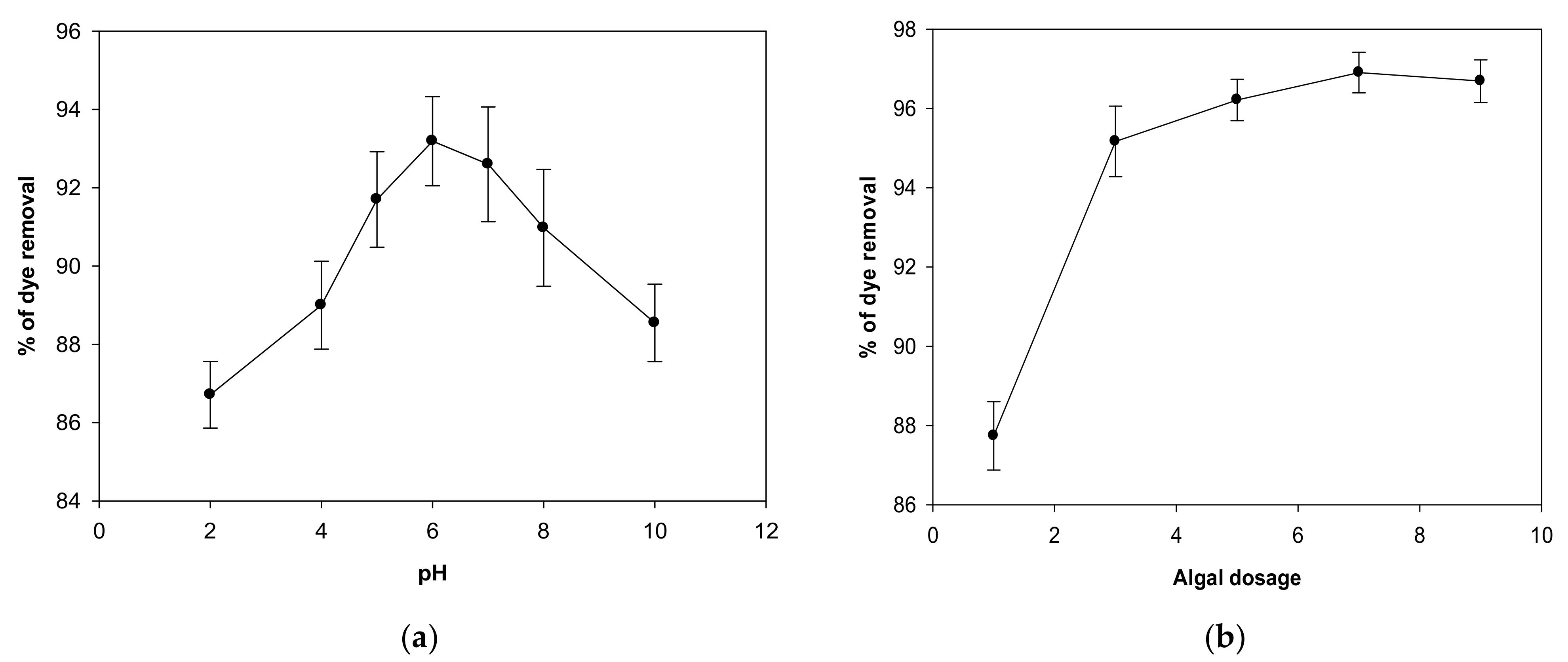
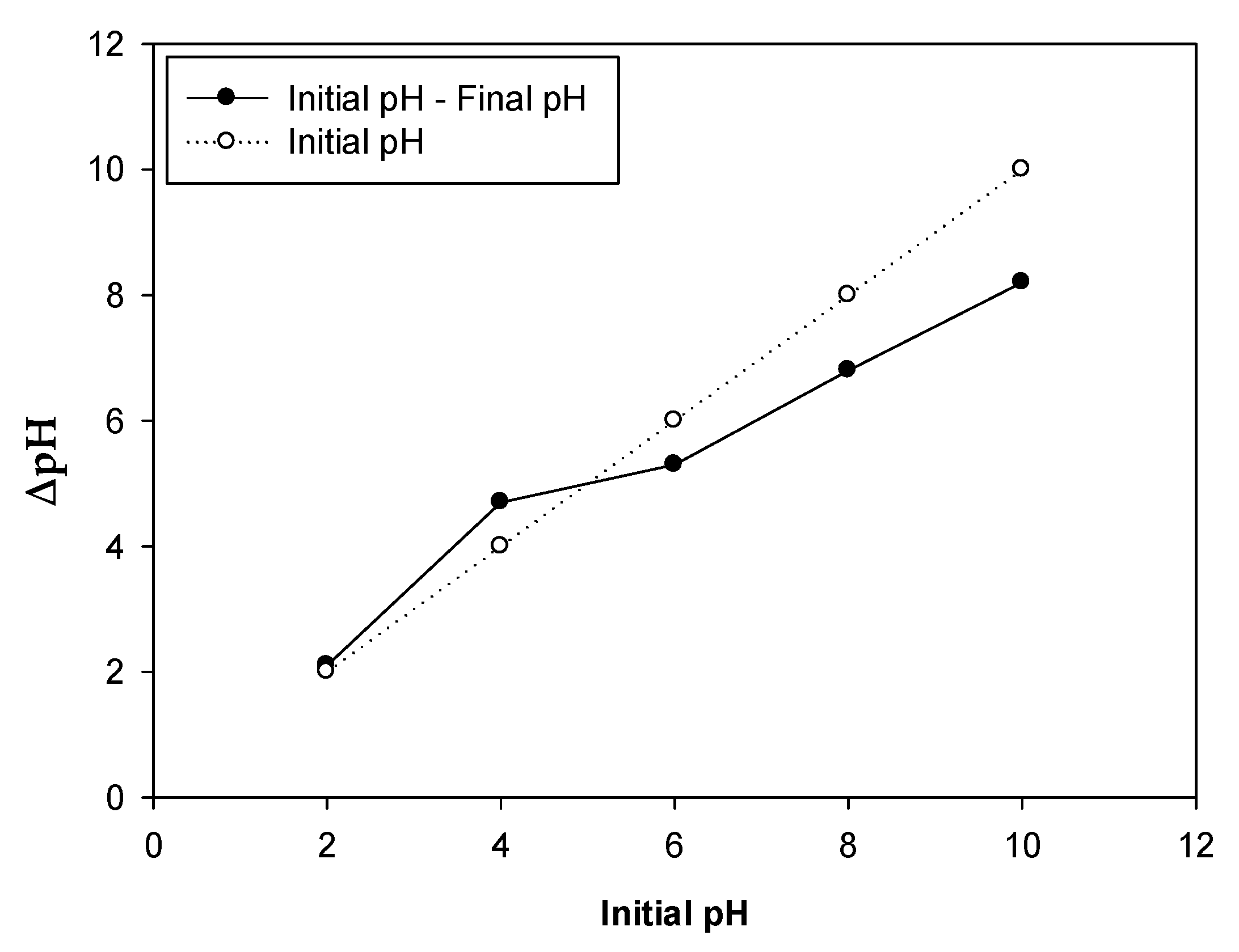
2.1. Impact of pH

The pH is the main variable in the adsorption process as it influences the charge and ionization of active groups in the biomaterial’s cell walls such as amino, carbonyl, phosphate, and carboxyl groups [22]. As a result, the kinetic and equilibrium isotherm characteristics of the adsorption process may be altered [23]. The impact of pH on the dye biosorption onto the biomass of C. tamariscifolia was tested utilizing different values of pH (2–10) and fixed biosorption conditions of 5 g/L biosorbent dose, an initial MV 2B concentration of 30 mg/L, and 90 min contact time at 25 °C. As seen in Figure 1a, the biosorption rate rose as the pH value increased up to 6. This may be clarified by assuming that the surface of algal biomass has a point of zero charge equal to 5.01. At low pH < pHpzc, the biosorbent is positively charged, causing electrostatic repulsion between the MV 2B and the algal surface. However, at high pH (6) > pHpzc, the algal surface can become negatively charged, increasing the biosorption capacity via electrostatic attractions [24]. A further rise in pH value to 10 led to a decrement in the dye biosorption onto the algal biomass since binding sites were restricted; hence, the rate of biosorption was not continually enhanced as the pH increased [17]. Because the highest removal percentage of dye (93.2%) was determined at pH 6, this pH was considered the best value for the following biosorption investigations.

2.2. Impact of Biomass Concentration

The influence of the biosorbent concentration on the bioremoval percentage of MV 2B dye by C. tamariscifolia biomass was examined using various biomass concentrations (1–9 g/L), an optimized pH value of 6, a fixed initial MV 2B concentration of 30 mg/L, and 90 min contact time at 25 °C, as presented in Figure 1b. The bioremoval percentage of MV 2B dye by C. tamariscifolia biomass increased from 87.7% to 96.9% when the algal dosage rose from 1 g/L to 7 g/L. There are direct correlations between the biosorbent dosage and biosorption process, meaning that the amount of binding sites increases with higher biosorbent dosage, resulting in effective dye biosorption [25]. In this respect, Yadav et al. [1] reported that increasing the biosorbent dosage from 25 to 200 mg/L enhanced the biosorption of methylene blue dye by Fucus vesiculosus from 66.06% to 98.96%. As a result, the transfer rate of the solute onto the sorbent surface increases, resulting in a split in the quantity of solute sorbed on the unit weight of biosorbent with an increasing biosorbent dose [26].
Although a rise in the biomass increases the availability of active sites, a decrease in Zn2+ ion biosorption occurred at an algal dosage greater than 7 g/L. This might be ascribed to partial agglomeration of algal biomass, which reduces the surface area and, hence, causes a decrease in the active binding sites [27].
Accordingly, the biosorbent dosage of 7 g/L was determined to be an optimal dose for the bioremoval of MV 2B by C. tamariscifolia biomass, and subsequent biosorption experiments were conducted at this value.

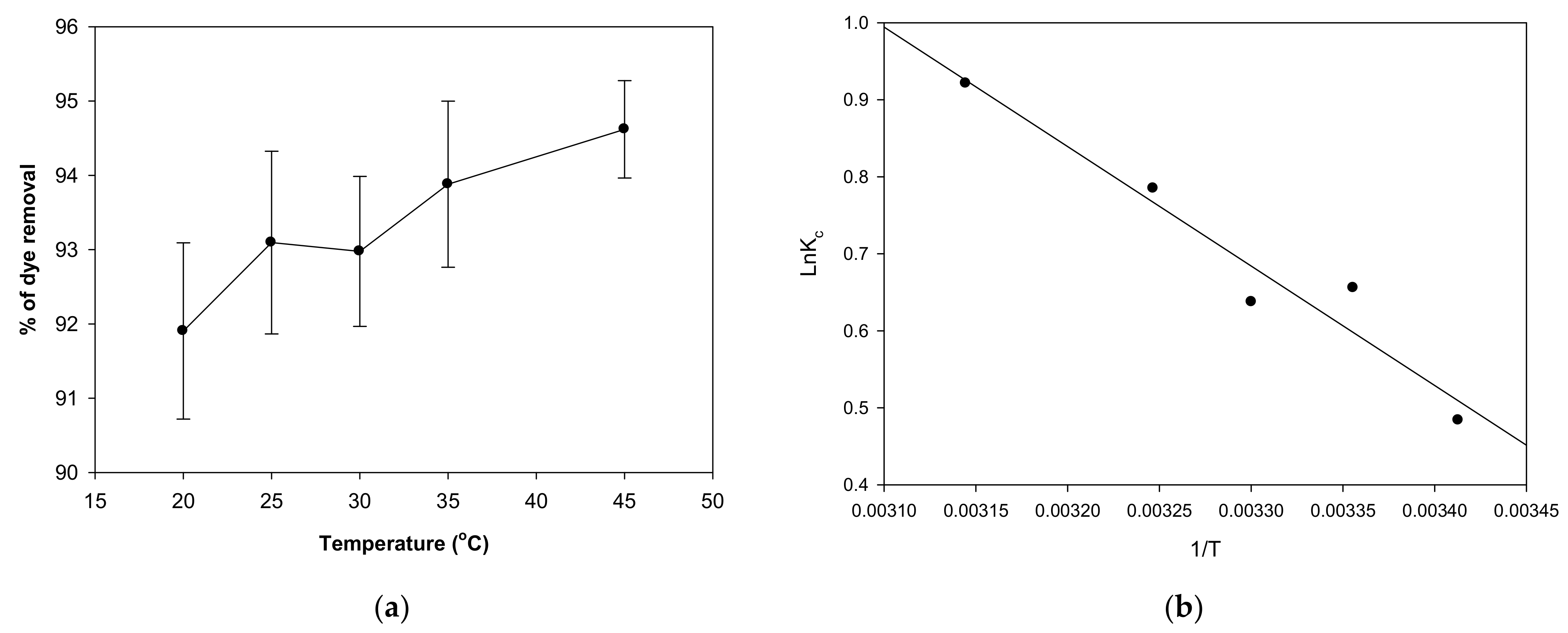
2.3. Impact of Temperature on MV 2B Biosorption and Thermodynamic Studies

Temperature is a crucial factor that influences the kinetic energy of molecules [28]. The influence of temperature on dye biosorption onto the C. tamariscifolia biomass was studied at various temperatures (20, 25, 30, 35, 45 °C) with an optimized pH of 6, an algal dose of 7 g/L, a fixed contact time of 90 min, and an initial MV 2B concentration of 30 mg/L. Figure 2a demonstrated that dye uptake enhanced with an increasing temperature, and the temperature value of 45 °C recorded the maximum removal percentage (94.6%), suggesting that the MV 2B biosorption onto the biosorbent is temperature-dependent. As the temperature rose, the dye molecule’s mobility increased. This indicates that when the temperature is elevated, the dye molecule effectively interacts with the surface of the biosorbent [29]. The increase in dye uptake with an increasing temperature was ascribed to either a rise in the number of active sites on the surface of the biosorbent or the increased affinity of dye molecules at active sites [30]. Additionally, higher temperatures frequently improve biosorption capacity due to the increased kinetic energy and surface activity of the solute. This mechanism shows that the biosorption reaction is endothermic.
The results of this experiment were effectively utilized to investigate the thermodynamics of the biosorption process. The results of the thermodynamics analysis are presented in Table 1 and Figure 2b. The negative values of Δ suggested that the biosorption process is spontaneous in nature. In addition, the reduction in Δ values when increasing the temperature showed that there is more driving force, resulting in quicker biosorption capacity, and biosorption is more feasible at higher temperatures [31].
In contrast, the positive ΔH° value (12.87 kJ/mol) showed that the biosorption of MV 2B dye onto the algal biomass is endothermic, and the positive ΔS° value (0.048 kJ/mol) indicated that the solid/solution interface is extremely random through the biosorption of MV 2B onto the C. tamariscifolia biomass [32].
This pattern (negative ΔG° and positive ΔH° and Δ values, as well as quicker biosorption capacity at higher temperatures) was also reported in previous literature for dye removal by other biosorbents [8,31].

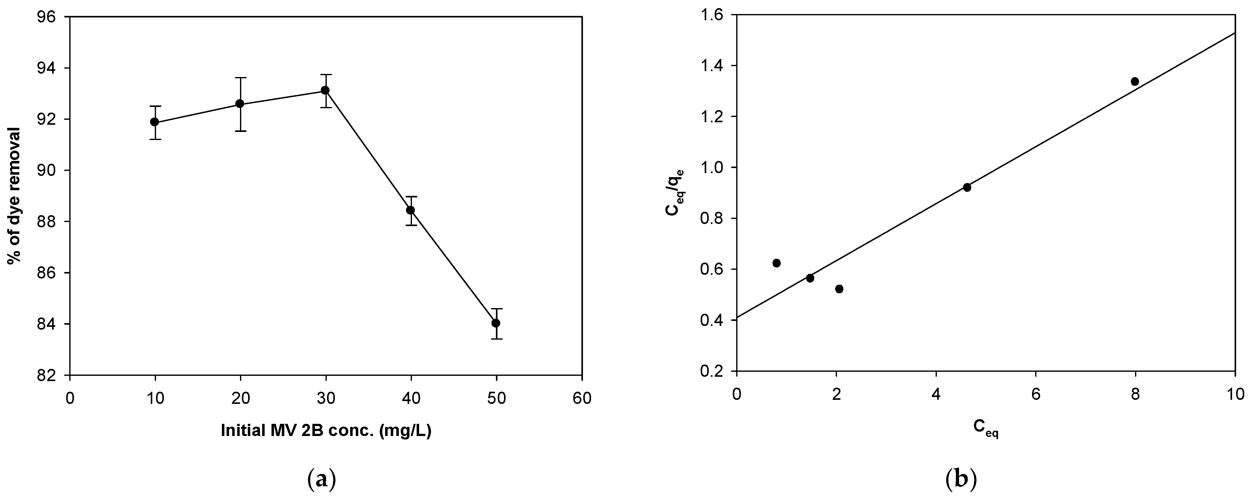
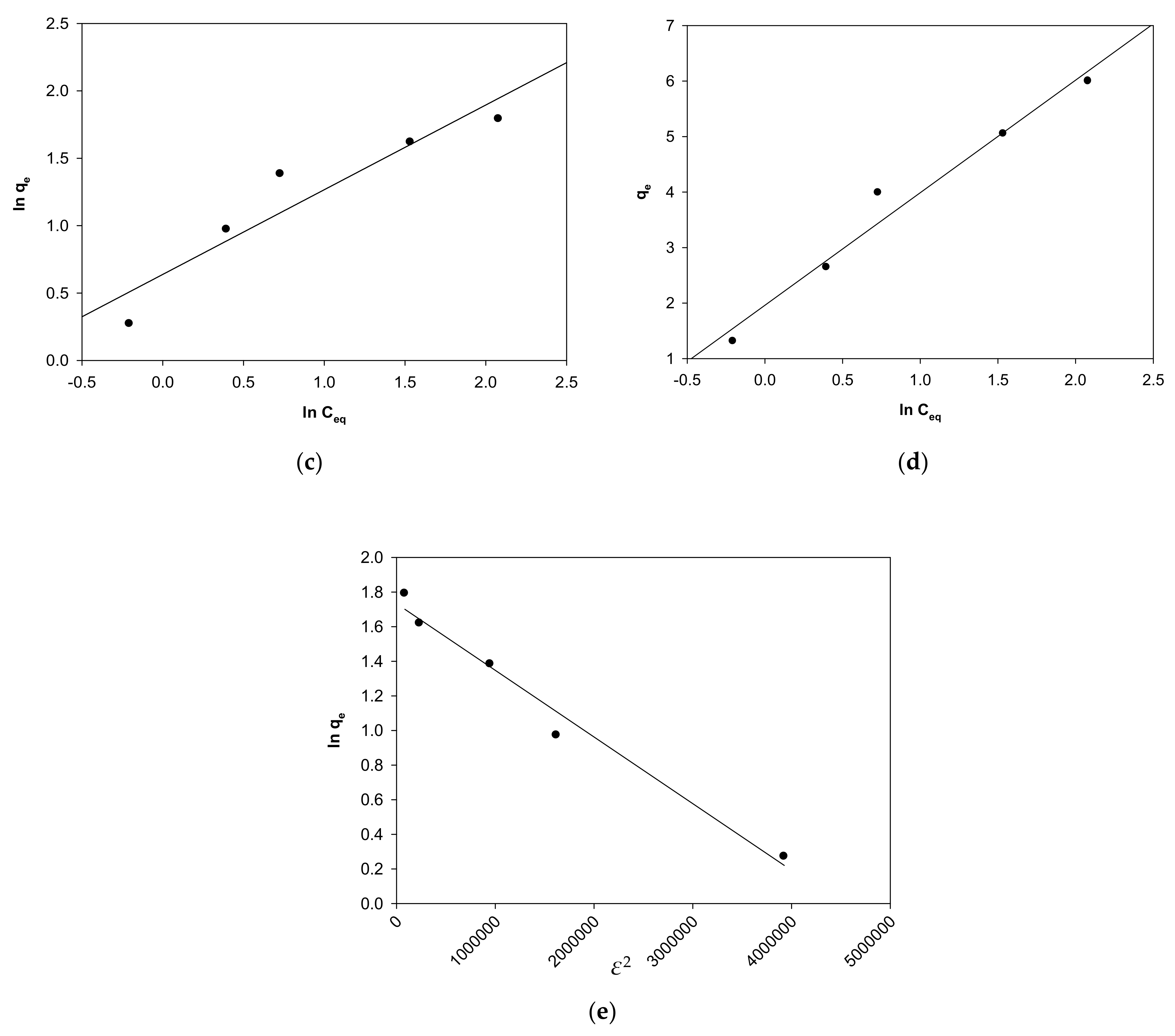
2.4. Impact of Initial MV 2B Concentration and Isothermal Studies

The dye concentration is a major driving force in attaining resistance to the mass transfer of dye between the biosorbents and the aqueous medium [33]. The influence of the initial MV 2B dye concentration on the biosorption process was conducted at different MV 2B concentrations (10–50 mg/L) and the optimum biosorption conditions of pH 6, a biosorbent dosage of 7 g/L, and a fixed contact time of 90 min at 25 °C. The elimination % of MV 2B dye by the C. tamariscifolia biomass was shown in Figure 3a. Increasing the MV 2B dye concentration from 10 mg/L to 30 mg/L increases the dye uptake from 91.8% to 93.1%. As a result, the optimal dye concentration was estimated to be 30 mg/L, and the maximum MV 2B elimination percentage was 93.1%. There are fewer dye molecules ready to be sorbed to a certain number of available active sites at a lower concentration of MV 2B dye. However, beyond the 30 mg/L dye concentration, the percentage of MV 2B dye removed by C. tamariscifolia biomass was reduced. Because all binding sites are saturated at higher initial dye concentrations, more dye molecules remain free in the solution, resulting in a lower removal %. Many researchers achieved similar results [8,34,35], reporting that increasing the initial dye concentrations reduced the biosorption process.
The results of this experiment were employed to estimate the isotherms of the biosorption process.
Adsorption equilibrium isotherms might highlight the correlation between the sorption capacity of sorbent and the adsorbate concentration in an aqueous medium at a certain temperature [36]. The Langmuir, Freundlich, Dubinin–Radushkevich, and Temkin models were applied in this work to investigate biosorption isotherms.
The Langmuir and Freundlich isotherm models were used to simulate the behavior of the isotherm and evaluate the interaction between the adsorbent and adsorbate [37].
By comparing the values of the determination coefficient, the Langmuir model is shown to be more appropriate for dye biosorption than the Freundlich model, since its R2 value (0.935; Figure 3b) is higher than that of the Freundlich model (R2 = 0.884; Figure 3c, Table 2). Therefore, the MV 2B dye biosorption onto C. tamariscifolia biomass occurs on monolayer surfaces with a homogenous pore distribution [38].
The maximum biosorption capacity (qmax) of the algal biomass was 10.0 mg/g, showing that C. tamariscifolia may be employed as an effective and sustainable biosorbent in the bioremoval of dyes. Furthermore, a higher value of the Langmuir constant (b = 0.24 L/mg) showed a greater affinity of dye molecules with the binding sites on C. tamariscifolia surface.
Table 3 compares the maximal biosorption capacity (qmax) of MV 2B dye for the C. tamariscifolia biomass and several different adsorbents available in the literature. It is clear that the biosorption capacity of C. tamariscifolia biomass is close to that of most other sorbents [19,20,39,40,41].
To determine the suitability of the biosorption of MV 2B dye onto the algal biomass, the separation factor (RL) equation was applied. It indicates the shape and feasibility of the biosorption isotherm [42]. The biosorption is linear if the value of RL higher than 1. However, the biosorption is irreversible if RL ≤ 0, and is favorable if 0 < RL < 1 [43]. The obtained value of RL for this investigation was 0.07–0.23 (Table 2), showing that the biosorption process of MV 2B dye was favorable.
The Temkin isotherm model assumed that the adsorption energy of the molecules in the layer gradually decreases with increasing surface coverage and supports the chemisorption process [44].
The high value of the determination coefficient (R2 = 0.971; Table 2, Figure 3d) indicated that MV 2B biosorption onto the surface of C. tamariscifolia biomass may involve a chemical biosorption process. This is in accordance with the data of Abdus-Salam et al. [45]. Additionally, the high value of adsorption heat (b = 1220 J/mol; Table 2) indicated a strong interaction between the biosorbent surface and the MV 2B dye molecules, which corresponds to a reduction in the sorption heat of the molecules in the layer [17].
The Dubinin–Radushkevich (D–R) adsorption model was used to identify how pores are filled, as well as to assess if the sorption process is chemical or physical in nature [46].
According to the D–R model, adsorption occurs physically via a weak intermolecular association if the biosorption energy (E) is less than 8 kJ/mol, and chemically via strong chemical bonds if the value of biosorption energy is between 8 and 16 kJ/mol [47]. As shown in Table 2, the dye biosorption onto the C. tamariscifolia biomass occurs chemically (E = 11.2 kJ/mol). In addition, the algal biomass demonstrated a high biosorption capacity for dye molecules (qo = 5.66 mg/g; Table 2). The high determination coefficient value (R2 = 0.979; Figure 3e, Table 2) suggested that the D–R adsorption model was exploited to represent the biosorption data well.

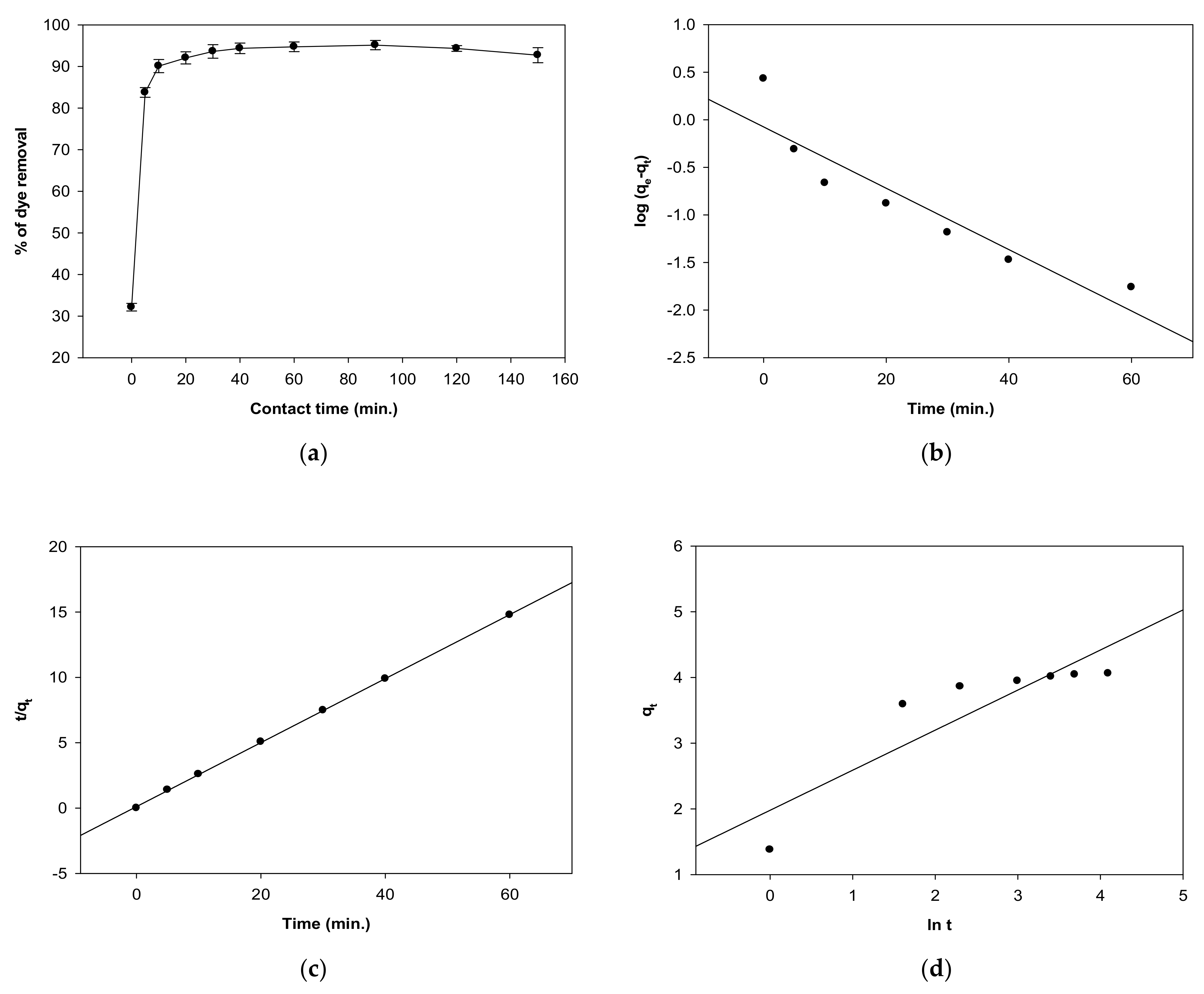
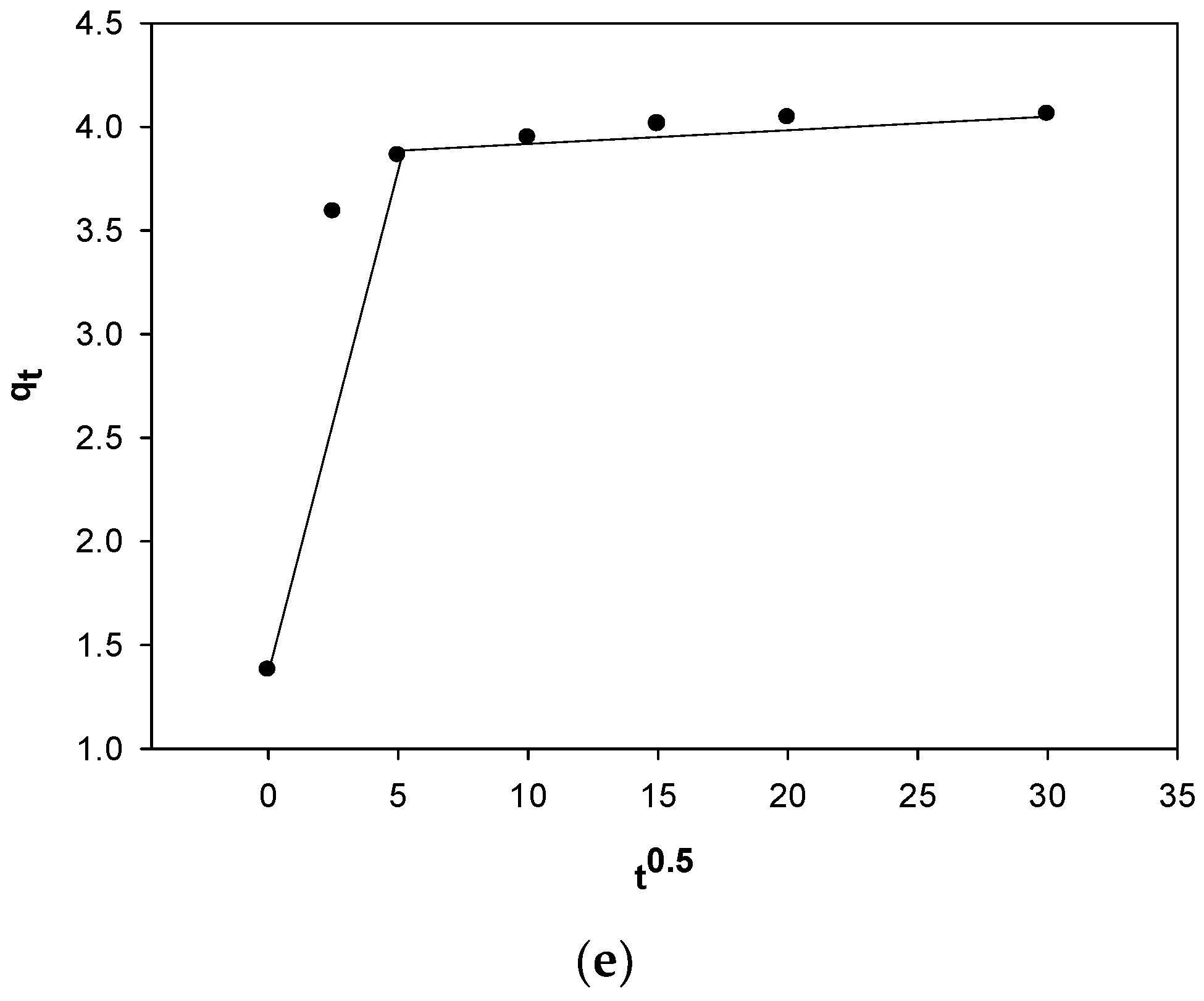
2.5. Impact of Contact Time on MV 2B Biosorption and Kinetic Studies

Contact time is among the most critical variables for efficient biosorption applications. The tests were conducted at 25 °C with variable contact times (0–150 min) and optimal conditions of pH 6, an algal dosage of 7 g/L, and an initial dye concentration of 30 mg/L. The uptake of MV 2B dye by algal biomass increased rapidly in the first 10 min of the reaction (Figure 4a). After that, the biosorption rate slowed, and equilibrium was reached at 60 min. This can be described by stating that the vacant binding site is initially plentiful. However, due to repulsive interactions between the molecules of the solute on the solid phase and bulk phases, it becomes difficult to occupy the remaining vacant surface spaces after some time [48]. As a result, the maximum removal percentage (94.7%) was attained after 60 min, followed by a fixed biosorption percentage.
The results of this experiment were utilized to describe biosorption kinetic models to further validate the experimental data.
Kinetic models are essential for determining the capacity or affinity of the biosorbent, which affects the residence time in the design of a biosorption process [49].
Several kinetic models were used, including pseudo-first, -second-order, intra-particle diffusion, and Elovich models.
In this study, the low value of the determination coefficient of the pseudo-first-order equation (R2 = 0.862; Figure 4b, Table 4) showed that the calculated value of the biosorption capacity (qe cal. = 1.19 mg/g) does not agree well with the experimental value (qe exp. = 4.08 mg/g). These results are in accordance with the data of Bonetto et al. [19] who stated that the calculated values of the adsorption capacity of the HNT–Fe3O4 composite for MV 2B dye differed from the experimental values. Consequently, the pseudo-first-order model is inadequate at explaining the biosorption kinetics data.
On the other hand, a high determination coefficient (0.999; Figure 4c) of the pseudo-second-order model and the very close calculated and experimental values of biosorption capacity (4.07 mg/g and 4.08 mg/g, respectively) demonstrated the model’s importance in describing the dye biosorption kinetics onto C. tamariscifolia biomass.
As a result, it was concluded that the second-order kinetic model better explained the biosorption process compared to the first-order model with a chemical biosorption process as a rate-controlling stage [50].
Moreover, the rate constants of the pseudo-first-order (k1) and second-order (k2) models were 0.014 min−1 and 0.53 g/mg min, respectively, indicating that the MV 2B dye biosorption onto C. tamariscifolia biomass was faster and more favorable (Table 4). These results were consistent with the data of prior investigations on the dyes’ biosorption using various biosorbents, including biorefinery waste of Sargassum latifolium [31], Codium decorticatum [51], lignocellulosic biomass (Luffa cylindrica) [52], and banana-stem-activated carbon [53].
The Elovich model, which is another model employed to explore the kinetics of chemisorption of gas onto solid sorbents, has been demonstrated to be successful in modeling several forms of adsorption.
In this study, the low value of the determination coefficient (R2 = 0.787; Table 4, Figure 4d) demonstrated that the Elovich kinetic model did not provide a suitable fit for the MV 2B biosorption onto C. tamariscifolia biomass.
The intraparticle diffusion model is connected to the sorbate diffusion to the inner pores of the sorbent’s surface as the rate-limiting stage.
Figure 4e exhibits that the results are not closely related to the intraparticle diffusion, and the plot does not pass through the origin. The graph revealed two linear parts, indicating that the intraparticle diffusion kinetic model is not only the rate-controlling phase in the MV 2B biosorption onto C. tamariscifolia biomass, but the biosorption process is also limited by external diffusion [49]. The first linear portion was, therefore, related to the mass transfer of MV 2B molecules from the liquid phase to the external surface of the C. tamariscifolia biomass (film diffusion), whereas the second portion depicts the dye molecule’s diffusion into the algal surface pores (intra-particle diffusion). Related results were reported for the biosorption of methyl violet by bagasse fly ash [54].
Furthermore, the first linear portion had a larger rate constant (Ki.1 = 0.496 mg g−1 min−1/2) and a shorter time than the second section (Ki.2 = 0.005 mg g−1 min−1/2; Table 4). Abou Oualid et al. [51] reported the same trend of mass transfer for crystal violet dye biosorption onto Codium decorticatum biomass.
According to the kinetic studies of the MV 2B biosorption onto C. tamariscifolia biomass, the biosorption process could involve chemical sorption, as indicated by the pseudo-second-order model, and the sorption process took place via the two mechanisms (intra-particle and film diffusion) occurring simultaneously as rate-limiting stages.

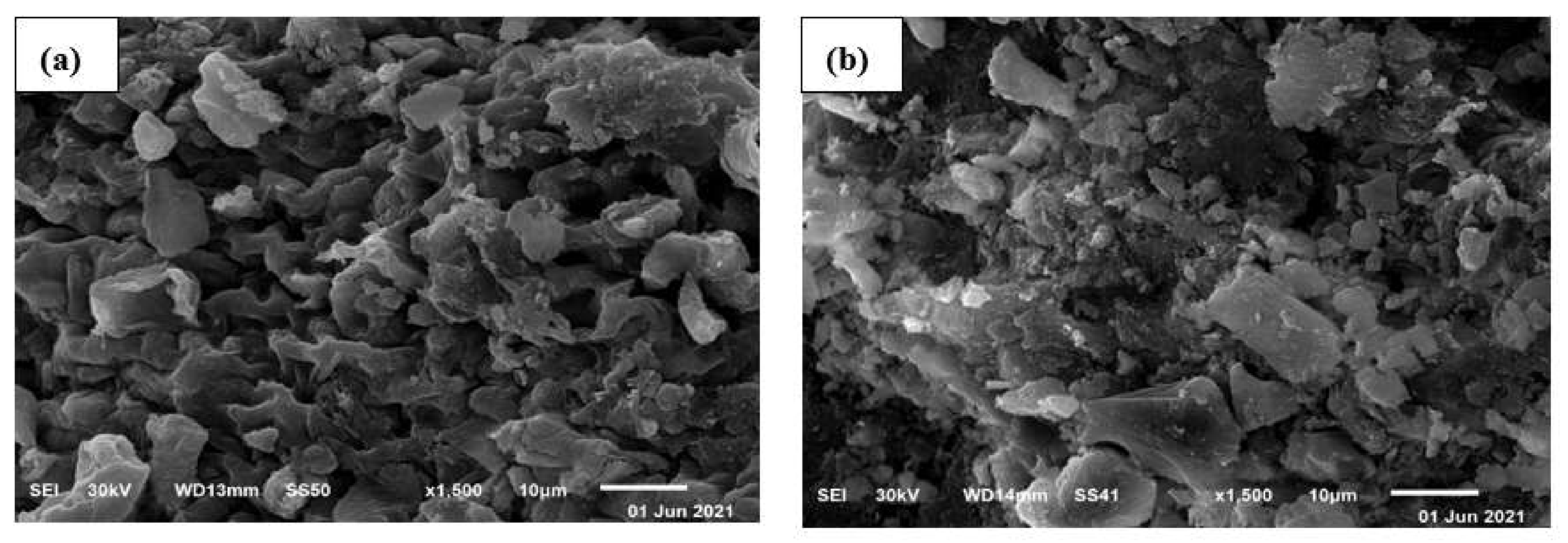
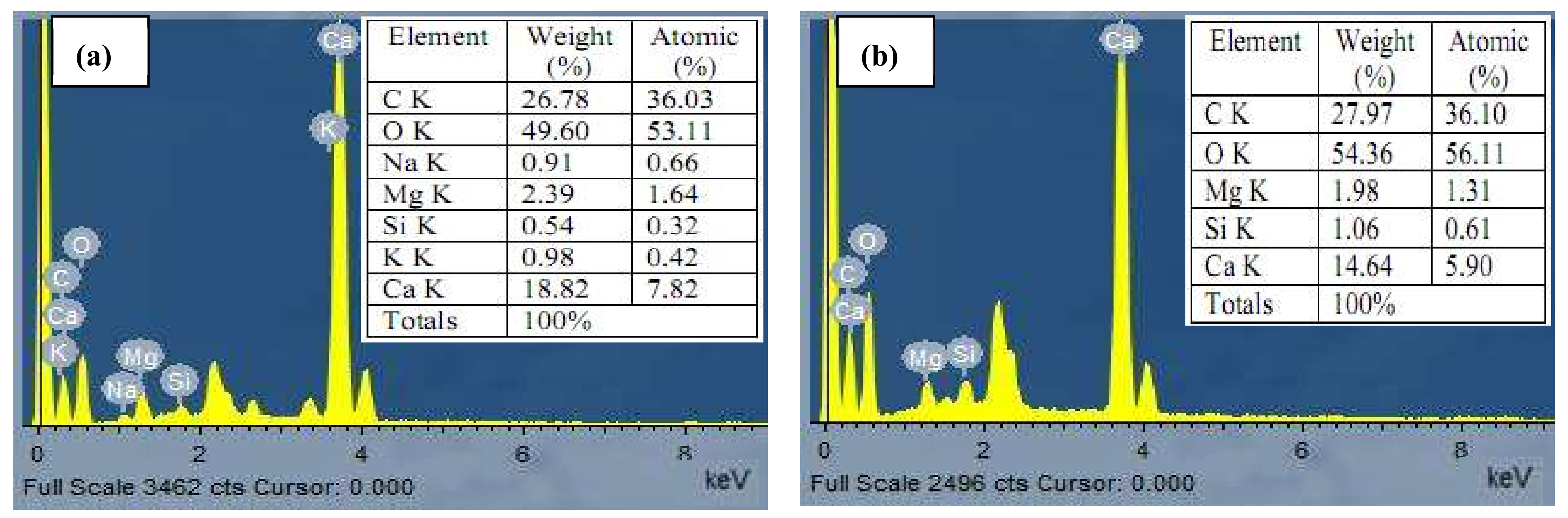
2.6. Characterization of Algal Biomass

2.6.1. SEM/EDX Analyses
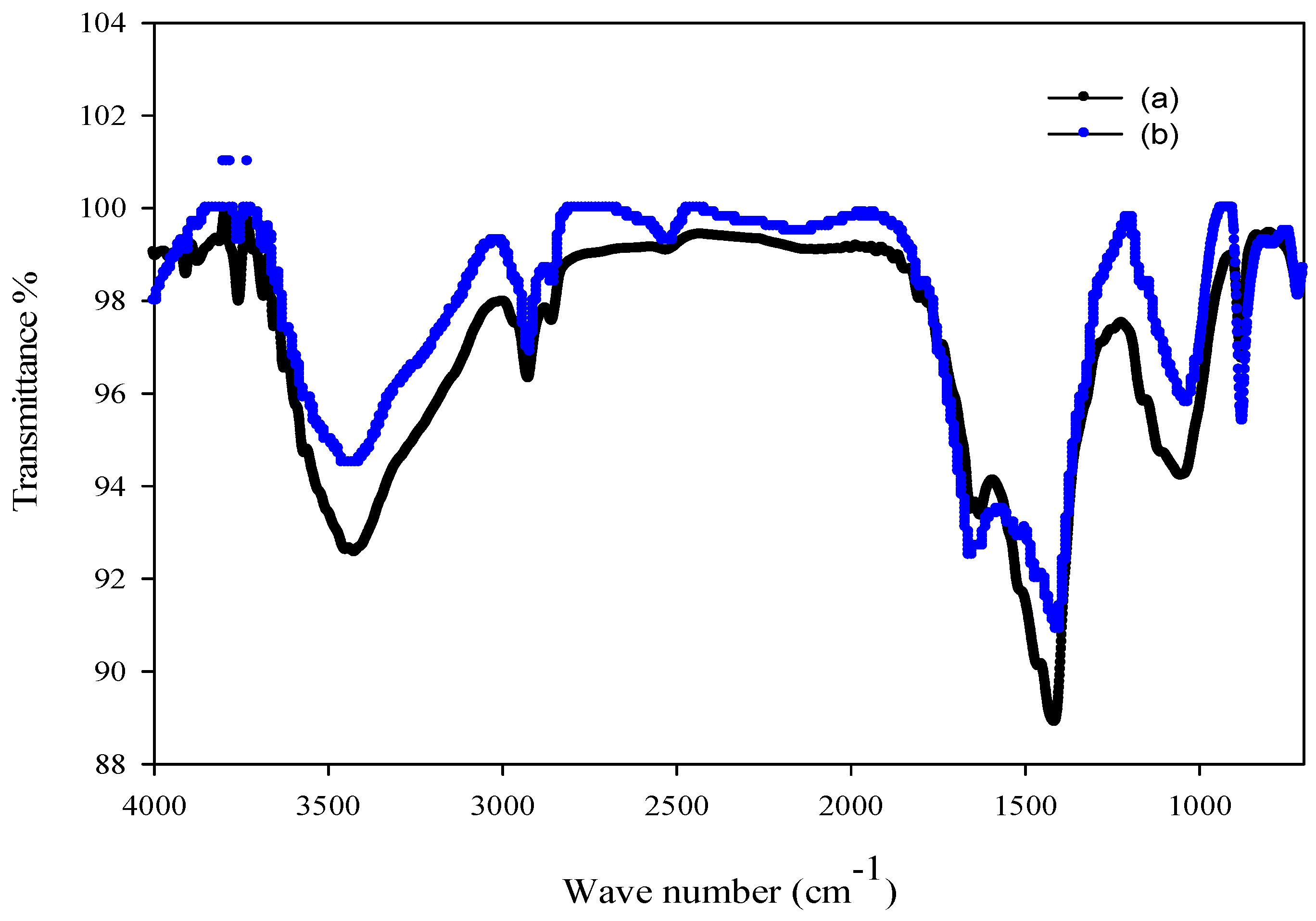
The morphological alterations of C. tamariscifolia biomass before and after dye biosorption were studied by SEM analysis. The micrograph of algal biomass revealed a vacant and rough surface with pores of varying shapes and sizes, providing a higher surface area for dye biosorption (Figure 5a) [55]. Figure 5b depicted the morphological alterations that occurred on the C. tamariscifolia biomass surface following the biosorption of MV 2B dye, which aggregated in various forms and sizes. This property might be the consequence of dye molecules forming a monolayer on the surface of the biosorbent. This aggregation pattern may explain the strong affinity of algal biomass for the MV 2B dye, demonstrating the efficacy of the biosorption process [56].
In this study, an EDX investigation was used to characterize the components deposited on the surface of algal biomass. EDX spectra of algal biomass before and after the MV 2B dye biosorption are depicted in Figure 6a,b.
It can be noted that the algal biomass includes elements such as carbon, oxygen, sodium, magnesium, silicon, potassium, and calcium. The primary constituents such as carbon and oxygen were increased after dye biosorption, but magnesium and calcium contents were reduced. Furthermore, some elements, such as sodium and potassium, cannot be detected in the spectrum. This might imply that ion exchange is among the mechanisms involved in dye sorption. In this respect, Escudero et al. [57] stated the absence of chloride following biosorption of methylene blue onto the Iridaea cordata biomass, and the sulfur content is greater than in raw algal biomass. Therefore, SEM/EDX studies confirmed the MV 2B dye biosorption onto C. tamariscifolia biomass.

2.6.2. FT-IR Study

The FT-IR investigation of the algal biomass revealed the functional groups present on the algal surface. Numerous peaks between 400 cm–1 and 4000 cm–1 were observed, as illustrated in Figure 7a,b. The N–H and O–H groups are responsible for the broad band at 3424 cm–1 shifting to 3449 cm–1. The bands at 2924 cm−1 and 2923 cm−1 were ascribed to the –CH group stretching vibrations [58]. Moreover, the peaks at 2856 cm−1 and 2855 cm−1 were ascribed to the C−H group of lipids [59]. The protein components of algal biomass were apparent as peaks at approximately 1652 cm−1 and 1653 cm−1 (I amide peaks), as well as a peak at 1517 cm−1 that was present only after biosorption of MV 2B dye (II amide peak). Following dye biosorption, the absorption peaks at 1627 cm−1 and 1052 cm−1 shifted to 1625 cm−1 and 1036 cm−1, respectively, and were ascribed to stretching vibrations of the –COOH group [60]. The band at 1414 cm−1 shifted to 1406 cm−1 after dye biosorption was related to C–H group vibrations as well as primary and secondary amines, indicating the existence of proteins and lipids [61]. Alkene vibrations were assigned to absorption peaks at 877 cm−1 and 876 cm−1 [62]. Moreover, the band at 609 cm−1 shifted to 594 cm−1 following MV 2B biosorption and was attributed to organic halide compounds [63]. Other absorption bands were detected at 714 cm−1 and 525 cm−1, which were related to the vibrations of the C–H and O–P–O groups, respectively [64].
Accordingly, it can be concluded that there is a shift in some active groups such as hydroxyl, amide, carboxyl, and carbonyl, which might be the consequence of electrostatic forces and the formation of hydrogen bonds between the dye molecules and the surface of algal biomass. These functional groups may compensate for the dye molecule’s biosorption onto the surface of C. tamariscifolia.

2.6.3. Determination of pHpzc

The estimation of the point of zero charge (pHPZC) of the algal biomass surface is critical to clarifying the mechanism of dye biosorption. The pHPZC value represents the point at which the algal biomass’s surface charge is neutral. As a result, the surface of biomass has a negative charge at pH levels above this point and a positive charge at lower pH levels. The pH change plot (∆pH) against the initial pH revealed that as the initial pH increased, the change in pH became more negative, and the zero value of ∆pH was attained at an initial pH = 5.01 (Figure 8), which is regarded as the pHPZC of C. tamariscifolia biomass.

2.7. Possible Mechanism of Dye Biosorption

Algae have a great surface area and high affinity during the biosorption process, which aids in their biosorption capability. Brown algal cell walls include a variety of polymers, including significant amounts of polysaccharides (alginate) and proteins with many functional groups [65].
The FT-IR data revealed considerable alterations in the wavenumber of the OH, NH, and –COOH groups after MV 2B dye biosorption, suggesting that these groups contribute to the biosorption process via the complexation mechanism. The existence of these functional groups has also been shown to represent a prominent part in the interaction between the algae and MV 2B dye molecules via an ion exchange mechanism that takes place between dye molecules and light ions including calcium, magnesium, sodium, silicon, and potassium, according to the EDX study. Moreover, the SEM image demonstrates that the surface of C. tamariscifolia is porous, suggesting that the biosorption of dye occurs on the pores of the algal surface.
The formation of hydrogen bonds during the biosorption process between the nitrogen atoms of the MV 2B dye and carboxyl and hydroxyl groups may also be suggested as a biosorption mechanism.
The biosorption of MV 2B dye can also occur via electrostatic interaction and the dye-hydrogen ion exchange mechanism because the algal surface is negatively charged (according to the pHPZC determination) at pH ≥ 5.01, facilitating its binding to positively charged MV 2B molecules.

3. Materials and Methods

3.1. Collection of Cystoseira tamariscifolia Seaweed

Brown seaweed Cystoseira tamariscifolia (Hudson) Papenfuss was obtained from the Red Sea shore, Jeddah, KSA. The seaweed was morphologically identified by Dr. M.A. Fawzy (Algae Laboratory of Taif University, Taif, Saudi Arabia), an expert in the taxonomy of macroalgae. The identification was based on the structure of the thallus, base, and apex, the presence/absence of aerocysts, and the patterns of blade branching. Voucher specimens were deposited at Taif University Herbarium (TUH), Taif, Saudi Arabia.
The samples were carefully rinsed with water to eliminate any pollutants. The algal biomass was then dried at 60 °C, pulverized in a mortar, and sieved [49]. For biosorption tests, an average size of 600–700 µm was employed. To increase the biosorption capacity of the algal biomass, 1 g of powdered sample was added to 100 mL of CaCl2. After that, the samples were rinsed with deionized water, dried at 60 °C, and kept in an airtight bottle.

3.2. Preparation of Stock Solution

The cationic organic dye, methyl violet 2B (C24H28N3Cl; Mr 393.95 g/mol, Sigma-Aldrich) was used as a biosorbate. The stock solution of MV 2B (1000 mg/L) was made by dissolving approximately 1 g of the MV 2B dye in 1000 mL of deionized water. Working solutions of various dye concentrations were made by diluting the stock solution with deionized water.

3.3. Biosorption Experiment

Batch biosorption tests were studied in volumetric flasks containing 100 mL of methyl violet 2B dye solution. The experiments were performed using various variables, such as pH (2–10), biomass dosage (1, 3, 5, 7, and 9 g/L), temperature (20, 25, 30, 35, and 45 °C), initial MV 2B dye concentration (10–50 mg/L), and contact time (0–150 min). The pH was set by adding 0.1 M sodium hydroxide and 0.1 M hydrochloric acid, and the mixture was agitated in a shaker at 170 rpm. Following biosorption, the samples were centrifuged for 10 min, and the final MV 2B concentrations in the solutions were quantified using a UV-vis spectrophotometer (Unico UV-2100; Rosemount, MN, USA) at 582 nm [19]. A calibration curve was carried out using various concentrations of MV 2B (1.0 to 10.0 mg/L). Triplicates were used for all dye estimations in the solution.
The following equations were used to calculate the MV 2B dye removal percentage and the amount adsorbed to the biosorbent surface (qe) [66]:
Removal   % = C i C e q   C i × 100
q e mg / g = C i C e q   V   M
where Ci and Ceq (mg/L) represent the initial MV 2B concentration and the concentration at equilibrium, respectively, M (g) is the weight of algal biomass, and V is the solution volume of the MV 2B dye (L).

3.4. Thermodynamic Analysis

A thermodynamic study was conducted at different temperatures (20, 25, 30, 35, and 45 °C), with an optimized pH of 6 and an algal dose of 7 g/L, as well as a fixed contact period of 90 min and a 30 mg/L initial MV 2B concentration. The following equations were exploited to calculate thermodynamic constants, including Δ (Gibbs free energy), Δ (entropy change), and Δ (enthalpy change):
Δ G ° = R T l n K C
    L n K C = Δ S R Δ H R T
Δ G ° = Δ H ° T   Δ S °
where Kc is the equilibrium constant, T is the temperature (K), and R is the universal gas constant (8.31 J/mol K). ΔS° and Δ values were estimated from the intercept and slope of the linear plot of lnKc against 1/T, respectively.

3.5. Modeling of Biosorption Isotherms

Several biosorption isotherms models including Langmuir, Freundlich, D–R, and Temkin were suitably fitted to characterize biosorption results for a broad range of adsorbate concentrations.
The biosorption process was performed at various dye concentrations (10–50 mg/L) and the optimal biosorption conditions of a 7 g/L biomass dosage, pH 6, and a constant contact period of 90 min at 25 °C.
The equation of the linearized Langmuir model is indicated by the following formula:
C e q q e = 1 q m a x b + C e q q m a x
where qmax (mg/g) represents the maximum biosorption capacity and b denotes the biosorbent-biosorbate affinity (L/mg). The intercept and slope of the Ceq/qe vs. Ceq plot can be used to assess the values of b and qmax, respectively.
The dimensionless constant known as the equilibrium parameter RL may be used to represent the main property of the Langmuir isotherm model, which is described as follows:
R L = 1 1 + b C 0  
where Co is the initial MV 2B concentration (mg/L).
The linearized Freundlich model is represented by the following formula:
l n   q e = l n   K f + 1 n   l n   C e q
where n and Kf signify the sorption intensity (mg/g) and the sorption capacity, respectively, and are derived from the plot of lnqe vs. lnCeq.
The linear representation of the Temkin isotherm model is defined by the following equations:
q e = B l n A + B   l n C e q
B = R T b  
where A represents the binding constant for the highest binding energy (L/mg) and b is the constant of Temkin isotherm model related to the heat of adsorption (J/mol).
The linearized D–R model is denoted as follows:
l n q e = l n q 0 β ε 2
where qo is the saturation biosorption capacity of the biosorbent, β is the free energy of the sorption process, and ε represents the potential for Polanyi and can be described as:
ε = R T 1 + 1 C e q
The mean adsorption energy (E; kJ/mol) may be estimated from the following formula:
E = 1 / 2 β

3.6. Modeling of Biosorption Kinetics

Various biosorption kinetic models might be utilized to evaluate the mechanism of biosorption of MV 2B dye onto C. tamariscifolia biomass comprising pseudo-first-order, -second-order, Elovich, and intraparticle diffusion models. The kinetic investigations were conducted at varied contact times (0–150 min) under optimum conditions of pH 6, a 7 g/L biomass dose, and a 30 mg/L initial concentration of MV 2B at 25 °C.
The following formula is a description of the linearized pseudo-first-order kinetic model:
L o g   q e q t = L o g   q e K 1   t 2.303
The plot of log (qeqt) vs. t is applied to determine the equilibrium biosorption capacity, qe (intercept), and the rate constant of the pseudo-first-order model, k1 (slope).
The linearized pseudo-second-order kinetic model is presented in Equation (15):
t q t = 1 K 2 q e 2 + t q e
where k2 is the rate constant of the pseudo-second-order model. A plot of t/qt and t gives the value of qe (slope) and the rate constant k2 (intercept).
The linearized Elovich kinetic model is defined by the following formula:
q t = 1 β ln α β + 1 β ln t
where α (mg/g min) denotes the initial rate of biosorption and β (g/mg) represents the constant of desorption. From the intercept and slope of the plot of qt against ln t, the constants α and β were calculated, respectively.
The linearized formula of the intraparticle diffusion model is given by Equation (17):
q t = K i t 1 / 2   + C i
where Ki denotes the rate constant of intraparticle diffusion (mg/g min1/2) and Ci represents the degree of surface thickness (mg/g). The Ki and Ci values can be estimated from the slope and intercept of the qt vs. t1/2 curve, respectively.

3.7. Characterization of Biosorbent

The morphology of the C. tamariscifolia biomass was evaluated using a scanning electron microscope (SEM; JEOL JSM-6510) and energy-dispersive X-ray spectroscopy (EDX; JEOL JEM-2100). Fourier transform infrared spectroscopy (FT-IR; Thermo Fisher Scientific model iS 10, Waltham, MA, USA) was used to determine the functional groups of the biosorbent before and after the biosorption process. For the determination of a point of zero charge (pHPZC), approximately 50 mL of the solution (pH 2–10) was constantly agitated with 7 g/L of C. tamariscifolia, and the final pH was determined [67].

4. Conclusions

Synthetic dyes are becoming more prevalent in wastewater, causing major health risks to humans. As a result, there is an ongoing increase in the study of effective techniques to ensure an environmental cleanup. The biosorption technique has attracted interest as one of the sustainable techniques for the treatment of wastewater.
In this work, the biosorption efficiency of the marine alga Cystoseira tamariscifolia for dye biosorption from synthetic aqueous solutions was examined. The biosorption process was optimized with respect to the pH, temperature, biomass dosage, initial MV 2B concentration, and contact time. The biosorption process was found to be random, endothermic, and feasible, based on the thermodynamic analysis. The biosorption isotherms fit the Langmuir model well, showing a homogeneous biosorption site distribution, and the Dubinin–Radushkevich and Temkin models also best fit the experimental results. Furthermore, the biosorption of MV 2B dye onto C. tamariscifolia biomass was well suited to the pseudo-second-order kinetic model. SEM/EDX analyses verified the binding of dye molecules on the algal surface, while the FT-IR study revealed the existence of hydroxyl, amides, carboxyl, and amine groups implicated in the dye’s biosorption onto C. tamariscifolia biomass. These findings demonstrated that C. tamariscifolia may be used to eliminate dyes from aqueous solutions in an efficient, sustainable, cost-effective, and eco-friendly manner.

Author Contributions

Conceptualization, writing—original draft, investigation, formal analysis, methodology, M.A.F.; writing—review and editing, supervision, N.K.A. and F.M.A.; investigation, supervision, software, validation, M.I.A.-Z., S.A. and S.H.A.H.; writing—review and editing, visualization, formal analysis. All authors have read and agreed to the published version of the manuscript.

Funding

This research received no external funding.

Institutional Review Board Statement

Not applicable.

Informed Consent Statement

Not applicable.

Data Availability Statement

Not applicable.

Acknowledgments

Princess Nourah bint Abdulrahman University Researchers Supporting Project number (PNURSP2022R84), Princess Nourah bint Abdulrahman University, Riyadh, Saudi Arabia. Taif University Researchers Supporting Project number (TURSP-2020/200), Taif University, Taif, Saudi Arabia.

Conflicts of Interest

There were no conflicts of interest for the authors.

References

  1. Yadav, M.; Thakore, S.; Jadeja, R. Removal of organic dyes using Fucus vesiculosus seaweed bioadsorbent an ecofriendly approach: Equilibrium, kinetics and thermodynamic studies. Environ. Chem. Ecotoxicol. 2022, 4, 67–77. [Google Scholar] [CrossRef]
  2. Chandhru, M.; Rani, S.K.; Vasimalai, N. Reductive degradation of toxic six dyes in industrial wastewater using diaminobenzoic acid capped silver nanoparticles. J. Environ. Chem. Eng. 2020, 8, 104225. [Google Scholar] [CrossRef]
  3. Ayele, A.; Getachew, D.; Kamaraj, M.; Suresh, A. Phycoremediation of synthetic dyes: An effective and eco-friendly algal technology for the dye abatement. J. Chem. 2021, 2021, 9923643. [Google Scholar] [CrossRef]
  4. Seow, T.W.; Lim, C.K.; Nor, M.H.M.; Mubarak, M.F.M.; Lam, C.Y.; Yahya, A.; Ibrahim, Z. Review on wastewater treatment technologies. Int. J. Appl. Environ. Sci. 2016, 11, 111–126. [Google Scholar]
  5. Fontana, I.B.; Peterson, M.; Cechinel, M.A.P. Application of brewing waste as biosorbent for the removal of metallic ions present in groundwater and surface waters from coal regions. J. Environ. Chem. Eng. 2018, 6, 660–670. [Google Scholar] [CrossRef]
  6. Sabnis, R.W. Handbook of Biological Dyes and Stains: Synthesis and Industrial Applications; John Wiley & Sons: Hoboken, NJ, USA, 2010. [Google Scholar]
  7. Dahri, M.K.; Kooh, M.R.R.; Lim, L.B. Removal of methyl violet 2B from aqueous solution using Casuarina equisetifolia needle. Int. Sch. Res. Not. 2013, 2013, 619819. [Google Scholar] [CrossRef] [Green Version]
  8. Mehr, H.V.; Saffari, J.; Mohammadi, S.Z.; Shojaei, S. The removal of methyl violet 2B dye using palm kernel activated carbon: Thermodynamic and kinetics model. Int. J. Environ. Sci. Technol. 2020, 17, 1773–1782. [Google Scholar] [CrossRef]
  9. Kooh, M.R.R.; Dahri, M.K.; Lim, L.B. Removal of the methyl violet 2B dye from aqueous solution using sustainable adsorbent Artocarpus odoratissimus stem axis. Appl. Water Sci. 2017, 7, 3573–3581. [Google Scholar] [CrossRef] [Green Version]
  10. Loulidi, I.; Boukhlifi, F.; Ouchabi, M.; Amar, A.; Jabri, M.; Kali, A.; Chraibi, S.; Hadey, C.; Aziz, F. Adsorption of crystal violet onto an agricultural waste residue: Kinetics, isotherm, thermodynamics, and mechanism of adsorption. Sci. World J. 2020. [Google Scholar] [CrossRef]
  11. Velmurugan, P.; Kumar, R.V.; Dhinakaran, G. Dye removal from aqueous solution using low cost adsorbent. Int. J. Environ. Sci. 2011, 1, 1492. [Google Scholar]
  12. Parmar, N.D.; Shukla, S.R. Decolourization of dye wastewater by microbial methods—A review. Indian J. Chem. Technol. (IJCT) 2018, 25, 315–323. [Google Scholar]
  13. Fawzy, M.A.; Issa, A.A. Bioremoval of heavy metals and nutrients from sewage plant by Anabaena oryzae and Cyanosarcina fontana. Int. J. Phytoremediat. 2016, 18, 321–328. [Google Scholar] [CrossRef] [PubMed]
  14. Alharbi, N.K.; Al-Zaban, M.I.; Albarakaty, F.M.; Abdelwahab, S.F.; Hassan, S.H.; Fawzy, M.A. Kinetic, Isotherm and Thermodynamic Aspects of Zn2+ Biosorption by Spirulina platensis: Optimization of Process Variables by Response Surface Methodology. Life 2022, 12, 585. [Google Scholar] [CrossRef] [PubMed]
  15. Romauld, S.I.; Venkataraghavan, R.; Yuvaraj, D.; Devi, V.I.; Hashika, S. Mycoremediation of Hydrocarbon and its products using Fusarium oxysporum. Res. J. Pharm. Technol. 2019, 12, 4216–4224. [Google Scholar] [CrossRef]
  16. Imran-Shaukat, M.; Wahi, R.; Ngaini, Z. The application of agricultural wastes for heavy metals adsorption: A meta-analysis of recent studies. Bioresour. Technol. Rep. 2022, 17, 100902. [Google Scholar] [CrossRef]
  17. Fawzy, M.A.; Darwish, H.; Alharthi, S.; Al-Zaban, M.I.; Noureldeen, A.; Hassan, S.H. Process optimization and modeling of Cd2+ biosorption onto the free and immobilized Turbinaria ornata using Box–Behnken experimental design. Sci. Rep. 2022, 12, 3256. [Google Scholar] [CrossRef]
  18. Kooh, M.R.R.; Dahri, M.K.; Lim, L.B. Removal of methyl violet 2B dye from aqueous solution using Nepenthes rafflesiana pitcher and leaves. Appl. Water Sci. 2017, 7, 3859–3868. [Google Scholar] [CrossRef] [Green Version]
  19. Bonetto, L.R.; Ferrarini, F.; De Marco, C.; Crespo, J.S.; Guégan, R.; Giovanela, M. Removal of methyl violet 2B dye from aqueous solution using a magnetic composite as an adsorbent. J. Water Process. Eng. 2015, 6, 11–20. [Google Scholar] [CrossRef] [Green Version]
  20. Mahini, R.; Esmaeili, H.; Foroutan, R. Adsorption of methyl violet from aqueous solution using brown algae Padina sanctae-crucis. Turk. J. Biochem. 2018, 43, 623–631. [Google Scholar] [CrossRef]
  21. Thomas, A.D.; Bohumil, V.; Alfonso, M. A review of the biochemistry of heavy metal biosorption by brown algae. Water Res. 2003, 37, 4311–4330. [Google Scholar]
  22. Abbas, S.H.; Ismail, I.M.; Mostafa, T.M.; Sulaymon, A.H. Biosorption of heavy metals: A review. J. Chem. Sci. Technol. 2014, 3, 74–102. [Google Scholar]
  23. Doan, V.D.; Tran, T.K.N.; Nguyen, A.T.; Tran, V.A.; Nguyen, T.D.; Le, V.T. Comparative study on adsorption of cationic and anionic dyes by nanomagnetite supported on biochar derived from Eichhornia crassipes and Phragmites australis stems. Environ. Nanotechnol. Monit. Manag. 2021, 16, 100569. [Google Scholar] [CrossRef]
  24. Ncibi, M.C.; Ben Hamissa, A.M.; Fathallah, A.; Kortas, M.H.; Baklouti, T.; Mahjoub, B.; Seffen, M. Biosorptive uptake of methylene blue using Mediterranean green alga Enteromorpha spp. J. Hazard. Mater. 2009, 170, 1050–1055. [Google Scholar] [CrossRef] [PubMed]
  25. Bhatia, D.; Sharma, N.R.; Singh, J.; Kanwar, R.S. Biological methods for textile dye removal from wastewater: A review. Crit. Rev. Environ. Sci. Technol. 2017, 47, 1836–1876. [Google Scholar] [CrossRef]
  26. Bhattacharjee, C.; Dutta, S.; Saxena, V.K. A review on biosorptive removal of dyes and heavy metals from wastewater using watermelon rind as biosorbent. Environ. Adv. 2020, 2, 100007. [Google Scholar] [CrossRef]
  27. El-Naggar, N.E.A.; Rabei, N.H.; El-Malkey, S.E. Eco-friendly approach for biosorption of Pb2+ and carcinogenic Congo red dye from binary solution onto sustainable Ulva lactuca biomass. Sci. Rep. 2020, 10, 16021. [Google Scholar] [CrossRef]
  28. Arief, V.O.; Trilestari, K.; Sunarso, J.; Indraswati, N.; Ismadji, S. Recent progress on biosorption of heavy metals from liquids using low cost biosorbents: Characterization, biosorption parameters and mechanism studies. CLEAN—Soil Air Water 2008, 36, 937–962. [Google Scholar] [CrossRef]
  29. Omokpariola, D.O. Experimental Modelling Studies on the removal of crystal violet, methylene blue and malachite green dyes using Theobroma cacao (Cocoa Pod Powder). J. Chem. Lett. 2021, 2, 9–24. [Google Scholar]
  30. Sharma, K.; Sharma, S.; Sharma, V.; Mishra, P.K.; Ekielski, A.; Sharma, V.; Kumar, V. Methylene Blue Dye Adsorption from Wastewater Using Hydroxyapatite/Gold Nanocomposite: Kinetic and Thermodynamics Studies. Nanomaterials 2021, 11, 1403. [Google Scholar] [CrossRef]
  31. Fawzy, M.A.; Gomaa, M. Low-cost biosorption of Methylene Blue and Congo Red from single and binary systems using Sargassum latifolium biorefinery waste/wastepaper xerogel: An optimization and modeling study. J. Appl. Phycol. 2021, 33, 675–691. [Google Scholar] [CrossRef]
  32. Fawzy, M.A.; Gomaa, M. Use of algal biorefinery waste and waste office paper in the development of xerogels: A low cost and eco-friendly biosorbent for the effective removal of Congo red and Fe (II) from aqueous solutions. J. Environ. Manag. 2020, 262, 110380. [Google Scholar] [CrossRef]
  33. Kankılıç, G.B.; Metin, A.Ü.; Aluç, Y. Investigation on phenol degradation capability of Scenedesmus regularis: Influence of process parameters. Environ. Technol. 2018, 41, 1065–1073. [Google Scholar] [CrossRef]
  34. Shojaei, S.; Khammarnia, S.; Shojaei, S.; Sasani, M. Removal of reactive red 198 by nanoparticle zero valent iron in the presence of hydrogen peroxide. J. Water Environ. Nanotechnol. 2017, 2, 129–135. [Google Scholar]
  35. Ruscasso, F.; Bezus, B.; Garmendia, G.; Vero, S.; Curutchet, G.; Cavello, I.; Cavalitto, S. Debaryomyces hansenii F39A as biosorbent for textile dye removal. Rev. Argent. Microbiol. 2021, 53, 257–265. [Google Scholar] [CrossRef]
  36. Fawzy, M.A.; Alharthi, S. Cellular responses and phenol bioremoval by green alga Scenedesmus abundans: Equilibrium, kinetic and thermodynamic studies. Environ. Technol. Innov. 2021, 22, 101463. [Google Scholar] [CrossRef]
  37. Tang, L.; Huang, S.; Wang, Y.; Liang, D.; Li, Y.; Li, J.; Wang, Y.; Xie, Y.; Wang, W. Highly efficient, stable, and recyclable hydrogen manganese oxide/cellulose film for the extraction of lithium from seawater. ACS Appl. Mater. Interfaces 2020, 12, 9775–9781. [Google Scholar] [CrossRef]
  38. Fu, J.; Xin, Q.; Wu, X.; Chen, Z.; Yan, Y.; Liu, S.; Wang, M.; Xu, Q. Selective adsorption and separation of organic dyes from aqueous solution on polydopamine microspheres. J. Colloid Interface Sci. 2016, 461, 292–304. [Google Scholar] [CrossRef]
  39. Jafari, S.; Azizian, S.; Jaleh, B. Enhancement of methyl violet removal by modification of TiO2 nanoparticles with AgI. J. Ind. Eng. Chem. 2012, 18, 2124–2128. [Google Scholar] [CrossRef]
  40. Wu, J.S.; Liu, C.H.; Chu, K.H.; Suen, S.Y. Removal of cationic dye methyl violet 2B from water by cation exchange membranes. J. Membr. Sci. 2008, 309, 239–245. [Google Scholar] [CrossRef]
  41. Ofomaja, A.E.; Ho, Y.S. Effect of temperatures and pH on methyl violet biosorption by Mansonia wood sawdust. Bioresour. Technol. 2008, 99, 5411–5417. [Google Scholar] [CrossRef]
  42. Ugbe, F.A.; Anebi, P.O.; Ikudayisi, V.A. Biosorption of an anionic dye, eosin yellow onto pineapple peels: Isotherm and thermodynamic study. Int. Ann. Sci. 2018, 4, 14–19. [Google Scholar] [CrossRef] [Green Version]
  43. Nnaji, C.C.; Agim, A.E.; Mama, C.N.; Emenike, P.C.; Ogarekpe, N.M. Equilibrium and thermodynamic investigation of biosorption of nickel from water by activated carbon made from palm kernel chaff. Sci. Rep. 2021, 11, 7808. [Google Scholar] [CrossRef]
  44. Foo, K.Y.; Hameed, B.H. Insights into the modeling of adsorption isotherm systems. Chem. Eng. J. 2010, 156, 2–10. [Google Scholar] [CrossRef]
  45. Abdus-Salam, N.; Ikudayisi-Ugbe, A.V.; Ugbe, F.A. Adsorptive removal of methylene blue from synthetic wastewater using date palm seeds, goethite and their composite. Acta Sci. Malays. (ASM) 2021, 5, 27–35. [Google Scholar] [CrossRef]
  46. Ahmad, M.A.; Ahmad, N.; Bello, O.S. Adsorption kinetic studies for the removal of synthetic dye using durian seed activated carbon. J. Dispers. Sci. Technol. 2015, 36, 670–684. [Google Scholar] [CrossRef]
  47. Isik, B.; Ugraskan, V.; Cankurtaran, O. Effective biosorption of methylene blue dye from aqueous solution using wild macrofungus (Lactarius piperatus). Sep. Sci. Technol. 2021, 57, 854–871. [Google Scholar] [CrossRef]
  48. Hasani, N.; Selimi, T.; Mele, A.; Thaçi, V.; Halili, J.; Berisha, A.; Sadiku, M. Theoretical, Equilibrium, Kinetics and Thermodynamic Investigations of Methylene Blue Adsorption onto Lignite Coal. Molecules 2022, 27, 1856. [Google Scholar] [CrossRef]
  49. Fawzy, M.A. Biosorption of copper ions from aqueous solution by Codium vermilara: Optimization, kinetic, isotherm and thermodynamic studies. Adv. Powder Technol. 2020, 31, 3724–3735. [Google Scholar] [CrossRef]
  50. Qian, H.; Huang, S.; Ba, Z.; Wang, W.; Yu, F.; Liang, D.; Xie, Y.; Wang, Y.; Wang, Y. HTO/Cellulose Aerogel for Rapid and Highly Selective Li+ Recovery from Seawater. Molecules 2021, 26, 4054. [Google Scholar] [CrossRef]
  51. Abou Oualid, H.; Abdellaoui, Y.; Laabd, M.; El Ouardi, M.; Brahmi, Y.; Iazza, M.; Abou Oualid, J. Eco-efficient green seaweed Codium decorticatum biosorbent for textile dyes: Characterization, mechanism, recyclability, and RSM optimization. ACS Omega 2020, 5, 22192–22207. [Google Scholar] [CrossRef]
  52. Boudechiche, N.; Mokaddem, H.; Sadaoui, Z.; Trari, M. Biosorption of cationic dye from aqueous solutions onto lignocellulosic biomass (Luffa cylindrica): Characterization, equilibrium, kinetic and thermodynamic studies. Int. J. Ind. Chem. 2016, 7, 167–180. [Google Scholar] [CrossRef] [Green Version]
  53. Karnakanti, S. Adsorption Kinetics of Malachite Green Dye Removal from Aqueous Solution by using Banana Stem. Int. J. Eng. Adv. Technol. 2021, 10, 215–220. [Google Scholar]
  54. Mall, I.D.; Srivastava, V.C.; Agarwal, N.K. Removal of Orange-G and Methyl Violet dyes by adsorption onto bagasse fly ash—Kinetic study and equilibrium isotherm analyses. Dyes Pigm. 2006, 69, 210–223. [Google Scholar] [CrossRef]
  55. Shakoor, S.; Nasar, A. Removal of methylene blue dye from artificially contaminated water using citrus limetta peel waste as a very low cost adsorbent. J. Taiwan Inst. Chem. Eng. 2016, 66, 154–163. [Google Scholar] [CrossRef]
  56. Litefti, K.; Freire, M.S.; Stitou, M.; González-Álvarez, J. Adsorption of an anionic dye (Congo red) from aqueous solutions by pine bark. Sci. Rep. 2019, 9, 16530. [Google Scholar] [CrossRef] [Green Version]
  57. Escudero, L.B.; Smichowski, P.N.; Dotto, G.L. Macroalgae of Iridaea cordata as an efficient biosorbent to remove hazardous cationic dyes from aqueous solutions. Water Sci. Technol. 2017, 76, 3379–3391. [Google Scholar] [CrossRef]
  58. Gokulan, R.; Avinash, A.; Prabhu, G.G.; Jegan, J. Remediation of remazol dyes by biochar derived from Caulerpa scalpelliformis—An eco-friendly approach. J. Environ. Chem. Eng. 2019, 7, 103297. [Google Scholar] [CrossRef]
  59. Fawzy, M.A.; Hifney, A.F.; Adam, M.S.; Al-Badaani, A.A. Biosorption of cobalt and its effect on growth and metabolites of Synechocystis pevalekii and Scenedesmus bernardii: Isothermal analysis. Environ. Technol. Innov. 2020, 19, 100953. [Google Scholar] [CrossRef]
  60. Zhuang, J.; Li, M.; Pu, Y.; Ragauskas, A.J.; Yoo, C.G. Observation of potential contaminants in processed biomass using fourier transform infrared spectroscopy. Appl. Sci. 2020, 10, 4345. [Google Scholar] [CrossRef]
  61. Queiroz, M.F.; Teodosio Melo, K.R.; Sabry, D.A.; Sassaki, G.L.; Rocha, H.A.O. Does the use of chitosan contribute to oxalate kidney stone formation? Mar. Drugs 2014, 13, 141–158. [Google Scholar] [CrossRef]
  62. Chen, Z.; Hay, J.N.; Jenkins, M.J. FTIR spectroscopic analysis of poly (ethylene terephthalate) on crystallization. Eur. Polym. J. 2012, 48, 1586–1610. [Google Scholar] [CrossRef]
  63. Michalak, I.; Mironiuk, M.; Marycz, K. A comprehensive analysis of biosorption of metal ions by macroalgae using ICP-OES, SEM-EDX and FTIR techniques. PLoS ONE 2018, 13, e0205590. [Google Scholar] [CrossRef] [PubMed]
  64. Sánchez-Ponce, L.; Granado-Castro, M.D.; Casanueva-Marenco, M.J.; Galindo-Riaño, M.D.; Díaz-de-Alba, M. Sherry wine industry by-product as potential biosorbent for the removal of Cr (VI) from aqueous medium. Biomass Convers. Biorefinery 2021, 1–19. [Google Scholar] [CrossRef]
  65. Deniaud-Bouët, E.; Hardouin, K.; Potin, P.; Kloareg, B.; Hervé, C. A review about brown algal cell walls and fucose-containing sulfated polysaccharides: Cell wall context, biomedical properties and key research challenges. Carbohydr. Polym. 2017, 175, 395–408. [Google Scholar] [CrossRef] [PubMed]
  66. Fawzy, M.A. Phycoremediation and adsorption isotherms of cadmium and copper ions by Merismopedia tenuissima and their effect on growth and metabolism. Environ. Toxicol. Pharmacol. 2016, 46, 116–121. [Google Scholar] [CrossRef]
  67. Jiang, R.; Tian, J.; Zheng, H.; Qi, J.; Sun, S.; Li, X. A novel magnetic adsorbent based on waste litchi peels for removing Pb (II) from aqueous solution. J. Environ. Manag. 2015, 155, 24–30. [Google Scholar] [CrossRef]
Figure 1. Impact of (a) pH and (b) biosorbent dose on the percentage of bioremoval of MV 2B by C. tamariscifolia biomass.
Sustainability 14 05285 g001
Figure 2. (a) Effect of temperature and (b) thermodynamic analysis for the bioremoval of MV 2B by C. tamariscifolia biomass.
Sustainability 14 05285 g002
Figure 3. (a) Impact of initial MV 2B dye concentration, (b) Langmuir, (c) Freundlich, (d) Temkin, and (e) D–R models for the bioremoval of MV 2B by C. tamariscifolia biomass.
Sustainability 14 05285 g003aSustainability 14 05285 g003b
Figure 4. (a) Impact of contact time, (b) Pseudo-first, (c) second-order, (d) Elovich, (e) intraparticle diffusion kinetic models for the removal of MV 2B dye by C. tamariscifolia biomass.
Sustainability 14 05285 g004aSustainability 14 05285 g004b
Figure 5. SEM (a) before and (b) after the biosorption of MV 2B dye onto C. tamariscifolia biomass.
Sustainability 14 05285 g005
Figure 6. EDX (a) before and (b) after the biosorption of MV 2B dye onto C. tamariscifolia biomass.
Sustainability 14 05285 g006
Figure 7. FTIR (a) before and (b) after the biosorption of MV 2B dye onto C. tamariscifolia biomass.
Sustainability 14 05285 g007
Figure 8. Plot of ∆pH against initial pH for C. tamariscifolia biomass.
Sustainability 14 05285 g008
Table 1. Parameters of thermodynamic analysis for the biosorption of MV 2B dye onto C. tamariscifolia.
Δ (kJ/mol)Δ (kJ/mol)Δ (kJ/mol)R2
293 K298 K303 K308 K318 K
−1.18−1.62−1.60−2.01−2.4312.870.0480.940
Table 2. Isotherm constants for the bioremoval of methyl violet 2B dye onto C. tamariscifolia.
LangmuirFreundlichTemkinD–R
qmax (mg/g)b
(L/mg)
RLR2Kf (mg/g)nR2BA
(L/mg)
b
(J/mol)
R2qo
(mg/g)
Β × 10−7 (mol2/J2)E (kJ/mol)R2
10.00.240.07–0.230.941.891.590.882.032.6412200.975.664.011.20.979
Table 3. Maximum MV 2B adsorption capacity (qmax) in comparison to various sorption materials.
Adsorbents Adsorption Conditionsqmax
(mg/g)
References
pHTemperature (oC)Adsorbent Dose (g/L)
HNT-Fe3O4 composite4.2251.520.4[19]
Padina sanctae-crucis825210.02[20]
TiO2P25-3014.6[39]
Modified cation exchange membrane-25-10.1[40]
Wood of Monsunya (tree)1025417.7[41]
Cystoseira tamariscifolia625710Present study
Table 4. Kinetic constants for the bioremoval of MV 2B dye onto C. tamariscifolia.
Pseudo-First OrderPseudo-Second OrderElovichIntra-Particle Diffusion
qe (exp.) (mg/g)qe (cal.) (mg/g)K1 (min−1)R2qe (cal.)
(mg/g)
K2
(g/mg min)
R2α
(mg/g min)
β
(g/mg)
R2Ki1
(mg/g min1/2)
Ki2
(mg/g min1/2)
Ci
(mg/g)
R21R22
4.071.190.0140.864.080.530.999756.51.640.7870.4964.06.050.830.803
Publisher’s Note: MDPI stays neutral with regard to jurisdictional claims in published maps and institutional affiliations.

