Article

Cellulolytic Bacillus Strain: Production Optimization Using Wheat Bran under Solid-State Fermentation and Investigation of Its Probiotic Potential

1
Laboratory Écologie et de Technologie Microbienne, Institut National des Sciences Appliquées et de Technologie (INSAT), University of Carthage, Tunis 1080, Tunisia
2
French School Victor Hugo, Gontardstraße 11, 60488 Frankfurt, Germany
3
Laboratory of Biotechnology and Bio-Geo Resources Valorization, Institut Superieur de Biotechnology de Sidi Thabet, University of Manouba, Manouba 2010, Tunisia
*
Author to whom correspondence should be addressed.
Sustainability 2023, 15(10), 8394; https://doi.org/10.3390/su15108394
Submission received: 30 November 2022 / Revised: 8 February 2023 / Accepted: 28 February 2023 / Published: 22 May 2023

Abstract

:
Attention to the association of cellulolytic bacteria with probiotic potential as an additive in animal feeding has risen in the last decades. Such additive use in livestock feed is essential in improving animal health, growth, and production performances. This study was designed to identify probiotic characteristics and test the cellulolytic ability of Bacillus strains isolated from the dromedary gastrointestinal tract. Thus, thirty-two Bacillus strains were tested for their cellulolytic ability on cellulose Congo-red agar media. Among the isolates, only the strain D1B3 showed the largest degradation zone (2.4 cm) and was identified as Bacillus amyloliquefacians by 16S rRNA gene sequence analysis. Solid-state fermentation (SSF) retained this strain for cellulase and biomass production using wheat bran as a substrate. The fermentation was optimized through a central composite design, by exploring three factors: incubation temperature, moisture ratio, and pH. Biomass and cellulose enzyme activity were selected as responses and corresponding regression coefficients were calculated. The optimal parameters were: liquid-to-solid ratio (1.19%), pH buffer (6.2), and incubation temperature (36.99 °C) to obtain the highest level of biomass and cellulose enzyme activity reaching a value of 9.828 log CFU/g and 0.0144 g/L.min, respectively. The potentiality of Bacillus amyloliquefacians D1B3 as a probiotic was examined in vitro. It also showed antimicrobial activity against Pseudomonas aeruginosa 9027, Klebsiella pneumoniae, and Escherichia coli 10536. The isolate tolerates low pH and bile salt (0.3% Oxygall). The hydrophobicity and coaggregation abilities were 1.7% and 69.79%, respectively. The results indicated that Bacillus amyloliquefacians D1B3 could be a potential probiotic additive for improving in vitro fermentation of wheat bran and suggests the possibility of combining the probiotic attributes of this strain with its cellulolytic ability to enhance the rumen fermentation of animal feed.

1. Introduction

The global feed enzymes (phytases, carbohydrases, and proteinases) market size is estimated to account for USD 1.3 billion in 2020 and is estimated to reach about USD 1.9 billion by 2025 with a compound annual growth rate (CAGR) of 8.1% [1].This growth is explained, on the one hand, by the increase in the demand of meat and dairy products by a growing world population; and, on the other hand, by the restriction of the use of antibiotics in animal production.
Among these enzymes, the market of carbohydrases—and especially xylanases and glucanasesis—is the fastest-growing one. In fact, carbohydrases were preferred by most animal feed manufactures and livestock producers due to their role in increasing the digestibility of nutrients [2].
Cellulase (β-glucanase) is an enzyme able to hydrolyze cellulose as a substrate for available sugars; thus, it has many applications, especially in animal-feed production. Supplementary cellulose in monogastric animals’ diets enhances intestinal health, prevents constipation, and improves animal performance [3]. However, herbivore animals lack digestive enzymes such as cellulase to degrade this supplementary cellulose and they totally depend on their gut microorganism having this digestive enzyme pattern [4,5].
Therefore, there is a higher demand for the enhanced digestibility of animal feeds by adding efficient cellulolytic microorganisms. Cellulases are part of the glycosyl hydrolase family, which includes endoglucanase and exoglucanase. These enzymes have acquired much interest owing to their large applications in paper, detergents, biofuel textiles, and animal feed industries [6,7]. Microorganisms, namely bacteria and fungi, can produce cellulase; and particularly, bacterial cellulase is in raising the demand because of its high growth rate and variety of habitats. For several years, cellulase-producing bacteria have been isolated from diverse sources, such as soils, vegetable matter, sugar-industry waste, the feces and gut microbiota of animals etc. [5,8,9]. In addition, several studies on cellulolytic microorganisms utilized the submerged culture because the cellulose substrate is a soluble form that facilitates the growth of microorganisms. However, the solid-state bioprocess (SSF) offers many advantages, such as its lower cost, direct contact between substrate and microorganism, and higher product yield [10,11].
Further, among microorganisms, Bacillus sp. was widely mentioned as a cellulase-producing bacteria [12,13,14] and also recently, as a probiotic candidate. As described by the World Health Organization, probiotics are live microorganisms that when administrated in adequate amounts, exhibited beneficial effects in the host organism. The most well-known as probiotics were lactic-acid bacteria, such as Lactobacillus and Enterococcus [15]. Bacillus were former spore bacteria; so, in this form, they have advantages over other bacteria such as Lactobacillus spp., including the possibility of being a storage product at room temperature, without the loss of the viability [13,16]. Furthermore, according to recent studies, Bacillus probiotics were extensively implicated in animal feed as growth promoters or as alternatives of antibiotics for livestock farming [15,17]. The use of Bacillus amyloliquefaciens as a probiotic in broiler chicken was investigated by Li et al. [18] they demonstrated that it can be also efficient on the immune function. Considering all these advantages of Bacillus strains and the fact that they were able to produce extracellular enzymes, namely cellulase, amylase, xylanase, and phytase, it would be interesting to find a new Bacillus strain with the ability to combine probiotic and cellulolytic properties.
When these probiotics bacteria colonize the digestive tract of animals, their enzymes such as cellulase are secreted and efficiently improved in the digestion of animal feed, especially for young animals [15].
In our study, cellulolytic Bacillus were previously isolated from the gastrointestinal tract of herbivore animals, such as the Tunisian dromedary [19].
The present study was conducted to assess the ability of the most efficient isolate, Bacillus amyloliquefacians (D1B3), to produce cellulase in SSF fermentation using wheat bran as substrate. An experimental factorial design was performed to optimize the production of biomass and cellulase with the variable of liquid-to-solid ratio, temperature, and pH of the buffer; and also, studied the pattern of mutual interaction between the variable. This study investigates for the first time cellulolytic Bacillus amyloliquefaciens isolated from the gastrointestinal gut of a dromedary as a potential animal probiotic that could enhance feed digestibility.

2. Material and Methods

2.1. Bacterial Strains and Cellulolytic Activity

Thirty-two Bacillus strains previously isolated from the intestinal microbiota of Tunisian dromedary belonging to the Laboratory of Ecology and Technology microbian culture collection (LETM) were tested. Strains were cultivated in Tryptic soy broth (TSB) (Merck Co., Darmstadt, Germany) and then, screened for their cellulolytic activity using the plate-base technique supplemented with Congo red (1%) according to Ariffin et al. [20]. Colonies presenting discoloration of Congo red were considered able to degrade cellulose, and the isolate showing the highest cellulase activity was taken for further study.

2.2. Identification of Cellulase-Producing Bacteria

The 16S rRNA gene sequence analysis identified the bacterial isolate with the highest cellulolytic activity. Total DNA was extracted using the phenol chlorophorm method described by Wilson [21] and modified by lysozyme (35 mg/mL) for cell lysis. 16SrDNA gene PCR amplification is released using the primer 16S forward (5′-AGAGTTTGATCCTGGCTCAG-3′) and the primer 16S reverse (5′-CTACGGCTACCTTCTTACGGA-3′). PCR amplicons were sequenced at the Biogenouest sequencing platform in LMBA (Campus Universitaire El Manar), and the sequence obtained was subjected to the NCBI website (http://www.ncbi.nlm.nih.gov/BLAST). This work’s partial 16S sequences of the identified strain were saved in GenBank and accessed on 23 June 2020.

2.3. Solid State Fermentation

2.3.1. Inoculum Preparation

The most efficient cellulase-producing isolate was cultured in TSB; then, incubated at 37 °C at 200 rpm for 16 h; and then, used as the inoculums for the solid-state fermentation.

2.3.2. Solid-State Fermentation

Experiments were conducted in sacrificed flasks containing 5 g of wheat bran supplemented the proper volume of salt solution (1%(NH4) SO4; 0.06% K2HPO4; 0.05% KH2PO4; 0.04 mg CuSO4.5H2O; and 0.02% MgSO4.7H2O); then, mixed and sterilized at 121°C, 20 min. The inoculum was introduced in each flask with a level of 10% and incubated for 96 h. The parameters optimized were as follows: liquid-to-solid ratio, incubation temperature, and initial pH. The responses were the biomass production (Y) expressed as log10 cfu/g and cellulase activity yields.

2.3.3. Enhanced Production of Cellulase and High Biomass of Bacillus amyloliquefaciens (D1B3) through Process Optimization

The response surface methodology was used to optimize the experimental conditions of the production of Bacillus amyloliquefaciens (D1B3) in wheat bran under SSF to obtain high biomass and cellulase activity yields. The studied parameters and their values ranges were: liquid-to-solid ratio (X1) from 0.7 to 2%, pH buffer (X2) from 6 to 7, and incubation temperature (X3) from 25 °C to 37 °C.

2.3.4. Statistical Analysis and Mathematic Models

The statistical analysis of the output variables obtained after running the experimental design and the calculation of the coefficients were performed with NemrodW® software version 9901.
A multiple regression model (second-order polynomial equation) was obtained to predict responses (Y). The following equation explains the behavior of the system:
Y = b 0 + b 1 X 1 + b 2 X 2 + b 3 X 3 + b 11 X 1 2 + b 22 X 2 2 + b 33 X 3 2 + b 12 X 1 X 2 + b 13 X 1 X 3 + b 23 X 2 X 3 ,
where X1, X2, and X3 refer to the independent coded variables; b0 to the offset term; b1, b2, and b3 to the linear effects; b11, b22, and b33 to the squared results; and b12, b23, and b13 to the interaction terms.

2.4. Analysis

2.4.1. Extraction of Crude Enzyme and Quantification of Total Cellulase Activity

Afterincubation, the source of the crude enzyme was obtained by centrifugation of each prepared culture. Whole cellulase (filter paper hydrolyzing activity–FPase) was determined as reported by Ghose [22].

2.4.2. Viable Cells Enumeration

After incubation, 10 g of each flask was mixed with 90 mL of sterile peptone water (0.1%). After serial 10-fold dilutions (0.1 mL) were plated into tryptic soy agar and incubated at 37 °C for 48 h under aerobic conditions, viable cell counts (CFU/g) were determined and expressed as log10 per gram.

2.5. Screening of Probiotic Properties of Cellulolytic Bacteria

2.5.1. Antibacterial and Antifungal Activities

Antimicrobial activities of the selected cellulolytic isolate were performed using the Agar’s well-diffusion-assay test [23,24]. Indicator microorganisms were Streptococcus faecalis (ATCC10541), Klebsiella pneumoniae (ATCC 10031), Streptococcus aureus (ATCC 6538), Staphylococcus epidermidis (ATCC 12228), Escherichia coli (ATCC 10536), Bacillus cereus (ATCC 11778), and Pseudomonas aeruginosa (ATCC 9027). In addition, Aspergillus spp. previously isolated from the surface of wheat seeds and characterized as Aspergillus spp. by a key based on the morphology of sporing structures and stored in the culture collection of Laboratoire d’Ecologie et de Technologie Microbienne LETMI (Tunisia) was also tested.

2.5.2. Hydrophobicity Test

The degree of hydrophobicity of the selected cellulolytic isolate was determined as described by Collado et al. [25]. The hydrophobicity of strain adhering to the solvent was calculated as % hydrophobicity = (1 − A1/A0) × 100.

2.5.3. Acid Tolerance

The acid tolerance of cellulolytic isolate was tested according to Guo et al. [26]. Bacterial cells were reaped from the overnight culture (18 h), then centrifugated at refrigerated conditions for 20 min (4000× g). The obtained pellets were washed twice with PBS buffer (0.8% of NaCl; 0.02% of KH2PO4; and 0.115% of Na2HPO4 (p/v), pH = 7), well homogenized, then transferred in different PBS solutions adjusted at pH (pH 2.5, 3.0, 4.0). PBS solutions were incubated at 37 °C for 0, 1, 2, 3, and 4 h; and viable cells counts were enumerated on Tryptic soy agar after incubation aerobically at 37 °C for 48 h.
The survival rate was expressed as the following Equation:
Survival rate % = [log cfu N1/log cfuN0] × 100
N1 and N0 were values of the total viable count, respectively, after and before treatment.

2.5.4. Bile Tolerance

Bile salts tolerance of each selected isolate was performed according to Du Toit et al. [27].

2.5.5. Coaggregation with Saccharomyces Cerevisiae

Coaggregation with Saccharomyces cerevisiae was investigated with a protocol of Collado et al. [25]. Coaggregation was calculated as
[(Asac + Aprob) − (Amix)/(Asac + Aprob)] × 100
where:
Asac: represents absorbance (600 nm) of de S. cerevisiae alone;
Aprob: represents absorbance (600 nm) of bacterial cells alone; and
Amix: represents absorbance (600 nm) of the mixed bacterial and S. cerevisiae suspensions.

2.6. Statistical Analysis

The statistical analysis was performed using a one-way analysis of variance (ANOVA), followed by Duncan’s test for the means comparisons and a p-value of less than 0.001 was considered significant.

3. Results and Discussion

3.1. Isolation of Cellulolytic Bacillus

Thirty-two spore-forming bacterial strains earlier isolated from the gut microbiota of dromadary were tested [19]. Unfortunately, only thirteen bacterial isolates were found to be positive on screening media (cellulose Congo-red agar) (Figure 1), which demonstrates that they were able to degrade cellulose (Table 1). From these 13 isolates, one isolate (D1B3) was the most efficient cellulase producer, showing a diameter of 2.4 cm; and was chosen for further studies (Table 1).

3.2. Morphologic and Genotypic Identification of Performed Cellulolytic Bacillus Isolate

The isolate D1B3 represents the most efficient cellulase producer. It was a rod-shaped, Gram-positive, and Haden oval spore in the center of the bacteria. In addition, it was catalase positive (can produce the enzyme catalase), which can ferment sugars such as glucose, lactose, and sucrose. The temperature tolerance test revealed that the isolate could grow from 15 to 60 °C. Genotypical identification confirmed that the strain is referred to as Bacillus amyloliquefaciens with a high similarity of 99%.

3.3. Screening of Probiotic Properties

3.3.1. Antimicrobial Activities

The isolate Bacillus amyloliquefaciens D1B3 exhibited antimicrobial activity against different indicators strains tested (Table 2). Against Aspergillus spp., the isolate showed a high diameter of inhibition 24 mm. The highest zone of inhibition presented with bacterial indicator strains was found against Pseudomonas aeruginosa (ATCC 9027) (27 mm), followed by a 24 mm zone against Klebsiella pneumoniae (ATCC 10031) and Escherichia coli (ATCC 10536). Antimicrobial activity is an important criteria to demonstrate the probiotic attribute; it prevents the colonization of intestinal cells by pathogens. In our case, Bacillus amyloliquefaciens D1B3 exhibited antagonist activity against one mould and three pathogenic bacteria. Nevertheless, no inhibition zones against Streptococcus faecalis (ATCC10541), Staphylococcus aureus (ATCC 6538), Staphylococcus epidermidis (ATCC 12228), and Bacillus cereus (ATCC 11778) are shown (Table 2).

3.3.2. Acid Tolerance

Acid tolerance of Bacillus amyloliquefaciens D1B3 was expressed as its survival rate in low pH (2.5, 3, and 4) compared with Lactobacillus plantarum 299 (commercial probiotic). The result is presented in Table 3. Our results showed that B. amyloliquefaciens D1B3 was acid tolerant to low pH and its survival rate was more than 90%. The Bacillus strain and commercial probiotic bacteria exhibited the same tolerance to acidic pH whatever the value. However, the survival rate of each bacteria decreases significantly when time contact increases (1, 2, and 3 h), p < 0.001. The previous study was found that B. amyloliquefaciens could survive in simulated gastric content at pH 2.0 [28]. Still after 3 h time of contact it was over than 70% in the study of Manhar et al. [29] which is the same as the findings of our case.

3.3.3. Bile Salt Tolerance

Bacillus amyloliquefaciens D1B3 tolerated bile salt (0.3%), showing a delay of growth over than 60 min compared with the culture without bile salt. So, it was be considered sensitive according to Chateau et al. [30]. During transit through the gastrointestinal (GI) tract, probiotic ingested bacteria must tolerate the toxicity of its compounds such as bile [28,31]. Despite this sensitivity, our results agreed with the previous one, which reported that Bacillus species could rarely tolerate the toxic effect of bile salts in the intestine, mainly vegetative cells. It can be suggested that the spore must be tested too [32]. In our case, the vegetative cell has demonstrated a possible growth in the presence of bile salts, but with a high delay compared to the control test. Our results correlate with those obtained with Barbosa et al. [33], who also assessed the survival rate of both vegetative cells and spores of several strains of Bacillus exposed to bile salts. This study found that vegetative cells from all the isolates were very sensitive to bile salts unlike spores. In addition, Mingmongkolchai and Panbangred [15] obtained the same findings. Lee et al. [28] reported that Bacillus amyloliquefaciens LN survived in the presence of bile salts with more than 12 h of incubation.

3.3.4. Cell Surface Hydrophobicity and Co-Aggregation Ability

The ability of probiotics to adhere to epithelial cells that were their target site has been suggested to be an essential criterion for their expression of functionality. The bacterial adhesion to hydrocarbons (BATH) is an indirect method to evaluate the surface hydrophobicity of bacteria [34,35]. Adhesion to xylen (apolar solvent) reflected the degree of hydrophobicity of the bacteria. In our finding, the percent cell surface hydrophobicity of Bacillus amyloliquefaciens D1B3 was 1.7% (Table 4) indicating that it was fully hydrophilic. Many previous studies on cell surfaces of microorganisms have shown that higher hydrophobicity is due to the presence of (glyco-) proteinaceous. In contrast, hydrophilic surfaces are related to the presence of polysaccharides [36]. So, it should be concluded that even relatively hydrophilic strains can remain adherent as reported by Valeriano et al. [37]. On the other hand, Bacillus amyloliquefaciens D1B3 presented a high percentage of coaggregation with Saccharomyces cervisiae (Table 4) 70%. This is because Saccharomyces cerevisiae cannot adhere to the intestinal wall; however, it was present at a high level in the intestine, particularly in a living form. These data, therefore, reflect the strong capacity for colonization of the intestinal lumen of our strain of Bacillus amyloliquefaciens D1B3 via its strong adhesion to Saccharomyces cerivisiae (a microorganism abundant in the intestines).
Co-aggregation between bacteria and yeasts contributed to the formation of mixed-species biofilms.
The coaggregation experiments also found that the Lactobacillus plantarum ML11-11 cells were highly adhesive to the Saccharomyces cerevisiae cells, and some factors on the LAB. and yeast cell surfaces participated in co-aggregation. This contributed to the mixed-species biofilm formation [38,39]. The property of our strain of Bacillus amyloliquefaciens D1B3 to produce biofilm may support its probiotic status.

3.3.5. Optimization Production of Cellulase and High Biomass of Bacillus Amyloliquefaciens (D1B3) in the Wheat Brain using SSF and through Process Optimization

There were 17 runs for optimizing the individual parameters in the current CDD design (Table 5). The data were analyzed by multiple regression analysis using NEMODW (9901_version).
The estimated values of model coefficients and the model validation parameters are shown in Table 5. Again, the Student’s test was used to determine each coefficient’s significance.
Six parameters for biomass response (Y1) were estimated to be statistically highly significant in Bacillus (D1B3) production under SSF.
The following equation was developed to express the predicted biomass growth:
Y 1 = 9.422 0.245   X 1 0.931   X 1 2 + 0.48   X 1 X 2 0.321   X 2 2 + 0.232   X 3
According to this equation, the biomass would be a function of the linear effect of temperature and the quadratic effect of the pH buffer and liquid-to-solid ratio (Table 6).
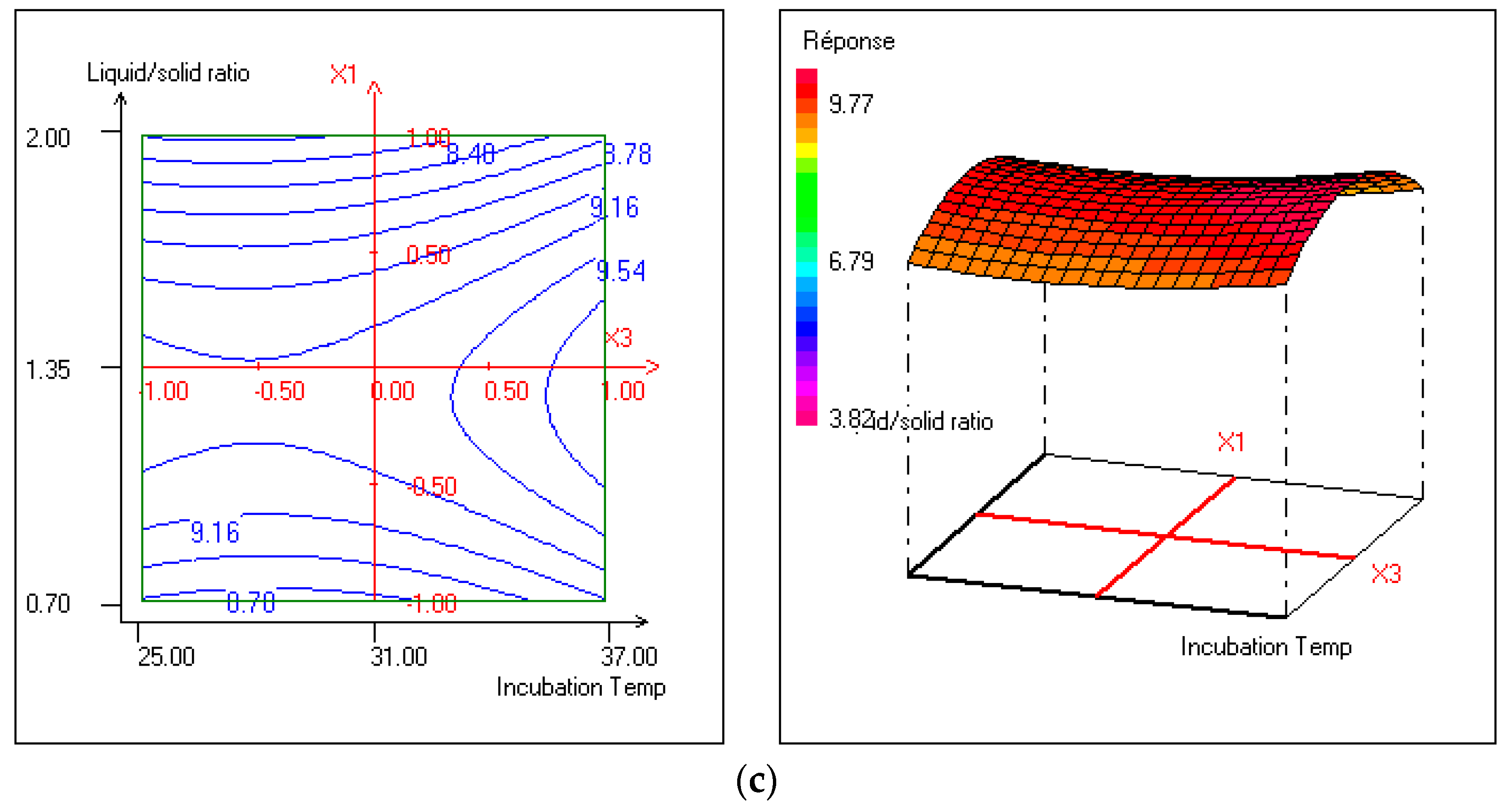
The negative coefficients b1 and b11 showed that biomass would increase with the decrease of the liquid-to-solid ratio under SSF, which means less added liquid to wheat bran enhances the growth of the strain. As shown in Figure 2a,c, the optimal values range of liquid-to-solid ratio is from 1 to 1.35%. This is also valid for the pH buffer parameter, which favors the biomass growth for values between 6 and 6.5. However, temperature positively affects this response (+0.232) and optimal values of the incubation temperature range from 32 to 37 °C.
Regarding the cellulose activity response (Y2), seven parameters were found to be statistically highly significant. So, predicted enzyme activity is given by the following equation:
Y 2 = 0.01098 0.00125   X 2 0.00075   X 3 0.00100   X 1 X 2 0.00100   X 1 X 3 0.00150   X 2 X 3 + 0.00251   X 3 2
According to this equation, cellulose activity would be function of the quadratic effect of temperature (X32), which means that the increased incubation temperature increases the enzyme activity. However, the pH buffer is shown to negatively impact this response (−0.00125) and the liquid-to-solid ratio seems to affect only when interacting with the pH factor.
As shown in Figure 3b, optimal cellulose activity would occur at a temperature range between 25 °C and 37 °C, while optimal pH values (Figure 3a,b) include a 6.12 to 6.3 values range.
According to the contour plots aand b (Figure 3), a higher liquid-to-solid ratio seems to lead to optimal cellulose activity (1.5 to 2%).
This current study aimed to find the maximum of biomass production of Bacillus amyloliquefaciens D1B3 as well as its cellulase activity, which means looking for optimum values of the experimental field’s factors.
Table 7 shows the optimal liquid-to-solid ratio (1.19%), the pH buffer (6.2), and the incubation temperature (36.99 °C). These conditions would lead to maximum response characteristics (Table 8).
The desirability is maximum (100%) for reaching a value of biomass close to 9.828 log CFU/g and a cellulase activity of about 0.0144 g/L.min.

4. Conclusions

Screening of cellulolytic bacteria earlier isolated from the gastrointestinal tract of dromedary using Congo-red agar media was carried out. One isolate D1B3 that presented the largest degradation zone (2.4 cm) was retained. This screened isolate was identified by 16S rDNA sequence analysis as Bacillus amyloliquefaciens D1B3 with 99% similarity. To be functional and to be able to accomplish its benefits, probiotics must tolerate all the conditions that prevail in the gastro-intestinal tract such as low pH and bile salts. In our case, Bacillus amyloliquefaciens D1B3 had attractive probiotic potentials, such as antimicrobial properties; and host-health-improving aspects, including the best tolerance against simulated gastro-intestinal conditions, namely gastric acid tolerance, with a survival rate over 90% and bile salts. In addition, for the first time, we statistically optimized the parameters associated with the high production of biomass and the cellulose enzyme using solid-state fermentation. The optimal parameters were: liquid-to-solid ratio (1.19%), the pH buffer (6.2), and the incubation temperature (36.99 °C) to obtain the highest level of biomass and cellulose enzyme activity reaching a value of 9.828 log CFU/g and 0.0144 g/l.min, respectively. These results suggested the potential utilization of Bacillus amyloliquefaciens D1B3 in animal feeding, via which we can enhance the digestibility of the feed and, at the same time, the probiotic effect. However, “in vivo” experiments are necessary to confirm this strain’s efficiency.

Author Contributions

Conceptualization, T.B.; Methodology, M.E. and A.A.; Software, A.A.; Investigation, M.Z. and M.H.; Resources, A.S. and A.C.; Writing—original draft, T.B. All authors have read and agreed to the published version of the manuscript.

Funding

This research received no external funding.

Institutional Review Board Statement

Not applicable.

Informed Consent Statement

Not applicable.

Data Availability Statement

The data used to support the findings of this study are available from the corresponding author upon request.

Conflicts of Interest

The authors declare no conflict of interest.

References

  1. Markets and markets.com. Feed Enzymes Market by Type (Phytase, Carbohydrase and Protease), Livestock (Poultry Swine, Ruminants, and Aquatic Animals), Source (Microrganism, Plant and Animal), form (Dry and Liquid) and Region-Global Forecast to 2025. 2020. Available online: https://www.marketsandmarkets.com/Market-Reports/feed-enzyme-market-1157.html (accessed on 23 June 2020).
  2. Adeola, O.; Cowieson, A.J. Board-invited review: Opportunities and challenges in using exogenous enzymes to improve nonruminant animal production. J. Anim. Sci. 2011, 89, 3189–3218. [Google Scholar] [CrossRef] [PubMed]
  3. Bimrew, A. Effect of common feed enzymes on nutrient utilization of monogastric animals. Int. J. Biotechnololgy Mol. Biol. Res. 2014, 5, 27–34. [Google Scholar] [CrossRef]
  4. Thapa, S.; Mishra, J.; Arora, N.; Mishra, P.; Li, H.; O’Hair, J.; Bhatti, S.; Zhou, S. Microbial cellulolytic enzymes: Diversity and biotechnology with reference to lignocellulosic biomass degradation. Rev. Environ. Sci. Biotechnol. 2020, 19, 621–648. [Google Scholar] [CrossRef]
  5. Nandy, G.; Chakraborti, M.; Shee, A.; Aditya, G.; Acharya, K. Gut Microbiota from Lower Groups of Animals: An Upcoming Source for Cellulolytic Enzymes with Industrial Potentials Bio-interface. Res. Appl. Chem. 2021, 11, 13614–13637. [Google Scholar]
  6. Budihal, S.R.; Agsar, D.; Patil, S.R. Enhanced production and application of acidothermophilic Streptomyces cellulose. Bioresour. Technol. 2016, 200, 706–712. [Google Scholar] [CrossRef]
  7. Chakraborty, D.; Sarkar, N.; Biswas, I.; Jacob, S. Molecular Aspect of Prokaryote and Eukaryote Cellulase and Thieir Modulation for Potential Application in Biofuel Production Genetic and Metabolic Engineering for Improved Buofuel Production from Lignocellulosic, Biomass; Elsevier: Amsterdam, The Netherlands, 2020; pp. 81–95. [Google Scholar]
  8. Doi, R.H. Cellulases of mesophilic microorganisms: Cellulosome and noncellulosome producers. Ann. N. Y. Acad. Sci. 2008, 1125, 267–279. [Google Scholar] [CrossRef]
  9. Islam, F.; Roy, N. Screening, purification and characterization of cellulase from cellulase producing bacteria in molasses. BMC Res. Notes 2018, 11, 445. [Google Scholar] [CrossRef]
  10. Manan, M.A.; Webb, C. Design aspect of solid state fermentation as applied to Microbial bioprocessing. J. Appl. Biotechnol. Bioeng. 2017, 4, 1–25. [Google Scholar]
  11. Bentil, J.A.; Thygesen, A.; Mensah, M.; Lange, L.; Meyer, A.S. Cellulase production by white-rot basidiomycetous fungi: Solid-state versus submerged cultivation. Appl. Microbiol. Biotechnol. 2018, 102, 5827–5839. [Google Scholar] [CrossRef]
  12. Croos, A.M.B.; Sarathadevi, R.; Kapilan, R. Isolation of cellulose producing Bacillus Cereus from Cow dung and determination of Kinetic properties of the crude enzyme. J. Nat. Sci. Found. Sri Lanka 2019, 47, 261–267. [Google Scholar] [CrossRef]
  13. Li, F.; Xie, Y.; Gao, X.; Shan, M.; Sun, C.; Niu, Y.D.; Shan, A. Screening of cellulose degradation bacteria from Min pigs and optimization of its cellulase production. Electron. J. Biotechnol. 2020, 48, 29–35. [Google Scholar] [CrossRef]
  14. Maravi, P.; Kumar, A. Optimization and statistical modeling of microbial cellulase production using submerged culture. J. Appl. Biol. Biotechnol. 2021, 9, 142–152. [Google Scholar]
  15. Mingmongkolchai, S.; Panbangred, W. Bacillus probiotics: An alternative to antibiotics for livestock production. J. Appl. Microbiol. 2018, 124, 1334–1346. [Google Scholar] [CrossRef]
  16. Simon, M. Cutting Bacillus probiotics. Food Microbiol. 2011, 28, 214–220. [Google Scholar]
  17. Khalid, F.; Khalid, A.; Fu, Y.; Hu, Q.; Zheng, Y.; Khan, S.; Wang, Z. Potential of Bacillus velezensis as a probiotic in animal feed: A review. J. Microbiol. 2021, 59, 627–633. [Google Scholar] [CrossRef]
  18. Li, Y.; Zhang, H.; Chen, Y.P.; Yang, M.X.; Zhang, L.L.; Lu, Z.X.; Zhou, Y.M.; Wang, T. Bacillus amyloliquefaciens supplementation alleviates immunological stress in lipopolysaccharide-challenged broilers at early age. Poult. Sci. 2015, 94, 1504–1511. [Google Scholar] [CrossRef]
  19. Sellami, A. Sélection et Caractérisation de Bactéries du genre Bacillus à Activité Cellulolytique. Master’s Thesis, University of Carthage, Tunis, Tunisia, 2011. [Google Scholar]
  20. Ariffin, H.; Abdullah, N.; UmKalsom, M.S.; Shirai, Y.; Hassan, M.A. Production and characterisation of cellulase by Bacilluspumilus EB3. Int. J. Eng. Technol. 2006, 3, 47–53. [Google Scholar]
  21. Wilson, K. Preparation of genomic DNA from bacteria. Curr. Protoc. Mol. Biol. 1989, 56, 241–245. [Google Scholar] [CrossRef]
  22. Ghose, T.K. Measurement of cellulase activities, Pure and Applied Chemistry. Biochem. Eng. Cent. 1987, 59, 257–268. [Google Scholar]
  23. Tagg, J.R.; Daani, A.S.; Wannamaker, L.W. Bacteriocins of gram-positive bacteria. Bacteriol. Rev. 1976, 40, 722–756. [Google Scholar] [CrossRef]
  24. Dalie, D.K.D.; Deschamps, A.M.; Atanasova-Penichon, V.; Richard-Forget, F. Potential of Pediococcus pentosaceus (L006) Isolated from Maize Leaf to Suppress Fumonisin-Producing Fungal Growth. J. Food Prot. 2010, 73, 1129–1137. [Google Scholar] [CrossRef] [PubMed]
  25. Collado, M.C.; Meriluoto, J.; Salminen, S. Adhesion and aggregation properties of probiotic and pathogen strains. Eur. Food Res. Technol. 2008, 226, 1065–1073. [Google Scholar] [CrossRef]
  26. Guo, Z.; Wang, J.; Yan, L.; Chen, W.; Liu, X.M.; Zhang, H.P. In vitro comparison of probiotic properties of Lactobacillus casei Zhang, a potential new probiotic, with selected probiotic strains. LWT-Food Sci. Technol. 2009, 42, 1640–1646. [Google Scholar] [CrossRef]
  27. Du Toit, M.; Franz, C.M.A.P.; Dicks, L.M.T.; Schillinger, U.; Haberer, P.; Warlies, B.; Ahrens, F.; Holzapfel, W.H. Characterisation and selection of probiotic lactobacilli for a preliminary minipig feeding trial and their effect on serumcholesterol levels, faeces pH and faeces moisture content. Int. J. Food Microbiol. 1998, 1–2, 93–104. [Google Scholar] [CrossRef]
  28. Lee, A.; Cheng, K.C.; Liu, J.R. Isolation and characterization of a Bacillus amyloliquefaciens strain with zearalenone removal ability and its probiotic potential. PLoS ONE 2017, 12, e0182220. [Google Scholar] [CrossRef]
  29. Manhar, A.K.; Saikia, D.; Bashir, Y.; Mech, R.K.; Nath, D.; Konwar, B.K.; Mandal, M. In vitro evaluation of celluloytic Bacillus amyloliquefaciens AMS1 isolated from traditional fermented soybean (Churpi) as an animal probiotic. Res. Vet. Sci. 2015, 99, 149–156. [Google Scholar] [CrossRef]
  30. Chateau, N.; Deschamps, A.M.; Sassi, A.H. Heterogeneity of bile salts resistance in the Lactobacillus isolates of a probiotic consortium. Lett. Appl. Microbiol. 1994, 18, 42–44. [Google Scholar] [CrossRef]
  31. Fontana, L.; Bermudez-Brito, M.; Plaza-Diaz, J.; Muñoz-Quezada, S.; Gil, A. Sources, isolation, characterisation and evaluation of probiotics. Britsh J. Nutr. 2012, 109, S35–S50. [Google Scholar] [CrossRef]
  32. Patel, A.K.; Ahire, J.J.; Pawar, S.P.; Chaudhari, B.L.; Chincholkar, S.B. Comparative accounts of probiotic characteristics of Bacillus spp. Isolated from food wastes. Food Res. Int. 2009, 42, 505–510. [Google Scholar] [CrossRef]
  33. Barbosa, T.M.; Serra, C.R.; La Ragione, R.M.; Woodward, M.J.; Henriques, A.O. Screening for Bacillus Isolates in the Broiler. Appl. Environ. Microbiol. 2005, 71, 968–978. [Google Scholar] [CrossRef]
  34. Rosenberg, M. Microbialadhesion to hydrocarbons: Twenty-five years of doing MATH. FEMS Microbiol. Lett. 2006, 262, 129–134. [Google Scholar] [CrossRef]
  35. Subbiahdoss, G.; Reimhult, E. Biofilm formation at oil-water interfaces is not a simple function of bacterial hydrophobicity. Colloids Surf. Bionterfaces 2020, 194, 1163. [Google Scholar] [CrossRef]
  36. Pelletier, C.; Bouley, C.; Cayuela, C.; Bouttier, S.; Bourlioux, P.; Bellon-Fontaine, M.N. Cell surface characteristics of Lactobacillus casei subsp. casei, Lactobacillus paracasei subsp. paracasei, and Lactobacillus rhamnosus strains. Rumen Appl. Environ. Microbiol. 1997, 63, 1725–1731. [Google Scholar] [CrossRef]
  37. Valeriano, V.D.; Parungao-Balolong, M.M.; Kang, D.-K. In vitro evaluation of the mucin-adhesion ability and probiotic potential of Lactobacillus mucosae LM1. J. Appl. Microbiol. 2014, 117, 485–497. [Google Scholar] [CrossRef]
  38. Furukawa, S.; Nojima, N.; Yoshida, K.; Hirayama, S.; Ogihara, H.; Morinaga, Y. The Importance of Inter-Species Cell-Cell Co-Aggregation between Lactobacillus plantarum ML11-11 and Saccharomyces cerevisiae BY4741 in Mixed-Species Biofilm Formation. Biosci. Biotechnol. Biochem. 2011, 75, 1430–1434. [Google Scholar] [CrossRef]
  39. Hirayama, S.; Nojima, N.; Furukawa, S.; Ogihara, H.; Morinaga, Y. Steric microstructure of mixed-species biofilm formed by interaction between Lactobacillus plantarum ML11-11 and Saccharomyces cerevisiae. Biosci. Biotechnol. Biochem. 2019, 83, 2386–2389. [Google Scholar] [CrossRef]
Figure 1. Clear degradation zone in cellulose Congo-red agar.
Sustainability 15 08394 g001
Figure 2. Contour plots and response surface for biomass (a) effects of pH buffer and liquid-to-solid ratio at fixed temperature, (b) effects of pH buffer and temperature at fixed liquid-to-solid ratio, and (c) effects of liquid-to-solid ratio and temperature at fixed pH buffer. Fixed factors are set at central levels.
Sustainability 15 08394 g002aSustainability 15 08394 g002b
Figure 3. Contour plots and response surface for cellulase activity (a) effects of pH buffer and liquid-to-solid ratio at fixed temperature, (b) effects of pH buffer and temperature at fixed liquid-to-solid ratio, and (c) effects of liquid-to-solid ratio and temperature at fixed pH buffer. Fixed factors are set at central levels.
Sustainability 15 08394 g003aSustainability 15 08394 g003b
Table 1. Diameter of clearing zone of cellulolytic bacteria on cellulose Congo-red agar.
Isolate CodeDiameter of Clear Zone of Hydrolysis (cm)
D1B11.1 ± 0.2
D1B32.4 ± 0.0
D1B42.2 ± 0.1
D1B71.8 ± 0.3
D1B101.1 ± 0.2
D1B121.6 ± 0.0
COL7B21.4 ± 0.4
COL7B52.1 ± 0.0
COL7B61.1 ± 0.0
COL7B72 ± 0.1
COL7B81.1 ± 0.0
COLB111.5 ± 0.2
C3-1B81.9 ± 0.2
Values are presented with standard deviation ± SD.
Table 2. Inhibition zone mm (±standard deviation) of Bacillus amyloliquefaciens D1B3.
Indicator Strains Zone of Inhibition(mm)
Aspergillus spp.24 ± (0.0)
Klebsiella pneumoniae 1003124 ± (2.0)
Escherichia coli 1053624 ± (0.0)
Pseudomonas aeruginosa 902727 ± (1.0)
Streptococcus faecalis 10541-
Staphylococcus aureus 6538-
Staphylococcus epidermidis 12228-
Bacillus cereus 11778-
- indicates no zone of inhibition.
Table 3. Survival rates of B. amyloliquefaciens and L. plantarum 299 under simulated gastric juice. Values are given as mean ± standard deviation. Significant differences are performed by ANOVA test at p < 0.001.
StrainspH 2.5pH 3pH 4
1 hBacillus amyloliquefaciens90.50 ± 0.15 b91.56 ± 0.09 b93.38 ± 0.1 b
Lactobacillus plantarum 29991.58 ± 0.33 a95.14 ± 0.07 a96.72 ± 0.03 a
2 hBacillus amyloliquefaciens81.44 ± 0.15 d87.46 ± 0.1 d89.73 ± 0.06 d
Lactobacillus plantarum 29984.92 ± 0.05 c89.87 ± 0.21 c91.26 ± 0.04 c
3 hBacillus amyloliquefaciens72.64 ± 0.08 f82.03 ± 0.14 f87.06 ± 0.04 e
Lactobacillus plantarum 29981.02 ± 0.06 e84.85 ± 0.07 e89.65 ± 0.02 d
Mean values (±standard deviation) within the same column followed by different superscript letters (a,b,c,d,e,f) differ significantly (p < 0.05).
Table 4. Cell surface hydrophobicity of B. amyloliquefaciens D1B3.
Strain% Hydrophobicity% Coaggregation
D1B31.70 ± 0.269.79 ± 0.1
Values are performed in triplicate and presented as mean ± standard deviation.
Table 5. Central composite design for the production of Bacillus amyloliquefaciens (D1B3): factors with observed responses.
TESTFactorsResponses
X1X2X3Y1Y2
Liquid-to-Solid Ratio (%)pH BufferTemperature (°C)Biomass (logcfu/g)Cellulase Activity (g/L.min)
10.706.0319.000.0113
22.006.0317.640.0126
30.707.0317.740.0106
42.007.0318.300.0092
50.706.5259.000.0133
62.006.5258.340.0160
70.706.5379.000.0134
82.006.5378.500.0116
91.356.0259.220.0141
101.357.0258.570.0138
111.356.0379.770.0164
121.357.0379.720.0102
131.356.53110.680.0100
141.356.5319.540.0113
151.356.5319.470.0106
161.356.5319.240.0108
171.356.5319.440.0109
Table 6. Parameters of the polynomial models representing the studied responses (biomass: Y1; and cellulose activities: Y2).
ModelY1Y2
Model ParametersCoefficientp-ValueCoefficientp-Value
b09.422***0.01098***
b1−0.245*0.00025n.s
b2−0.162n.s−0.00125***
b30.232*−0.00075**
b11−0.931***0.00001n.s
b22−0.321*0.00001n.s
b330.219n.s0.00251***
b120.480**−0.00100**
b130.040n.s−0.00100**
b230.150n.s−0.00150**
Model Validation
Significance level (%)*****
Df66
Sum of squares6.2155.98 × 10−5
Mean square0.69066.64 × 10−6
R20.9450.975
R2A0.8620.938
*** Significant at the level 99.9%. ** Significant at the level 99%. * Significant at the level 95%; n.s: not significant. Df: degrees of freedom.
Table 7. Optimal conditions for Bacillus amyloliquefaciens (D1B3) production under SSF.
VariableValueFactorValue
X1−0.243642Liquid-to-solid ratio1.19
X2−0.606961pH buffer6.20
X30.998078Temperature36.99
Table 8. Predicted values of the responses at optimum conditions.
ResponseNameValued (i) %Weightdi min %di max %
Y1Biomass9.8281001100100
Y2Enzyme activity0.01441001100100
Desirability 100 100100
Disclaimer/Publisher’s Note: The statements, opinions and data contained in all publications are solely those of the individual author(s) and contributor(s) and not of MDPI and/or the editor(s). MDPI and/or the editor(s) disclaim responsibility for any injury to people or property resulting from any ideas, methods, instructions or products referred to in the content.