Article

Aspartic Acid-Based Nano-Copper Induces Resilience in Zea mays to Applied Lead Stress Via Conserving Photosynthetic Pigments and Triggering the Antioxidant Biosystem

1
Department of Botany, University of Peshawar, Peshawar 25100, Khyber Pakhtunkhwa, Pakistan
2
Department of Plant Sciences, Faculty of Biological Sciences, Quaid-i-Azam University Islamabad, Islamabad 45320, Islamabad Capital Territory, Pakistan
3
Department of Botany, Bacha Khan University, Charsadda 24420, Khyber Pakhtunkhwa, Pakistan
4
Department of Mechanical and Aerospace Engineering, University of California, Los Angeles, CA 90095, USA
5
Department of Clinical Pharmacy, College of Pharmacy, King Saud University, Riyadh 11451, Saudi Arabia
*
Authors to whom correspondence should be addressed.
Sustainability 2023, 15(16), 12186; https://doi.org/10.3390/su151612186
Submission received: 6 June 2023 / Revised: 14 July 2023 / Accepted: 25 July 2023 / Published: 9 August 2023

Abstract

:
Heavy metal stress, including lead, adversely affects the growth and yield of several economically important crops, leading to food challenges and significant economic losses. Ameliorating plant responses to various environmental stresses is one of the promising areas of research for sustainable agriculture. In this study, we evaluated the effect of aspartic acid-functionalized copper nanoparticles on the photosynthetic efficiency and antioxidation system of maize plants under Pb toxicity. The ion reduction method was employed for the synthesis of CuNPs, using ascorbic acid as the reducing agent and aspartic acid as the surface functionalizing agent. Isolated experiments under laboratory and field conditions were performed using a randomized complete block design (RCBD). Seeds primed in water, 1.0, 5.0, and 10 µg/mL of Asp-CuNPs were sown under 0, 500, and 1000 mg/L Pb stress in laboratory conditions, while primed seeds along with foliar-applied Asp-CuNP plants were grown in a field under applied Pb stress, and the obtained data were statistically analyzed using TWANOVA. The laboratory experiment shows that Asp-CuNPs act both as a plant growth regulator (PGR) and plant growth inhibitor (PGI), depending upon their concentration, whereby Asp-CuNPs act as a PGR at a concentration of 1 µg/mL ≤ X ≤10 µg/mL. The field experiment confirms that seed priming and foliar spraying with Asp-CuNPs activate embryos and enhance plant growth in a dose-dependent manner. In addition, Asp-CuNPs (10 µg/mL) significantly increase chlorophyll content to 0.87 mg/g from 0.53 mg/g (untreated) when plants were exposed to Pb toxicity at 1000 mg/kg of soil. It is noteworthy that Asp-CuNPs induce resilience to Pb toxicity (1000 mg/kg of soil) in plants by reducing its root absorption from 3.68 mg/kg (0 µg/mL Asp-CuNPs) to 1.72 mg/kg with the application of 10 µg/mL Asp-CuNPs. Additionally, histochemical analyses with NBT and hydrogen peroxide revealed that ROS accretion in plants treated with Asp-CuNPs declined because of the augmentation of antioxidant enzyme (POD, SOD, APOX, etc.) activities under Pb toxicity. Our findings suggest that amino acid-functionalized copper nanoparticles regulate plant defensive mechanisms related to lead tolerance, which is a promising approach for the induction of resistivity to heavy metal stress.

1. Introduction

Nanotechnology is a novel frontier for agriculture with the application of nano-sized fertilizers and plant growth regulators [1,2]. The productivity of crops can be boosted by the development of appropriate growth-promoting nanoparticles [3,4,5]. Early studies suggest that the nanoparticles at an optimum dose may improve plant development, seed germination, nutrient utilization effectiveness, yield, and soil pollution reduction [6,7,8]. By altering the regulation of gene expression, nanoparticles’ interactions with plant cells change the biochemical pathways and promote the growth and development of plants [9,10]. There have been reports on how plant cells can change because of ROS generated by nanoparticles [11,12,13,14]. Plants have the ability to absorb nanoparticles through different routes, including stomata, root hairs, and cracks on the leaf surface [15,16]. Once nanoparticles enter the plant, they can be transported through diffusion, bulk flow, and phloem loading. The transportation of nanoparticles is influenced by various factors, such as their size, shape, surface properties, solution pH, and the presence of other substances [17,18]. Previous research has utilized various techniques, such as leaf spraying, root application, branch injection, and seed treatment, to demonstrate the uptake of nanoparticles by plants [19,20,21]. If the particle sizes of nanomaterials are smaller than the sizes of cell wall pores, they may be able to enter plant cells directly through the structures that resemble sieves in the cell walls [22]. Utilizing nanoparticles as nanofertilizers plays a crucial role in promoting valuable and effective crop production. These nanofertilizers have been found to have significant positive effects on plant growth, crop yield, and plant tolerance, particularly when used in appropriate concentrations [23,24]. The application of various metallic nanoparticles, such as zinc (Zn-NPs), copper (Cu-NPs), and iron (Fe-NPs), has been shown to enhance plant growth and improve the photosynthetic rate and seedling growth [25,26]. Furthermore, studies have demonstrated the beneficial impact of hydroxyapatite nanoparticles on maize plant growth under salt-stress conditions [27]. In another study, the application of silver nanoparticles (AgNPs) resulted in an upregulation of genes related to the IAA, NCED3 (9-cis carotenoid dioxygenase), and RD22 proteins in Arabidopsis thaliana under abiotic stress [28].
Moreover, the application of zinc nanoparticles significantly enhanced the size of the stem and vascular cylinder by increasing their length and width. Furthermore, it resulted in an increase in the thickness of both the cortex and vascular cylinder [29]. Previous studies have reported that ZnO nanoparticles (NPs) can mitigate oxidative damage in different crop plants. In stressed Leucaena leucocephala, ZnO NPs were found to decrease the level of malondialdehyde (MDA), a marker of oxidative stress, while simultaneously enhancing the activities of catalase (CAT) and superoxide dismutase (SOD) [30,31]. The unrestricted progression of industrialization and urbanization during the past decades has given rise to severe environmental problems, among which heavy metals are one of the most prevalent threats to the environment [32]. Human activities in developing agriculture and industry, including the excessive use of agrochemicals, disposal of sludge, melting operations, and the generation of large-scale effluent water, contribute significantly to the global contamination of soil and water with heavy metals, which is a matter of particular concern due to the long-lasting presence of these toxic metals in the environment [33,34,35,36,37,38]. Plants exhibit various symptoms when exposed to heavy metals, including reduced root and shoot lengths, lowered chlorophyll and protein levels, electrolyte leakage, chlorosis, increased production of ROS, MDA accumulation, senescence, imbalances in water and nutrient uptake, and eventual plant death [39,40]. When cells are exposed to heavy metals, several organelles are affected, such as the cell membrane, mitochondria, nucleus, lysosomes, endoplasmic reticulum, enzymes, and metabolites involved in the healing process following injury [41].
Lead is currently among the significant heavy metal pollutants, being toxic for all kinds of life forms, and is of global concern [42]. It is considered one of the most hazardous heavy metals, can maintain high concentrations for up to 150 years, and has a soil retention duration ranging from 150 to 5000 years [43]. Lead not only induces hepatotoxicity, neurotoxicity, and nephrotoxicity in animals, including humans, but also constrains plant growth via inhibiting photosynthesis, water balance, mineral nutrition, generation of ROS, and enzyme activities [44,45]. The application of amino acid biostimulants is a novel frontier in modern agriculture for mitigating toxicity caused by abiotic stresses, including heavy metal stress [46,47]. Amino acids not only act as precursors for the biosynthesis of phytohormones as well as for photosynthetic pigments but also as a signaling factor for the progression of different physiological processes, such as antioxidant metabolism, regulation of soil nitrogen absorption, and root development [48]. Hence, this study was designed with the hypothesis that exogenous application of aspartic acid-functionalized copper nanoparticles can induce resistivity to induced lead stress in Zea mays.

2. Materials and Methods

The research study was conducted at the Department of Botany, University of Peshawar, Pakistan in 2021 to assess the growth performance of Zea mays plants under lead stress with the application of Asp-CuNPs.

2.1. Synthesis and Characterization of Asp-CuNPs

Following the schematic illustration of microwave-assisted synthesis of Asp-CuNPs (Figure 1), briefly, a solution of 1.0 mM L-ascorbic acid was combined with a 0.1 mM aqueous CuSO4 solution. The reaction mixture was then subjected to microwave irradiation for a duration of 5 min. Following the reaction, the resulting suspension of nano-copper (characterized by its red color) was subjected to centrifugation and washing, and subsequently resuspended in dH2O. A capping agent in the form of a 1.0 mM aqueous solution of aspartic acid was added dropwise to the suspension while continuously stirring the mixture on a magnetic stirrer plate for a period of 48 h. The resulting suspension of aspartic acid-functionalized copper nanoparticles (Asp-CuNPs) was centrifuged at 10,000 rpm for 20 m, washed with deionized water, dried in a vacuum drier, and subjected to characterization [49,50].

2.1.1. X-ray Diffraction (XRD) Analysis

The X-ray diffraction (XRD) pattern of Asp-CuNPs was examined using a JEOL JDX 3532 X-ray diffractometer (CRL, University of Peshawar). The diffraction pattern of Asp-CuNPs was documented and recorded by a nickel monochromator sorting the wave at a tube voltage of 40 kV and tube current of 30 mA with Cu-Kα radiation (λ = 1.5406 Å).

2.1.2. Scanning Electron Microscopy (SEM)

At CRL, University of Peshawar, dried Asp-CuNPs were coated on carbon tape, subjected to gold coating using an auto fine coater (Spi-module sputter coater), and examined for morphological features using FE-SEM (JSM-5910-JEOL-JAPAN).

2.1.3. Energy Dispersive X-ray Spectroscopy (EDS)

To determine its elemental makeup, the oven-dried (50 °C) Asp-CuNP pulverized mass was used to conduct EDX analysis using an Oxford Inca 200 SEM instrument equipped with a Thermo EDX attachment (CRL, University of Peshawar).

2.1.4. Thermogravimetric Analysis (TGA)

At CRL, the University of Peshawar, a thermogravimetric/differential thermal analyzer (TG/DTA) Diamond from PerkinElmer (Shelton, CT, USA) was used to test the thermal stability of Asp-CuNPs. An amount of 5.145 g of material was tested using TGA at a rate of 10.0 °C/min in a temperature range between 30 and 600 °C.

2.1.5. Infrared Spectroscopy

To make a sample pellet, Asp-CuNPs were thoroughly dried, coupled with KBr, and placed in a hydraulic pellet press. FT-IR spectroscopy was employed to examine the samples in the range of 4000 to 400 wavenumbers cm−1 using “PerkinElmer Spectrophotometer FT-IR Spectrum One”.

2.2. Plant Material and Growth Conditions

For determining the germination indices, a laboratory experiment was performed on Petri plates in RCBD (Table 1 layout of factorial treatments). Five replicates with 25 seeds were germinated under the application of various doses of Asp-CuNPs (0, 1, 5, and 10 mg/L) and induced lead stress (0, 500, and 1000 mg/L) in 12 cm diameter Petri plates with double folds of Whatman filter paper in an incubator at 25 °C. Germination indices in the form of time to 50% germination (T50), mean time taken to complete germination (MGT), final germination percentage (FGP), and germination index of Zea mays seeds were calculated using the following equations:
T   50 = T a + N t 2 N a T b T a [ N b N a ]
where Nt is the total number of seeds germinated, Na and Nb are the cumulative germinated seed at respective times (day) of Ta and Tb when Na < Nt/2 < Nb.
MGT = [ Ni × Ti ] Nt
where Ni is the number of seeds germinated on the day, (Ti) is the time (days), and Nt is the total number of germinated seeds.
F G P = G   m a x N ]
where G max is the total number of germinated seeds and N is the total number of seeds.
G I = G i D i + + G f D f
where Gi is the initial count of germinated seeds on the initial day (Di) and Gf is the final count of germinated seeds on the final day (Df).
Similarly, for the field experiment, seeds of Zea mays were primed (treated) in 0, 1, 5, and 10 µg/mLof Asp-CuNPs and were sown at the screen house of the Department of Botany, University of Peshawar, Pakistan located at 34.0086 °N, 71.4878 °E. Pots, filled with clay, sand, and farmyard manure at 3:1:1 with ten seeds in each and triplicated in RCBD (Table 1), were subjected to average minimum and maximum monthly temperatures of 18 °C and 35 °C. Pots were also contaminated with lead acetate at a dose of 0, 500, and 1000 mg/kg of soil. After the completion of emergence, seedlings/plants were treated via the foliar application of 0, 1, 5, and 10 mg/L of Asp-CuNPs, and an experiment was performed using RCB design by following the given layout.
Harvested plants were initially subjected to data collection for vegetative growth parameters (shoot length, average root length, number of leaves, and fresh and dry biomass). Fresh plant specimens were also subjected to biochemical analysis as described below.

2.2.1. Determination of Photosynthetic Pigment Contents

Photosynthetic pigments (chlorophyll (Chl) a and b and carotenoid) were determined following the procedure of Lichtenthaler [34]. Leaf samples (500 mg) were homogenized in 10 mL of 80% (v/v) pre-chilled acetone with the assistance of a magnetic stirrer at room temperature (22 ± 3 °C) for 15 min. The homogenates were centrifuged at 6000 rpm for 15 min. The supernatant was collected, and the residue was extracted again following the same procedure. The collected supernatants were combined and tested for the optical densities (OD) at 420, 645, and 663 nm for quantification of total carotenoids and Chl a and b, respectively, using the following Equations (5)–(7):
C h l   a m g / g = 12.25 × O D   a t   663     2.79 × O D   a t   645 × V 1000 × L W
C h l   b m g / g = 21.5 × O D   a t   645     5.1 × O D   a t   663 × V 1000 × L W
C a r o t e n o i d m g / g = 1000 × O D   a t   663     2.79 × O D   a t   645 × V 1000 × L W

2.2.2. Leaf Protein Contents

For determining protein content, fresh leaves (500 mg) were homogenized in phosphate buffer (pH 7.5) and centrifuged at 5000 rpm. Na2CO3 (alkaline), Na-K tartrate (aqu.), and CuSO4·5H2O (aqu.) were added to the supernatant (0.1 mL). An amount of 0.1 mL of Folin phenol was added to the reaction mixture on a magnetic stirrer, incubated for 30 min, and subsequently the OD of the reaction mixture was measured at 650 nm. Protein concentration was assessed in relation to a standard curve of bovine serum albumen [51,52].

2.2.3. Estimation of Total Soluble Sugar

Fresh leaves (500 mg) were homogenized in distilled water and centrifuged at 5000 rpm for 5 min. An amount of 1 mL of 30% (w/v) phenol was added to the reaction mixture followed by incubation at room temperature. Conc. H2SO4 was added to this sample, and after 4 h of incubation, the OD was recorded at 420 nm [53,54]. The sugar concentration was determined against the standard curve of glucose.

2.2.4. Proline Contents of Leaves

The homogenates of leaves (500 mg) were treated for 1 h in a water bath with 3% aqueous sulphosilicyclic acid and acid ninhydrin (100 °C). The reaction was terminated in an ice bath, and 4 mL of toluene was used to extract the mixture. Toluene-containing chromophore was evacuated from the aqueous phase and warmed to room temperature, and the optical density (OD) against toluene as a blank was recorded at 520 nm [55]. The standard curve was used to calculate the proline concentration.

2.2.5. Extraction of Antioxidant Enzymes

Fresh leaves (500 mg) were homogenized in 15 mL of 0.05 N phosphate buffer (pH 7.0) containing PVPP and 0.1 M EDTA. The homogenate was centrifuged at 10,000 rpm for 15 min and the supernatant was used for SOD EC 1.15.1.1, CAT EC 1.11.1.6, and POD EC1.11.1.X, and APX EC1.11.1.11 activities.
To measure SOD activity, the reaction mixture (3 mL) comprised of 13 mM methionine, 0.075 mM NBT, 0.1 mM EDTA, 0.002 M riboflavin, and 0.1 mL of enzyme extract in 50 mM phosphate buffer (pH 7.8) in the tube was placed below a light chamber for 15 min and the OD was read at 560 nm. For POD activity, a mixture containing 0.1 mL of enzyme extract, 1.35 mL of 100 mM MES buffer (pH 5.5), 0.05% H2O2, and 0.1 mL of phenylene diamine was evaluated for change in absorbance at 485 nm for 3 min. The POD activity was presented as delta OD 485 nm/min mg protein [56]. To determine the activity of APX, a reaction mixture containing enzyme extract, 0.05 M potassium phosphate buffer (pH 7), 0.5 mM ascorbate, 0.1 mM H2O2, and 0.1 mM EDTA was tested for the decline in OD value at 290 nm for 3 min [57]. For CAT activity, 50 mM K3PO4 buffer (pH 7.0), 2 mL of the diluted mixture, and 1 mL of 10 mM H2O2 were added to the enzyme extract. The resulting sample was tested for the decrease in optical density at 240 nm, indicating hydrogen peroxide decomposition [58,59].

2.2.6. Determination of Phenol Content

The total phenolic content was estimated using Folin phenol reagent and gallic acid as a standard. Leaves were homogenized in dH2O, to which 0.5 mL of diluted Folin phenol reagent was added, and the mixture was incubated for 5 min. Finally, 1 mL of Na2CO3 (7.5% W/V) was added to this reaction mixture, and after incubation for 2 h, the OD was recorded at 576 nm [60].

2.2.7. Determination of Pb Concentration

A wet digestion method was employed for the extraction of Pb from the plant sample [61]. Leaves (1 g) were digested with nitric acid in a conical flask at 150 °C. The concentration of Pb was determined by subjecting the resulting sample to atomic absorption spectroscopy (AAS 700, Perkin Emler, Waltham, MA, USA).

2.3. Statistics

The data were analyzed using two-factor factorial ANOVA using Mstate C followed by an LSD post hoc test; p ≤ 0.05 were considered statistically significant responses. The results were presented as 95% CL (as standard error bars) of the mean. The graphs for characterization as well as for biochemical parameters were plotted in Origin Pro 9.1.

3. Results

3.1. Characterization of Asp-CuNPs

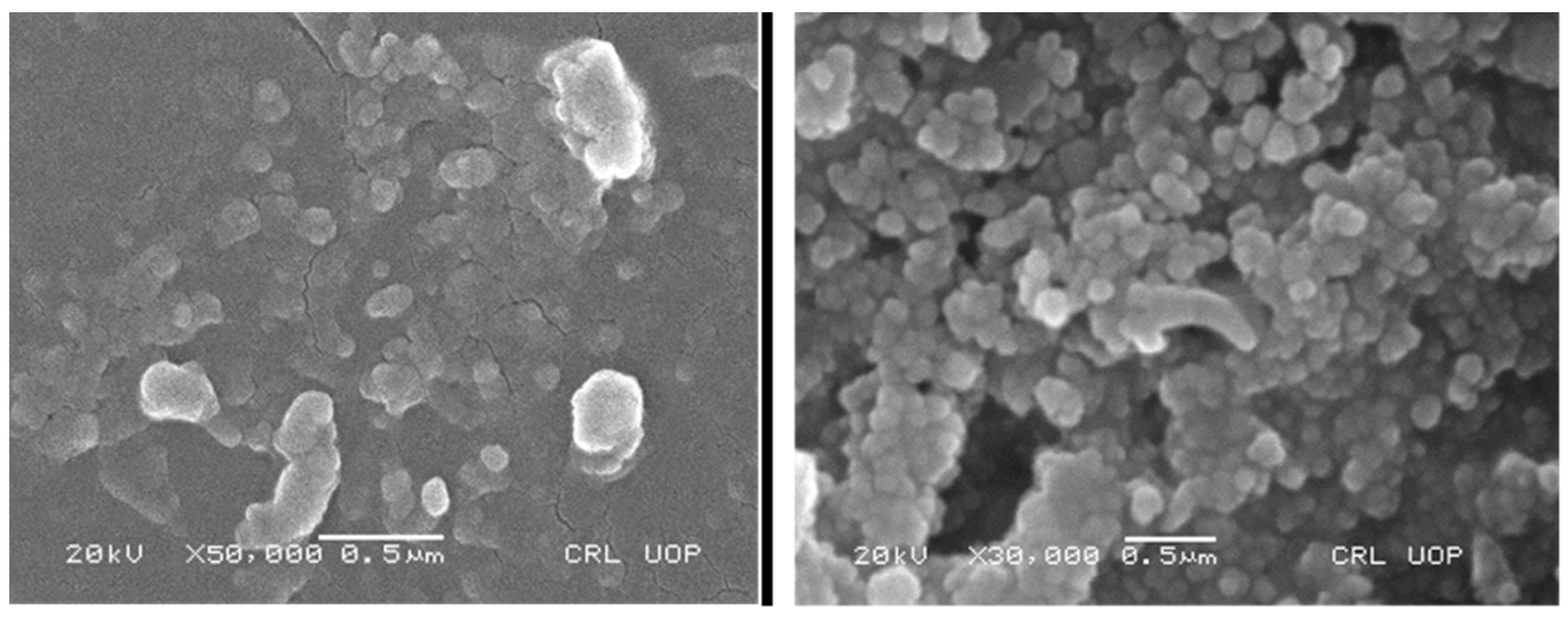
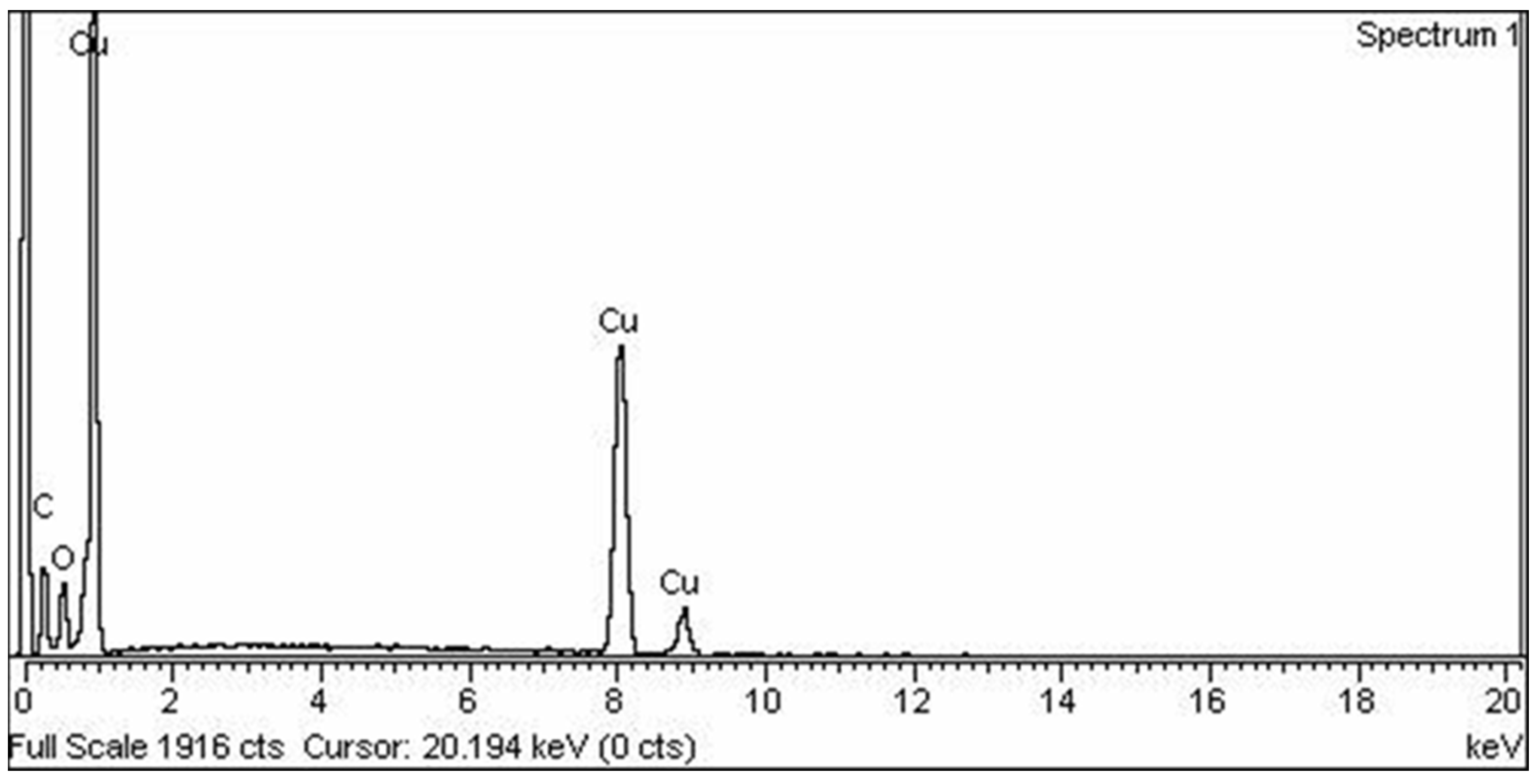
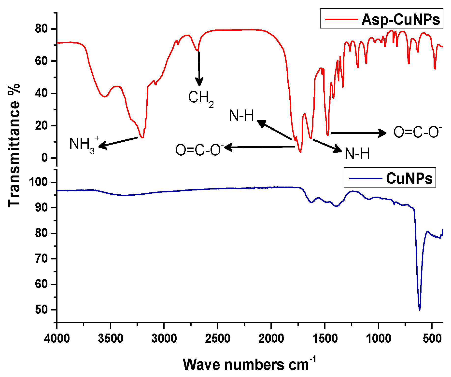
Images obtained using scanning electron microscopy (SEM) showed spherical morphology with particulate sizes ranging from 46 to 120 nm, with a mean particulate size of 76.78 nm (Figure 2). EDS was used to validate the elemental mapping of Asp-CuNPs. Strong signals of elemental copper at 1.8 and 8.8 KeV suggest that copper ions have been reduced in the aqueous solution by bio-reducing agents (ascorbic acid). The capped amino acid is represented by signals for carbon, oxygen, and nitrogen (Figure 3). Thermogravimetric analysis (TGA) was employed to investigate the thermal stability of Asp-CuNPs over a temperature range spanning from 25 °C to 400 °C. The TGA graph exhibited an initial decrease in the rate of mass change around 100 °C, which corresponded to the evaporation of moisture (desolvation) from the sample. A subsequent decrease in mass slope was observed over the temperature range from 180 °C to 230 °C, attributed to the decomposition of the capping agent surrounding the central metal core. Notably, a significant and abrupt increase in the mass slope was observed beyond 250 °C, indicating the oxidation of the copper metal (Figure 4). The XRD crystallograph (Figure 5) authenticated the crystalline nature of ASP-CuNPs where copper was reported to be present in two different oxidizing states (Cu0 and Cu2O). The characteristic diffraction intensities around 2Ɵ; 38, 42, and 64 represent the lattice of oxidized copper (Cu2O), whereas diffraction intensities around 2Ɵ; 44, 48, and 73 represent the FCC lattice geometry of metallic copper (Cu0). FT-IR analysis was carried out for validating the chemical conformation of Asp-CuNPs through functional group profiling. The FT-IR spectrum detected the presence of N-H, C=O, C-O, and C-H bonding in aspartic acid-capped copper nanoparticles characteristic of aspartic acid. The N-H, C=O, and C-H functional groups absorb infrared radiations at wavenumbers 3202.44 cm−1, 1727.45 cm−1, and 2688.88 cm−1, respectively. Figure 6 shows the presence of characteristic vibrational stretches in the IR spectrum of Asp-CuNPs for amino and carboxyl functional groups, which were not detected in non-capped CuNPs validating the surface functionalization of CuNPs with aspartic acid.

3.1.1. Germination Indices and Seedling Growth

The effect of Asp-CuNPs on germination indices (T50, MGT, FGP, and GI) of Zea mays under induced lead application was found to be statistically significant (p < 0.05). T50 values (days) were significantly increased under Pb application in a dose-dependent manner (5.33 days at 1000 mg/kg of soil Pb application). With the application of Asp-CuNPs, the number of days to 50 percent germination (T50) significantly declined, i.e., 2.26 days at 10 µg/mL Asp-CuNPs (Table 2). Moreover, aspartic acid-capped copper nanoparticles (Asp-CuNPs) in conjunction with lead stress (T11–T12), resulted in a significantly reduced time to 50% germination (T50), 3.49 and 3.74, respectively, suggesting the effective mitigation of Pb toxicity via engineered amino acid-based nano-PGR. Similarly, Pb stress prolonged the mean germination time (MGT), with the highest MGT of 7.28 days observed with the 1000 mg/L Pb treatment. Asp-CuNPs restored the prolonged MGT values, and the shortest MGT of 3.97 days was observed at T10 (10 µg/mL Asp-CuNPs) under 1000 mg/L Pb stress. Similarly, the maximum final germination percentage (100%) was observed at T10 (10 µg/mL Asp-CuNPs) followed by 96% at T7. FGP was significantly inhibited by lead toxicity (56%) at its highest experimental dose (1000 mg/L), which was ameliorated to 96% via seed priming with 5 µg/mL of Asp-CuNP suspension. The germination index (GI) varied significantly among the treatments, with the highest GI of 36.74% at T7 and 33.57% at T10. The lowest GI of 19.29% was observed at T3 (1000 mg/L Pb), followed by 21.38% at T1. However, Pb and Asp-CuNPs had no significant impact on radial growth in maize seedlings. The longest radial length of 5.26 cm was observed at T4 (1 µg/mL of Asp-CuNPs), while the shortest length of 3.44 cm was recorded at T3 (1000 mg/L Pb). Pre-sowing treatment of seeds with Asp-CuNPs significantly influenced plumule length, with the highest length of 6.74 cm at T10 (10 µg/mL Asp-CuNPs) under no Pb stress. These findings suggest that the application of engineered Asp-CuNPs potentially mitigates Pb toxicity and restores the germination indices and seedling growth.

3.1.2. Response of Zea mays under Field Conditions

Significant (p < 0.05) growth inhibition was observed for the shoot, root, and leaf lengths in plants grown under 1000 mg/kg soil Pb compared to plants grown under a control Pb level (T1, T4, T7, and T10). Priming with Asp-CuNPs and their foliar application significantly promotes vegetative growth by ameliorating plant resistivity to induced Pb toxicity. The results displayed in Table 3 show that high Pb soil levels (T2 and T3) induced stunted growth by inhibiting both shoot and root growth, while treating the plant with various concentrations of Asp-CuNPs (T4–T12) significantly improved the vegetative growth of Zea mays even under high lead toxicity levels. The synergistic effect of pre-sowing and foliar application of Asp-CuNPs significantly increased shoot and root length, with the response in the order of treatment doses being 0 µg/mL < 1 µg/mL < 5 µg/mL > 10 µg/mL of Asp-CuNPs. Similarly, leaf length was also improved via the application of Asp-CuNPs, and hence a maximum leaf area of 33.18 cm was recorded in plants treated with 5 µg/mL Asp-CuNPs followed by plants treated with 1 µg/mL Asp-CuNPs (31.38 cm) and 5 µg/mL Asp-CuNPs with 500 mg/L Pb (30.08 cm), respectively. A significant restoration of plant fresh (5.36 g and 5.06 g at T4 and T7) and dry biomass (0.78 g and 0.73 g at T4 and T7) was achieved under the synergistic effect of pre-sowing treatment and exogenous foliar application of Asp-CuNP at the experimental doses of 1.0 µg/mL and 5.0 µg/mL, which were significantly reduced to 4.18 g and 0.69 g, respectively, due to Pb toxicity (Table 3).

3.1.3. Biochemical Response of Zea mays

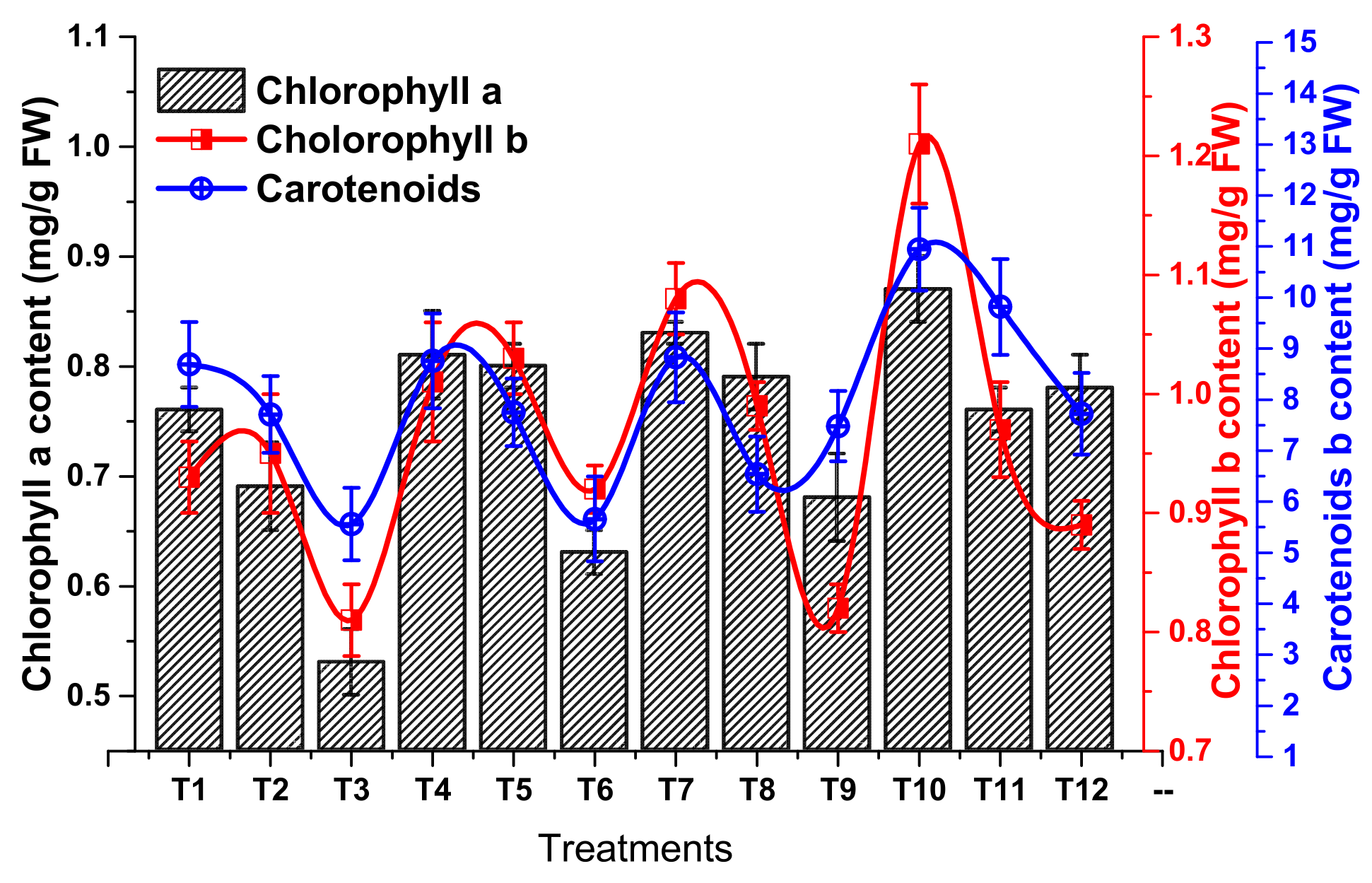
Significant declines in the amount of photosynthetic pigments were reported in plants exposed to induced Pb stress (Chlorophyll a, b, and carotenoids). Both chlorophyll a and b content revealed dynamic changes with soil Pb levels. Maximum chlorophyll a (0.87 mg/g) and chlorophyll b (1.24 mg/kg) contents were noted in plants treated exogenously with 10 µg/mL of Asp-CuNPs. Soil Pb levels of 1000 mg/kg had significantly declined levels of chlorophyll a (0.53 mg/g) and chlorophyll b (0.80 mg/g) compared to the control condition (Figure 7). A similar trend of a drop in carotenoid content was also observed due to Pb toxicity, which was significantly restored via the application of Asp-CuNPs (Figure 7).

3.1.4. Response of the Oxidation/Antioxidation System of Zea mays

Leaf protein, proline, and sugar contents were positively affected by Asp-CuNPs and negatively correlated to soil Pb level (Figure 8). Interestingly, protein, proline, and sugar contents were found to be higher in response to 5.0 µg/mL of Asp-CuNPs (T7) than 10.0 µg/mL of Asp-CuNPs (T10). Moreover, the activity of antioxidant enzymes, i.e., POD and SOD, was observed to be increased under high Pb toxicity levels from 30.11 and 36.73 IU/g FW in the control group to 51.87 and 58.33 IU/g FW under 1000 mg/kg Pb application, respectively. Both POD and SOD activities were restored with the application of 10.0 µg/mL of Asp-CuNPs, to 38.14 and 40.22 IU/g FW, respectively (Figure 9). Similarly, the activities of CAT and APX were also significantly elevated to 7.42 and 6.89 IU/g FW under a higher soil Pb level (1000 mg/kg) compared to the control soil Pb level. The foliar application of Asp-CuNPs at a dose of 10 µg/mL significantly restored the activity of both CAT and APOX to 5.25 and 5.18 IU/g FW under higher soil Pb levels via countering the oxidative stress induced by Pb toxicity (Figure 10). The total phenol content in plants grown under 500 and 1000 mg/kg soil Pb levels was significantly raised to 1.38 mg/g DW and 1.53 mg/g DW, respectively, compared to the plants grown under the T1 condition (control Pb level). Phenolic content was relatively higher in plants treated with 1.0 µg/mL of Asp-CuNPs than 10 µg/mL, reflecting that a higher dose of Asp-CuNPs is more potent in stabilizing plant phenolic content under induced Pb stress (Figure 11). Likewise, Figure 11 indicates a significant drop in plant Pb level to 0.94 mg/g DW when treated with 10 µg/mL of Asp-CuNPs (T12) compared to 3.68 mg/g DW under 0 µg/mL Asp-CuNP application (T3). Generally, pre-sowing treatment and foliar application of aspartic acid-capped copper nanoparticles leads to a substantial decline in plant Pb level compared to the non-treated plants.

4. Discussion

An earlier study suggests that amino acids act as activators that bind to the enhancer of genes involved in cytokinin homeostasis in plants, including Zea mays, which leads to the up-regulation of cytokinin production. It is well known that plant growth hormone-like cytokinin is important in embryo activation and stimulating meristematic cells [62]. Our results demonstrate that priming seeds with amino acid-capped CuNPs at an experimental dose of 5 µg/mL acts as a more potent growth regulator for the early onset of germination and optimizing germination indices than 10 µg/mL under Pb toxicity. Similarly, our results revealed a prompt decline in photosynthetic pigments, i.e., chlorophyll a, b, and carotenoids, under the influence of Pb stress. Previous studies suggested that plant lead exposure shows lessening in photosynthetic activity due to CO2 deficiency resulting from stomatal closure, restrained chlorophyll and carotenoid synthesis, deformation of the chloroplast ultra-structure, obstruction of the thylakoid electron transport system, and impairment with enzymes associated with the Calvin cycle [63]. Pb causes alterations in glycolipids, specifically affecting monogalactosyldiacylglycerol, which is closely linked to changes in chloroplast membrane permeability [60]. Heavy metals, including Pb, deactivate enzymes by binding to cysteine residues. They also induce lipid peroxidation, resulting in the downregulation of peroxidases and damage to thylakoid membranes [64]. Pb also constraints the synthesis of chlorophyll by competing with the uptake of essential nutrients like Mg and Fe, and enhancing chlorophyllase activity. It also inhibits the photosynthetic apparatus through its strong binding tendencies for protein N and S ligands [65]. Figure 7 and Figure 8 show that Asp-CuNPs reversed the decline in photosynthetic pigments in plants due to Pb toxicity.
Our findings agree with those of the authors of [66], who reported enhanced photosynthetic activity in soybean under the application of amino acids. Moreover, the activity of antioxidant enzymes, i.e., POD, SOD, CAT, and APX, was increased to 36.73 IU/g FW, 58.33 IU/g FW, 7.42 IU/g FW, and 6.89 IU/g FW respectively under 1000 mg/kg lead stress (Figure 8 and Figure 9). This increase is the result of the overproduction of ROS in response to high Pb concentrations to protect the plant from oxidative damage produced by heavy metal stress [67]. The peroxidase family’s key genes play a crucial role in the plant cell’s enzyme defense system, actively scavenging reactive oxygen species (ROS) during stress [68]. Lead toxicity enhances the production of ROS, including H2O2, which subsequently triggers secondary oxidative stress, leading to inhibition during the early stages of growth [69]. The optimization of antioxidant enzymes with the application of Asp-CuNPs may be attributed to the antioxidant nature of amino acids, including aspartic acid, as well as the production of other antioxidants like alpha-tocopherol, glutathione, phenolic compounds, ascorbic acid, etc. Further, CuNPs also impair the absorption of Pb by the roots in Zea mays, hence coping with the ROS production in plant tissues.

5. Conclusions

Ascorbic acid acts as a reducing agent for the reduction in transition metal ions (Cu2+) providing bases for core metal nanoparticles. Amino acid (aspartic acid) has the tendency for capping the metal surface and hence acts as a stabilizing as well as a surface functionalizing agent. Pb significantly reduced the germination rate and germination indices as well as the vegetative growth and biochemical growth parameters in Zea mays. Asp-CuNPs act as PGR at relatively low doses, as they activate embryos, reduce the time for germination, and enhance seedling as well as plant growth. Asp-CuNPs at a higher dose (10 µg/mL) were least effective, revealing relatively low values of root, shoot, and leaf lengths, plant fresh and dry biomasses, etc. Asp-CuNPs enhance plant resistivity to heavy metal toxicity by increasing chlorophyll and carotenoid contents and optimizing the activity of antioxidant enzymes.

Author Contributions

R.U. generated the idea. Experimental facilities and supervision were provided by R.U. and Z.U. The experimental study was performed by R.U. and Z.U.; R.U. and J.I. wrote the manuscript. R.U., Z.U., J.I. and W.C., helped with software, manuscript editing, and revision. A.A. reviewed and edited the manuscript and provided funds. All authors have read and agreed to the published version of the manuscript.

Funding

The authors would like to extend their sincere appreciation to the Researchers Supporting Project Number (RSP2023R350), King Saud University, Riyadh, Saudi Arabia.

Institutional Review Board Statement

Not applicable.

Informed Consent Statement

Not applicable.

Data Availability Statement

All the raw data of this study can be obtained from the corresponding authors upon reasonable request.

Acknowledgments

The authors would like to extend their sincere appreciation to the Researchers Supporting Project Number (RSP2023R350), King Saud University, Riyadh, Saudi Arabia.

Conflicts of Interest

The authors declare no conflict of interest.

References

  1. Khan, S.; Yu, H.; Li, Q.; Gao, Y.; Sallam, B.N.; Wang, H.; Liu, P.; Jiang, W. Exogenous Application of Amino Acids Improves the Growth and Yield of Lettuce by Enhancing Photosynthetic Assimilation and Nutrient Availability. Agronomy 2019, 9, 266. [Google Scholar] [CrossRef] [Green Version]
  2. Ullah, Z.; Gul, F.; Iqbal, J.; Abbasi, B.A.; Kanwal, S.; Chalgham, W.; El-Sheikh, M.A.; Diltemiz, S.E.; Mahmood, T. Biogenic Synthesis of Multifunctional Silver Oxide Nanoparticles (Ag2ONPs) Using Parieteria alsinaefolia Delile Aqueous Extract and Assessment of Their Diverse Biological Applications. Microorganisms 2023, 11, 1069. [Google Scholar] [CrossRef]
  3. Kowalczyk, K.; Zielony, T.; Gajewski, M. Effect of Aminoplant and Asahi on yield and quality of lettuce grown on rockwool. Conf. Biostimulators Mod. Agric. 2008, 2, 7–8. [Google Scholar]
  4. Sharma, P.; Dubey, R.S. Lead toxicity in plants. Braz. J. Plant Physiol. 2005, 17, 35–52. [Google Scholar] [CrossRef] [Green Version]
  5. Abbasi, B.A.; Iqbal, J.; Mahmood, T.; Qyyum, A.; Kanwal, S. Biofabrication of iron oxide nanoparticles by leaf extract of Rhamnus virgata: Characterization and evaluation of cytotoxic, antimicrobial and antioxidant potentials. Appl. Organomet. Chem. 2019, 33, e4947. [Google Scholar] [CrossRef]
  6. Sasidharan, D.; Namitha, T.; Johnson, S.P.; Jose, V.; Mathew, P. Synthesis of silver and copper oxide nanoparticles using Myristica fragrans fruit extract: Antimicrobial and catalytic applications. Sustain. Chem. Pharm. 2020, 16, 100255. [Google Scholar] [CrossRef]
  7. Hernandez-Viezcas, J.A.; Castillo-Michel, H.; Servin, A.D.; Peralta-Videa, J.R.; Gardea-Torresdey, J.L. Spectroscopic verification of zinc absorption and distribution in the desert plant Prosopis juliflora-velutina [Velvet mesquite] treated with ZnO nanoparticles. Chem. Eng. J. 2011, 170, 346–352. [Google Scholar] [CrossRef] [Green Version]
  8. Jadoun, S.; Arif, R.; Jangid, N.K.; Meena, R.K. Green synthesis of nanoparticles using plant extracts: A review. Environ. Chem. Lett. 2021, 19, 355–374. [Google Scholar] [CrossRef]
  9. Mittal, D.; Kaur, G.; Singh, P.; Yadav, K.; Ali, S.A. Nanoparticle-based sustainable agriculture and food science: Recent advances and future outlook. Front. Nanotechnol. 2020, 2, 579954. [Google Scholar] [CrossRef]
  10. Gao, M.; Chang, J.; Wang, Z.; Zhang, H.; Wang, T. Advances in transport and toxicity of nanoparticles in plants. J. Nanobiotechnol. 2023, 21, 75. [Google Scholar] [CrossRef]
  11. Ajmal, M.; Ullah, R.; Muhammad, Z.; Khan, M.N.; Kakar, H.A.; Kaplan, A.; Okla, M.K.; Saleh, I.A.; Kamal, A.; Abdullah, A.; et al. Kinetin Capped Zinc Oxide Nanoparticles Improve Plant Growth and Ameliorate Resistivity to Polyethylene Glycol (PEG)-Induced Drought Stress in Vigna radiata (L.) R. Wilczek (Mung bean). Molecules 2023, 28, 5059. [Google Scholar] [CrossRef]
  12. Hu, J.; Guo, H.; Li, J.; Gan, Q.; Wang, Y.; Xing, B. Comparative impacts of iron oxide nanoparticles and ferric ions on the growth of Citrus maxima. Environ. Pollut. 2017, 221, 199–208. [Google Scholar] [CrossRef] [Green Version]
  13. Fatima, F.; Hashim, A.; Anees, S. Efficacy of nanoparticles as nanofertilizer production: A review. Environ. Sci. Pollut. Res. 2021, 28, 1292–1303. [Google Scholar] [CrossRef]
  14. Ul Haq, T.; Ullah, R.; Khan, M.N.; Nazish, M.; Almutairi, S.M.; Rasheed, R.A. Seed Priming with Glutamic-Acid-Functionalized Iron Nanoparticles Modulating Response of Vigna radiata (L.) R. Wilczek (Mung bean) to Induce Osmotic Stress. Micromachines 2023, 14, 736. [Google Scholar] [CrossRef]
  15. Alabdallah, N.M.; Hasan, M.M.; Hammami, I.; Alghamdi, A.I.; Alshehri, D.; Alatawi, H.A. Green Synthesized Metal Oxide Nanoparticles Mediate Growth Regulation and Physiology of Crop Plants under Drought Stress. Plants 2021, 10, 1730. [Google Scholar] [CrossRef]
  16. Rasheed, A.; Li, H.; Tahir, M.M.; Mahmood, A.; Nawaz, M.; Shah, A.N.; Aslam, M.T.; Negm, S.; Moustafa, M.; Hassan, M.U.; et al. The Role of Nanoparticles in Plant Biochemical, Physiological, and Molecular Responses under Drought Stress: A Review. Front. Plant Sci. 2022, 13, 976179. [Google Scholar] [CrossRef]
  17. Faryal, S.; Ullah, R.; Khan, M.N.; Ali, B.; Hafeez, A.; Jaremko, M.; Qureshi, K.A. Thiourea-Capped Nanoapatites Amplify Osmotic Stress Tolerance in Zea mays L. by Conserving Photosynthetic Pigments, Osmolytes Biosynthesis and Antioxidant Biosystems. Molecules 2022, 27, 5744. [Google Scholar] [CrossRef]
  18. Pérez-Labrada, F.; Hernández-Hernández, H.; López-Pérez, M.C.; González-Morales, S.; Benavides-Mendoza, A.; Juárez-Maldonado, A. Nanoparticles in Plants: Morphophysiological, Biochemical, and Molecular Responses. In Plant Life under Changing Environment; Elsevier: Amsterdam, The Netherlands, 2020; pp. 289–322. [Google Scholar] [CrossRef]
  19. Semida, W.M.; Abdelkhalik, A.; Mohamed, G.F.; Abd El-Mageed, T.A.; Abd El-Mageed, S.A.; Rady, M.M.; Ali, E.F. Foliar Application of Zinc Oxide Nanoparticles Promotes Drought Stress Tolerance in Eggplant (Solanum melongena L.). Plants 2021, 10, 421. [Google Scholar] [CrossRef]
  20. Dimkpa, C.O.; Andrews, J.; Fugice, J.; Singh, U.; Bindraban, P.S.; Elmer, W.H.; Gardea-Torresdey, J.L.; White, J.C. Facile Coating of Urea with Low-Dose ZnO Nanoparticles Promotes Wheat Performance and Enhances Zn Uptake Under Drought Stress. Front. Plant Sci. 2020, 11, 168. [Google Scholar] [CrossRef] [Green Version]
  21. Zahedi, S.M.; Karimi, M.; Da Silva, J.A.T. The use of nanotechnology to increase quality and yield of fruit crops. J. Sci. Food Agric. 2020, 100, 25–31. [Google Scholar] [CrossRef]
  22. Kim, S.; Lee, S.; Lee, I. Alteration of phytotoxicity and oxidant stress potential by metal oxide nanoparticles in Cucumis sativus. Water Air Soil Pollut 2012, 223, 2799–2806. [Google Scholar] [CrossRef]
  23. Kohli, S.K.; Khanna, K.; Bhardwaj, R.; Abda_Allaha, E.F.; Ahmad, P.; Corpas, F.J. Assessment of subcellular ROS and NO metabolism in higher plants: Multifunctional signalling molecules. Antioxidants 2019, 8, 641. [Google Scholar] [CrossRef] [Green Version]
  24. Mansoor, S.; Wani, O.A.; Lone, J.F.; Manhas, S.; Kour, N.; Alam, P.; Ahmad, A.; Ahmad, P. Reactive oxygen species in plants: From source to sink. Antioxidants 2022, 11, 225. [Google Scholar] [CrossRef]
  25. Kaya, C.; Ashraf, M.; Alyemeni, M.N.; Ahmad, P. Responses of nitric oxide and hydrogen sulfide in regulating oxidative defence system in wheat plants grown under cadmium stress. Physiol. Plant. 2020, 168, 345–360. [Google Scholar] [CrossRef]
  26. Kaya, C.; Okant, M.; Ugurlar, F.; Alyemeni, M.N.; Ashraf, M.; Ahmad, P. Melatonin-mediated nitric oxide improves tolerance to cadmium toxicity by reducing oxidative stress in wheat plants. Chemosphere 2019, 225, 627–638. [Google Scholar] [CrossRef]
  27. Muhammad, L.; Khan, A.; Zhou, Y.; He, M.; Alrefaei, A.F.; Khan, M.; Ali, S. Physiological and ultrastructural changes in Dendranthema morifolium cultivars exposed to different cadmium stress conditions. Agriculture 2023, 13, 317. [Google Scholar] [CrossRef]
  28. Niyoifasha, C.J.; Borena, B.M.; Ukob, I.T.; Minh, P.N.; Al Azzawi, T.N.I.; Imran, M.; Ali, S.; Inthavong, A.; Mun, B.-G.; Lee, I.-J.; et al. Alleviation of Hg-, Cr-, Cu-, and Zn-Induced Heavy Metals Stress by Exogenous Sodium Nitroprusside in Rice Plants. Plants 2023, 12, 1299. [Google Scholar] [CrossRef]
  29. Mortezagholi, B.; Movahed, E.; Fathi, A.; Soleimani, M.; Mirhosseini, A.F.; Zeini, N.; Khatami, M.; Naderifar, M.; Kiasari, B.A.; Zareanshahraki, M. Plant-mediated synthesis of silver-doped zinc oxide nanoparticles and evaluation of their antimicrobial activity against bacteria cause tooth decay. Microsc. Res. Tech. 2022, 85, 3553–3564. [Google Scholar] [CrossRef]
  30. Krishnaraj, C.; Jagan, E.G.; Ramachandran, R.; Abirami, S.M.; Mohan, N.; Kalaichelvan, P.T. Effect of biologically synthesized silver nanoparticles on Bacopa monnieri [Linn.] Wettst. Plant growth metabolism. Proc. Biochem. 2012, 47, 651–658. [Google Scholar] [CrossRef]
  31. Liu, R.; Lal, R. Potentials of engineered nanoparticles as fertilizers for increasing agronomic productions. A review. Sci. Total Environ. 2015, 514, 131–139. [Google Scholar] [CrossRef]
  32. Mousavi Kouhi, S.M.; Lahouti, M.; Ganjeali, A.; Entezari, M.H. Comparative effects of ZnO nanoparticles, ZnO bulk particles, and Zn2+ on Brassica napus after long-term exposure: Changes in growth, biochemical compounds, antioxidant enzyme activities, and Zn bioaccumulation. Water Air Soil Pollut 2015, 226, 364. [Google Scholar] [CrossRef]
  33. Shankar, S.; Rhim, J. Effect of copper salts and reducing agents on characteristics and antimicrobial activity of copper nanoparticles. Mater. Lett. 2014, 132, 307–311. [Google Scholar] [CrossRef]
  34. Mukherjee, A.; Sun, Y.; Morelius, E.; Tamez, C.; Bandyopadhyay, S.; Niu, G.; White, J.C.; Peralta-Videa, J.R.; Gardea-Torresdey, J.L. Differential toxicity of bare and hybrid ZnO nanoparticles in green pea [Pisum sativum L.]: Life cycle study. Front. Plant Sci. 2016, 6, 1242. [Google Scholar] [CrossRef] [Green Version]
  35. Rostami, F.; Ehsanpour, A.A. Application of silver thiosulfate (STS) on silver accumulation and protein pattern of potato (Solanum tubersum L.) under in vitro culture. Ann. Appl. Biol. 2009, 38, 46–54. [Google Scholar]
  36. Hasan, S.A.; Fariduddin, Q.; Ali, B.; Hayat, S.; Ahmad, A. Cadmium: Toxicity and tolerance in plants. J. Env. Biol. 2009, 30, 165–174. [Google Scholar]
  37. Mustafa, H.; Ilyas, N.; Akhtar, N.; Raja, N.I.; Zainab, T.; Shah, T.; Ahmad, P. Biosynthesis and characterization of titanium dioxide nanoparticles and its effects along with calcium phosphate on physicochemical attributes of wheat under drought stress. Ecotoxicol. Environ. Saf. 2021, 223, 112519. [Google Scholar] [CrossRef]
  38. Nejad, A.R.; Hatamian, M.; Kafi, M.; Souri, M.K.; Shahbazi, K. Interaction of Lead and Nitrate on Growth Characteristics of Ornamental Judas Tree [Cercis siliquastrum L.]. Open Agric. 2018, 3, 670–677. [Google Scholar] [CrossRef]
  39. Korkmaz, N.; Ceylan, Y.; Taslimi, P.; Karadağ, A.; Bülbül, A.S.; Şen, F. Biogenic nano silver: Synthesis, characterization, antibacterial, antibiofilms, and enzymatic activity. Adv. Powder Technol. 2020, 31, 2942–2950. [Google Scholar] [CrossRef]
  40. Vasantharaj, S.; Sripriya, N.; Shanmugavel, M.; Manikandan, E.; Gnanamani, A.; Senthilkumar, P. Surface active gold nanoparticles biosynthesis by new approach for bionanocatalytic activity. J. Photochem. Photobiol. B Biol. 2018, 179, 119–125. [Google Scholar] [CrossRef]
  41. Guemari, F.; Laouini, S.E.; Rebiai, A.; Bouafia, A.; Meneceur, S.; Tliba, A.; Majrashi, K.A.; Alshareef, S.A.; Menaa, F.; Barhoum, A. UV-Visible Spectroscopic Technique-Data Mining Tool as a Reliable, Fast, and Cost-Effective Method for the Prediction of Total Polyphenol Contents: Validation in a Bunch of Medicinal Plant Extracts. Appl. Sci. 2022, 12, 9430. [Google Scholar] [CrossRef]
  42. Bankaji, I.; Kouki, R.; Dridi, N.; Ferreira, R.; Hidouri, S.; Duarte, B.; Sleimi, N.; Caçador, I. Comparison of Digestion Methods Using Atomic Absorption Spectrometry for the Determination of Metal Levels in Plants. Separations 2023, 10, 40. [Google Scholar] [CrossRef]
  43. Yu, J.; Zhao, F.; Shao, W.; Ge, C.; Li, W. Shape-controllable and versatile synthesis of copper nanocrystals with amino acids as capping agents. Nanoscale 2015, 7, 8811–8818. [Google Scholar] [CrossRef]
  44. Ryu, H.; Cho, H.; Choi, D.; Hwang, I. Plant hormonal regulation of nitrogen-fixing nodule organogenesis. Mol. Cell 2012, 34, 117–126. [Google Scholar] [CrossRef] [Green Version]
  45. Teixeira, W.F.; Fagan, E.B.; Soares, L.H.; Soares, J.N.; Reichardt, K.; Neto, D.D. Seed and Foliar Application of Amino Acids Improve Variables of Nitrogen Metabolism and Productivity in Soybean Crop. Front. Plant Sci. 2018, 9, 396. [Google Scholar] [CrossRef] [Green Version]
  46. Forde, B.G. Glutamate signalling in roots. J. Exp. Bot. 2014, 65, 779–787. [Google Scholar] [CrossRef] [Green Version]
  47. Aslam, M.; Aslam, A.; Sheraz, M.; Ali, B.; Ulhassan, Z.; Najeeb, U.; Zhou, W.; Gill, R.A. Lead Toxicity in Cereals: Mechanistic Insight into Toxicity, Mode of Action, and Management. Front. Plant Sci. 2021, 11, 587785. [Google Scholar] [CrossRef]
  48. Pirzadah, T.B.; Malik, B.; Tahir, I.; Hakeem, K.R.; Alharby, H.F.; Rehman, R.U. Lead Toxicity Alters the Antioxidant Defense Machinery and Modulates the Biomarkers in Tartary Buckwheat Plants. Int. Biodeterior. Biodegrad. 2020, 151, 104992. [Google Scholar] [CrossRef]
  49. Zulfiqar, U.; Farooq, M.; Hussain, S.; Maqsood, M.; Hussain, M.; Ishfaq, M. Lead Toxicity in Plants: Impacts and Remediation. J. Environ. Manag. 2019, 250, 109557. [Google Scholar] [CrossRef]
  50. Kanwal, A.; Farhan, M.; Sharif, F.; Hayyat, M.U.; Shahzad, L.; Ghafoor, G.Z. Effect of industrial wastewater on wheat germination, growth, yield, nutrients and bioaccumulation of lead. Sci. Rep. 2020, 10, 11361. [Google Scholar] [CrossRef]
  51. Gent, L.; Forde, B.G. How do plants sense their nitrogen status? J. Exp. Bot. 2017, 68, 2531–2539. [Google Scholar] [CrossRef] [Green Version]
  52. Guo, N.; Zhang, S.; Gu, M.; Xu, G. Function, transport, and regulation of amino acids: What is missing in rice? Crop. J. 2021, 9, 530–542. [Google Scholar] [CrossRef]
  53. Sonoda, Y.; Ikeda, A.; Saiki, S.; Yamaya, T.; Yamaguchi, J. Feedback regulation of the ammonium transporter gene family AMT1 by glutamine in rice. Plant Cell Physiol. 2003, 44, 1396–1402. [Google Scholar] [CrossRef] [Green Version]
  54. Woodward, A.W.; Bartel, B. Auxin: Regulation, action, and interaction. Ann. Bot. 2005, 95, 707–735. [Google Scholar] [CrossRef] [Green Version]
  55. Kan, C.C.; Chung, T.Y.; Juo, Y.A.; Hsieh, M.H. Glutamine rapidly induces the expression of key transcription factor genes involved in nitrogen and stress responses in rice roots. BMC Genom. 2015, 16, 731. [Google Scholar] [CrossRef] [Green Version]
  56. O’Leary, B.M.; Oh, G.G.K.; Lee, C.P.; Millar, A.H. Metabolite regulatory interactions control plant respiratory metabolism via Target of Rapamycin (TOR) kinase activation. Plant Cell 2019, 32, 666–682. [Google Scholar] [CrossRef]
  57. Forde, B.G.; Roberts, M.R. Glutamate receptor-like channels in plants: A role as amino acid sensors in plant defence? F1000prime Rep. 2014, 6, 37. [Google Scholar] [CrossRef]
  58. Weiland, M.; Mancuso, S.; Baluska, F. Signalling via glutamate and GLRs in Arabidopsis thaliana. Funct. Plant Biol. 2015, 43, 1–25. [Google Scholar] [CrossRef]
  59. Teixeira, W.F.; Fagan, E.B.; Soares, L.H.; Umburanas, R.C.; Reichardt, K.; Neto, D.D. Foliar and seed application of amino acids a_ects the antioxidant metabolism of the soybean crop. Front. Plant Sci. 2017, 8, 327. [Google Scholar] [CrossRef] [Green Version]
  60. Rizwan, M.; Ali, S.; ur Rehman, M.Z.; Javed, M.R.; Bashir, A. Lead toxicity in cereals and its management strategies: A critical review. Water Air Soil Pollut. 2018, 229, 211. [Google Scholar] [CrossRef]
  61. Akhtar, S.; Khan, Z.I.; Ahmad, K.; Nadeem, M.; Ejaz, A.; Hussain, M.I.; Ashraf, M.A. Assessment of lead toxicity in diverse irrigation regimes and potential health implications of agriculturally grown crops in Pakistan. Agric. Water Manag. 2022, 271, 107743. [Google Scholar] [CrossRef]
  62. Nile, S.H.; Thiruvengadam, M.; Wang, Y.; Samynathan, R.; Shariati, M.A.; Rebezov, M.; Nile, A.; Sun, M.; Venkidasamy, B.; Xiao, J.; et al. Nano-priming as emerging seed priming technology for sustainable agriculture—Recent developments and future perspectives. J. Nanobiotechnol. 2022, 20, 254. [Google Scholar] [CrossRef]
  63. Ullah, R.; Sher, S.; Muhammad, Z.; Jan, S.A.; Nafees, M. Modulating response of sunflower [helianthus annus L.] to induced salinity stress through application of engineered urea functionalized hydroxyapatite nanoparticles. Micro. Res. Tech. 2021, 85, 244–252. [Google Scholar] [CrossRef]
  64. Ali, B.; Mwamba, T.M.; Gill, R.A.; Yang, C.; Ali, S.; Daud, M.K. Improvement of element uptake and antioxidative defense in Brassica napus under lead stress by application of hydrogen sulfide. Plant Growth Regul. 2014, 74, 261–273. [Google Scholar] [CrossRef]
  65. Feng, X.H.; Zhai, L.M.; Tan, W.F.; Liu, F.; He, J.Z. Adsorption and redox reactions of heavy metals on synthesized Mn oxide minerals. Environ. Pollut. 2007, 147, 366–373. [Google Scholar] [CrossRef]
  66. Spagnoletti, F.N.; Spedalieri, C.; Kronberg, F.; Giacometti, R. Extracellular biosynthesis of bactericidal Ag/AgCl nanoparticles for crop protection using the fungus Macrophomina phaseolina. J. Environ. Manag. 2019, 231, 457–466. [Google Scholar] [CrossRef]
  67. Hussain, F.; Hadi, F.; Rongliang, Q. Effects of zinc oxide nanoparticles on antioxidants, chlorophyll contents, and proline in Persicaria hydropiper L. and its potential for Pb phytoremediation. Environ. Sci. Pollut. Res. 2021, 28, 34697–34713. [Google Scholar] [CrossRef]
  68. Hasanuzzaman, M.; Nahar, K.; Rahman, A.; Mahmud, J.A.; Alharby, H.F.; Fujita, M. Exogenous glutathione attenuates lead-induced oxidative stress in wheat by improving antioxidant defense and physiological mechanisms. J. Plant Interact. 2018, 13, 203–212. [Google Scholar] [CrossRef]
  69. Ashraf, U.; Tang, X. Yield and quality responses, plant metabolism and metal distribution pattern in aromatic rice under lead (Pb) toxicity. Chemosphere 2017, 176, 141–155. [Google Scholar] [CrossRef]
Figure 1. Schematic illustration of microwave-assisted synthesis of Asp-CuNPs.
Sustainability 15 12186 g001
Figure 2. Scanning electron micrographs of Asp-CuNPs showing spherical dimensions.
Sustainability 15 12186 g002
Figure 3. EDX spectroscopic analysis of Asp-CuNPs reveals signals of Cu as the core of nanoparticles, whereas signals of C and O represent the presence of a capping agent (amino acid).
Sustainability 15 12186 g003
Figure 4. TGA graph of Asp-CuNPs showing the thermal stability of NPs up to 180 °C. The decline in the mass curve up to 100 °C represents the desolvation of NPs, whereas mass decline around 180 °C represents the decomposition of the surfactant.
Sustainability 15 12186 g004
Figure 5. XRD crystallograph of Asp-CuNPs showing the biphasic lattice state with copper in two different oxidation states as metallic copper and copper oxide.
Sustainability 15 12186 g005
Figure 6. FT-IR spectrum of CuNPs and Asp-CuNPs showing characteristic vibrational stretches of an amino group and carboxyl group associated with aspartic acid.
Sustainability 15 12186 g006
Figure 7. Effect of various doses of Asp-CuNPs and soil Pb levels on the carotenoid content of Zea mays.
Sustainability 15 12186 g007
Figure 8. Various osmolyte contents of leaves of Zea mays under the application of different doses of Asp-CuNPs and soil Pb levels.
Sustainability 15 12186 g008
Figure 9. The activity of superoxide dismutase (SOD) and peroxidase (POD) of Zea mays under the application of different doses of Asp-CuNPs and soil Pb levels.
Sustainability 15 12186 g009
Figure 10. The activity of catalase (CAT) and ascorbate peroxidase (APOX) of Zea mays under the application of different doses of Asp-CuNPs and soil Pb levels.
Sustainability 15 12186 g010
Figure 11. Effect of different doses of Asp-CuNPs and soil Pb levels on plant phenolic and Pb contents in Zea mays.
Sustainability 15 12186 g011
Table 1. Showing the layout of factorial treatments of various concentrations of Asp-CuNPs and Pb.
Controls (Hydro-Primed/
Hydro-Sprayed)
Asp-CuNP Treatments (Priming/Foliar Spray)
1.0 µg/mL5.0 µg/mL10.0 µg/mL
T1T2T3T4T5T6T7T8T9T10T11T12
Pb level (mg/kg of soil)/mg/L
05001000050010000500100005001000
Table 2. Effect of various concentrations of Asp-CuNPs on germination indices and seedling growth of Zea mays under Pb stress.
TreatmentsT50 (Days)MGT (Days)FGPGIRadial Length (cm)Plumule Length (cm)
T13.38 bc4.74 de80 cd21.38 ef4.976.43 a
T24.05 b6.33 b72 d24.35 de4.784.92 abc
T35.33 a7.28 a56 e19.29 f3.443.95 c
T43.14 bc4.11 de88 bc30.91 bc5.266.42 a
T53.62 bc5.92 bc80 cd25.63 de4.95.52 abc
T64.22 ab6.55 b76 d24.20 de4.095.77 abc
T72.83 cd3.97 ef96 ab36.74 a4.86.93 a
T83.76 bc4.67 de80 cd32.11 b4.295.75 abc
T93.98 bc5.18 cd80 cd30.66 bc3.754.31 bc
T102.26 d3.29 f100 a33.57 ab5.096.74 a
T113.49 bc4.49 de88 bc27.69 cd4.836.34 ab
T123.74 bc4.92 cd80 cd24.37 de4.974.69 abc
LSD at α 0.051.221.0411.694.51NS2.14
T1 = (0 µg/mL NPs + control Pb), T2 = (0 µg/mL NPs +500 mg/L Pb), T3 = (0 µg/mL NPs +1000 mg/L Pb), T4 = (1.0 µg/mL NPs + Control Pb), T5 = (1.0 µg/mL NPs + 500 mg/L Pb), T6 = (1.0 µg/mL NPs + 1000 mg/L Pb), T7 = (5.0 µg/mL NPs + control Pb), T8 = (5.0 µg/mL NPs + 500 mg/L Pb), T9 = (5.0 µg/mL NPs + 1000 mg/L Pb), T10 = (10 µg/mL NPs + control Pb), T11 = (10 µg/mL NPs +_500 mg/L Pb), T12 = (10 µg/mL NPs + 1000 mg/L Pb).
Note: Means not sharing a letter within the column are significantly different at p < 0.05. T50 = time to 50% germination; MGT = mean germination time; FGP = final germination percentage; and GI = germination index.
Table 3. Effect of various concentrations of Asp-CuNPs on the vegetative growth of Zea mays under Pb stress.
TreatmentsShoot Length
(cm)
Root Length
(cm)
Leaf Length (cm)Plant Fresh Biomass (g)Plant Dry Biomass (g)
T112.33 ± 1.226.31 ± 0.7626.73 ± 1.184.81 ± 0.540.71 ± 0.09
T210.27 ± 1.096.12 ± 1.0429.14 ± 0.834.63 ± 0.690.69 ± 0.11
T309.59 ± 0.825.73 ± 1.2126.13 ± 1.244.18 ± 0.380.63 ± 0.13
T413.62 ± 1.567.18 ± 1.0831.38 ± 1.485.36 ± 0.770.78 ± 0.09
T512.03 ± 1.886.51 ± 1.1928.81 ± 1.034.73 ± 0.830.74 ± 0.12
T612.16 ± 0.746.06 ± 0.9227.45 ± 0.914.85 ± 0.530.63 ± 0.09
T712.83 ± 1.658.28 ± 2.5033.81 ± 1.845.06 ± 0.920.73 ± 0.11
T811.33 ± 1.187.36 ± 0.8830.08 ± 0.924.69 ± 0.580.69 ± 0.07
T910.96 ± 0.797.11 ± 1.2227.22 ± 1.364.57 ± 0.490.68 ± 0.14
T1010.73 ± 1.226.59 ± 0.7327.59 ± 0.814.73 ± 0.820.67 ± 0.08
T1110.62 ± 0.956.36 ± 1.2628.18 ± 1.384.61 ± 0.680.66 ± 0.16
T1210.50 ± 0.847.18 ± 1.1727.06 ± 0.834.69 ± 0.890.68 ± 0.09
T1 = (0 µg/mL NPs + control Pb), T2 = (0 µg/mL NPs +500 mg/kgPb), T3 = (0 µg/mL NPs +1000 mg/kg Pb), T4 = (1.0 µg/mL NPs + Control Pb), T5 = (1.0 µg/mL NPs + 500 mg/kg Pb), T6 = (1.0 µg/mL NPs + 1000 mg/kg Pb), T7 = (5.0 µg/mL NPs + control Pb), T8 = (5.0 µg/mL NPs + 500 mg/kg Pb), T9 = (5.0 µg/mL NPs + 1000 mg/kg Pb), T10 = (10 µg/mL NPs + control Pb), T11 = (10 µg/mL NPs +_500 mg/kg Pb), T12 = (10 µg/mL NPs + 1000 mg/kg Pb).
Disclaimer/Publisher’s Note: The statements, opinions and data contained in all publications are solely those of the individual author(s) and contributor(s) and not of MDPI and/or the editor(s). MDPI and/or the editor(s) disclaim responsibility for any injury to people or property resulting from any ideas, methods, instructions or products referred to in the content.