Abstract
Badminton is very popular on college campuses. In badminton, the anterior cruciate ligament of the players has a higher risk of injury. There are many studies investigating the impact of fatigue on the injury of professional athletes, but few studies focused on college students. We hypothesized that the knee joint would experience greater ground reaction forces, valgus moments, and flexion moments of lunge contact in amateur after fatigue than those indicators before fatigue. Ten male badminton amateurs were enrolled in this study. They performed a lunge to hit the shuttlecock at the designated position and then quickly returned to the starting position before and after fatigue. Fatigue was induced by repeated isokinetic flexion/extension of the knee. Lower body kinematics and ground reaction force (GRF) were collected and further used to calculate the lower body joint moments from initial contact to maximum knee flexion. Compared to the pre-fatigue condition, the peak flexion moment (p = 0.012) and peak abduction moment of knee joint (p = 0.01), and maximum horizontal ground reaction force (p = 0.027) increased significantly at the initial contact (p = 0.01). After muscle fatigue, the knee buckling moment and valgus moment increased significantly at initial contact, and the horizontal backward maximum GRF also increased significantly. These changes might increase the injury risk of anterior cruciate ligament (ACL). The fatigue of the muscles around the knee joint did not change the maximum GRF in the vertical direction at the moment of contact. Combined with the results of our study, badminton coaches and teachers should increase the training of lower extremity muscle strength and endurance in our daily class and training, and also should pay special attention to the coordinated development of muscles.
1. Introduction
In 1992, badminton officially became an Olympic sport, and became popular in many countries around the world [1]. Chen et al. pointed out that there are 220 million badminton fans in the world [2]. Due to the relatively low threshold of badminton, it has a high public acceptance. Badminton is an intermittent sport [3]. In the process of playing badminton, there are high intensity, fast displacement and hitting moments, and there are also short, low intensity rest of moments. From the center of the court to the front of the net and the baseline, players run mainly in four directions, namely left front, right front, left back and right back [4]. The most frequently used step in front of the net was the lunge. According to the research by Kuntze and colleagues, lunges accounted for about 15% in a badminton singles match [5], which was one of the most frequently used movement form. Studies had shown that frequent, rapid lunges exert a high load on the lower limbs [6].
Non-contact injuries to the anterior cruciate ligament (ACL) of the knee have a high injury rate in badminton [7]. Through previous studies, it could be found that knee ACL injury mainly occurs in the last stage of the game [8,9]. Fatigue is an important causes of non-contact ACL injury [7]. Mejane et al. investigated biomechanical characters during landing missions in female recreational athletes and indicated that neuromuscular fatigue may alter knee kinematics during landing, reduce knee buffering capacity during landing and increase the risk of non-contact cruciate ligament injury [10]. Muscle fatigue would lead to muscle activation delay after fatigue, and would result in decreased motor control and increased knee laxity [9]. The delay of knee muscle activity had been identified as a major risk factor for knee instability and the risk of cruciate ligament injury [8,11].
It had been found that the injury of ACL injury during lunges is mainly caused by excessive forward leaning of the calf at the moment of contact, which cause the knee joint to receive a large horizontal backward reaction force from the ground. At the same time, knee valgus is one of the main causes of ACL injury during exercise [12]. Cadaveric studies had shown that the ACL injury was always accompanied with the internal rotation of the tibia [13]. The investigators demonstrated that this condition might be caused by the concave geometry of the medial tibial section, combined with the slightly convex shape of the lateral tibial section. and resulted in the posterior sliding of the lateral femoral condyle [14].
The gastrocnemius muscles play a major role in supporting the knee joint during the landing, and the hamstrings, gastrocnemius, and soleus are key to protect the knee joint during dynamic movement [15,16]. The soleus and hamstrings provide 28%~32% of the force to maintain knee stability during the jumping landing task [15]. During the lunges, quadriceps muscle contractions caused forward vector forces that caused displacement of the femur relative to the tibia, in which the lateral femoral condyle moved backward and the tibia moved forward and rotates internally, ultimately leading to rupture of the cruciate ligament [12]. The strong quadriceps muscle compresses the knee joint and produces secondary shear forces on the tibia. The hamstring of contraction can reduce the traction effect of the quadriceps muscle and thus improve the strain resistance of the ACL [17].
There are many studies investigating the impact of fatigue on the injury of professional athletes, but few studies focused on college students. Considering the popular of badminton, and the university as the last stage of school sports, it is more important to popularize the knowledge of scientific sports to students. In this study, badminton amateurs were asked to complete forehand and backhand strokes before and after fatigue. The changes of biomechanical characteristics of lower limbs, various joint range of motion of lower limbs, ground reaction force (GRF) and other related parameters before and after fatigue were compared. We hypothesized that the knee joint would experience greater ground reaction forces, valgus moments, and shear forces at the moment of lunge contact, and the muscle activation delay would be more pronounced in amateur after fatigue than before fatigue.
2. Materials and Methods
2.1. Participants
Effect sizes were calculated from previous research with methods that closely resembled this study [16,18]. Based on an alpha of 0.05 and 80% power, twelve healthy male participants were recruited from Ningbo University. All subjects had no history of lower extremity injury in the last year, and also had no history of lower extremity surgery, no strenuous activity 48 hours prior to the experiment, no muscle soreness or fatigue on the day of the experiment. All participants were right-handed. The testing procedure and purposes were explained to each subject before testing. The age, body weight, body height, and experience with badminton (i.e., number of years playing the game) were recorded. With a mean (standard deviation] age of 22.9 [0.6] years, height of 1. 75 [0.6] m, weight of 74.2 [6.9] kg and experience with badminton of 3.3 [0.25] years. An informed consent form approved by the institutional review board of Ningbo University was signed by each subject. In order to minimize the bias of individual differences in footsteps and overhead strokes, an experienced coach who was blinded to the topic of this study from the university’s badminton team modeled the appropriate actions to ensure that subjects’ uniform movements during the recording.
2.2. Instrumentation
Eight infrared cameras 3-dimensional motion analysis system (Vicon, Oxford Metrics Ltd., Oxford, UK) at 200 Hz was used to collect the kinematic variables of the participants during lunges. GRFs were collected at 1000Hz using an AMTI force plate (model OR6; AMTI, Watertown, MA, USA) that was embedded in the floor and was synchronized with the Vicon system for simultaneous collection. The retroreflective marker trajectories of the right lower limbs were filtered through a Butterworth filter at a cut-off frequency of 6 Hz and the GRF signals were smoothed at 50Hz in the visual 3D software (4.00.20, C-Motion Inc., Germantown, MD, USA). The intersegmental dynamics model was used to calculate kinematics and internal joint moment values of the knee in the sagittal plane based on the filtered marker coordinate data and force data in an inverse dynamic solution.
Thirty-eight reflective markers were applied to the participant to collect kinematic data. A standing calibration trial was recorded with markers placed at the following locations: left shoulder peak and Right shoulder peak, Sternum, left anterior superior iliac spine and right anterior superior iliac spine, V Sacral, Right Thigh Upper, Right Thigh Front, Right Thigh Rear, Right Knee Lateral, Right Knee Medial, Right Shank Upper, Right Shank Front, Right Shank Rear, Right Ankle Lateral, Right Ankle Medial, Right Heel, Right Midfoot Lateral, Right Toe Lateral, Right Toe Tip, Right Toe Med, Right Midfoot Sup, Left Thigh Upper, Left Thigh Front, Left Thigh Rear, Left Knee Lateral, Left Knee Medial, Left Shank Upper, Left Shank Front, Left Shank Rear, Left Ankle Lateral, Left Ankle Medial, Left Heel, Left Midfoot Lateral, Left Toe Lateral, Left Toe Tip, Left Toe Med, Left Midfoot Sup (Figure 1 and Figure 2).
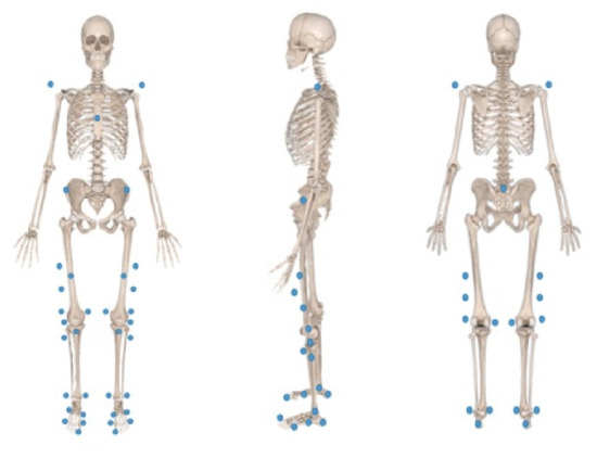
Figure 1.
Reflective marker’s location.
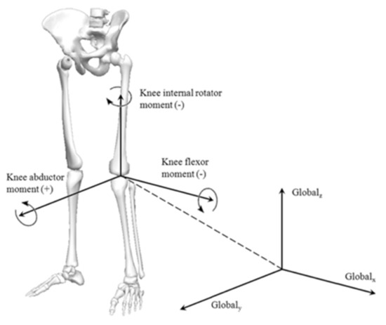
Figure 2.
Definition of experimental coordinate system.
2.3. Experimental Procedure
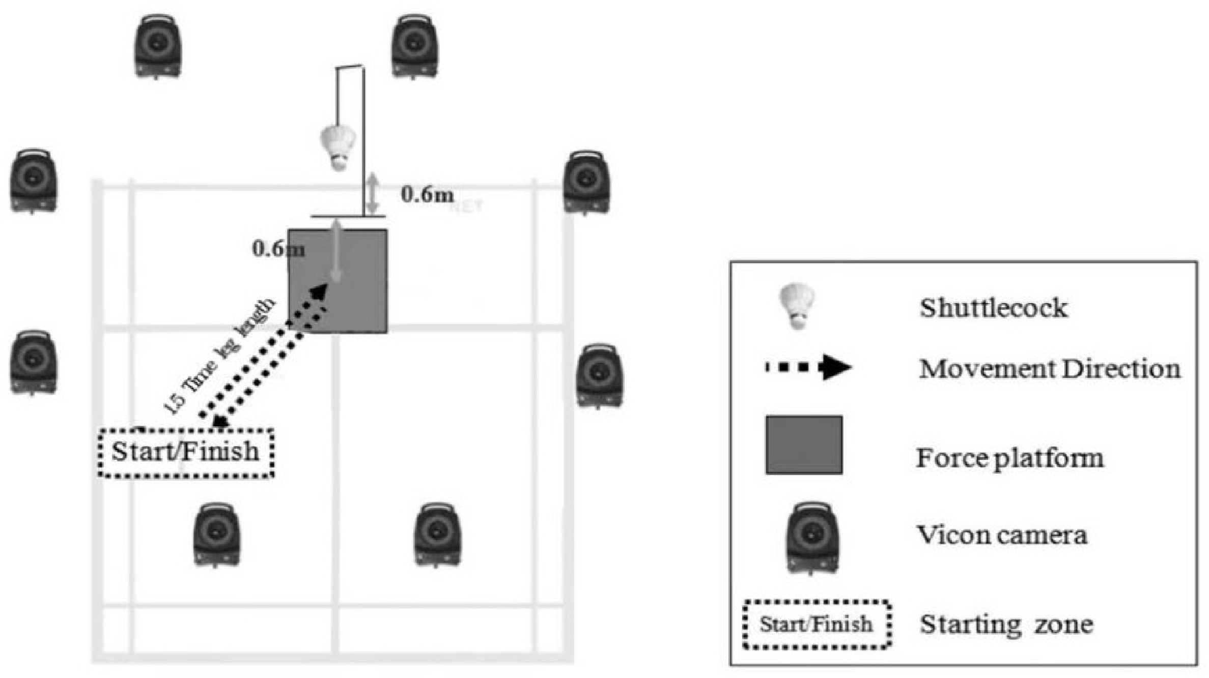
The starting position of each participant was set at a distance of 1.5 times of the individual’s leg length (measured between the anterior superior iliac spine and the ground) at an angle of 45 degrees with respect to the y-axis of the force platforms [5]. In order to better simulate the actual situation in badminton sparring, a shuttlecock was suspended 0.60 m above the ground, at a distance of 0.60 m from the center of the force platform as specified by the previous study [19] (Figure 3). The subjects hit the badminton with one lunge to the specified position, and then quickly returned to the starting position. The above action was considered as an effective action.
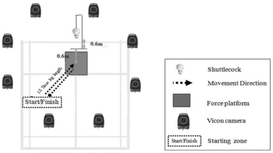
Figure 3.
Experimental set-up for forehand forward lunges.
The sequences of tested sides were randomized across participants. Participants wore a pair of same badminton shoes and tight shorts for the test. Each subject conducted the same warm-up for ten minutes before the experiment. After warm-up, the same assistant pasted the reflection markers. Each participant before data collection had a chance to try lunges three times to reduce experimental error. Eight successful trials were collected for each participant. Then, the participants were led to the isokinetic instrument to fatigue the lower limb muscle. After fatigue, the same data acquisition process was performed again.
2.4. Fatigue Protocol
An isokinetic dynamometer was utilized to produce fatigue in right lower limb muscles after completion of the first forward lunge trials. We chose the isokinetic dynamometer as the instrument for muscle fatigue because of its ability to quantify the production of muscle force. All subjects performed multiple knee flexion and extension exercises using the CONTREX isokinetic dynamometer to fatigue the muscles around the knee joint. Maximal muscle strength of extensor and flexor muscles in the knee joint was measured by isometric contractions before fatigue and averaged over three tests. The degree of fatigue was assessed by the average torque of knee joint flexion and extension.
After the maximum muscle strength was tested, the subjects began to implement the fatigue program after a three minutes rest. The subjects performed continuous flexion and extension exercise at the speed of 60°/s and 120°/s. Each group of flexion and extension exercise lasted for three minutes, with a 30 seconds rest between each group. It was defined as fatigue when the muscle strength of the flexor and extensor of the knee joint decreased to 50% of the maximum muscle strength in three consecutive groups [9] (Figure 4).
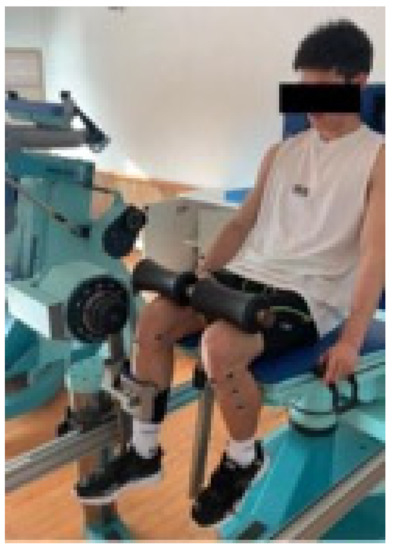
Figure 4.
Fatigue protocol setup.
2.5. Data Analysis
Data analysis was performed using Nexus software (Vicon) and Visual3D (version 3.26, C-Motion Inc., USA). Firstly, the collected kinematics and dynamics data were filtered and preprocessed by nexus software. Secondly, the obtained data was imported into the Visual3D software for inverse dynamic calculation. The previous research suggested that the badminton lunge could be divided into three stages based on the ground reaction force appearing on the force platform [5]. The first lunge landing phase was from the beginning of preparation to the time when the ground reaction force appeared. The second buffer phase was from the time when the ground reaction appeared to the time when the ground reaction was stable. The third departure phase was from the time when the ground reaction was stable to the time when the ground reaction disappeared. Combining knee joint flexion angle and ground reaction force, what we mainly analyze in the lunge movement was from the time when the initial contact with the ground (threshold value on the force plate of 10 N) to maximum knee flexion angle. We mainly analyzed the changes of kinematics and dynamics of the knee joint before and after fatigue in this movement phase. The maximum loading rate of peak was the slope between 20% and 80% of the peak magnitude during the initial loading period (from foot strike to the maximum GRF), and the mean loading rate of peak was the slope between 0% and 100% of the peak magnitude during the initial loading period [20]. We used SPSS 24.0 (IBM Corp., Armonk, NY, USA) to analyze all data obtained. Paired T-test was used to compare the joint torque and GRFs between the pre-fatigue and post-fatigue. Significant differences were set at a level of 0.05.
3. Results
The vertical GRF was shown in Table 1. The results showed that the mean loading rate of peak before and after fatigue showed significant statistical difference. As shown in Figure 5, the vertical GRF was lower after fatigue at the phase from 0% to about 10% stance phase. No significant difference was found in max loading rate of peak or maximum vertical GRF.

Table 1.
Vertical GRF.
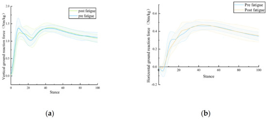
Figure 5.
(a) Description of what is vertical ground reaction force curves; (b) Description of what is horizontal ground reaction force curves.
Comparison of the horizontal backward GRF before and after fatigue in Table 2 showed that max loading rate of peak decreased after fatigue (P = 0.026; pre: 13.16 and post: 9.25 BW). The statistical results also indicated that the maximum horizontal GRF was significantly greater after fatigue (P = 0.027; pre-fatigue: 0.49 and post-fatigue: 0.53 Nm/kg). No significant difference was found in mean loading rate of peak in the horizontal direction.
Table 2.
Horizontal GRF.
The moment on the knee joint during lunges was showed in Table 3. The peak knee flexion moment (P = 0.012) and peak knee abduction moment (P = 0.01) at initial contact both increased significantly after fatigue. The adduction moment of the knee increased after fatigue, but no statistical significance was found (P = 0.46).
Table 3.
Normalized knee moment (BW·Nm/kg).
As shown in Table 4, there was no significant difference in knee flexion at IC or range of motion.
Table 4.
Knee range of motion (Deg).
4. Discussion
Now an increasing number of people play badminton. Both athletes and recreational players attempt to optimize their sport performance, thus increasing the risk of injury. Prevention of sports-related injuries was an important challenge. We were interested in the effect of fatigue developed by exhaustive badminton on lower extremity kinematics. Because in the late stage of sports activities and competitions, the incidence of sports injuries was high [21]. The first objective of this study was to investigate the effects of fatigue on the biomechanics of lower limbs in college badminton fans during lunging at the net, and the second purpose was to investigate how those changes might increase the risk of ACL injury.
The results of this study supported the hypothesis that the biomechanical characteristics of the lunge change significantly after fatigue. After fatigue, the horizontal backward maximum GRF is significantly higher. This might be cause by fatigue in the muscles around the knee joint. The strength of quadriceps muscle was still higher than that of hamstring and gastrocnemius muscles after the gastrocnemius, hamstring and quadriceps muscles were fatigued, it led to an increase in stress of knee joint during flexion movement, and the increase in horizontal backward maximum GRF. the lower extremity contacts the ground, the load of the cruciate ligament changes due to the change in the absorption of the backward GRF. Previous studies demonstrated that in vivo maximum ACL loading in a landing task occurred at time when the peak impact vertical GRF occurs [22], and that the peak horizontal GRF and vertical GRFs occurred at the same time [23]. Increasing the peak impact posterior GRF would also increase ACL loading and thus increasing the risk of non-contact ACL injury.
During the lunge, the hamstrings and gastrocnemius muscles did concentric contraction, while the quadriceps muscles did eccentric contraction. After fatigue, the balance between the strength of the muscles doing concentric contractions and the strength of the muscles doing eccentric contractions during lunges was broken. So shear forces were generated in the knee joint. This shear force causes an increase in tibial anterior displacement [24]. The study of Podraza and White [25] revealed that the force product by the quadriceps, gastrocnemius and hamstrings was likely utilized to improve joint kinematics. They reduced the strain exerted on the ACL and other knee ligaments during single-leg landing. In the badminton lunge, the gastrocnemii’s main function may be co-contracting with the quadriceps to elevate joint compression and to protect the knee and ACL from external joint loading [26]. The load is distributed to the ACL when the gastrocnemii exhibit smaller muscle forces [27].
By comparing the dynamic results of the knee joint in this study, we could find that the flexion moment and external rotation moment of the knee joint increased significantly at the moment of contact after fatigue. Similar with our results, Krosshaug reported an increased knee abduction moment in fatigued and injured athletes [28,29]. The link between valgus loading and resultant increases in ACL strain had been demonstrated experimentally through both cadaveric and in vivo research [30]. The present study extended these findings to badminton. Our results found that the abduction moment of the knee joint increased significantly after fatigue, which was consistent with the results of previous studies. During dynamic knee abduction, the medial collateral ligament, medial patellofemoral ligament, and ACL were mainly used to prevent excessive knee abduction [31]. Tensile strain increased on the medial collateral ligament and the ACL when a knee abduction moment was present [32]. The lunge was used frequently in the sport of badminton. Fatigue accumulated continuously and slowly on the frontal plane. The ability of the knee ligaments to absorb GRFs would deteriorate due to the accumulation of fatigue, this condition became a risk factor for ACL injury. Therefore, knee valgus angle and moment would be one of the factors of predicting the risk of ACL injury. It was also important to note that our studies found the trends in peak knee adduction moment increased but variables were no statistical significance. The reason for this result was mainly caused by the different positions of the shot, our forehand lunge in front of the net was not near the center line of the net, but in front of the right front net, where the knee joint of the batting players was more susceptible to the influence of valgus moment.
Experimental results in Table 4 showed that there was no significant difference in knee joint flexion angle before and after fatigue at the moment when the forehand badminton lunges touchdown. This result was consistent with previous studies to some extent. Hewett, Myer et al. found that there was no significant difference in knee flexion kinematics and knee flexion angle between injured athletes and uninjured athletes. Valldecabres et al [33] also found a decrease in the knee peak angle after fatigue, but there was no significant difference. Combined with previous studies, it could be inferred that knee flexion angle was not the decisive factor for increasing ACL injury. There was no significant difference in knee flexion angle, it may also be related to our fatigue protocol that only made quadriceps femoris, hamstring tendon and gastrocnemius muscles fatigue [34]. The studies of Jonhagen [35] and Halvorsen [8] have shown that small changes in hip joint angle might compensate the changes in knee flexion angle, which could be used to explain the sightly decreased knee flexion angle that did not reach a level of significance during batting after fatigue.
This type of landing might increase ACL strain and the support evidence suggested that more injuries occur at the later stages of a competition. Although the decrease was small, it may be an attempt to increase stability of knee after fatigue, because decreased knee flexion could prevent valgus and landing with less knee flexion did not require so much eccentric muscle strength. With less knee flexion, subjects could rely more on the bony architecture and noncontractile tissues surrounding the knee, such as tightened collateral ligaments. The decrease of knee flexion angle, combined with previous research demonstrated that increase of anterior tibial translation with fatigue [13] might increase the risk of ACL injury during the latter stages of competition.
Through Figure 5, we could find that from the appearance of GRF to the GRF reaching its maximum value in the vertical direction, the GRF curve remained almost the same between pre- and post- fatigue in the first half, but in the latter half the GRF curve in pre-fatigue was significantly higher than that in post-fatigue. This phenomenon led to significant difference in the mean loading rate of peak. Our findings were consistent with those of previous studies in which subjects exhibited a lower loading rate in the vertical direction after fatigue [36]. Concerning with this result, we speculate that when the muscles around the knee joint were fatigued, more muscle units were recruited by the nerves to maintain body balance at the moment of the foot touching the ground. The speed of the touchdown process slowed down and the time to reach the peak value of GRF increased, it caused the mean loading rate reduced. No significant difference was found in the maximum GRF perpendicular to the ground at the moment of contact by analyzing the pre-and post-fatigue data. This result was consistent with the result of Winter’s (1984) research, indicating that the external force applies to the lower limbs during lunge contact do not change significantly after fatigue [37]. Although the muscles around the knee joint would change the biomechanical performance of the knee joint after fatigue, and the fatigued muscles would affect the normal support ability of the knee joint, but the ankle plantar flexion moment increased to ensure the total prop moment remained constant.
Our study also had some limitations. Firstly, we did not combine the changes of biomechanical factors of hip joint and knee joint after fatigue in the process of analyzing the causes of knee injury. Second, only the muscles around the right knee joint were fatigued due to the limitations of the experimental equipment, it might lead to compensatory phenomena of other muscles in the process of exercise, so it might have a certain impact on the experimental results. Third, in the absence of EMG measurement, we would be able to accurately evaluate the kinematics of muscle changes, in the future research could make use of constant speed meter design a set of fatigue schemes to make the muscles more approach to the situation of muscle fatigue of badminton so it could evaluate the actual situation of the muscle fatigue more objective and carried out biomechanical analysis. Furthermore, the generalization of the study results was limited to the specific fatigue protocol utilized in this study. The results from this study did not hold true for all forms of exercise fatigue.
5. Conclusions
Our research results showed that badminton enthusiasts were greatly particularly affected by muscle fatigue when they hit the ball in front of the bow step net. After fatigue, the knee buckling torque and valgus moment increased significantly at the moment of contact with the ground, and the horizontal backward maximum GRF also increased significantly. Due to the effect of other joint compensation, the flexion angle of the knee joint did not change significantly, but the risk of ACL injury did not decrease. The fatigue of the muscles around the knee joint did not change the maximum GRF in the vertical direction at the moment of contact.
Combined with the results of our study on college badminton fans, we could confirm that the fatigue of quadriceps femoris, gastrocnemius and hamstring muscles increased the risk of ACL injury in badminton. Therefore, badminton coaches and teachers should increase the training of lower extremity muscle strength and endurance in our daily class and training, and also should pay special attention to the coordinated development of muscles.
Author Contributions
Conceptualization, J.Y., J.L. and Y.G.; Methodology, Z.L., X.Z., Y.Z., Z.L. and J.W.; Writing—original draft, Z.L.; Writing—review & editing, C.Y. and J.Y.; All authors have read and agreed to the published version of the manuscript.
Funding
This research received no external funding.
Institutional Review Board Statement
The study was conducted according to the guidelines of the Declaration of Helsinki and was approved by the ethics committee of the Research Academy of Grand Health, Ningbo University (RAGH20220600021; June 2022).
Informed Consent Statement
Informed consent was obtained from all subjects involved in the study.
Data Availability Statement
Data available on request due to restrictions privacy. The data presented in this study may be available on request from the corresponding author.
Acknowledgments
The authors thank the other investigators, the staff, and the participants of the study for their valuable contributions.
Conflicts of Interest
The authors declare no conflict of interest.
References
- Laffaye, G.; Phomsoupha, M.; Dor, F. Changes in the game characteristics of a badminton match: A longitudinal study through the Olympic game finals analysis in men’s singles. J. Sport. Sci. Med. 2015, 14, 584–590. [Google Scholar]
- Chen, T.L.-W.; Wang, Y.; Wong, D.W.-C.; Lam, W.-K.; Zhang, M. Joint contact force and movement deceleration among badminton forward lunges: A musculoskeletal modelling study. Sport. Biomech. 2022, 21, 1249–1261. [Google Scholar] [CrossRef] [PubMed]
- Abian-Vicen, J.; Castanedo, A.; Abian, P.; Gonzalez-Millan, C.; Salinero, J.J.; Del Coso, J. Influence of successive badminton matches on muscle strength, power, and body-fluid balance in elite players. Int. J. Sports Physiol. Perform. 2014, 9, 689–694. [Google Scholar] [CrossRef] [PubMed]
- Hong, Y.; Wang, S.J.; Lam, W.K.; Cheung, J.T. Kinetics of badminton lunges in four directions. J. Appl. Biomech. 2014, 30, 113–118. [Google Scholar] [CrossRef]
- Kuntze, G.; Mansfield, N.; Sellers, W. A biomechanical analysis of common lunge tasks in badminton. J. Sports Sci. 2010, 28, 183–191. [Google Scholar] [CrossRef]
- Morgan, K.D.; Donnelly, C.J.; Reinbolt, J.A. Elevated gastrocnemius forces compensate for decreased hamstrings forces during the weight-acceptance phase of single-leg jump landing: Implications for anterior cruciate ligament injury risk. J. Biomech. 2014, 47, 3295–3302. [Google Scholar] [CrossRef]
- Tamura, A.; Akasaka, K.; Otsudo, T.; Sawada, Y.; Okubo, Y.; Shiozawa, J.; Toda, Y.; Yamada, K. Fatigue Alters Landing Shock Attenuation During a Single-Leg Vertical Drop Jump. Orthop. J. Sports Med. 2016, 4, 1–7. [Google Scholar] [CrossRef]
- James, C.R.; Scheuermann, B.W.; Smith, M.P. Effects of two neuromuscular fatigue protocols on landing performance. J. Electromyogr. Kinesiol. 2010, 20, 667–675. [Google Scholar] [CrossRef]
- Rozzi, S.L.; Lephart, S.M.; Fu, F.H. Effects of muscular fatigue on knee joint laxity and neuromuscular characteristics of male and female athletes. J. Athl. Train 1999, 34, 106–114. [Google Scholar]
- Mejane, J.; Faubert, J.; Romeas, T.; Labbe, D.R. The combined impact of a perceptual-cognitive task and neuromuscular fatigue on knee biomechanics during landing. Knee 2019, 26, 52–60. [Google Scholar] [CrossRef]
- Harato, K.; Morishige, Y.; Niki, Y.; Kobayashi, S.; Nagura, T. Fatigue and recovery have different effects on knee biomechanics of drop vertical jump between female collegiate and recreational athletes. J. Orthop. Surg. Res. 2021, 16, 739. [Google Scholar] [CrossRef]
- Koga, H.; Nakamae, A.; Shima, Y.; Iwasa, J.; Myklebust, G.; Engebretsen, L.; Bahr, R.; Krosshaug, T. Mechanisms for noncontact anterior cruciate ligament injuries: Knee joint kinematics in 10 injury situations from female team handball and basketball. Am. J. Sports Med. 2010, 38, 2218–2225. [Google Scholar] [CrossRef]
- Olsen, O.E.; Myklebust, G.; Engebretsen, L.; Bahr, R. Injury mechanisms for anterior cruciate ligament injuries in team handball: A systematic video analysis. Am. J. Sports Med. 2004, 32, 1002–1012. [Google Scholar] [CrossRef]
- Meyer, E.G.; Haut, R.C. Anterior cruciate ligament injury induced by internal tibial torsion or tibiofemoral compression. J. Biomech. 2008, 41, 3377–3383. [Google Scholar] [CrossRef]
- Mokhtarzadeh, H.; Yeow, C.H.; Hong Goh, J.C.; Oetomo, D.; Malekipour, F.; Lee, P.V. Contributions of the soleus and gastrocnemius muscles to the anterior cruciate ligament loading during single-leg landing. J. Biomech. 2013, 46, 1913–1920. [Google Scholar] [CrossRef]
- Lam, W.K.; Ding, R.; Qu, Y. Ground reaction forces and knee kinetics during single and repeated badminton lunges. J. Sports Sci. 2017, 35, 587–592. [Google Scholar] [CrossRef]
- Boden, B.P.; Sheehan, F.T. Mechanism of non-contact ACL injury. J. Orthop. Res. 2022, 40, 531–540. [Google Scholar] [CrossRef]
- Cohen, J. Statistical Power Analysis for the Behavioral Sciences, 2nd ed.; Routledge: Abingdon, UK, 1988. [Google Scholar]
- Nielsen, M.H.; Lund, J.N.; Lam, W.K.; Kersting, U.G. Differences in impact characteristics, joint kinetics and measurement reliability between forehand and backhand forward badminton lunges. Sports Biomech. 2020, 19, 547–560. [Google Scholar] [CrossRef]
- Willy, R.; Pohl, M.B.; Davis, I.S. Calculation of vertical load rates in the absence of vertical impact peaks. In Proceedings of the American Society of Biomechanics Meeting, Ann Arbor, MI, USA, 5–9 August 2008. [Google Scholar]
- Feagin, J.A.J.; Lambert, K.L.; Cunningham, R.R.; Anderson, L.M.; Riegel, J.; King, P.H.; Vangenderen, L. Consideration of the Anterior Cruciate Ligament Injury in Skiing. Clin. Orthop. Relat. Res. 1987, 216, 13–18. [Google Scholar] [CrossRef]
- Lamontagne, M.; Benoit, D.L.; Ramsey, D.K.; Caraffa, A.; Cerulli, G. What Can We Learn from In Vivo Biomechanical Investigations of Lower Extremity? In Proceedings of the XXIII International Symposium of Biomechanics in Sports, Beijing, China, 22–27 August 2005. [Google Scholar]
- Yu, B.; Lin, C.F.; Garrett, W.E. Lower extremity biomechanics during the landing of a stop-jump task. Clin. Biomech. Mar. 2006, 21, 297–305. [Google Scholar] [CrossRef]
- Lam, W.K.; Lee, K.K.; Park, S.K.; Ryue, J.; Yoon, S.H.; Ryu, J. Understanding the impact loading characteristics of a badminton lunge among badminton players. PLoS ONE 2018, 13, e0191493. [Google Scholar] [CrossRef] [PubMed]
- Podraza, J.T.; White, S.C. Effect of knee flexion angle on ground reaction forces, knee moments and muscle co-contraction during an impact-like deceleration landing: Implications for the non-contact mechanism of ACL injury. The Knee 2010, 17, 291–295. [Google Scholar] [CrossRef] [PubMed]
- Winter, D.A. Overall principle of lower limb support during stance phase of gait. J. Biomech. 1980, 13, 923–927. [Google Scholar] [CrossRef] [PubMed]
- Hewett, T.E.; Myer, G.D.; Ford, K.R. Anterior cruciate ligament injuries in female athletes: Part 1, mechanisms and risk factors. Am. J. Sports Med. 2006, 34, 299–311. [Google Scholar] [CrossRef]
- Hewett, T.E.; Myer, G.D.; Ford, K.R.; Heidt, R.S., Jr.; Colosimo, A.J.; McLean, S.G.; van den Bogert, A.J.; Paterno, M.V.; Succop, P. Biomechanical measures of neuromuscular control and valgus loading of the knee predict anterior cruciate ligament injury risk in female athletes: A prospective study. Am. J. Sports Med. 2005, 33, 492–501. [Google Scholar] [CrossRef]
- Kristianslund, E.; Krosshaug, T.; van den Bogert, A.J. Effect of low pass filtering on joint moments from inverse dynamics: Implications for injury prevention. J. Biomech. 2012, 45, 666–671. [Google Scholar] [CrossRef]
- Donelon, T.A.; Dos’Santos, T.; Pitchers, G.; Brown, M.; Jones, P.A. Biomechanical Determinants of Knee Joint Loads Associated with Increased Anterior Cruciate Ligament Loading During Cutting: A Systematic Review and Technical Framework. Sports Med. Open. 2020, 6, 53. [Google Scholar] [CrossRef]
- Markolf, K.L.; Gorek, J.F.; Kabo, J.M.; Shapiro, M.S. Direct measurement of resultant forces in the anterior cruciate ligament. An in vitro study performed with a new experimental technique. JBJS 1990, 72, 557–567. [Google Scholar] [CrossRef]
- Garrett, W.E.; Yu, B. Anterior cruciate ligament injury mechanisms and risk factors. J. Orthop. Sports Phys. Ther. 2007, 37, A10–A11. [Google Scholar]
- Valldecabres, R.; De Benito, A.M.; Littler, G.; Richards, J. An exploration of the effect of proprioceptive knee bracing on biomechanics during a badminton lunge to the net, and the implications to injury mechanisms. Peer J. 2018, 6, e6033. [Google Scholar] [CrossRef]
- Roth, R.; Donath, L.; Zahner, L.; Faude, O. Acute Leg and Trunk Muscle Fatigue Differentially Affect Strength, Sprint, Agility, and Balance in Young Adults. J. Strength Cond. Res. 2021, 35, 2158–2164. [Google Scholar] [CrossRef] [PubMed]
- Jonhagen, S.; Halvorsen, K.; Benoit, D.L. Muscle activation and length changes during two lunge exercises: Implications for rehabilitation. Scand. J. Med. Sci. Sports 2009, 19, 561–568. [Google Scholar] [CrossRef] [PubMed]
- Liederbach, M.; Kremenic, I.J.; Orishimo, K.F.; Pappas, E.; Hagins, M. Comparison of landing biomechanics between male and female dancers and athletes, part 2: Influence of fatigue and implications for anterior cruciate ligament injury. Am. J. Sports Med. 2014, 42, 1089–1095. [Google Scholar] [CrossRef] [PubMed]
- Winter, D.A. Kinematic and kinetic patterns in human gait: Variability and compensating effects. Hum. Mov. Sci. 1984, 3, 51–76. [Google Scholar] [CrossRef]


Disclaimer/Publisher’s Note: The statements, opinions and data contained in all publications are solely those of the individual author(s) and contributor(s) and not of MDPI and/or the editor(s). MDPI and/or the editor(s) disclaim responsibility for any injury to people or property resulting from any ideas, methods, instructions or products referred to in the content. |
© 2023 by the authors. Licensee MDPI, Basel, Switzerland. This article is an open access article distributed under the terms and conditions of the Creative Commons Attribution (CC BY) license (https://creativecommons.org/licenses/by/4.0/).