Article

Improvement and Stability of Soil Organic Carbon: The Effect of Earthworm Mucus Organo-Mineral Associations with Montmorillonite and Hematite

1
Yunnan Provincial Key Laboratory of Soil Carbon Sequestration and Pollution Control, Faculty of Environmental Science & Engineering, Kunming University of Science & Technology, Kunming 650500, China
2
Faculty of Modern Agricultural Engineering, Kunming University of Science & Technology, Kunming 650500, China
3
School of Biological Sciences, University of Western Australia, Perth, WA 6009, Australia
*
Author to whom correspondence should be addressed.
Sustainability 2024, 16(13), 5458; https://doi.org/10.3390/su16135458
Submission received: 5 May 2024 / Revised: 12 June 2024 / Accepted: 24 June 2024 / Published: 27 June 2024
(This article belongs to the Special Issue Recent Advances in Environmental Geochemistry)

Abstract

:
Improving soil carbon storage and stability plays an important role in the development of sustainable agricultural production and mitigating climate change. Earthworms are widely distributed in soil environments; earthworm mucus (EM) can interact with natural mineral materials to form EM–mineral association, enriching soil carbon storage. However, it is unclear how minerals affect the formation and oxidation degradation of EM–mineral associations. Herein, the interactions between EM and natural mineral materials (hematite ore (HO) and montmorillonite (MT)) were investigated. The carbon stability of EM–mineral associations was analyzed based on their chemical oxidative resistance. EM interacted with HO/MT through ligand exchange, hydrogen bonding interaction, and electrostatic attraction. Compared to EM that was extracted under pH 5.0 (EM5) or 9.0 (EM9), EM obtained at pH7 (EM7) contained more protein and polysaccharide components, and was greatly adsorbed by HO/MT. Moreover, EM showed a stronger sorption affinity to MT than HO. The stronger oxidation resistance of EM–MT than EM–HO was revealed by its higher carbon retention, suggesting the vital role of MT in protecting biogenically excreted organic carbon from degradation. Earthworms in neutral environments could substantially promote the establishment of organo-mineral associations. This study provides guidance for promoting soil carbon sequestration through agricultural management and is beneficial to the sustainability of the soil.

1. Introduction

Soil is the largest active carbon pool in terrestrial ecosystems, which contains over three times more carbon than the atmosphere or terrestrial vegetation, and plays an important role in the carbon cycle and mitigating climate change [1,2]. As one of the most common and widely distributed soil animals, earthworms are considered the “ecosystem engineer” and affect the physico–chemical properties of soil, in turn affecting plant growth [3]. Earthworms greatly promote the transformation of plant residues to soil organic matter, significantly increasing the amount of soil particulate organic matter [4]. Furthermore, earthworms can secrete cutaneous mucus as a lubricant when they move in the soil [5]. Earthworm mucus (EM) consists of low molecular weight water-soluble carbohydrates, proteins, lipids, and polysaccharides [6]. EM is thus easily decomposed by soil microorganisms as a source of available plant nutrients. Meanwhile, EM can be adsorbed on soil particles to form organic–mineral complexes or soil aggregates. Being an important source of soil organic carbon, studies have shown that 1 g of earthworms can produce 5.6 mg of EM, which accounts for 0.2–0.5% of total animal C every day [7]. Therefore, understanding the contribution of EM to organic–mineral complexes is essential for the turnover of soil organic matter and carbon accumulation.
As two major components in soil, organic substances and secondary soil minerals can combine to form mineral-associated organic matter (MAOM) [8,9], which accounts for 91% of the total soil carbon [10]. MAOM persists in soil for hundreds to thousands of years and is considered a long-term stable OM [11]. Meanwhile, MAOM is thought to be the smallest unit of soil aggregates and plays an essential role in the stability of soil aggregates [12]. The forming of MAOM is regulated by the properties of dissolved organic matter (DOM) and minerals [13]. Most studies have focused on the surface interaction of minerals and DOM released from the decomposition of plant and faunal tissues and debris [12,14], as well as pyrogenic carbon [15,16]. Studies also reported the interaction of minerals with microbially produced organic matter such as extracellular polymeric substances [17,18], of which mechanisms include ligand exchange, hydrogen bond, van der Waals force, electron donor–acceptor action, etc. [10,19,20,21]. However, information on the interaction of biogenically excreted organic matter especially EM with soil minerals is limited.
Currently, it has been shown that EM can promote the formation of organo-mineral associations in soil [22]. However, studies on how the EM components affect organo-mineral associations are still scarce. Guhra et al. evidenced that 21–36% of organic carbon in EM could adsorb strongly with soil minerals [22]. Notably, composition differences in EM may be influenced by soil pH, resulting in its interaction with minerals. Therefore, we aim to contribute to a better understanding of the characterization of EM components and the role of EM–mineral associations in the transformation of EM in the soil carbon cycle. To explore the process of the carbon cycle in the soil ecological environment, the carbon stability of EM–mineral associations has to be analyzed. The objectives of this work were to address (i) the composition changes of EM obtained at different pH stimuli; (ii) the interactions between EM and typical minerals (hematite and montmorillonite); and (iii) the chemical stability of EM–mineral associations through chemical oxidation. The expected results can provide a better understanding of the interaction between EM and minerals to elucidate the impact of minerals on the transformation of earthworm-derived soil organic carbon.

2. Materials and Methods

2.1. Preparation of Samples

Earthworms (Eisenia fetida) were purchased from Dazu Ecological Agriculture Co., Ltd., Hunan, China. Mature earthworms (about 350 ± 2.3 mg biomass) were selected to extract EM. Before the extraction of EM, earthworms were washed with deionized water at least 5 times and then placed on a wet filter paper for 2 days to completely empty the residual food in their guts and thus avoid the impact of earthworm castings. At the end of the emptying period, these earthworms were rinsed with deionized water and then transferred to a 500 mL beaker. Earthworms can survive between pH 3.0 and 9.1 [23,24]. To compare the components and properties of EM under different pH levels, EM was extracted at pH levels of 5.0, 7.0, and 9.0 which represent acidity, neutrality, and alkalinity, respectively. A ratio of 1.3 g (earthworm biomass): 1 mL extraction solution (pH-adjusted deionized water with NaOH and HCl) was used; these were named EM5, EM7, and EM9, respectively. Triplicate extractions were performed under the dark condition with extraction solutions (20 °C). The extracted EM was centrifuged twice (10,000 g, 30 min at 4 °C) to remove the remaining impurities. The obtained EM were frozen overnight at −20 °C and subsequently lyophilized.
Natural mineral materials, raw montmorillonite (MT) and hematite ore (HO) are widely used in soil improvement and remediation [25,26] and thus were used to investigate the formation and carbon stability of EM–mineral associations. The mineral components of MT/HO were analyzed using X-ray diffraction (XRD; SmartLab SE, Rigaku, Japan) spectra and X-ray fluorescence (XRF; Zetium, PANalytical, The Netherlands). The XRF data provided information on the different element contents in MT/HO (Table S1). The XRD analysis revealed that montmorllonite and hematite were the main mineral composition in MT and HO, respectively (Figure S1). See the Supplementary Materials for more details. To avoid the effect of granulometric fractions on sorption behaviors, MT and HO were ground to a particle size of 0.15 mm with a 100-mesh sieve. In addition, to remove residual carbon impurities, MT and HO were pretreated with hydrogen peroxide (H2O2, 30%) until no obvious bubbles appeared. Then, the MT and HO were collected via centrifugation (2500 r/min, 5 min) and washed 5 times with deionized water. The pretreated MT and HO were oven-dried at 80 °C.

2.2. Composition Characterization of EM

The elemental compositions of EM were determined using an elemental analyzer (MicroCube, Elementar, Germany). The composition of the functional groups (in % of total C) of EM was analyzed via 13C NMR (DSX 200, Bruker, Billerica, MA, USA) at 50.3 MHz, and FTIR (Varian 640-IR) was used in the transmission mode with KBr pellets (0.2 mg HA in 200 mg KBr). The spectra were collected within the range of 4000–400 cm−1 with 16 scans at a resolution of 8 cm−1. Triplicate measurements of the zeta potential values of EM were conducted using a zeta potential analyzer (Nano ZEN 3600, Malvern, UK) at 25 °C. The pH values of EM were adjusted with HCl and NaOH to 4.0, 5.0, 6.0, 7.0, and 8.0.
The polysaccharides of EM were determined via the phenol–sulfuric acid method [27]. Glucose powder (0.1 g) was dissolved with ultrapure water in a 1000 mL volumetric flask to obtain a 100 mg/L stock solution. The standard curve was applied with 0 mL, 0.1 mL, 0.2 mL, 0.3 mL, 0.4 mL, 0.6 mL, 0.8 mL, and 1.0 mL glucose solutions (100 mg/L) until 10 mL, with the addition of ultrapure water until 1.0 mL. Then, 1 mL of 5% phenol solution and 5 mL of sulfuric acid were added to the standard samples, which then reacted in a water bath (96 °C) for 20 min. The absorbance of samples was measured at 490 nm after cooling at room temperature. The polysaccharide content of the 0.1 mL EM solution (133 g/L) was measured using the same approach, and the content was finally calculated according to the standard curve.
Protein contents in EM were quantified using the Bradford method [28]. Firstly, 100 mg of Coomassie blue G-250 was dissolved in 50 mL of 95% ethanol and then mixed with 100 mL of 85% phosphoric acid to obtain 200 mL of stock solution; this was finally stocked in a 4 °C refrigerator. Bovine serum albumin was dissolved in normal saline (0.5 g/L) to obtain protein standard samples. After 5 min of mixing at room temperature (20 °C), the absorbance (595 nm) of samples was measured using UV–Vis spectrophotometer (UV-2600, Shimadzu, Japan).

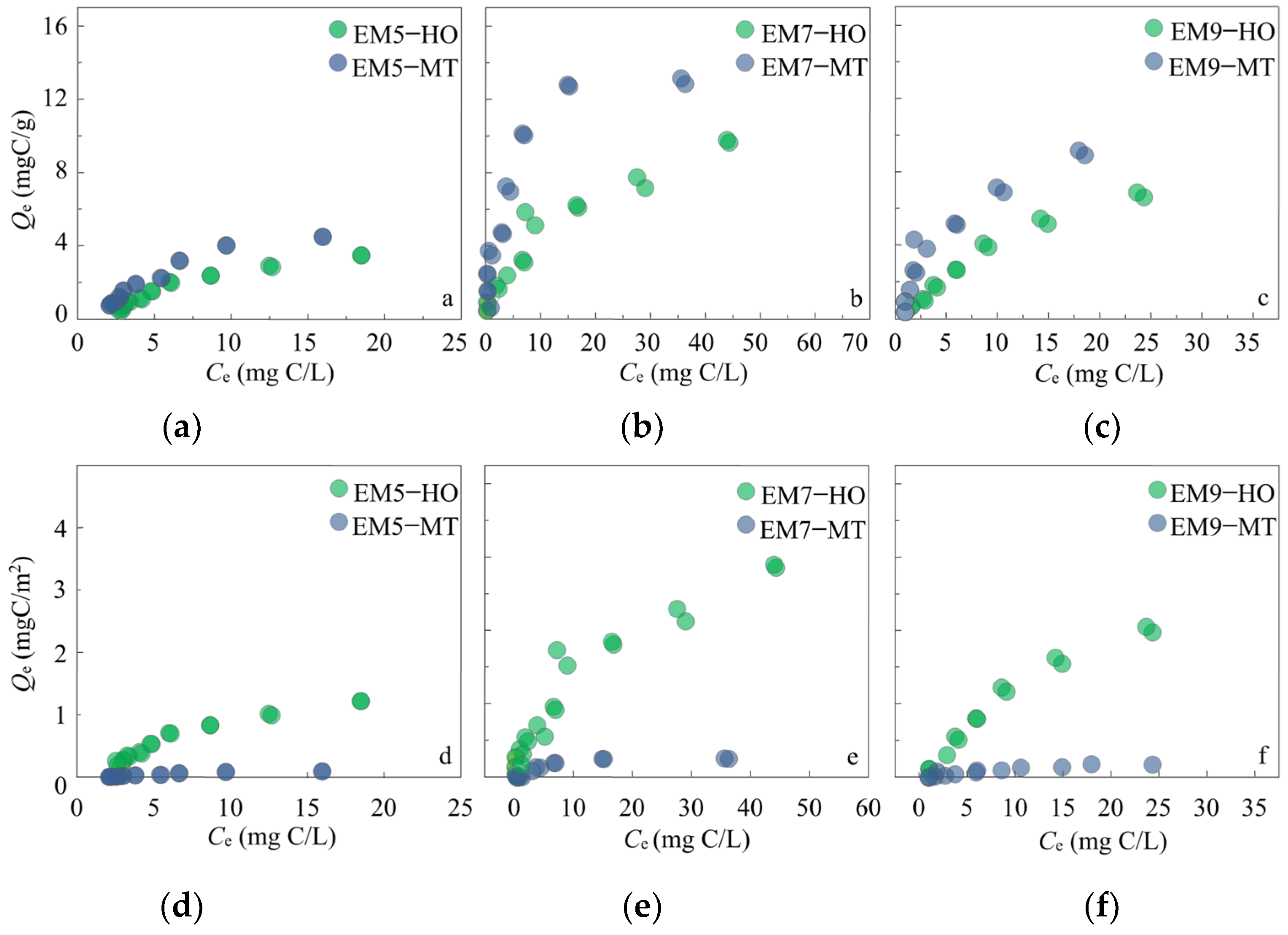
2.3. EM–Mineral Sorption Experiment

Batch adsorption experiments [29] for EM were conducted using HO or MT. The stock solution (150 mg/L) for EM5, EM7, and EM9 was dissolved in a background solution containing 1 mM NaCl to maintain constant ionic strength, and 0.05% NaN3 was diluted in the background solution as a biocide to obtain ten different concentrations within the range of 10 to 133 mg/L. The aqueous/solid ratio in the sorption experiment was 2.5:1 (mg/mL) to ensure 20–80% sorption of the initially applied sorbates at equilibrium. All sorption experiments were conducted in 6 mL glass vials capped with Teflon-lined screw caps, and they were shaken at 25 °C under dark conditions in a shaker (200 r/min) until equilibrium. All experiments were run in duplicate. According to our preliminary test, 24 h was long enough for apparent equilibrium. EM–mineral associations were obtained through centrifugation (2500 r/min), and the supernatants were sampled for adsorbate quantification with UV absorbance (280 nm) to obtain the aqueous phase concentration at equilibrium (Ce, mg C/L). The sorption of the aromatic groups of EMs on HO/MT was analyzed using the decrease of UV absorbance (254 nm) in supernatants. The concentrations of the solid phase (Qe, mg C/g) were calculated according to Equation (1) [30]:
Q e = ( C 0 C e ) × V / M
where Qe (mg C/g) and Ce (mg/L) are the solid- and liquid-phase equilibrium concentrations of EM, C0 (mg/L) is the initial concentration in liquid-phase, V is the solution volume (mL), and M is natural mineral material quality (mg).
The Langmuir model (Equation (2)) [30] and Freundlich model (Equation (3)) [30] were employed to fit the sorption isotherms using Sigmaplot 10.0. The model is expressed as follows:
1 Q e = 1 Q m a x + 1 ( K L · Q m a x · C e )
where Qe (mg C/g) and Ce (mg/L) are the solid- and liquid-phase equilibrium concentrations of EM, respectively; Qmax is the maximum solid sorption concentration of EM (mg C/g); KL is the Langmuir sorption coefficient.
L o g   C b = l o g   K F + n   l o g   C f
where Cb (mg/kg C) and Cf (mg/L) are the bound and free concentrations of chemicals, KF is Freundlich sorption coefficient [(μg/kg)/(μg/L)n] and n is the nonlinearity factor.

2.4. The Oxidation Resistance of EM–Mineral Associations

The EM–mineral associations were prepared using EM5, EM7, and EM9 with HO or MT (aqueous/solid ratio, 2.5 mg/1 mL), and they are named EM5-HO, EM7-HO, EM9-HO, EM5-MT, and EM7-MT, EM9-MT, respectively. The elemental composition of EM–mineral associations was analyzed using an elemental analyzer (MicroCube, Elementar, Germany), and the composition of the functional groups of EM–mineral associations was measured using an FTIR spectrometer. EM–mineral associations were oxidized using the NaClO solution (1.5 mL) in 5 mL vials at room temperature (20 °C) for 3 or 5 days. Then, the solid materials of EM–mineral associations were washed with deionized water 5 times to remove the residue of NaClO. The supernatants were discarded via centrifugation at 2500 r/min for 10 min. The solid materials of EM–mineral associations were freeze-dried to detect the elemental composition using the above-mentioned elemental analyzer. The residual fraction of carbon contents (W, %) in EM–mineral associations after oxidation was calculated using Equation (3):
W   ( % ) = M × N / ( M 0 N 0 ) × 100 %
where M0 (mg) and N0 (mg/g) are the weight and carbon content of EM–mineral associations, respectively; M (mg) and N (mg/g) are the weight and carbon content of the solid materials of EM–mineral associations after oxidation.

3. Results and Discussion

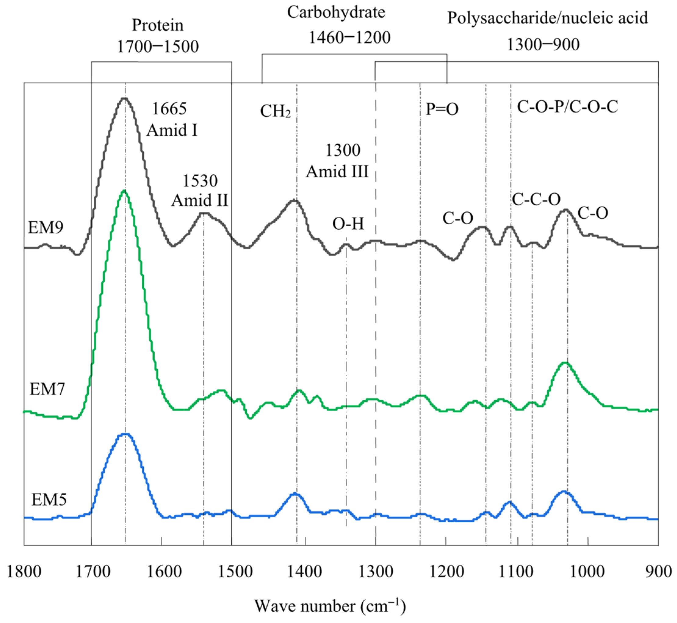
3.1. Characterization of Various pH Stimuli-Responsive EM

EM mainly consisted of carbohydrates, polysaccharides, and proteins [22,31]. According to the analysis of EMs components (Table S2), the total proteins and polysaccharides of EMs were 0.255–0.394 mg mL−1 and 0.042–0.068 mg mL−1, respectively. EM7 showed the highest contents of protein and polysaccharide, followed by EM9, and the lowest content was observed with respect to EM5. The results suggested that pH7 was more suitable for earthworm activity. In addition, EM7 showed the lowest H/C atom ratio (9.27) and highest (N+O)/C atom ratio (0.85) compared to EM9 and EM5 (Table S3), indicating that EM7 contained more aromatic and hydrophilic components.
The components of EM were further analyzed via FTIR spectra (Figure 1) and 13C NMR quantification (Table 1). Based on the analysis of FTIR spectra, the main components of EM were identified as proteins (1700–1500 cm−1), carbohydrates (1460–1200 cm−1), polysaccharides, and nucleic acids (1300–900 cm−1). In the protein region, significant peaks at 1665 cm−1 were observed in three EM samples, representing the protein-specific amide I band, which resulted from the C=O stretching of peptide groups in proteins [32,33]. The EM absorption peaks observed at 1530 cm−1 represent the N-H bending and C-N stretching of protein components at the amide II band. The band at 1300 cm−1 belonged to the amide III band [34], and all three EM samples exhibited relatively weak absorption at this band. EM7 showed an obviously stronger response than EM5 and EM9 at 1252 cm−1, which represents the asymmetric P=O stretching of the phosphodiester backbone of nucleic acid or phosphorylated proteins [32]. The band at 1078 cm−1 is attributed to the vibration of the symmetric and stretched phosphate C-O-P band and the C-O-C ring of polysaccharides, and the band at 1028 cm−1 is attributed to C–O asymmetric stretching [34,35]. Therefore, EM7 contained the higher contents of amides and carboxyl and aromatic groups, and the lower aliphatic components and carbohydrates (65–110 ppm) [36] compared to EM5 and EM9, which was further evidenced via 13C-NMR quantification. The differences in the components in EM might be a characteristic of the ecological adaptability of earthworms, indicating that the pH had influenced the components in the secreted EMs.

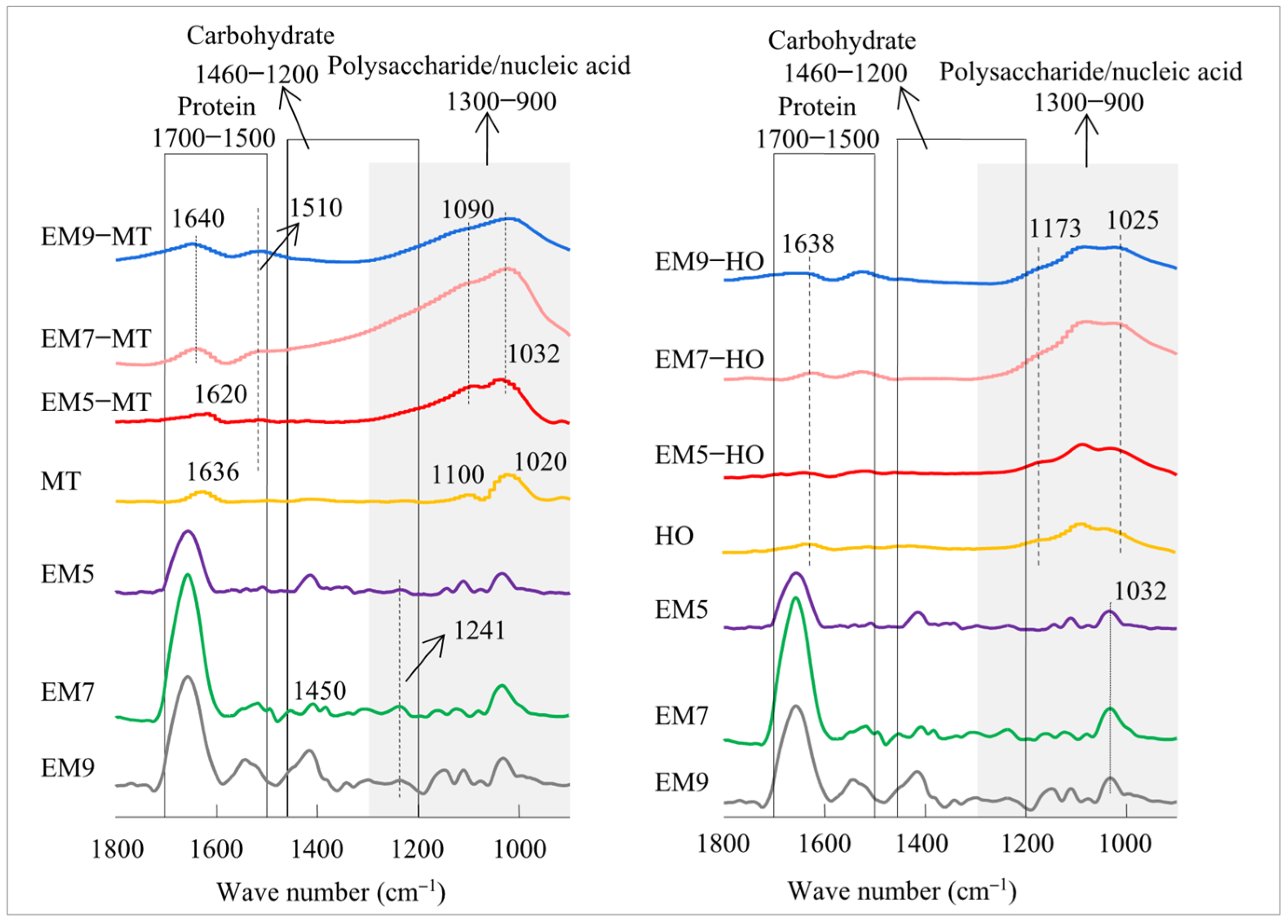
3.2. The Formation of EM–Mineral Associations

EM sorption isotherms (Figure 2) were fitted using the Langmuir and Freundlich models. The Langmuir model provided higher correlation coefficients (R2) within the range of 0.94–0.98 than those obtained by the Freundlich model (0.91–0.96, Table 2). Thus, the Langmuir model was employed to describe the sorption behavior of EM in MT/HO. The Langmuir constants (KL) and theoretical maximum sorption capacity (Qmax) of EM on MT/HO were obtained via model fitting. A greater KL value indicated stronger sorption affinities between the mineral and EM [37]. Compared to HO, MT showed the highest EM Qmax value within the range of 9.55–15.59 mg C/g and KL was 6.58–13.61, indicating the strong sorption affinity of EM in MT. The MT had a significantly higher specific surface area than HO (Table S4). The sorption capacity of EM on MT obviously decreased after specific surface area normalization (Figure 2d–f), indicating that the higher specific surface area of MT greatly provided more sorption sites for EM. In contrast, the contribution of the specific surface area of HO to EM sorption played a relatively minor role.
The UV absorbance of supernatants was measured to analyze the alteration of EM components in EM–mineral sorption systems. Compared to EM5 and EM9, EM7 showed the strongest UV absorbance at 254 nm (Figure 3), suggesting the highest aromaticity [38,39]. The absorbance at 254 nm significantly decreased after sorption, especially in EM7–mineral reaction systems, indicating the higher sorption of aromatic components in EM7. Furthermore, compared to bare MT/HO, EM–mineral associations showed a lower atom ratio of H/C and a higher value of (N+O)/C and nitrogen element contents (Table 3 and Table S3), suggesting that aromatic groups, hydrophilic groups, and protein components were adsorbed in EM–mineral associations. Among all EMs, MT/HO presented the highest sorption capacity to EM7, (Figure 2, Table 2), while less EM5 was adsorbed on the MT/HO. The lower value of H/C and the higher value of (N+O)/C indicated that EM7–mineral associations contained more aromatic and polar groups than EM5 and EM9, especially for MT (Table 3). Therefore, compared to HO, MT adsorbed more protein components, and more aromatic, hydrophilic groups were adsorbed on MT compared to HO.
The formation of EM–mineral associations was indirectly evidenced by the determination of the surface charge of EM–mineral associations via zeta potential measurements in comparison to the bare MT/HO (Figure 4). The HO and MT were negatively charged in EM–mineral systems within the pH range of 6.45–8.83. The zeta potentials of EM7 and EM9 were mostly positive within this pH range, while EM5 was mostly negative. Zeta potentials were the index for colloid charges; the higher negative charge indicated EM5’s higher electronic double-layer potential [40]. These results indicated that EM7 and EM9 could interact with HO or MT through electrostatic interactions. The electrostatic repulsion between EM5 and MT/HO was stronger than the electrostatic attraction, thus resulting in its low sorption affinity to MT/HO. The carboxyl (pKa = 4.43 ± 0.03), phosphate (pKa = 6.4 9 ± 0.08), and amino or hydroxyl (pKa = 9.11 ± 0.41) groups of EM could increase the surface negative charge. The protein components comprised dielectric colloids with both carboxyl and amino groups. The isoelectric point (IEP) of the protein is about 7.2–7.36 [41,42]. The amino acid molecules of proteins comprise dipolar molecules, and the molecules have both positive (ammonium) ions (NH3+-R-COOH) and negative (carboxylate) ions (NH2-R-COO-) [43,44]. Therefore, during the sorption process, the protein components of EM could become positively charged when pH values are lower than the isoelectric point, and the proteins are then adsorbed on MT/HO via electrostatic attraction [22].
It has been reported that proteins could be maximally adsorbed on minerals when the reaction pH value is close to the IEP of proteins [43,44]. Compared to MT, the pH values of EM–HO were closer to the proteins’ IEP. Therefore, more EM protein components could be adsorbed on HO. In addition, the lower negative zeta potential in EM–MT indicated that the less positively charged proteins of EM were adsorbed on MT than HO (Figure 4c). In contrast, the higher negative zeta potential ranges of EM–HO suggested a stronger electrostatic interaction between EM and HO. In particular, the negative zeta potential of the EM9-HO was substantially lower than EM9-MT. Correspondingly, the higher atom ratio of N/C in EM9 (0.191, Table S3) than that in EM7 (0.177) and EM5 (0.181) suggested more NH3+-R-COOH groups were contained in EM9. These results indicated that the electrostatic attraction between HO and EM was stronger than that for MT. Electrostatic attraction played a more important role in the sorption of EM for HO than for MT.
The sorption of protein components from EM on MT and HO was also evidenced by FTIR spectra. (Figure 5). In particular, the N-H or C-N vibration shifted the peaks of EM-MT at 1510 cm−1, and the amide II band of proteins shifted to 1591 cm−1 after sorption. The sorption of protein components on MT could induce structural stress on MT and expand the interlayer space [45]. The results of zeta potential and Kolman, et al. [46] also indicate the important role of electrostatic interactions between proteins and HO. In addition, studies reported that the formation of hydrogen bonds between the protein carbonyl and Al-OH groups of HO, which enhances the adsorption of proteinaceous constituents onto HO [47]. Furthermore, the increased responses for C=C stretching at around 1640 cm−1 in EM–mineral associations (Figure 5) suggested the sorption of the aromatic components. The functional groups of aromatic components may greatly enhance the hydrogen bond and cation–pi interactions with clay minerals, because of the electron–donor–acceptor pairs [48]. In addition, the peak at 1020 cm−1 with respect to MT shifted to 1032 cm−1 (Figure 5), suggesting the sorption of polysaccharides in EM [49,50,51]. It has been reported that the polyhydroxy structures of polysaccharides could interact with MT through hydrogen bonding interactions [42]. Moreover, compared to bare MT/HO, EM–mineral associations showed the obvious FTIR response between 900 cm−1 and 1300 cm−1, indicating the adsorption of nucleic acid components on MT/HO. The sorption of phosphate groups in nucleic acid components from EM on MT/HO might involve the formation of inner-sphere surface complexes through ligand exchange [52,53,54].
Among three EMs, EM–mineral associations which were formed by EM7 showed the highest response at 900–1300 cm−1. Compared to EM–OH, the significantly stronger absorption peaks of nucleic acid were observed in the EM7-MT association, suggesting that more inner-sphere complexes were formed between MT and the phosphate groups in EM.

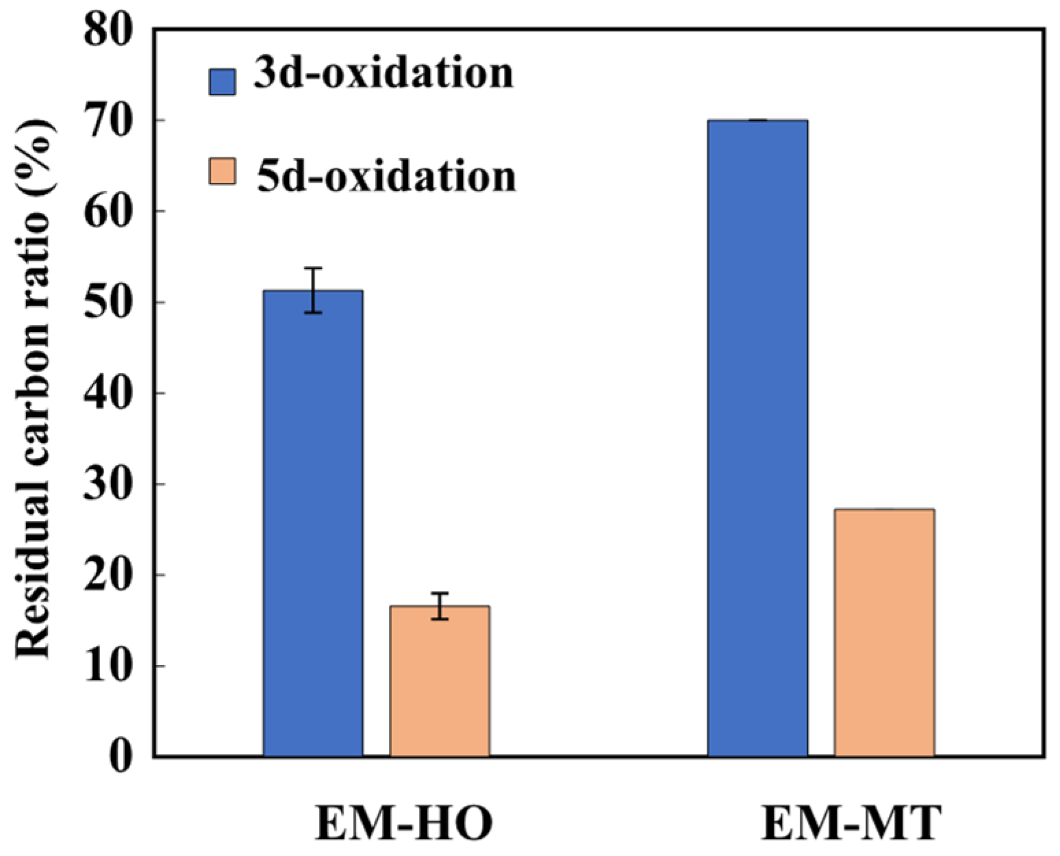
3.3. Carbon Stability of EM–Mineral Associations for Oxidation Resistance

To exclude the effect of original carbon contents on the oxidation, the original carbon contents of complexes were kept the same before the oxidation. Although EM7 was effectively adsorbed on MT/HO with the highest sorption capacity, whether such sorption was stable was unclear. To explore the carbon stability for chemical oxidation resistance of EM–mineral associations, the proportion of carbon loss in EM7–mineral associations upon 3 or 5 days of oxidation was calculated (Figure 6). Oxidation treatment reduced the carbon content of EM7–mineral associations, particularly after 5 days of oxidation (Table 4). The obvious differences in the residual carbon ratio (%) revealed different oxidation resistance levels between the two EM–mineral associations (Figure 6). The retention ratio of the carbon of EM7–mineral associations was within the range of 16.57–70.02% for oxidation (Table S5). After 3 days of oxidation, the carbon retention ratio of EM-HO was substantially reduced to 51.31%. However, MT retained 70.02% of carbon, even with respect to the longer oxidation time (5 days), and it accounted for 27.26% of the remaining carbon (Table S5). In addition, after oxidation treatment, the (N+O)/C values of EM7–mineral associations increased from 2.17–3.22 to 2.21–4.27, and the H/C value decreased from 0.58–0.75 to 0.32–0.51. Notably, H/C value of EM7-HO obviously decreased from 0.75 to 0.32 after the oxidation, but the H/C value for EM7-MT hardly changed (from 0.58 to 0.51). Meanwhile, the change in the (N+O)/C value of EM7-HO was slight after oxidation.
The higher carbon retention ratios of EM7-MT compared to EM7-HO suggested that MT could strongly protect EM from oxidation. The higher carbon retention for EM7-MT could be attributed to the stronger sorption affinity and the more inner-sphere complexes between MT and EM7. It was demonstrated that the inner-sphere complexes of organic matter/minerals were relatively more stable during organic carbon transformation [55]. In contrast, based on previous discussion, EM7-HO was mainly formed via surface electrostatic attraction, which might play a weak role in protecting carbon from oxidation. In addition, compared to the inner-sphere complexes, EM components adsorbed on HO surface were more likely to react with NaClO, and thus suggested the weaker protection of HO to EM degradation than MT.

4. Conclusions

The current study analyzed the formation and carbon stability of EM–MT, and EM–HO with respect to oxidation resistance. The carbohydrates, polysaccharides, and proteins were the main components of Eisenia fetida EM. Compared to acidic and alkaline environments, the mucus secreted by earthworms under neutral conditions contained more proteins and polysaccharide components and thus greatly promoted the formation of EM–mineral associations. EM interacted with HO and MT mainly through electrostatic attraction, ligand exchange, and hydrogen bond interactions. Electrostatic attraction could be the main sorption mechanism of EM on HO, while MT interacted with EM mainly through hydrogen bond interaction and ligand exchange. MT exhibited stronger sorption affinities and higher sorption capacities for EM. MT increased the oxidation resistance of EM and greatly protected organic carbon from degradation. This work revealed the important role of MT in protecting EM from oxidation degradation and improving the formation of organo-mineral associations in soils. The results emphasized the protection of natural mineral materials in soil relative to the organic carbon cycle.

Supplementary Materials

The following supporting information can be downloaded at: https://www.mdpi.com/article/10.3390/su16135458/s1, Figure S1: The X-ray diffraction (XRD) spectra of HO (a) and MT (b); Table S1: The element content of MT and HO (%); Table S2: Content of main components of EMs; Table S3: The elemental compositions and atomic ratios of EMs; Table S4: The basic properties of MT and HO; Table S5: The carbon retention ratios of EM7–minerals associations after 3 days or 5.

Author Contributions

Y.L.: Methodology, Data curation, Formal analysis, Writing—original draft, Writing—review & editing. S.F.: Investigation, Methodology, Data curation, Writing—original draft. L.W.: Formal analysis, Writing—review & editing. C.L.: Methodology, Formal analysis. H.P.: Methodology, Formal analysis, Writing—review & editing. X.H.: Writing—review & editing. D.Z.: Writing—review & editing. F.L.: Conceptualization, Investigation, Writing—original draft, Writing—review & editing, Funding acquisition, Project administration. All authors have read and agreed to the published version of the manuscript.

Funding

This research was financially supported by the National Natural Science Foundation of China (42167030, 42267028, and 41807382), Yunnan Major Scientific and Technological Projects (202202AG050019) and Yunnan Science and Technology Planning Project (202303AC100010).

Institutional Review Board Statement

Not applicable.

Informed Consent Statement

Not applicable.

Data Availability Statement

Data is contained within the article and Supplementary Materials.

Conflicts of Interest

The authors declare no conflicts of interest.

References

  1. Schmidt, M.W.; Torn, M.S.; Abiven, S.; Dittmar, T.; Guggenberger, G.; Janssens, I.A.; Kleber, M.; Kögel-Knabner, I.; Lehmann, J.; Manning, D.A. Persistence of soil organic matter as an ecosystem property. Nature 2011, 478, 49–56. [Google Scholar] [CrossRef] [PubMed]
  2. Lal, R. Soil carbon sequestration to mitigate climate change. Geoderma 2004, 123, 1–22. [Google Scholar] [CrossRef]
  3. Singh, S.; Singh, J.; Vig, A.P. Earthworm as ecological engineers to change the physico-chemical properties of soil: Soil vs. vermicast. Ecol. Eng. 2016, 90, 1–5. [Google Scholar] [CrossRef]
  4. Bohlen, P.J.; Parmelee, R.W.; Allen, M.F.; Ketterings, Q.M. Differential Effects of Earthworms on Nitrogen Cycling from Various Nitrogen-15-Labeled Substrates. Soilence Soc. Am. J. 1999, 63, 882–890. [Google Scholar] [CrossRef]
  5. Abail, Z.; Sampedro, L.; Whalen, J.K. Short-term carbon mineralization from endogeic earthworm casts as influenced by properties of the ingested soil material. Appl. Soil Ecol. 2017, 116, 79–86. [Google Scholar] [CrossRef]
  6. Pathma, J.; Sakthivel, N.J.S. Microbial diversity of vermicompost bacteria that exhibit useful agricultural traits and waste management potential. SpringerPlus 2012, 1, 26. [Google Scholar] [CrossRef] [PubMed]
  7. Scheu, S. Mucus excretion and carbon turnover of endogeic earthworms. Biol. Fertil. Soils 1991, 12, 217–220. [Google Scholar] [CrossRef]
  8. Kennedy, M.J.; Pevear, D.R.; Hill, R.J. Mineral surface control of organic carbon in black shale. Science 2002, 295, 657–660. [Google Scholar] [CrossRef]
  9. Mayer, L.M. Relationship between mineral surfaces and organic carbon concentrations in soils and sediments. Chem. Geol. 1994, 114, 347–363. [Google Scholar] [CrossRef]
  10. Kalbitz, K.; Schwesig, D.; Rethemeyer, J.; Matzner, E. Stabilization of dissolved organic matter by sorption to the mineral soil. Soil Biol. Biochem. 2005, 37, 1319–1331. [Google Scholar] [CrossRef]
  11. Angst, G.; Mueller, K.E.; Castellano, M.J.; Vogel, C.; Wiesmeier, M.; Mueller, C.W. Unlocking complex soil systems as carbon sinks: Multi-pool management as the key. Nat. Commun. 2023, 14, 2967. [Google Scholar] [CrossRef]
  12. Totsche, K.U.; Amelung, W.; Gerzabek, M.H.; Guggenberger, G.; Klumpp, E.; Knief, C.; Lehndorff, E.; Mikutta, R.; Peth, S.; Prechtel, A. Microaggregates in soils. J. Plant Nutr. Soil Sci. 2018, 181, 104–136. [Google Scholar] [CrossRef]
  13. Jones, K.D.; Tiller, C.L. Effect of solution chemistry on the extent of binding of phenanthrene by a soil humic acid: A comparison of dissolved and clay bound humic. Environ. Sci. Technol. 1999, 33, 580–587. [Google Scholar] [CrossRef]
  14. Kögel-Knabner, I.; Guggenberger, G.; Kleber, M.; Kandeler, E.; Kalbitz, K.; Scheu, S.; Eusterhues, K.; Leinweber, P. Organo-mineral associations in temperate soils: Integrating biology, mineralogy, and organic matter chemistry. J. Plant Nutr. Soil Sci. 2008, 171, 61–82. [Google Scholar] [CrossRef]
  15. Whitman, T.; Zhu, Z.; Lehmann, J. Carbon mineralizability determines interactive effects on mineralization of pyrogenic organic matter and soil organic carbon. Environ. Sci. Technol. 2014, 48, 13727–13734. [Google Scholar] [CrossRef]
  16. Woo, S.H.; Enders, A.; Lehmann, J. Microbial mineralization of pyrogenic organic matter in different mineral systems. Org. Geochem. 2016, 98, 18–26. [Google Scholar] [CrossRef]
  17. Cao, Y.; Wei, X.; Cai, P.; Huang, Q.; Rong, X.; Liang, W. Preferential adsorption of extracellular polymeric substances from bacteria on clay minerals and iron oxide. Colloids Surf. B Biointerfaces 2011, 83, 122–127. [Google Scholar] [CrossRef]
  18. Fang, L.; Cao, Y.; Huang, Q.; Walker, S.L.; Cai, P. Reactions between bacterial exopolymers and goethite: A combined macroscopic and spectroscopic investigation. Water Res. 2012, 46, 5613–5620. [Google Scholar] [CrossRef] [PubMed]
  19. Gerzabek, M.H.; Aquino, A.J.A.; Balboa, Y.I.E.; Galicia-Andrés, E.; Grančič, P.; Oostenbrink, C.; Petrov, D.; Tunega, D. A contribution of molecular modeling to supramolecular structures in soil organic matter. J. Plant Nutr. Soil Sci. 2022, 185, 44–59. [Google Scholar]
  20. Liu, X.; Lu, X.; Wang, R.; Zhou, H.; Xu, S. Surface complexes of acetate on edge surfaces of 2: 1 type phyllosilicate: Insights from density functional theory calculation. Geochim. Cosmochim. Acta 2008, 72, 5896–5907. [Google Scholar] [CrossRef]
  21. Zhai, H.; Wang, L.; Putnis, C.V. Molecular-scale investigations reveal noncovalent bonding underlying the adsorption of environmental DNA on mica. Environ. Sci. Technol. 2019, 53, 11251–11259. [Google Scholar] [CrossRef] [PubMed]
  22. Guhra, T.; Stolze, K.; Schweizer, S.; Totsche, K.U. Earthworm mucus contributes to the formation of organo-mineral associations in soil. Soil Biol. Biochem. 2020, 145, 107785. [Google Scholar] [CrossRef]
  23. Di Carlo, E.; Boullemant, A.; Poynton, H.; Courtney, R. Exposure of earthworm (Eisenia fetida) to bauxite residue: Implications for future rehabilitation programmes. Sci. Total Environ. 2020, 716, 137126. [Google Scholar] [CrossRef] [PubMed]
  24. Zhang, J.-E.; Yu, J.; Ouyang, Y. Activity of Earthworm in Latosol Under Simulated Acid Rain Stress. Bull. Environ. Contam. Toxicol. 2015, 94, 108–111. [Google Scholar] [CrossRef]
  25. Allen, B.; Hajek, B. Mineral occurrence in soil environments. Miner. Soil Environ. 1989, 1, 199–278. [Google Scholar]
  26. Wang, H.; Li, X.; Chen, Y.; Li, Z.; Hedding, D.W.; Nel, W.; Ji, J.; Chen, J. Geochemical behavior and potential health risk of heavy metals in basalt-derived agricultural soil and crops: A case study from Xuyi County, eastern China. Sci. Total Environ. 2020, 729, 139058. [Google Scholar] [CrossRef] [PubMed]
  27. Dubois, M.; Gilles, K.A.; Hamilton, J.K.; Rebers, P.A.; Smith, F. Colorimetric method for determination of sugars and related substances. Anal. Chem. 1956, 28, 350–356. [Google Scholar] [CrossRef]
  28. Bradford, M.M. A rapid and sensitive method for the quantitation of microgram quantities of protein utilizing the principle of protein-dye binding. Anal. Biochem. 1976, 72, 248–254. [Google Scholar] [CrossRef] [PubMed]
  29. Chen, S.; Klotzbuecher, T.; Lechtenfeld, O.J.; Hong, H.; Liu, C.; Kaiser, K.; Mikutta, C.; Mikutta, R. Legacy Effects of Sorption Determine the Formation Efficiency of Mineral-Associated Soil Organic Matter. Environ. Sci. Technol. 2022, 56, 2044–2053. [Google Scholar] [CrossRef]
  30. Wang, L.; Li, H.; Yang, Y.; Zhang, D.; Wu, M.; Pan, B.; Xing, B. Identifying structural characteristics of humic acid to static and dynamic fluorescence quenching of phenanthrene, 9-phenanthrol, and naphthalene. Water Res. 2017, 122, 337–344. [Google Scholar] [CrossRef]
  31. Zhang, D.; Chen, Y.; Ma, Y.; Guo, L.; Sun, J.; Tong, J. Earthworm epidermal mucus: Rheological behavior reveals drag-reducing characteristics in soil. Soil Tillage Res. 2016, 158, 57–66. [Google Scholar] [CrossRef]
  32. Omoike, A.; Chorover, J. Spectroscopic study of extracellular polymeric substances from Bacillus subtilis: Aqueous chemistry and adsorption effects. Biomacromolecules 2004, 5, 1219–1230. [Google Scholar] [PubMed]
  33. Lei, J.; Yi, Y.; Lin, X.J.; Ying, L.; Yong, L. Effects of earthworm casts on sorption-desorption, degradation, and bioavailability of nonylphenol in soil. Environ. Sci. Pollut. Res. 2018, 25, 1–10. [Google Scholar]
  34. Abdulla, H.; Minor, E.C.; Dias, R.F.; Hatcher, P.G. Changes in the compound classes of dissolved organic matter along an estuarine transect: A study using FTIR and 13C NMR. Geochim. Cosmochim. Acta 2010, 74, 3815–3838. [Google Scholar] [CrossRef]
  35. Omoike, A.; Chorover, J. Adsorption to goethite of extracellular polymeric substances from Bacillus subtilis. Geochim. Cosmochim. Acta 2006, 70, 827–838. [Google Scholar]
  36. Aguiar, N.; Novotny, E.; Oliveira, A.; Rumjanek, V.; Olivares, F.; Canellas, L. Prediction of humic acids bioactivity using spectroscopy and multivariate analysis. J. Geochem. Explor. 2013, 129, 95–102. [Google Scholar] [CrossRef]
  37. Mikutta, R.; Mikutta, C.; Kalbitz, K.; Scheel, T.; Kaiser, K.; Jahn, R. Biodegradation of forest floor organic matter bound to minerals via different binding mechanisms. Geochim. Cosmochim. Acta 2007, 71, 2569–2590. [Google Scholar] [CrossRef]
  38. Peuravuori, J.; Pihlaja, K. Molecular size distribution and spectroscopic properties of aquatic humic substances. Anal. Chim. Acta 1997, 337, 133–149. [Google Scholar] [CrossRef]
  39. Chin, Y.-P.; Aiken, G.R.; Danielsen, K.M. Binding of pyrene to aquatic and commercial humic substances: The role of molecular weight and aromaticity. Environ. Sci. Technol. 1997, 31, 1630–1635. [Google Scholar] [CrossRef]
  40. Pan, B.; Ghosh, S.; Xing, B. Dissolved organic matter conformation and its interaction with pyrene as affected by water chemistry and concentration. Environ. Sci. Technol. 2008, 42, 1594–1599. [Google Scholar] [CrossRef]
  41. Elzinga, E.J.; Sparks, D.L. Phosphate adsorption onto hematite: An in situ ATR-FTIR investigation of the effects of pH and loading level on the mode of phosphate surface complexation. J. Colloid Interface Sci. 2007, 308, 53–70. [Google Scholar] [CrossRef] [PubMed]
  42. Olsson, R.; Giesler, R.; Persson, P. Adsorption mechanisms of glucose in aqueous goethite suspensions. J. Colloid Interface Sci. 2011, 353, 263–268. [Google Scholar] [CrossRef] [PubMed]
  43. Barral, S.; Villa-García, M.; Rendueles, M.; Díaz, M. Interactions between whey proteins and kaolinite surfaces. Acta Mater. 2008, 56, 2784–2790. [Google Scholar] [CrossRef]
  44. Johnston, C.T.; Premachandra, G.S.; Szabo, T.; Lok, J.; Schoonheydt, R.A. Interaction of Biological Molecules with Clay Minerals: A Combined Spectroscopic and Sorption Study of Lysozyme on Saponite. Langmuir 2012, 28, 611–619. [Google Scholar] [CrossRef] [PubMed]
  45. Nkoh, J.N.; Hong, Z.; Lu, H.; Li, J.; Xu, R. Adsorption of amino acids by montmorillonite and gibbsite: Adsorption isotherms and spectroscopic analysis. Appl. Clay Sci. 2022, 219, 106437. [Google Scholar] [CrossRef]
  46. Kolman, K.; Makowski, M.M.; Golriz, A.A.; Kappl, M.; Piglowski, J.; Butt, H.-J.; Kiersnowski, A. Adsorption, Aggregation, and Desorption of Proteins on Smectite Particles. Langmuir 2014, 30, 11650–11659. [Google Scholar] [CrossRef] [PubMed]
  47. Yu, W.H.; Li, N.; Tong, D.S.; Zhou, C.H.; Lin, C.X.; Xu, C.Y. Adsorption of proteins and nucleic acids on clay minerals and their interactions: A review. Appl. Clay Sci. 2013, 80–81, 443–452. [Google Scholar] [CrossRef]
  48. Wang, F.; Jin, L.; Guo, C.; Min, L.; Zhang, P.; Sun, H.; Zhu, H.; Zhang, C. Enhanced heavy metals sorption by modified biochars derived from pig manure. Sci. Total Environ. 2021, 786, 147595. [Google Scholar] [CrossRef]
  49. Ren, L.; Edwards, P.J.; Perera, C.O.; Hemar, Y. Structural features of a novel polysaccharide isolated from a New Zealand Maori mushroom Iliodiction cibarium. Carbohydr. Res. 2015, 406, 19–26. [Google Scholar] [CrossRef]
  50. Kozarski, M.; Klaus, A.; Nikšić, M.; Vrvić, M.M.; Todorović, N.; Jakovljević, D.; Van Griensven, L.J. Antioxidative activities and chemical characterization of polysaccharide extracts from the widely used mushrooms Ganoderma applanatum, Ganoderma lucidum, Lentinus edodes and Trametes versicolor. J. Food Compos. Anal. 2012, 26, 144–153. [Google Scholar]
  51. Kaewnarin, K.; Suwannarach, N.; Kumla, J.; Choonpicharn, S.; Tanreuan, K.; Lumyong, S. Characterization of polysaccharides from wild edible mushrooms from Thailand and their antioxidant, antidiabetic, and antihypertensive activities. Int. J. Med. Mushrooms 2020, 22, 221–233. [Google Scholar] [CrossRef] [PubMed]
  52. Troyer, L.D.; Maillot, F.; Wang, Z.; Wang, Z.; Mehta, V.S.; Giammar, D.E.; Catalano, J.G. Effect of phosphate on U (VI) sorption to montmorillonite: Ternary complexation and precipitation barriers. Geochim. Cosmochim. Acta 2016, 175, 86–99. [Google Scholar] [CrossRef]
  53. Borgnino, L.; Giacomelli, C.E.; Avena, M.J.; De Pauli, C.P. Phosphate adsorbed on Fe (III) modified montmorillonite: Surface complexation studied by ATR-FTIR spectroscopy. Colloids Surf. A Physicochem. Eng. Asp. 2010, 353, 238–244. [Google Scholar] [CrossRef]
  54. Tertre, E.; Hofmann, A.; Berger, G. Rare earth element sorption by basaltic rock: Experimental data and modeling results using the “Generalised Composite approach”. Geochim. Cosmochim. Acta 2008, 72, 1043–1056. [Google Scholar] [CrossRef]
  55. Oren, A.; Chefetz, B. Sorptive and desorptive fractionation of dissolved organic matter by mineral soil matrices. J. Environ. Qual. 2012, 41, 526–533. [Google Scholar] [CrossRef]
Figure 1. FTIR spectra of EM. The spectra were collected at 4000–400 cm−1, but only the spectral range of 1800–900 cm−1, which showed obvious differences, was presented.
Sustainability 16 05458 g001
Figure 2. The sorption isotherm diagrams in MT/HO for EM5 (a), EM7 (b), and EM9 (c); the standardization diagrams of specific surface areas in MT/HO for EM5 (d), EM7 (e), and EM9 (f). Two repetitions for each treatment.
Sustainability 16 05458 g002
Figure 3. The UV absorbance (254 nm) of supernatants in EM–mineral systems after sorption. The error bar represents the standard error (n = 2).
Sustainability 16 05458 g003
Figure 4. Effect of pH conditions on the zeta potential of EM (a), zeta potential measurements for EM–HO (b), EM–MT (c). The error bar represents the standard error (n = 2).
Sustainability 16 05458 g004
Figure 5. FTIR spectra of all combinations in EM–mineral systems. The spectra were collected at 4000–400 cm−1, but only the spectral range of 1800–900 cm−1, which showed obvious differences, is presented.
Sustainability 16 05458 g005
Figure 6. Residual carbon ratio (%) after the oxidation of different batches of EM7-HO and EM7-MT. The error bar represents the standard deviation of the mean. The error bar represents the standard error (n = 2).
Sustainability 16 05458 g006
Table 1. Composition of functional groups (in % of total C) in EM obtained via 13C NMR spectra.
Sample200–160 ppm160–110 ppm110–90 ppm90–65 ppm65–45 ppm45–0 ppm
Amides, CarboxylAromatic, OlefinicAnomericO-AlkylαC of Amino AcidsAliphatic
EM527.6%24.6%2.0%15.9%15.5%14.4%
EM728.7%32.1%1.8%13.9%14.2%9.3%
EM927.4%27.6%2.1%14.3%13.6%15.0%
Table 2. Langmuir model and Freundlich model fitting results of EMs sorption in different mineral-containing materials.
SamplesLangmuirFreundlich
QmaxaKLR2KfnR2
EM5-HO7.344.870.980.850.430.93
EM7-HO11.777.280.961.130.920.95
EM9-HO8.646.730.950.610.580.92
EM5-MT9.556.580.971.780.770.95
EM7-MT15.5913.610.973.791.710.91
EM9-MT11.938.820.942.971.30.92
a Qmax (mg C/g) is the maximum solid sorption concentration of EM; KL is the Langmuir sorption coefficient; R2 is the Coefficient of Determination; KF is Freundlich sorption coefficient [(μg/kg)/(μg/L)n] and n is the nonlinearity factor.
Table 3. Physicochemical properties of EM-mineral associations.
SamplespHC%N%H%O%(N+O)/CH/C
EM5-HO6.861.950.280.264.311.781.61
EM5-MT7.142.190.420.145.221.960.74
EM7-HO8.382.690.600.177.592.330.75
EM7-MT8.672.910.870.1411.503.220.58
EM9-HO8.292.510.270.175.831.860.83
EM9-MT8.812.730.570.109.992.920.45
Table 4. Elemental composition and atomic ratio of EM7–mineral associations before and after 5 days of oxidation.
OxidationSamplesC%N%H%O%(N+O)/CH/C
BeforeEM7-HO2.690.580.177.092.170.75
EM7-MT2.910.870.1411.53.220.58
AfterEM7-HO0.540.40.011.142.210.32
EM7-MT0.890.020.045.044.270.51
Disclaimer/Publisher’s Note: The statements, opinions and data contained in all publications are solely those of the individual author(s) and contributor(s) and not of MDPI and/or the editor(s). MDPI and/or the editor(s) disclaim responsibility for any injury to people or property resulting from any ideas, methods, instructions or products referred to in the content.

