Article

Green Synthesis of Pure Superparamagnetic Fe3O4 Nanoparticles Using Shewanella sp. in a Non-Growth Medium

by
Saranath Parthasarathy
and
T. S. Chandra
*
Department of Biotechnology, Bhupat & Jyoti Mehta School of Biosciences, Indian Institute of Technology Madras, Chennai 600036, Tamil Nadu, India
*
Author to whom correspondence should be addressed.
Sustainability 2024, 16(15), 6278; https://doi.org/10.3390/su16156278
Submission received: 23 June 2024 / Revised: 19 July 2024 / Accepted: 22 July 2024 / Published: 23 July 2024

Abstract

:
Conventional wet chemical methods for the synthesis of superparamagnetic magnetite nanoparticles (MNPs) are energy-intensive and environmentally unsustainable. Green synthesis using bacteria is a less-explored approach to MNP production. Large-scale biosynthesis of MNPs has heretofore been conducted using extremophilic bacteria that exhibit low growth rates and/or require strict temperature control. However, a decrease in material and energy costs would make such bioprocesses more sustainable. In this study, Shewanella putrefaciens CN-32, an iron-reducing bacterium, was employed to reduce amorphous iron oxyhydroxide and synthesize MNPs in a non-growth medium at ambient temperature and pressure. The synthesis was conducted using plain saline solution (0.85% NaCl) to avoid impurities in the products. X-ray diffraction and transmission electron microscopy indicated that the reduction products were MNPs with a pseudo-spherical shape and 6 ± 2 nm average size. Magnetometry showed that the particles were superparamagnetic with maximum saturation magnetization of 73.8 emu/g, which is comparable to that obtained via chemical synthesis methods. Using less than a quarter of the raw materials employed in a typical chemical co-precipitation method, we obtained a maximum yield of 3.473 g/L (>5-fold increase). These findings demonstrate that our simple and ecofriendly process can help overcome the current barriers for large-scale synthesis of high-purity magnetic nanopowders.

1. Introduction

Magnetic nanoparticles (NPs) are among the most prominent nanomaterials that have been tested for applications in diverse fields such as biomedicine [1], catalysis [2], and agriculture [3]. Among such materials, magnetite (Fe3O4) nanoparticles (MNPs) are recognized as the most versatile, owing to their inherent biocompatibility and tunable size-dependent properties such as saturation magnetization and surface area [1]. Notwithstanding the scope for their commercialization, the large-scale manufacture of metal oxide NPs, such as MNPs, using conventional wet chemical methods, including chemical co-precipitation, sol–gel synthesis, and thermal decomposition, is currently limited owing to issues such as negative environmental impact, high cost, and variations in product properties across batches [4,5]. This is due to the cost and toxicity of the solvents, reducing agents, and/or stabilizers involved in such approaches, in addition to their limited control on particle shape, size, and dispersity [6]. Methods such as hydrothermal synthesis also require considerable energy input [7]. Top-down approaches such as mechanical milling, sputtering, chemical etching, laser ablation, electroexplosion, and lithography can be used to obtain NPs while controlling their shape and size. However, these methods generally require specialized equipment and huge energy input; hence, their cost of production is high. Moreover, they may produce waste that is hazardous in nature. Additionally, most top-down approaches are not conducive to the synthesis of extremely small NPs (<10 nm) [6,8]. Ganapathe et al. [9] compared different chemical and biological approaches for the synthesis of MNPs. They concluded that although solvothermal synthesis offers the greatest control of the shape, size, and crystallinity of the synthesized NPs, it is also the most expensive approach at large scales. Hence, co-precipitation is a more preferable option owing to its simplicity and cost-effectiveness. On the other hand, the main drawbacks of co-precipitation are that the products exhibit low crystallinity, polydispersivity, and broad size distribution, resulting in low saturation magnetization values ranging from ~30 to ~50 emu/g [9]. Biological synthesis using bacteria has been suggested as the ideal solution to these problems [10].
Biosynthesis is an eco-friendly process that produces fairly mono-dispersed MNPs that are more biocompatible and have a milder impact on the environment than their chemically synthesized counterparts [11]. This is because microorganisms, plants, and their extracts themselves act as the reducing and/or stabilizing agents, and the process takes place in benign solvents such as water. Hence, such approaches are environmentally friendly and inexpensive for nanostructure fabrication [12]. Plant-mediated synthesis requires steps such as intermittent raw material acquisition and extract preparation. Moreover, the synthesis is often carried out in solvents such as lower alcohols [12]. Heating may also be required. In contrast, microbial synthesis has greater potential for large-scale NP synthesis. Micro-organisms are abundant in nature; once obtained, they can be continuously cultivated in the manufacturing plant itself. Microbial cells can also be reused after a batch of synthesis. They are hence more renewable raw materials and are logistically less demanding than plants. Certain micro-organisms are also versatile enough to synthesize complex bimetallic NPs, which are difficult to produce using conventional chemical and physical methods [6]. Among microbes, bacteria exhibit better potential than fungi for use in industrial NP synthesis because they grow more rapidly; they can hence shorten the production time, and thereby, the cost of synthesis [10].
Despite these advantages, only 1% of microbially synthesized nanomaterials have been commercialized to date [13], possibly due to low rates of production and poor yield [14]. Many bacteria that produce copious amounts of NPs are extremophiles that only grow optimally under complex conditions in terms of temperature, pH, and medium composition (metals, dissolved oxygen, salt, etc.) [15]. It is hence a challenge to use them for large-scale production of NPs. For example, magnetotactic bacteria such as Magnetospirillum gryphiswaldense MSR-1 require strict control of dissolved oxygen levels to ensure optimal growth [16]. Moreover, they produce magnetic NPs in intracellular organelles called magnetosomes. Therefore, employing them for MNP synthesis would entail extra steps of cell breakdown and purification from the cellular debris. Scaling up of microbial bioprocesses may also be encumbered by high energy requirements; hence, it has been suggested that room-temperature biosynthesis would make the process more economical and energy-sustainable [17]. Furthermore, similar to chemical synthesis processes, the reactors, metal salts, and media used for microbial growth are expensive [13]. Therefore, to scale up such laboratory-level bioprocesses to an industrial setup, the production must be cost-effective and sustainable.
Campaña et al. [6] have pointed out that the key parameters that affect the yield of NPs are incubation time, biomass concentration, precursor type, and choice of bacterial species. Therefore, sustainable inexpensive large-scale MNP production requires bacteria that produce a high yield of particles extracellularly while consuming minimal media. Considering these factors, dissimilatory iron-reducing bacteria (DIRB) emerge as ideal candidates. DIRB such as Shewanella, Geobacter, and Thermoanaerobacter spp. can conserve energy by coupling the oxidation of organic compounds with the reduction of oxides and hydroxides of iron to magnetite [18]. The mechanisms of magnetite biomineralization by these bacteria were among the earliest to be studied and applied [19]. Since these DIRB produce MNPs extracellularly, they can help avoid extra steps and costs associated with product purification. However, species of the Geobacter and Thermoanaerobacter genera, such as Geobacter sulfurreducens and Thermoanaerobacter ethanolicus TOR-39, are obligate anaerobes. Therefore, they exhibit lower growth rates compared with aerobic bacteria. Moreover, T. ethanolicus TOR-39 is a thermophile (it grows optimally at 60 °C [20]). It hence requires more energy input compared with mesophiles.
MNP biosynthesis using DIRB is generally a two-step process: a pre-inoculum of cells is prepared and then used for metal reduction. Therefore, the promotion of bacterial growth would cut down production time. However, growing cells under nutrient-rich conditions can divert metabolic reactions away from iron reduction. Another issue is that growth media components such as bicarbonates and phosphates can influence the final iron oxide phase and induce the precipitation of poorly magnetic carbonate- and phosphate-containing Fe minerals such as siderite and vivianite, respectively [21,22]. This decreases the yield of MNPs. Hence, profitable large-scale MNP synthesis using DIRB requires a system that generates biomass in a short period of time and produces maximum yield of the target particles.
Therefore, mesophilic Shewanella spp. are prime candidates for an industrial microbial system capable of MNP synthesis at ambient temperature, pressure, and pH. To overcome the aforementioned hurdles in scaling up MNP biosynthesis using DIRB, a proof-of-concept experiment was conducted wherein Shewanella putrefaciens CN-32 was used to reduce amorphous iron oxyhydroxide to MNPs in a two-step process. This strain is a well-characterized metal-reducing bacterium [23]. The aim of the experiment was to synthesize pure MNPs as fast as possible with minimal energy and material inputs. Since S. putrefaciens CN-32 is a mesophile (optimal growth temperature: 30 °C), it does not require energy inputs for temperature maintenance as in the case of thermophiles or psychrophiles. Moreover, it is a facultative anaerobe, and hence can grow both aerobically and anaerobically. Therefore, a pre-inoculum of cells was first prepared under aerobic conditions and then iron reduction was carried out under non-growth anaerobic conditions in plain saline solution at room temperature and atmospheric pressure, thus reducing the overall time required for NP biosynthesis and avoiding impurities in the reduction products. No exogenous electron carriers (e.g., anthraquinone disulfonate), reducing agents (e.g., cysteine), trace elements, or buffers were added to the anaerobic medium; this decreased the overall capital expenditure. Therefore, this is a cost-effective bacterial system for the synthesis of pure superparamagnetic (SP) MNPs.

2. Materials and Methods

2.1. Culture Source and Maintenance Conditions

Shewanella putrefaciens CN-32 (ATCC BAA-453) was obtained courtesy of Prof. Kenneth Nealson, University of Southern California, CA, USA. The bacterial cells were maintained as glycerol (40% v/v; HiMedia Laboratories Pvt Ltd, Thane (W), India) stocks at −80 °C and grown oxically in Tryptone Soya Broth (TSB; HiMedia Laboratories) at 30 °C to prepare inocula for iron reduction.

2.2. Preparation and Characterization of Precursor Iron Oxyhydroxide

The crystallinity of Fe(III) oxides affects the availability of ferric ions for dissimilatory reduction by microorganisms; highly crystalline oxides are recalcitrant to reduction under nutrient-poor conditions [24]. Therefore, a poorly crystalline precursor for magnetite synthesis, ferric iron oxyhydroxide (FeOOH), was chosen for the reduction experiments in this study. It was prepared by adding 10 M NaOH (Qualigens, Thermo Fisher Scientific India Pvt Ltd., Mumbai, India) drop-wise to a 0.4 M FeCl3·6H2O (Qualigens) solution while mixing with a magnetic stirrer (SPINOT, Tarsons Products Ltd., Kolkata, India) at 10 Hz (600 revolutions per min) until the pH reached 7 [25]. The reddish-brown precipitates formed were stirred overnight for uniform oxidation. Then, they were centrifuged (Eppendorf India Pvt Ltd., Chennai, India) at 6655× g for 10 min, washed, and re-suspended in sterile distilled water. This was repeated thrice to remove residual chloride ions. The suspension was made up to a final concentration of 0.4 M FeOOH. To characterize the precursor, a suspension sample was centrifuged (Eppendorf) and freeze-dried (ATS VirTis Freeze Dryer, SP Industries, Warminster, PA, USA) to obtain a fine powder. The crystal structure, morphology, and magnetic nature of this powder were analyzed using X-ray diffraction (XRD; Bruker D8 Advance, Bruker India Scientific Pvt Ltd., Bengaluru, India), transmission electron microscopy (TEM; CM12, Philips/FEI, Hillsboro, OR, USA), and vibrating sample magnetometry (VSM; 7410S, Lake Shore Cryotronics Inc, Westerville, OH, USA) at room temperature, respectively.

2.3. Preparation of Inoculum

Shewanella putrefaciens CN-32 cells were grown in TSB (pH 7.5 ± 0.2) using a shaker-incubator (ORBITEK, Scigenics Biotech Pvt Ltd., Chennai, India) set at 30 °C and 3 Hz (180 r/min) shaking speed for 12 h (the onset of the culture’s stationary phase). Early-stationary-phase cells were centrifuged (Eppendorf) at 6655× g for 10 min and the supernatant was discarded. The cells were then washed with and suspended in saline (0.85% NaCl, Qualigens) at a concentration of 5 × 10⁸ cells/mL for use in iron reduction.

2.4. Reduction of Iron Oxide

The reaction mixture used for iron reduction experiments comprised washed S. putrefaciens CN-32 cells suspended in saline solution, sodium lactate as the sole carbon source, and FeOOH as the sole electron acceptor. Anaerobic culture conditions were set up using a modification of the Hungate technique [25,26]. Saline was boiled to remove dissolved oxygen and flushed with O2-free N2 gas while being cooled. A manifold fitted with hypodermic needles (DispoVan, Hindustan Syringes & Medical Devices Ltd., New Delhi, India) was used for flushing by inserting the ends of the needles into the mouths of the flasks/vials used. Aliquots (47 mL) of saline were pipetted into 100 mL serum vials while flushing. The vials were sealed with butyl rubber stoppers, crimped, and sterilized by autoclaving. To initiate the reduction reaction, 1 mL each of the bacterial cell suspension, 1 M sodium lactate (Qualigens), and 0.4 M FeOOH were injected into the vials using sterile disposable syringes (DispoVan); thus, the final volume of the reaction mixture was 50 mL. The final concentrations of the reactants were 1 × 10⁷ cells/mL, 20 mM sodium lactate, and 8 mM FeOOH (hereinafter referred to as FH-8). A control vial without the bacterial cell suspension was also prepared. Similarly, vials with 16 and 24 mM precursor (FH-16 and FH-24, respectively) were prepared to determine the maximum amount of FeOOH that can be reduced at this scale. The vials were incubated (ORBITEK) at 30 °C while shaking at 3 Hz (180 r/min) for 24 h. All the iron-reduction experiments were conducted in triplicate. The culture characteristics data presented herein are an average of the triplicate values obtained.

2.5. Estimation of Lactate and Soluble Fe2+ Ion Concentrations

Media samples were taken at different intervals using sterile syringes (DispoVan) to record the time course of iron reduction by monitoring the pH, lactate utilization, and soluble ferrous (Fe2+) ion concentration. The medium pH was measured using a pH meter (Hanna Equipments India Pvt Ltd, Navi Mumbai, India). The lactate concentration was determined using high-performance liquid chromatography (HPLC; JASCO International Co, Ltd, Tokyo, Japan) with an Aminex Amino Acids column (Bio-Rad Laboratories India Pvt Ltd, Gurugram, India) and 0.005 M H2SO4 (Qualigens) as the mobile phase.
The soluble Fe2+ ion concentration was measured using the ferrozine assay (modified from the protocol of Kim et al. [27]). Firstly, 0.5 mL of the reaction mixture was filtered through a 0.2 µm filter paper (Whatman Cytiva, Chicago, IL, USA) and the filtrate was mixed with 1 mL of 0.5 N HCl (Qualigens). This mixture was kept at room temperature for 15 min. Finally, 1 mL of ferrozine solution (1 g/L in 50 mmol/L HEPES buffer, pH 7.0; Sisco Research Laboratories Pvt Ltd, Mumbai, India) was added to the mixture and reacted for 15 min before measuring the absorbance at 562 nm in a UV–Vis spectrometer (ATI UNICAM UV4-100, Cambridge, UK). The soluble Fe2+ ion concentration was then determined using a standard curve. Ferrous sulphate tetrahydrate (Qualigens) was used to prepare the standard Fe2+ solutions (concentration ranging from 200 to 1000 µM with increments of 200 µM).

2.6. Characterization of MNPs

At the end of incubation, reaction mixtures were decanted, and the precipitates were washed with de-ionized water five times and freeze-dried (ATS VirTis) to fine powders, which were then characterized. A PANalytical X’Pert PRO XRD unit (Malvern Panalytical Ltd, Malvern, UK) was used for XRD analysis. Powder samples were mounted in epoxy, polished, and ion-milled for high-resolution transmission electron microscopy (HR-TEM) and energy-dispersive X-ray spectroscopy (EDS) analyses using a Philips CM20 instrument (Philips/FEI). VSM was carried out in a Lake Shore 7410S unit at room temperature.

3. Results

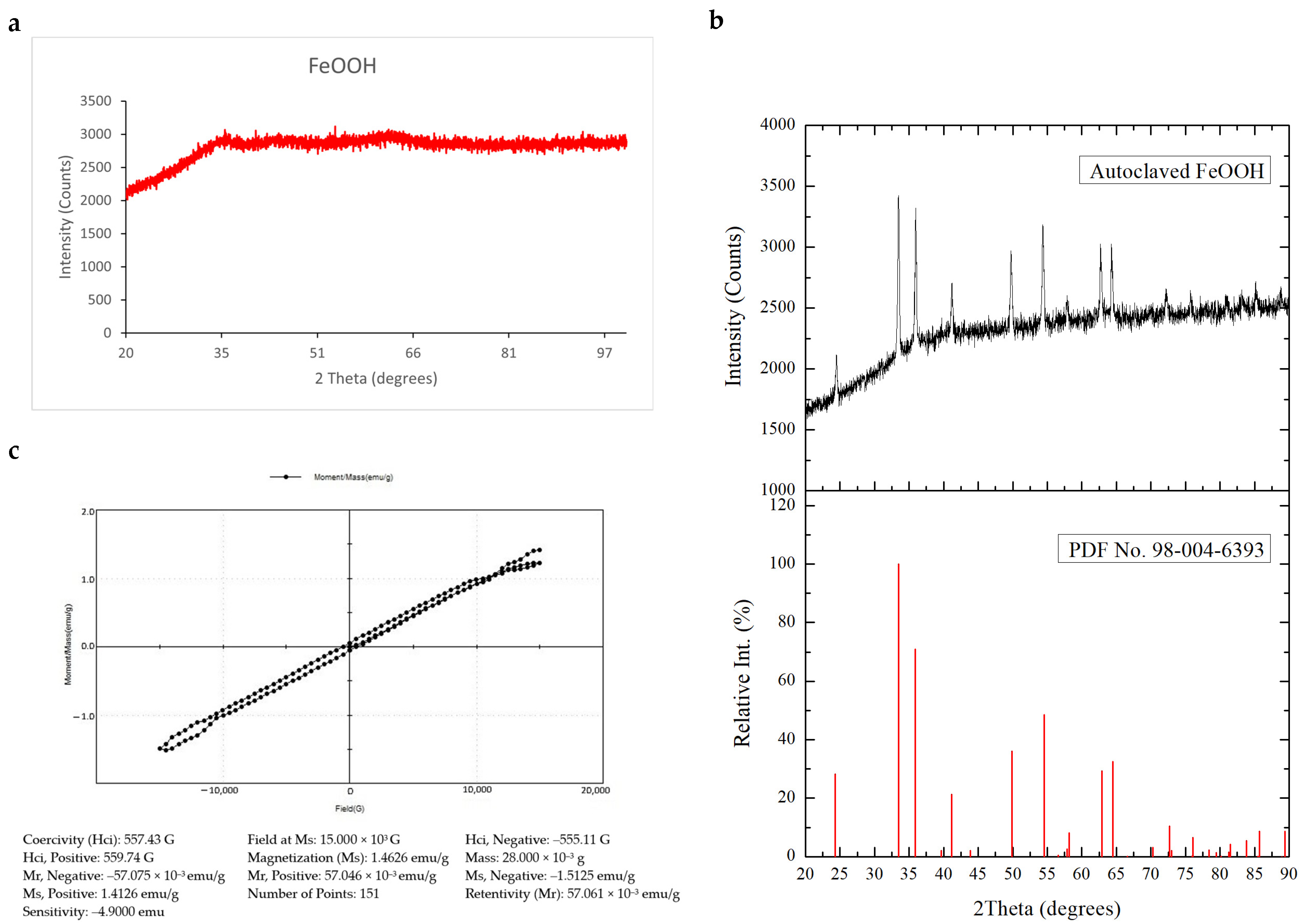
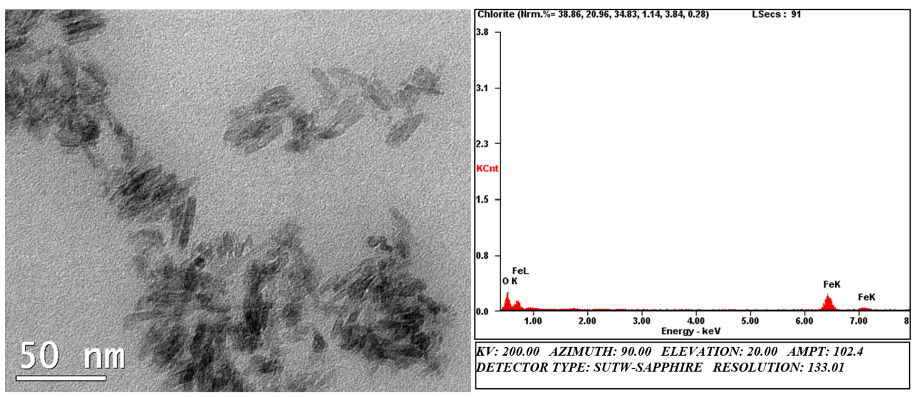
3.1. Characterization of FeOOH Precursor Particles

The XRD pattern (Figure 1a) of the precursor showed no sharp planes, confirming that the compound was amorphous in nature. Autoclaving increased the crystallinity of the precursor, as indicated by the appearance of a few peaks in the diffractogram (Figure 1b), which matched with those of hexagonal Fe2O3 (hematite). Hence, the precursor was used for experiments without autoclaving. The magnetization curve of the un-autoclaved sample exhibited no characteristic hysteresis loop of a ferromagnetic or paramagnetic material, indicating that the compound was non-magnetic at room temperature (Figure 1c).
TEM analysis showed that the FeOOH particles were nanorods with an average width of 2 nm and average length of 26 nm (Figure 2). The chemical composition of the particles was analyzed using EDS, which elicited characteristic signals for only Fe and O.

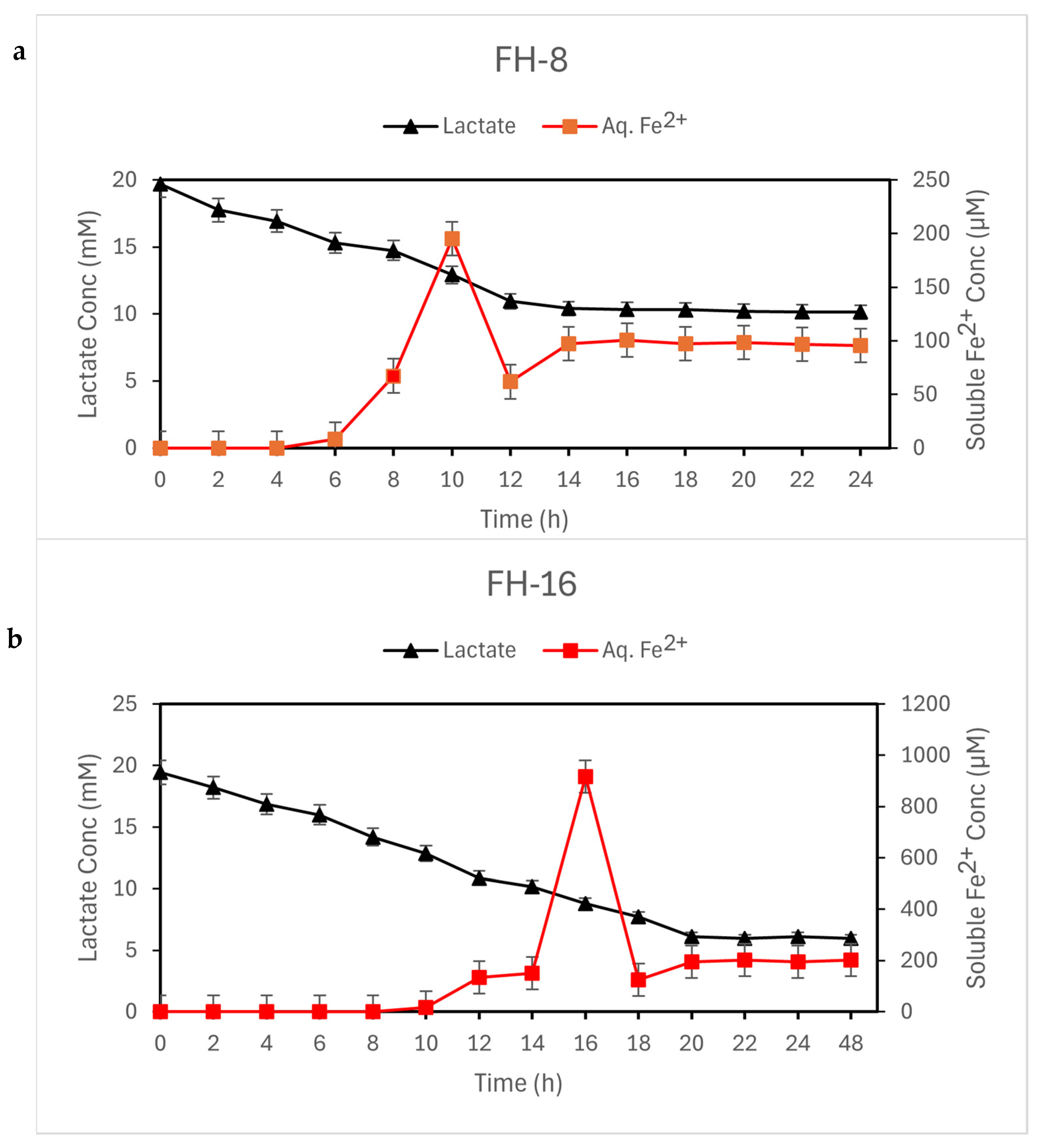
3.2. Culture Characteristics

There was no significant change in the pH (7.65) of the reaction mixture throughout the reduction experiment. In the FH-8 and FH-16 vials, the soluble ferrous ion concentration increased with the incubation time, indicating the reduction and dissolution of ferric iron in the FeOOH precursor by S. putrefaciens CN-32 (Figure 3). The color of the medium changed from the initial reddish brown to dark brown and finally to black. The drop in the soluble Fe2+ ion concentration (Figure 3) in the medium corresponded with the complete transformation of the suspension into black magnetic precipitates. This occurred at 12 h in the FH-8 vials and 18 h in the FH-16 vials. At the end of incubation, the concentrations of lactate and soluble Fe2+ in the medium were 10.133 mM and 95.37 µM, respectively in the FH-8 vials, and 5.98 mM and 201.11 µM, respectively in the FH-16 vials. More lactate was utilized and a higher concentration of soluble Fe2+ ions was produced in the FH-16 vials because they had twice the initial concentration of precursor than that in the FH-8 vials.
These results indicate that S. putrefaciens CN-32 could reduce ferric ions in the insoluble precursor to ferrous ions and induce the precipitation of a magnetic mineral under the non-growth conditions of this experiment. Lactate concentration dropped with incubation time to become nearly constant after the formation of the magnetic precipitates. The subsequent slight increase in ferrous ion concentration suggests that S. putrefaciens CN-32 reduced some ferric ions on the magnetic product surface under these conditions. However, this reduction did not lead to further transformation of the mineral product.
Control vials with only the precursor and lactate (no inoculum) in saline did not exhibit any change in color or magnetic behavior during the incubation period (Figure 4). Thus, it was confirmed that the use of an unsterilized precursor did not affect the experimental outcome and that the reduction activity was biotically mediated by the S. putrefaciens CN-32 inoculum alone.
The medium color in the FH-24 vials darkened slightly but did not change to black. The precipitates were not strongly attracted to a magnet as in the other experimental vials even after 75 d of incubation. Doubling the inoculum concentration also had no effect on the outcome, indicating that only partial iron reduction occurred under this ratio of carbon source to precursor. Table 1 presents the time taken for the formation of black precipitates in different experimental vials.

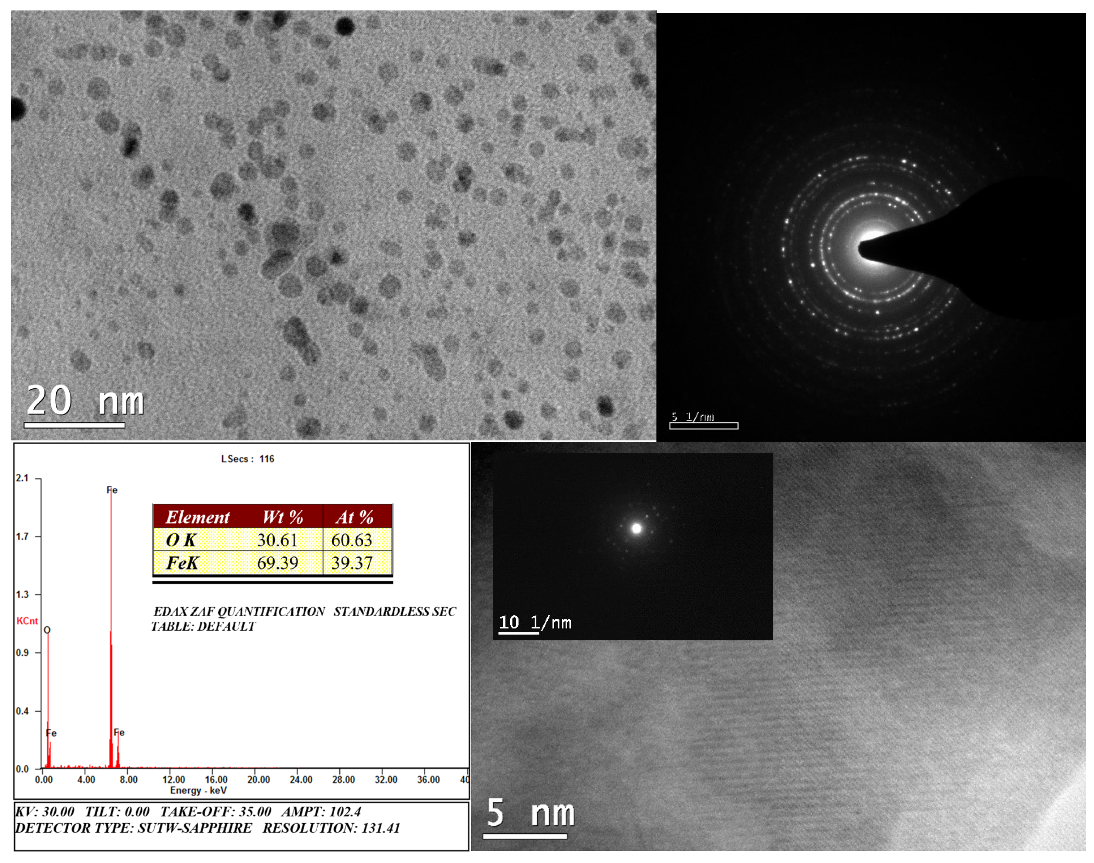
3.3. Characterization of Magnetic Products

The black, magnetic precipitates formed in the FH-8 and FH-16 vials exhibited similar characteristics. Their crystal structure was confirmed using XRD (Figure 5a). The diffractogram peaks appearing at 2θ of 30.440, 35.713, 43.370, 53.869, 57.403, 62.957, and 74.605 corresponded to those of cubic Fe3O4 (PDF No. 98-011-1045). The average crystallite size was calculated, using the Scherrer equation, to be 7.93 nm. The precipitates were strongly attracted to a magnet (Figure 5b).
The magnetization curves of the product samples showed hysteresis loops typical of superparamagnetic materials, with no appreciable coercivity (Figure 5c). The highest Ms value recorded was 73.8 emu/g (FH-16).
TEM imaging (Figure 6) showed that the MNPs formed were fairly monodispersed with low aggregation. They were pseudo-spherical in shape, with an average diameter of 6 ± 2 nm. The TEM diffraction spectrum suggested that the particle surface was polycrystalline. Furthermore, the lattice fringes observed under HR-TEM implied a highly crystalline core. EDS analysis of the MNPs elicited peaks for Fe and O, with no characteristic signals of impurities.
Shewanella putrefaciens CN-32 completely reduced FeOOH to pure superparamagnetic MNPs within 12–18 h under the experimental conditions described. Based on the weight of the magnetite collected at the end of incubation, the yield was estimated to be 1.806 g/L (7.8 mM; FH-8) and 3.473 g/L (15 mM; FH-16). Considering the amount of Fe2+ ions remaining in the solution after magnetite precipitation (~100–200 µM; Figure 3), it was essentially a 100% conversion. This is in line with the results obtained by Salas et al. [28]. It is notable that this bacterium reduced the same precursor to magnetic precipitates within 8–12 h (8–16 mM precursor) under growth conditions in our tests using a defined medium containing yeast extract, vitamins, and minerals (Table S1 in the Supplementary Material). Hence, the advantages of the non-growth bacterial system (cost and product purity) were achieved without much increase in production time.

4. Discussion

Iron reduction/oxidation of amorphous/crystalline precursors leading to magnetite formation has been studied using a number of bacterial strains (Table 2). There have also been a few attempts at large-scale MNP synthesis using DIRB [29,30]. However, in most cases, the products were reported to be mixtures containing different levels of Fe-bearing minerals such as magnetite, maghemite, hematite, geothite, lepidocrocite, siderite, vivianite, and green rust. Moreover, some of the strains employed are obligate anaerobes or extremophiles that require precise control of temperature, pH, and medium composition. Their production times vary from a few days to weeks. These factors can significantly add to the cost of synthesis on an industrial scale.
Furthermore, in some cases of pure Fe3O4 formation, the synthesized particles exhibited diameters in the single-domain (SD) range [29,35]. SPMNPs are preferred over single-domain (SD) MNPs for biomedical applications because they can be guided using an external magnetic field but do not agglomerate after the field is removed. This loss of interparticle magnetic dipolar interactions also lowers the risk of thrombus formation. Furthermore, the heating power exhibited by SPMNPs under biologically tolerable magnetic field strengths makes them beneficial for magnetic hyperthermia to treat tumors [36]. Although ferri- and ferromagnetic NPs exhibit superior heat dissipation, they might form agglomerates and induce capillary embolization. This limits the in vivo application of SDMNPs. Moreover, the heating effect of SPMNPs can be enhanced by controlling their crystal growth or adding metal dopants [37].
Biosynthesis using plant extracts is widely considered as more economical than chemical methods because it eschews the use of costly reducing agents or stabilizers. However, its ability to control particle shape and size is currently limited [38]. Altaf et al. [39] conducted a cost analysis of MNP synthesis using extracts of Moringa olifera leaves. They calculated that the cost of chemicals involved in this method was 75.3% and 94.6% less than that for chemically synthesized and commercially available magnetite, respectively. They obtained an MNP yield of 4.55 g using 3.99 g NaOH, 5.3 g FeCl3·6H2O, and 2.65 g FeCl2·4H2O at a cost of USD 33.03; however, they considered only chemical costs in their analysis but not other physical costs (such as extract preparation and heating). In comparison, our method does not involve heating or the use of FeCl2·4H2O. Therefore, according to Equations (1) and (2), our method can yield 4.26 g MNPs using 5.3 g FeCl3·6H2O, 2.35 g NaOH, and 2.207 g lactate at a cost of USD 31.01 (Table 3).
FeCl3 + 3NaOH -> FeOOH·H2O + 3NaCl
6FeOOH·H2O + lactate + H2O -> 2Fe3O4 + 2O2 + 6H2O + acetate + 2H2 + CO2
Table 3 summarizes the advantages of our bacterial system over other methods of MNP synthesis in terms of cost and sustainability.
The non-growth–associated magnetite biomineralization (NGAMB) system that we have formulated in this study exhibits the following features that make it advantageous over previously used biological and chemical methods of MNP synthesis.
  • It eschews the temperature control requirements for psychrophilic/thermophilic species.
  • Since S. putrefaciens CN-32 is a facultative anaerobe, greater quantities of pre-inoculum cells can be prepared quickly under aerobic conditions before the bioreduction process, which would increase the quantity of iron oxide/hydroxides that can be reduced in a batch and reduce overall production time.
  • The reaction is carried out in plain saline. Thus, impurities (from growth media) that may affect the crystal structure of the reduction products can be avoided. Hence, pure magnetite crystals can be produced without undesired side products. Moreover, no toxic waste is generated.
  • The MNPs obtained are superparamagnetic with saturation magnetization comparable to that of other biologically and chemically synthesized MNPs [40], making them ideal for biomedical applications.
  • This system also permits easy experimentation to study the effects of dopants on the crystal structure of biomagnetites, which can be tailored for use in different fields such as catalysis and bioremediation.
  • Furthermore, since Shewanella spp. are capable of using more than 10 different metals as terminal electron acceptors for growth [41], this system can be expanded to produce bimetallic and higher mixed metallic nanoparticles (including ferrites).
In summary, the NGAMB that we have employed in this study enhances the scope for economical large-scale green synthesis of highly pure MNPs. Since we have not used any complex or defined media for MNP synthesis, the end products of the reaction are only magnetite, cells, and saline (with minuscule amounts of lactate/acetate, which are non-toxic). The bacterial culture generates no other waste. Magnetite can be easily separated from the culture via magnetic decantation. The cells in saline can either be reused for MNP synthesis or preparation of S. putrefaciens CN-32 inoculum. Therefore, the environmental impact of our system is nil compared with that of physical/chemical fabrication. As indicated by Sadhukhan et al. [42], further experiments are underway in our lab to determine alternative precursors (such as ferric sulphate instead of chloride) for more sustainable and efficient microbial reduction to magnetite. Scale-up studies and cost–benefit analyses with different ratios of inoculum:electron donor:electron acceptor are necessary to deploy our NGAMB system at an industrial level.

Supplementary Materials

The following supporting information can be downloaded at: https://www.mdpi.com/article/10.3390/su16156278/s1, Table S1: Defined growth medium (pH 7.3 ± 0.2) used for dissimilatory iron reduction by Shewanella putrefaciens CN-32.

Author Contributions

S.P.: Methodology, investigation, formal analysis, validation, visualization, writing—original draft, writing—review and editing. T.S.C.: Conceptualization, methodology, supervision, writing—review and editing. All authors have read and agreed to the published version of the manuscript.

Funding

This research did not receive any specific grant from funding agencies in the public, commercial, or not for profit sectors.

Institutional Review Board Statement

Not applicable.

Informed Consent Statement

Not applicable.

Data Availability Statement

The original contributions presented in the study are included in the article, further inquiries can be directed to the corresponding author.

Acknowledgments

S.P. is thankful to MHRD, Government of India for the HTRA fellowship to support this research. The authors thank the Department of Chemical Engineering, Sophisticated Analytical Instrument Facility (SAIF), and Central XRD—IIT Madras for the product characterization support.

Conflicts of Interest

The authors declare no conflicts of interest.

References

  1. Natarajan, S.; Harini, K.; Gajula, G.P.; Sarmento, B.; Neves-Petersen, M.T.; Thiagarajan, V. Multifunctional Magnetic Iron Oxide Nanoparticles: Diverse Synthetic Approaches, Surface Modifications, Cytotoxicity towards Biomedical and Industrial Applications. BMC Mat. 2019, 1, 2. [Google Scholar] [CrossRef]
  2. Díez, A.G.; Rincón-Iglesias, M.; Lanceros-Méndez, S.; Reguera, J.; Lizundia, E. Multicomponent Magnetic Nanoparticle Engineering: The Role of Structure-Property Relationship in Advanced Applications. Mater. Today Chem. 2022, 26, 101220. [Google Scholar] [CrossRef]
  3. Saqib, S.; Zaman, W.; Ayaz, A.; Habib, S.; Bahadur, S.; Hussain, S.; Muhammad, S.; Ullah, F. Postharvest Disease Inhibition in Fruit by Synthesis and Characterization of Chitosan Iron Oxide Nanoparticles. Biocatal. Agric. Biotechnol. 2020, 28, 101729. [Google Scholar] [CrossRef]
  4. Vangijzegem, T.; Lecomte, V.; Ternad, I.; Van Leuven, L.; Muller, R.N.; Stanicki, D.; Laurent, S. Superparamagnetic Iron Oxide Nanoparticles (SPION): From Fundamentals to State-of-the-Art Innovative Applications for Cancer Therapy. Pharmaceutics 2023, 15, 236. [Google Scholar] [CrossRef]
  5. Koo, K.N.; Ismail, A.F.; Othman, M.H.D.; Bidin, N.; Rahman, M.A. Preparation and Characterization of Superparamagnetic Magnetite (Fe3O4) Nanoparticles: A Short Review. Malaysian, J. Fundam. Appl. Sci. 2019, 15, 23–31. [Google Scholar] [CrossRef]
  6. Campaña, A.L.; Saragliadis, A.; Mikheenko, P.; Linke, D. Insights into the Bacterial Synthesis of Metal Nanoparticles. Front. Nanotechnol. 2023, 5, 1216921. [Google Scholar] [CrossRef]
  7. Saqib, S.; Zaman, W.; Ullah, F.; Majeed, I.; Ayaz, A.; Hussain Munis, M.F. Organometallic Assembling of Chitosan-Iron Oxide Nanoparticles with Their Antifungal Evaluation Against Rhizopus oryzae. Appl. Organomet. Chem. 2019, 33, e5190. [Google Scholar] [CrossRef]
  8. Fu, X.; Cai, J.; Zhang, X.; Li, W.D.; Ge, H.; Hu, Y. Top-Down Fabrication of Shape-Controlled, Monodisperse Nanoparticles for Biomedical Applications. Adv. Drug Deliv. Rev. 2018, 132, 169–187. [Google Scholar] [CrossRef]
  9. Ganapathe, L.S.; Mohamed, M.A.; Mohamad Yunus, R.; Berhanuddin, D.D. Magnetite (Fe3O4) Nanoparticles in Biomedical Application: From Synthesis to Surface Functionalisation. Magnetochemistry 2020, 6, 68. [Google Scholar] [CrossRef]
  10. Carmona, M.; Poblete-Castro, I.; Rai, M.; Turner, R.J. Opportunities and Obstacles in Microbial Synthesis of Metal Nanoparticles. Microb. Biotechnol. 2023, 16, 871–876. [Google Scholar] [CrossRef]
  11. Xu, W.; Yang, T.; Liu, S.; Du, L.; Chen, Q.; Li, X.; Dong, J.; Zhang, Z.; Lu, S.; Gong, Y.; et al. Insights into the Synthesis, Types and Application of Iron Nanoparticles: The Overlooked Significance of Environmental Effects. Environ. Int. 2022, 158, 106980. [Google Scholar] [CrossRef]
  12. Jacinto, M.J.; Silva, V.C.; Valladão, D.M.S.; Souto, R.S. Biosynthesis of Magnetic Iron Oxide Nanoparticles: A Review. Biotechnol. Lett. 2021, 43, 1–12. [Google Scholar] [CrossRef]
  13. Kapoor, R.T.; Salvadori, M.R.; Rafatullah, M.; Siddiqui, M.R.; Khan, M.A.; Alshareef, S.A. Exploration of Microbial Factories for Synthesis of Nanoparticles—A Sustainable Approach for Bioremediation of Environmental Contaminants. Front. Microbiol. 2021, 12, 658294. [Google Scholar] [CrossRef]
  14. Narayanan, K.B.; Sakthivel, N. Biological Synthesis of Metal Nanoparticles by Microbes. Adv. Colloid Interface Sci. 2010, 156, 1–13. [Google Scholar] [CrossRef]
  15. Atalah, J.; Espina, G.; Blamey, L.; Muñoz-Ibacache, S.A.; Blamey, J.M. Advantages of Using Extremophilic Bacteria for the Biosynthesis of Metallic Nanoparticles and its Potential for Rare Earth Element Recovery. Front. Microbiol. 2022, 13, 855077. [Google Scholar] [CrossRef]
  16. Sun, J.-B.; Zhao, F.; Tang, T.; Jiang, W.; Tian, J.; Li, Y.; Li, J.L. High-Yield Growth and Magnetosome Formation by Magnetospirillum gryphiswaldense MSR-1 in an Oxygen-Controlled Fermentor Supplied Solely with Air. Appl. Microbiol. Biotechnol. 2008, 79, 389. [Google Scholar] [CrossRef]
  17. Gahlawat, G.; Choudhury, A.R. A Review on the Biosynthesis of Metal and Metal Salt Nanoparticles by Microbes. RSC Adv. 2019, 9, 12944–12967. [Google Scholar] [CrossRef]
  18. Esther, J.; Sukla, L.B.; Pradhan, N.; Panda, S. Fe (III) Reduction Strategies of Dissimilatory Iron Reducing Bacteria. Korean J. Chem. Eng. 2015, 32, 1–14. [Google Scholar] [CrossRef]
  19. Abhilash; Revati, K.; Pandey, B.D. Microbial Synthesis of Iron-Based Nanomaterials—A Review. Bull. Mater. Sci. 2011, 34, 191–198. [Google Scholar] [CrossRef]
  20. Roh, Y.; Liu, S.V.; Li, G.; Huang, H.; Phelps, T.J.; Zhou, J. Isolation and Characterization of Metal-Reducing Thermoanaerobacter Strains from Deep Subsurface Environments of the Piceance Basin, Colorado. Appl. Environ. Microbiol. 2002, 68, 6013–6020. [Google Scholar] [CrossRef]
  21. Fredrickson, J.K.; Zachara, J.M.; Kennedy, D.W.; Dong, H.; Onstott, T.C.; Hinman, N.W.; Li, S. Biogenic Iron Mineralization Accompanying the Dissimilatory Reduction of Hydrous Ferric Oxide by a Groundwater Bacterium. Geochim. Cosmochim. Acta 1998, 62, 3239–3257. [Google Scholar] [CrossRef]
  22. Wang, Q.; Wei, Z.; Yi, X.; Tang, J.; Feng, C.; Dang, Z. Biogenic Iron Mineralization of Polyferric Sulfate by Dissimilatory Iron Reducing Bacteria: Effects of Medium Composition and Electric Field Stimulation. Sci. Total Environ. 2019, 684, 466–475. [Google Scholar] [CrossRef]
  23. Yang, J.; Ju, P.; Dong, X.; Duan, J.; Xiao, H.; Tang, X.; Zhai, X.; Hou, B. Green Synthesis of Functional Metallic Nanoparticles by Dissimilatory Metal-Reducing Bacteria “Shewanella”: A Comprehensive Review. J. Mater. Sci. Technol. 2023, 158, 63–76. [Google Scholar] [CrossRef]
  24. Lovley, D.R.; Holmes, D.E.; Nevin, K.P. Dissimilatory Fe(III) and Mn(IV) Reduction. In Advances in Microbial Physiology; Elsevier: Amsterdam, The Netherlands, 2004; Volume 49, pp. 219–286. [Google Scholar]
  25. Lovley, D.R.; Phillips, E.J.P. Organic Matter Mineralization with Reduction of Ferric Iron in Anaerobic Sediments. Appl. Environ. Microbiol. 1986, 51, 683–689. [Google Scholar] [CrossRef]
  26. Miller, T.L.; Wolin, M.J. A Serum Bottle Modification of the Hungate Technique for Cultivating Obligate Anaerobes. Appl. Microbiol. 1974, 27, 985–987. [Google Scholar] [CrossRef]
  27. Kim, G.T.; Hyun, M.S.; Chang, I.S.; Kim, H.J.; Park, H.S.; Kim, B.H.; Kim, S.D.; Wimpenny, J.W.T.; Weightman, A.J. Dissimilatory Fe(III) Reduction by an Electrochemically Active Lactic Acid Bacterium Phylogenetically Related to Enterococcus gallinarum Isolated from Submerged Soil. J. Appl. Microbiol. 2005, 99, 978–987. [Google Scholar] [CrossRef]
  28. Salas, E.C.; Berelson, W.M.; Hammond, D.E.; Kampf, A.R.; Nealson, K.H. The Impact of Bacterial Strain on the Products of Dissimilatory Iron Reduction. Geochim. Cosmochim. Acta 2010, 74, 574–583. [Google Scholar] [CrossRef]
  29. Moon, J.-W.; Rawn, C.J.; Rondinone, A.J.; Love, L.J.; Roh, Y.; Everett, S.M.; Lauf, R.J.; Phelps, T.J. Large-Scale Production of Magnetic Nanoparticles Using Bacterial Fermentation. J. Ind. Microbiol. Biotechnol. 2010, 37, 1023–1031. [Google Scholar] [CrossRef]
  30. Byrne, J.M.; Muhamadali, H.; Coker, V.S.; Cooper, J.; Lloyd, J.R. Scale-up of the Production of Highly Reactive Biogenic Magnetite Nanoparticles Using Geobacter sulfurreducens. J. R. Soc. Interface 2015, 12, 20150240. [Google Scholar] [CrossRef]
  31. Zhang, C.; Liu, S.; Phelps, T.J.; Cole, D.R.; Horita, J.; Fortier, S.M.; Elless, M.; Valley, J.W. Physiochemical, Mineralogical, and Isotopic Characterization of Magnetite-Rich Iron Oxides Formed by Thermophilic Iron-Reducing Bacteria. Geochim. Cosmochim. Acta 1997, 61, 4621–4632. [Google Scholar] [CrossRef]
  32. Roh, Y.; Zhang, C.-L.; Vali, H.; Lauf, R.J.; Zhou, J.; Phelps, T.J. Biogeochemical and Environmental Factors in Fe Biomineralization: Magnetite and Siderite Formation. Clays Clay Miner. 2003, 51, 83–95. [Google Scholar] [CrossRef]
  33. Shimizu, M.; Zhou, J.; Schröder, C.; Obst, M.; Kappler, A.; Borch, T. Dissimilatory Reduction and Transformation of Ferrihydrite-Humic Acid Coprecipitates. Environ. Sci. Technol. 2013, 47, 13375–13384. [Google Scholar] [CrossRef]
  34. Bingjie, O.; Xiancai, L.; Huan, L.; Juan, L.; Tingting, Z.; Xiangyu, Z.; Jianjun, L.; Rucheng, W. Reduction of Jarosite by Shewanella oneidensis MR-1 and Secondary Mineralization. Geochim. Cosmochim. Acta 2014, 124, 54–71. [Google Scholar] [CrossRef]
  35. Miot, J.; Li, J.; Benzerara, K.; Sougrati, M.T.; Ona-Nguema, G.; Bernard, S.; Jumas, J.-C.; Guyot, F. Formation of Single Domain Magnetite by Green Rust Oxidation Promoted by Microbial Anaerobic Nitrate-Dependent Iron Oxidation. Geochim. Cosmochim. Acta 2014, 139, 327–343. [Google Scholar] [CrossRef]
  36. Rajan, A.; Sahu, N.K. Review on Magnetic Nanoparticle-Mediated Hyperthermia for Cancer Therapy. J. Nanoparticle Res. 2020, 22, 319. [Google Scholar] [CrossRef]
  37. Laurent, S.; Dutz, S.; Häfeli, U.O.; Mahmoudi, M. Magnetic Fluid Hyperthermia: Focus on Superparamagnetic Iron Oxide Nanoparticles. Adv. Colloid Interface Sci. 2011, 166, 8–23. [Google Scholar] [CrossRef]
  38. Saif, S.; Tahir, A.; Chen, Y. Green Synthesis of Iron Nanoparticles and Their Environmental Applications and Implications. Nanomaterials 2016, 6, 209. [Google Scholar] [CrossRef] [PubMed]
  39. Altaf, S.; Zafar, R.; Zaman, W.Q.; Ahmad, S.; Yaqoob, K.; Syed, A.; Khan, A.J.; Bilal, M.; Arshad, M. Removal of Levofloxacin from Aqueous Solution by Green Synthesized Magnetite (Fe3O4) Nanoparticles Using Moringa olifera: Kinetics and Reaction Mechanism Analysis. Ecotoxicol. Environ. Saf. 2021, 226, 112826. [Google Scholar] [CrossRef]
  40. Yeary, L.W.; Moon, J.-W.; Love, L.J.; Thompson, J.R.; Rawn, C.J.; Phelps, T.J. Magnetic Properties of Biosynthesized Magnetite Nanoparticles. IEEE Trans. Magn. 2005, 41, 4384–4389. [Google Scholar] [CrossRef]
  41. Rajput, V.D.; Minkina, T.; Kimber, R.L.; Singh, V.K.; Shende, S.; Behal, A.; Sushkova, S.; Mandzhieva, S.; Lloyd, J.R. Insights into the Biosynthesis of Nanoparticles by the Genus Shewanella. Appl. Environ. Microbiol. 2021, 87, e01390-21. [Google Scholar] [CrossRef]
  42. Sadhukhan, J.; Joshi, N.; Shemfe, M.; Lloyd, J.R. Life Cycle Assessment of Sustainable Raw Material Acquisition for Functional Magnetite Bionanoparticle Production. J. Environ. Manag. 2017, 199, 116–125. [Google Scholar] [CrossRef] [PubMed]
Figure 1. X-ray diffraction (XRD) spectra of FeOOH precursor (a) before autoclaving and (b) after autoclaving, and (c) magnetization curve of unautoclaved precursor used for experiments.
Sustainability 16 06278 g001
Figure 2. Transmission electron microscopy (TEM) and energy-dispersive X-ray spectroscopy (EDS) analysis of FeOOH precursor.
Sustainability 16 06278 g002
Figure 3. Ferrous (Fe2+) ion dissolution and lactate utilization by Shewanella putrefaciens CN-32 in saline. Initial concentrations were 1 × 10⁷ cells/mL, 20 mM lactate, and (a) 8 mM or (b) 16 mM precursor. Data presented are averages of triplicate results; the variation was ≤ 5%.
Sustainability 16 06278 g003
Figure 4. Representative control (without bacterial inoculum) and experimental serum vials after incubation. The reddish-brown FeOOH precursor was transformed into black magnetic precipitates by bacterial action. The control vials showed no change.
Sustainability 16 06278 g004
Figure 5. (a) Representative XRD spectrum, (b) image, and (c) magnetization curve of black precipitates at the end of incubation.
Sustainability 16 06278 g005
Figure 6. Representative TEM and EDS analysis images of magnetic products.
Sustainability 16 06278 g006
Table 1. Effect of precursor concentration on time taken for magnetic product formation by Shewanella putrefaciens CN-32.
VialPrecursor Concentration (mM)Inoculum Concentration (cells/mL)Product CharacteristicTime (h)
FH-881 × 107Magnetic12
FH-16161 × 107Magnetic18
FH-24241 × 107Weakly magnetic>1800
FH-24242 × 107Weakly magnetic>1800
Table 2. Purity and characteristics of magnetite nanoparticles (MNPs) synthesized using bacteria.
StrainTypeProduct CharacteristicsTotal Production TimeReference
Enrichment culturesTAs aMixture>2 d[31]
Thermoanaerobacter ethanolicus TOR-39TA aSD e magnetite>2 d[32]
Shewanella alga BrYMFA bMagnetiteNA
Shewanella alga NV-1PFA cMagnetite>5 d
Shewanella
pealeana W3-7-1
PFA cSP f magnetiteNA
Shewanella
putrefaciens strain CN32
MFA bMixture>2 d[33]
Shewanella oneidensis MR-1MFA bMixture>2 d[34]
Acidovorax sp. strain BoFeN1AnaerobeMixture with SD e magnetite>2 d[35]
Thermoanaerobacter sp. TOR-39TA aSD e magnetite>2 d[29]
Geobacter sulfurreducensMA dMixture>2 d[30]
Shewanella
putrefaciens strain CN32
MFA bSP f magnetite1–2 dThis study
a TA: Thermophilic anaerobe; b MFA: Mesophilic facultative anaerobe; c PFA: Psychrotolerant facultative anaerobe; d MA: Mesophilic anaerobe; e SD: Single-domain; f SP: Superparamagnetic.
Table 3. Comparison of cost and sustainability aspects of MNP synthesis using different methods.
Material/AspectUnit Cost (USD)Chemically Synthesized
(Ref. [39])
Green Synthesized
(Ref. [39])
This Study
Amount Used (g)Cost (USD)NP Yield (g)Amount Used (g)Cost (USD)NP Yield (g)Amount Used (g)Cost (USD)NP Yield (g)
FeCl3.6H2O5.7619.8114.040.805.330.524.555.330.524.26
FeCl2.4H2O0.72627.0319.622.651.92--
NaOH0.153.990.593.990.592.350.35
Nitrogen gas0.010/L500 (mL)0.005--500 (mL)0.005
Lactate0.063----2.2070.14
Heating requirement YesYesNo
Waste generated Spent liquorSpent liquorRe-usable cells and saline
Total Chemical Cost (USD) 134.25 33.03 31.01
Disclaimer/Publisher’s Note: The statements, opinions and data contained in all publications are solely those of the individual author(s) and contributor(s) and not of MDPI and/or the editor(s). MDPI and/or the editor(s) disclaim responsibility for any injury to people or property resulting from any ideas, methods, instructions or products referred to in the content.

