1. Introduction
Liubao tea is prepared from Wuzhou large tea leaves from China. It is a specialty tea generated through the processes of natural fermentation, pile fermentation, drying, autoclaving, aging, and other characteristics. Therefore, Liubao tea is a kind of black tea that is post-fermented [
1]. This tea is named according to its geographical origin, namely, Liubao County, Wuzhou City, Guangxi Province, China. Liubao tea was historically used as a preventive medicine [
2]. The majority of research studies focus on Pu’er tea, Hunan black tea, and Fuzhuan tea, whereas investigations of relevant technology and functions of Liubao tea are limited [
3]. Recent studies showed that Liubao tea imparts lipid-lowering effects, regulates glucose and lipid metabolism, possesses anti-oxidation activity, and regulates immune function and intestinal flora. These health benefits come from the components of Liubao tea, which include polyphenols, flavonoids, caffeine, free amino acids, and soluble sugars [
4,
5,
6].
The liver is an important metabolic organ, and damage to this organ can cause severe harm to the human body, which includes liver injury due to chemicals such as higher alcohol intake, drug side effects, and environmental toxic chemicals, ultimately leading to cirrhosis and liver cancer [
7]. Carbon tetrachloride (CCl
4) is a common chemical inducer of liver damage in the laboratory. CCl
4 triggers the production of high levels of inflammatory cytokines in liver cells during liver injury, which aggravates inflammation and liver damage. Simultaneously, CCl
4 can induce the formation of Cl
− and CCl
3− in liver cell microsomes, leading to lipid peroxidation of liver microsomes, resulting in lipid peroxidation and destruction of cell membranes, and ultimately, liver damage [
8].
Active oxygen free radicals cause oxidative stress, which is a common pathophysiological mechanism of liver diseases. Oxidative stress could cause hepatic damage by inducing membrane lipid peroxidation that changes biofilm function, as well as inducing covalent combinations with biological macromolecules, and destruction of enzyme activities (such as tumor necrosis factor-α (TNF-α) and nuclear factor κ-light-chain-enhancer of activated B cells (NF-κB)) [
9]. Oxidative stress plays an important role in fatty liver, viral hepatitis, liver fibrosis, and other liver diseases [
10]. Energy metabolism in organisms utilizes oxygen as an electronic acceptor in the process of aerobic metabolism, which inevitably produces reactive oxygen species (ROS). ROS has a dual effect, which is closely related to the regulation of some physiological active substances and the inflammatory immune process, but excessive ROS can easily lead to oxidative stress [
11]. The mitochondrial respiratory chain complex uses electron transfer to produce ATP, which is the main source of ROS. The liver is rich in mitochondria, and is, therefore, the main organ susceptible to ROS attack, and oxidative stress has a close relationship with most liver damage [
12]. ROS can also initiate a variety of cytokines such as transforming growth factor-beta (TGF-β), interleukin-8 (IL-8), and NF-κB. These cytokines can lead to infiltration of neutrophils, enhance inflammatory response, and ultimately lead to liver cell injury [
13].
Tea polyphenols are a very important component of tea. Studies showed that tea polyphenols have a strong scavenging effect on oxygen free radicals [
14,
15]. Tea polyphenols can sequester lipid peroxidation free radicals during the peroxidation process, lower polyphenolic free-radical content, and interrupt free-radical oxidation chain reactions, thereby effectively removing free radicals [
14]. Simultaneously, tea polyphenols can activate and enhance the activity of various antioxidant enzymes such as superoxide dismutase (SOD), glutathione peroxidase (GSH-Px), and catalase (CAT), as well as efficiently eliminate free radicals [
16]. Tea polyphenols can prevent lipid peroxidation caused by CCl
4, as well as avoid the damage of the membrane structure and function of liver cells caused by the covalent binding of CCl
4 and liver microsomal lipids and proteins [
17]. In addition, tea polyphenols impart a protective effect on obstructive jaundice liver injury caused by peroxidation, acute liver injury caused by cadmium poisoning, alcoholic liver injury, and liver cancer [
18]. In addition, except for certain reports of catechins and other individual tea polyphenols on liver injury protective effects, the characteristics of the tea polyphenols and polyphenol composition analysis remain unclear, including those of Liubao tea [
19].
This study utilized CCl4 in establishing a chemical liver injury mouse model to investigate the preventive effect of PLT. We also employed molecular biology methods to test the indices of serum and liver tissue, and the preventive mechanism of PLT on liver injury was elucidated. The results of this study may facilitate the development and utilization of PLT in food processing and the manufacture of health products.
4. Discussion
Liubao tea is a non-toxic food and meets the requirements of food safety according to the standards of food toxicology [
25]. Liupao tea was shown to have weight-loss effects [
3], and Liupao tea polyphenols were also found to inhibit weight gain in mice in this study. Therefore, there were individual differences in the body weight of mice before carbon-tetrachloride (CTC) treatment. Liver injuries may result in harmful and sometimes life-threatening effects to the body. Liver quality and liver index, which are indices of CTC-induced liver injury, were used in the present study [
26]. The results show that PLT can reduce the liver quality and liver indices of mice with liver injury, and these effects are similar to those using the liver injury drug, silymarin.
ALT and AST are expressed by hepatocytes; ALT is secreted into the cytoplasm, whereas AST is mainly produced in the mitochondria of hepatocytes. Damage to cells due to hepatitis, myocarditis, and pancreatitis induces ALT to enter the bloodstream. However, during severe damage, AST also enters the bloodstream [
27]. Thus, a significant increase in ALT and AST levels indicates liver damage [
28]. Liver injury can lead to the transfer of fatty acids to the liver, resulting in increased intrahepatic TG content, and TG levels also reflect the degree of liver lipid peroxidation [
29]. In this study, PLT was found to inhibit the increase in ALT, AST, and TG levels caused by carbon tetrachloride liver injury. ALT, AST, and TG are the most typical clinical liver function indicators [
27,
28]. Observing the influence of these indicators can judge whether the liver function is normal or not. It could be seen that PLT had a certain effective role in restoring normal liver function.
CCl
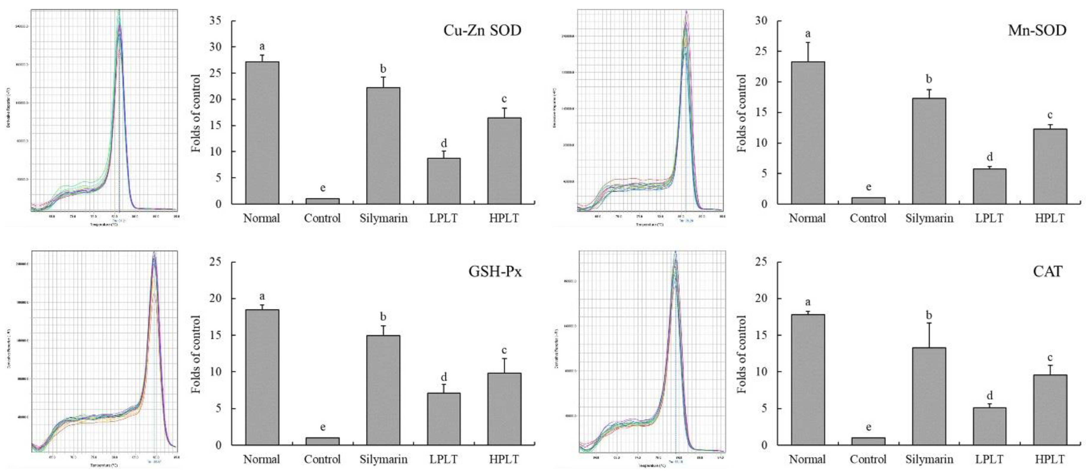
4 will lead to the body’s oxidation; the body utilizes two defenses, namely, non-enzymatic and enzymatic, to prevent oxidative damage, including regulation of SOD, CAT, and GSH-Px, which are the main mechanisms for enzymatic oxidation [
30]. SOD catalyzes superoxide radicals and is capable of scavenging free radicals, whereas CAT and SOD synergistically enhance the role of free radicals [
31]. GSH-Px is an important enzyme that catalyzes the decomposition of hydrogen peroxide, which in turn, protects cell membranes and prevents cell damage [
32]. MDA is a metabolite of lipid peroxidation; a high content of MDA accumulates in the body after liver injury [
33]. In this study, we found that PLT could significantly regulate the levels of SOD, GSH-Px, and MDA in the body caused by liver injury, thereby protecting the liver from the effects of carbon tetrachloride.
CCl
4 induces oxidization and liver inflammation, resulting in a significant increase in serum IL-6, IL-12, TNF-α, and IFN-γ levels in mice [
21]. IL-6 is a factor secreted by T helper 2 (Th2) cells and is involved in the humoral immune response. An increase in Th2 levels may result in visceral dysfunction [
34]. IL-6 promotes the differentiation, proliferation, and antibody production of T lymphocytes. It can also change intracellular G cell activity and upregulate neutrophil function, as well as enhance inflammatory reactions of the body [
35]. IL-12 is an activating factor of natural killer (NK) cells, and its effect is the most intense. High rates of apoptosis in hepatocytes and excessive immune response during liver injury further aggravate the condition, which is related to the fact that IL-12 increases the cytotoxicity of cluster of differentiation 8 (CD8)
+ T cells [
36]. Binding of TNF-α and liver cell membrane TNF-α receptor 1 (TNF-αR1) can induce intracellular double-stranded DNA to fragment, thereby resulting in stem cell apoptosis. In addition, TNF-α triggers inflammatory responses by activating NF-κB, which exacerbates liver injury [
37]. IFN-γ is a proinflammatory cytokine that increases the sensitivity of hepatocytes to TNF-α, rendering hepatocytes to further damage [
38]. Oxidative stress after liver tissue damage can cause an imbalance in the level of inflammatory cytokines such as TNF-α, IL-1β, and IL-6, which increases in the levels of TNF-α, IL-1β, and IL-6 in the liver [
39]. Through the detection of inflammatory cytokines, we also found that PLT could inhibit inflammation by reducing the level of inflammatory factors, thereby reducing liver injury.
Mn-SOD and Gu/Zn-SOD are SOD isomers [
40]. Mn-SOD is an SOD radical scavenger in the mitochondria [
41]. Gu/Zn-SOD is an SOD free radical scavenger in the cytoplasm and takes Cu
2+ and Zn
2+ as its active center [
42]. The liver and heart are organs that are rich in mitochondria, and Mn-SOD activity markedly decreases after CCl
4-induced liver injury [
43]. The same result was obtained in this study. Gu/Zn-SOD can purify the toxic effects of O
2− in the body, and protect the visceral tissues [
44]. Studies showed that CCl
4 causes oxidative stress reactions in the body, resulting in the excessive production of free radicals. Mn-SOD and Gu/Zn-SOD can inhibit free radicals in the body, and play a preventive role in liver injury [
45,
46]. CAT is an important antioxidant enzyme in the body. CAT can eliminate H
2O
2 in the body, thereby inhibiting oxidative stress, reducing the body’s oxidation caused by carbon tetrachloride, and inhibiting liver injury [
47]. Through the detection of gene and protein expression, it was further found that PLT could regulate the expressions of oxidation-related proteins in tissues, thereby reducing the damage caused by oxidative stress to tissues, thus protecting the liver.
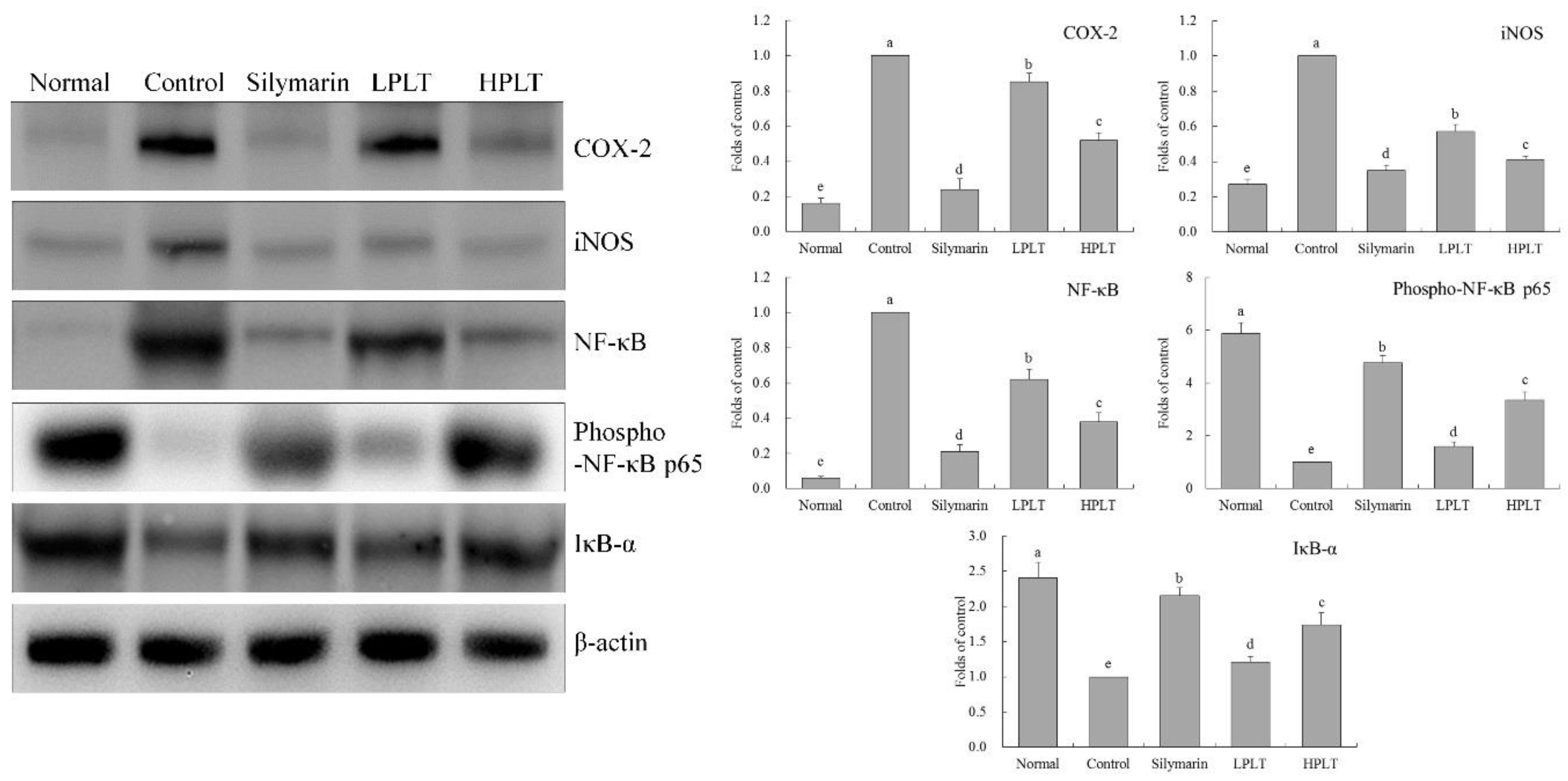
NF-κB is a key factor in the regulation of inflammatory response, including inflammation-related IL-6 and TNF-α; these are upregulated during inflammation. Under normal circumstances, NF-κB and IκB-α in the bound state show an inactivation of both. NF-κB and IκB-α in extrinsic inflammatory conditions lead to the inhibition of inflammation by binding to IκB-α to activate NF-κB [
48]. Meanwhile, through phosphorylation of NF-κB, it can reduce the promotion of inflammation of NF-κB and alleviate tissue damage [
49]. NO is a highly active oxidant that is produced in the liver by activated NOS in liver cells, and promotes the high expression of the
iNOS gene upon liver damage progression. In liver injury, oxidative stress occurs in hepatocytes, and a large number of inflammatory factors are released [
50]. iNOS is an important inflammatory factor, and iNOS is very active in inflammation. iNOS-induced NO also promotes further damage to the liver [
51]. COX-2 is also an important inflammatory factor; the tissue is not expressed under normal conditions. COX-2 expression raises after liver injury, whereby Kupffer cells are activated, and COX-2 upregulation exacerbates the liver inflammation [
52]. Further experiments showed that PLT could regulate the expressions of COX-2, iNOS, NF-κB, p-NF-κB p65, and IκB-α, alleviating the liver injury caused by inflammation and carbon tetrachloride.
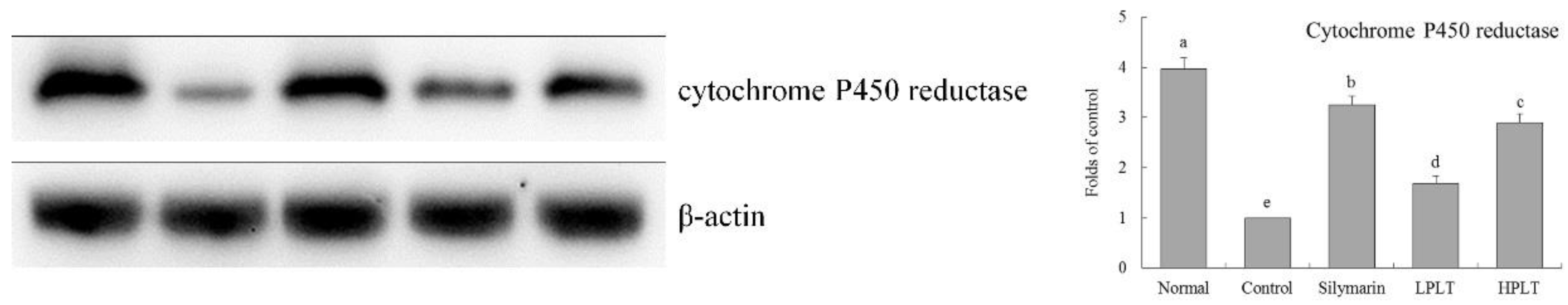
Most foreign compounds depend on metabolism by P450 in the liver. When carbon tetrachloride induces acute liver injury, lipid peroxidation and a large number of free radicals are produced in the liver, resulting in a decrease in activity of cytochrome P450. At the same time, carbon tetrachloride directly inhibits the synthesis of the cytochrome P450 enzyme, and the decrease in cytochrome P450 enzyme activity directly or indirectly leads to a decrease in detoxification ability of the liver, thereby aggravating liver injury [
53]. Upon carbon tetrachloride treatment, PLT could raise the activity of cytochrome P450 reductase, and inhibited the liver injury.
Gallic acid (GA) can inhibit oxidative stress and cytotoxicity to improve liver injury. The activation of hepatic stellate cells (HSCs) caused by liver injury is an important part of liver fibrosis [
54]. GA can also induce HSCs to produce O
2−, OH
−, and H
2O
2, thereby inducing oxidative stress that selectively kills HSCs [
55]. Catechin also affects free-radical scavenging by reducing the content of MDA and increasing the activity of SOD [
56]. Catechin has inhibitory effects on chronic hepatitis [
57]. IL-1β can induce hepatic acute-phase protein synthesis, thereby affecting normal liver activity [
58], while caffeine suppresses the production of inflammatory molecules, thus preventing the activating of the immune system in IL-1β [
59], which may play a role in liver protection. Animal model studies showed that caffeine could also inhibit acute alcoholic liver injury possibly by imparting antioxidant effects and inhibiting the expressions of IL-1β and TNF-α [
60]. EC also has antioxidant effects that influence cardiovascular disease, hypertension, cancer, and obesity [
61]. EC could reduce the inflammatory-related expression of NF-κB, iNOS, and TNF-α [
62]. Oxidative stress is considered to be the main cause of CCl
4-induced liver damage. CCl
4 is metabolized by cytochrome P450 in hepatocytes to produce three chloromethyl radicals. These radicals cause lipid peroxidation and lipid peroxidation products, which cause liver cell damage and promote the formation of fibrous tissue. EGCG plays a role in anti-CCl
4-induced liver fibrosis in rats through its antioxidant capacity [
63]. The effects of EGCG on anti-liver injury are also reflected in the inhibition of TNF-α and IFN-γ by EGCG expression, thus preventing further immune damage caused by TNF-α and IFN-γ [
64]. GCG can inhibit oxidative damage to tissues and protect viscera from oxidative damage [
65]. ECG has anti-cancer effects, possibly stronger than EGCG [
66]. ECG has better melanin inhibition effects than EGCG, and ECG shows antioxidant effects, which are greater than EGCG [
67]. These active components are combined together to form PLT, thereby strongly inhibiting liver injury. PLT is a mixture with substantial biological activity. Its action may be the combined action of many substances, and its specific mechanism needs further study.
In this study, toxic carbon tetrachloride was used to simulate chemical-induced liver injury, and the observed effects remained at the laboratory level. In order to better prove this study’s argument, future research on the human body is expected. In addition, the role of PLT in liver injury needs to be further studied, which will be conducive to more obvious discoveries of the link between its active components and their mechanisms. At the same time, in view of the mechanism of PLT, it is necessary to verify the mechanism more accurately for the differences across PLT components in the future.


