Systematic Review and Meta-Analysis: Accuracy of Both Gamma Delta+ Intraepithelial Lymphocytes and Coeliac Lymphogram Evaluated by Flow Cytometry for Coeliac Disease Diagnosis
Abstract
:1. Introduction
2. Methods
2.1. Search Strategy and Study Selection
2.2. Outcome Assessment
2.2.1. Data Extraction
2.2.2. Study Methodological Quality
2.2.3. Data Synthesis and Statistical Analysis
3. Results
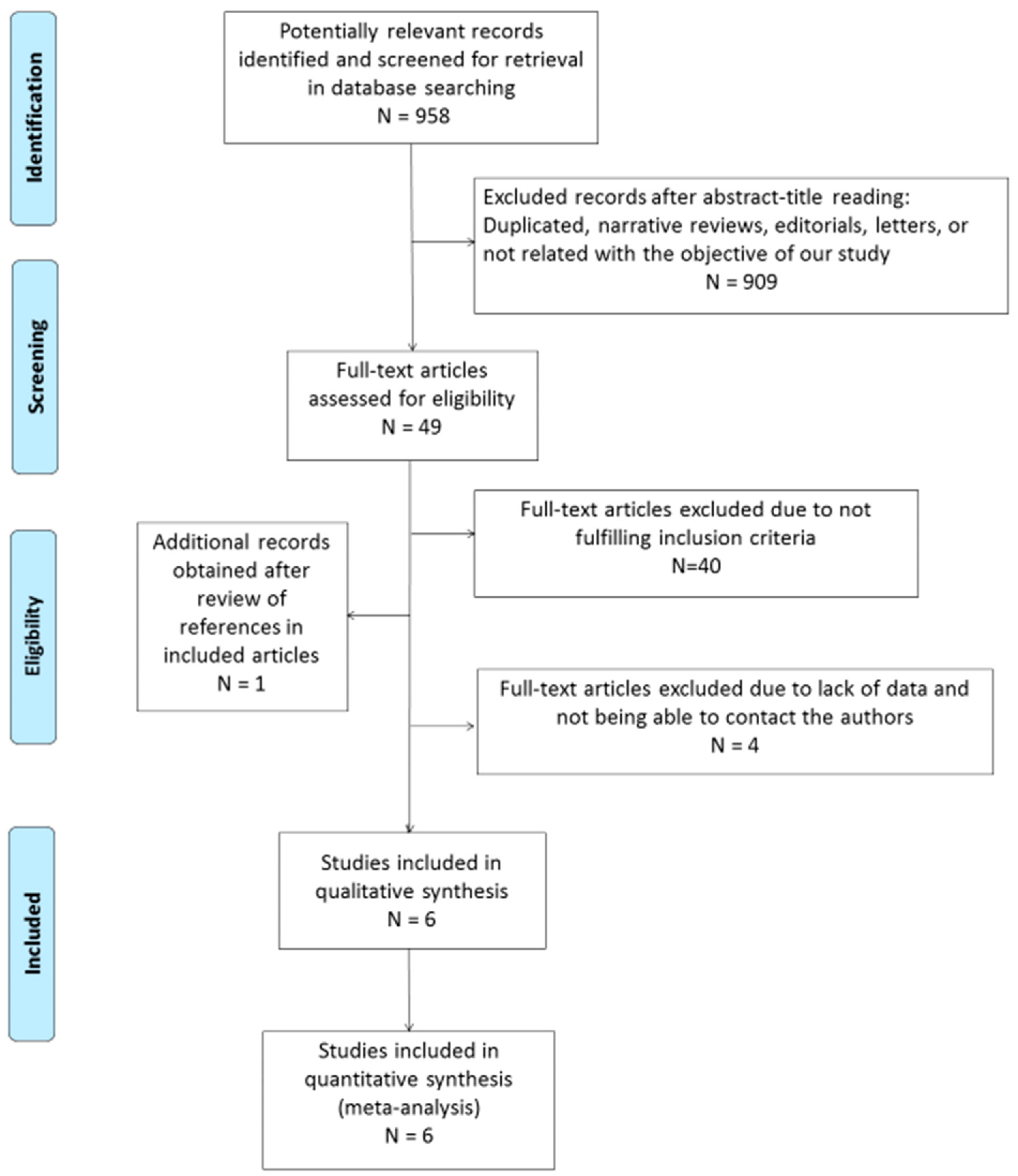
3.1. Search Results
3.2. Description of the Included Studies
3.3. Qualitative Analysis of Included Studies
3.4. Quantitative Analysis of Included Studies
3.5. TCRγδ+ IEL in Diseased Controls with Non-Coeliac Atrophy
3.6. TCRγδ+ IEL Evolution after a Gluten-Free Diet
4. Discussion
5. Conclusions
Supplementary Materials
Author Contributions
Funding
Acknowledgments
Conflicts of Interest
References
- Singh, P.; Arora, A.; Strand, T.A.; Leffler, D.A.; Catassi, C.; Green, P.H.; Kelly, C.P.; Ahuja, V.; Makharia, G.K. Global Prevalence of Celiac Disease: Systematic Review and Meta-analysis. Clin. Gastroenterol. Hepatol. 2018, 16, 823–836. [Google Scholar] [CrossRef] [PubMed]
- Lundin, K.E.A.; Qiao, S.-W.; Snir, O.; Sollid, L.M. Coeliac disease – from genetic and immunological studies to clinical applications. Scand. J. Gastroenterol. 2015, 50, 708–717. [Google Scholar] [CrossRef] [PubMed]
- Ludvigsson, J.F.; Bai, J.C.; Biagi, F.; Card, T.R.; Ciacci, C.; Ciclitira, P.J.; Green, P.H.R.; Hadjivassiliou, M.; Holdoway, A.; Van Heel, D.; et al. Diagnosis and management of adult coeliac disease: Guidelines from the British Society of Gastroenterology. Gut 2014, 63, 1210–1228. [Google Scholar] [CrossRef] [PubMed]
- Husby, S.; Koletzko, S.; Korponay-Szabó, I.; Mearin, M.; Phillips, A.; Shamir, R.; Troncone, R.; Giersiepen, K.; Branski, D.; Catassi, C.; et al. European Society for Pediatric Gastroenterology, Hepatology, and Nutrition Guidelines for the Diagnosis of Coeliac Disease. J. Pediatr. Gastroenterol. Nutr. 2012, 54, 136–160. [Google Scholar] [CrossRef] [PubMed]
- Al-Toma, A.; Volta, U.; Auricchio, R.; Castillejo, G.; Sanders, D.S.; Cellier, C.; Mulder, C.J.; Lundin, K. European Society for the Study of Coeliac Disease (ESsCD) guideline for coeliac disease and other gluten-related disorders. United Eur. Gastroenterol. J. 2019, 7, 583–613. [Google Scholar] [CrossRef] [PubMed]
- Abrams, J.A.; Diamond, B.; Rotterdam, H.; Green, P.H.R. Seronegative Celiac Disease: Increased Prevalence with Lesser Degrees of Villous Atrophy. Dig. Dis. Sci. 2004, 49, 546–550. [Google Scholar] [CrossRef] [PubMed]
- Dickey, W.; Hughes, D.F.; McMillan, S.A. Dissapearance of endomysial antibodies in treated celiac disease does not indicate histological recovery. Am. J. Gastroenterol. 2000, 95, 712–714. [Google Scholar]
- Vivas, S.; De Morales, J.M.R.; Fernandez, M.; Hernando, M.; Herrero, B.; Casqueiro, J.; Gutierrez, S. Age-Related Clinical, Serological, and Histopathological Features of Celiac Disease. Am. J. Gastroenterol. 2008, 103, 2360–2365. [Google Scholar] [CrossRef]
- Mooney, P.D.; Aziz, I.; Sanders, D.S. Non-celiac gluten sensitivity: Clinical relevance and recommendations for future research. Neurogastroenterol. Motil. 2013, 25, 864–871. [Google Scholar] [CrossRef]
- Fernández-Bañares, F.; Carrasco, A.; Rosinach, M.; Arau, B.; García-Puig, R.; González, C.; Tristán, E.; Zabana, Y.; Esteve, M. A Scoring System for Identifying Patients Likely to Be Diagnosed with Low-Grade Coeliac Enteropathy. Nutrients 2019, 11, 1050. [Google Scholar] [CrossRef]
- Fernandez-Bañares, F.; Farré, C.; Carrasco, A.; Marine, M.; Esteve, M. New tools for the Diagnosis of Celiac Disease. In Advances in the Understanding of Gluten related Pathology and the Evolution of Gluten-Free Foods; Omnia Publisher SL: Barcelona, Spain, 2015; Volume 25, pp. 259–276. [Google Scholar]
- Brandtzaeg, P.; Halstensen, T.; Kett, K.; Krajči, P.; Kvale, D.; Rognum, T.; Scott, H.; Sollid, L. Immunobiology and immunopathology of human gut mucosa: Humoral immunity and intraepithelial lymphocytes. Gastroenterology 1989, 97, 1562–1584. [Google Scholar] [CrossRef]
- Järvinen, T.T.; Kaukinen, K.; Laurila, K.; Kyrönpalo, S.; Rasmussen, M.; Mäki, M.; Korhonen, H.; Reunala, T.; Collin, P. Intraepithelial lymphocytes in celiac disease. Am. J. Gastroenterol. 2003, 98, 1332–1337. [Google Scholar] [CrossRef] [PubMed]
- Calleja, S.; Vivas, S.; Santiuste, M.; Arias, L.; Hernando, M.; Nistal, E.; Casqueiro, J.; De Morales, J.G.R. Dynamics of Non-conventional Intraepithelial Lymphocytes—NK, NKT, and γδ T—In Celiac Disease: Relationship with Age, Diet, and Histopathology. Dig. Dis. Sci. 2011, 56, 2042–2049. [Google Scholar] [CrossRef] [PubMed]
- Camarero, C.; Eiras, P.; Asensio, A.; Leon, F.; Olivares, F.; Escobar, H.; Roy, G. Intraepithelial lymphocytes and coeliac disease: Permanent changes in CD3−/CD7+ and T cell receptor γβ subsets studied by flow cytometry. Acta Paediatr. 2000, 89, 285–290. [Google Scholar] [CrossRef] [PubMed]
- Roy, G. Intestinal intraepithelial lymphocytes contain a CD3-CD7+ subset expressing natural killer markers and a singular pattern of adhesion molecules. Scand. J. Immunol. 2000, 52, 1–6. [Google Scholar]
- León, F.; Roldán, E.; Sanchez, L.; Camarero, C.; Bootello, A.; Roy, G. Human small-intestinal epithelium contains functional natural killer lymphocytes. Gastroenterology. 2003, 125, 345–356. [Google Scholar] [CrossRef]
- León, F. Flow cytometry of intestinal intraepithelial lymphocytes in celiac disease. J. Immunol. Methods 2011, 363, 177–186. [Google Scholar] [CrossRef]
- Leon, F.; Eiras, P.; Roy, G.; Camarero, C. Intestinal intraepithelial lymphocytes and anti-transglutaminase in a screening algorithm for coeliac disease. Gut 2002, 50, 740–741. [Google Scholar] [CrossRef]
- Moher, D.; Liberati, A.; Tetzlaff, J.; Altman, D.G. Preferred Reporting Items for Systematic Reviews and Meta-Analyses: The PRISMA Statement. J. Clin. Epidemiol. 2009, 62, 1006–1012. [Google Scholar] [CrossRef]
- Whiting, P.F.; Rutjes, A.W.; Westwood, M.E.; Leeflang, M.M.; Sterne, J.A.; Mallett, S.; Deeks, J.J.; Reitsma, J.B.; Bossuyt, P.M. QUADAS-2: A Revised Tool for the Quality Assessment of Diagnostic Accuracy Studies FREE. Ann. Intern. Med. 2011, 155, 529. [Google Scholar] [CrossRef]
- Lee, J.A.; Spidlen, J.; Boyce, K.; Cai, J.; Crosbie, N.; Dalphin, M.; Furlong, J.; Gasparetto, M.; Goldberg, M.; Goralczyk, E.M.; et al. MIFlowCyt: The Minimum Information about a Flow Cytometry Experiment. Cytom. Part A 2008, 73, 926–930. [Google Scholar] [CrossRef] [PubMed]
- Dwamena, B.A. MIDAS: Stata module for meta-analytical integration of diagnostic test accuracy studies. In Statistical Software Components S456880; Boston College Department of Economics: Boston, MA, USA, 2009. [Google Scholar]
- Fernandez-Bañares, F.; Carrasco, A.; Garcia-Puig, R.; Rosinach, M.; González, C.; Alsina, M.; Loras, C.; Salas, A.; Viver, J.M.; Esteve, M. Intestinal Intraepithelial Lymphocyte Cytometric Pattern Is More Accurate than Subepithelial Deposits of Anti-Tissue Transglutaminase IgA for the Diagnosis of Celiac Disease in Lymphocytic Enteritis. PLoS ONE 2014, 9, e101249. [Google Scholar] [CrossRef] [PubMed]
- Valle, J.; Morgado, J.M.T.; Ruiz-Martín, J. Flow cytometry of duodenal intraepithelial lymphocytes improves diagnosis of celiac disease in difficult cases. United Eur. Gastroenterol. J. 2017, 5, 819–826. [Google Scholar] [CrossRef] [PubMed]
- Saborido, R.; Martinón, N.; Regueiro, A.; Crujeiras, V.; Eiras, P.; Leis, R. Intraepithelial lymphocyte immunophenotype: A useful tool in the diagnosis of celiac disease. J. Physiol. Biochem. 2018, 74, 153–158. [Google Scholar] [CrossRef] [PubMed]
- Nijeboer, P.; Van Gils, T.; Reijm, M.; Ooijevaar, R.; Lissenberg-Witte, B.I.; Bontkes, H.J.; Mulder, C.J.; Bouma, G. Gamma-Delta T Lymphocytes in the Diagnostic Approach of Coeliac Disease. J. Clin. Gastroenterol. 2019, 53, e208–e213. [Google Scholar] [CrossRef] [PubMed]
- Rosinach, M.; Carrasco, A.; Gonzalo, V. Evolución del linfograma intraepithelial celíaco después de la dieta sin gluten en pacientes con enteritis linfocítica (abstract). Gastroenterol. Hepatol. 2017, 40, 237. [Google Scholar]
- Ravelli, A.; Villanacci, V. Tricks of the trade: How to avoid histological Pitfalls in celiac disease. Pathol. Res. Pr. 2012, 208, 197–202. [Google Scholar] [CrossRef] [PubMed]
- Fernández-Bañares, F.; Arau, B.; Dieli-Crimi, R.; Rosinach, M.; Nuñez, C.; Esteve, M. Systematic Review and Meta-analysis Show 3% of Patients With Celiac Disease in Spain to be Negative for HLA-DQ2.5 and HLA-DQ8. Clin. Gastroenterol. Hepatol. 2017, 15, 594–596. [Google Scholar] [CrossRef]
- Aziz, I.; Peerally, M.F.; Barnes, J.H.; Kandasamy, V.; Whiteley, J.C.; Partridge, D.; Sanders, D.S. The clinical and phenotypical assessment of seronegative villous atrophy; a prospective UK centre experience evaluating 200 adult cases over a 15-year period (2000-2015). Gut 2017, 66, 1563–1572. [Google Scholar] [CrossRef]
- Bai, J.C.; Fried, M.; Corazza, G.R.; Schuppan, D.; Farthing, M.; Catassi, C.; Fasano, A. World Gastroenterology Organisation Global Guidelines: Celiac Disease. 2016. Available online: http://www.worldgastroenterology.org/guidelines/global-guidelines/celiac-disease (accessed on 15 August 2019).
- Rosinach, M.; Fernández-Bañares, F.; Carrasco, A.; Ibarra, M.; Temiño, R.; Salas, A.; Esteve, M. Double-Blind Randomized Clinical Trial: Gluten versus Placebo Rechallenge in Patients with Lymphocytic Enteritis and Suspected Celiac Disease. PLoS ONE 2016, 11, e0157879. [Google Scholar] [CrossRef]
- Voort JL, V.; Murray, J.A.; Lahr, B.D.; Van Dyke, C.T.; Kroning, C.M.; Moore, S.B.; Wu, T.T. Lymphocytic duodenosis and the spectrum of coeliac disease. Am. J. Gastroenterol. 2009, 104, 142–148. [Google Scholar] [CrossRef] [PubMed]
- Santaolalla, R.; Fernández-Bañares, F.; Rodriguez, R.; Alsina, M.; Rosinach, M.; Marine, M.; Farre, C.; Salas, A.; Forné, M.; Loras, C.; et al. Diagnostic value of duodenal antitissue transglutaminase antibodies in gluten-sensitive enteropathy. Aliment. Pharmacol. Ther. 2008, 27, 820–829. [Google Scholar] [CrossRef] [PubMed]
- Grupo de Trabajo del Protocolo Para el Diagnóstico Precoz de la Enfermedad Celíaca. Protocolo Para el Diagnóstico Precoz de la Enfermedad Celíaca. Ministerio de Sanidad, Servicios Sociales e Igualdad. Servicio de Evaluación del Servicio Canario de la Salud (SESCS). 2018. Available online: https://www.mscbs.gob.es/profesionales/prestacionesSanitarias/publicaciones/DiagnosticoCeliaca.htm (accessed on 15 August 2019).
- Fernández-Bañares, F.; Esteve, M. Gamma-delta T lymphocytes in the diagnostic approach of celiac disease. J. Clin. Gastroenterol. 2019, 22. [Google Scholar] [CrossRef] [PubMed]
- De Andrés, A.; Camarero, C.; Roy, G. Distal duodenum versus duodenal bulb: Intraepithelial lymphocytes have something to say in celiac disease diagnosis. Dig. Dis. Sci. 2015, 60, 1004–1009. [Google Scholar] [CrossRef] [PubMed]
- Van Wanrooij, R.L.; Müller, D.M.; Neefjes-Borst, E.A. Optimal strategies to identify aberrant intra-epithelial lymphocytes in refractory coeliac disease. J. Clin. Immunol. 2014, 34, 828–835. [Google Scholar] [CrossRef] [PubMed]
- Gahlot GP, S.; Das, P.; Baloda, V.; Singh, A.; Vishnubhatla, S.; Gupta, S.D.; Makharia, G.K. Duodenal mucosal immune cells in treatment-naive adult patients with celiac disease having different histological grades and controls. Indian J. Pathol. Microbiol. 2019, 62, 399–404. [Google Scholar]
- Steenholt, J.V.; Nielsen, C.; Baudewijn, L.; Staal, A.; Rasmussen, K.S.; Sabir, H.J.; Barington, T.; Husby, S.; Toft-Hansen, H. The composition of T cell subtypes in duodenal biopsies are altered in coeliac disease patients. PLoS ONE 2017, 12, e0170270. [Google Scholar] [CrossRef]
- Tosco, A.; Maglio, M.; Paparo, F.; Greco, L.; Troncone, R.; Auricchio, R. Discriminant Score for Celiac Disease Based on Immunohistochemical Analysis of Duodenal Biopsies. J. Pediatr. Gastroenterol. Nutr. 2015, 60, 621–625. [Google Scholar] [CrossRef]
- Bhagat, G.; Naiyer, A.J.; Shah, J.G.; Harper, J.; Jabri, B.; Wang, T.C.; Manavalan, J.S. Small intestinal CD8+TCRgammadelta+NKG2A+ intraepithelial lymphocytes have attributes of regulatory cells in patients with celiac disease. J. Clin. Invest. 2008, 118, 281–293. [Google Scholar] [CrossRef]
- Remes-Troche, J.M.; Adames, K.; Castillo-Rodal, A.I.; Ramírez, T.; Barreto-Zuñiga, R.; López-Vidal, Y.; Uscanga, L.F. Intraepithelial gammadelta+ lymphocytes: A comparative study between celiac disease, small intestinal bacterial overgrowth, and irritable bowel syndrome. J. Clin. Gastroenterol. 2007, 41, 671–676. [Google Scholar] [CrossRef]


| Study ID | Patients | Controls | Age | γδ+ IEL (%) | CD3− IEL (%) | Accuracy of Increased TCR γδ+ | Accuracy of Coeliac Lymphogram | N. of Subjects with Abnormal Tests |
|---|---|---|---|---|---|---|---|---|
| Camarero 2000 [15] | 40 CD plus 14 CD on GFD | 59 non-CD (dyspepsia, diarrhea, H. pylori gastritis) (55 normal histology and 4 enteropathy) | 0–18 y | CD Patients: 28 ± 13% (SD) Controls: Mean, 8%; median 5.5% (P10–90: 2.4–21%) P < 0.01 | CD patients: 4.9 ± 7.9% (SD) Controls: Mean, 42%; median, 47% (14.4–67.3%) P < 0.01 | Not available (N.A.) | S, 94.4%; Sp, 95% (using an equation derived from a logistic regression analysis) | CD lymphogram: 51/54 CD 3/59 controls |
| Calleja 2011 [14] | 66 CD | 112 non-CD (dyspepsia, gastroesophageal reflux disease, iron deficiency anemia) with normal histology and negative serology | Children (median age, 5 y, 1 to 14) and adults (median age, 42 y, 15 to 73) | CD patients: 29 ± 1.4% (SEM) Controls: 5.3 ± 0.4% (SEM) P < 0.001 | CD patients: 5.6 ± 1.4% (SEM) Controls: 23.1 ± 1% (SEM) P < 0.001 | S, 97%; Sp, 95.5% | S, 88%; Sp, 100% | Increase in γδ+: 64/66 CD 5/112 controls CD lymphogram: 58/66 CD 0/112 controls |
| Fernández-Bañares 2014 [24] | 50 CD plus 12 potential CD | 23 non-CD (8 non-CD atrophy plus 15 H. pylori lymphocytic enteritis) | Children and adults (mean age, 29 y) | CD patients: Median, 23% (P25–75: 19–33) Potential CD: Median, 34% (20–37%) Controls: Median, 5% (4–7%) P < 0.001 | CD patients: Median, 4% (2–6%) Potential CD: Median, 6% (4–8%) Controls: Median, 22% (15–30%) P < 0.001 | S, 97%; Sp, 91% | S, 85%; Sp, 100% | Increase in γδ+: 60/62 CD 2/23 controls CD lymphogram: 53/62 CD 0/23 controls |
| Valle 2017 [25] | 161 CD | 147 non-CD (negative serology, no atrophy) | 95 children (median age 7 y, 0 to 13) and 66 adults (median age 34 y, 14 to 74) | CD patients: Median, 27% (P25–75: 2–62%) Controls: Median 5%, (0–22%) P < 0.001 | CD patients: Median, 2% (0–8%) Controls: Median, 20% (1–90%) P < 0.001 | NA | Children: S, 96%; Sp, 95% Adults: S, 89%; Sp, 96% | CD lymphogram: Children: 91/95 CD; 1/22 controls Adults: 59/66 CD; 5/125 controls |
| Saborido 2018 [26] | 81 CD | 10 non-CD (symptoms of CD, antigliadin antibodies (AGA)+, normal histology, negativization of AGA and symptom resolution on follow-up) | Children; median age, 5 y (1 to 16) | CD patients: Mean, 32.9 ± 13.2% (SD) Controls: Mean, 7.5 ± 9.8% P < 0.001 | CD patients: 3.7 ± 8.8% (SD) Controls: 42.4 ± 17.6% P < 0.001 | S, 99%; Sp, 90% | S, 96%; Sp, 100% | Increase in γδ+: 80/81 CD 1/10 controls CD lymphogram: 78/81 CD 0/10 controls |
| Nijeboer 2019 [27] | 95 CD plus 118 CD on GFD | 89 non-CD (symptoms of CD, negative serology and normal histology) | Adults; median age, 53 y (14 to 81) | CD patients: Median, 18.5% (range, 1–58) Controls: Median, 6% (1–15) P < 0.001 | N.A. | S, 66.3%; Sp, 96.6% | N.A. | Increase in γδ+: 67/95 CD 3/89 controls |
| Study ID | Sample | Treatment for IEL Isolation | Gating Strategy | TCRγδ+ Definition | CD3− Definition |
|---|---|---|---|---|---|
| Camarero 2000 [15] | Duodenum or proximal jejunum | RPMI 10% FBS, 1 mM DTT, 1mM EDTAShacker, 60 min, RT | IEL: CD45+, lowSSC | CD45+TCRγδ+ | CD45+CD3−CD7+ |
| Calleja 2011 [14] | 3 biopsies distal duodenum | RPMI, 1 mM DTT, 1 mM EDTA60 min | IEL: CD45+, lowSSC, CD103+ | CD45+TCRγδ+ CD103+ | CD45+CD3−CD103+ |
| Fernández-Bañares 2014 [24] | 1 biopsy 2nd part of duodenum | HBSS, 1mM DTT, 1 mM EDTAShacker, 90 min, RT | IEL: CD45+, lowSSC | CD45+TCRγδ+ | CD45+CD3− |
| Valle 2017 [25] | 2 biopsies 2nd part of duodenum | RPMI 10% FBS, 1 mM DTT, 1mM EDTAShacker, 60 min, RT | IEL: CD45+, lowSSC | CD45+TCRγδ+ | CD45+CD3−CD103+ |
| Saborido 2018 [26] | 1 biopsy | RPMI 10% FBS, 1 mM DTT, 1mM EDTA60 min | IEL: CD45+, lowSSC, CD103+ | CD45+TCRγδ+ CD103+ | CD45+CD3−CD103+ |
| Nijeboer 2019 [27] | 6 biopsies | PBS, 1 mM DTT, 1 mM EDTA60 min | IEL: CD45+, lowSSC | CD45+TCR γδ + | CD45+surfaceCD3−intrCD3+ CD7+ |
| Study | Risk of Bias | Concerns Regarding Applicability | |||||
|---|---|---|---|---|---|---|---|
| Patient Selection | Index Test | Reference Standard | Flow and Timing | Patient Selection | Index Test | Reference Standard | |
| Camarero 2000 [15] |  |  |  |  |  |  |  |
| Calleja 2011 [14] |  |  |  |  |  |  |  |
| Fernández-Bañares 2014 [24] |  |  |  |  |  |  |  |
| Valle 2017 [25] |  |  |  |  |  |  |  |
| Saborido 2018 [26] |  |  |  |  |  |  |  |
| Nijeboer 2019 [27] |  |  |  |  |  |  |  |
 Low risk;
  High risk.
| Study | Sample Size | Time on a GFD | Mean Age (Years) | TCRγδ+ Cut-Off for CD | Baseline TCRγδ+ IEL (%) | After-GFD TCRγδ+ IEL (%) |
|---|---|---|---|---|---|---|
| Calleja [14] | 21 | 1 year | Adults | >12% | Mean (SEM), 24.9 (3.3) (100% Marsh 3) | Mean (SEM), 25.7 (3.0) (14% Marsh 3a) |
| Saborido [26] | 30 | 5.4 ± 1.6 years | Mean 10.3 (range, 6–18) | >10% | Mean (SD), 35.9 (16.4); Median, 36.5 (IQR, 25–75) (0% Marsh 3) | |
| Camarero [15] | 14 | >2 years | Mean 7 (range, 3–14) | >10% | Median, 22 (IQR, 19–36) (0% Marsh 3) | |
| Rosinach [28] | 18 | >1 year | Mean 25 (range, 5–65) | >8.5% | Mean (SEM), 25.1 (2.4) (100% Marsh 3) | Mean (SEM), 28.2 (2.6) (16% Marsh 3) |
| Nijeboer [27] | 118 | NA | Median 53 (range, 12–79) | >13% | Median, 19 (0% Marsh 3) |
© 2019 by the authors. Licensee MDPI, Basel, Switzerland. This article is an open access article distributed under the terms and conditions of the Creative Commons Attribution (CC BY) license (http://creativecommons.org/licenses/by/4.0/).
Share and Cite
Fernández-Bañares, F.; Carrasco, A.; Martín, A.; Esteve, M. Systematic Review and Meta-Analysis: Accuracy of Both Gamma Delta+ Intraepithelial Lymphocytes and Coeliac Lymphogram Evaluated by Flow Cytometry for Coeliac Disease Diagnosis. Nutrients 2019, 11, 1992. https://doi.org/10.3390/nu11091992
Fernández-Bañares F, Carrasco A, Martín A, Esteve M. Systematic Review and Meta-Analysis: Accuracy of Both Gamma Delta+ Intraepithelial Lymphocytes and Coeliac Lymphogram Evaluated by Flow Cytometry for Coeliac Disease Diagnosis. Nutrients. 2019; 11(9):1992. https://doi.org/10.3390/nu11091992
Chicago/Turabian StyleFernández-Bañares, Fernando, Ana Carrasco, Albert Martín, and Maria Esteve. 2019. "Systematic Review and Meta-Analysis: Accuracy of Both Gamma Delta+ Intraepithelial Lymphocytes and Coeliac Lymphogram Evaluated by Flow Cytometry for Coeliac Disease Diagnosis" Nutrients 11, no. 9: 1992. https://doi.org/10.3390/nu11091992
APA StyleFernández-Bañares, F., Carrasco, A., Martín, A., & Esteve, M. (2019). Systematic Review and Meta-Analysis: Accuracy of Both Gamma Delta+ Intraepithelial Lymphocytes and Coeliac Lymphogram Evaluated by Flow Cytometry for Coeliac Disease Diagnosis. Nutrients, 11(9), 1992. https://doi.org/10.3390/nu11091992


