
Bone Mineral Density, Body Composition, and Metabolic Health of Very Low Birth Weight Infants Fed in Hospital Following Current Macronutrient Recommendations during the First 3 Years of Life

 , , , , , ,
Abstract
:1. Introduction
- (a)
- To longitudinally evaluate growth, body composition, bone mineral density, and metabolic health outcomes from six months to three years of life in a prospective cohort of VLBW infants starting at 36 weeks postmenstrual age, relative to term infants.
- (b)
- To test in these infants whether growth status near discharge (LTEA vs. ATEA) predicts later growth retardation, bone mineral density, abdominal obesity, insulin resistance, or other associated metabolic health outcomes, relative to term infants, during the first three years of life.
- (c)
- To analyze whether growth status near discharge (LTEA vs. ATEA) still predicts neurocognitive and motor-developmental outcome at two years of age.
2. Materials and Methods
2.1. Study Design and Population
2.2. Anthropometry and Analytical Measurements
2.3. Dual-Energy X-ray Absorptiometry
2.4. Clinical Characteristics
2.5. Neurodevelopment Outcome
2.6. Statistical Analysis
3. Results
3.1. Characteristics of the Study Population
3.2. Anthropometric Evolution
3.3. Body Composition and Bone Mineral Density
3.4. Metabolic Outcome
3.5. Neurodevelopmental Outcome
4. Discussion
4.1. Growth
4.2. Body Composition
4.3. Bone Mineral Density
4.4. Metabolic Outcome
4.5. Neurodevelopmental Outcome
4.6. Strengths and Limitations
5. Conclusions
Author Contributions
Funding
Institutional Review Board Statement
Informed Consent Statement
Data Availability Statement
Acknowledgments
Conflicts of Interest
References
- American Academy of Pediatrics. Nutritional needs of preterm infants. In Pediatric Nutrition Handbook, 6th ed.; Kleinman, R.E., Ed.; American Academy of Pediatrics: Elk Grove Village, IL, USA, 2009; pp. 79–112. [Google Scholar]
- Embleton, N.D. Early nutrition and later outcomes in preterm infants. World Rev. Nutr. Diet. 2013, 106, 26–32. [Google Scholar] [CrossRef] [PubMed]
- Bracewell, M.A.; Hennessy, E.M.; Wolke, D.; Marlow, N. The EPICure study: Growth and blood pressure at 6 years of age following extremely preterm birth. Arch. Dis. Child. Fetal Neonatal Ed. 2008, 93, F108–F114. [Google Scholar] [CrossRef]
- Guellec, I.; Lapillonne, A.; Marret, S.; Picaud, J.C.; Mitanchez, D.; Charkaluk, M.L.; Fresson, J.; Arnaud, C.; Flamant, C.; Cambonie, G.; et al. Effect of Intra- and Extrauterine Growth on Long-Term Neurologic Outcomes of Very Preterm Infants. J. Pediatr. 2016, 175, 93–99. [Google Scholar] [CrossRef]
- Zozaya, C.; Diaz, C.; Saenz de Pipaon, M. How Should We Define Postnatal Growth Restriction in Preterm Infants? Neonatology 2018, 114, 177–180. [Google Scholar] [CrossRef]
- Agostoni, C.; Buonocore, G.; Carnielli, V.P.; De Curtis, M.; Darmaun, D.; Decsi, T.; Domellof, M.; Embleton, N.D.; Fusch, C.; Genzel-Boroviczeny, O.; et al. Enteral nutrient supply for preterm infants: Commentary from the European Society of Paediatric Gastroenterology, Hepatology and Nutrition Committee on Nutrition. J. Pediatr. Gastroenterol. Nutr. 2010, 50, 85–91. [Google Scholar] [CrossRef]
- Mihatsch, W.A.; Braegger, C.; Bronsky, J.; Cai, W.; Campoy, C.; Carnielli, V.; Darmaun, D.; Desci, T.; Domellof, M.; Embleton, N.; et al. ESPGHAN/ESPEN/ESPR/CSPEN guidelines on pediatric parenteral nutrition. Clin. Nutr. 2018, 37, 2303–2305. [Google Scholar] [CrossRef] [Green Version]
- Saenz de Pipaon, M.; Martinez-Biarge, M.; Dorronsoro, I.; Salas, S.; Madero, R.; Martos, G.A.; Argente, J.; Quero, J. Growth in preterm infants until 36 weeks’ postmenstrual age is close to target recommendations. Neonatology 2014, 106, 30–36. [Google Scholar] [CrossRef]
- Rochow, N.; Landau-Crangle, E.; So, H.Y.; Pelc, A.; Fusch, G.; Dabritz, J.; Gopel, W.; Fusch, C. Z-score differences based on cross-sectional growth charts do not reflect the growth rate of very low birth weight infants. PLoS ONE 2019, 14, e0216048. [Google Scholar] [CrossRef] [PubMed] [Green Version]
- Andrews, E.T.; Ashton, J.J.; Pearson, F.; Beattie, R.M.; Johnson, M.J. Early postnatal growth failure in preterm infants is not inevitable. Arch. Dis. Child. Fetal Neonatal Ed. 2019, 104, F235–F241. [Google Scholar] [CrossRef] [PubMed]
- Maas, C.; Mathes, M.; Bleeker, C.; Vek, J.; Bernhard, W.; Wiechers, C.; Peter, A.; Poets, C.F.; Franz, A.R. Effect of Increased Enteral Protein Intake on Growth in Human Milk-Fed Preterm Infants: A Randomized Clinical Trial. JAMA Pediatrics 2017, 171, 16–22. [Google Scholar] [CrossRef] [PubMed] [Green Version]
- Martin, C.R.; Brown, Y.F.; Ehrenkranz, R.A.; O’Shea, T.M.; Allred, E.N.; Belfort, M.B.; McCormick, M.C.; Leviton, A.; Extremely Low Gestational Age Newborns Study, I. Nutritional practices and growth velocity in the first month of life in extremely premature infants. Pediatrics 2009, 124, 649–657. [Google Scholar] [CrossRef] [Green Version]
- Wells, J.C.; Fewtrell, M.S. Measuring body composition. Arch. Dis. Child. 2006, 91, 612–617. [Google Scholar] [CrossRef] [Green Version]
- Johnson, M.J.; Wootton, S.A.; Leaf, A.A.; Jackson, A.A. Preterm birth and body composition at term equivalent age: A systematic review and meta-analysis. Pediatrics 2012, 130, e640–e649. [Google Scholar] [CrossRef] [PubMed] [Green Version]
- Fewtrell, M.S.; Lucas, A.; Cole, T.J.; Wells, J.C. Prematurity and reduced body fatness at 8-12 y of age. Am. J. Clin. Nutr. 2004, 80, 436–440. [Google Scholar] [CrossRef] [PubMed] [Green Version]
- Breukhoven, P.E.; Kerkhof, G.F.; Willemsen, R.H.; Hokken-Koelega, A.C. Fat mass and lipid profile in young adults born preterm. J. Clin. Endocrinol. Metab. 2012, 97, 1294–1302. [Google Scholar] [CrossRef] [Green Version]
- Isganaitis, E. Developmental Programming of Body Composition: Update on Evidence and Mechanisms. Curr. Diab. Rep. 2019, 19, 60. [Google Scholar] [CrossRef] [PubMed]
- Nordman, H.; Jaaskelainen, J.; Voutilainen, R. Birth Size as a Determinant of Cardiometabolic Risk Factors in Children. Horm. Res. Paediatr. 2020, 93, 144–153. [Google Scholar] [CrossRef]
- Kerkhof, G.F.; Willemsen, R.H.; Leunissen, R.W.; Breukhoven, P.E.; Hokken-Koelega, A.C. Health profile of young adults born preterm: Negative effects of rapid weight gain in early life. J. Clin. Endocrinol. Metab. 2012, 97, 4498–4506. [Google Scholar] [CrossRef] [PubMed] [Green Version]
- Euser, A.M.; Finken, M.J.; Keijzer-Veen, M.G.; Hille, E.T.; Wit, J.M.; Dekker, F.W.; Dutch, P.-C.S.G. Associations between prenatal and infancy weight gain and BMI, fat mass, and fat distribution in young adulthood: A prospective cohort study in males and females born very preterm. Am. J. Clin. Nutr. 2005, 81, 480–487. [Google Scholar] [CrossRef] [PubMed] [Green Version]
- Embleton, N.D.; Korada, M.; Wood, C.L.; Pearce, M.S.; Swamy, R.; Cheetham, T.D. Catch-up growth and metabolic outcomes in adolescents born preterm. Arch. Dis. Child. 2016, 101, 1026–1031. [Google Scholar] [CrossRef]
- Markopoulou, P.; Papanikolaou, E.; Analytis, A.; Zoumakis, E.; Siahanidou, T. Preterm Birth as a Risk Factor for Metabolic Syndrome and Cardiovascular Disease in Adult Life: A Systematic Review and Meta-Analysis. J. Pediatr. 2019, 210, 69–80. [Google Scholar] [CrossRef]
- Roggero, P.; Gianni, M.L.; Amato, O.; Orsi, A.; Piemontese, P.; Cosma, B.; Morlacchi, L.; Mosca, F. Postnatal growth failure in preterm infants: Recovery of growth and body composition after term. Early Hum. Dev. 2008, 84, 555–559. [Google Scholar] [CrossRef]
- Patel, P.; Abate, N. Body fat distribution and insulin resistance. Nutrients 2013, 5, 2019–2027. [Google Scholar] [CrossRef] [PubMed]
- Tsang, T.W.; Briody, J.; Kohn, M.; Chow, C.M.; Singh, M.F. Abdominal fat assessment in adolescents using dual-energy X-ray absorptiometry. J. Pediatr. Endocrinol. Metab. 2009, 22, 781–794. [Google Scholar] [CrossRef] [PubMed]
- Poizat, G.; Alexandre, C.; Al Rifai, S.; Riffault, L.; Crepin, D.; Benomar, Y.; Taouis, M. Maternal resistin predisposes offspring to hypothalamic inflammation and body weight gain. PLoS ONE 2019, 14, e0213267. [Google Scholar] [CrossRef] [PubMed]
- Muller, T.D.; Nogueiras, R.; Andermann, M.L.; Andrews, Z.B.; Anker, S.D.; Argente, J.; Batterham, R.L.; Benoit, S.C.; Bowers, C.Y.; Broglio, F.; et al. Ghrelin. Mol. Metab. 2015, 4, 437–460. [Google Scholar] [CrossRef] [PubMed]
- Misra, M.; Miller, K.K.; Almazan, C.; Ramaswamy, K.; Aggarwal, A.; Herzog, D.B.; Neubauer, G.; Breu, J.; Klibanski, A. Hormonal and body composition predictors of soluble leptin receptor, leptin, and free leptin index in adolescent girls with anorexia nervosa and controls and relation to insulin sensitivity. J. Clin. Endocrinol. Metab. 2004, 89, 3486–3495. [Google Scholar] [CrossRef] [Green Version]
- de Kieviet, J.F.; Piek, J.P.; Aarnoudse-Moens, C.S.; Oosterlaan, J. Motor development in very preterm and very low-birth-weight children from birth to adolescence: A meta-analysis. JAMA 2009, 302, 2235–2242. [Google Scholar] [CrossRef] [PubMed]
- Franz, A.R.; Pohlandt, F.; Bode, H.; Mihatsch, W.A.; Sander, S.; Kron, M.; Steinmacher, J. Intrauterine, early neonatal, and postdischarge growth and neurodevelopmental outcome at 5.4 years in extremely preterm infants after intensive neonatal nutritional support. Pediatrics 2009, 123, e101–e109. [Google Scholar] [CrossRef] [PubMed] [Green Version]
- Alexander, G.R.; Himes, J.H.; Kaufman, R.B.; Mor, J.; Kogan, M. A United States national reference for fetal growth. Obstet. Gynecol. 1996, 87, 163–168. [Google Scholar] [CrossRef]
- Bell, M.J.; Ternberg, J.L.; Feigin, R.D.; Keating, J.P.; Marshall, R.; Barton, L.; Brotherton, T. Neonatal necrotizing enterocolitis. Therapeutic decisions based upon clinical staging. Ann. Surg. 1978, 187, 1–7. [Google Scholar] [CrossRef] [PubMed]
- Volpe, J.J. Neurology of the Newborn; Saunders: Philadelphia, PA, USA, 2001. [Google Scholar]
- de Vries, L.S.; Eken, P.; Dubowitz, L.M. The spectrum of leukomalacia using cranial ultrasound. Behav. Brain Res. 1992, 49, 1–6. [Google Scholar] [CrossRef]
- An International Classification of Retinopathy of Prematurity. The Committee for the Classification of Retinopathy of Prematurity. Arch. Ophthalmol. 1984, 102, 1130–1134. [Google Scholar] [CrossRef] [PubMed]
- Richardson, D.K.; Gray, J.E.; McCormick, M.C.; Workman, K.; Goldmann, D.A. Score for Neonatal Acute Physiology: A physiologic severity index for neonatal intensive care. Pediatrics 1993, 91, 617–623. [Google Scholar]
- Richardson, D.K.; Corcoran, J.D.; Escobar, G.J.; Lee, S.K. SNAP-II and SNAPPE-II: Simplified newborn illness severity and mortality risk scores. J. Pediatr. 2001, 138, 92–100. [Google Scholar] [CrossRef]
- Pfister, K.M.; Ramel, S.E. Linear growth and neurodevelopmental outcomes. Clin. Perinatol. 2014, 41, 309–321. [Google Scholar] [CrossRef]
- Rawlings, D.J.; Cooke, R.J.; McCormick, K.; Griffin, I.J.; Faulkner, K.; Wells, J.C.; Smith, J.S.; Robinson, S.J. Body composition of preterm infants during infancy. Arch. Dis. Child. Fetal Neonatal Ed. 1999, 80, F188–F191. [Google Scholar] [CrossRef] [Green Version]
- Darendeliler, F.; Bas, F.; Bundak, R.; Coban, A.; Sancakli, O.; Eryilmaz, S.K.; Kucukemre, B.; Disci, R.; Gokcay, G.; Aki, S.; et al. Insulin resistance and body composition in preterm born children during prepubertal ages. Clin. Endocrinol. 2008, 68, 773–779. [Google Scholar] [CrossRef]
- Willemsen, R.H.; de Kort, S.W.; van der Kaay, D.C.; Hokken-Koelega, A.C. Independent effects of prematurity on metabolic and cardiovascular risk factors in short small-for-gestational-age children. J. Clin. Endocrinol. Metab. 2008, 93, 452–458. [Google Scholar] [CrossRef] [Green Version]
- Hernandez, M.I.; Rossel, K.; Pena, V.; Cavada, G.; Avila, A.; Iniguez, G.; Mericq, V. Leptin and IGF-I/II during the first weeks of life determine body composition at 2 years in infants born with very low birth weight. J. Pediatr. Endocrinol. Metab. 2012, 25, 951–955. [Google Scholar] [CrossRef] [PubMed]
- Gallo, S.; Vanstone, C.A.; Weiler, H.A. Normative data for bone mass in healthy term infants from birth to 1 year of age. J. Osteoporos. 2012, 2012, 672403. [Google Scholar] [CrossRef]
- Lapillonne, A.A.; Glorieux, F.H.; Salle, B.L.; Braillon, P.M.; Chambon, M.; Rigo, J.; Putet, G.; Senterre, J. Mineral balance and whole body bone mineral content in very low-birth-weight infants. Acta Paediatr. Suppl. 1994, 405, 117–122. [Google Scholar] [CrossRef]
- Wauben, I.P.; Atkinson, S.A.; Grad, T.L.; Shah, J.K.; Paes, B. Moderate nutrient supplementation of mother’s milk for preterm infants supports adequate bone mass and short-term growth: A randomized, controlled trial. Am. J. Clin. Nutr. 1998, 67, 465–472. [Google Scholar] [CrossRef] [Green Version]
- Pieltain, C.; de Halleux, V.; Senterre, T.; Rigo, J. Prematurity and bone health. World Rev. Nutr. Diet. 2013, 106, 181–188. [Google Scholar] [CrossRef]
- Fewtrell, M.S.; Prentice, A.; Jones, S.C.; Bishop, N.J.; Stirling, D.; Buffenstein, R.; Lunt, M.; Cole, T.J.; Lucas, A. Bone mineralization and turnover in preterm infants at 8–12 years of age: The effect of early diet. J. Bone Miner. Res. 1999, 14, 810–820. [Google Scholar] [CrossRef]
- Abou Samra, H.; Stevens, D.; Binkley, T.; Specker, B. Determinants of bone mass and size in 7-year-old former term, late-preterm, and preterm boys. Osteoporos. Int. 2009, 20, 1903–1910. [Google Scholar] [CrossRef]
- Zamora, S.A.; Belli, D.C.; Rizzoli, R.; Slosman, D.O.; Bonjour, J.P. Lower femoral neck bone mineral density in prepubertal former preterm girls. Bone 2001, 29, 424–427. [Google Scholar] [CrossRef]
- Martinez-Mesa, J.; Restrepo-Mendez, M.C.; Gonzalez, D.A.; Wehrmeister, F.C.; Horta, B.L.; Domingues, M.R.; Menezes, A.M. Life-course evidence of birth weight effects on bone mass: Systematic review and meta-analysis. Osteoporos. Int. 2013, 24, 7–18. [Google Scholar] [CrossRef] [PubMed]
- Balasuriya, C.N.D.; Evensen, K.A.I.; Mosti, M.P.; Brubakk, A.M.; Jacobsen, G.W.; Indredavik, M.S.; Schei, B.; Stunes, A.K.; Syversen, U. Peak Bone Mass and Bone Microarchitecture in Adults Born With Low Birth Weight Preterm or at Term: A Cohort Study. J. Clin. Endocrinol. Metab. 2017, 102, 2491–2500. [Google Scholar] [CrossRef] [Green Version]
- Buttazzoni, C.; Rosengren, B.; Tveit, M.; Landin, L.; Nilsson, J.A.; Karlsson, M. Preterm Children Born Small for Gestational Age are at Risk for Low Adult Bone Mass. Calcif. Tissue Int. 2016, 98, 105–113. [Google Scholar] [CrossRef] [PubMed]
- Hovi, P.; Andersson, S.; Jarvenpaa, A.L.; Eriksson, J.G.; Strang-Karlsson, S.; Kajantie, E.; Makitie, O. Decreased bone mineral density in adults born with very low birth weight: A cohort study. PLoS Med. 2009, 6, e1000135. [Google Scholar] [CrossRef] [Green Version]
- Xie, L.F.; Alos, N.; Cloutier, A.; Beland, C.; Dubois, J.; Nuyt, A.M.; Luu, T.M. The long-term impact of very preterm birth on adult bone mineral density. Bone Rep. 2019, 10, 100189. [Google Scholar] [CrossRef]
- Pohlandt, F. Prevention of postnatal bone demineralization in very low-birth-weight infants by individually monitored supplementation with calcium and phosphorus. Pediatr. Res. 1994, 35, 125–129. [Google Scholar] [CrossRef] [Green Version]
- Trotter, A.; Pohlandt, F. Calcium and phosphorus retention in extremely preterm infants supplemented individually. Acta Paediatrica 2002, 91, 680–683. [Google Scholar] [CrossRef]
- Lapillonne, A.; Griffin, I.J. Feeding preterm infants today for later metabolic and cardiovascular outcomes. J. Pediatr. 2013, 162, S7–S16. [Google Scholar] [CrossRef] [PubMed]
- Yanni, D.; Darendeliler, F.; Bas, F.; Kucukemre Aydin, B.; Coban, A.; Ince, Z. The role of leptin, soluble leptin receptor, adiponectin and visfatin in insulin sensitivity in preterm born children in prepubertal ages. Cytokine 2013, 64, 448–453. [Google Scholar] [CrossRef] [PubMed]
- Buske-Kirschbaum, A.; Krieger, S.; Wilkes, C.; Rauh, W.; Weiss, S.; Hellhammer, D.H. Hypothalamic-pituitary-adrenal axis function and the cellular immune response in former preterm children. J. Clin. Endocrinol. Metab. 2007, 92, 3429–3435. [Google Scholar] [CrossRef] [PubMed] [Green Version]
- Cutfield, W.S.; Regan, F.A.; Jackson, W.E.; Jefferies, C.A.; Robinson, E.M.; Harris, M.; Hofman, P.L. The endocrine consequences for very low birth weight premature infants. Growth Horm IGF Res 2004, 14 (Suppl. A), S130–S135. [Google Scholar] [CrossRef]
- Ortiz-Espejo, M.; Perez-Navero, J.L.; Olza, J.; Munoz-Villanueva, M.C.; Aguilera, C.M.; Gil-Campos, M. Changes in plasma adipokines in prepubertal children with a history of extrauterine growth restriction. Nutrition 2013, 29, 1321–1325. [Google Scholar] [CrossRef] [PubMed]
- Ong, K.K.; Kennedy, K.; Castaneda-Gutierrez, E.; Forsyth, S.; Godfrey, K.M.; Koletzko, B.; Latulippe, M.E.; Ozanne, S.E.; Rueda, R.; Schoemaker, M.H.; et al. Postnatal growth in preterm infants and later health outcomes: A systematic review. Acta Paediatr. 2015, 104, 974–986. [Google Scholar] [CrossRef] [Green Version]
- Chung, H.R. Screening and management of thyroid dysfunction in preterm infants. Ann. Pediatr. Endocrinol. Metab. 2019, 24, 15–21. [Google Scholar] [CrossRef] [PubMed]


| Characteristics | Patients Group | Birth | 36 Weeks Postmenstrual Age |
|---|---|---|---|
| Weight (g) | Term (n = 34) | 3299 ± 462 †,γ | |
| ATEA (n = 39) | 1112 ± 214 | 2141± 219 * | |
| LTEA (n = 55) | 1025 ± 279 | 1686± 245 | |
| Length (cm) | Term | 49.2 ± 2.2 †,γ | |
| ATEA | 36.0 ± 4.0 | 43.8 ± 1.4 * | |
| LTEA | 35.7 ± 3.4 | 40.8 ± 2.3 | |
| Head circumference (cm) | Term | 35.2 ± 1.6 †,γ | |
| ATEA | 25.9 ± 2.0 | 31.8 ± 1.3 * | |
| LTEA | 25.2 ± 2.7 | 30.3 ± 1.2 | |
| Weight z score | Term | −0.37 ± 0.93 † | |
| ATEA | −0.46 ± 0.49 * | −1.6 ± 0.3 * | |
| LTEA | −1.19 ± 0.66 | −2.6 ± 0.5 | |
| Gestational age (weeks) | Term | 39 ± 1 †,γ | |
| ATEA | 28 ± 2 * | ||
| LTEA | 29 ± 3 | ||
| Sex (male, %) | Term | 68 | |
| ATEA | 49 | ||
| LTEA | 42 | ||
| Mid arm circumference (cm) | ATEA | 8.8 ± 0.5 * | |
| LTEA | 8.0 ± 0.7 |
| LTEA (n = 45) | ATEA (n = 31) | |
|---|---|---|
| IGF-1 (ng/mL) | 75 ± 43 | 82 ± 47 |
| IGFBP-1 (ng/mL) | 37 ± 32 | 67 ± 113 |
| IGFBP-3 (µg/mL) | 1.45 ± 0.42 | 1.39 ± 0.37 |
| Glucose (mg/dl) | 81 ± 22 | 84 ± 19 |
| Insulin (mcU/mL) | 7 ± 5 * | 11 ± 7 |
| HOMA-IR | 1.49 ± 1.33 * | 2.36 ± 1.78 |
| Cholesterol (mg/dL) | 111 ± 37 | 101 ± 26 |
| Triglycerides (mg/dL) | 122 ± 51 | 95 ± 43 |
| Leptin (ng/mL) | 2.61 ± 3.08 | 3.05 ± 2.62 |
| Leptin receptor (U/mL) | 64 ± 27 | 58 ± 24 |
| Adiponectin (µg/mL) | 27 ± 11 | 29 ± 7 |
| Resistin (ng/mL) | 31 ± 15 | 29 ± 18 |
| Total Ghrelin (pg/mL) | 2122 ± 970 | 2011 ± 890 |
| Acyl Ghrelin (pg/mL) | 102 ± 82 | 98 ± 75 |
| IL6 (pg/mL) | 3.92 ± 3.56 | 5.21 ± 4.13 |
| Cortisol (µg/dL) | 11 ± 8 | 11 ± 10 |
| Anthro-Pometry | Group | 6 m | 12 m | 18 m | 24 m | 36 m | P1 | ||
|---|---|---|---|---|---|---|---|---|---|
| Interaction Group * Time | Group Effect | Time Effect | |||||||
| Weight (g) | Term | 7686 ± 1010 (34) † | 9770 ± 1307 (29) †,γ | 11079 ± 1406 (17) †,γ | 12553 ± 1405 (31) †,γ | 14969 ± 1925 (28) †,γ | <0.01 | <0.001 | <0.001 |
| ATEA | 7299 ± 823 (31) * | 8768 ± 1026 (28) * | 9999 ± 1266 (17) | 11553 ± 1336 (28) | 13260 ± 1645 (20) | ||||
| LTEA | 6306 ± 979 (51) | 8085 ± 1213 (43) | 9474 ± 1201 (40) | 10572 ± 1493 (46) | 12117 ± 1640 (41) | ||||
| Length (cm) | Term | 68.0 ± 2.7 † | 76.1 ± 2.3 †,γ | 84 ± 4 †,γ | 89 ± 4 †,γ | 97 ± 4 †,γ | NS | <0.001 | <0.001 |
| ATEA | 66.5 ± 3.1 * | 73.7 ± 2.6 | 80 ± 3 | 87 ± 4 * | 93 ± 4 | ||||
| LTEA | 63.5 ± 3.2 | 72.1 ± 3.1 | 79 ± 3 | 84 ± 4 | 91 ± 4 | ||||
| HC (cm) | Term | 44.2 ± 1.7 † | 47.2 ± 1.6 † | 48.8 ± 1.6 † | 49.6 ± 1.8 † | 50.8 ± 1.8 †,γ | NS | <0.001 | <0.001 |
| ATEA | 43.7 ± 1.2 * | 46.3 ± 1.2 * | 47.9 ± 1.3 | 48.9 ± 1.4 * | 49.7 ± 1.1 * | ||||
| LTEA | 42.5 ± 1.8 | 45.2 ± 1.8 | 46.7 ± 1.5 | 47.5 ± 1.7 | 48.4 ± 1.8 | ||||
| Mid arm circum-ference (cm) | Term | 14.5 ± 1.1(34) † | 15.4 ± 1.4 (28) † | 15.3 ± 1.4 (17) | 16.4 ± 1.2 (30) † | 16.9 ± 1.4 (28) † | <0.01 | <0.001 | <0.001 |
| ATEA | 14.4 ± 0.9 (29) * | 14.7 ± 1.1 (28) | 15.4 ± 1.3 (17) | 15.8 ± 1.3 (22) | 16.3 ± 1.1 (18) | ||||
| LTEA | 13.2 ± 1.3 (50) | 14.1 ± 1.2 (43) | 14.8 ± 1.2 (39) | 15.2 ± 1.0 (40) | 15.5 ± 1.1 (38) | ||||
| Sub-scapular skinfold (cm) | Term | 7.1 ± 1.5(34) | 6.8 ± 1.3 (29) | 6.3 ± 1.5 (17) | 6.3 ± 1.4 (30) | 6.2 ± 1.4 (28) | NS | <0.001 | <0.001 |
| ATEA | 6.9± 1.4 (31) | 6.3 ± 1.3 (27) | 6.1 ± 1.3 (17) | 5.8 ± 1.3 (22) | 5.6 ± 1.4 (17) | ||||
| LTEA | 6.2 ± 1.4 (50) | 5.6 ± 1.0 (43) | 5.5 ± 1.2 (39) | 5.3 ± 1.0 (39) | 5.1 ±1.0 (38) | ||||
| Tricipital skinfold (cm) | Term | 7.9 ± 1.9(34) † | 7.8 ± 2.5 (29) † | 6.3 ± 1.5 (17) | 8.2 ± 1.8 (30) | 8.6 ± 1.9 (28) †,γ | <0.01 | NS | <0.001 |
| ATEA | 8.2 ± 1.8 (31) * | 7.3 ± 1.6 (27) | 6.8 ± 1.3 (17) | 7.5 ± 1.5 (22) | 7.3 ± 1.3 (17) | ||||
| LTEA | 6.8 ± 1.3 (50) | 6.7 ± 1.5 (43) | 6.9 ± 1.2 (39) | 7.5 ± 1.5 (39) | 7.3 ±1.5 (38) | ||||
| Bicipital skinfold (cm) | Term | 6.8 ± 1.7(34) † | 5.8 ± 1.6 (29) | 5.6 ± 1.6 (16) | 5.8 ± 1.5 (30) | 5.8 ± 1.3 (28) | NS | <0.01 | <0.001 |
| ATEA | 6.3 ± 1.2 (31) | 5.8 ± 1.7 (27) | 5.4 ± 1.6 (17) | 5.4 ± 1.1 (22) | 5.3 ± 1.3 (17) | ||||
| LTEA | 5.6 ± 1.2 (50) | 5.3 ± 1.4 (43) | 5.1 ± 1.1 (39) | 5.4 ± 1.1 (39) | 5.2 ±1.1 (37) | ||||
| Suprailiac skinfold (cm) | Term | 7.1 ± 1.8(34) | 6.3 ± 1.9 (29) | 5.2 ± 1.2(15) | 6.4 ± 1.7 (30) | 5.8 ± 1.9 (28) | NS | NS | <0.001 |
| ATEA | 7.4 ± 1.9 (31) | 6.2 ± 1.8 (27) | 6.7 ± 2.3 (17) | 6.3 ± 1.9 (22) | 6.6 ± 2.1 (17) | ||||
| LTEA | 6.7 ± 1.8 (50) | 5.9 ± 1.7 (42) | 6.0 ± 1.9 (39) | 6.0 ± 1.4 (39) | 6.1 ±1.9 (38) | ||||
| Group | 6 m | 12 m | 18 m | 24 m | 36 m | P1 | |||
|---|---|---|---|---|---|---|---|---|---|
| Interaction Group * Time | Group Effect | Time Effect | |||||||
| FM (g) | Term | 2234 ± 681(22) †,γ | 2446 ± 865(25) † | 2756 ± 854(15) † | 2787 ± 840 (24) † | 3407 ± 1036(25) † | <0.05 | <0.001 | <0.001 |
| ATEA | 1813 ± 625(26) * | 2008 ± 681(25) | 2241 ± 809(17) | 2591 ± 667(14) | 2949 ± 920(13) | ||||
| LTEA | 1282 ± 597(46) | 1660 ± 580(43) | 1877 ± 589(34) | 2045 ± 580(35) | 2323 ± 588(34) | ||||
| FFM (g) | Term | 6059 ± 729 † | 7451 ± 939 † | 8939 ± 795 | 9581 ± 839 † | 11517 ± 1234 † | <0.05 | <0.001 | <0.001 |
| ATEA | 5982 ± 517 * | 7117 ± 642 | 8049 ± 952 | 9221 ± 876 | 10891 ± 1349 | ||||
| LTEA | 5495 ± 675 | 6832 ± 844 | 7880 ± 790 | 8824 ± 978 | 9796 ± 1213 | ||||
| FM (%) | Term | 27 ± 5(22) †,γ | 24 ± 5(25) † | 23 ± 6(15) | 22 ± 5(24) † | 22 ± 4(25) | <0.01 | <0.01 | NS |
| ATEA | 23 ± 6(26) | 22 ± 6(25) | 21 ± 6(17) | 22 ± 4(14) | 21 ± 5(13) | ||||
| LTEA | 18 ± 6(46) | 19 ± 5(43) | 19 ± 4(34) | 19 ± 4(35) | 19 ± 4(34) | ||||
| FFM (%) | Term | 74 ± 6 †,γ | 76 ± 5 † | 77 ± 6 | 78 ± 5 † | 78 ± 4 | <0.05 | <0.01 | NS |
| ATEA | 77 ± 6(26) | 78 ± 6 | 79 ± 6 | 78 ± 4 | 79 ± 5 | ||||
| LTEA | 82 ± 6(46) | 81 ± 5 | 81 ± 4 | 81 ± 4 | 81 ± 4 | ||||
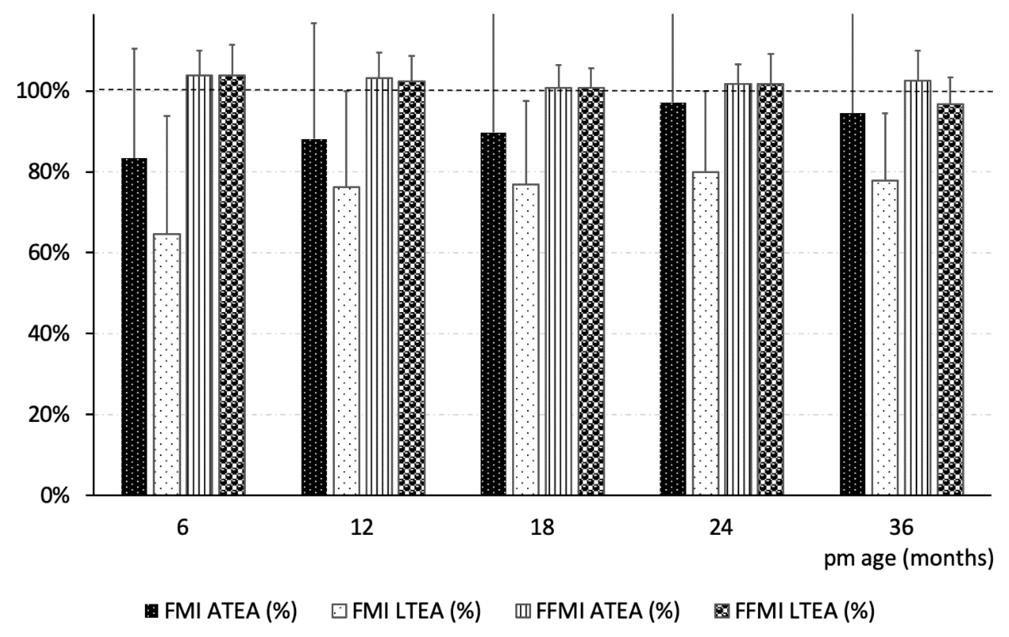
| FMI (g/cm2) | Term | 0.48 ± 0.13 †,γ | 0.42 ± 0.13 † | 0.39 ± 0.11 | 0.35 ± 0.09 | 0.36 ± 0.09 | <0.05 | <0.01 | <0.001 |
| ATEA | 0.40 ± 0.13 | 0.37 ± 0.12 | 0.35 ± 0.12 | 0.34 ± 0.09 | 0.34 ± 0.10 | ||||
| LTEA | 0.31 ± 0.14 | 0.32 ± 0.10 | 0.30 ± 0.08 | 0.28 ± 0.07 | 0.28 ± 0.06 | ||||
| FFMI (g/cm2) | Term | 1.31 ± 0.12 (22) | 1.28 ± 0.13 (25) | 1.26 ± 0.05 (15) | 1.21 ± 0.05 (24) | 1.21 ± 0.08 (24) | NS | NS | <0.001 |
| ATEA | 1.36 ± 0.08 (25) | 1.32 ± 0.08 (25) | 1.27 ± 0.07 (16) | 1.23 ± 0.06 (14) | 1.24 ± 0.09 (13) | ||||
| LTEA | 1.36 ± 0.10 (46) | 1,31 ± 0.08 (43) | 1.27 ± 0.06 (34) | 1.23 ± 0.09 (35) | 1.17 ± 0.08 (34) | ||||
| BMD (g/cm2) | Term | 0.57 ± 0.05(30) †,γ | 0.63 ± 0.04(25) | 0.69 ± 0.05(15) † | 0.71 ± 0.05(24) | 0.77 ± 0.05(25) | NS | <0.001 | <0.001 |
| ATEA | 0.54 ± 0.05(27) | 0.60 ± 0.04(25) | 0.67 ± 0.05(17) | 0.70 ± 0.03(14) | 0.75 ± 0.04(13) | ||||
| LTEA | 0.52 ± 0.05(46) | 0.60 ± 0.04(43) | 0.65 ± 0.06(33) | 0.69 ± 0.04(35) | 0.74 ± 0.04(34) | ||||
| BMC (g) | Term | 167 ± 39 †,γ | 244 ± 46 † | 339 ± 55 †,γ | 401 ± 73 †,γ | 547 ± 89 †,γ | <0.001 | <0.001 | <0.001 |
| ATEA | 129 ± 31 | 210 ± 41 | 293 ± 67 | 377 ± 53 | 482 ± 64 | ||||
| LTEA | 106 ± 29 | 196 ± 44 | 281 ± 55 | 345 ± 56 | 421 ± 65 | ||||
| BMC/length2 (g/mm2) | Term | 3.62 ± 0.78 †,γ(30) | 4.16 ± 0.66 (25) | 4.79 ± 0.54 (15) | 5.05 ± 0.59 (24) | 5.76 ± 0.68 † (24) | <0.001 | <0.001 | <0.001 |
| ATEA | 2.90 ± 0.66 (27) | 3.87 ± 0.59 (25) | 4.69 ± 0.65 (16) | 5.00 ± 0.54 (14) | 5.49 ± 0.53 (13) | ||||
| LTEA | 2.61 ± 0.62 (46) | 3.73 ± 0.66 (43) | 4.50 ± 0.68 (34) | 4.79 ± 0.64 (35) | 5.04 ± 0.55 (34) | ||||
| TF (g) | Term | 681 ± 230 (30) †,γ | 683 ± 368 (25) † | 768 ± 300(15) † | 728 ± 288 (24) † | 984 ± 370 (25) † | <0.01 | <0.001 | <0.001 |
| ATEA | 512 ± 237 (27) | 538 ± 251 (25) | 604 ± 312 (17) | 674 ± 229 (14) | 865 ± 378 (13) | ||||
| LTEA | 358 ± 186 (46) | 440 ± 199 (43) | 480 ± 219(34) | 519 ± 209 (35) | 680 ± 250(34) | ||||
| PTF (%) | Term | 28 ± 3 | 26 ± 6 | 27 ± 3 | 26 ± 3 | 28 ± 4 | NS | NS | <0.001 |
| ATEA | 27 ± 4 | 26 ± 6 | 26 ± 5 | 26 ± 3 | 29 ± 4 | ||||
| LTEA | 28 ± 5 | 26 ± 4 | 25 ± 4 | 25 ± 4 | 29 ± 5 | ||||
| Patient Group | 6 m | N | 12 m | N | 18 Months | N | 24 Months | N | 36 Months | N | P 1 | |||
|---|---|---|---|---|---|---|---|---|---|---|---|---|---|---|
| Interaction Group * Time | Group Effect | Time Effect | ||||||||||||
| IGF-1 (ng/mL) | Term | 94 ± 48 | 34 | 94 ± 52 | 26 | 59 ± 22 | 16 | 67 ± 39 | 29 | 78 ± 30 | 29 | NS | <0.05 | NS |
| ATEA | 108 ± 55 | 29 | 117 ± 101 | 26 | 88 ± 70 | 14 | 95 ± 57 | 18 | 88 ± 46 | 17 | ||||
| LTEA | 103 ± 67 | 48 | 114 ± 83 | 40 | 91 ± 43 | 35 | 88 ± 46 | 37 | 87 ± 34 | 40 | ||||
| IGFBP-1 (ng/mL) | Term | 52 ± 36 | 34 | 65 ± 30 γ | 26 | 43 ± 24 | 15 | 43 ± 14 | 19 | NS | NS | <0.05 | ||
| ATEA | 50 ± 28 | 29 | 51 ± 25 | 23 | 41 ± 12 | 6 | 58 ± 30 | 4 | ||||||
| LTEA | 49 ± 29 | 49 | 55 ± 31 | 37 | 45 ± 17 | 18 | 37 ± 6 | 7 | ||||||
| IGFBP-3 (µg/mL) | T | 2.41 ± 0.5 | 34 | 2.56 ± 0.71 | 25 | 2.39 ± 0.82 | 17 | 2.35 ± 0.80 | 28 | 2.61 ± 0.51 | 29 | NS | NS | <0.01 |
| ATEA | 2.29 ± 0.51 | 29 | 2.38 ± 0.72 | 25 | 2.18 ± 0.91 | 15 | 2.37 ± 0.80 | 18 | 2.70 ± 0.61 | 17 | ||||
| LTEA | 2.37 ± 0.71 | 50 | 2.58 ± 0.54 | 36 | 2.43 ± 0.9 | 37 | 2.25 ± 0.82 | 41 | 2.63 ± 0.69 | 39 | ||||
| Plasma glucose (mg/dL) | T | 77 ± 12 | 8 | 75 ± 8 | 26 | 73 ± 9 | 17 | 76 ± 6 | 29 | 80 ± 5 | 29 | NS | NS | <0.01 |
| ATEA | 80 ± 6 | 16 | 76 ± 5 | 25 | 75 ± 7 | 14 | 77 ± 7 | 18 | 78 ± 13 | 17 | ||||
| LTEA | 77 ± 7 | 31 | 74 ± 8 | 41 | 76 ± 9 | 40 | 79 ± 8 | 43 | 79 ± 7 | 41 | ||||
| Insulin (mcU/mL) | T | 2.64 ± 1.28 | 34 | 2.51 ± 1.24 | 28 | 2.44 ± 1.43 | 17 | 2.60 ± 1.07 | 29 | 3.85 ± 1.70 | 29 | NS | NS | <0.001 |
| ATEA | 2.99 ± 1.69 | 27 | 2.56 ± 1.33 | 26 | 2.58 ± 1.24 | 15 | 3.45 ± 1.86 | 18 | 3.70 ± 1.64 | 17 | ||||
| LTEA | 3.00 ± 1.62 | 46 | 2.66 ± 1.53 | 39 | 2.84 ± 1.44 | 36 | 3.79 ± 1.77 | 39 | 3.49 ± 2.12 | 40 | ||||
| HOMA | Term | 0.5085 ± 0.195 | 8 | 0.469 ± 0.244 | 25 | 0.454 ± 0.319 | 17 | 0.492 ± 0.220 | 29 | 0.775 ± 0.370 | 29 | NS | NS | <0.001 |
| ATEA | 0.680 ± 0.421 | 14 | 0.497 ± 0.288 | 24 | 0.488 ± 0.253 | 14 | 0.673 ± 0.395 | 18 | 0.768 ± 0.390 | 16 | ||||
| LTEA | 0.604 ± 0.338 | 26 | 0.493 ± 0.319 | 37 | 0.556 ± 0.329 | 36 | 0.746 ± 0.398 | 39 | 0.695 ± 0.451 | 40 | ||||
| Cholesterol (mg/dL) | Term | 132 ± 33 | 26 | 146 ± 25 | 28 | 151 ± 28 | 17 | 152 ± 27 | 29 | 150 ± 27 | 28 | NS | NS | <0.001 |
| ATEA | 138 ± 25 | 27 | 144 ± 38 | 26 | 159 ± 38 | 15 | 159 ± 32 | 18 | 158 ± 32 | 17 | ||||
| LTEA | 135 ± 24 | 46 | 159 ± 29 | 41 | 158 ± 28 | 40 | 160 ± 35 | 42 | 165 ± 30 | 38 | ||||
| Triglycerides (mg/dL) | Term | 105 ± 33 | 25 | 116 ± 51 | 27 | 81 ± 37 | 17 | 70 ± 16 | 29 | 68 ± 22 | 28 | NS | NS | <0.001 |
| ATEA | 116 ± 41 | 27 | 108 ± 41 | 26 | 104 ± 61 | 15 | 78 ± 26 | 18 | 72 ± 26 | 17 | ||||
| LTEA | 114 ± 45 | 44 | 107 ± 47 | 41 | 87 ± 32 | 40 | 79 ± 29 | 42 | 67 ± 19 | 38 | ||||
| Leptin (ng/mL) | T | 5.60 ± 2.74 † | 34 | 4.40 ± 4.38 | 29 | 2.87 ± 1.21 | 17 | 3.30 ± 1.71 | 29 | 3.49 ± 1.52 | 29 | <0.05 | NS | <0.001 |
| ATEA | 6.16 ± 3.54 * | 30 | 4.31 ± 3.01 * | 27 | 3.75 ± 1.95 | 15 | 3.53 ± 1.84 | 18 | 4.32 ± 2.69 | 17 | ||||
| LTEA | 4.21 ± 1.80 | 50 | 3.18 ± 1.59 | 43 | 3.40 ± 1.41 | 39 | 3.60 ± 1.56 | 42 | 3.28 ± 1.54 | 40 | ||||
| sOB-R (U/mL) | Term | 66 ± 21 | 34 | 72 ± 30 | 28 | 66 ± 17 | 17 | 72 ± 19 | 29 | 50 ± 13 | 29 | NS | NS | <0.001 |
| ATEA | 70 ± 21 | 30 | 76 ± 27 | 25 | 76 ± 20 | 14 | 66 ± 21 | 18 | 54 ± 17 | 17 | ||||
| LTEA | 69 ± 23 | 51 | 79 ± 20 | 42 | 76 ± 23 | 37 | 68 ± 19 | 42 | 57 ± 13 | 40 | ||||
| Leptin/sOB-R | Term | 0.12 ± 0.18 † | 34 | 0.08 ± 0.06 † | 28 | 0.05 ± 0.02 | 17 | 0.05 ± 0.03 | 29 | 0.08 ± 0.06 | 29 | NS | NS | <0.001 |
| ATEA | 0.11 ± 0.12 | 31 | 0.08 ± 0.13 | 25 | 0.06 ± 0.04 | 15 | 0.07 ± 0.06 | 19 | 0.11 ± 0.13 | 18 | ||||
| LTEA | 0.07 ± 0.05 | 49 | 0.05 ± 0.04 | 41 | 0.06 ± 0.06 | 36 | 0.06 ± 0.03 | 41 | 0.06 ± 0.04 | 39 | ||||
| Adiponectin (µg/mL) | Term | 27 ± 9 | 32 | 23 ± 6 | 28 | 28 ± 11 | 17 | 25 ± 8 | 29 | 21 ± 7 | 28 | <0.05 | NS | <0.05 |
| ATEA | 25 ± 9 | 30 | 24 ± 9 | 26 | 22 ± 6 | 15 | 20 ± 5 | 18 | 20 ± 5 | 17 | ||||
| LTEA | 26 ± 7 | 50 | 24 ± 9 | 44 | 22 ± 7 | 37 | 24 ± 8 | 42 | 25 ± 9 | 40 | ||||
| Resistin (ng/mL) | T | 15 ± 10 | 34 | 19 ± 8 | 25 | 22 ± 9 | 15 | 18 ± 9 | 27 | 18 ± 8 | 23 | NS | <0.05 | <0.05 |
| ATEA | 17 ± 12 | 29 | 17 ± 6 | 25 | 17 ± 11 | 13 | 19 ± 10 | 16 | 19 ± 7 | 15 | ||||
| LTEA | 23 ± 16 | 50 | 24 ± 12 | 41 | 19 ± 9 | 39 | 20 ± 10 | 40 | 21 ± 11 | 35 | ||||
| Total Ghrelin (pg/mL) | T | 3107 ± 1951 | 34 | 2977 ± 1120 | 28 | 2805 ± 1702 | 17 | 2406 ± 778 | 29 | 2055 ± 702 | 29 | NS | NS | <0.001 |
| ATEA | 3465 ± 1739 | 30 | 3414 ± 1342 | 27 | 2563 ± 991 | 15 | 2166 ± 868 | 18 | 1923 ± 658 | 17 | ||||
| LTEA | 3222 ± 1385 | 50 | 3234 ± 1215 | 44 | 2947 ± 1559 | 39 | 2343 ± 1050 | 42 | 2354 ± 1226 | 39 | ||||
| Ghrelin acylate (pg/mL) | T | 95 ± 51 | 34 | 85 ± 39 | 28 | 62 ± 24 | 17 | 62 ± 24 | 29 | 43 ± 31 | 29 | NS | NS | <0.001 |
| ATEA | 82 ± 47 | 29 | 79 ± 52 | 27 | 64 ± 49 | 15 | 58 ± 27 | 18 | 44 ± 29 | 17 | ||||
| LTEA | 102 ± 64 | 51 | 66 ± 36 | 43 | 57 ± 27 | 39 | 53 ± 23 | 40 | 47 ± 35 | 40 | ||||
| IL6 (pg/mL) | T | 2.77 ± 3.5 | 31 | 3.01 ± 2.96 | 21 | 2.25 ± 1.29 | 7 | 1.82 ± 1.19 | 14 | NS | <0.05 | NS | ||
| ATEA | 1.08 ± 0.83 | 21 | 1.73 ± 1.23 | 17 | 0.97 ± 0.45 | 7 | 1.48 ± 1.76 | 6 | ||||||
| LTEA | 2.45 ± 2.14 | 40 | 3.11 ± 2.97 | 33 | 1.88 ± 1.74 | 23 | 2.85 ± 3.16 | 11 | ||||||
| Cortisol (µg/dL) | T | 13 ± 5 | 28 | 12 ± 4 | 29 | 10 ± 2 | 17 | 10 ± 4 | 29 | 9 ± 3 | 28 | NS | <0.01 | <0.01 |
| ATEA | 11 ± 6 | 27 | 11 ± 6 | 27 | 14 ± 4 | 15 | 11 ± 3 | 18 | 8 ± 2 | 16 | ||||
| LTEA | 9 ± 5 | 49 | 10 ± 5 | 42 | 9 ± 4 | 39 | 9 ± 4 | 41 | 8 ± 2 | 33 | ||||
Publisher’s Note: MDPI stays neutral with regard to jurisdictional claims in published maps and institutional affiliations. |
© 2021 by the authors. Licensee MDPI, Basel, Switzerland. This article is an open access article distributed under the terms and conditions of the Creative Commons Attribution (CC BY) license (http://creativecommons.org/licenses/by/4.0/).
Share and Cite
Mihatsch, W.; Dorronsoro Martín, I.; Barrios-Sabador, V.; Couce, M.L.; Martos-Moreno, G.Á.; Argente, J.; Quero, J.; Saenz de Pipaon, M. Bone Mineral Density, Body Composition, and Metabolic Health of Very Low Birth Weight Infants Fed in Hospital Following Current Macronutrient Recommendations during the First 3 Years of Life. Nutrients 2021, 13, 1005. https://doi.org/10.3390/nu13031005
Mihatsch W, Dorronsoro Martín I, Barrios-Sabador V, Couce ML, Martos-Moreno GÁ, Argente J, Quero J, Saenz de Pipaon M. Bone Mineral Density, Body Composition, and Metabolic Health of Very Low Birth Weight Infants Fed in Hospital Following Current Macronutrient Recommendations during the First 3 Years of Life. Nutrients. 2021; 13(3):1005. https://doi.org/10.3390/nu13031005
Chicago/Turabian StyleMihatsch, Walter, Izaskun Dorronsoro Martín, Vicente Barrios-Sabador, María L. Couce, Gabriel Á. Martos-Moreno, Jesús Argente, José Quero, and Miguel Saenz de Pipaon. 2021. "Bone Mineral Density, Body Composition, and Metabolic Health of Very Low Birth Weight Infants Fed in Hospital Following Current Macronutrient Recommendations during the First 3 Years of Life" Nutrients 13, no. 3: 1005. https://doi.org/10.3390/nu13031005