Article

Evaluation of 2’-Fucosyllactose and Bifidobacterium longum Subspecies infantis on Growth, Organ Weights, and Intestinal Development of Piglets

1
Division of Nutritional Sciences, University of Illinois, Urbana, IL 61801, USA
2
Department of Food Science and Human Nutrition, University of Illinois, Urbana, IL 61801, USA
3
IFF Health and Biosciences, 02460 Kantvik, Finland
4
IFF R&D—Enabling Technologies, Advanced Analytical, 8220 Brabrand, Denmark
5
IFF Health and Biosciences, Saint Louis, MO 63110, USA
6
Department of Animal Sciences, University of Illinois, Urbana, IL 61801, USA
*
Author to whom correspondence should be addressed.
Nutrients 2022, 14(1), 199; https://doi.org/10.3390/nu14010199
Submission received: 18 November 2021 / Revised: 29 December 2021 / Accepted: 30 December 2021 / Published: 31 December 2021
(This article belongs to the Section Pediatric Nutrition)

Abstract

:
Human milk is rich in oligosaccharides that influence intestinal development and serve as prebiotics for the infant gut microbiota. Probiotics and 2’-fucosyllactose (2’-FL) added individually to infant formula have been shown to influence infant development, but less is known about the effects of their synbiotic administration. Herein, the impact of formula supplementation with 2’-fucosyllactose (2’-FL) and Bifidobacterium longum subsp. infantis Bi-26 (Bi-26), or 2’-FL + Bi-26 on weight gain, organ weights, and intestinal development in piglets was investigated. Two-day-old piglets (n = 53) were randomized in a 2 × 2 design to be fed a commercial milk replacer ad libitum without (CON) or with 1.0 g/L 2’-FL. Piglets in each diet were further randomized to receive either glycerol stock alone or Bi-26 (109 CFU) orally once daily. Body weights and food intake were monitored from postnatal day (PND) 2 to 33/34. On PND 34/35, animals were euthanized and intestine, liver and brain weights were assessed. Intestinal samples were collected for morphological analyses and measurement of disaccharidase activity. Dry matter of cecum and colon contents and Bifidobacterium longum subsp. infantis abundance by RT-PCR were also measured. All diets were well tolerated, and formula intake did not differ among the treatment groups. Daily body weights were affected by 2’-FL, Bi-26, and day, but no interaction was observed. There was a trend (p = 0.075) for greater total body weight gain in CON versus all other groups. Jejunal and ascending colon histomorphology were unaffected by treatment; however, there were main effects of 2’-FL to increase (p = 0.040) and Bi-26 to decrease (p = 0.001) ileal crypt depth. The addition of 2’-FL and/or Bi-26 to milk replacer supported piglet growth with no detrimental effects on body and organ weights, or intestinal structure and function.

1. Introduction

The postnatal period is a critical time for gastrointestinal and immune development, which are stimulated by bioactive components in human milk (HM) and the gut microbiota [1]. The bioactive components in HM include among others the prebiotic human milk oligosaccharides (HMOs) [2,3] and the milk microbiota [4]. The HMOs are the third most abundant solid component of HM after lipid and lactose, but their concentrations are influenced by maternal genetics, stage of lactation, seasonality and geographic location [5]. 2’-fucosyllactose (2’-FL), a neutral 2-linked fucosylated oligosaccharide [4], is one of the predominant HMO, but its synthesis is influenced by maternal secretor status [5]. 2’-FL concentration averages 2–3 g/L in HM of secretor mothers, but is undetectable in the milk of non-secretors [5].
HMOs resist gastrointestinal digestion [6,7], allowing intact HMO to influence various aspects of infant development. In the intestine, 2’-FL dose-dependently decreased cell proliferation and increased sucrase activity in cultured cells [8,9], suggesting a shift towards differentiation. In vivo, dietary HMO increased cecal barrier function in mice [10]. In the colon, HMOs are metabolized by the microbiota, and some species of Bifidobacterium and Bacteroides utilize HMOs as their primary energy source [11].
Bifidobacterium species predominate in fecal samples of breastfed infants due in part to their ability to utilize HMOs and to develop cross-feeding relationships with butyrate-producing bacteria, such as those belonging to the phylum Firmicutes [11,12]. In particular, Bifidobacterium longum subsp. infantis (B. infantis) contains a complement of genes that support utilization of both fucosylated and sialylated HMOs [13]. Metabolism of HMO by B. infantis upregulates solute-binding proteins with an affinity for HMOs genes and glycosidases, which further aids in its ability to breakdown HMOs, specifically 2’-FL [12,13,14]. Metabolism of 2’-FL by B. infantis produces acetate, resulting in cross feeding to other bacteria.
While B. infantis appears to have co-evolved with HMO in a synbiotic relationship, surveys of the fecal microbiota have shown that the relative abundance of Bifidobacterium, particularly B. infantis, in the feces of breastfed infants in the US [15] is lower than that of infants in the developing world [16,17]. Thus, administration of B. infantis to breastfed infants has gained popularity as a commercially available probiotic due to its associated benefits [18,19,20]. Among commercially available products is the B. longum subsp. infantis strain Bi-26 (Bi-26) from the Danisco Global Culture Collection, which was shown to metabolize fucosylated HMOs more rapidly and efficiently than the type strain of B. infantis ATCC 15697 [21,22]. Thus, co-administration of 2’-FL and Bi-26 have the potential to confer benefits to formula-fed infants; however, this has not been tested in vivo.
Thus, the goal of this study was to evaluate the effects of individual and synbiotic supplemental 2’-FL and Bi-26 to formula on body weight gain, organ growth and gastrointestinal development and function in the piglet model. The pig is a widely accepted preclinical model due to more shared similarities in gut anatomy and physiology to the human infant than do rodent models [23,24,25]. We hypothesized that 2’-FL and Bi-26 alone or in combination would be well tolerated and support normal piglet growth, and aimed to investigate any synergy between 2’-FL and Bi-26. Based on previous evidence [8,9,10], we further hypothesized that 2’-FL would promote intestinal development in vivo.

2. Materials and Methods

2.1. Study Design

The Institutional Animal Care and Use Committee at the University of Illinois approved all animal procedures (Protocol 17286). Naturally farrowed piglets from a commercial swine herd remained with the sows until postnatal day 2 (PND 2) before being transported to a specialized neonatal pig rearing system at the Piglet Nutrition and Cognition Laboratory (PNCL) on the University of Illinois campus. A total for 63 male piglets were randomly assigned to treatment groups by equalizing initial body weight and genetics (i.e., litter of origin) across treatments, such that an equal number of pigs from each litter was assigned to each treatment group in every cohort. The treatment structure included a 2 × 2 factorial arrangement of dietary [commercial milk replacer ad libitum without (CON) or with 1.0 g/L 2’-FL (FL)] and probiotic treatments. Within each dietary treatment, piglets were further randomized to receive either glycerol solution alone (BI) or Bi-26 (109 CFU) (FLBI) orally once daily. The experiment was conducted in five cohorts. Piglets of only one sex (male) were chosen to reduce any source of variation; although it was recently demonstrated that body and organ growth does not differ for pigs in the age range used in this study [26].
All piglets were monitored continuously using home-cage cameras that permitted asynchronous viewing of animal health and well-being throughout the study. Additionally, animal caretakers who were blinded to experimental treatment identity performed twice-daily in-person health checks (0800 and 1600 h) that included the following outcomes for individual pigs: (1) fecal consistency (1 = solid; 2 = semisolid; 3 = loose; 4 = watery), (2) body condition (5-point scale), (3) visual inspection of feeding system and milk bowl, (4) piglet responsiveness upon approach and general demeanor (lethargy; yes/no), and (5) evidence of piglet vomiting (yes/no). In total, 10 piglets were removed from the study due to failure to thrive unrelated to experimental treatments. The number removed per cohort was: Cohort 1 (n = 3); Cohort 2 (n = 0); Cohort 3 (n = 2); Cohort 4 (n = 3); and Cohort 5 (n = 2). The final number per treatment group was: CON (n = 12); BI (n = 14); FL (n = 15); and FLBI (n = 12).

2.2. Experimental Diets

Two milk replacer diets (CON and FL) were manufactured in powder form by TestDiet (Purina Mills, St. Louis, MO, USA). The composition was identical to ProNurse (Land O’Lakes, North Arden Hills, MN, USA) with the exception that the CON diet had added 0.532% lactose to match the 2’-FL content in the test diet (Table 1). The 2’-FL was supplied as Care4U 2’-fucosyllactose (International Flavors & Fragrances; New York, NY, USA). Diets were shipped to the University of Illinois as color-coded bags, so that researchers working with the piglets were blinded to the dietary treatment. Throughout the study, experimental milk replacer powders were reconstituted fresh each day with 200 g of dry power being added to 800 mL of tap water. Analyzed values are presented for the experimental dietary treatments both in the powder form and after the powder was reconstituted into a liquid form (Table 1). Milk was dispensed into clean reservoirs and automatically delivered to piglets using a pump beginning at 10 am each day and ending at 6 am the following day (i.e., ad libitum access to treatment diets over a 20 h feeding period). Milk disappearance was calculated per pig based on the initial and final reservoir weights before and after the daily feeding cycle, respectively. Based on milk disappearance quantified the preceding day, a sufficient volume of reconstituted milk replacer treatment was dispensed into individual reservoirs per pig to ensure ad libitum access to milk each day.

2.3. Probiotic Treatment

Piglets in the CON and FL diet groups were further randomized to receive probiotic or no probiotic, resulting in four experimental treatment groups (CON, BI, FL, FLBI). Aliquots of Bi-26 (Bi-26 50B 100 GM, FloraFIT® Probiotics; Danisco USA Inc., Madison, WI, USA) at the dose of 109 CFU/pig/d was solubilized prior to the start of the study in bacterial glycerol stock (12.1% glycerol) and frozen at −80 °C until administration. From PND 2-PND 12, the probiotic dose was thawed and orally administered using a syringe prior to the first feed of the day. Piglets not receiving probiotic treatment received an equal dose of glycerol solution. From PND 13 to PND 33 or 34, piglets received either probiotic treatment or glycerol solution mixed with approximately 10 mL of milk in their bowl prior to the first feeding of the day. The study duration was based on known changes in brain structure and development from previous studies [27].

2.4. Sample Collection

Each morning, piglets were weighed, and any remaining formula was measured. On PND 34 or 35, piglets were sedated with an intramuscular injection of Telazol® (Tiletamine HCl and Zolazepam HCl, 3.5 mg/kg BW each, Pfizer Animal Health, Fort Dodge, IA, USA). Piglets were then euthanized by an intracardiac injection of sodium pentobarbital (72 mg/kg BW; Euthasol, by Virbac, Westlake, TX, USA). The brain and liver were removed and weighed. The small intestine was cut at 10% and 85% from the proximal end to give 3 segments corresponding to the duodenum, jejunum and ileum, respectively. A 25 cm section of jejunum and ileum were cut longitudinally, and mucosal scrapings were obtained and frozen in liquid nitrogen and stored at −80 °C. In addition, the large intestine was excised, and segments were cut from the most proximal and distal parts corresponding to the ascending colon (AC) and rectum (RC), respectively. Five-centimeter pieces of each intestinal segment were fixed in formalin for 48 h for histomorphological analyses.

2.5. Intestine Histomorphology

Formalin-fixed sections of the jejunum, ileum and AC were processed into paraffin blocks, which were sectioned, placed on glass slides and stained with hematoxylin and eosin for the measurement of small intestinal villus length and width, villus area, crypt depth and width, crypt volume, villus-to-crypt ratio, and surface area. Mucosa thickness was measured in AC. Villus area and crypt volume were calculated by multiplying width by villus height or crypt depth, respectively. Tissue processing and staining, and all measurements were performed by Dr. Fred Hoerr, a board-certified veterinary pathologist (Veterinary Diagnostic Pathology, LLC., Fort Valley, VA, USA).

2.6. Disaccharidase Activities

Intestinal lactase and sucrase activities were measured as previously described [28,29]. Mucosal homogenates were prepared and incubated with 0.6 M lactose (Thermo Fischer Scientific, Hanover Park, IL, USA) or 0.3 M sucrose (Thermo Fisher Scientific, Hanover Park, IL, USA) buffer (dissolved in 0.0625 maleate buffer) for 60 min at 37 °C. The reactions were stopped by adding 2.0% zinc sulfate (Thermo Fisher Scientific, Hanover Park, IL, USA) and 1.8% barium hydroxide (Thermo Fisher Scientific, Hanover Park, IL, USA), and the amount of glucose release was detected by using a glucose oxidase reagent (Thermo Fisher Scientific, Hanover Park, IL, USA). Enzyme activity is expressed as µmoles glucose/minute/g protein. Protein was measured in intestinal homogenates using the Bradford method (Bio-Rad, Hercules, CA, USA).

2.7. Dry Matter of Luminal Contents

Dry matter (DM) of AC, cecum (CE) and RC contents were assessed on the samples according to the AOAC method [30]. Initial (~4 g) and final weights of contents were recorded after drying and % DM was calculated.

2.8. DNA Extraction from Luminal Contents

Microbial whole genomic DNA was isolated from AC and DC contents by the QIAamp DNA Stool Mini Kit (Qiagen, Hilden, Germany) in combination with the FastPrep-24 bead beating system (MP Biomedicals, Santa Ana, CA, USA) as previously described [31]. DNA concentration was determined with a Qubit 3.0 Fluorometer (Life Technologies, Carlsbad, CA, USA) and DNA quality was confirmed with agarose electrophoresis.

2.9. qPCR for Bifidobacterium spp. and B. infantis

Absolute abundances of Bifidobacterium spp. and B. infantis were quantified in AC and RC with the use of primers specific for Bifidobacterium spp. [32] and B. infantis [33], respectively. Real-time qPCR was performed by QuantStudio 7 Real-Time PCR systems (Thermo Fisher Scientific, Hanover Park, IL, USA) using SYBR Green Master Mix (Applied Biosystems, Foster City, CA, USA). All experiments were performed in triplicated with a total reaction volume of 10 μL, containing 5 μL of 2 × Power SYBR Green Master Mix, 0.5 μM of each primer, 1 μL (1 mg/mL) bovine serum albumin (BSA), 1 μL of DEPC water and 2 μL of DNA template diluted 1:30 or 1:15. The cycling conditions were as follows: 50 °C for 2 min, 90 °C for 10 minutes followed by 40 cycles of 95 °C for 15 s, 60 °C or 55 °C for 20 s for Bifidobacterium spp. or B. infantis, respectively, and 72 °C for 45 s. Following amplification, a dissociation step was included to assess the melting profile of amplified products. Standard curves (10–108 16S ribosomal RNA (rRNA) gene copies/reaction) were generated using purified 2.1 TOPO-TA plasmids containing full length 16S rRNA gene of B. infantis (Thermo Fisher Scientific, Hanover Park, IL, USA). Data were processed with QuantStudio Real-Time PCR Software V1.3 (Thermo Fisher Scientific, Hanover Park, IL, USA) and expressed as log10 16S rRNA gene copies.

2.10. Statistical Analysis

Statistical analyses were performed using SAS version 9.4 (SAS Institute Inc., Cary, NC, USA) using the PROC MIXED procedure. BW and formula intake were analyzed using a repeated-measure analysis of variance (ANOVA). Organ weights, disaccharidase activity, intestine histomorphology, and qPCR abundance data were analyzed using a 2-way ANOVA. Both statistical models included a nested model for random effects with replicate nested within sow. Both models included fixed effects of diet, probiotic and the interaction between diet and probiotic with Dunnett’s post hoc test to compare statistical difference between the control group and experimental treatment groups. The occurrence of B. infantis among treatments was analyzed by Fisher’s exact test using the PROC FREQ procedure in SAS. If samples fell outside of ± three standard deviations from the mean, they were excluded as outliers. The following data were transformed to meet the assumptions of normality and confirmed post hoc: jejunal and ileal disaccharidase activity and abundance of Bifidobacterium spp. and B. infantis. Significance was set at p < 0.05 and trends are reported as p < 0.10. Data are presented as means ± standard error of the mean (SEM) unless otherwise stated.

3. Results

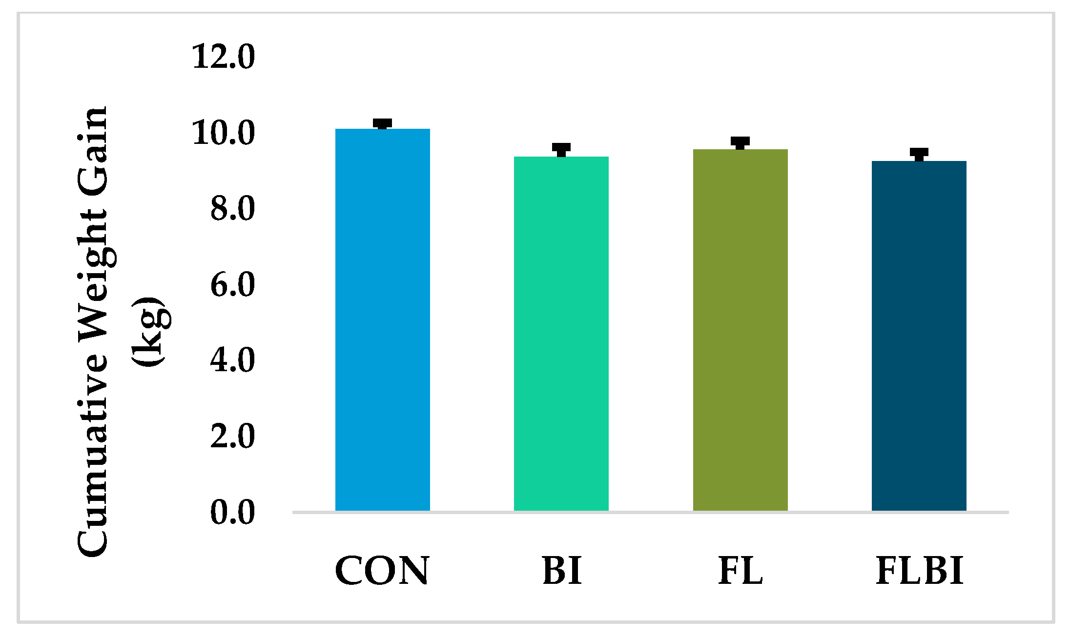
3.1. Tolerance, Weight Gain, Organ Weights and Intestinal Length and Weight

Experimental diets and probiotic treatments were successfully administered, and Bi-26 stored frozen in glycerol stock remained viable throughout the study (Table S1). There was no significant treatment differences in any of the assessed behavioral or general demeanor outcomes and piglets in all groups demonstrated a typical pattern of body weight gain for artificially reared animals in the PNCL facility [26]. Although animals were allowed ad libitum access to the diets, relative formula intake over the course of the study did not differ between treatments, with an average of 316.9 ± 11.6 mL/kg BW/d (p = 0.419). Daily weight gain was affected by probiotic and diet, but without interactive effect (Figure S1). Total weight gain from PND2 to PND33 showed a statistical trend (p = 0.075) on the interactive effects of diet and probiotic, with CON piglets showing the greatest total body weight gain (Figure 1). Brain weights adjusted per kg BW tended (p = 0.060) to differ, but neither liver weight or small or large intestinal weight or length differed based on absolute or adjusted values (Table S2).

3.2. Intestine Histomorphology and Disaccharidase Activity

Jejunal morphology was similar across treatments (Table S3). In the ileum, there were no effects of treatment on villus morphology (Table 2), but crypt width (p = 0.071) and crypt volume (p = 0.069) were numerically greater in piglets fed the FL diet (Table 2) and the interaction between diet and probiotic was statistically significant at trend level. Crypt depth showed a main effect of probiotic, with shorter crypts in the BI treatment group than the CON and FL groups (p = 0.006), as well as a main effect of diet, with crypt depth in the FL group being less than CON. (Table 2). Mucosa thickness in the AC was similar across all treatment groups (Table S4).
Lactase and activity in jejunum and ileum were similar across all treatments (Table S5), but the addition of 2’-FL to the diet resulted in a significant increase in sucrase activity (Table 3).

3.3. Dry Matter of Luminal Contents and qPCR for Bifidobacterium spp. and B. infantis

Dry matter was significantly higher in RC than CE and AC (p < 0.001) (Figure S2). However, there was no differences in dry matter due to an experimental treatment (p = 0.752). The absolute quantity of Bifidobacterium spp. was similar across treatments in AC and RC contents (Table S6). B. infantis was detected in ≥50% of BI and FLBI piglets, but in none of the CON or FL piglets (Table 4). The percent of AC and RC samples from piglets in the BI and FLBI groups with detectable B. infantis was similar (p = 0.172). In both the AC and RC, B. infantis abundance was greater in the BI and FLBI than CON or FL, but there were no differences in the abundance of B. infantis between the BI and FLBI groups (Table 4).

4. Discussion

HMOs are complex glycans [5] that withstand digestion of the upper gastrointestinal tract [6,7] to act as a prebiotic in the lower gastrointestinal tract [11,12], with physiological benefits for the infant [1,2,3,34]. HMO are proposed to influence intestinal development [8,9,10], as well as microbiota composition by promoting growth of Bifidobacterium species, including B. infantis [11,12,13]. Infant formulas that contain prebiotics, such as galactooligosaccharides (GOS) and fructooligosaccharides, support Bifidobacterium growth are currently commercially available [34,35]. However, the chemical structures of commercial prebiotics added to formula and HMOs differ substantially, and it has been suggested that HMOs may be better suited to the infant’s developing gastrointestinal system [36]. Piglets and infants share similarities in gastrointestinal anatomy and metabolism [23,24,25], and piglets display a closer similarity in gut microbiota composition to humans than do rodents [37]. Thus, they serve as a relevant preclinical model to study prebiotics and probiotics and have been used extensively by our group [38,39,40] and others [41,42] to investigate the biological effects of individual HMOs and B. infantis [43]. The present study is the first to compare the individual and combined effects of HMO and B. infantis in the piglet model.
Supplementation of 2’-FL at 1 g/L and Bi-26 at 109 CFU/d were well tolerated and supported normal growth throughout the study. CON piglets tended to gain more weight than FLBI and BI had lower feed efficiency than CON. Data on the effect of HMO on weight gain are conflicting. In suckling rat pups, weight gain was greater by the end of the study in those who received 2’-FL (0.2 g/100 g body weight) vs. the control group, who received a gavage of only mineral water [44]. This dose was estimated to be similar to the daily consumption of 2’-FL of human infants [45]. Data from human infants differ depending on whether the HMO was supplemented to infant formula [46,47] or studies examined associations between HMO concentrations in milk and infant weight gain [48,49,50]. In randomized controlled trials, infants fed formula containing 2’-FL alone (0.2 or 1.0 g/L) [46] or with lacto-N-neotetraose (LNnT) (1.0 g/L 2’-FL and 0.5 g/L LNnT) [47] had similar growth to infants fed formula alone. In contrast, a study of breastfed infants demonstrated that the abundance of two fucosylated HMOs, 2’-FL and difucosyl-lactose (DFlac), were associated with increased weight velocity at 5 months, whereas LNnT was negatively associated with height-for-age Z-scores [48]. In contrast, concentrations of other HMOs, including lacto-N-fucopentaose I and LNnT, were positively associated with weight and body composition at 6-months of age in breastfed infants; however, there were no associations with 2’-FL [50]. Lastly, a meta-analysis of the modulation of the microbiota and weight gain showed that probiotic supplementation in infants has little effect on weight gain [51]. The present study showed that supplementation with 2’-FL and Bi-26 individually or together lowered daily weight gain and together tended to lower total body weight gain compared to the CON formula. However, it should be noted that piglets demonstrated a typical growth pattern for piglets raised in the artificial rearing system in our facility [26] and total weight gain in the treatment groups was only ~5% lower than control; thus, it is likely that they difference body weight would not be of clinical relevance.
To assess the impact of 2’-FL and/or Bi-26 supplementation on intestine function and development, intestinal morphology and disaccharidase activities were assessed. Previous research has shown that pooled HMOs or 2’-FL alone at biologically relevant concentrations inhibited intestinal cell proliferation and increased alkaline phosphatase activity, a marker of cellular differentiation, in vitro [8,9]. In suckling rats, daily oral administration of 0.2 g 2’-FL/100 g BW increased jejunal villus height and area on day 8 postpartum compared to suckling rat pups not administered 2’-FL [44]. In the current study, 1 g/L 2’-FL had no effect on jejunal or ileal villus height, but ileal crypt width and area tended to be greater in piglets fed 2’-FL. Our study is in agreement with previous work which evaluated formula supplementation with 4 g/L HMOs, of which 40% was 2’-FL, showed no impact of HMO on pig jejunal or ileal morphology following rotavirus infection [38]. An increase in ileal sucrase activity in response to dietary 2’FLwas observed, which was also shown in vitro [8]. However, two other pig studies with HMO supplementation did not detect any effect on small intestinal disaccharidase activity [39,41]. Thus, more studies are needed to understand the impact of 2’-FL on sucrase activity in the developing intestine. In terms of Bi-26, the only statistically significant effect was a reduction in ileal crypt depth. Shorter crypts could indicate lower proliferation, however, no differences in villus height were observed. Taken together, these findings suggest that 2’-FL and Bi-26 had little impact on intestinal development in the piglet.
Lastly, we analyzed the absolute abundance and detectability of B. infantis in the AC and RC. A previous study from our lab has shown successful colonization of B. infantis ATCC 15697 at 3.0 × 10⁹ per day in a piglet model [43]. Herein, the abundance (Log10 copies/g feces) of the Bifidobacterium genera was similar to previous studies investigating the impact of diet on the piglet microbiota [39,43,52] and were unaffected by treatment with Bi-26 and 2’-FL. However, primers designed specifically for B. infantis demonstrated that B. infantis was only detected in the AC and RC of piglets in the BI and FLBI groups, verifying that the correct treatments were administered throughout the study. Although only BI and FLBI piglets had detectible levels of B. infantis, it was not detected in samples from all piglets. This is likely due to the fact that the piglets were administered their last dose of B. infantis on the morning of the day prior to sample collection. Thus, B. infantis had persisted for more than 24 h in some, but not all piglets. In infants, the B. infantis EVC001 strain at 1.8 × 10¹⁰ CFU have been shown to colonize the gut even after supplementation stops, as long as the infants continued to be breastfed [19]. The current study used a different strain of B. infantis (Bi-26) than the human study and those infants were receiving a complex mixture of HMO through breastfeeding [19]. The Bi-26 strain of B. infantis metabolized fucosylated HMOs more rapidly and efficiently than the strain type B. infantis ATCC 15697 in vitro [21,22], however, there were no differences in the presence or abundance (log10 counts/g feces) of B. infantis in either the AC or RC of the BI and FLBI groups. This finding suggests that in vitro evidence may not translate to a synbiotic effect of 2’-FL when co-administered with Bi-26 in vivo. A similar finding was observed by Musilova and colleagues, where the synbiotic effects of HMO and B. infantis that were observed in vitro, were not seen in humanized mice [53]. Although the native piglet microbiota has a lower abundance of Bifidobacterium than the human infant [39,43,52], this study, and our previous work [43], demonstrate that the piglet intestine will accept the probiotic administration of B. infantis without adverse effect. Thus, the piglet is a robust model in which to study the bioactivity of probiotics and host-microbe interactions [54].

5. Conclusions

The American Academy of Pediatrics recommends exclusive breastfeeding for the first 6 months of life [55], however; only a quarter of infants in the US meet this goal [56]. Thus, there is a desire to improve the composition of infant formula in order to narrow the differences in microbial diversity, gut health and immune outcomes between breast and formula-fed infants, and oligosaccharides and Bifidobacterium have been identified as prime candidates [57,58,59]. In summary, this study demonstrates that the addition of 2’-FL and/or Bi-26 is well tolerated in piglets. Piglets administered 2’-FL and/or Bi-26 showed a normal growth pattern and did not demonstrate any adverse effects on overall intestine, liver or brain growth. Region-specific effects of supplementation were observed within the intestine. 2’-FL tended to increase crypt depth and increased sucrase activity in the ileum, whereas Bi-26 decreased ileal crypt depth and increased lactase activity in the jejunum. Thus, the addition of 2’-FL and/or Bi-26 could provide the formula-fed infant the benefits of the bioactive components that infant formulas typically lack; such a formula should be compared to breastfeeding as a benchmark.

Supplementary Materials

The following are available online at https://www.mdpi.com/article/10.3390/nu14010199/s1, Figure S1: Daily body weight gain, Figure S2: Fecal dry matter; Table S1: Viability of Bi-26 stock; Table S2: Absolute and relative organ lengths and weights. Table S3 Jejunum histomorphology; Table S4: Ascending colon histomorphology; Table S5: jejunum and ileum lactase activity; Table S6: Abundance of Bifidobacterium.

Author Contributions

S.M.D. and R.N.D. obtained funding for the research; S.M.D., R.N.D., J.H., H.M.J., A.C.O. and R.M. designed the study; V.C.D., M.H.M. and M.W. participated in sample collection and analysis and statistical analysis, V.C.D. prepared the manuscript. All authors have read and agreed to the published version of the manuscript.

Funding

This research was funded by DuPont Nutrition Biosciences ApS (fully owned by IFF), Copenhagen, Denmark. After initial contributions to the study design, the funders had no role in the collection, analyses, or interpretation of data.

Institutional Review Board Statement

All of the procedures described were approved by the Institutional Animal Care and Use Committee at the University of Illinois Urbana-Champaign.

Data Availability Statement

Data will be available upon request.

Acknowledgments

The authors would like to thank Adam Jones for his assistance conducting the animal experiment.

Conflicts of Interest

V.C.D., M.H.M. and M.W. declare no conflict of interest. S.M.D. and R.N.D. received grant funding from IFF; J.H., H.M.J. and A.C.O. are employees of IFF. R.M. was employed by IFF at the time the study was conducted and is currently employed at Benson Hill, St. Louis, MO 63132, USA.

References

  1. Carr, L.E.; Virmani, M.D.; Rosa, F.; Munblit, D.; Matazel, K.S.; Elolimy, A.A.; Yeruva, L. Role of human milk bioactives on infants’ gut and immune health. Front. Immunol. 2021, 12, 604080. [Google Scholar] [CrossRef] [PubMed]
  2. Salamone, M.; Di Nardo, V. Effects of human milk oligosaccharides (HMOs) on gastrointestinal health. Front. Biosci. 2020, 12, 183–198. [Google Scholar] [CrossRef]
  3. Donovan, S.M.; Comstock, S.S. Human milk oligosaccharides influence neonatal mucosal and systemic immunity. Ann. Nutr. Metab. 2016, 69, 42–51. [Google Scholar] [CrossRef] [PubMed]
  4. Lyons, K.E.; Ryan, C.A.; Dempsey, E.M.; Ross, R.P.; Stanton, C. Breast Milk, a source of beneficial microbes and associated benefits for infant health. Nutrients 2020, 12, 1039. [Google Scholar] [CrossRef] [PubMed]
  5. Azad, M.B.; Robertson, B.; Atakora, F.; Becker, A.B.; Subbarao, P.; Moraes, T.J.; Mandhane, P.J.; Turvey, S.E.; Lefebvre, D.L.; Sears, M.R.; et al. Human milk oligosaccharide concentrations are associated with multiple fixed and modifiable maternal characteristics, environmental factors, and feeding practices. J. Nutr. 2018, 148, 1733–1742. [Google Scholar] [CrossRef]
  6. Engfer, M.B.; Stahl, B.; Finke, B.; Sawatzki, G.; Daniel, H. Human milk oligosaccharides are resistant to enzymatic hydrolysis in the upper gastrointestinal tract. Am. J. Clin. Nutr. 2000, 71, 1589–1596. [Google Scholar] [CrossRef] [Green Version]
  7. Gnoth, M.J.; Kunz, C.; Kinne-Saffran, E.; Rudloff, S. Human milk oligosaccharides are minimally digested in vitro. J. Nutr. 2000, 130, 3014–3020. [Google Scholar] [CrossRef]
  8. Holscher, H.D.; Bode, L.; Tappenden, K.A. Human milk oligosaccharides influence intestinal epithelial cell maturation in vitro. J. Pediatr. Gastroenterol. Nutr. 2017, 64, 296–301. [Google Scholar] [CrossRef] [PubMed]
  9. Holscher, H.D.; Davis, S.R.; Tappenden, K.A. Human milk oligosaccharides influence maturation of human intestinal Caco-2Bbe and HT-29 cell lines. J. Nutr. 2014, 144, 586–591. [Google Scholar] [CrossRef] [Green Version]
  10. Lee, S.; Goodson, M.L.; Vang, W.; Rutkowsky, J.; Kalanetra, K.; Bhattacharya, M.; Barile, D.; Raybould, H.E. Human milk oligosaccharide 2’-fucosyllactose supplementation improves gut barrier function and signaling in the vagal afferent pathway in mice. Food Funct. 2021, 12, 8507–8521. [Google Scholar] [CrossRef]
  11. Underwood, M.A.; German, J.B.; Lebrilla, C.B.; Mills, D.A. Bifidobacterium longum subspecies infantis: Champion colonizer of the infant gut. Pediatr. Res. 2015, 77, 229–235. [Google Scholar] [CrossRef] [Green Version]
  12. Rivière, A.; Selak, M.; Lantin, D.; Leroy, F.; De Vuyst, L. Bifidobacteria and butyrate-producing colon bacteria: Importance and strategies for their stimulation in the human gut. Front. Microbiol. 2016, 7, 979. [Google Scholar] [CrossRef] [Green Version]
  13. Thomson, P.; Medina, D.A.; Garrido, D. Human milk oligosaccharides and infant gut bifidobacteria: Molecular strategies for their utilization. Food Microbiol. 2018, 75, 37–46. [Google Scholar] [CrossRef]
  14. Garrido, D.; Kim, J.H.; German, J.B.; Raybould, H.E.; Mills, D.A. Oligosaccharide binding proteins from Bifidobacterium longum subsp. infantis reveal a preference for host glycans. PLoS ONE 2011, 6, e17315. [Google Scholar] [CrossRef] [PubMed] [Green Version]
  15. Casaburi, G.; Duar, R.M.; Brown, H.; Mitchell, R.D.; Kazi, S.; Chew, S.; Cagney, O.; Flannery, R.L.; Sylvester, K.G.; Frese, S.A.; et al. Metagenomic insights of the infant microbiome community structure and function across multiple sites in the United States. Sci. Rep. 2021, 11, 1472. [Google Scholar] [CrossRef] [PubMed]
  16. Henrick, B.M.; Yao, X.-D.; Nasser, L.; Roozrogousheh, A.; Rosenthal, K.L. Breastfeeding behaviors and the innate immune system of human Milk: Working together to protect infants against inflammation, HIV-1, and other infections. Front. Immunol. 2017, 8, 1631. [Google Scholar] [CrossRef] [Green Version]
  17. Davis, J.C.; Lewis, Z.T.; Krishnan, S.; Bernstein, R.M.; Moore, S.E.; Prentice, A.M.; Mills, D.A.; Lebrilla, C.B.; Zivkovic, A.M. Growth and morbidity of Gambian infants are influenced by maternal milk oligosaccharides and infant gut microbiota. Sci. Rep. 2017, 7, 40466. [Google Scholar] [CrossRef]
  18. Smilowitz, J.T.; Moya, J.; Breck, M.A.; Cook, C.; Fineberg, A.; Angkustsiri, K.; Underwood, M.A. Safety and tolerability of Bifidobacterium longum subspecies infantis EVC001 supplementation in healthy term breastfed infants: A phase I clinical trial. BMC Pediatr. 2017, 17, 133. [Google Scholar] [CrossRef]
  19. Frese, S.A.; Hutton, A.A.; Contreras, L.N.; Shaw, C.A.; Palumbo, M.C.; Casaburi, G.; Xu, G.; Davis, J.C.C.; Lebrilla, C.B.; Henrick, B.M.; et al. Persistence of supplemented Bifidobacterium longum subsp. infantis EVC001 in breastfed infants. mSphere 2017, 2, 00501–005017. [Google Scholar] [CrossRef] [PubMed] [Green Version]
  20. Henrick, B.M.; Rodriguez, L.; Lakshmikanth, T.; Pou, C.; Henckel, E.; Arzoomand, A.; Olin, A.; Wang, J.; Mikes, J.; Tan, Z.; et al. Bifidobacteria-mediated immune system imprinting early in life. Cell 2021, 184, 3884–3898.e11. [Google Scholar] [CrossRef]
  21. Zabel, B.; Yde, C.C.; Roos, P.; Marcussen, J.; Jensen, H.M.; Salli, K.; Hirvonen, J.; Ouwehand, A.C.; Morovic, W. Novel genes and metabolite trends in Bifidobacterium longum subsp. infantis Bi-26 metabolism of human milk oligosaccharide 2’-fucosyllactose. Sci. Rep. 2019, 9, 7983. [Google Scholar] [CrossRef]
  22. Zabel, B.E.; Gerdes, S.; Evans, K.C.; Nedveck, D.; Singles, S.K.; Volk, B.; Budinoff, C. Strain-specific strategies of 2’-fucosyllactose, 3-fucosyllactose, and difucosyllactose assimilation by Bifidobacterium longum subsp. infantis Bi-26 and ATCC 15697. Sci. Rep. 2020, 10, 15919. [Google Scholar] [CrossRef]
  23. Roura, E.; Koopmans, S.J.; Lallès, J.P.; Le Huerou-Luron, I.; de Jager, N.; Schuurman, T.; Val-Laillet, D. Critical review evaluating the pig as a model for human nutritional physiology. Nutr. Res. Rev. 2016, 29, 60–90. [Google Scholar] [CrossRef]
  24. Burrin, D.; Sangild, P.T.; Stoll, B.; Thymann, T.; Buddington, R.; Marini, J.; Olutoye, O.; Shulman, R.J. Translational advances in pediatric nutrition and gastroenterology: New insights from pig models. Annu. Rev. Anim. Biosci. 2020, 15, 321–354. [Google Scholar] [CrossRef] [Green Version]
  25. Meurens, F.; Summerfield, A.; Nauwynck, H.; Saif, L.; Gerdts, V. The pig: A model for human infectious diseases. Trends Microbiol. 2012, 20, 50–57. [Google Scholar] [CrossRef] [PubMed]
  26. Vu, V.; Dilger, R.; Donovan, S.M.; Li, Q.; Gross, G.; Brink, L.; Fleming, S. Developing a reference framework for typical development in the young pig. Curr. Dev. Nutr. 2021, 5, 546. [Google Scholar] [CrossRef]
  27. Fleming, S.A.; Mudd, A.T.; Hauser, J.; Yan, J.; Metairon, S.; Steiner, P.; Donovan, S.M.; Dilger, R.N. Dietary oligofructose alone or in combination with 2’-Fucosyllactose differentially improves recognition memory and hippocampal mRNA expression. Nutrients 2020, 12, 2131. [Google Scholar] [CrossRef] [PubMed]
  28. Hartke, J.L.; Monaco, M.H.; Wheeler, M.B.; Donovan, S.M. Effect of a short-term fast on intestinal disaccharidase activity and villus morphology of piglets suckling insulin-like growth factor-I transgenic sows. J. Anim. Sci. 2005, 83, 2404–2413. [Google Scholar] [CrossRef] [PubMed]
  29. Dudley, M.A.; Jahoor, F.; Burrin, D.G.; Reeds, P.J. Brush-border disaccharidase synthesis in infant pigs measured in vivo with [2H3]. Am. J. Physiol. 1994, 267, G1128–G1134. [Google Scholar] [CrossRef]
  30. Wang, M.; Radlowski, E.C.; Monaco, M.H.; Fahey, G.C., Jr.; Gaskins, H.R.; Donovan, S.M. Mode of delivery and early nutrition modulate microbial colonization and fermentation products in neonatal piglets. J. Nutr. 2013, 143, 795803. [Google Scholar] [CrossRef] [Green Version]
  31. Li, M.; Bauer, L.L.; Chen, X.; Wang, M.; Kuhlenschmidt, T.B.; Kuhlenschmidt, M.S.; Fahey, G.C., Jr.; Donovan, S.M. Microbial composition and in vitro fermentation patterns of human milk oligosaccharides and prebiotics differ between formula-fed and sow-reared piglets. J. Nutr. 2012, 142, 681–689. [Google Scholar] [CrossRef] [Green Version]
  32. Kok, R.G.; de Waal, A.; Schut, F.; Welling, G.W.; Weenk, G.; Hellingwerf, K.J. Specific detection and analysis of a probiotic Bifidobacterium strain in infant feces. Appl. Environ. Microbiol. 1996, 62, 3668–3672. [Google Scholar] [CrossRef] [Green Version]
  33. Matsuki, T.; Watanabe, K.; Tanaka, R.; Fukuda, M.; Oyaizu, H. Distribution of bifidobacterial species in human intestinal microflora examined with 16S rRNA-gene-targeted species-specific primers. Appl. Environ. Microbiol. 1999, 65, 4506–4512. [Google Scholar] [CrossRef] [PubMed] [Green Version]
  34. Davis, E.C.; Dinsmoor, A.M.; Wang, M.; Donovan, S.M. Microbiome composition in pediatric populations from birth to adolescence: Impact of diet and prebiotic and probiotic interventions. Dig. Dis. Sci. 2020, 65, 706–722. [Google Scholar] [CrossRef] [PubMed] [Green Version]
  35. Borewicz, K.; Suarez-Diez, M.; Hechler, C.; Beijers, R.; de Weerth, C.; Arts, I.; Penders, J.; Thijs, C.; Nauta, A.; Lindner, C.; et al. The effect of prebiotic fortified infant formulas on microbiota composition and dynamics in early life. Sci. Rep. 2019, 21, 2434. [Google Scholar] [CrossRef] [Green Version]
  36. Scott, K.P.; Martin, J.C.; Duncan, S.H.; Flint, H.J. Prebiotic stimulation of human colonic butyrate-producing bacteria and bifidobacteria, in vitro. FEMS Microbiol. Ecol. 2014, 87, 30–40. [Google Scholar] [CrossRef] [Green Version]
  37. Heinritz, S.; Mosenthin, R.; Weiss, E. Use of pigs as a potential model for research into dietary modulation of the human gut microbiota. Nutr. Res. Rev. 2013, 26, 191–209. [Google Scholar] [CrossRef] [Green Version]
  38. Li, M.; Monaco, M.H.; Wang, M.; Comstock, S.S.; Kuhlenschmidt, T.B.; Fahey, G.C., Jr.; Miller, M.J.; Kuhlenschmidt, M.S.; Donovan, S.M. Human milk oligosaccharides shorten rotavirus-induced diarrhea and modulate piglet mucosal immunity and colonic microbiota. ISME J. 2014, 8, 1609–1620. [Google Scholar] [CrossRef] [Green Version]
  39. Monaco, M.H.; Wang, M.; Pan, X.; Li, Q.; Richards, J.D.; Chichlowski, M.; Berg, B.M.; Dilger, R.N.; Donovan, S.M. Evaluation of Sialyllactose supplementation of a prebiotic-containing formula on growth, intestinal development, and bacterial colonization in the neonatal piglet. Curr. Dev. Nutr. 2018, 2, nzy067. [Google Scholar] [CrossRef]
  40. Wang, M.; Monaco, M.H.; Hauser, J.; Yan, J.; Dilger, R.N.; Donovan, S.M. Bovine milk oligosaccharides and human milk oligosaccharides modulate the gut microbiota composition and volatile fatty acid concentrations in a preclinical neonatal model. Microorganisms 2021, 9, 884. [Google Scholar] [CrossRef] [PubMed]
  41. Cilieborg, M.S.; Sangild, P.T.; Jensen, M.L.; Østergaard, M.V.; Christensen, L.; Rasmussen, S.O.; Mørbak, A.L.; Jørgensen, C.B.; Bering, S.B. α1,2-Fucosyllactose does not improve intestinal function or prevent Escherichia coli F18 diarrhea in newborn pigs. J. Pediatr. Gastroenterol. Nutr. 2017, 64, 310–318. [Google Scholar] [CrossRef] [PubMed] [Green Version]
  42. Hanlon, P.R.; Thorsrud, B.A. A 3-week pre-clinical study of 2’-fucosyllactose in farm piglets. Food Chem. Toxicol. 2014, 74, 343–348. [Google Scholar] [CrossRef]
  43. Reznikov, E.A.; Comstock, S.S.; Hoeflinger, J.L.; Wang, M.; Miller, M.J.; Donovan, S.M. Dietary bovine lactoferrin reduces Staphylococcus aureus in the tissues and modulates the immune response in piglets systemically infected with S. aureus. Curr. Dev. Nutr. 2017, 2, nzy001. [Google Scholar] [CrossRef] [PubMed] [Green Version]
  44. Azagra-Boronat, I.; Massot-Cladera, M.; Mayneris-Perxachs, J.; Knipping, K.; Van’t Land, B.; Tims, S.; Stahl, B.; Garssen, J.; Franch, À.; Castell, M.; et al. Immunomodulatory and prebiotic effects of 2’-fucosyllactose in suckling rats. Front. Immunol. 2019, 31, 1773. [Google Scholar] [CrossRef] [Green Version]
  45. Vandenplas, Y.; Berger, B.; Carnielli, V.P.; Ksiazyk, J.; Lagström, H.; Sanchez Luna, M.; Migacheva, N.; Mosselmans, J.M.; Picaud, J.C.; Possner, M.; et al. Human milk oligosaccharides: 2’-fucosyllactose (2’-FL) and lacto-N-neotetraose (LNnT) in infant formula. Nutrients 2018, 10, 1161. [Google Scholar] [CrossRef] [Green Version]
  46. Marriage, B.J.; Buck, R.H.; Goehring, K.C.; Oliver, J.S.; Williams, J.A. Infants fed a lower calorie formula with 2’FL showed growth and 2’FL uptake like breast-fed infants. J. Pediatr. Gastroenterol. Nutr. 2015, 61, 649–658. [Google Scholar] [CrossRef] [Green Version]
  47. Puccio, G.; Alliet, P.; Cajozzo, C.; Janssens, E.; Corsello, G.; Sprenger, N.; Wernimont, S.; Egli, D.; Gosoniu, L.; Steenhout, P. Effects of infant formula with human milk oligosaccharides on growth and morbidity: A randomized multicenter trial. J. Pediatr. Gastroenterol. Nutr. 2017, 64, 624–631. [Google Scholar] [CrossRef] [Green Version]
  48. Larsson, M.W.; Lind, M.V.; Laursen, R.P.; Yonemitsu, C.; Larnkjær, A.; Mølgaard, C.; Michaelsen, K.F.; Bode, L. Human milk oligosaccharide composition is associated with excessive weight gain during exclusive breastfeeding—An explorative study. Front. Pediatr. 2019, 18, 297. [Google Scholar] [CrossRef] [Green Version]
  49. Alderete, T.L.; Autran, C.; Brekke, B.E.; Knight, R.; Bode, L.; Goran, M.I.; Fields, D.A. Associations between human milk oligosaccharides and infant body composition in the first 6 mo of life. Am. J. Clin. Nutr. 2015, 102, 1381–1388. [Google Scholar] [CrossRef] [PubMed]
  50. Berger, P.K.; Plows, J.F.; Jones, R.B.; Alderete, T.L.; Yonemitsu, C.; Ryoo, J.H.; Bode, L.; Goran, M.I. Human milk oligosaccharides and Hispanic infant weight gain in the first 6 months. Obesity 2020, 28, 1519–1525. [Google Scholar] [CrossRef]
  51. Dror, T.; Dickstein, Y.; Dubourg, G.; Paul, M. Microbiota manipulation for weight change. Microb. Pathog. 2017, 106, 146–161. [Google Scholar] [CrossRef]
  52. Sattler, V.A.; Bayer, K.; Schatzmayr, G.; Haslberger, A.G.; Klose, V. Impact of a probiotic, inulin, or their combination on the piglets’ microbiota at different intestinal locations. Benef. Microbes 2015, 6, 473–483. [Google Scholar] [CrossRef] [PubMed]
  53. Musilova, S.; Modrackova, N.; Hermanova, P.; Hudcovic, T.; Svejstil, R.; Rada, V.; Tejnecky, V.; Bunesova, V. Assessment of the synbiotic properites of human milk oligosaccharides and Bifidobacterium longum subsp. infantis in vitro and in humanised mice. Benef. Microbes 2017, 8, 281–289. [Google Scholar] [CrossRef] [PubMed]
  54. Wang, M.; Donovan, S.M. Human microbiota-associated swine: Current progress and future opportunities. ILAR J. 2015, 56, 63–73. [Google Scholar] [CrossRef] [PubMed]
  55. Section on Breastfeeding. Breastfeeding and the use of human milk. Pediatrics 2012, 129, e827–e841. [Google Scholar] [CrossRef] [Green Version]
  56. Centers for Disease Control and Prevention. Breastfeeding Report Card-United States. 2020. Available online: https://www.cdc.gov/breastfeeding/data/reportcard.htm (accessed on 7 April 2021).
  57. Almeida, C.C.; Mendonça Pereira, B.F.; Leandro, K.C.; Costa, M.P.; Spisso, B.F.; Conte-Junior, C.A. Bioactive compounds in infant formula and their effects on infant nutrition and health: A systematic literature review. Int. J. Food Sci. 2021, 2021, 8850080. [Google Scholar] [CrossRef]
  58. Kosmerl, E.; Rocha-Mendoza, D.; Ortega-Anaya, J.; Jiménez-Flores, R.; García-Cano, I. Improving human health with milk fat globule membrane, lactic acid bacteria, and bifidobacteria. Microorganisms 2021, 9, 341. [Google Scholar] [CrossRef]
  59. Chichlowski, M.; German, J.B.; Lebrilla, C.B.; Mills, D.A. The influence of milk oligosaccharides on microbiota of infants: Opportunities for formulas. Ann. Rev. Food Sci. Technol. 2011, 2, 331–351. [Google Scholar] [CrossRef] [Green Version]
Figure 1. Total body weight gain of piglets. Total body weight gain between PND 2 from PND 33. Data are expressed as means ± SEM. n = 12–15. Experimental treatment tended (p = 0.075) to impact total weight gain. Abbreviations: BI, control diet + 109 CFU Bi-26/day; CON, control diet; FL, control formula + 1.0 g/L 2’-FL; FLBI, control formula + 1.0 g/L 2’-FL + 109 CFU Bi-26/day; PND, postnatal day; SEM, standard error of the mean.
Nutrients 14 00199 g001
Table 1. Nutritional composition of experimental pig milk replacer treatments 1.
Dietary Treatment 2
CONFLCONFL
Dietary FormPowderPowderLiquidLiquid
Unitsg/100 g DMg/100 g DMg/Lg/L
Dry matter97.2297.21194.44194.42
Organic matter90.7590.84176.45176.611
Lactose40.0539.1677.8876.13
2’-fucosyllactoseND0.63ND1.22
Ash9.259.1617.9917.81
Phosphorus0.730.721.431.40
Calcium0.950.921.851.79
Acid-hydrolyzed fat26.2926.3251.1251.17
Crude protein23.7123.9546.1046.56
Total amino acids23.5624.7245.8148.06
Essential amino acids
Arginine0.660.681.281.32
Histidine0.470.480.910.93
Isoleucine1.421.442.762.80
Leucine2.452.494.764.84
Lysine2.213.104.306.03
Methionine0.420.450.820.87
Phenylalanine0.890.911.731.77
Threonine1.481.502.882.92
Tryptophan0.430.440.840.86
Valine1.371.392.662.70
Non-essential amino acids
Alanine1.111.122.162.18
Aspartic acid 32.392.424.654.70
Cyst(e)ine 40.500.510.970.99
Glutamic acid 53.923.947.627.66
Glycine0.500.500.970.97
Proline1.411.402.742.72
Serine1.071.102.082.14
Tyrosine0.680.701.321.36
1 Abbreviations: CON, control treatment; DM, dry matter; FL, 2’-fucosyllactose treatment; ND, not detected; 2 Dietary treatment powders were produced starting with a commercially available milk replacer (ProNurse Multi-species Milk Replacer; Land O’Lakes, Arden Hills, MN, USA) and dry-blended by TestDiet (St. Louis, MO, USA). Both dietary treatments reserved 0.532% of the formulation for either lactose (CON) or 2’-fucosyllactose (FL; supplied as Care4U 2’-fucosyllactose; International Flavors & Fragrances; New York, NY, USA); 3 Analysis does not distinguish between aspartic acid and asparagine, so the reported value is summative; 4 Analysis does not distinguish between the reduced (cysteine) plus oxidized (cystine; disulfide) forms, so the reported value is summative; 5 Analysis does not distinguish between glutamic acid and glutamine, so the reported value is summative.
Table 2. Ileum Histomorphology.
p-Value
CONBIFLFLBIDietProbioticInteraction
Villus length (μm)529 ± 25.6555 ± 34.4605 ± 39.4542 ± 37.50.93710.35020.5789
Villus width (μm)127 ± 5.5115 ± 4.9127 ± 4.5124 ± 3.40.32340.13000.2957
Villus area (μm2)65.5 ± 3.764.8 ± 6.676 ± 4.967.4 ± 5.30.25640.40620.4613
Crypt width (μm)38.4 ± 0.841.6 ± 1.442.3 ± 1.141.3 ± 1.70.22850.54320.0709
Crypt depth (μm)168 ± 7.0145 ± 6.0184 ± 7.5159 ± 4.30.04020.00060.6804
Crypt volume (μm3)5853 ± 3205728 ± 3627080 ± 4435961 ± 3660.09620.06590.0686
Surface area (μm2)205 ± 28204 ± 27.3238 ± 15.2212 ± 17.20.24990.43240.4893
Villus-to-crypt ratio3.6 ± 0.33.6 ± 0.33.2 ± 0.23.5 ± 0.20.34830.54800.5369
Data are expressed as means ± SEM. n = 9–12. Abbreviations: BI, control diet + 109 CFU Bi-26/day; CON, control diet; FL, control formula + 1.0 g/L 2’-FL; FLBI, control formula +1.0 g/L 2’-FL +109 CFU Bi-26/day; ILE, ileum; PND, postnatal day; SEM, standard error of the mean.
Table 3. Jejunum and Ileum Sucrase Activity.
p-Value
CONBIFLFLBIDietProbioticInteraction
Jejunum25.5 ± 0.522.3 ± 3.523.3 ± 2.420.6 ± 3.90.63020.33960.6227
Ileum14.1 ± 1.713.9 ± 2.021.6 ± 3.625.0 ± 4.60.01160.79030.5158
Data are expressed as means ± SEM. n = 8–12. Abbreviations: BI, control diet + 109 CFU Bi-26/day; CON, control diet; FL, control formula + 1.0 g/L 2’-FL; FLBI, control formula + 1.0 g/L 2’-FL + 109 CFU Bi-26/day; ILE, ileum; PND, postnatal day; SEM, standard error of the mean.
Table 4. Presence and Absolute Abundance of Gene Copies of B. infantis in Ascending Colon and Recal Contents.
PresenceAbundance (No Imputations) *
Positive/Total of Animal (%)Log10 Copies/g Feces
Ascending Colon ContentsRectal ContentsAscending Colon ContentsRectal Contents
CON0/10 (0) a0/12 (0) aBLDBLD
BI6/12 (50) b8/13 (62) b7.58 ± 0.348.03 ± 0.13
FL0/15 (0) a0/15 (0) aBLDBLD
FLBI9/12 (75) b8/12 (67) b7.46 ± 0.117.40 ± 0.38
Data are expressed as means ± SEM. n = 10–15; different letter superscripts indicates values within a column differ by treatment (p < 0.05) by Fisher’s Exact Test; * Values for samples that had detectable levels of B. infantis; Abbreviations: BLD, below level of detection; BI, control diet + 109 CFU Bi-26/day; CON, control diet; FL, control formula + 1.0 g/L 2’-FL; FLBI, control formula + 1.0 g/L 2’-FL + 109 CFU Bi-26/day; SEM, standard error of the mean.
Publisher’s Note: MDPI stays neutral with regard to jurisdictional claims in published maps and institutional affiliations.

