
The Weak Relationship between Vitamin D Compounds and Glucose Homeostasis Measures in Pregnant Women with Obesity: An Exploratory Sub-Analysis of the DALI Study

 , , , , , , , , , , ,
  and
and
Abstract
:1. Introduction
2. Materials and Methods
2.1. Study Population
2.2. Data Collection and Assessments
2.3. Measurements
- The corresponding disposition indices: at fasting, QUICKI* HOMA-β and 1/HOMA-IR* HOMA-β and post-challenge, Matsuda*AUCins/glu, and Matsuda*Stumvoll phase 1.
- Additional commonly used glucose homeostasis indices were also calculated as complementary information.
- Post-challenge insulin sensitivity using oral glucose insulin sensitivity (OGIS) [51].
- The corresponding disposition index (OGIS*IGI) plus those for combining OGIS and IGI with pregnancy-validated indices (Matsuda*IGI, OGIS*AUCins/glu, OGIS*Stumvoll phase 1).
2.4. Statistical Analysis
3. Results
4. Discussion
Strengths/Limitations
5. Conclusions
Supplementary Materials
Author Contributions
Funding
Institutional Review Board Statement
Informed Consent Statement
Data Availability Statement
Conflicts of Interest
References
- Pittas, A.G.; Lau, J.; Hu, F.B.; Dawson-Hughes, B. Review: The role of vitamin D and calcium in type 2 diabetes. A systematic review and meta-analysis. J. Clin. Endocrinol. Metab. 2007, 92, 2017–2029. [Google Scholar] [CrossRef] [PubMed]
- Haddad, J.G.; Matsuoka, L.Y.; Hollis, B.W.; Hu, Y.Z.; Wortsman, J. Human plasma transport of vitamin D after its endogenous synthesis. J. Clin. Investig. 1993, 91, 2552–2555. [Google Scholar] [CrossRef] [PubMed] [Green Version]
- Zarei, A.; Hulley, P.A.; Sabokbar, A.; Javaid, M.K.; Morovat, A. 25-Hydroxy- and 1A,25-Dihydroxycholecalciferol Have Greater Potencies Than 25-Hydroxy- and 1A,25-Dihydroxyergocalciferol in Modulating Cultured Human and Mouse Osteoblast Activities. PLoS ONE 2016, 11, e0165462. [Google Scholar] [CrossRef] [Green Version]
- Tripkovic, L.; Lambert, H.; Hart, K.; Smith, C.; Giselda, B.; Penson, S.; Chope, G.; Hyppönen, E.; Berry, J.; Vieth, J. Comparison of vitamin D2 and vitamin D3 supplementation in raising serum 25-hydroxyvitamin D status: A systematic review and meta-analysis. Am. J. Clin. Nutr. 2012, 95, 1357–1364. Available online: http://www.academia.edu/download/31819832/Birkenmaier__Revista_Iberoamericana_2008.pdf. [CrossRef] [PubMed] [Green Version]
- Maestro, B.; Campion, J.; Dávila, N.; Calle, C. Stimulation by 1,25-Dihydroxyvitamin D3 of Insulin Receptor Expression and Insulin Responsiveness for Glucose Transport in U-937 Human Promonocytic Cells. Endocr. J. 2000, 47, 383–391. [Google Scholar] [CrossRef] [PubMed] [Green Version]
- Maestro, B.; Molero, S.; Bajo, S.; Dávila, N.; Calle, C. Transcriptional activation of the human insulin receptor gene by 1,25-dihydroxyvitamin D3. Cell Biochem. Funct. 2002, 20, 227–232. [Google Scholar] [CrossRef] [PubMed]
- Pittas, A.G.; Joseph, N.A.; Greenberg, A.S. Adipocytokines and Insulin Resistance. J. Clin. Endocrinol. Metab. 2004, 89, 447–452. [Google Scholar] [CrossRef] [Green Version]
- Gysemans, C.; Cardozo, A.K.; Callewaert, H.; Giulietti, A.; Hulshagen, L.; Bouillon, R.; Eizirik, D.L.; Mathieu, C. 1,25-Dihydroxyvitamin D3 Modulates Expression of Chemokines and Cytokines in Pancreatic Islets: Implications for Prevention of Diabetes in Nonobese Diabetic Mice. Endocrinology 2005, 146, 1956–1964. [Google Scholar] [CrossRef]
- Norman, A.W.; Frankel, B.J.; Heldt, A.M.; Grodsky, G.M. Vitamin D Deficiency Inhibits Pancreatic Secretion of Insulin. Science 1980, 209, 823–825. [Google Scholar] [CrossRef]
- Zeitz, U.; Weber, K.; Soegiarto, D.W.; Wolf, E.; Balling, R.; Erben, R.G. Impaired insulin secretory capacity in mice lacking a functional vitamin D receptor. FASEB J. 2003, 17, 509–511. [Google Scholar] [CrossRef]
- Bland, R.; Markovic, D.; Hills, C.E.; Hughes, S.V.; Chan, S.L.; Squires, P.E.; Hewison, M. Expression of 25-hydroxyvitamin D3-1α-hydroxylase in pancreatic islets. J. Steroid Biochem. Mol. Biol. 2004, 89-90, 121–125. [Google Scholar] [CrossRef] [PubMed]
- Clark, S.A.; Stumpf, W.E.; Sar, M. Effect of 1,25 Dihydroxy vitamin D3 on Insulin Secretion. Diabetes 1981, 30, 382–386. [Google Scholar] [CrossRef]
- Kjalarsdottir, L.; Tersey, S.; Vishwanath, M.; Chuang, J.; Posner, B.; Mirmira, R.; Repa, J.J. 1,25-Dihydroxyvitamin D3 enhances glu-cose-stimulated insulin secretion in mouse and human islets: A role for transcriptional regulation of voltage-gated calcium channels by the vitamin D receptor. J. Steroid Biochem. Mol. Biol. 2019, 185, 17–26. [Google Scholar] [CrossRef] [PubMed] [Green Version]
- Bornstedt, M.E.; Gjerlaugsen, N.; Olstad, O.K.; Berg, J.P.; Bredahl, M.K.; Thorsby, P.M. Vitamin D metabolites influence expression of genes concerning cellular viability and function in insulin producing β-cells (INS1E). Gene 2020, 746, 144649. [Google Scholar] [CrossRef] [PubMed]
- Bailey, D.; Veljkovic, K.; Yazdanpanah, M.; Adeli, K. Analytical measurement and clinical relevance of vitamin D3 C3-epimer. Clin. Biochem. 2013, 46, 190–196. [Google Scholar] [CrossRef] [PubMed]
- Mao, D.; Yuen, L.-Y.; Ho, C.-S.; Wang, C.-C.; Tam, C.H.-T.; Chan, M.H.-M.; Lowe, W.L.; Ma, R.C.-W.; Tam, W.-H. Maternal and Neonatal 3-epi-25-hydroxyvitamin D Concentration and Factors Influencing Their Concentrations. J. Endocr. Soc. 2021, 6. [Google Scholar] [CrossRef] [PubMed]
- Brown, A.J.; Ritter, C.S.; Weiskopf, A.S.; Vouros, P.; Sasso, G.J.; Uskokovic, M.R.; Wang, G.; Satyanarayana, G.R. Isolation and identification of 1α-hydroxy-3-epi-vitamin D3, a potent suppressor of parathyroid hormone secretion. J. Cell Biochem. 2005, 96, 569–578. [Google Scholar] [CrossRef]
- Nakagawa, K.; Sowa, Y.; Kurobe, M.; Ozono, K.; Siu–Caldera, M.-L.; Reddy, G.; Uskokovic, M.R.; Okano, T. Differential activities of 1α,25-dihydroxy-16-ene-vitamin D3 analogs and their 3-epimers on human promyelocytic leukemia (HL-60) cell differentiation and apoptosis. Steroids 2001, 66, 327–337. [Google Scholar] [CrossRef]
- Chailurkit, L.-O.; Aekplakorn, W.; Srijaruskul, K.; Ongphiphadhanakul, B. Discrepant association of serum C-3 epimer of 25-hydroxyvitamin D versus non-epimeric 25-hydroxyvitamin D with serum lipid levels. Lipids Health Dis. 2016, 15, 157. [Google Scholar] [CrossRef] [Green Version]
- Zheng, J.S.; Imamura, F.; Sharp, S.J.; Van Der Schouw, Y.T.; Sluijs, I.; Gundersen, T.E.; Ardanaz, E.; Boeing, H.; Bonet, C.; Humberto Gómez, J. Association of plasma Vitamin D metabolites with incident type 2 diabetes: EPIC-InterAct case-cohort study. J. Clin. Endocrinol. Metab. 2019, 104, 1293–1303. [Google Scholar] [CrossRef]
- Xia, J.; Song, Y.; Wu, J.; Hinkle, S.; Li, M.; Tsai, M.Y.; Zhang, C. Abstract P266: Biomarkers of Vitamin D3 C-3 Epimers During Pregnancy and the Risk of Gestational Diabetes Mellitus: A Longitudinal Study in a Multiracial Cohort. Circulation 2019, 139. [Google Scholar] [CrossRef]
- Baynes, K.C.R.; Boucher, B.J.; Feskens, E.J.M.; Kromhout, D. Vitamin D, glucose tolerance and insulinaemia in elderly men. Diabetologia 1997, 40, 344–347. [Google Scholar] [CrossRef] [PubMed] [Green Version]
- Kamycheva, E.; Jorde, R.; Figenschau, Y.; Haug, E. Insulin sensitivity in subjects with secondary hyperparathyroidism and the effect of a low serum 25-hydroxyvitamin D level on insulin sensitivity. J. Endocrinol. Investig. 2007, 30, 126–132. [Google Scholar] [CrossRef] [PubMed]
- Chiu, K.C.; Chu, A.; Go, V.L.W.; Saad, M.F. Hypovitaminosis D is associated with insulin resistance and β cell dysfunction. Am. J. Clin. Nutr. 2004, 79, 820–825. [Google Scholar] [CrossRef] [Green Version]
- Kayaniyil, S.; Vieth, R.; Retnakaran, R.; Knight, J.A.; Qi, Y.; Gerstein, H.C.; Perkins, B.A.; Harris, S.B.; Zinman, B.; Hanley, A.J. Association of vitamin D with insulin resistance and β-cell dysfunction in subjects at risk for type 2 diabetes. Diabetes Care 2010, 33, 1379–1381. [Google Scholar] [CrossRef] [Green Version]
- Dutta, D.; Mondal, S.A.; Choudhuri, S.; Maisnam, I.; Reza, A.H.H.; Bhattacharya, B.; Chowdhury, S.; Mukhopadhyay, S. Vitamin-D supplementation in prediabetes reduced progression to type 2 diabetes and was associated with decreased insulin resistance and systemic inflammation: An open label randomized prospective study from Eastern India. Diabetes Res. Clin. Pract. 2014, 103, e18–e23. [Google Scholar] [CrossRef]
- Niroomand, M.; Fotouhi, A.; Irannejad, N.; Hosseinpanah, F. Does high-dose vitamin D supplementation impact insulin resistance and risk of development of diabetes in patients with pre-diabetes? A double-blind randomized clinical trial. Diabetes Res. Clin. Pract. 2018, 148, 1–9. [Google Scholar] [CrossRef]
- Jorde, R.; Sollid, S.T.; Svartberg, J.; Schirmer, H.; Joakimsen, R.M.; Njølstad, I.; Fuskevåg, O.M.; Figenschau, Y.; Hutchinson, M.Y. Vitamin D 20 000 IU per Week for Five Years Does Not Prevent Progression from Prediabetes to Diabetes. J. Clin. Endocrinol. Metab. 2016, 101, 1647–1655. [Google Scholar] [CrossRef] [Green Version]
- Pittas, A.G.; Dawson-Hughes, B.; Sheehan, P.; Ware, J.H.; Knowler, W.C.; Aroda, V.R.; Brodsky, I.; Ceglia, L.; Chadha, C.; Chatterjee, R.; et al. Vitamin D Supplementation and Prevention of Type 2 Diabetes. N. Engl. J. Med. 2019, 381, 520–530. [Google Scholar] [CrossRef] [Green Version]
- Wagner, C.L.; McNeil, R.; Hamilton, S.A.; Winkler, J.; Cook, C.R.; Warner, G.; Bivens, B.; Davis, D.J.; Smith, P.G.; Murphy, M.; et al. A randomized trial of vitamin D supplementation in 2 community health center networks in South Carolina. Am. J. Obstet. Gynecol. 2012, 208, 137.e1–137.e13. [Google Scholar] [CrossRef] [Green Version]
- Davidson, M.B.; Duran, P.; Lee, M.L.; Friedman, T.C. High-dose vitamin D supplementation in people with prediabetes and hypo-vitaminosis D. Diabetes Care 2013, 36, 260–266. [Google Scholar] [CrossRef] [PubMed] [Green Version]
- Mitri, J.; Dawson-Hughes, B.; Hu, F.B.; Pittas, A.G. Effects of vitamin D and calcium supplementation on pancreatic β cell function, insulin sensitivity, and glycemia in adults at high risk of diabetes: The Calcium and Vitamin D for Diabetes Mellitus (CaDDM) randomized controlled trial. Am. J. Clin. Nutr. 2011, 94, 486–494. [Google Scholar] [CrossRef] [PubMed] [Green Version]
- Gagnon, C.; Daly, R.M.; Carpentier, A.; Lu, Z.X.; Shore-Lorenti, C.; Sikaris, K.; Jean, S.; Ebeling, P.R. Effects of combined calcium and vitamin D sup-plementation on insulin secretion, insulin sensitivity and β-cell function in multi-ethnic vitamin D-deficient adults at risk for type 2 diabetes: A pilot randomized, placebo-controlled trial. PLoS ONE 2014, 9, e109607. [Google Scholar] [CrossRef] [PubMed]
- Lemieux, P.; Weisnagel, S.J.; Caron, A.Z.; Julien, A.S.; Morisset, A.S.; Carreau, A.M.; Poirier, J.; Tchernof, A.; Robitaille, J.; Bergeron, J. Effects of 6-month Vitamin D supple-mentation on insulin sensitivity and secretion: A randomised, placebo-controlled trial. Eur. J. Endocrinol. 2019, 181, 287–299. [Google Scholar] [CrossRef]
- Asemi, Z.; Samimi, M.; Tabassi, Z.; Shakeri, H.; Esmaillzadeh, A. Retracted: Vitamin D Supplementation Affects Serum High-Sensitivity C-Reactive Protein, Insulin Resistance, and Biomarkers of Oxidative Stress in Pregnant Women. J. Nutr. 2013, 143, 1432–1438. [Google Scholar] [CrossRef] [PubMed] [Green Version]
- Tehrani, H.G.; Mostajeran, F.; Banihashemi, B. Effect of Vitamin D Supplementation on the Incidence of Gestational Diabetes. Adv. Biomed. Res. 2017, 6. [Google Scholar] [CrossRef]
- Shahgheibi, S.; Farhadifar, F.; Pouya, B. The effect of vitamin D supplementation on gestational diabetes in high-risk women: Results from a randomized placebo-controlled trial. J. Res. Med. Sci. 2018, 21, 2. [Google Scholar]
- Corcoy, R.; Mendoza, L.C.; Simmons, D.; Desoye, G.; Adelantado, J.; Chico, A.; Devlieger, R.; van Assche, A.; Galjaard, S.; Timmerman, D.; et al. The DALI vitamin D randomized controlled trial for gestational diabetes mellitus prevention: No major benefit shown besides vitamin D sufficiency. Clin. Nutr. 2019, 39, 976–984. [Google Scholar] [CrossRef] [Green Version]
- Palacios, C.; Kostiuk, L.; Peña-Rosas, J. Vitamin D supplementation for women during pregnancy: Summary of a Cochrane review. Cochrane Database Syst. Rev. Database Syst. Rev. 2019, 7, CD008873. [Google Scholar]
- Zhang, Y.; Gong, Y.; Xue, H.; Xiong, J.; Cheng, G. Vitamin D and gestational diabetes mellitus: A systematic review based on data free of Hawthorne effect. BJOG Int. J. Obstet. Gynaecol. 2018, 125, 784–793. [Google Scholar] [CrossRef] [Green Version]
- Chan, K.Y.; Wong, M.M.H.; Pang, S.S.H.; Lo, K.K.H. Dietary supplementation for gestational diabetes prevention and management: A meta-analysis of randomized controlled trials. Arch. Gynecol. Obstet. 2021, 303, 1381–1391. [Google Scholar] [CrossRef] [PubMed]
- Kirwan, J.P.; Huston-Presley, L.; Kalhan, S.C.; Catalano, P.M. Clinically Useful Estimates of Insulin Sensitivity During Pregnancy. Diabetes Care 2001, 24, 1602–1607. [Google Scholar] [CrossRef] [PubMed] [Green Version]
- Powe, C.E.; Locascio, J.J.; Gordesky, L.H.; Florez, J.C.; Catalano, P.M. Oral Glucose Tolerance Test-Based Measures of Insulin Secretory Response in Pregnancy. J. Clin. Endocrinol. Metab. 2022. [Google Scholar] [CrossRef] [PubMed]
- Simmons, D.; Jelsma, J.G.; Galjaard, S.; Devlieger, R.; van Assche, A.; Jans, G.; Corcoy, R.; Adelantado, J.M.; Dunne, F.; Desoye, G.; et al. Results From a European Multicenter Randomized Trial of Physical Activity and/or Healthy Eating to Reduce the Risk of Gestational Diabetes Mellitus: The DALI Lifestyle Pilot. Diabetes Care 2015, 38, 1650–1656. [Google Scholar] [CrossRef] [PubMed] [Green Version]
- Simmons, D.; Devlieger, R.; Van Assche, A.; Jans, G.; Galjaard, S.; Corcoy, R.; Adelantado, J.M.; Dunne, F.; Desoye, G.; Harreiter, J.; et al. Effect of Physical Activity and/or Healthy Eating on GDM Risk: The DALI Lifestyle Study. J. Clin. Endocrinol. Metab. 2016, 102, 903–913. [Google Scholar] [CrossRef] [PubMed] [Green Version]
- Jelsma, J.M.; van Poppel, M.N.; Galjaard, S.; Desoye, G.; Corcoy, R.; Devlieger, R.; van Assche, A.; Timmerman, D.; Jans, G.; Harreiter, J.; et al. DALI: Vitamin D and lifestyle intervention for gestational diabetes mellitus (GDM) prevention: An European multicentre, randomised trial—Study protocol. BMC Pregnancy Childbirth 2013, 13, 16. [Google Scholar] [CrossRef] [PubMed] [Green Version]
- Matthews, D.R.; Hosker, J.P.; Rudenski, A.S.; Naylor, B.A.; Treacher, D.F.; Turner, R.C. Homeostasis model assessment: Insulin resistance and β-cell function from fasting plasma glucose and insulin concentrations in man. Diabetologia 1985, 28, 412–419. [Google Scholar] [CrossRef] [Green Version]
- Katz, A.; Nambi, S.S.; Mather, K.; Baron, A.D.; Follmann, D.A.; Sullivan, G.; Quon, M.J. Quantitative Insulin Sensitivity Check Index: A Simple, Accurate Method for Assessing Insulin Sensitivity in Humans. J. Clin. Endocrinol. Metab. 2000, 85, 2402–2410. [Google Scholar] [CrossRef]
- Matsuda, M.; DeFronzo, R.A. Insulin sensitivity indices obtained from oral glucose tolerance testing: Comparison with the euglycemic insulin clamp. Diabetes Care 1999, 22, 1462–1470. [Google Scholar] [CrossRef]
- Stumvoll, M.; Mitrakou, A.; Pimenta, W.; Jenssen, T.; Yki-Järvinen, H.; Van Haeften, T.; Renn, W.; Gerich, J. Use of the oral glucose tolerance test to assess insulin release and insulin sensitivity. Diabetes Care 2000, 23, 295–301. [Google Scholar] [CrossRef] [Green Version]
- Mari, A.; Pacini, G.; Murphy, E.; Ludvik, B.; Nolan, J.J. A Model-Based Method for Assessing Insulin Sensitivity from the Oral Glucose Tolerance Test. Diabetes Care 2001, 24, 539–548. [Google Scholar] [CrossRef] [PubMed] [Green Version]
- Pjillips, D.; Clark, P.; Hales, C.; Osmond, C. Understanding Oral Glucose Tolerance: Comparison of Glucose or Insulin Measurements During the Oral Glucose Tolerance Test with Specific Measurements of Insulin Resistance and Insulin Secretion. Diabet Med. 1994, 11, 286–292. [Google Scholar] [CrossRef] [PubMed]
- Maghbooli, Z.; Hossein-Nezahd, A.; Karimi, F.; Shafaei, A.; Larijani, B. Correlation between vitamin D3 deficiency and insulin resistance in pregnancy. Diabetes Metab. Res. Rev. 2008, 24, 27–32. [Google Scholar] [CrossRef] [PubMed]
- Karamali, M.; Beihaghi, E.; Mohammadi, A.; Asemi, Z. Effects of High-Dose Vitamin D Supplementation on Metabolic Status and Pregnancy Outcomes in Pregnant Women at Risk for Pre-Eclampsia. Horm. Metab. Res. 2015, 47, 867–872. [Google Scholar]
- Gedik, O.; Akahn, S. Effects of vitamin D deficiency and repletion on insulin and glucagon secretion in man. Diabetologia 1986, 29, 142–145. [Google Scholar] [CrossRef]
- Inomata, S.; Kadowaki, S.; Yamatani, T.; Fukase, M.; Fujita, T. Effect of 1 alpha (OH)-vitamin D3 on. Bone Miner. 1983, 1, 187–192. [Google Scholar]
- Chun, R.F.; Hernandez, I.; Pereira, R.; Swinkles, L.; Huijs, T.; Zhou, R.; Liu, N.Q.; Shieh, A.; Guemes, M.; Mallya, S.M.; et al. Differential Responses to Vitamin D2 and Vitamin D3 Are Associated with Variations in Free 25-Hydroxyvitamin D. Endocrinology 2016, 157, 3420–3430. [Google Scholar] [CrossRef] [Green Version]
- Barbarawi, M.; Zayed, Y.; Barbarawi, O.; Bala, A.; Alabdouh, A.; Gakhal, I.; Rizk, F.; Alkasasbeh, M.; Bachuwa, G.; Manson, J.E. Effect of Vitamin D Supplementation on the Incidence of Diabetes Mellitus. J. Clin. Endocrinol. Metab. 2020, 105, 2857–2868. [Google Scholar] [CrossRef]
- Zhang, Y.; Tan, H.; Tang, J.; Li, J.; Chong, W.; Hai, Y.; Feng, Y.; Lunsford, L.D.; Xu, P.; Jia, D.; et al. Effects of Vitamin D Supplementation on Prevention of Type 2 Diabetes in Patients with Prediabetes: A Systematic Review and Meta-analysis. Diabetes Care 2020, 43, 1650–1658. [Google Scholar] [CrossRef]
- Rasouli, N.; Brodsky, I.G.; Chatterjee, R.; Kim, S.H.; Pratley, R.E.; Staten, M.A.; Pittas, A.G.; Ceglia, L.; Chadha, C.; Dawson-Hughes, B.; et al. Effects of Vitamin D Supplementation on Insulin Sensitivity and Secretion in Prediabetes. J. Clin. Endocrinol. Metab. 2021, 107, 230–240. [Google Scholar] [CrossRef]
- Corcoy, R.; Mendoza, L.C.; Simmons, D.; Desoye, G.; Mathiesen, E.R.; Kautzky-Willer, A.; Damm, P.; Dunne, F.P.; Wender-Ozegowska, E.; Lapolla, A.; et al. Re: Vitamin D and gestational diabetes mellitus: A systematic review based on data free of Hawthorne effect. BJOG Int. J. Obstet. Gynaecol. 2018, 125, 784–793. [Google Scholar] [CrossRef] [PubMed] [Green Version]

| Variables | Median (P25–75) or n (%) | n |
|---|---|---|
| Maternal age (years) | 32.1 (28.4–36.0) | 912 |
| Height (cm) | 166 (161–170) | 912 |
| Gestational age at baseline (weeks) | 15.0 (13.2–16.7) | 912 |
| Ethnicity (Caucasian) | 784 (86.2) | 910 |
| Family history of DM | 228 (25.0) | 912 |
| Previous pregnancies | 567 (62.3) | 910 |
| GDM | 58 (10.4) | 558 |
| Stillbirth | 64 (11.5) | 558 |
| Congenital anomalies | 24 (4.3) | 560 |
| Macrosomia | 120 (21.6) | 556 |
| BMI according to pregnancy period | ||
| Pre-pregnancy BMI (kg/m2) | 32.9 (30.6–36.2) | 912 |
| <20 weeks | 33.7 (31.6–36.8) | 911 |
| 24–28 weeks | 34.9 (33.1–37.9) | 655 |
| 35–37 weeks | 36.6 (34.2–39.2) | 500 |
| Variable | <20 Weeks | 24–28 Weeks | 35–37 Weeks | |||
|---|---|---|---|---|---|---|
| Median (P25-75) or n (%) | n | Median (P25-75) or n (%) | n | Median (P25-75) or n (%) | n | |
| Vitamin D compounds | ||||||
| 25OHDtotal (nmol/L) | 61.8 (43.5–79.3) | 912 | 70.4 (47.9–98.7) | 660 | 71.1 (45.7–101.4) | 502 |
| 25OHD2 (nmol/L) | 0.025 (0.025–8.310) ‡ | 912 | 0.025 (0.025–8.299) | 660 | 0.025 (0.025–6.027) | 502 |
| 25OHD3 (nmol/L) | 60.1 (42.0–77.2) | 912 | 68.3 (46.8–97.0) | 660 | 67.2 (42.5–99.0) | 502 |
| C3-epimer (+) | 323 (36%) | 896 | 296 (46%) | 643 | 233 (47.6%) | 490 |
| Vitamin D sufficiency (≥50 nmol/L) | 605 (66.3%) | 912 | 476 (72.1%) | 660 | 348 (69.3%) | 502 |
| Glucose | ||||||
| FPG (mmol/L) | 4.7 (4.4–5.0) | 912 | 4.6 (4.3–4.8) | 660 | 4.5 (4.2–4.8) | 502 |
| 1 h PG (mmol/L) | 7.0 (5.8–8.3) | 902 | 7.6 (6.6–8.8) | 652 | 8.2 (7.1–9.2) | 485 |
| 2 h PG (mmol/L) | 6.0 (5.2–6.9) | 904 | 6.2 (5.4–7.0) | 652 | 6.4 (5.6–7.2) | 485 |
| Fasting glucose homeostasis indexes | ||||||
| HOMA-IR (mUI/L*mmol/L) | 2.8 (2.1–3.9) | 892 | 3.0 (2.2–4.1) | 645 | 3.3 (2.4–4.4) | 491 |
| QUICKI (1/(log μUI/mL + log mg/dL) | 0.33 (0.31–0.34) | 892 | 0.32 (0.31–0.34) | 645 | 0.32 (0.31–0.33) | 491 |
| HOMA-β (mUI/mmol) | 237 (177–340) | 890 | 287 (206–405) | 642 | 348 (252–528) | 488 |
| Fasting DI | ||||||
| 76.7 (58.6–108.9) | 890 | 92.9 (67.3–127) | 642 | 111 (81.9–164.5) | 488 |
| 79.8 (60–114) | 890 | 88.9 (72.1–130.8) | 642 | 100 (72.1–153) | 488 |
| Post-challenge glucose homeostasis indexes | ||||||
| Matsuda (μUI/mg) | 3.1 (2.1–4.3) | 408 | 2.6 (1.9–3.5) | 311 | 2.2 (1.6–2.9) | 220 |
| Stumvoll phase 1 (pmol) | 1623 (1257–2117) | 845 | 1843 (1368–2347) | 622 | 2200 (1732–2775) | 470 |
| AUCins/glu (μUI/mmol | 12.7 (9.0–19.0) | 414 | 14.8 (10.3–19.5) | 311 | 17.6 (13.0–23.4) | 224 |
| Post-challenge DI | ||||||
| 38.4 (29.8–47.6) | 408 | 34.9 (30.0–43.2) | 310 | 38.1 (29.9–46.1) | 220 |
| 4892 (4029–5979) | 408 | 4595 (3836–5469) | 311 | 4576 (3940–5599) | 220 |
| HiP | 245 (27.1%) | 904 | 140 (21.5%) | 652 | 97 (20%) | 486 |
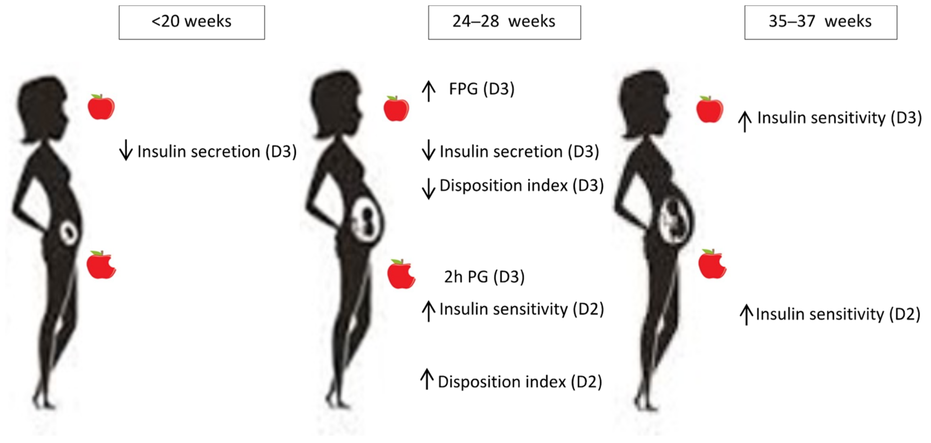
| Outcome Variables | β Values/OR for Significant Associations | ||||||||
|---|---|---|---|---|---|---|---|---|---|
| <20 Weeks | 24–28 Weeks | 35–37 Weeks | |||||||
| 25OHD2 | 25OHD3 | C3-Epimer | 25OHD2 | 25OHD3 | C3-Epimer | 25OHD2 | 25OHD3 | C3-Epimer | |
| Fasting | |||||||||
| FPG | 0.124 * | 0.111 * | |||||||
| 1/HOMA-IR (sens) | 0.127 * | ||||||||
| QUICKI (sens) | |||||||||
| HOMA-β (sec) | −0.117 * | −0.145 ** | |||||||
| Fasting DI | |||||||||
| −0.124 * | −0.148 ** | |||||||
| −0.093 * | ||||||||
| Post-challenge | |||||||||
| 1 h PG | |||||||||
| 2 h PG | 0.120* | ||||||||
| Matsuda (sens) | 0.149 * | ||||||||
| Stumvoll phase 1 (sec) | |||||||||
| AUCins/glu (sec) | |||||||||
| Post-challenge DI | |||||||||
| |||||||||
| −0.103 * | 0.168 * | 0.239 ** | ||||||
| |||||||||
Publisher’s Note: MDPI stays neutral with regard to jurisdictional claims in published maps and institutional affiliations. |
© 2022 by the authors. Licensee MDPI, Basel, Switzerland. This article is an open access article distributed under the terms and conditions of the Creative Commons Attribution (CC BY) license (https://creativecommons.org/licenses/by/4.0/).
Share and Cite
Mendoza, L.C.; Harreiter, J.; Desoye, G.; Simmons, D.; Adelantado, J.M.; Kautzky-Willer, A.; Zawiejska, A.; Wender-Ozegowska, E.; Lapolla, A.; Dalfra, M.G.; et al. The Weak Relationship between Vitamin D Compounds and Glucose Homeostasis Measures in Pregnant Women with Obesity: An Exploratory Sub-Analysis of the DALI Study. Nutrients 2022, 14, 3256. https://doi.org/10.3390/nu14163256
Mendoza LC, Harreiter J, Desoye G, Simmons D, Adelantado JM, Kautzky-Willer A, Zawiejska A, Wender-Ozegowska E, Lapolla A, Dalfra MG, et al. The Weak Relationship between Vitamin D Compounds and Glucose Homeostasis Measures in Pregnant Women with Obesity: An Exploratory Sub-Analysis of the DALI Study. Nutrients. 2022; 14(16):3256. https://doi.org/10.3390/nu14163256
Chicago/Turabian StyleMendoza, Lilian Cristina, Jürgen Harreiter, Gernot Desoye, David Simmons, Juan M. Adelantado, Alexandra Kautzky-Willer, Agnieszka Zawiejska, Ewa Wender-Ozegowska, Annunziata Lapolla, Maria G. Dalfra, and et al. 2022. "The Weak Relationship between Vitamin D Compounds and Glucose Homeostasis Measures in Pregnant Women with Obesity: An Exploratory Sub-Analysis of the DALI Study" Nutrients 14, no. 16: 3256. https://doi.org/10.3390/nu14163256