Review

The Role and Mechanism of Gut Microbiota in Pulmonary Arterial Hypertension

1
Heart Center and Beijing Key Laboratory of Hypertension, Beijing Chaoyang Hospital, Capital Medical University, Beijing 100020, China
2
Medical Research Center, Beijing Institute of Respiratory Medicine and Beijing Chaoyang Hospital, Capital Medical University, Beijing 100020, China
3
Department of Cardiology, Beijing Chaoyang Hospital, Capital Medical University, Beijing 100020, China
4
State Key Laboratory of Cardiovascular Disease, Fuwai Hospital, National Center for Cardiovascular Disease, Peking Union Medical College, Chinese Academy of Medical Sciences, Beijing 100032, China
*
Authors to whom correspondence should be addressed.
Nutrients 2022, 14(20), 4278; https://doi.org/10.3390/nu14204278
Submission received: 12 September 2022 / Revised: 7 October 2022 / Accepted: 11 October 2022 / Published: 13 October 2022
(This article belongs to the Section Clinical Nutrition)

Abstract

:
Pulmonary arterial hypertension (PAH) is a malignant pulmonary vascular disease characterized by increased pulmonary vascular resistance, pulmonary vasoconstriction, and right ventricular hypertrophy. Recent developments in genomics and metabolomics have gradually revealed the roles of the gut microbiota (GM) and its metabolites in cardiovascular diseases. Accumulating evidence reveals that the GM plays important roles in the occurrence and development of PAH. Gut microbiota dysbiosis directly increases the gut permeability, thereby facilitating pathological bacterial translocation and allowing translocation of bacterial products such as lipopolysaccharides from the gut into circulation. This process aggravates pulmonary perivascular inflammation and exacerbates PAH development through the endothelial–mesenchymal transition. Additionally, a shift in the composition of PAH also affects the gut metabolites. Changes in gut metabolites, such as decreased short-chain fatty acids, increased trimethylamine N-oxide, and elevated serotonin, contribute to pulmonary perivascular inflammation and pulmonary vascular remodeling by activating several signaling pathways. Studies of the intestinal microbiota in treating pulmonary hypertension have strengthened linkages between the GM and PAH. Probiotic therapy and fecal microbiota transplantation may supplement existing PAH treatments. In this article, we provide new insight for diagnosing, preventing and treating PAH by adding to the current knowledge of the intestinal flora mechanisms and its metabolites efficacy involved in PAH.

1. Introduction

Pulmonary arterial hypertension (PAH) is a malignant pulmonary vascular disease characterized by augmented pulmonary vascular resistance, pulmonary vasoconstriction, and right ventricular hypertrophy, ultimately resulting in right heart failure and death. The main pathophysiological mechanisms of PAH are endothelial injury, pulmonary vascular remodeling, and orthotopic thrombosis [1]. Owing to the lack of screening methods and biomarkers for early detection, patients with PAH often develop severe right ventricular dysfunction at the time of diagnosis [2]. The 1-, 3-, and 5-year survival rates for patients with PAH are 90.4%, 76.2%, and 65.4%, respectively, thereby enhancing the society economic burden in China and Western [3]. Moreover, current pharmacological therapies are expensive and non-curative, only modestly increasing patients’ exercise capacity and reducing hospital admissions [4]. Since 2005, no new therapeutic pathways for treating PAH have been identified [5]. Therefore, new approaches are urgently needed for early diagnosis and more efficient curative treatment for patients with PAH.
In the intestinal system, many bacteria, archaea, protists, fungi, and viruses form an ecological community known as the gut microbiota (GM). The microbiome produces plenty of metabolites with biological effects, thereby maintaining gut barrier function and homeostasis [6,7]. Gut microbes regulate digestion, metabolism, and innate immunity and prevent colonization of pathogens, while the gut supplies them with the proper environment for survival [8,9,10,11].
Dysbiosis of the GM has been described in multiple cardiovascular diseases, including atherosclerosis, hypertension, and platelet hyperactivity [12]. Accumulated evidence has indicated that gut integrity, the composition of GM, and its metabolites play important roles in the development of systemic inflammation, therefore modulating various cardiovascular disease risk factors. For example, the gut microbiota metabolite trimethylamine N-oxide (TMAO) directly constricts arterioles and sensitizes blood pressure levels in Ang II-induced hypertensive mice. [13]. At the same time, TMAO exerts pro-atherosclerotic effects via a variety of mechanisms [14]. In contrast, short-chain fatty acids (SCFAs) protect from cardiac damage and reduce atherosclerosis in experimental hypertension [15]. However, for PAH, a systemic disease involving many organs in addition to the lungs, such as the central nervous system, immune system, bone marrow, and intestines, the GM remains an understudied area [16]. Recent research indicates an association between the gut microflora and PAH, sparking great interest among scholars. On the one hand, persistent pulmonary hypertension leads to right ventricular hypertrophy, right heart failure, and systemic venous congestion, which alters the intestinal flora; on the other hand, changes in the GM greatly affect the intestinal barrier function [17]. When the gut barrier is impaired, intestinal Gram-negative bacteria release lipopolysaccharides (LPS) into the bloodstream, causing Toll-like receptor 4 (TLR4)-mediated endotoxemia [18,19], which induces pulmonary perivascular inflammatory responses and thus promotes the development of PAH. Furthermore, variations in gut microbial metabolites facilitate perivascular inflammation and the proliferation of pulmonary vascular smooth muscle cells, thereby initiating lung injury and pulmonary vascular remodeling, and finally leading to PAH [20,21,22] (Figure 1). However, the specific interaction between gut dysbiosis and PAH has not been clearly clarified. This review explores the relationship among intestinal flora, gut microbial metabolites, and PAH, and we discuss potential therapeutic strategies for PAH by reconstructing the GM.

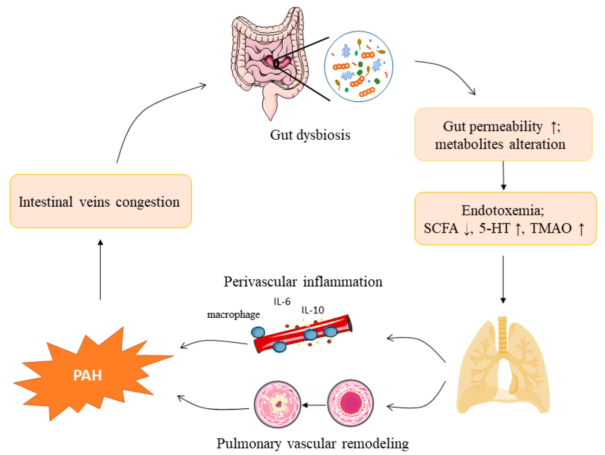
2. The Gut Microbiota and Pulmonary Arterial Hypertension

2.1. Gut Microbiota Composition

The five predominant phyla in the human gut microbiome are Bacteroidetes, Firmicutes, Actinobacteria, Proteobacteria, and Verrucomicrobia [23]. In healthy guts, anaerobic Bacteroidetes and Firmicutes account for >90% of the total bacterial species, whereas Proteobacteria and Actinobacteria are the most abundant phyla in the circulating microbiome [24]. The number of genes in these microorganisms exceeds the total number of human genes by >100 times, greatly enriching the gene pool and metabolic functions of their human hosts. A stable intestinal microbiome is fundamental for preserving the intact epithelial barrier and preventing microbiota constituents from translocating to the epithelium, spreading to and infecting other distant areas of the body [25]. However, the GM differs between healthy and unhealthy individuals [16]. Gut microbiota activity and composition are leading factors influencing human health and causing diseases [26]. Diet, drugs, and lifestyles can affect and cause disproportions in the intestinal flora. Gut dysbiosis occurs when the number of beneficial bacteria decreases or the number of harmful bacteria increases, causing an imbalance between the GM and host, and eventually leading to diseases such as diabetes mellitus, malignant tumors, hypertension, and pulmonary hypertension [27,28].

2.2. Shifts in the Gut Microbiota Composition in Pulmonary Arterial Hypertension

Recent reports suggest a link between PAH and the GM. Gut microbiota dysbiosis was observed in rat models of experimental PAH, including Sugen 5416-induced, chronic hypoxia-induced, and monocrotaline (MCT)-induced rat models [29,30]. Similarly, intestinal microbiota imbalances have been detected in patients with pulmonary hypertension [31,32]. Long-term PAH can result in chronic right heart failure and systemic venous congestion. Congestion of the intestinal veins leads to reduced bowel perfusion, increased intestinal permeability, and gut bacterial and/or endotoxin translocation [33,34]. With the emerging role of the GM in health and diseases, the gut–lung axis provides new insight into the PAH pathogenesis and may be a novel therapeutic target for PAH.
Experimental animals induced via hypoxia/Sugen 5416 or intraperitoneal injection of MCT are widely used animal models of PAH. Wistar rats treated with hypoxia/Sugen 5416 exhibited an increased Firmicutes-to-Bacteroidetes ratio, a hallmark of gut dysbiosis, with a lower abundance of Bacteroidetes [29]. Moreover, gut genera that function as acetate and butyrate producers were depleted by hypoxia/Sugen 5416 and showed decreased serum acetate levels [29]. Monocrotaline-induced PAH rats also showed disordered gut microbiota [30]. It was found that compared with the control group, MCT-induced PAH rats showed higher relative abundances of Firmicutes, Proteobacteria, and Actinobacteria but lower relative abundances of Bacteroidota and Spirochaetota. Moreover, modifying the gut microbiota with antibiotics suppressed the increase in right ventricular systolic pressure, right ventricular hypertrophy, and pulmonary vascular remodeling in hypoxia/Sugen 5416-induced rats [28].
Consistent with the results from PAH animals, patients with PAH also exhibit altered gut microbiome profiles. Using metagenomic analyses, Kim et al. revealed the fecal microbial features of 18 patients with type I PAH and 13 controls without PAH [31]. Alpha diversity and bacterial richness and evenness were reduced in the intestinal flora of patients with type I PAH [31]. Bifidobacterium, a genus of acetate producers, was overrepresented, whereas Coprococcus, Butyrivibrio, Lachnospiraceae, and Eubacterium, which are butyrate-producing bacteria, and Akkermansia and Bacteroides, which are propionate-producing bacteria, were reduced in patients with type I PAH [31]. A subsequent study on 11 patients with chronic thromboembolic pulmonary hypertension (CTEPH) and 22 matched controls showed that patients with CTEPH had different gut microbiota compositions [32]. The butyrate-producing bacteria, Roseburia and Faecalibacterium, were also significantly reduced in patients with CTEPH [32]. Overall, these studies showed dysregulated gut microbiota in both animals and patients of PAH (Table 1), which may contribute to the development of PAH.

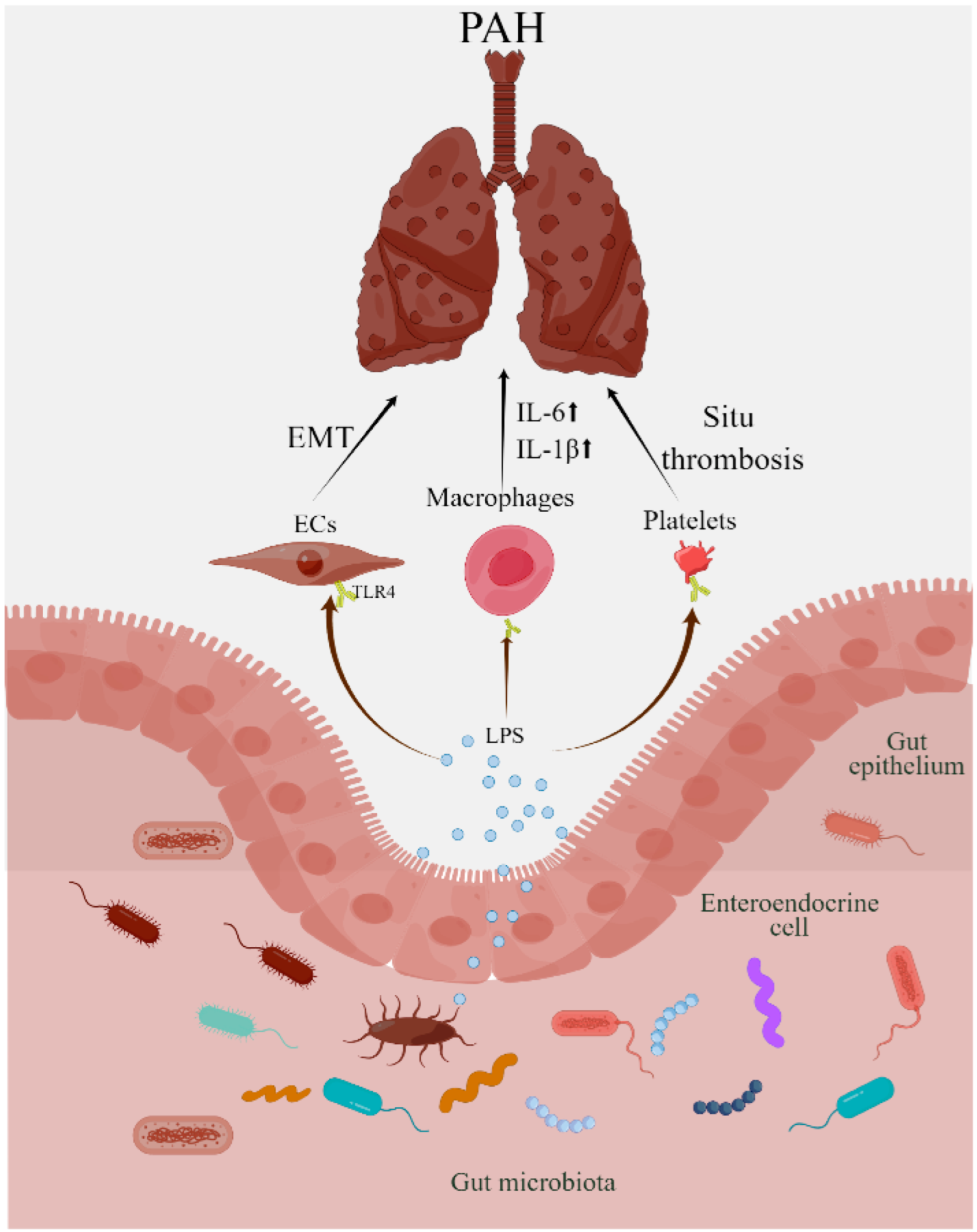
2.3. Lipopolysaccharides in Pulmonary Arterial Hypertension

The GM plays a major role in maintaining the normalization of intestinal permeability [7]. The intestinal mucosal layer, a component of the intestinal barrier, covers the intestinal cells like a shell and protects them from mechanical, chemical, and biological attacks [37]. The GM is essential for the formation of a proper mucosal layer, and its composition notably influences the intestinal mucus [38]. In a study with germ-free mice, researchers could experimentally aspirate only a few filled goblet cells and a small volume of intestinal mucus, because that mucus fails to mature and develop into the proper structure in the absence of bacteria or their metabolites [39]. Imbalanced intestinal flora in PAH rats alters the intestinal permeability, including fewer mucin-producing goblet cells, shortened villus lengths, and increased intestinal fibrosis and muscular tissue [40]. Decreases in SCFA-producing bacteria, such as Coprococcus, Butyrivibrio, Lachnospiraceae, Eubacterium, and Clostridia, in patients with PAH may epigenetically modify gut epithelial cells and consequently increase gut permeability. Patients with PAH exhibit increased plasma biomarkers for gut leakiness and inflammation, indicating that impaired mucosal barriers and increased permeability exist in these patients [6].
Increased gut permeability caused by gut dysbiosis allows commensal bacteria to translocate from the enteric cavity into circulation and promotes the generation of peripheral blood bacterial products, such as LPS. Lipopolysaccharides, also known as endotoxins, are the main structural components of Gram-negative bacteria and strongly affect foreign pathogen detection by host cells. Lipopolysaccharide levels were significantly increased in CTEPH-, type I PAH-, and MCT-induced animals [29,31,32]. As a pathogen-associated molecular pattern, LPS is recognized by pathogen recognition receptors, including TLRs. TLR4, the major signaling receptor for LPS, plays a vital role in LPS-induced inflammatory responses [41].
Toll-like receptor 4 is expressed in lung parenchymal and nonparenchymal cells, including endothelial cells (ECs), smooth muscle cells (SMCs), macrophages, and platelets. Toll-like receptor 4 and its downstream signaling pathways participate in PAH pathogenesis [42]. Several mechanisms underlie the effect of TLR4 activation on PAH development. As a component of innate immunity, TLR4-induced inflammation is an important mechanism triggering PAH. Lipopolysaccharides stimulate macrophages through TLR4 and initiate a series of signaling events, including activation of IκB kinase-activated nuclear factor κ-light-chain-enhancer of activated B cells (NF-κB), the mitogen-activated protein kinase (MAPK) family c-Jun N-terminal kinases, extracellular signal-regulated kinases (ERKs), and p38 [43]. Therefore, in chronic hypoxia-induced PAH rats, the depletion of inflammatory macrophages prevented remodeling of the pulmonary vascular extracellular matrix and development of pulmonary hypertension [44,45].
Activation of NF-κB, the central switchboard of inflammation, results in the release of cytokines (e.g., interleukin [IL]-4, IL-13, tumor necrosis factor-α, and IL-1β) and chemokines (e.g., IL-6 and IL-8) [46], which promote the development of PAH by regulating pulmonary vascular remodeling. In bone morphogenetic protein receptor type II-deficient mice chronically treated with LPS, TLR4 was upregulated in pulmonary arterial SMCs (PASMCs) and the lungs, and thus stimulated IL-6 and IL-8 production and led to pulmonary vascular remodeling [47]. Cytokines directly control pulmonary vascular cell proliferation, migration, and differentiation. For example, IL-6 overexpression promoted the growth and proliferation of porcine aortic ECs and PASMCs, and IL-6 knockout mice were resistant to hypoxia-induced PAH [48].
In addition to inflammation, TLRs on ECs also contribute to PAH development. ECs are widely involved in PAH-related pathological processes, including vasoconstriction, inflammation, cell viability, cell growth, and cell differentiation [49]. The effects of TLR signaling on endothelial dysfunction are multifaceted. TLR4 signaling increases the levels of vascular endothelial growth factors, which are central angiogenic molecules that regulate EC proliferation, migration, and tube formation during angiogenesis [50]. In addition, TLR signaling promotes PAH development through endothelial–mesenchymal transition (EMT), a process by which ECs acquire a mesenchymal cell phenotype. Increasing studies have established that EMT gives rise to the dysfunctional endothelial phenotype in patients with PAH [51,52,53]. Toll-like receptor 4 activation enhanced EMT and promoted the expression of the progenitor cell marker c-kit (CD117) in mouse pulmonary ECs, indicating a potential connection between TLR4 and EMT [54].
Furthermore, TLRs on platelets may also play important roles in PAH. After bonding to TLR4, LPS promoted P-selectin, IL-1β, and ATP expression in platelets, which may promote in situ thrombosis, a feature of human idiopathic PAH [55,56]. Genetic deletion of TLR4 on platelets in mice attenuates hypoxia-induced pulmonary hypertension [57]. However, hypoxia downregulates TLR4 expression, and TLR4 knockout mice spontaneously develop PH, suggesting that the role of TLR4 is protective in PAH [58]. These results suggest that LPS-TLR4 plays a pivotal role in PAH development (Figure 2). Given the importance of LPS, a pathophysiological link may exist between gut dysbiosis and PAH.

2.4. Gut Microbial Metabolites and Pulmonary Arterial Hypertension

Another mechanism by which the microbiota participates in PAH progression is through metabolites. Most nutrients are digested and absorbed in the small intestine, while gut bacteria ferment undigested dietary fibers, proteins, and peptides in the cecum and colon. Microbiota-derived metabolites, including SCFAs, tryptophan (5-HT), and TMAO, have received much attention for their roles in diseases [13,59,60].

2.5. Short-Chain Fatty Acids

Short-chain fatty acids are derived from indigestible dietary fibers in the intestines and are the major products of microbial fermentative activity in the gut [61]. The principal SCFAs in the gut include acetate, propionate, and butyrate, whose concentrations are in a ratio of 3:1:1, respectively [62]. Short-chain fatty acids are widely distributed throughout the intestines, with acetate and propionate in the small and large intestines and butyrate mostly in the colon and cecum [63]. Several factors contribute to SCFA production, including diet, GM diversity, and the number of commensal bacteria [63]. Certain gut bacteria regulate the microbial conversion of undigested fibers to SCFAs. Under the action of enteric bacteria, pyruvate produces acetate via the acetyl-CoA pathway, while several Firmicutes species generate butyrate via the acetyl-CoA pathway. Bacteroidetes produce propionate via the succinate pathway, and Firmicutes produce propionate via the lactate pathway [64]. A few gut bacteria, including Faecalibacterium prausnitzii, Eubacterium (E.) rectale, E. hallii, and Ruminococcus (R.) bromii, participate in most butyrate production [65]. Resistant starch fermentation, regulated by R. bromii, greatly promotes butyrate production in the colon [66]. Colonic epithelial cells absorb most SCFAs, which are transported to the liver via the portal vein and metabolized by hepatocytes. A few SCFAs pass through the liver, and a low but measurable concentration can be detected in the systemic circulation [67]. After entering circulation, SCFAs affect metabolism and the functions of peripheral tissues, such as the lungs. They also regulate immune responses and inflammation, which may affect PAH development [63,68].
Short-chain fatty acids regulate immune responses and inflammation via two pathways. One is the activation of free fatty acid 2 (FFA2), FFA3, and GPR109A receptors, and the other is the inhibition of the generation of histone deacetylases (HDACs) [67].
The human genome encompasses 800 G-protein-coupled receptors (GPRs), and a cluster of four GPR genes (GPR40–GPR43) are closely associated with SCFAs. These GPRs are also called free fatty acid receptors (FFARs) because they can sense free fatty acids. GPR41 and GPR43 have been renamed FFAR2 and FFAR3, respectively [61]. G-protein-coupled receptor 41 and GPR43 are differentially expressed on cell membranes and are involved in diverse cellular functions [69,70]. GPR41 is mainly expressed in blood vessels on ECs, and GPR43 is mainly expressed on immune cells. GPR41 is also expressed on immune cells but at lower levels than GPR43 [71]. SCFAs can regulate immune responses by activating GPRs (e.g., neutrophils and macrophages), which are expressed on most immune cells. For example, in models of colitis, arthritis, and asthma, GPR43 deficiency boosted the synthesis of inflammatory mediators and recruitment of immune cells, thus promoting inflammatory responses [72].
HDACs are a group of deacetylating enzymes that remove acetyl groups from both histone and non-histone protein complexes to regulate gene expression [73]. Butyrate and propionate have key roles in suppressing the production of class I and class IIa HDACs and downregulating silence information regulator 1 expression [74]. By specifically inhibiting HDAC1 and HDAC3, butyrate and propionate are non-competitive HDAC inhibitors [67].
Histone deacetylase inhibitors are potent agents for reducing inflammatory activities in inflammatory diseases [75,76]. For example, HDAC3 promotes the expression of inflammatory genes and recruits monocytes to inflammation sites by regulating NF-kB activity [77]. Suberoylanilide hydroxamic acid, a potent HDAC inhibitor with anti-inflammatory properties, attenuates LPS-induced expression of NF-κB-regulated cytokines [78]. Acetate-producing bacteria are decreased in PAH. One study found notable differences in SCFA-producing bacterial genera between controls and patients with PAH. Acetate levels are also reduced in the serum of PAH-induced rats. This change parallels the differences observed in the types of acetate-producing bacteria [29].
Therefore, SCFAs affect PAH via several mechanisms. Firstly, reduced SCFAs weaken gut barrier functions and favor oxidative metabolism, which further increases the likelihood of gut inflammation and leakiness. In fact, SCFA-producing bacteria, such as Coprococcus, Butyrivibrio, and Lachnospiraceae, are decreased, and gut permeability is increased in patients with PAH [31]. As mentioned, the mucosal layer is the primary physical barrier against pathogen invasion and is mainly composed of mucins secreted by goblet cells in the epithelium. SCFAs are closely associated with mucins. In vitro, SCFAs promoted goblet cells to produce mucins by increasing mucin gene expression [79]. In vivo, administering probiotic supplements boosted SCFA production and markedly enhanced intestinal barrier functions in mice with antibiotic-induced dysbiosis [80]. Short-chain fatty acids also protect against endothelial barrier dysfunction. Tight junctions (TJs) combine with epithelial cells to regulate paracellular transport. Lowered expression of TJ proteins (i.e., occludin, claudin, JAMs, and ZO-1) increases gut permeability. Researchers have demonstrated that SCFAs strengthen barrier functions by regulating TJ protein expression in several disease models [80,81,82,83]. Moreover, SCFAs affect both intestinal and vascular ECs [20]. Short-chain fatty acids upregulated the expression of TJ proteins (ZO-1, occludin, and claudin-1) and a cytoplasmic adaptor protein (cingulin) in rhesus monkey vascular ECs to protect the integrity of the vascular epithelial barrier.
Secondly, SCFAs exert anti-inflammatory effects on chemotaxis and leukocyte recruitment; these effects have been demonstrated in in vitro experiments and animal models [84]. SCFAs activate or induce differentiation of T-regulatory cells either via activation of GPCRs or epigenetic modifications by inhibiting HDACs [85,86]. SCFAs block the NF-kB pathway in macrophages to arrest inflammatory responses and promote T-cell differentiation to enhance IL-10 production [71]. SCFAs boost the generation of colonic T-regulatory cells [87] and attenuate hypoxia-induced pulmonary vascular remodeling by regulating cytokine production in the lungs [20]. In conclusion, SCFAs play unique roles in PAH development; however, the specific molecular mechanisms require further investigation.

2.6. Trimethylamine N-Oxide

The GM transforms dietary nutrients, such as phosphatidylcholine, choline, L-carnitine, and betaine from beef and fish, into trimethylamine. Eight bacterial species involved in trimethylamine production have been identified and belong to Firmicutes and Proteobacteria [88]. Most trimethylamine enters the circulatory system and is subsequently oxidized to TMAO by hepatic flavin-containing monooxygenase, while excess TMA is directly decomposed into dimethylamine or methane [89]. Therefore, the GM composition notably affects TMAO production from dietary sources [90].
Trimethylamine N-oxide is a risk factor for several cardiovascular diseases such as hypertension, heart failure, and atherosclerosis [13,14,91]. Recent research revealed that bacterial communities producing TMAO metabolites were increased in PAH patients and animals [29,31,32]. Because TMAO synthesis relies on the intestinal microbial flora, changes in the GM directly alter circulating TMAO levels.
Trimethylamine N-oxide levels were elevated in high-risk patients with idiopathic PAH and MCT-induced rat models but not in low-risk patients with idiopathic PAH or hypoxia-induced mouse models, indicating that TMAO expression was increased only in severe PAH [21]. Furthermore, 3,3-dimethyl-1-butanol (DMB), a TMAO synthesis inhibitor, alleviated the pathological changes in PAH. DMB treatment reduced right ventricular systolic pressure, maximum right atrial pressure, right ventricular hypertrophy, and pulmonary vascular thickness in MCT-induced rat models [21], suggesting that TMAO plays an important role in accelerating PAH progression and may be a therapeutic target for treating PAH.
Regarding its mechanism, Wang et al. found that DMB prevented macrophages from secreting inflammatory factors and thereby relieved pulmonary vascular damage [21]. It was also noticed that TMAO significantly intensified PASMC proliferation and macrophage migration, which are important contributors to PAH development and progression [21].

2.7. 5-Hydroxytryptophan

Serotonin (5-hydroxytryptophan [5-HT]), a natural vasoconstrictor and vascular SMC mitogen, is essential in the PAH pathobiology. Patients with both idiopathic and secondary pulmonary hypertension exhibit increased plasma serotonin levels [92]. Tryptophan is the only substrate for serotonin synthesis. Tryptophan hydroxylase (TPH), the rate-limiting enzyme in serotonin biosynthesis, converts tryptophan to the intermediate, L-5-hydroxytryptophan (5-HTP), which is then transformed into 5-HT. There are two active TPH isoforms: TPH1 and TPH2. A majority of peripheral serotonin is synthesized by TPH1 in the gut, while TPH2 participates in the manufacture of central serotonin and is mostly expressed in the central nervous system. Over 99% of total 5-HT in the circulatory system is taken up by platelets after being released from the intestines. The rate of peripheral 5-HT synthesis is limited by TPH1 levels, which directly affect the plasma serotonin concentration. Previous studies have demonstrated that the GM plays a role in the regulation of 5-HT metabolism in the colon, which in turn might affect peripheral blood concentration of serotonin [93,94,95]. The GM promotes 5-HT biosynthesis by elevating TPH1 expression in colonic ECs and regulate both colon and serum levels of 5-HT.
Serotonin, a natural vasoconstrictor and vascular SMC mitogen, is essential in the PAH pathobiology. Serotonin acts on the pulmonary vasculature via interactions with GPRs (5-HT 1B, 2A and 2B). Substantial evidence suggests that activation of 1B-R, 2A-R, and 2B-R subtypes modulates pulmonary vascular tone. In rodent models, the 5-HT2B receptor has therapeutic effects on heritable PAH owing to its antagonism [96]. In experimental PAH, 5-HT2B receptors play crucial roles in the development of endovascular injury caused by bone marrow-derived proangiogenic cells [97].
The effect of 5-HT1B receptors is achieved via two signaling pathways after activation. First, activated 5-HT receptors induce the expression of downstream RhoA [98]. As a member of the Rho family of small GTPases, RhoA regulates various cellular responses, including contraction, migration, and growth, as well as gene expression and differentiation [99]. RhoA is activated by exchanging GDP for GTP and translocates to the plasma membrane to stimulate downstream effectors such as Rho-kinase. Rho-kinase activation inhibits the expression of myosin light-chain phosphatase by phosphorylating the myosin-binding subunit of myosin light-chain phosphatase. Consequently, Ca2+ becomes more sensitive to contraction in vascular SMCs. Furthermore, cytosolic Ca2+ increases contraction and myosin light-chain kinase activity [100]. RhoA/Rho-kinase affects nuclear translocation of ERK1/ERK2 during 5HT1BR-mediated mitogenesis [101]. Therefore, GM-serotonin-RhoA/ROCK signaling exerts important roles in the pathogenesis of pulmonary hypertension.
Second, serotonin induces cellular Src-related kinase-regulated Nox1-induced reactive oxygen species (ROS) and nuclear factor (erythroid-derived 2)-like 2 (Nrf-2) dysregulations by activating 5-HT1B receptors. Consequently, post-translational oxidative protein modification is increased, and redox-sensitive signaling pathways are activated. ROS production in the lungs contributes to experimental pulmonary hypertension [102].
The serotonin transporter (SERT or 5-HTT) is a monoamine transporter protein that delivers serotonin into cells. Evidence suggests that 5-HT’s mitogenic effect depends on SERT activity [103]. Transgenic mice with SERT overexpression were more susceptible to pulmonary hypertension [104], and administering SERT inhibitors prevented hypoxia-induced pulmonary vascular remodeling in wild-type animals [105]. Similarly, the 5-HTT inhibitors, fluoxetine and citalopram, impeded human PASMC growth in vitro by blocking SERT expression [106]. miR-361-3p overexpression suppressed serotonin-induced human PASMC proliferation by lowering SERT levels [107], indicating that SERT induced PASMC proliferation. Because SERT mediates indolamine uptake in SMCs, the mitogenic effect of 5-HT on PASMCs is ascribed to 5-HT internalization by SERT. Serotonin entering the cytoplasm of PASMCs is likely decomposed by MAO-A or NADPH oxidases into ROS.

3. Gut Microbiota-Directed Therapies for Pulmonary Arterial Hypertension

Considering the important contribution of GM in PAH development described earlier, reconstructing the imbalanced gut by probiotics, prebiotics, or fecal microbiota transplantation may be effective therapeutic strategies for PAH.

3.1. Prebiotics and Probiotics

Probiotics are live microorganisms that benefit host health when administered in adequate amounts. Probiotics with non-strain-specific claims include Bifidobacterium adolescentis, B. animalis, B. bifidum, B. breve, B. longum, Lactobacillus acidophilus, L. casei, L. fermentum, L. gasseri, L. johnsonii, L. paracasei, L. plantarum, L. rhamnosus, and L. salivarius [108]. Wedgwood et al. showed that rat pups with PAH induced by postnatal growth restriction presented gut dysbiosis that was attenuated by administering the probiotic L. reuteri DSM 17938 [109]. L. reuteri reduces proinflammatory cytokine levels and simultaneously blocks TLR4 signaling via the NF-κB pathway [110]. In children with acute lung injury, a probiotic formulation of Eosinophil L. and L. acidophilus significantly improved pulmonary artery pressure [111]. Thus, PAH is at least partly driven by dysbiosis in the developing gut, and combining probiotics with established approaches may provide a more effective therapy for treating PAH. However, precise probiotic strains that are both safe and effective in lowering pulmonary arterial pressure and preventing PAH remains a great challenge in clinical application and is worth exploring.
Prebiotics are nondigestible carbohydrates that modulate microbiota activity [112]. Studies show that they selectively enhance the growth of beneficial bacteria such as Lactobacillus and Bifidobacterium. They also exert a strong anti-inflammatory effect to deter harmful intestinal pathogens. Inulin is a prebiotic that promotes beneficial bacterial growth and stimulates SCFA production. In a randomized controlled trial, dietary inulin or inulin propionate supplementation attenuated propionate release from the colon in obese patients [113]. Similarly, obese patients can lose weight on an inulin-enriched diet [114]. However, no study to date has demonstrated the role of prebiotics in PAH.

3.2. Fecal Microbiota Transplantation

Fecal microbiota transplantation (FMT) involves transferring functional bacteria from healthy donor feces into the gastrointestinal tracts of patients to repair an imbalanced GM. Filtered feces collected from healthy donors or recipients (autologous FMT) are transplanted into the intestines of patients with certain diseases. Fecal microbiota transplantation from ACE2-knock-in mice to wild-type mice protected wild-type mice from hypoxia-induced cardiopulmonary pathophysiology and mitigated gut pathology [36]. However, although FMT is currently a reliable treatment for patients with Clostridioides difficile colitis [115], the safety (especially in the long term) and efficacy of microbiota-targeting interventions to treat PAH remain uncertain. The U.S. Food and Drug Administration recently approved a phase I trial evaluating the safety and feasibility of FMT in treating PAH [116]. In this open-label trial, 12 patients diagnosed with PAH will take once-daily FMT capsules for 7 days to determine whether FMT is safe and feasible for these patients. This is the first step in investigating the effectiveness of FMT as a treatment for PAH. As FMT is more effective than probiotics or prebiotics in reconstructing the GM, the therapeutic effect of FMT for PAH may be better; however, large-scale, multicenter clinical studies are needed to obtain a definitive conclusion.

4. Conclusions

Gut dysbiosis and PAH form a circularity interaction. A shift in gut microbiota composition causes an increase in gut permeability, thereby contributing to bacteria translocation and metabolic endotoxemia. Gut dysbiosis also changes the circulatory levels of gut metabolites such as TMAO, 5-HT, and SCFAs. Collectively, endotoxin and metabolic products exert on pulmonary vasculature, causing pulmonary perivascular inflammation, pulmonary vascular remodeling, and eventually developing the PAH. Meanwhile, long-term PAH leads to intestinal vein congestion, which in turn contributes to gut dysbiosis. However, more research is needed to investigate and verify an etiological link between the PAH pathogenesis and changes in the gut microbiota. Reconstructing the GM by prebiotics, probiotics, or FMT may provide new effective therapeutic strategies for PAH. Nevertheless, it is imperative to uncover the specific bacterial species and metabolic pathways in the context of PAH in future studies.

Author Contributions

Conceptualization, Y.-H.C.; writing—original draft preparation, Y.-H.C.; writing—review and editing, X.-Y.L.; visualization, W.Y.; supervision, L.-K.M.; project administration, J.-C.Z., X.-Y.L. All authors have read and agreed to the published version of the manuscript.

Funding

This study was supported by the General Program and the National Major Research Plan Training Program of the National Natural Science Foundation of China (No. 82170302, 82070325, 92168117; 81770253; 91849111); the Beijing Hospitals Authority Youth Program (No. QML20200305), the Beijing Natural Science Foundation (No. 7222138, 7222068; 7162069); the Reform and Development Program of Beijing Institute of Respiratory Medicine (No. ysrh2022010, ysrh2022002, ysrh2022007); and Clinical Research Incubation Program of Beijing Chaoyang Hospital Affiliated to Capital Medical University (CYFH202209).

Institutional Review Board Statement

Not applicable.

Informed Consent Statement

Not applicable.

Data Availability Statement

Not applicable.

Conflicts of Interest

All authors declare that they have no conflict of interest. Figure 2 was created by Figdraw (www.figdraw.com), accessed on 31 August 2022.

References

  1. McLaughlin, V.V.; McGoon, M.D. Pulmonary Arterial Hypertension. Circulation 2006, 114, 1417–1431. [Google Scholar] [CrossRef] [PubMed] [Green Version]
  2. Wedgwood, S.; Gerard, K.; Halloran, K.; Hanhauser, A.; Monacelli, S.; Warford, C.; Thai, P.N.; Chiamvimonvat, N.; Lakshminrusimha, S.; Steinhorn, R.H.; et al. Intestinal Dysbiosis and the Developing Lung: The Role of Toll-Like Receptor 4 in the Gut-Lung Axis. Front. Immunol. 2020, 11, 357. [Google Scholar] [CrossRef] [PubMed]
  3. Farber, H.W.; Miller, D.P.; Poms, A.D.; Badesch, D.B.; Frost, A.E.; Rouzic, E.M.-L.; Romero, A.J.; Benton, W.W.; Elliott, C.G.; McGoon, M.D.; et al. Five-Year Outcomes of Patients Enrolled in the REVEAL Registry. Chest 2015, 148, 1043–1054. [Google Scholar] [CrossRef] [Green Version]
  4. Anand, V.; Roy, S.S.; Archer, S.L.; Weir, E.K.; Garg, S.K.; Duval, S.; Thenappan, T. Trends and Outcomes of Pulmonary Arterial Hypertension–Related Hospitalizations in the United States: Analysis of the Nationwide Inpatient Sample Database From 2001 Through 2012. JAMA Cardiol. 2016, 1, 1021. [Google Scholar] [CrossRef]
  5. Humbert, M.; Guignabert, C.; Bonnet, S.; Dorfmüller, P.; Klinger, J.R.; Nicolls, M.R.; Olschewski, A.J.; Pullamsetti, S.S.; Schermuly, R.T.; Stenmark, K.R.; et al. Pathology and Pathobiology of Pulmonary Hypertension: State of the Art and Research Perspectives. Eur. Respir. J. 2019, 53, 1801887. [Google Scholar] [CrossRef] [Green Version]
  6. Kwant, C.T.; Ruiter, G.; Noordegraaf, A.V. Malnutrition in Pulmonary Arterial Hypertension. Curr. Opin. Pulm. Med. 2019, 25, 405–409. [Google Scholar] [CrossRef]
  7. Paone, P.; Cani, P.D. Mucus Barrier, Mucins and Gut Microbiota: The Expected Slimy Partners? Gut 2020, 69, 2232–2243. [Google Scholar] [CrossRef] [PubMed]
  8. Kamada, N.; Seo, S.; Chen, G.Y.; Núñez, G. Role of the Gut Microbiota in Immunity and Inflammatory Disease. Nat. Rev. Immunol. 2013, 13, 321–335. [Google Scholar] [CrossRef] [PubMed]
  9. Clements, S.J.; Carding, S.R. Diet, the Intestinal Microbiota, and Immune Health in Aging. Crit. Rev. Food Sci. 2018, 58, 651–661. [Google Scholar] [CrossRef] [Green Version]
  10. Martin-Gallausiaux, C.; Marinelli, L.; Blottière, H.M.; Larraufie, P.; Lapaque, N. SCFA: Mechanisms and Functional Importance in the Gut. Proc. Nutr. Soc. 2021, 80, 37–49. [Google Scholar] [CrossRef]
  11. Callejo, M.; Barberá, J.A.; Duarte, J.; Perez-Vizcaino, F. Impact of Nutrition on Pulmonary Arterial Hypertension. Nutrients 2020, 12, 169. [Google Scholar] [CrossRef] [Green Version]
  12. Brown, J.M.; Hazen, S.L. Microbial Modulation of Cardiovascular Disease. Nat. Rev. Microbiol. 2018, 16, 171–181. [Google Scholar] [CrossRef] [PubMed]
  13. Jiang, S.; Shui, Y.; Cui, Y.; Tang, C.; Wang, X.; Qiu, X.; Hu, W.; Fei, L.; Li, Y.; Zhang, S.; et al. Gut Microbiota Dependent Trimethylamine N-Oxide Aggravates Angiotensin II–Induced Hypertension. Redox Biol. 2021, 46, 102115. [Google Scholar] [CrossRef] [PubMed]
  14. Koeth, R.A.; Wang, Z.; Levison, B.S.; Buffa, J.A.; Org, E.; Sheehy, B.T.; Britt, E.B.; Fu, X.; Wu, Y.; Li, L.; et al. Intestinal Microbiota Metabolism of l-carnitine, a Nutrient in Red Meat, Promotes Atherosclerosis. Nat. Med. 2013, 19, 576–585. [Google Scholar] [CrossRef] [PubMed] [Green Version]
  15. Bartolomaeus, H.; Balogh, A.; Yakoub, M.; Homann, S.; Markó, L.; Höges, S.; Tsvetkov, D.; Krannich, A.; Wundersitz, S.; Avery, E.G.; et al. Short-Chain Fatty Acid Propionate Protects from Hypertensive Cardiovascular Damage. Circulation 2019, 139, 1407–1421. [Google Scholar] [CrossRef] [PubMed]
  16. Oliveira, A.C.; Richards, E.M.; Raizada, M.K. Pulmonary Hypertension: Pathophysiology beyond the Lung. Pharmacol. Res. 2020, 151, 104518. [Google Scholar] [CrossRef]
  17. Allam-Ndoul, B.; Castonguay-Paradis, S.; Veilleux, A. Gut Microbiota and Intestinal Trans-Epithelial Permeability. Int. J. Mol. Sci. 2020, 21, 6402. [Google Scholar] [CrossRef] [PubMed]
  18. Hug, H.; Mohajeri, M.H.; La Fata, G. Toll-Like Receptors: Regulators of the Immune Response in the Human Gut. Nutrients 2018, 10, 203. [Google Scholar] [CrossRef] [Green Version]
  19. Young, K.C.; Hussein, S.M.A.; Dadiz, R.; DeMello, D.; Devia, C.; Hehre, D.; Suguihara, C. Toll-Like Receptor 4–Deficient Mice are Resistant to Chronic Hypoxia-Induced Pulmonary Hypertension. Exp. Lung Res. 2010, 36, 111–119. [Google Scholar] [CrossRef] [PubMed]
  20. Karoor, V.; Strassheim, D.; Sullivan, T.; Verin, A.; Umapathy, N.S.; Dempsey, E.C.; Frank, D.N.; Stenmark, K.R.; Gerasimovskaya, E. The Short-Chain Fatty Acid Butyrate Attenuates Pulmonary Vascular Remodeling and Inflammation in Hypoxia-Induced Pulmonary Hypertension. Int. J. Mol. Sci. 2021, 22, 9916. [Google Scholar] [CrossRef]
  21. Huang, Y.; Lin, F.; Tang, R.; Bao, C.; Zhou, Q.; Ye, K.; Shen, Y.; Liu, C.; Hong, C.; Yang, K.; et al. Gut Microbial Metabolite Trimethylamine N-Oxide Aggravates Pulmonary Hypertension. Am. J. Respir. Cell Mol. Biol. 2022, 66, 452–459. [Google Scholar] [CrossRef] [PubMed]
  22. Song, J.; Zheng, S.; He, R.; Gui, L.; Lin, M.; Sham, J.S.K. Serotonin and Chronic Hypoxic Pulmonary Hypertension Activate a NADPH Oxidase 4 and TRPM2 Dependent Pathway for Pulmonary Arterial Smooth Muscle Cell Proliferation and Migration. Vasc. Pharmacol. 2021, 138, 106860. [Google Scholar] [CrossRef]
  23. Qin, J.; Li, R.; Raes, J.; Arumugam, M.; Burgdorf, K.S.; Manichanh, C.; Nielsen, T.; Pons, N.; Levenez, F.; Yamada, T.; et al. A human Gut Microbial Gene Catalogue Established by Metagenomic Sequencing. Nature 2010, 464, 59–65. [Google Scholar] [CrossRef] [Green Version]
  24. Zmora, N.; Zilberman-Schapira, G.; Suez, J.; Mor, U.; Dori-Bachash, M.; Bashiardes, S.; Kotler, E.; Zur, M.; Regev-Lehavi, D.; Brik, R.B.; et al. Personalized Gut Mucosal Colonization Resistance to Empiric Probiotics is Associated with Unique Host and Microbiome Features. Cell 2018, 174, 1388–1405. [Google Scholar] [CrossRef] [PubMed] [Green Version]
  25. Plé, C.; Breton, J.; Daniel, C.; Foligné, B. Maintaining Gut Ecosystems for Health: Are Transitory Food Bugs Stowaways or Part of the Crew? Int. J. Food Microbiol. 2015, 213, 139–143. [Google Scholar] [CrossRef]
  26. Marques, F.Z.; Mackay, C.R.; Kaye, D.M. Beyond gut feelings: How the Gut Microbiota Regulates Blood Pressure. Nat. Rev. Cardiol. 2018, 15, 20–32. [Google Scholar] [CrossRef] [PubMed]
  27. Yan, X.; Jin, J.; Su, X.; Yin, X.; Gao, J.; Wang, X.; Zhang, S.; Bu, P.; Wang, M.; Zhang, Y.; et al. Intestinal Flora Modulates Blood Pressure by Regulating the Synthesis of Intestinal-Derived Corticosterone in High Salt-Induced Hypertension. Circ. Res. 2020, 126, 839–853. [Google Scholar] [CrossRef] [PubMed]
  28. Gavin, P.G.; Mullaney, J.A.; Loo, D.; Cao, K.L.; Gottlieb, P.A.; Hill, M.M.; Zipris, D.; Hamilton-Williams, E.E. Intestinal Metaproteomics Reveals Host-Microbiota Interactions in Subjects at Risk for Type 1 Diabetes. Diabetes Care 2018, 41, 2178–2186. [Google Scholar] [CrossRef] [Green Version]
  29. Callejo, M.; Mondejar-Parreño, G.; Barreira, B.; Izquierdo-Garcia, J.L.; Morales-Cano, D.; Esquivel-Ruiz, S.; Moreno, L.; Cogolludo, Á.; Duarte, J.; Perez-Vizcaino, F. Pulmonary Arterial Hypertension Affects the Rat Gut Microbiome. Sci. Rep. 2018, 8, 9681. [Google Scholar] [CrossRef] [PubMed] [Green Version]
  30. Hong, W.; Mo, Q.; Wang, L.; Peng, F.; Zhou, Y.; Zou, W.; Sun, R.; Liang, C.; Zheng, M.; Li, H.; et al. Changes in the Gut Microbiome and Metabolome in a Rat Model of Pulmonary Arterial Hypertension. Bioengineered 2021, 12, 5173–5183. [Google Scholar] [CrossRef] [PubMed]
  31. Kim, S.; Rigatto, K.; Gazzana, M.B.; Knorst, M.M.; Richards, E.M.; Pepine, C.J.; Raizada, M.K. Altered Gut Microbiome Profile in Patients with Pulmonary Arterial Hypertension. Hypertension 2020, 75, 1063–1071. [Google Scholar] [CrossRef]
  32. Ikubo, Y.; Sanada, T.J.; Hosomi, K.; Park, J.; Naito, A.; Shoji, H.; Misawa, T.; Suda, R.; Sekine, A.; Sugiura, T.; et al. Altered Gut Microbiota and Its Association with Inflammation in Patients with Chronic Thromboembolic Pulmonary Hypertension: A Single-Center Observational Study in Japan. BMC Pulm. Med. 2022, 22, 138. [Google Scholar] [CrossRef] [PubMed]
  33. Sandek, A.; Bjarnason, I.; Volk, H.; Crane, R.; Meddings, J.B.; Niebauer, J.; Kalra, P.R.; Buhner, S.; Herrmann, R.; Springer, J.; et al. Studies on Bacterial Endotoxin and Intestinal Absorption Function in Patients with Chronic Heart Failure. Int. J. Cardiol. 2012, 157, 80–85. [Google Scholar] [CrossRef] [PubMed]
  34. Nagatomo, Y.; Tang, W.H.W. Intersections between Microbiome and Heart Failure: Revisiting the Gut Hypothesis. J. Card. Fail. 2015, 21, 973–980. [Google Scholar] [CrossRef] [PubMed] [Green Version]
  35. Sanada, T.J.; Hosomi, K.; Shoji, H.; Park, J.; Naito, A.; Ikubo, Y.; Yanagisawa, A.; Kobayashi, T.; Miwa, H.; Suda, R.; et al. Gut Microbiota Modification Suppresses the Development of Pulmonary Arterial Hypertension in an SU5416/Hypoxia Rat Model. Pulm. Circ. 2020, 10, 1365052046–1765618565. [Google Scholar] [CrossRef]
  36. Sharma, R.K.; Oliveira, A.C.; Yang, T.; Karas, M.M.; Li, J.; Lobaton, G.O.; Aquino, V.P.; Robles-Vera, I.; de Kloet, A.D.; Krause, E.G.; et al. Gut Pathology and Its Rescue by ACE2 (Angiotensin-Converting Enzyme 2) in Hypoxia-Induced Pulmonary Hypertension. Hypertension 2020, 76, 206–216. [Google Scholar] [CrossRef] [PubMed]
  37. Etienne-Mesmin, L.; Chassaing, B.; Desvaux, M.; De Paepe, K.; Gresse, R.; Sauvaitre, T.; Forano, E.; de Wiele, T.V.; Schuller, S.; Juge, N.; et al. Experimental Models to Study Intestinal Microbes-Mucus Interactions in Health and Disease. Fems Microbiol. Rev. 2019, 43, 457–489. [Google Scholar] [CrossRef] [PubMed] [Green Version]
  38. Jakobsson, H.E.; Rodriguez-Pineiro, A.M.; Schutte, A.; Ermund, A.; Boysen, P.; Bemark, M.; Sommer, F.; Backhed, F.; Hansson, G.C.; Johansson, M.E. The Composition of the Gut Microbiota Shapes the Colon Mucus Barrier. Embo Rep. 2015, 16, 164–177. [Google Scholar] [CrossRef]
  39. Rodriguez-Pineiro, A.M.; Johansson, M.E. The Colonic Mucus Protection Depends on the Microbiota. Gut Microbes 2015, 6, 326–330. [Google Scholar] [CrossRef] [Green Version]
  40. Sharma, R.K.; Oliveira, A.C.; Yang, T.; Kim, S.; Zubcevic, J.; Aquino, V.; Lobaton, G.O.; Goel, R.; Richards, E.M.; Raizada, M.K. Pulmonary Arterial Hypertension-Associated Changes in Gut Pathology and Microbiota. ERJ Open Res. 2020, 6, 253–2019. [Google Scholar] [CrossRef]
  41. Park, B.S.; Lee, J. Recognition of Lipopolysaccharide Pattern by TLR4 Complexes. Exp. Mol. Med. 2013, 45, e66. [Google Scholar] [CrossRef] [PubMed] [Green Version]
  42. Bhagwani, A.; Thompson, A.A.R.; Farkas, L. When Innate Immunity Meets Angiogenesis—The Role of Toll-Like Receptors in Endothelial Cells and Pulmonary Hypertension. Front. Med. 2020, 7, 352. [Google Scholar] [CrossRef] [PubMed]
  43. Roy, A.; Srivastava, M.; Saqib, U.; Liu, D.; Faisal, S.M.; Sugathan, S.; Bishnoi, S.; Baig, M.S. Potential Therapeutic Targets for Inflammation in Toll-Like Receptor 4 (TLR4)-Mediated Signaling Pathways. Int. Immunopharmacol. 2016, 40, 79–89. [Google Scholar] [CrossRef] [PubMed]
  44. Tian, W.; Jiang, X.; Tamosiuniene, R.; Sung, Y.K.; Qian, J.; Dhillon, G.; Gera, L.; Farkas, L.; Rabinovitch, M.; Zamanian, R.T.; et al. Blocking Macrophage Leukotriene B4 Prevents Endothelial Injury and Reverses Pulmonary Hypertension. Sci. Transl. Med. 2013, 5, 117r–200r. [Google Scholar] [CrossRef] [PubMed] [Green Version]
  45. Frid, M.G.; Brunetti, J.A.; Burke, D.L.; Carpenter, T.C.; Davie, N.J.; Reeves, J.T.; Roedersheimer, M.T.; van Rooijen, N.; Stenmark, K.R. Hypoxia-Induced Pulmonary Vascular Remodeling Requires Recruitment of Circulating Mesenchymal Precursors of a Monocyte/Macrophage Lineage. Am. J. Pathol. 2006, 168, 659–669. [Google Scholar] [CrossRef] [Green Version]
  46. Lawrence, T. The Nuclear Factor NF- B Pathway in Inflammation. Csh. Perspect. Biol. 2009, 1, a1651. [Google Scholar] [CrossRef] [PubMed] [Green Version]
  47. Soon, E.; Crosby, A.; Southwood, M.; Yang, P.; Tajsic, T.; Toshner, M.; Appleby, S.; Shanahan, C.M.; Bloch, K.D.; Pepke-Zaba, J.; et al. Bone Morphogenetic Protein Receptor Type II Deficiency and Increased Inflammatory Cytokine Production. A Gateway to Pulmonary Arterial Hypertension. Am. J. Respir. Crit. Care Med. 2015, 192, 859–872. [Google Scholar] [CrossRef] [Green Version]
  48. Steiner, M.K.; Syrkina, O.L.; Kolliputi, N.; Mark, E.J.; Hales, C.A.; Waxman, A.B. Interleukin-6 Overexpression Induces Pulmonary Hypertension. Circ. Res. 2009, 104, 236–244. [Google Scholar] [CrossRef] [PubMed]
  49. Evans, C.E.; Cober, N.D.; Dai, Z.; Stewart, D.J.; Zhao, Y. Endothelial Cells in the Pathogenesis of Pulmonary Arterial Hypertension. Eur. Respir. J. 2021, 58, 2003957. [Google Scholar] [CrossRef] [PubMed]
  50. Paolillo, R.; Iovene, M.R.; Romano, C.C.; Rizzo, A. Induction of VEGF and MMP-9 Expression by Toll-Like Receptor 2/4 in Human Endothelial Cells Infected with Chlamydia Pneumoniae. Int. J. Immunopathol. Pharmacol. 2012, 25, 377–386. [Google Scholar] [CrossRef] [PubMed]
  51. Good, R.B.; Gilbane, A.J.; Trinder, S.L.; Denton, C.P.; Coghlan, G.; Abraham, D.J.; Holmes, A.M. Endothelial to Mesenchymal Transition Contributes to Endothelial Dysfunction in Pulmonary Arterial Hypertension. Am. J. Pathol. 2015, 185, 1850–1858. [Google Scholar] [CrossRef]
  52. Ranchoux, B.; Antigny, F.; Rucker-Martin, C.; Hautefort, A.; Péchoux, C.; Bogaard, H.J.; Dorfmüller, P.; Remy, S.; Lecerf, F.; Planté, S.; et al. Endothelial-to-Mesenchymal Transition in Pulmonary Hypertension. Circulation 2015, 131, 1006–1018. [Google Scholar] [CrossRef] [Green Version]
  53. Suzuki, T.; Carrier, E.J.; Talati, M.H.; Rathinasabapathy, A.; Chen, X.; Nishimura, R.; Tada, Y.; Tatsumi, K.; West, J. Isolation and Characterization of Endothelial-to-Mesenchymal Transition Cells in Pulmonary Arterial Hypertension. Am. J. Physiol.-Lung C. 2018, 314, L118–L126. [Google Scholar] [CrossRef] [PubMed]
  54. Suzuki, T.; Tada, Y.; Nishimura, R.; Kawasaki, T.; Sekine, A.; Urushibara, T.; Kato, F.; Kinoshita, T.; Ikari, J.; West, J.; et al. Endothelial-to-Mesenchymal Transition in Lipopolysaccharide-Induced Acute Lung Injury Drives a Progenitor Cell-Like Phenotype. Am. J. Physiol.-Lung C. 2016, 310, L1185–L1198. [Google Scholar] [CrossRef] [PubMed] [Green Version]
  55. Andonegui, G.; Kerfoot, S.M.; McNagny, K.; Ebbert, K.V.J.; Patel, K.D.; Kubes, P. Platelets Express Functional Toll-Like Receptor-4. Blood 2005, 106, 2417–2423. [Google Scholar] [CrossRef] [PubMed]
  56. Zhang, G.; Han, J.; Welch, E.J.; Ye, R.D.; Voyno-Yasenetskaya, T.A.; Malik, A.B.; Du, X.; Li, Z. Lipopolysaccharide Stimulates Platelet Secretion and Potentiates Platelet Aggregation via TLR4/MyD88 and the cGMP-Dependent Protein Kinase Pathway. J. Immunol. 2009, 182, 7997–8004. [Google Scholar] [CrossRef] [PubMed] [Green Version]
  57. Bauer, E.M.; Chanthaphavong, R.S.; Sodhi, C.P.; Hackam, D.J.; Billiar, T.R.; Bauer, P.M. Genetic Deletion of Toll-Like Receptor 4 on Platelets Attenuates Experimental Pulmonary Hypertension. Circ. Res. 2014, 114, 1596–1600. [Google Scholar] [CrossRef] [PubMed] [Green Version]
  58. Ma, L.; Ambalavanan, N.; Liu, H.; Sun, Y.; Jhala, N.; Bradley, W.E.; Dell’Italia, L.J.; Michalek, S.; Wu, H.; Steele, C.; et al. TLR4 Regulates Pulmonary Vascular Homeostasis and Remodeling via Redox Signaling. Front. Biosci. 2016, 21, 397–409. [Google Scholar]
  59. Chen, L.; He, F.J.; Dong, Y.; Huang, Y.; Wang, C.; Harshfield, G.A.; Zhu, H. Modest Sodium Reduction Increases Circulating Short-Chain Fatty Acids in Untreated Hypertensives. Hypertension 2020, 76, 73–79. [Google Scholar] [CrossRef] [PubMed]
  60. Taleb, S. Tryptophan Dietary Impacts Gut Barrier and Metabolic Diseases. Front. Immunol. 2019, 10, 2113. [Google Scholar] [CrossRef]
  61. Koh, A.; De Vadder, F.; Kovatcheva-Datchary, P.; Bäckhed, F. From Dietary Fiber to Host Physiology: Short-Chain Fatty Acids as Key Bacterial Metabolites. Cell 2016, 165, 1332–1345. [Google Scholar] [CrossRef] [PubMed]
  62. Fernandes, J.; Su, W.; Rahat-Rozenbloom, S.; Wolever, T.M.S.; Comelli, E.M. Adiposity, Gut Microbiota and Faecal Short Chain Fatty Acids are Linked in Adult Humans. Nutr. Diabetes 2014, 4, e121. [Google Scholar] [CrossRef] [PubMed] [Green Version]
  63. Sun, M.; Wu, W.; Liu, Z.; Cong, Y. Microbiota Metabolite Short Chain Fatty Acids, GPCR, and Inflammatory Bowel Diseases. J. Gastroenterol. 2017, 52, 1–8. [Google Scholar] [CrossRef] [PubMed]
  64. Louis, P.; Hold, G.L.; Flint, H.J. The Gut Microbiota, Bacterial Metabolites and Colorectal Cancer. Nat. Rev. Microbiol. 2014, 12, 661–672. [Google Scholar] [CrossRef]
  65. Louis, P.; Young, P.; Holtrop, G.; Flint, H.J. Diversity of Human Colonic Butyrate-Producing Bacteria Revealed by Analysis of the Butyryl-CoA:Acetate CoA-Transferase Gene. Environ. Microbiol. 2010, 12, 304–314. [Google Scholar] [CrossRef]
  66. Ze, X.; Duncan, S.H.; Louis, P.; Flint, H.J. Ruminococcus Bromii is a Keystone Species for the Degradation of Resistant Starch in the Human Colon. ISME J. 2012, 6, 1535–1543. [Google Scholar] [CrossRef] [Green Version]
  67. Li, M.; van Esch, B.; Wagenaar, G.; Garssen, J.; Folkerts, G.; Henricks, P. Pro- and Anti-inflammatory Effects of Short Chain Fatty Acids on Immune and Endothelial Cells. Eur. J. Pharmacol. 2018, 831, 52–59. [Google Scholar] [CrossRef]
  68. Marchesi, J.R.; Adams, D.H.; Fava, F.; Hermes, G.D.A.; Hirschfield, G.M.; Hold, G.; Quraishi, M.N.; Kinross, J.; Smidt, H.; Tuohy, K.M.; et al. The Gut Microbiota and Host Health: A New Clinical Frontier. Gut 2016, 65, 330–339. [Google Scholar] [CrossRef] [Green Version]
  69. Kim, M.H.; Kang, S.G.; Park, J.H.; Yanagisawa, M.; Kim, C.H. Short-Chain Fatty Acids Activate GPR41 and GPR43 on Intestinal Epithelial cells to Promote Inflammatory Responses in Mice. Gastroenterology 2013, 145, 396–406. [Google Scholar] [CrossRef] [PubMed]
  70. Aoyama, M.; Kotani, J.; Usami, M. Butyrate and Propionate Induced Activated or Non-Activated Neutrophil Apoptosis via HDAC Inhibitor Activity but without Activating GPR-41/GPR-43 Pathways. Nutrition 2010, 26, 653–661. [Google Scholar] [CrossRef]
  71. Kim, S.; Kim, J.; Park, B.O.; Kwak, Y.S. Perspectives on the Therapeutic Potential of Short-Chain Fatty Acid Receptors. BMB Rep. 2014, 47, 173–178. [Google Scholar] [CrossRef]
  72. Puddu, A.; Sanguineti, R.; Montecucco, F.; Viviani, G.L. Evidence for the Gut Microbiota Short-Chain Fatty Acids as Key Pathophysiological Molecules Improving Diabetes. Mediat. Inflamm. 2014, 2014, 1–9. [Google Scholar] [CrossRef] [PubMed] [Green Version]
  73. Delcuve, G.P.; Khan, D.H.; Davie, J.R. Roles of Histone Deacetylases in Epigenetic Regulation: Emerging Paradigms from Studies with Inhibitors. Clin. Epigenetics 2012, 4, 5. [Google Scholar] [CrossRef] [Green Version]
  74. Schilderink, R.; Verseijden, C.; de Jonge, W.J. Dietary Inhibitors of Histone Deacetylases in Intestinal Immunity and Homeostasis. Front. Immunol. 2013, 4, 226. [Google Scholar] [CrossRef] [PubMed] [Green Version]
  75. Das Gupta, K.; Shakespear, M.R.; Iyer, A.; Fairlie, D.P.; Sweet, M.J. Histone Deacetylases in Monocyte/Macrophage Development, Activation and Metabolism: Refining HDAC Targets for Inflammatory and Infectious Diseases. Clin. Transl. Immunol. 2016, 5, e62. [Google Scholar] [CrossRef] [Green Version]
  76. Tang, J.; Yan, H.; Zhuang, S. Histone Deacetylases as Targets for Treatment of Multiple Diseases. Clin. Sci. 2013, 124, 651–662. [Google Scholar] [CrossRef] [PubMed] [Green Version]
  77. Leus, N.G.; Zwinderman, M.R.; Dekker, F.J. Histone Deacetylase 3 (HDAC 3) as Emerging Drug Target in NF-κB-Mediated Inflammation. Curr. Opin. Chem. Biol. 2016, 33, 160–168. [Google Scholar] [CrossRef] [PubMed] [Green Version]
  78. Li, M.; van Esch, B.C.A.M.; Henricks, P.A.J.; Folkerts, G.; Garssen, J. The Anti-inflammatory Effects of Short Chain Fatty Acids on Lipopolysaccharide- or Tumor Necrosis Factor α-Stimulated Endothelial Cells via Activation of GPR41/43 and Inhibition of HDACs. Front. Pharmacol. 2018, 9, 533. [Google Scholar] [CrossRef] [Green Version]
  79. Burger-van Paassen, N.; Vincent, A.; Puiman, P.J.; van der Sluis, M.; Bouma, J.; Boehm, G.; van Goudoever, J.B.; van Seuningen, I.; Renes, I.B. The Regulation of Intestinal Mucin MUC2 Expression by Short-Chain Fatty Acids: Implications for Epithelial Protection. Biochem. J. 2009, 420, 211–219. [Google Scholar] [CrossRef] [Green Version]
  80. Hagihara, M.; Kuroki, Y.; Ariyoshi, T.; Higashi, S.; Fukuda, K.; Yamashita, R.; Matsumoto, A.; Mori, T.; Mimura, K.; Yamaguchi, N.; et al. Clostridium butyricum Modulates the Microbiome to Protect Intestinal Barrier Function in Mice with Antibiotic-Induced Dysbiosis. iScience 2020, 23, 100772. [Google Scholar] [CrossRef] [Green Version]
  81. Pan, L.L.; Niu, W.; Fang, X.; Liang, W.; Li, H.; Chen, W.; Zhang, H.; Bhatia, M.; Sun, J. Clostridium butyricum Strains Suppress Experimental Acute Pancreatitis by Maintaining Intestinal Homeostasis. Mol. Nutr. Food Res. 2019, 63, e1801419. [Google Scholar] [CrossRef] [PubMed]
  82. Li, H.; Sun, J.; Du, J.; Wang, F.; Fang, R.; Yu, C.; Xiong, J.; Chen, W.; Lu, Z.; Liu, J. Clostridium Butyricum Exerts a Neuroprotective Effect in a Mouse Model of Traumatic Brain Injury via the Gut-Brain Axis. Neurogastroenterol. Motil. 2018, 30, e13260. [Google Scholar] [CrossRef] [PubMed]
  83. Shang, H.; Sun, J.; Chen, Y.Q. Clostridium Butyricum CGMCC0313.1 Modulates Lipid Profile, Insulin Resistance and Colon Homeostasis in Obese Mice. PLoS ONE 2016, 11, e154373. [Google Scholar] [CrossRef] [PubMed] [Green Version]
  84. Mackay, C.R.; Artis, D.; Yu, D.; Xavier, R.J.; Ng, A.; Mackay, F.; Sierro, F.; Kranich, J.; Rolph, M.S.; Teixeira, M.M.; et al. Regulation of Inflammatory Responses by Gut Microbiota and Chemoattractant Receptor GPR43. Nature 2009, 461, 1282–1286. [Google Scholar]
  85. Yuille, S.; Reichardt, N.; Panda, S.; Dunbar, H.; Mulder, I.E. Human Gut Bacteria as Potent Class I Histone Deacetylase Inhibitors in Vitro through Production of Butyric Acid and Valeric Acid. PLoS ONE 2018, 13, e201073. [Google Scholar] [CrossRef] [Green Version]
  86. Tan, J.; McKenzie, C.; Potamitis, M. The Role of Short-Chain Fatty Acids in Health and Disease. Adv. Immunol. 2014, 121, 91–119. [Google Scholar]
  87. Thenappan, T.; Khoruts, A.; Chen, Y.; Weir, E.K. Can Intestinal Microbiota and Circulating Microbial Products Contribute to Pulmonary Arterial Hypertension? Am. J. Physiol.-Heart C 2019, 317, H1093–H1101. [Google Scholar] [CrossRef]
  88. Wang, Z.; Klipfell, E.; Bennett, B.J.; Koeth, R.; Levison, B.S.; DuGar, B.; Feldstein, A.E.; Britt, E.B.; Fu, X.; Chung, Y.; et al. Gut Flora Metabolism of Phosphatidylcholine Promotes Cardiovascular Disease. Nature 2011, 472, 57–63. [Google Scholar] [CrossRef] [PubMed] [Green Version]
  89. Bennett, B.J.; Vallim, T.Q.D.A.; Wang, Z.; Shih, D.M.; Meng, Y.; Gregory, J.; Allayee, H.; Lee, R.; Graham, M.; Crooke, R.; et al. Trimethylamine-N-Oxide, a Metabolite Associated with Atherosclerosis, Exhibits Complex Genetic and Dietary Regulation. Cell Metab. 2013, 17, 49–60. [Google Scholar] [CrossRef] [Green Version]
  90. Romano, K.A.; Vivas, E.I.; Amador-Noguez, D.; Rey, F.E. Intestinal Microbiota Composition Modulates Choline Bioavailability from Diet and Accumulation of the Proatherogenic Metabolite Trimethylamine-N-Oxide. Mbio 2015, 6, e2481. [Google Scholar] [CrossRef] [Green Version]
  91. Zhang, Y.; Wang, Y.; Ke, B.; Du, J. TMAO: How Gut Microbiota Contributes to Heart Failure. Transl. Res. 2021, 228, 109–125. [Google Scholar] [CrossRef]
  92. Egermayer, P.; Town, G.I.; Peacock, A.J. Role of Serotonin in the Pathogenesis of Acute and Chronic Pulmonary Hypertension. Thorax 1999, 54, 161–168. [Google Scholar] [CrossRef] [Green Version]
  93. Ge, X.; Ding, C.; Zhao, W.; Xu, L.; Tian, H.; Gong, J.; Zhu, M.; Li, J.; Li, N. Antibiotics-Induced Depletion of Mice Microbiota Induces Changes in Host Serotonin Biosynthesis and Intestinal Motility. J. Transl. Med. 2017, 15, 13. [Google Scholar] [CrossRef]
  94. Reigstad, C.S.; Salmonson, C.E.; III, J.F.R.; Szurszewski, J.H.; Linden, D.R.; Sonnenburg, J.L.; Farrugia, G.; Kashyap, P.C. Gut Microbes Promote Colonic Serotonin Production through an Effect of Short-Chain Fatty Acids on Enterochromaffin Cells. FASEB J. 2015, 29, 1395–1403. [Google Scholar] [CrossRef] [Green Version]
  95. Yano, J.M.; Yu, K.; Donaldson, G.P.; Shastri, G.G.; Ann, P.; Ma, L.; Nagler, C.R.; Ismagilov, R.F.; Mazmanian, S.K.; Hsiao, E.Y. Indigenous Bacteria from the Gut Microbiota Regulate Host Serotonin Biosynthesis. Cell 2015, 161, 264–276. [Google Scholar] [CrossRef] [Green Version]
  96. West, J.D.; Carrier, E.J.; Bloodworth, N.C.; Schroer, A.K.; Chen, P.; Ryzhova, L.M.; Gladson, S.; Shay, S.; Hutcheson, J.D.; Merryman, W.D.; et al. Serotonin 2B Receptor Antagonism Prevents Heritable Pulmonary Arterial Hypertension. PLoS ONE 2016, 11, e148657. [Google Scholar] [CrossRef]
  97. Bloodworth, N.C.; Clark, C.R.; West, J.D.; Snider, J.C.; Gaskill, C.; Shay, S.; Scott, C.; Bastarache, J.; Gladson, S.; Moore, C.; et al. Bone Marrow–Derived Proangiogenic Cells Mediate Pulmonary Arteriole Stiffening via Serotonin 2B Receptor Dependent Mechanism. Circ. Res. 2018, 123, e51–e64. [Google Scholar] [CrossRef]
  98. Guilluy, C.; Eddahibi, S.; Agard, C.; Guignabert, C.; Izikki, M.; Tu, L.; Savale, L.; Humbert, M.; Fadel, E.; Adnot, S.; et al. RhoA and Rho Kinase Activation in Human Pulmonary Hypertension: Role of 5-HT signaling. Am. J. Respir. Crit. Care Med. 2009, 179, 1151–1158. [Google Scholar] [CrossRef] [Green Version]
  99. Vega, F.M.; Ridley, A.J. Rho GTPases in cancer cell biology. FEBS Lett. 2008, 582, 2093–2101. [Google Scholar] [CrossRef] [Green Version]
  100. Somlyo, A.P.; Somlyo, A.V. Ca2+ Sensitivity of Smooth Muscle and Nonmuscle Myosin II: Modulated by G Proteins, Kinases, and Myosin Phosphatase. Physiol. Rev. 2003, 83, 1325–1358. [Google Scholar] [CrossRef] [Green Version]
  101. Liu, Y.; Suzuki, Y.J.; Day, R.M.; Fanburg, B.L. Rho Kinase–Induced Nuclear Translocation of ERK1/ERK2 in Smooth Muscle Cell Mitogenesis Caused by Serotonin. Circ. Res. 2004, 95, 579–586. [Google Scholar] [CrossRef] [PubMed] [Green Version]
  102. Hood, K.Y.; Mair, K.M.; Harvey, A.P.; Montezano, A.C.; Touyz, R.M.; MacLean, M.R. Serotonin Signaling Through the 5-HT1B Receptor and NADPH Oxidase 1 in Pulmonary Arterial Hypertension. Arterioscler. Thromb. Vasc. Biol. 2017, 37, 1361–1370. [Google Scholar] [CrossRef] [PubMed] [Green Version]
  103. Eddahibi, S.; Humbert, M.; Fadel, E.; Raffestin, B.; Darmon, M.; Capron, F.; Simonneau, G.; Dartevelle, P.; Hamon, M.; Adnot, S. Serotonin Transporter Overexpression is Responsible for Pulmonary Artery Smooth Muscle Hyperplasia in Primary Pulmonary Hypertension. J. Clin. Investig. 2001, 108, 1141–1150. [Google Scholar] [CrossRef]
  104. MacLean, M.R.; Deuchar, G.A.; Hicks, M.N.; Morecroft, I.; Shen, S.; Sheward, J.; Colston, J.; Loughlin, L.; Nilsen, M.; Dempsie, Y.; et al. Overexpression of the 5-Hydroxytryptamine Transporter Gene. Circulation 2004, 109, 2150–2155. [Google Scholar] [CrossRef] [PubMed] [Green Version]
  105. Marcos, E.; Adnot, S.; Pham, M.H.; Nosjean, A.; Raffestin, B.; Hamon, M.; Eddahibi, S. Serotonin Transporter Inhibitors Protect against Hypoxic Pulmonary Hypertension. Am. J. Respir. Crit. Care Med. 2003, 168, 487–493. [Google Scholar] [CrossRef] [PubMed] [Green Version]
  106. Marcos, E.; Fadel, E.; Sanchez, O.; Humbert, M.; Dartevelle, P.; Simonneau, G.; Hamon, M.; Adnot, S.; Eddahibi, S. Serotonin-Induced Smooth Muscle Hyperplasia in Various Forms of Human Pulmonary Hypertension. Circ. Res. 2004, 94, 1263–1270. [Google Scholar] [CrossRef] [Green Version]
  107. Zhang, Y.; Chen, Y.; Chen, G.; Zhou, Y.; Yao, H.; Tan, H. Upregulation of MiR-361-3p Suppresses Serotonin-Induced Proliferation in Human Pulmonary Artery Smooth Muscle Cells by Targeting SERT. Cell. Mol. Biol. Lett. 2020, 25, 45. [Google Scholar] [CrossRef]
  108. Hill, C.; Guarner, F.; Reid, G.; Gibson, G.R.; Merenstein, D.J.; Pot, B.; Morelli, L.; Canani, R.B.; Flint, H.J.; Salminen, S.; et al. Expert Consensus Document. The International Scientific Association for Probiotics and Prebiotics Consensus Statement on the Scope and Appropriate Use of the Term Probiotic. Nat. Rev. Gastroenterol. Hepatol. 2014, 11, 506–514. [Google Scholar] [CrossRef] [Green Version]
  109. Wedgwood, S.; Warford, C.; Agvatisiri, S.R.; Thai, P.N.; Chiamvimonvat, N.; Kalanetra, K.M.; Lakshminrusimha, S.; Steinhorn, R.H.; Mills, D.A.; Underwood, M.A. The Developing Gut–Lung Axis: Postnatal Growth Restriction, Intestinal Dysbiosis, and Pulmonary Hypertension in a Rodent Model. Pediatr. Res. 2020, 87, 472–479. [Google Scholar] [CrossRef]
  110. Liu, Y.; Fatheree, N.Y.; Mangalat, N.; Rhoads, J.M. Lactobacillus Reuteri Strains Reduce Incidence and Severity of Experimental Necrotizing Enterocolitis via Modulation of TLR4 and NF-κB Signaling in the Intestine. Am. J. Physiol.-Gastr. Liver Physiol. 2012, 302, G608–G617. [Google Scholar]
  111. Wang, Y.; Gao, L.; Yang, Z.; Chen, F.; Zhang, Y. Effects of Probiotics on Ghrelin and Lungs in Children with Acute Lung Injury: A Double-Blind Randomized, Controlled Trial. Pediatr Pulmonol. 2018, 53, 197–203. [Google Scholar] [CrossRef] [PubMed]
  112. Blaut, M. Relationship of Prebiotics and Food to Intestinal Microflora. Eur. J. Nutr. 2002, 41, i11–i16. [Google Scholar] [CrossRef] [PubMed]
  113. Chambers, E.S.; Byrne, C.S.; Morrison, D.J.; Murphy, K.G.; Preston, T.; Tedford, C.; Garcia-Perez, I.; Fountana, S.; Serrano-Contreras, J.I.; Holmes, E.; et al. Dietary Supplementation with Inulin-Propionate Ester or Inulin Improves Insulin Sensitivity in Adults with Overweight and Obesity with Distinct Effects on the Gut Microbiota, Plasma Metabolome and Systemic Inflammatory Responses: A Randomised Cross-Over Trial. Gut 2019, 68, 1430–1438. [Google Scholar]
  114. Hiel, S.; Gianfrancesco, M.A.; Rodriguez, J.; Portheault, D.; Leyrolle, Q.; Bindels, L.B.; Gomes Da Silveira Cauduro, C.; Mulders, M.D.G.H.; Zamariola, G.; Azzi, A.; et al. Link between Gut Microbiota and Health Outcomes in Inulin -Treated Obese Patients: Lessons from the Food4Gut Multicenter Randomized Placebo-Controlled Trial. Clin. Nutr. 2020, 39, 3618–3628. [Google Scholar] [CrossRef] [PubMed]
  115. Nood, V.E.; Vrieze, A.; Nieuwdorp, M.; Fuentes Enriquez De Salamanca, S.; Zoetendal, E.G.; Vos, D.W.M.; Visser, C.E.; Kuijper, E.J.; Bartelsman, J.F.; Tijssen, J.G.; et al. Duodenal Infusion of Donor Feces for Recurrent Clostridium difficile. N. Engl. J. Med. 2013, 368, 407–415. [Google Scholar] [CrossRef]
  116. Moutsoglou, D.M. 2021 American Thoracic Society BEAR Cage Winning Proposal: Microbiome Transplant in Pulmonary Arterial Hypertension. Am. J. Respir. Crit. Care Med. 2022, 205, 13–16. [Google Scholar] [CrossRef]
Figure 1. The roles and underlying mechanisms of gut microbiota in pulmonary arterial hypertension. A shift in gut microbiota composition causes an increase in gut permeability, which leads to bacteria translocation and metabolic endotoxemia. Gut dysbiosis also changes the circulatory levels of gut metabolites such as TMAO, 5-HT, and SCFAs. Collectively, endotoxin and metabolic products exert on pulmonary vasculature, causing pulmonary perivascular inflammation, pulmonary vascular re-modeling, and eventually PAH. Long-term PAH leads to intestinal vein congestion, which in turn contributes to gut dysbiosis. TMAO: trimethylamine N-oxide; 5-HT: 5-hydroxytryptophan; SCFAs: short-chain fatty acids; IL-6: interleukin-6; IL-10: interleukin-10; PAH: pulmonary arterial hypertension, ↑ increased; ↓ decreased.
Nutrients 14 04278 g001
Figure 2. Increased gut permeability leads to translocation of gut bacterial products, such as LPS, into circulation. LPS is recognized by TLR4 expression in endothelial cells, macrophages, and platelets. After blinding to TLR4, LPS promotes endothelial–mesenchymal transition, macrophage inflammatory reaction, platelet in situ thrombosis, and finally PAH. LPS: lipopolysaccharide; TLR4: Toll-like receptor 4, ↑ increased.
Nutrients 14 04278 g002
Table 1. Shifts in the gut microbiota composition in pulmonary arterial hypertension.
Experimental Models/PopulationsEffects and ObservationPreclinical or ClinicalRef.
SUGEN5416/hypoxia rat model↑Firmicutes, Actinobacteria, Cyanobacteria
↓Bacteroides, Akkermansia
PreclinicalTakayuki J. Sanada et al. (2020)
[35]
MCT-induced rat model↑Firmicutes, Proteobacteria, Actinobacteria
↑Allobaculum, Ralstonia, Bifidobacterium
↓Bacteroidota, Spirochaetota
↓Lactobacillus, Romboutsia
PreclinicalWei Hong et al. (2021)
[30]
mice with hypoxia↑F/B, Proteobacteria, Prevotella, Oscillospira, Ruminococcus
↓Lactobacillus
PreclinicalRavindra K. Sharma et al. (2020)
[36]
SUGEN5416/hypoxia rat model↓Bacteroidetes, Odoribacteraceae
↑Firmicutes, Peptostreptococcaceae
PreclinicalMaría Callejo et al. (2018)
[29]
Patients with type I PAH↑S. parasanguinis, R. gnavus, C. aerofaciens
↑Collinsella, Blautia
↓B. crossotus, B. cellulosilyticus, E. siraeum, B. vulgatus, A. muciniphila, ↓B. intestinihominis
ClinicalSeungbum Kim et al. (2020)
[31]
Patients with CTEPH↓Faecalibacterium, Roseburia, FusicatenibacterClinicalYumiko Ikubo et al. (2022)
[32]
Abbreviations: MCT, monocrotaline; PAH, pulmonary arterial hypertension; CTEPH, chronic thromboembolic pulmonary hypertension; F/B, Firmicutes/Bacteroides, ↑ increased; ↓ decreased.
Publisher’s Note: MDPI stays neutral with regard to jurisdictional claims in published maps and institutional affiliations.

