Review

Methodological Aspects of Indirect Calorimetry in Patients with Sepsis—Possibilities and Limitations

by
Weronika Wasyluk
1,2,*,
Agnieszka Zwolak
1,
Joop Jonckheer
3,
Elisabeth De Waele
3,4,5 and
Wojciech Dąbrowski
6
1
Department of Internal Medicine in Nursing, Faculty of Health Sciences, Medical University of Lublin, Witold Chodźko Street 7, 20-093 Lublin, Poland
2
Doctoral School, Medical University of Lublin, 20-093 Lublin, Poland
3
Intensive Care, UZ Brussel, University Hospital Brussels (UZB), 1090 Jette, Belgium
4
Department of Nutrition, UZ Brussel, University Hospital Brussels (UZB), 1090 Jette, Belgium
5
Faculty of Medicine and Pharmacy, Vrije Universiteit Brussel, 1090 Brussels, Belgium
6
Department of Anaesthesiology and Intensive Care, Medical University of Lublin, 20-954 Lublin, Poland
*
Author to whom correspondence should be addressed.
Nutrients 2022, 14(5), 930; https://doi.org/10.3390/nu14050930
Submission received: 22 January 2022 / Revised: 17 February 2022 / Accepted: 18 February 2022 / Published: 22 February 2022
(This article belongs to the Section Clinical Nutrition)

Abstract

:
The aim of the review was to analyse the challenges of using indirect calorimetry in patients with sepsis, including the limitations of this method. A systematic review of the literature was carried out. The analysis concerned the methodology and presentation of research results. In most studies assessing energy expenditure, energy expenditure was expressed in kcal per day (n = 9) and as the mean and standard deviation (n = 7). Most authors provided a detailed measurement protocol, including measurement duration (n = 10) and device calibration information (n = 7). Ten papers provided information on the day of hospitalisation when the measurements were obtained, nine on patient nutrition, and twelve on the criteria for inclusion and exclusion of participants from the study. Small study group sizes and study at a single centre were among the most cited limitations. Studies assessing energy expenditure in patients with sepsis by indirect calorimetry differ in the methodology and presentation of results, and their collective analysis is difficult. A meta-analysis of the results could enable multi-site and large patient evaluation. Standardisation of protocols and presentation of all collected data would enable their meta-analysis, which would help to achieve greater knowledge about metabolism in sepsis.

1. Introduction

Sepsis is defined as a “life-threatening organ dysfunction caused by a dysregulated host response to infection” [1]. Dysregulated host response includes, among others, changes in basic metabolic processes and limitations in the body’s metabolic capacity [2,3]. In addition, hormonal disorders and circulating cytokines occurring in sepsis cause insulin resistance and promote lipolysis and proteolysis, the latter of which can lead to cachexia [3,4]. This endogenous energy production is not measureable in clinical practice and therefore only 70% of resting energy expenditure (REE) should be given during the first days. The metabolic changes induced by sepsis itself and the initiated therapies [5] are only measurable by indirect calorimetry as the predicting equations do not take these into consideration. Therefore, according to both the European Society for Clinical Nutrition and Metabolism (ESPEN) and the American Society for Parenteral and Enteral Nutrition (ASPEN), indirect calorimetry (IC) is the recommended method for assessing the energy needs of critically ill, mechanically ventilated patients [6,7,8,9,10].
However several issues compromise the use of IC during sepsis: oxygen enrichment (FiO2 > 60%), organ support therapies such as renal replacement or liver support therapy and extracorporeal membrane oxygenation (ECMO) (may change blood gas levels or affect acid-base balance), unstable pH, and unstable body temperature [10].
The aim of the study was to analyse these challenges of using IC in patients with sepsis and to highlight limitations of this method that may reduce its clinical utility and limit reliability of studies using it. The presentation of the methodology and results of this analysis will be preceded by a summary of current knowledge about the evolution of the definition of sepsis, and the theoretical basis of IC. This information is essential for a thorough understanding of the limitations described below.

2. Materials and Methods

A review of the literature was carried out between March and May 2020. The literature search process was conducted as for a systematic review in accordance with the PRISMA guidelines [11]. Articles were searched for in PubMed/Medline database. The following search formula was used: (sepsis [Title/Abstract]) OR (septic [Title/Abstract]) AND (indirect calorimetry [Title/Abstract]). The review was carried out in several stages. In the first stage, the titles and abstracts of the papers were reviewed to exclude review articles, congress annals, guidelines, case reports, studies not using IC and studies that did not include patients with sepsis/septic shock, or in which the inclusion criteria remained unclear. In the case of any doubt, the entire article was analysed before making a decision. In the second stage, the full content of articles describing research on sepsis and using IC was read, after which animal studies, studies with children, and studies with healthy subjects were excluded. After reviewing the content of the articles, studies older than 20 years were also excluded from the detailed analysis due to insufficient data being up to date.
The inclusion criteria for studies included in the final analysis were original articles describing studies using the IC method in adult (with the exception of two studies in which the patient age range was 15–85 years) patients diagnosed with sepsis or septic shock, with full texts in English and not older than 20 years. Exclusion criteria were no use of IC in the study, no participation of patients with sepsis/septic shock in the study, unclear criteria for inclusion in the study, animal studies, studies with children, studies with healthy subjects, and studies older than 20 years.
A data extraction form has been designed and used for extract data from eligible studies.After completing each form, data compliance with the study was checked. The data from the forms has been compiled in tabular form, which has been partially presented in this review. The synthesis of results was carried out in a descriptive form, comparing individual aspects of the analysed studies. The discussion summarises the main results of the review, their potential practical application, and the advantages and limitations of this review.

3. Theory

3.1. Energy Expenditure

Energy expenditure (EE) has three main components: basal energy expenditure (BEE), diet-induced thermogenesis (DIT), and activity-induced energy expenditure (AEE). BEE is the amount of energy required to maintain basic metabolic activity of cells and life functions, such as respiration and body temperature; DIT is the heat production associated with substrate oxidation during energy uptake (EN and PN); while AEE is the EE associated with physical activity. These three components can be combined to form successive levels of EE–the sum of BEE and DIT is resting energy expenditure (REE), and the sum of BEE, DIT, and AEE is total energy expenditure (TEE) [10,12,13]. When discussing this issue, it is worth noting that sometimes REE literature is identified as BEE because these terms are often used interchangeably [14]. However, in the strict sense, BEE means the lowest level of EE and its measurement should be performed in very demanding conditions, including post 8-h sleep, post 10-h fast (or 12-h, depending on the source of information), complete resting posture, free from physiological and mental stress, and a thermally neutral, quiet and shaded room [10,13]. The above requirements make this measurement impossible for critically ill people. By contrast, measurements of REE are accompanied by fewer constraints (to be discussed in the following section).

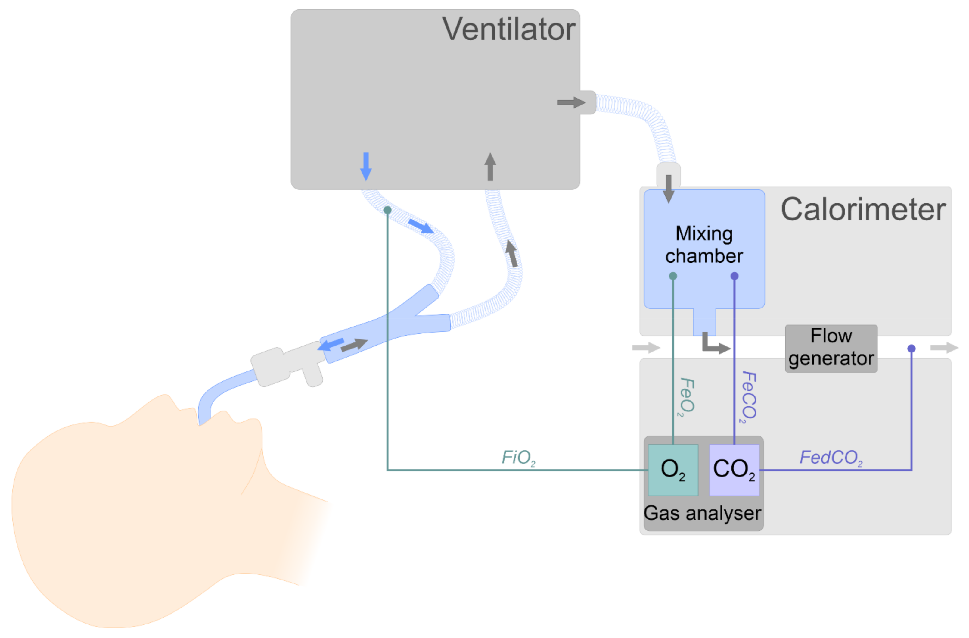
3.2. Indirect Calorimetry

IC is recognised as the gold standard for EE assessment in various populations, including in critically ill patients [6,7], cancer patients [15], and polymorbid internal medicine patients [16]. Determination of EE by the IC method requires measurement of oxygen concentration in inhaled (FiO2) and expired (FeO2) air, CO2 concentration in exhaled air (FeCO2), and the volume of exhaled gas per minute (Figure 1). These data allow the calculation of oxygen consumption (VO2) and CO2 production (VCO2), which can be used to calculate the EE using the Weir equation as described below [10,12].
The basis for calculations in IC is a modified Weir equation enabling the calculation of EE based on oxygen consumption (VO2), carbon dioxide production (VCO2), and urinary nitrogen (uN2) [18]. Due to the small share of urinary nitrogen in the actual EE in critically ill patients (the error caused by the use of respiratory functions alone was estimated at about 4%) and problematic collection of urine samples with potential additional error, a shortened version of the equation is commonly used, which omits this parameter [12,19,20]:
E E   k c a l d = V O 2 × 3.941 + V C O 2 × 1.11 × 1440
where: VO2—oxygen consumption [L/min]; VCO2—carbon dioxide production [L/min].
Under ideal conditions, VO2 and VCO2 could be calculated as the difference in the volume of inhaled and exhaled air multiplied by the concentrations of the respective gases as shown below:
V O 2 = V i × F i O 2 V e × F e O 2  
V C O 2 = V e × F e C O 2 V i × F i C O 2  
where: Vi—volume of inhaled air, Ve—volume of exhaled air, FiO2/FiCO2—fraction of inspired oxygen/carbon dioxide, and FeO2/FeCO2—fraction of expired oxygen/carbon dioxide.
However, in practice, due to technical difficulties in measuring the small difference between the volumes of inhaled and exhaled air, the volume of inhaled air (relatively more difficult to measure) is calculated using the Haldane transformation [12].
The Haldane transformation method is based on the assumption that nitrogen (N2) is not consumed or produced during breathing, so every minute the volume of inhaled N2 is equal to the volume of expired N2 [21]. Using the above assumptions, the following equations can be formulated:
V i × 1 F i O 2 F i C O 2 = V e × 1 F e O 2 F e C O 2
It can be transformed (if FiCO2 of 0.03–0.05% is ignored) into the following equation for calculating VO2:
V O 2 = 1 F e O 2 F e C O 2 × F i O 2 F e O 2 × V e 1 F i O 2
Haldane transformation allows the simplification of measurement systems by omitting the measurement of the volume of inhaled air. However, its use also introduces one of the main limitations in the use of IC in critically ill patients. “1−FiO2” in the denominator of the equation means that an increase in FiO2 above 60% is associated with a significant error in VO2 calculations. Therefore, in patients requiring high oxygen levels in inhaled air, the possibility of measuring EE by the IC method is limited [12,22].

3.3. Rules for Measuring

Correct measurement is of paramount importance for the reliability of EE measured by the IC method. The effectiveness of IC requires the following conditions to be met: proper patient preparation, correct calorimeter preparation, optimal measurement conditions, and analysis of results by experienced specialists.
Due to the demanding requirements related to patient preparation for BEE measurement (described earlier), REE measurement is usually performed for critically ill patients. This measurement should take place a minimum of five hours from a meal (or during continuous feeding), a minimum of four hours from caffeine intake, two hours from alcohol or nicotine intake, after 30 min of rest, and a minimum of two hours from moderate physical activity [10,23]. During the examination, the patient should be in a supine position and in neutral ambient conditions (temperature 27–29 °C) [10,24,25]. Some authors indicate that the measurement should take place in conditions of silence and soft lighting; however, the impact of noise on EE has not been studied so far [26,27]. In clinical situations where the possibilities of adequate preparation for the study may be limited, it is possible to liberalise, e.g., fasting time, obtaining a result sufficiently accurate for clinical purposes; however, measurements obtained in such conditions should not be used for research on REE, as they may differ as much as 100 kcal from actual REE values [23].
Technical aspects that may affect the accuracy of the results obtained include appropriate calibration and validation of the calorimeter. Accurate gas and flow rate measurements are crucial for obtaining a reliable EE. Calibration of gas analysers according to the manufacturer’s recommendations is one of the most important aspects for a correctly carried out measurement [12,27,28].
The measurement should be obtained for 30 min, or until steady state is reached. Steady state is determined by the degree of variation of VO2 and VCO2 over a period of time and can be determined when the calculated coefficient of variation (CV) % for VO2 and VCO2 is <5% within 5 min or <10% within 25 min (or 30 min, depending on the source) of the measurement. Achieving steady state is important for the accuracy of the obtained EE measurement [10,27,29,30].
When performing an EE measurement using the IC method, circumstances that could lead to inaccurate results should also be excluded. These include air leakage–patients with chest drainage, mechanically ventilated patients with high positive end expiratory pressure (PEEP > 10–12 cm H2O), endotracheal tube cuff leak or the presence of bronchopleural fistula may be at risk of this [10,12,31,32]. Inaccurate results can also be caused by mechanical ventilation with FiO2 >60%. As described earlier, due to the widespread use of a mathematical operation called the Haldane transformation, such a high concentration of oxygen in the inspiratory gas mixture can generate inaccurate results [10,12,17,22,33]. Moreover, the presence of gases other than oxygen, CO2, and N2 in the breathing mixture (e.g., gases used in therapy or anaesthesia) may reduce the accuracy of the measurement obtained [10,12,34,35].
The usefulness of IC is also limited in patients in unstable condition because the measurement will only reflect the temporary and not the general metabolic state. Examples include agitated or involuntary patient movements, patients with unstable body temperature (>±1 °C at <1 h) or patients with unstable acid-base status. This also includes patients with changes in FiO2 administration, nutrient administration, or medication administration (especially in sedation and analgesia), and those who have recently undergone invasive procedures [10,12,23,35]. Additionally, some therapies used on patients may change blood gas levels or affect acid-base balance. These include extracorporeal membrane oxygenation (ECMO), renal replacement therapy, and Molecular Adsorbent Recirculating System (MARS, liver support therapy) [10,35]. The measurement of EE in patients with ECMO is theoretically possible by combining ventilator gas and ECMO analysis [36,37]. The co-authors of this review also proposed blood gas analysis from the continuous renal replacement therapy (CRRT) system as a solution enabling indirect calorimetry to be performed in patients treated with this method [38,39]. At the same time, Jonckheer et al. [39] showed that CRRT leads to a relatively small change in the measurement of EE by IC.
Interpretation and validation of test results is another important element of a properly performed EE measurement. RQ can be used as a tool to identify measurement inaccuracies. RQ < 0.7 or >1.0 (or <0.67; >1.3, depending on the source) may indicate incorrect measurement compared to the presence of air leaks, hypo- or hyperventilation, inaccuracy of the measurement system, or improper preparation of the patient for examination (prolonged fasting, excessive energy consumption, extreme pain, or agitation) [10,14,23,35,40].
The clinical context of the assessment of a metabolic state by IC should be incorporated in the interpretation of the test results, as with any medical monitor in ICU. For example, high levels of intravenous lipids in an analgesia strategy, such as propofol, should also be kept in mind when the medical nutrition prescription is designed: a large quantity of unintentional calories could be present, especially when a low concentration form of propofol is used [41].
Practical feasibility of IC in a non-research setting has been questioned for many years but has been tackled by the literature [42]. Several clinical, real life conditions can influence the results of IC. The ICALIC research group [10] discusses these influences but concludes that IC, even when influenced by small deviations due to treatment interventions, remains a better way to assess energy expenditure than predictive equations or body weight based calculations. Widespread increase in the use of this technology proves this concept.
Oshima et al. [10] proposed checkpoints for successful IC in a list that can be easily adapted to prepare a protocol for measuring EE by IC.

4. Results

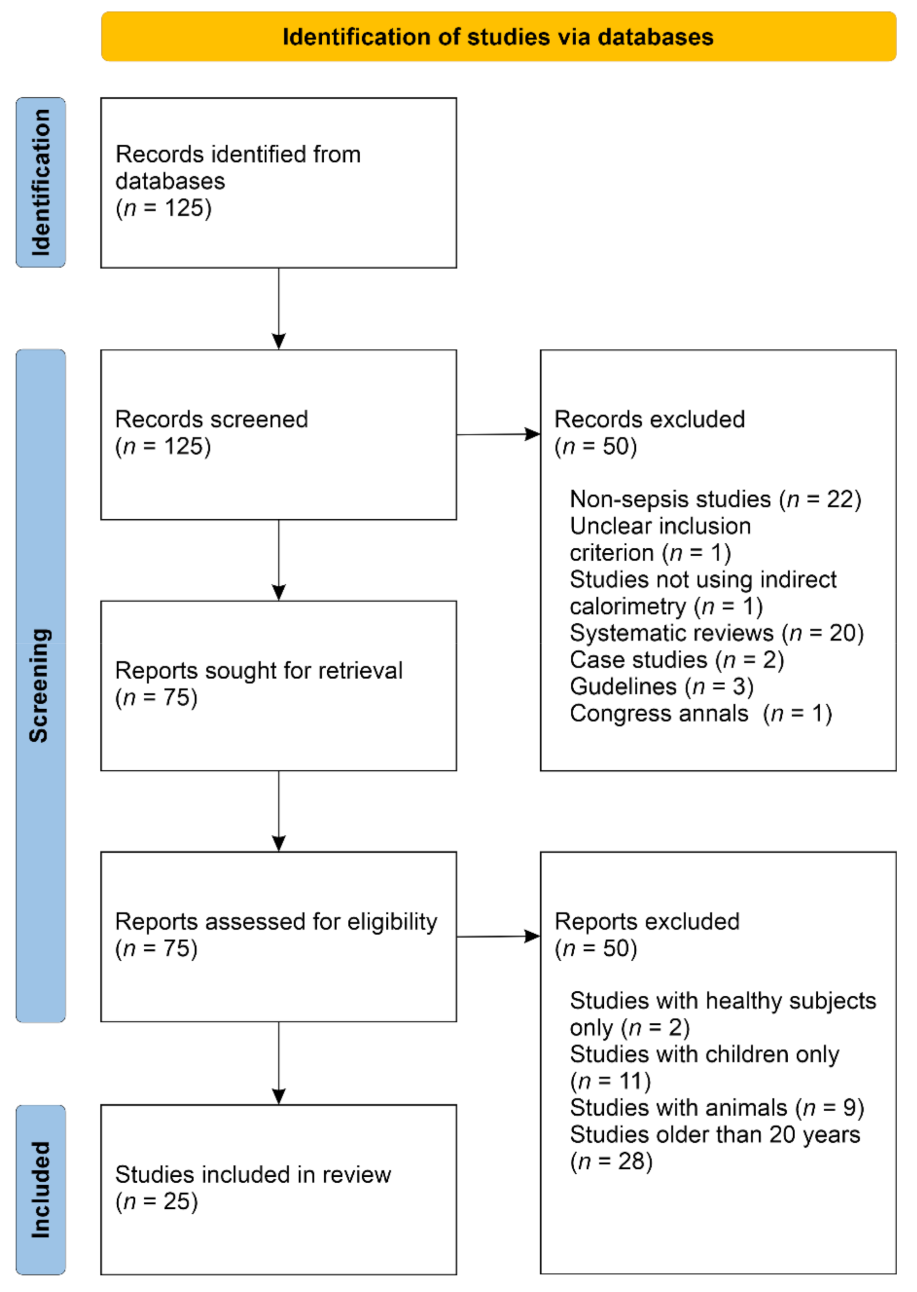
4.1. Systematic Review

In the search conducted in PubMed/Medline, using the described methodology, 125 articles were identified. Twenty-five articles were included in this review. Both included and excluded articles are presented in the PRISMA workflow (Figure 2) [11]. The 25 articles selected represent studies in which IC was used in adult patients with sepsis or septic shock. Of these, 18 studies included patients with only sepsis or septic shock, while the remaining 7 studies included other patients, but some of them presented the data in a way that allowed analysis of data on septic patients separately.

4.2. Aims and Types of Research

Table 1 presents a summary of the objectives of the analysed studies. Despite the use of the IC method, only some studies aimed to assess patient EE, while in others, IC was used as a tool for measuring VO2 for the needs of the study (n = 9) [43,44,45,46,47,48,49,50,51]. The purpose of some studies that assessed EE was to evaluate energy metabolism in patients with sepsis (and possibly its relationship with disease severity and prognosis), compare the results obtained by various methods or develop new predictive equations. Nine such studies were identified [52,53,54,55,56,57,58,59,60]. In other studies, EE was measured to assess its change under the influence of the interventions tested–hyperinsulinaemic clamp [61,62,63], liberation from mechanical ventilation [64], continuous renal replacement therapy [65], early exercise [66], or cardiac selective beta adrenergic [67]. At least sixteen of the studies analysed were prospective. Details are presented in Table 1.

4.3. Energy Expenditure and Respiratory Quotient

In studies assessing EE (n = 16), in 12 of the studies, the estimated EE was described as REE, while in the remaining four, it remained undefined. Five of the 16 EE assessments did not include patients only with sepsis, while the results of two of the studies were presented in a way that made it impossible to analyse the results of septic patients separately. The remaining 14 studies are summarised in Table 2 [68,69,70,71], which compares selected features of the examined groups (or groups of patients with sepsis in the case of studies by Menegueti et al. [53], Zauner et al. [59], and Uehara et al. [60]), sources of criteria on the basis of which sepsis/septic shock was diagnosed, type of device used in the study, day at the ICU, where the measurement was obtained, nutritional support during the measurement period, EE, and RQ results. Further analysis will involve the 14 studies presented in Table 2. In most of the studies shown in Table 2, EE was expressed in kcal per day (n = 9) as the mean and standard deviation (n = 7). The average values obtained in individual studies demonstrated a range from 1414 ± 134 kcal/day in the Gore and Wolfe study [67], to 2179 ± 354 kcal/day in the study by Rusavy et al. [61] (Figure 3).
Some authors gave EE in kcal/kg/24 h (n = 2), kJ/min/ m2 (n = 1), kcal/kg FFM/min (n = 1), or did not provide the value at all (n = 2); moreover, in some papers, the results were given in the form of median and range, or median and interquartile range (n = 2). Of the 14 studies compared in Table 2, RQ was given in eight, of which six presented the data in the form of mean and standard deviation. The mean RQ presented in the studies ranged from 0.76 ± 0.12 [52] to 0.99 ± 0.06 [67] (Figure 4).

4.4. Energy Expenditure Measurement Protocol

Most study authors provided a detailed measurement protocol, including measurement duration (n = 10) and device calibration information (n = 7). Ten papers provided information on the day of hospitalisation in which the measurements were obtained (Table 2), from the measurement carried out after admission to the ward [59,65], to the twenty-third day in a multi-day study by Uehara et al. [60]. In most studies, measurements were obtained during the first week of the patient’s stay in the ward (n = 10). Nine articles provide information on patient nutrition during the measurement period (Table 2). In six cases, feeding was suspended prior to measurement. The number of hours for which nutrition was suspended varied depending on the study, from “at least 1.5 h” in the studies of Wu et al. [55,65], up to 9 h in the study of Rusavy et al. [62], and all-night fasting in the research of Saeed et al. [63]. In two studies, IC measurements were performed before feeding [53,59], and in one, feeding was given during the measurements [67]. All of the 14 analysed studies provided the name of the calorimetry device, which in five cases was the Deltatrac II®; for many years, it has been recognised as a reference device (Table 2).

4.5. Criteria for the Diagnosis of Sepsis and Septic Shock

Nine studies provided the source of the criteria used to diagnose sepsis and septic shock. In older studies (published up to, and including 2008), the criteria established at the ACCP and SCCM conference in 1991 (SEPSIS-1) were used. Later studies used the criteria established at the 2001 SEPSIS-2 conference and Surviving Sepsis Campaign 2008 and 2012.

4.6. Criteria for Participating in the Study

Of the 14 analysed articles, 12 presented information on the criteria for inclusion and exclusion of participants from the study. The most common exclusion criterion was the FiO2 criterion (n = 9), which is known to be required for a correct IC measurement result. The other exclusion criteria directly related to IC technical requirements were PEEP (n = 4), no chest drain (n = 3), and no bronchopleural fistula (n = 1). The criterion of the lower age limit of participants (18 (n = 5) or 15 (n = 2) years) appeared relatively often, while the criterion of the upper age limit appeared only twice (80 and 85 years). A list of exclusion criteria from individual studies is presented in Table 3.

4.7. Limitations of the Analysed Studies

Limitations of the described studies were given in only half (n = 7) of the fourteen analysed works. They are listed in Table 4. The most frequently occurring limitations included the small size of the study group (n = 3) (the groups in the study had 16, 27, and 62 patients) [54,55,65], as well as equal mention that the study was conducted at a single centre [53,55,65]. Doubts were also expressed as to whether one measurement per day, most often obtained at a fixed time for all patients, would be representative of daily EE [52,54,55]. In two works, it was noted that patients admitted to ICU may be at various stages of the disease, which could have initially progressed e.g. when the patient was in another ward and their condition was not severe enough to qualify them for treatment in ICU [55,64]. Some of the other limitations mentioned included: no protocol to control patient nutrition [52], EE measurement only on admission [53], difficulties in obtaining steady state [54], variability in sedation management and body mass [64], and no assessment of the impact of medical procedures on EE [55].

5. Discussion

5.1. Indirect Calorimetry

Considering the importance of accurate determination of EE in patients with sepsis, a review of the literature was carried out, trying to compare the methodology of previous studies using IC in this group of patients. A total of 25 papers meeting accepted criteria were identified, but only 14 of them described EE in patients with sepsis or septic shock. Others used IC as the method to determine VO2 for the needs of the study, or referred to a broader group of patients, and results for patients with sepsis could not be distinguished.

5.2. Energy Expenditure

Fourteen studies measuring EE in patients with sepsis were analysed. Significant variability was noted in the forms of data presentation of measured EE: it was presented in various units as well as in various statistical forms (average, median), which makes it impossible to compare the presented results or their meta-analysis. In addition, not all studies measured EE as REE, although according to study protocols, including suspension of nutritional support a few hours before testing, suggests that the measured EE corresponds to REE. Comparing individual works is also hampered by incomplete information pertaining to the characteristics of the studied groups with respect to parameters, such as sex, body weight, BMI, and severity of the patient’s condition (assessed on the APACHE II scale), which also affect EE. In displaying parameters describing the studied groups, the formula may be the work of Takemae et al. [52] who presented the detailed characteristics of the studied groups separately for women and separately for men, which may be important in studies of metabolism and EE. The advantage is that in most of the studies analysed, the APACHE II scale was chosen as an indicator of the severity of the patient’s condition, which facilitated the analysis of results.
A total of 12 of the 14 analysed studies provided information on criteria for inclusion and exclusion of participants from the studies. This information is valuable for two reasons: firstly, it allows for even more accurate characteristics of the examined group to be included, and secondly, it assures the correctness of the results obtained; n the case of the analysed works, many of the exclusion criteria were directly related to IC technical limitations, and the lack of exclusion would thus lead to unreliable measurements.
Furthermore, information on the protocol for measuring EE by IC varied from one work to another. High volatility was characterised by, among other factors, the number of hours for which nutrition was suspended, which may be important for the results obtained (for both REE and RQ). It should be emphasised that the studies with the longest period of feeding suspension (up to nine hours–Rusavy et al. [62] and all-night fasting–Saeed et al. [63]) concerned carbohydrate metabolism, which may be the reason for such a long suspension of nutrition. Moreover, not all works contained information about device calibration performance, measurement duration, and achievement of the steady state, which are important for the possible use of test results as a reference point or inclusion in meta-analyses. Oshima et al. [10] have proposed checkpoints in a list for a successful IC that can be easily adapted to develop a protocol for measuring EE by IC in patients.
Another important aspect that appeared in most studies was the day of illness on which the measurement was performed. This information is particularly important for high-dynamic diseases such as sepsis because it has been shown (including in the Uehara et al. [60] study analysed in this review) that EE is characterised by variability depending on the stage of the disease. However, as noted in two studies, some limitations should be taken into account, namely, that patients can be admitted to the ICU at various stages of the disease, after having been initially treated in another ward. This problem can be addressed, e.g., by analysing the patient’s medical history. It is also valuable to know whether IC is performed in patients undergoing mechanical ventilation, both due to a different measurement technique, and the demonstrated impact of liberation from mechanical ventilation on the REE of patients with sepsis [64].

5.3. Definition of Sepsis

In addition, when analysing the results of published studies, attention should be paid to the definition used for sepsis. As described in the introduction, both the view on pathophysiology and the definitions of sepsis and septic shock have changed over the years. Before comparing the results obtained in different studies, it is necessary to make sure that they actually relate to the same population, e.g., the definition of severe sepsis, appearing in research from a few years ago, corresponds to the current definition of sepsis [1]. The evolution of the definition of sepsis in modern times is presented in Figure 5.

5.4. Limitations of the Discribed Studies

In addition to concerns about not knowing the stage of the disease and associated physiological and therapeutic parameters, other study limitations explained by the authors of the studies included that fact that the research was conducted at a single centre and that the size of the examined group was small. Data associated with change in REE [5] were not presented.
Unification of the protocol for measuring EE by IC, as well as the form and detail of data presentation, would overcome this limitation through a meta-analysis of the results of research conducted across many centres; this can prove to be a milestone in research on energy metabolism in sepsis.
It is worth remembering that the EE measurement was only a tool, and not a goal in the research works discussed. Therefore, further studies with EE as a primary outcome parameter should be performed to shed light on this issue.

5.5. Limitations of This Review

This work has some limitations, which include a review of literature within only one database and the use of a search formula covering only titles and abstracts, which may result in the exclusion of some studies. However, this does not appear to significantly change the results obtained; a review within one database allowed for the analysis of many aspects of the methodology of studies using indirect calorimetry in patients with sepsis and to identify potential problems hindering the meta-analysis of these studies.

6. Conclusions

Sepsis, as a condition in which severe metabolic disorders occur, requires adequate nutritional support. The current nutritional strategy for these patients is focused on EN optimisation, and IC is considered the gold standard in assessing energy requirements. IC is a valuable method for optimising nutritional care in critically ill patients. There are studies assessing the EE of patients with sepsis by IC, but due to differences in data presentation and study protocols, their collective analysis is difficult. Due to the common study limitations of single centres and small study group sizes across the studies analysed, a meta-analysis of the results could enable evaluation covering many centres and a much larger group of patients. Standardisation of the research protocol and the form and manner in which results are presented, would allow a comprehensive meta-analysis of the data to provide deeper insights into energy metabolism in sepsis. Furthermore, the limitations discussed above should prompt scientists to thoughtfully design clinical trials, and clinicians to interpret results with care.

Author Contributions

Conceptualization, W.W.; methodology, W.W., A.Z., J.J., E.D.W., W.D.; literature search process, W.W.; writing—original draft preparation, W.W.; writing—review and editing, W.W., A.Z., J.J., E.D.W., W.D.; visualization, W.W.; supervision, E.D.W., W.D. All authors have read and agreed to the published version of the manuscript.

Funding

This research received no external funding.

Institutional Review Board Statement

Not applicable.

Informed Consent Statement

Not applicable.

Data Availability Statement

Not applicable.

Conflicts of Interest

The authors declare no conflict of interest.

References

  1. Singer, M.; Deutschman, C.S.; Seymour, C.W.; Shankar-Hari, M.; Annane, D.; Bauer, M.; Bellomo, R.; Bernard, G.R.; Chiche, J.-D.; Coopersmith, C.M.; et al. The Third International Consensus Definitions for Sepsis and Septic Shock (Sepsis-3). JAMA J. Am. Med. Assoc. 2016, 315, 801–810. [Google Scholar] [CrossRef]
  2. De Waele, E.; Malbrain, M.L.N.G.; Spapen, H. Nutrition in Sepsis: A Bench-to-Bedside Review. Nutrients 2020, 12, 395. [Google Scholar] [CrossRef] [Green Version]
  3. Wasyluk, W.; Zwolak, A. Metabolic Alterations in Sepsis. J. Clin. Med. 2021, 10, 2412. [Google Scholar] [CrossRef] [PubMed]
  4. Wasyluk, W.; Wasyluk, M.; Zwolak, A. Sepsis as a Pan-Endocrine Illness—Endocrine Disorders in Septic Patients. J. Clin. Med. 2021, 10, 2075. [Google Scholar] [CrossRef] [PubMed]
  5. Mtaweh, H.; Aguero, M.J.S.; Campbell, M.; Allard, J.P.; Pencharz, P.; Pullenayegum, E.; Parshuram, C.S. Systematic review of factors associated with energy expenditure in the critically ill. Clin. Nutr. ESPEN 2019, 33, 111–124. [Google Scholar] [CrossRef] [PubMed] [Green Version]
  6. Singer, P.; Blaser, A.R.; Berger, M.M.; Alhazzani, W.; Calder, P.C.; Casaer, M.P.; Hiesmayr, M.; Mayer, K.; Montejo, J.C.; Pichard, C.; et al. ESPEN guideline on clinical nutrition in the intensive care unit. Clin. Nutr. 2019, 38, 48–79. [Google Scholar] [CrossRef] [Green Version]
  7. McClave, S.A.; Taylor, B.E.; Martindale, R.G.; Warren, M.M.; Johnson, D.R.; Braunschweig, C.; McCarthy, M.S.; Davanos, E.; Rice, T.W.; Cresci, G.A.; et al. Guidelines for the Provision and Assessment of Nutrition Support Therapy in the Adult Critically Ill Patient: Society of Critical Care Medicine (SCCM) and American Society for Parenteral and Enteral Nutrition (ASPEN). JPEN J. Parenter. Enter. Nutr. 2016, 40, 159–211, Correction in JPEN J. Parenter. Enter. Nutr. 2016, 40, 1200. [Google Scholar] [CrossRef]
  8. Heidegger, C.P.; Berger, M.M.; Graf, S.; Zingg, W.; Darmon, P.; Costanza, M.C.; Thibault, R.; Pichard, C. Optimisation of energy provision with supplemental parenteral nutrition in critically ill patients: A randomised controlled clinical trial. Lancet 2013, 381, 385–393. [Google Scholar] [CrossRef]
  9. Singer, P.; Hiesmayr, M.; Biolo, G.; Felbinger, T.W.; Berger, M.M.; Goeters, C.; Kondrup, J.; Wunder, C.; Pichard, C. Pragmatic approach to nutrition in the ICU: Expert opinion regarding which calorie protein target. Clin. Nutr. 2014, 33, 246–251. [Google Scholar] [CrossRef]
  10. Oshima, T.; Berger, M.M.; De Waele, E.; Guttormsen, A.B.; Heidegger, C.-P.; Hiesmayr, M.; Singer, P.; Wernerman, J.; Pichard, C. Indirect calorimetry in nutritional therapy. A position paper by the ICALIC study group. Clin. Nutr. 2016, 36, 651–662. [Google Scholar] [CrossRef]
  11. Page, M.J.; McKenzie, J.E.; Bossuyt, P.M.; Boutron, I.; Hoffmann, T.C.; Mulrow, C.D.; Shamseer, L.; Tetzlaff, J.M.; Akl, E.A.; Brennan, S.E.; et al. The PRISMA 2020 statement: An updated guideline for reporting systematic reviews. BMJ 2021, 372, n71. [Google Scholar] [CrossRef]
  12. Haugen, H.A.; Chan, L.-N.; Li, F. Indirect Calorimetry: A Practical Guide for Clinicians. Nutr. Clin. Pract. 2007, 22, 377–388. [Google Scholar] [CrossRef]
  13. Psota, T.; Chen, K.Y. Measuring energy expenditure in clinical populations: Rewards and challenges. Eur. J. Clin. Nutr. 2013, 67, 436–442. [Google Scholar] [CrossRef] [Green Version]
  14. Thomas, N.; Das Gupta, R.; Ramachandran, R.; Venkatesan, P.; Anoop, S.; Joseph, M. Indirect calorimetry: From bench to bedside. Indian J. Endocrinol. Metab. 2017, 21, 594–599. [Google Scholar] [CrossRef]
  15. Arends, J.; Bachmann, P.; Baracos, V.; Barthelemy, N.; Bertz, H.; Bozzetti, F.; Fearon, K.; Hütterer, E.; Isenring, E.; Kaasa, S.; et al. ESPEN guidelines on nutrition in cancer patients. Clin. Nutr. 2017, 36, 11–48. [Google Scholar] [CrossRef] [Green Version]
  16. Gomes, F.; Schuetz, P.; Bounoure, L.; Austin, P.; Ballesteros-Pomar, M.; Cederholm, T.; Fletcher, J.; Laviano, A.; Norman, K.; Poulia, K.-A.; et al. ESPEN guidelines on nutritional support for polymorbid internal medicine patients. Clin. Nutr. 2017, 37, 336–353. [Google Scholar] [CrossRef] [Green Version]
  17. Takala, J.; Keinänen, O.; Väisänen, P.; Kari, A. Measurement of gas exchange in intensive care: Laboratory and clinical validation of a new device. Crit. Care Med. 1989, 17, 1041–1047. [Google Scholar] [CrossRef]
  18. Weir, J.B.D.B. New methods for calculating metabolic rate with special reference to protein metabolism. J. Physiol. 1949, 109, 1–9. [Google Scholar] [CrossRef]
  19. Ferrannini, E. The theoretical bases of indirect calorimetry: A review. Metabolism 1988, 37, 287–301. [Google Scholar] [CrossRef]
  20. Bursztein, S.; Saphar, P.; Singer, P.; Elwyn, D.H. A mathematical analysis of indirect calorimetry measurements in acutely ill patients. Am. J. Clin. Nutr. 1989, 50, 227–230. [Google Scholar] [CrossRef] [Green Version]
  21. Wilmore, J.H.; Costill, D.L. Adequacy of the Haldane transformation in the computation of exercise VO2 in man. J. Appl. Physiol. 1973, 35, 85–89. [Google Scholar] [CrossRef]
  22. Ultman, J.S.; Bursztein, S. Analysis of error in the determination of respiratory gas exchange at varying FIO2. J. Appl. Physiol. Respir. Environ. Exerc. Physiol. 1981, 50, 210–216. [Google Scholar] [CrossRef]
  23. Compher, C.; Frankenfield, D.; Keim, N.; Roth-Yousey, L.; Evidence Analysis Working Group. Best Practice Methods to Apply to Measurement of Resting Metabolic Rate in Adults: A Systematic Review. J. Am. Diet. Assoc. 2006, 106, 881–903. [Google Scholar] [CrossRef]
  24. Lichtenbelt, W.D.V.M.; Frijns, A.J.H.; Van Ooijen, M.J.; Fiala, D.; Kester, A.M.; Van Steenhoven, A.A. Validation of an individualised model of human thermoregulation for predicting responses to cold air. Int. J. Biometeorol. 2006, 51, 169–179. [Google Scholar] [CrossRef]
  25. Ueno, S.; Ikeda, K.; Tai, T.; Tai, T. Metabolic Rate Prediction in Young and Old Men by Heart Rate, Ambient Temperature, Weight and Body Fat Percentage. J. Occup. Health 2014, 56, 519–525. [Google Scholar] [CrossRef] [Green Version]
  26. McClave, S.A.; Martindale, R.G.; Kiraly, L. The use of indirect calorimetry in the intensive care unit. Curr. Opin. Clin. Nutr. Metab. Care 2013, 16, 202–208. [Google Scholar] [CrossRef]
  27. Mtaweh, H.; Tuira, L.; Floh, A.A.; Parshuram, C.S. Indirect Calorimetry: History, Technology, and Application. Front. Pediatr. 2018, 6, 257. [Google Scholar] [CrossRef]
  28. Macfarlane, D.J. Automated Metabolic Gas Analysis Systems. Sports Med. 2001, 31, 841–861. [Google Scholar] [CrossRef]
  29. Cunningham, K.F.; Aeberhardt, L.E.; Wiggs, B.R.; Phang, P. Appropriate interpretation of indirect calorimetry for determining energy expenditure of patients in intensive care units. Am. J. Surg. 1994, 167, 547–549. [Google Scholar] [CrossRef]
  30. McClave, S.; Spain, D.; Skolnick, J.; Lowen, C.; Kleber, M.; Wickerham, P.; Vogt, J.; Looney, S. Achievement of steady state optimizes results when performing indirect calorimetry. J. Parenter. Enter. Nutr. 2003, 27, 16–20. [Google Scholar] [CrossRef]
  31. El-Orbany, M.; Salem, M.R. Endotracheal Tube Cuff Leaks: Causes, consequences, and management. Anesth. Analg. 2013, 117, 428–434. [Google Scholar] [CrossRef] [PubMed]
  32. Singer, P.; Singer, J. Clinical Guide for the Use of Metabolic Carts: Indirect calorimetry—No longer the orphan of energy estimation. Nutr. Clin. Pract. 2015, 31, 30–38. [Google Scholar] [CrossRef] [PubMed] [Green Version]
  33. Weissman, C.; Sardar, A.; Kemper, M. In vitro evaluation of a compact metabolic measurement instrument. J. Parenter. Enter. Nutr. 1990, 14, 216–221. [Google Scholar] [CrossRef] [PubMed]
  34. Delsoglio, M.; Achamrah, N.; Berger, M.M.; Pichard, C. Indirect Calorimetry in Clinical Practice. J. Clin. Med. 2019, 8, 1387. [Google Scholar] [CrossRef] [Green Version]
  35. Rattanachaiwong, S.; Singer, P. Indirect calorimetry as point of care testing. Clin. Nutr. 2019, 38, 2531–2544. [Google Scholar] [CrossRef]
  36. De Waele, E.; Van Zwam, K.; Mattens, S.; Staessens, K.; Diltoer, M.; Honoré, P.M.; Czapla, J.; Nijs, J.; La Meir, M.; Huyghens, L.; et al. Measuring resting energy expenditure during extracorporeal membrane oxygenation: Preliminary clinical experience with a proposed theoretical model. Acta Anaesthesiol. Scand. 2015, 59, 1296–1302. [Google Scholar] [CrossRef]
  37. Wollersheim, T.; Frank, S.; Müller, M.; Skrypnikov, V.; Carbon, N.; Pickerodt, P.; Spies, C.; Mai, K.; Spranger, J.; Weber-Carstens, S. Measuring Energy Expenditure in extracorporeal lung support Patients (MEEP)—Protocol, feasibility and pilot trial. Clin. Nutr. 2017, 37, 301–307. [Google Scholar] [CrossRef]
  38. Jonckheer, J.; Spapen, H.; Debain, A.; Demol, J.; Diltoer, M.; Costa, O.; Lanckmans, K.; Oshima, T.; Honoré, P.M.; Malbrain, M.; et al. CO2 and O2 removal during continuous veno-venous hemofiltration: A pilot study. BMC Nephrol. 2019, 20, 222. [Google Scholar] [CrossRef] [Green Version]
  39. Jonckheer, J.; Demol, J.; Lanckmans, K.; Malbrain, M.; Spapen, H.; De Waele, E. MECCIAS trial: Metabolic consequences of continuous veno-venous hemofiltration on indirect calorimetry. Clin. Nutr. 2020, 39, 3797–3803. [Google Scholar] [CrossRef]
  40. Matarese, L.E. Indirect Calorimetry: Technical Aspects. J. Am. Diet. Assoc. 1997, 97, S154–S160. [Google Scholar] [CrossRef]
  41. Terblanche, E.; Remmington, C. Observational study evaluating the nutritional impact of changing from 1% to 2% propofol in a cardiothoracic adult critical care unit. J. Hum. Nutr. Diet. Off. J. Br. Diet. Assoc. 2020, 34, 413–419. [Google Scholar] [CrossRef]
  42. Achamrah, N.; Delsoglio, M.; De Waele, E.; Berger, M.M.; Pichard, C. Indirect calorimetry: The 6 main issues. Clin. Nutr. 2020, 40, 4–14. [Google Scholar] [CrossRef]
  43. Auxiliadora-Martins, M.; Coletto, F.A.; Campos, A.D.; Basile-Filho, A. Indirect calorimetry can be used to measure cardiac output in septic patients? Acta Cir. Bras. 2008, 23, 118–125. [Google Scholar] [CrossRef]
  44. Natalini, G.; Schivalocchi, V.; Rosano, A.; Taranto, M.; Pletti, C.; Bernardini, A. Norepinephrine and metaraminol in septic shock: A comparison of the hemodynamic effects. Intensiv. Care Med. 2005, 31, 634–637. [Google Scholar] [CrossRef]
  45. Marson, F.; Auxiliadora-Martins, M.; Coletto, F.A.; Campos, A.D.; Basile-Filho, A. Correlation between Oxygen Consumption Calculated Using Fick’s Method and Measured with Indirect Calorimetry in Critically Ill Patients. Arq. Bras. Cardiol. 2004, 82, 72–76. [Google Scholar] [CrossRef] [Green Version]
  46. Fernandes, C.J.; Akamine, N.; De Marco, F.V.; De Souza, J.A.; Lagudis, S.; Knobel, E. Red blood cell transfusion does not increase oxygen consumption in critically ill septic patients. Crit. Care 2001, 5, 362–367. [Google Scholar] [CrossRef] [Green Version]
  47. Sakka, S.G.; Reinhart, K.; Wegscheider, K.; Meier-Hellmann, A. Variability of splanchnic blood flow in patients with sepsis. Intensiv. Care Med. 2001, 27, 1281–1287. [Google Scholar] [CrossRef]
  48. Schaffartzik, W.; Sanft, C.; Schaefer, J.H.; Spies, C. Different dosages of dobutamine in septic shock patients: Determining oxygen consumption with a metabolic monitor integrated in a ventilator. Intensiv. Care Med. 2000, 26, 1740–1746. [Google Scholar] [CrossRef]
  49. Broccard, A.; Hurni, J.-M.; Eckert, P.; Liaudet, L.; Schaller, M.-D.; Lazor, R.; Perret, C.; Feihl, F. Tissue oxygenation and hemodynamic response to no synthase inhibition in septic shock. Shock 2000, 14, 35–40. [Google Scholar] [CrossRef] [Green Version]
  50. Sakka, S.G.; Reinhard, K.; Wegscheider, K.; Meier-Hellmann, A. Is the placement of a pulmonary artery catheter still justified solely for the measurement of cardiac output? J. Cardiothorac. Vasc. Anesth. 2000, 14, 119–124. [Google Scholar] [CrossRef]
  51. Opdam, H.; Bellomo, R. Oxygen consumption and lactate release by the lung after cardiopulmonary bypass and during septic shock. Crit. Care Resusc. J. Australas. Acad. Crit. Care Med. 2000, 2, 181–187. [Google Scholar]
  52. Takemae, A.; Takazawa, T.; Kamiyama, J.; Kanamoto, M.; Tobe, M.; Hinohara, H.; Kunimoto, F.; Saito, S. A novel prediction equation of resting energy expenditure for Japanese septic patients. J. Crit. Care 2020, 56, 236–242. [Google Scholar] [CrossRef]
  53. Menegueti, M.G.; De Araújo, T.R.; Laus, A.M.; Martins-Filho, O.A.; Basile-Filho, A.; Auxiliadora-Martins, M. Resting Energy Expenditure and Oxygen Consumption in Critically Ill Patients with vs without Sepsis. Am. J. Crit. Care 2019, 28, 136–141. [Google Scholar] [CrossRef]
  54. Panitchote, A.; Thiangpak, N.; Hongsprabhas, P.; Hurst, C. Energy expenditure in severe sepsis or septic shock in a Thai Medical Intensive Care Unit. Asia Pac. J. Clin. Nutr. 2017, 26, 794–797. [Google Scholar]
  55. Wu, C.; Wang, X.; Yu, W.; Tian, F.; Liu, S.; Li, P.; Li, J.; Li, N. Hypermetabolism in the Initial Phase of Intensive Care Is Related to a Poor Outcome in Severe Sepsis Patients. Ann. Nutr. Metab. 2015, 66, 188–195. [Google Scholar] [CrossRef] [PubMed]
  56. Basile-Filho, A.; Auxiliadora-Martins, M.; Marson, F.; Evora, P.R.B. An easy way to estimate energy expenditure from hemodynamic data in septic patients. Acta Cir. Bras. 2008, 23, 112–117. [Google Scholar] [CrossRef] [Green Version]
  57. Auxiliadora-Martins, M.; Coletto, F.A.; Martins-Filho, O.A.; Marchini, J.S.; Basile-Filho, A. 13CO2 recovery fraction in expired air of septic patients under mechanical ventilation. Braz. J. Med. Biol. Res. 2008, 41, 563–570. [Google Scholar] [CrossRef] [PubMed] [Green Version]
  58. Dvir, D.; Cohen, J.; Singer, P. Computerized energy balance and complications in critically ill patients: An observational study. Clin. Nutr. 2006, 25, 37–44. [Google Scholar] [CrossRef] [PubMed]
  59. Zauner, C.; Schuster, B.I.; Schneeweiss, B. Similar metabolic responses to standardized total parenteral nutrition of septic and nonseptic critically ill patients. Am. J. Clin. Nutr. 2001, 74, 265–270. [Google Scholar] [CrossRef] [PubMed] [Green Version]
  60. Uehara, M.; Plank, L.D.; Hill, G.L. Components of energy expenditure in patients with severe sepsis and major trauma. Crit. Care Med. 1999, 27, 1295–1302. [Google Scholar] [CrossRef] [PubMed]
  61. Rušavý, Z.; Macdonald, I.A.; Sramek, V.; Lacigova, S.; Tesinsky, P.; Novak, I. Glycemia Influences on Glucose Metabolism in Sepsis during Hyperinsulinemic Clamp. J. Parenter. Enter. Nutr. 2005, 29, 171–175. [Google Scholar] [CrossRef]
  62. Rusavy, Z.; Sramek, V.; Lacigova, S.; Novak, I.; Tesinsky, P.; Macdonald, I.A. Influence of insulin on glucose metabolism and energy expenditure in septic patients. Crit. Care 2004, 8, R213–R220. [Google Scholar] [CrossRef] [Green Version]
  63. Saeed, M.; Carlson, G.L.; Little, R.A.; Irving, M.H. Selective impairment of glucose storage in human sepsis. Br. J. Surg. 1999, 86, 813–821. [Google Scholar] [CrossRef]
  64. Lee, P.S.-P.; Lee, K.L.; Betts, J.A.; Law, K.I. Metabolic Requirement of Septic Shock Patients before and after Liberation from Mechanical Ventilation. J. Parenter. Enter. Nutr. 2016, 41, 993–999. [Google Scholar] [CrossRef]
  65. Wu, C.; Wang, X.; Yu, W.; Li, P.; Liu, S.; Li, J.; Li, N. Short-term consequences of continuous renal replacement therapy on body composition and metabolic status in sepsis. Asia Pac. J. Clin. Nutr. 2016, 25, 300–307. [Google Scholar]
  66. Hickmann, C.E.; Roeseler, J.; Castanares-Zapatero, D.; Herrera, E.I.; Mongodin, A.; Laterre, P.-F. Energy expenditure in the critically ill performing early physical therapy. Intensiv. Care Med. 2014, 40, 548–555. [Google Scholar] [CrossRef]
  67. Gore, D.C.; Wolfe, R.R. Hemodynamic and metabolic effects of selective β1 adrenergic blockade during sepsis. Surgery 2006, 139, 686–694. [Google Scholar] [CrossRef]
  68. Levy, M.M.; Fink, M.P.; Marshall, J.C.; Abraham, E.; Angus, D.; Cook, D.; Cohen, J.; Opal, S.M.; Vincent, J.-L.; Ramsay, G. 2001 SCCM/ESICM/ACCP/ATS/SIS International Sepsis Definitions Conference. Crit. Care Med. 2003, 31, 1250–1256. [Google Scholar] [CrossRef]
  69. Dellinger, R.P.; Levy, M.M.; Rhodes, A.; Annane, D.; Gerlach, H.; Opal, S.M.; Sevransky, J.E.; Sprung, C.L.; Douglas, I.S.; Jaeschke, R.; et al. Surviving Sepsis Campaign: International guidelines for management of severe sepsis and septic shock: 2012. Crit. Care Med. 2013, 41, 580–637. [Google Scholar] [CrossRef]
  70. Dellinger, R.P.; Levy, M.M.; Carlet, J.M.; Bion, J.; Parker, M.M.; Jaeschke, R.; Reinhart, K.; Angus, D.C.; Brun-Buisson, C.; Beale, R.; et al. Surviving Sepsis Campaign: International guidelines for management of severe sepsis and septic shock: 2008. Intensiv. Care Med. 2007, 34, 17–60. [Google Scholar] [CrossRef] [Green Version]
  71. Bone, R.C.; Balk, R.A.; Cerra, F.B.; Dellinger, R.P.; Fein, A.M.; Knaus, W.A.; Schein, R.M.; Sibbald, W.J. Definitions for sepsis and organ failure and guidelines for the use of innovative therapies in sepsis. Chest 1992, 101, 1644–1655. [Google Scholar] [CrossRef] [Green Version]
Figure 1. Indirect calorimetry scheme with the use of the mixing chamber technique in a mechanically ventilated patient (based on the Deltatrac Metabolic Monitor® calorimeter). FiO2 is measured from the ventilator’s inspiratory limb, while FeO2 and FeCO2 are measured from the mixing chamber. The gas from the mixing chamber is removed through a system with a constant flow of gas, in which it is diluted with ambient air. The CO2 fraction in the diluted exhaust gas (FedCO2) is measured. VCO2 is calculated as the product of constant flow (Q) and FedCO2 (VCO2 = FedCO2 × Q). VO2 is calculated using the Haldane transformation. EE is calculated using the Weir equation [10,17]. Abbreviations: CO2—carbon dioxide; EE—energy expenditure; FeCO2—fraction of exhaled CO2; FedCO2—fraction of exhaled CO2 after dilution; FeO2—fraction of exhaled oxygen; FiO2—fraction of inhaled oxygen; O2—oxygen; Q—flow; VO2—volume of consumed oxygen; VCO2—volume of produced CO2.
Nutrients 14 00930 g001
Figure 2. PRISMA flow diagram of the literature search process [11].
Nutrients 14 00930 g002
Figure 3. Comparison of mean energy expenditure (EE) measured by indirect calorimetry in patients with sepsis or septic shock in selected studies. [52,53,54,55,56,57,59,60,61,62,63,64,65,67] Some studies have reported more than one EE value. Time and conditions for obtaining measurements in individual tests may be different. Two studies reported the median instead of the mean. The details of the studies are presented in Table 2. * The value in the chart is the median EE. ** The authors did not publish EE at all or as kcal/24 h.
Nutrients 14 00930 g003
Figure 4. Comparison of mean respiratory quotient (RQ) measured by indirect calorimetry in patients with sepsis or septic shock in selected studies. [52,53,54,55,56,57,59,60,61,62,63,64,65,67] Some studies have reported more than one RQ value. Time and conditions for obtaining measurements in individual tests may be different. Two studies reported the median instead of the mean. The details of the studies are presented in Table 2. * The value in the chart is the median RQ. ** The authors did not publish RQ.
Nutrients 14 00930 g004
Figure 5. Comparison of old and new sepsis definitions. The first definition of sepsis and classification of clinical conditions associated with it were presented as a result of the American College of Chest Physicians (ACCP) and Society of Critical Care Medicine (SCCM) conference in 1991 (SEPSIS-1) [71]. Due to reservations, mainly related to the non-specificity of the definition of systemic inflammatory response syndrome (SIRS), a conference called SEPSIS-2 was organised in 2001, during which the definitions established during SEPSIS-1 were maintained and an extended list of possible symptoms of systemic inflammation in response to infection was proposed [68]. A further need to update the sepsis nomenclature led to the publication of The Third International Consensus Definitions for Sepsis and Septic Shock (SEPSIS-3) in 2016, including new definitions of sepsis and septic shock [1].
Nutrients 14 00930 g005
Table 1. Summary of studies in patients with sepsis/septic shock in which indirect calorimetry was used.
ReferenceType of StudyObjective of the StudyOnly Septic Patients
Takemae et al. (2020) [52]Retrospective observational studyDevelopment of new equations to estimate the total EE of Japanese patients with sepsis.Yes
Menegueti et al. (2019) [53]Observational cross-sectional studyAssessment of whether REE, respiratory quotient, oxygen consumption, and carbon dioxide production (measured by IC) differ in critically ill patients with sepsis compared to critically ill patients without sepsis.No
Panitchote et al. (2017) [54]Prospective observational studyAssessment of the correlation between REE of patients with sepsis/septic shock, measured by IC and estimated using predictive equations.Yes
Lee et al. (2017) [64]NDIdentification of the difference in EE and substrate utilisation by patients during and upon liberation from mechanical ventilation.Yes
Wu et al. (2016) [65]Prospective observational studyAssessment of the short-term consequence of continuous renal replacement therapy on body composition and pattern of EE.Yes
Wu et al. (2015) [55]Prospective observational studyAssessment of the incidence of hypermetabolism, defined as high REE, in severe sepsis ICU patients, and evaluate the suitability of excessive RRE as a risk factor of their clinical outcome.Yes
Hickmann et al. (2014) [66]Prospective observational studyDetermining the impact of early exercise on energy requirements to adjust caloric intake accordingly in critically ill patients.No
Auxiliadora-Martins et al. (2008) [43]Prospective clinical studyComparison of two different CO monitoring systems based on the thermodilution principle (Thermo-CO) and IC (Fick mixed-CO) in septic patients.Yes
Basile-Filho et al. (2008) [56]Prospective clinical studyComparison of REE obtained by IC and the REE calculated by predictive equations (Brandi and Liggett) using the oxygen consumption obtained by Fick‘s method in septic patients.Yes
Auxiliadora-Martins et al. (2008) [57]Prospective clinical studyEvaluation of the 13CO2 recovery fraction in expired air after continuous intravenous infusion of NaH13CO2, in critically ill patients with sepsis under mechanical ventilation (calculation of substrate oxidation).Yes
Gore et al. (2006) [67]NDInvestigating the haemodynamic and metabolic effects of cardiac selective beta adrenergic blockade in septic patients.Yes
Dvir et al. (2006) [58]Prospective observational studyMeasuring the daily cumulative energy balance in critically ill patients receiving mechanical ventilation using a bedside computerised information system, and to assess its impact on outcome.No
Rusavy et al. (2005) [61]NDComparing the effects of 2 blood glucose levels (5 and 10 mmol/L) under hyperinsulinemic conditions, and the effect of glycaemia 5 mmol/L with extremely high insulinaemia on glucose metabolism and EE in septic patients.Yes *
Natalini et al. (2005) [44]Open-label, controlled clinical trialComparison of the effects of noradrenaline and metaraminol on haemodynamics in septic shock patients.Yes
Rusavy et al. (2004) [62]NDComparing the effects of two levels of insulinaemia on glucose metabolism and EE in septic patients and volunteers.Yes *
Marson et al. (2004) [45]Prospective studyComparison of oxygen consumption index measured by using IC with a portable metabolic cart and calculated according to Fick‘s principle in critically ill patients.No
Fernandes et al. (2001) [46]Interventional, prospective, randomised, controlled studyEvaluation of the haemodynamic and oxygen utilisation effects of haemoglobin infusion on critically ill septic patients.Yes
Sakka et al. (2001) [47]Prospective clinical studyExamining the variability of splanchnic blood flow during a 4-h period of unchanged global haemodynamics in patients with sepsis.Yes
Zauner et al. (2001) [59]Prospective, clinical cohort studyEvaluation of the energy and substrate metabolism in septic and non-septic critically ill patients in the resting state and during the administration of standardised total parenteral nutrition.No
Schaffartzik et al. (2000) [48]Prospective clinical studyComparison of oxygen consumption obtained from breathing gases by IC with a metabolic monitor integrated with a respirator and oxygen consumption obtained by the Fick principle in patients with sepsis after an increase in oxygen delivery induced by positive inotropic support.Yes
Broccard et al. (2000) [49]NDEvaluation of the tissue oxygenation and haemodynamic effects of NOS inhibition in clinical severe septic shock.Yes
Sakka et al. (2000) [50]Prospective clinical studyComparison of four clinical techniques of measuring cardiac output in critically ill patients: pulmonary artery thermodilution, transpulmonary aortic thermodilution, Fick principle-derived, and continuous pulmonary artery measurements.Yes
Opdam et al. (2000) [51]Prospective observational studyDetermining whether there is a correlation between lung lactate release and lung oxygen consumption by studying adult intensive care patients, either after cardiopulmonary bypass or with septic shock.No
Uehara et al. (1999) [60]Prospective studyObtaining accurate values for the components of EE in critically ill patients with sepsis or trauma during the first 2 weeks after admission to the ICU.No
Saeed et al. (1999) [63]NDAssessment of the effect of sepsis on total glucose utilisation, oxidation and storage, and the energetic costs of these metabolic processes.Yes *
* Septic patients and healthy volunteers as a control group. The content of the table contains quoted information from the articles, with possible modifications. The type of study was categorised according to the study authors’ declarations. Abbreviations: 13C—labeled carbon; CO—cardiac output; CO2—carbon dioxide; EE—energy expenditure; IC—indirect calorimetry; ICU—Intensive care unit; NaH13CO2—labeled bicarbonate; ND—no data; NOS—nitric oxide synthase; REE—resting energy expenditure; RQ—respiratory quotient.
Table 2. Comparison of results and some aspects of the methodology in studies using indirect calorimetry in patients with sepsis or septic shock.
ReferenceDiagnosisCriteria for Sepsis AND Septic ShockSample Size% of WomenAge (years)Body Mass (kg)BMI (kg/m2)APACHE II (points)Mechanical Ventilation (%)DeviceNutrition during ICDay of MeasurementEE (kcal/24 h)EE (kcal/kg/24 h)RQ
Takemae et al. (2020) [52]Severe sepsisSEPSIS-2 [68]
SSC 2012 [69]
420%68 ± 1460 ± 1422.2 ± 4.724.2 ± 5.8100%M-COVX®
(Datex-Ohmeda, Helsinki, Finland)
≥4 h between changes in the feeding method and IC1st day of the intubation periodNDND0.78 ± 0.09
24100%60 ± 1648 ± 1620.4 ± 5.327.6 ± 6.00.77 ± 0.9
190%66 ± 1362 ± 1023.0 ± 2.926.9 ± 5.70.81 ± 0.11
10100%56 ± 1560 ± 1725.1 ± 7.134.8 ± 8.00.76 ± 0.12
Menegueti et al. (2019) [53]Sepsis/septic shockSSC 2008 [70]9142%58 (19–89) m(r)ND26 (17–45) m(r)25 (9–47) m(r)100%Deltatrac II®
(Datex-Ohmeda)
IC before the beginning of nutritionFirst 48 h of admission1430 (540–2420) m(r)ND0.82 (0.6–1.24) m(r)
Panitchote et al. (2017) [54]Severe sepsis/septic shockND1644%71.6 ± 5.5ND22.0 ± 2.926.9 ± 4.0100%Engström Carestation®
(GE Healthcare, Chicago, IL, USA)
ND24 h1488 ± 26126.7 ± 5.3ND
48 h1459 ± 270
72 h1560 ± 363
Lee et al. (2017) [64]Septic shockND3743%69 ± 1059.01 ± 7.63ND22 m100%CCM Express® (Medical Graphics Corporation, St Paul, MN, USA)Suspended 4 h before ICND2090 ± 489NDND
Wu et al. (2016) [65]Sepsis and CRRT requirementSSC 2012 [69]2741%48.2 ± 22.062.8 ± 14.722.0 ± 1.4ND48.1%Metabolic cart
(Cosmed, Roma, Italy)
Suspended ≥1.5 h before ICAt admissionND27.9 ± 5.90.81 ± 0.06
Before CRRT a29.9 ± 5.60.82 ± 0.06
6 h after CRRT a26.6 ± 4.30.86 ± 0.05
Wu et al. (2015) [55]Severe sepsis/septic shockSSC 2012 [69]6235%57.1 ± 19.579.1 ± 10.321.6 ± 3.120.2 ± 4.137.5%Metabolic cart
(Med Graphics)
Suspended ≥1.5 h before IC1st, 2nd, 3rd, 4th, 5th dayNDNDND
Basile-Filho et al. (2008) [56]Septic shockSEPSIS-1 [71]1527%41.3 ± 18.968.5 ± 9.2ND22.6 ± 7.2100%Deltatrac II®
(Datex–Ohmeda)
ND3rd–5th day1669 ± 271ND0.82 ± 0.11
Auxiliadora-Martins et al. (2008) [57]Sepsis/septic shockSEPSIS-1 [71]1060%55.1 ± 19NDND25.9 ± 7.4100%Deltatrac II®
(Datex-Ohmeda)
ND2nd–5th day1587 ± 430 bND0.79 ± 0.10
Gore et al. (2006) [67]SepsisND6ND41 ± 781 ± 18ND17 ± 2100%Delta Trac®
(Sensormedics, Yorba Linda, CA, USA)
EN
40 cal/h
during IC
ND1414 ± 134ND0.99 ± 0.06
Rusavy et al. (2005) [61]SepsisND10NDNDNDND18.4 ± 2.12100%Deltatrac II®
(Datex, Instrumentarium, Helsinki, Finland)
NDND2179± 354NDND
Rusavy et al. (2004) [62]SepsisND20ND65 (52–68) m(IQR)ND26 (24.6–27.8) m(IQR)20.2 (18.3–22.4) m(IQR)100%Deltatrac II®
(Datex-Ohmeda)
Suspended 9 h before IC3rd–7th day2116 (1880–2455) m(IQR)ND0.79 (0.77–0.85) m(IQR)
Zauner et al. (2001) [59]Severe sepsis/septic shockSEPSIS-1 [71]1443%57.5 ± 12.9271.4 ± 12.724.1 ± 4.2ND cNDMMC 2900®
(SensorMedics)
TPN
was started after the first IC
At admissionND dND0.77 ± 0.05
2nd day0.84 ± 0.05
7th day0.86 ± 0.05
Uehara et al. (1999) [60]Severe sepsisSEPSIS-1 [71]1233%67 (25–84) m(r)Day 0: 78.4 ± 3.8
Day 5: 74.2 ± 3.3
Day 10: 70.2 ± 3.4
mean±SEM
ND23 (15–34) m(r)100%Deltatrac
MBM-100®
(Datex/Instrumentarium)
ND2nd
3rd
4th
5t
6th
7th
8th
9th
10th
11th
12th
23rd day
1859 ± 140
1840 ± 119
1864 ± 139
1988 ± 121
2047 ± 141
2091 ± 140
2022 ± 150
2061 ± 138
2036 ± 147
1947 ± 126
2013 ± 140
1770 ± 116
mean±SEM
NDND
Saeed et al. (1999) [63]SepsisSEPSIS-1 [71]2442%52.2 ± 15.677.2 ± 11.7NDNDNDDeltratrac®
(Datex)
PN
overnight fast before IC
NDND eNDND
a Length of ICU stay before CRRT, days (mean ± SD) 6.7±4.8. b 1587 ± 430 kcal/min according to the authors, probably a mistake in terms of time unit. c The APACHE III score was used (70.2 ± 11.1). d The results are given as kJ · min−1 m−2 (Day 0—2.65 ± 0.5; Day 2—2.69 ± 0.5; Day 7—2.55 ± 0.7). e REE was expressed relative to FFM (kcal per kg FFM per min). m(r) Median (range). m(IQR) Median (IQR). The values in the table are given as mean ± SD, unless otherwise stated. Abbreviations: APACHE II—Acute Physiology and Chronic Health Evaluation II; BMI—body mass index; CRRT—continuous renal replacement therapy; EN—enteral nutrition; FFM—fat free mass; IC—indirect calorimetry; IQR—interquartile range; ND—no data; PN—parenteral nutrition; REE—resting energy expenditure; RQ—respiratory quotient; SD—standard deviation; SEM—standard error of the mean; TPN—total parenteral nutrition.
Table 3. Summary of study exclusion criteria.
ReferenceTakemae et al. (2020) [52]Menegueti et al. (2019) [53]Panitchote et al. (2017) [54]Lee et al. (2017) [64]Wu et al. (2016) b [65]Wu et al. (2015) [55]Basile-Filho et al. (2008) [56]Auxiliadora-Martins et al. (2008) [57]Gore et al. (2006) c [67]Rusavy et al. (2005) d [61]Rusavy et al. (2004) d [62]Zauner et al. (2001) [59]Uehara et al. (1999) e [60]Saeed et al. (1999) d [63]
Exclusion
Criterion a
Age (years)<18 <18<18<18<18<15
>80
<15
>85
Chest tube/drain+++
Bronchopleural fistula +
PEEP (cm H2O)>12>14>12 >12
FiO2≥0.6>0.6>0.6 >0.6> 0.6>0.6>0.6 >0.7>0.55
MAP (mm Hg) <50<50 <70<75
Diuresis (ml/h) <50<50
Cardiac index <3<3
Respiratory rate (breath/min)>35
Lactate (mmol/L) ↑ trend↑ trend>5
Changes in buffer base in 12 h >10%>10%
Haemodialysis+ + ++
CRRT+
ECMO+
Brain death + +
Pregnancy ++
Endocrine/metabolic disorders + + +
Triacylglycerol (mmol/L) >5.1
Oliguric renal insufficiency +
Haemodynamic shock +
Major pulmonary complications +
Malignant disease +
Significant postoperative bleeding +
Isolation protocol +
Comfort care directives +
Expected ICU stay (days) <5
Corticosteroid treatment + +
Catecholamine treatment +
β-adrenoceptor antagonist treatment +
Thyroid hormones treatment +
Clinical conditions resulting in false data of body composition parameters +
Refusal to participate +
a Some exclusion criteria are based on inclusion criteria. b Body composition was also assessed. c The authors do not provide criteria for inclusion and exclusion from the study; a brief description of patients is available: All subjects had a MAP > 70 mm Hg without inotropic support. Urine output > 0.5 cc/kg/hour on all subjects at the time of study. No subject was hypoxic (O2 saturation ≤ 94%) or severely acidotic (pH ≤ 7.32). d Glucose metabolism was also assessed. e The authors do not provide inclusion and exclusion criteria except criteria for entry into this study, for patients with sepsis, were those of the ACCP/SCCM Consensus Conference. +, the criterion was used in the study; ↑, increasing. Abbreviations: CRRT—continuous renal replacement therapy; ECMO—extracorporeal membrane oxygenation; FiO2—fraction of inspired oxygen; ICU—Intensive care unit; MAP—mean arterial pressure; PEEP—positive end-expiratory pressure.
Table 4. Limitations of the analysed studies.
ReferenceLimitations
Takemae et al. (2020) [52]No specific protocol to control nutrition during patient intubation;
A small number of REE data were acquired per day.
Menegueti et al. (2019) [53]The REE was measured only at admission to the ICU;
The study was conducted in a single centre
Panitchote et al. (2017) [54]Difficulties in obtaining steady state;
The small sample size;
The IC was measured only 6 h per day and did not occur randomly during the day;
Activities were not recorded during the measurements.
Lee et al. (2017) [64]Heterogeneous nature of the cohort;
Patients whose disease progression warrants admission to the ICU can be in their late and more severe stages;
Variability in sedation management and body mass.
Wu et al. (2016) [65]A short-term self-control study in surgical ICU–mortality outcomes of enrolled patients were not followed;
A small-size study at a single department;
Plasma cytokine concentration and ultrafiltration were not tested due to operational difficulties.
Wu et al. (2015) [55]The effect of medical procedures on the REE determination has not been evaluated in each individual patient included;
The IC measurement was performed around noon every day;
A single centre, small sample study;
Some patients entered the ICU directly without prior hospitalisation, while others were admitted from the ward or postoperatively–the included patients were at various stages in the course of their disease.
Basile-Filho et al. (2008) [56]ND
Auxiliadora-Martins et al. (2008) [57]ND
Gore et al. (2006) [67]ND
Rusavy et al. (2005) [61]ND
Rusavy et al. (2004) [62]The volunteers were younger, and had lower fasting glycaemia and EE–increased age decreases insulin sensitivity;
Calculation of carbohydrate and fat utilisation on the basis of nonprotein RQ can lead to errors if the rates of gluconeogenesis and ketogenesis are changing.
Zauner et al. (2001) [59]ND
Uehara et al. (1999) [60]ND
Saeed et al. (1999) [63]ND
The table presents the limitations of the analysed studies provided by the authors. The content of the table contains direct information quoted from articles, with possible modifications. Abbreviations: EE—energy expenditure; IC—indirect calorimetry; ICU—Intensive care unit; ND—no data; REE—resting energy expenditure; RQ—respiratory quotient.
Publisher’s Note: MDPI stays neutral with regard to jurisdictional claims in published maps and institutional affiliations.

