Review

Antioxidant Nutraceutical Strategies in the Prevention of Oxidative Stress Related Eye Diseases

by
Umberto Rodella
1,2,†,
Claudia Honisch
3,†,
Claudio Gatto
2,
Paolo Ruzza
3,* and
Jana D’Amato Tóthová
2,*
1
Fondazione Banca degli Occhi del Veneto Onlus (FBOV), 30174 Zelarino, Italy
2
Research and Development, AL.CHI.MI.A. S.R.L., Viale Austria 14, 35020 Ponte San Nicoló, Italy
3
Institute of Biomolecular Chemistry of CNR (ICB-CNR), Via F. Marzolo, 1, 35131 Padova, Italy
*
Authors to whom correspondence should be addressed.
These authors contributed equally to this work.
Nutrients 2023, 15(10), 2283; https://doi.org/10.3390/nu15102283
Submission received: 14 April 2023 / Revised: 4 May 2023 / Accepted: 9 May 2023 / Published: 12 May 2023
(This article belongs to the Section Nutritional Epidemiology)

Abstract

:
This review aims to discuss the delicate balance between the physiological production of reactive oxygen species and the role of antioxidant nutraceutical molecules in managing radicals in the complex anatomical structure of the eye. Many molecules and enzymes with reducing and antioxidant potential are present in different parts of the eye. Some of these, such as glutathione, N-acetylcysteine, α-lipoic acid, coenzyme Q10, and enzymatic antioxidants, are endogenously produced by the body. Others, such as plant-derived polyphenols and carotenoids, vitamins B2, C, and E, zinc and selenium, and omega-3 polyunsaturated fatty acids, must be obtained through the diet and are considered essential nutrients. When the equilibrium between the production of reactive oxygen species and their scavenging is disrupted, radical generation overwhelms the endogenous antioxidant arsenal, leading to oxidative stress-related eye disorders and aging. Therefore, the roles of antioxidants contained in dietary supplements in preventing oxidative stress-based ocular dysfunctions are also discussed. However, the results of studies investigating the efficacy of antioxidant supplementation have been mixed or inconclusive, indicating a need for future research to highlight the potential of antioxidant molecules and to develop new preventive nutritional strategies.

Graphical Abstract

1. Introduction

Energy production in aerobic organisms mainly depends on oxygen metabolism, occurring in mitochondria. Oxygen is abundant within cells and readily accepts free electrons generated by normal metabolism generating various kinds of reactive oxygen species (ROS), such as the superoxide anion radical (O2•−), hydrogen peroxide (H2O2), and the hydroxyl radical (HO).
Additionally, ROS may be produced by other cellular components, including enzymes such as xanthine oxidase, cyclooxygenases, lipoxygenases, and the cytochrome P450-system [1].
Under physiological and relatively low concentrations, ROS participates in intra- and inter-cellular signalling pathways inducing reversible post-translational modifications of critical regulatory elements, which, in turn, control many cellular functions, including proliferation, differentiation, migration, and survival [2,3]. On the other hand, excessive ROS generation leads to uncontrolled reactions with other molecules, such as lipids, carbohydrates, and proteins, altering their functionality.
To control ROS production and to keep their levels at relatively low and safe concentrations, cells are endowed with an endogenous antioxidant system that comprises both enzymatic and non-enzymatic components [4]. These molecules shield against ROS, preventing the latter from oxidizing other biological molecules. For example, superoxide dismutase (SOD, a metalloprotein that catalyses the dismutation of superoxide radical into hydrogen peroxide and molecular oxygen), catalase (CAT, a hemoprotein deputed to the conversion of peroxides into water and oxygen), peroxiredoxins, glutathione peroxidases (GPXs, a family of selenoproteins that exploit GSH as electron donor to allow the reduction of hydrogen peroxide to alcohol and water), glutathione reductase (GR) and the thioredoxin system are among the enzymatic components of the endogenous antioxidant system [5]. Non-enzymatic components are represented by a collection of antioxidant molecules acting at different cellular compartments, such as ascorbic acid (vitamin C), alpha-tocopherol (vitamin E), retinol (vitamin A), molecules containing thiol groups (glutathione -GSH- and lipoic acid), as well as lipophilic substances (such as Coenzyme Q10 and polyunsaturated omega-3 fatty acids).
The present review focuses on the role of antioxidant molecules in nutraceutical formulations available in ocular health and oxidative stress-related disease prevention.

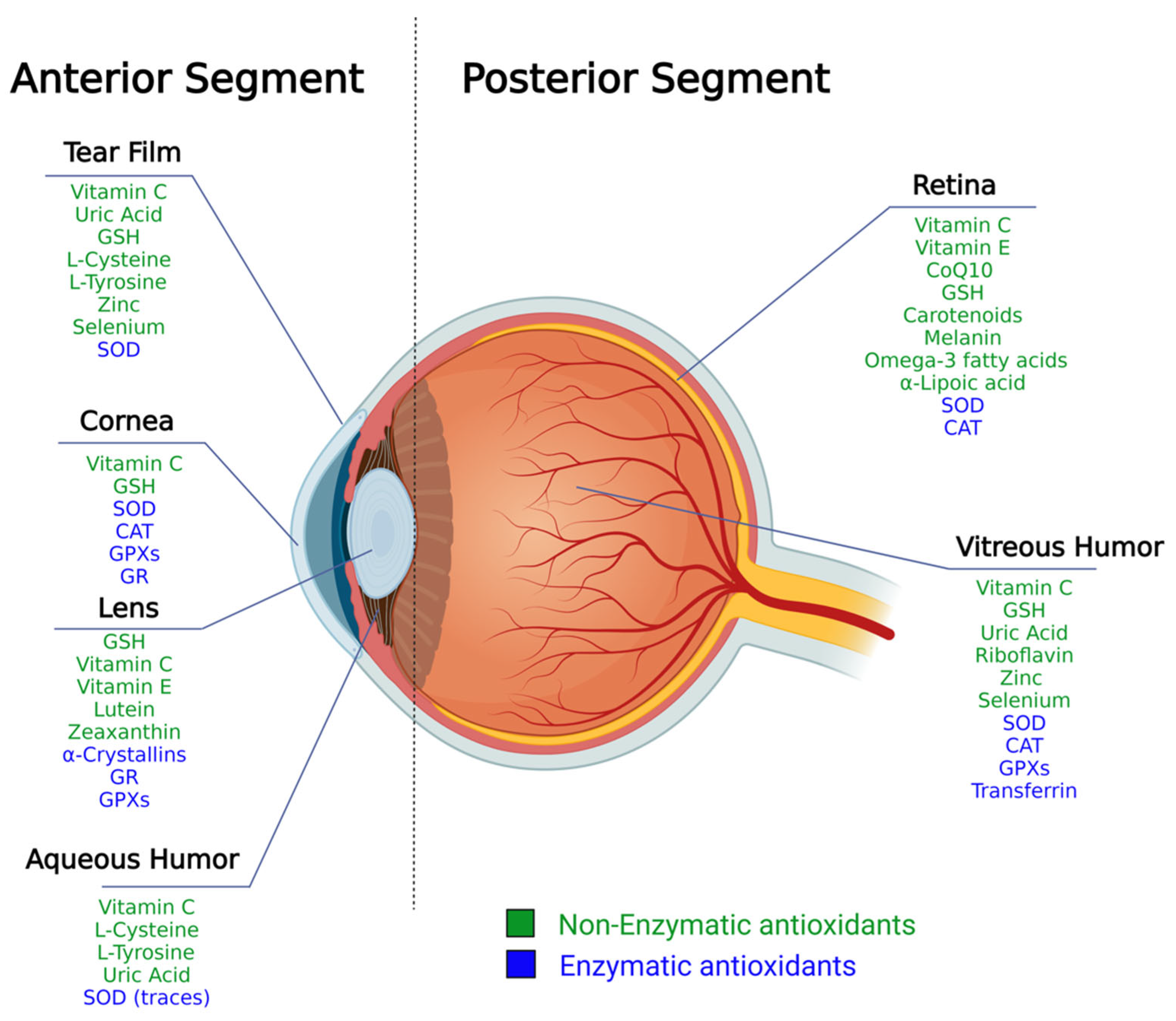
2. The Structure of the Eye and Ocular Antioxidant Defense System

The eye is a very complex organ of the human body that can be divided into the anterior and the posterior segments [6]. In the following chapters, the different molecules composing the antioxidant arsenals of the different anatomical compartments of the eye are presented (Figure 1).

2.1. Antioxidants Arsenal in the Anterior Segment

The anterior segment includes the ocular surface, the anterior chamber, the iris, and the lens. The ocular surface, which represents the interface towards the environment, provides cornea, conjunctiva, lacrimal and meibomian glands, nasolacrimal duct and eyelids [26]. The ocular surface is humectated by the tear film, a watery environment rich in electrolytes and proteins, resulting in a muco-aqueous lubricant gel; the outermost layer of the tear film is composed of lipids (meibum), which prevents evaporation.
Different antioxidant molecules were reported in human tears, with ascorbic acid being the most abundant (665 μM) and uric acid the second (328 μM). These two compounds account for ~50% of the total antioxidant activity; other small molecules, including GSH (107 μM), L-cysteine (48 μM) and L-tyrosine (45 μM) make up the rest [7]. Zinc and selenium were also identified in the tear film [8]. However, the only reported antioxidant enzyme in the tear film is SOD [9]. On the contrary, many enzymatic antioxidants have been documented in the cornea. These include SODs, CAT, GPXs, GR, glucose-6-phosphate dehydrogenase (G6PD), along with members of the aldehyde dehydrogenase (ALDH) superfamily [10].
Among the non-enzymatic antioxidants in the cornea, ascorbic acid, commonly known as vitamin C, is an excellent antioxidant agent that can donate a proton and form a relatively stable ascorbyl free radical. In addition, vitamin C was shown to be an effective scavenger of oxygen and nitrogen oxide species, such as superoxide radical ion, hydrogen peroxide, hydroxyl radical, and singlet oxygen. Its oxidation gives rise to dehydroascorbate, which is then reconverted to ascorbate, using GSH or NADPH as electron donors [9] (Figure 2).
The ascorbate accumulation in the eye is achieved primarily by active transport through the iris-ciliary body into aqueous humour and subsequent transport into the cornea and lens.
The cornea presents millimolar concentrations of GSH (4~7 mM) that, unlike ascorbate, can be synthesized via its de novo biosynthetic pathway mediated by two cytosolic enzymes, glutamate-cysteine ligase, and glutathione synthetase. It represents the most abundant cellular non-protein thiol and can directly scavenge hydroxyl radicals and superoxide [11].
GSH converts superoxide (O2•−) to H2O2 while undergoing self-oxidation (GSSG). H2O2 can then be converted into water by CAT or GPXs. Finally, GSSG is recycled to GSH by GR in the presence of NADPH as an electron donor (Figure 3). This redox cycle also protects lens proteins from the action of ROS.
The crystalline lens is a highly elastic, circular, biconvex, transparent body composed of long and thin cells named lens fibers, highly rich in water-soluble proteins called crystallins [12]. These cells lack cellular organelles, neural innervation and blood vessels, increasing tissue transparency and reducing the production of ROS, also in relation to the low oxygen tension of the lens [27].
Crystallin proteins combine both structural roles and chaperone activity. Indeed, the α-isoform could bind damaged proteins, sequestering misfolded or damaged aggregation-prone proteins and preventing the formation of light-scattering species [28].
Mammalian crystallins are intrinsically resistant to photo-damage due to the presence of tryptophans (Trp), tyrosines (Tyr) and phenylalanines (Phe) in their sequences, residues capable of UV absorption and radical photochemistry [29,30]. Trp has the highest specific absorption of protein amino acids at 280 nm. It dissipates UV-B radiation through energy transfer and internal conversion, thereby protecting the structures behind the lens, particularly the retina, from UV damage [31]. Besides Trp, Cys, Tyr and His residues can participate in excited state radical chemistry that could lead to photochemical covalent crosslinking of the involved proteins [32].
Oxidative processes in the healthy eye lens are controlled by both α-crystallin proteins and physiologically occurring water-soluble antioxidants.
GSH, ascorbic acid, GR and GPx are potent endogenous radical scavengers in lens fiber cells. GSH is the most abundant, reaching about 10 mM concentration in the lens [13].
The anterior chamber, located between the iris and the cornea’s innermost surface, serves as the rear support for the cornea and is filled with a clear fluid. The aqueous humor that provides nutrition removes excretory products from metabolism, transports neurotransmitters, stabilizes the ocular structure and contributes to regulating the homeostasis of the lens and cornea [33]. In addition, ROS can be continuously generated in the aqueous humor through hydrogen peroxide, superoxide anion, singlet oxygen, and peroxyl radicals. In human aqueous humor the antioxidant defence system is represented by small molecules, including ascorbic acid (530 μM), L-tyrosine (78 μM), uric acid (43 μM), L-cysteine (14.3 μM) and GSH (5.5 μM) [14], and only trace amounts of SOD [15].

2.2. Antioxidants Arsenal in the Posterior Segment

The posterior segment of the eye is represented by all-optical structures behind the lens, including the vitreous, retina, choroid and optic nerve.
The retina is a complex structure, rich in blood vessels and a dense network of neurons that capture and process light by virtue of the visual pigments. It is a highly energy-consuming tissue that relies on abundant mitochondrial oxidative phosphorylation and aerobic glycolysis to sustain the energy demand. Indeed, the retina has the highest oxygen consumption rate (per gram of tissue) in the body [34], which can produce a considerable amount of ROS in addition to that generated by exposure to visible and UV light. The retina developed significant defence strategies against ROS to overcome this situation, including SOD and CAT enzymes, GSH, vitamin C, carotenoids (lutein and zeaxanthin) and other pigments [16]. A vital antioxidant role is also played by ocular melanin, a highly heterogeneous and complex polymer normally present in the iris, choroid and retinal pigment epithelium [17]. Melanin exerts a protective effect on ocular cells and tissues using physical and biochemical mechanisms. In the anterior segment of the eye globe, it mainly acts as a photo-screen, while in the posterior segment, the mechanism appears to be predominantly biochemical [35]. It was suggested to be a weak antioxidant and free radical scavenger. Still, most likely, it is involved in the inactivation of the autofluorescent membrane-bound lipofuscin, an essential source of oxidative stress since it absorbs light and generates light-induced ROS [36].
The vitreous is a viscoelastic gel in the eye’s posterior chamber, with a high water content (99%) and few resident cells [37]. It occupies about 80% of the eye globe volume. It presents a vast array of both an enzymatic and non-enzymatic antioxidant profile, including SOD, GPX and CAT, as well as GSH, metal-chelating proteins (such as transferrin), uric acid, ascorbic acid, riboflavin and trace metals (zinc and selenium) [38].
Among metals crucial for the antioxidant system, zinc protects sulfhydryl groups from oxidation and competes with iron and copper ions for binding to cell membranes and proteins, displacing these potential ROS-generating metals; in addition, zinc increases the activation of antioxidant proteins, such as GSH and SOD [39]. Another essential metal is selenium, which acts indirectly through incorporation in selenoproteins having antioxidant properties.

3. Oxidative Stress and Eye Pathologies

When antioxidant patrolling is inadequate and/or ROS production is excessive, the delicate balance between ROS generation and ROS scavenging executed by antioxidants leads to “oxidative stress”, a chronic condition in which ROS overwhelm antioxidant shield activity. In this scenario, progressive cell dysfunctionality follows the uncontrolled reaction of ROS with cellular proteins, lipids, and other cellular molecules.
Oxidative stress has been implicated in aging and a variety of age-related chronic diseases, including several eye pathologies, such as Dry Eye Disease (DED), cataracts, glaucoma, Age-related Macular Degeneration (AMD) and Diabetic Retinopathy (DR) [40].
DED is a disorder involving the ocular surface, in which an excess of inflammatory cytokines increases ROS production, disrupting the healthy tear film [41].
A cataract occurs when the ordered arrangement of crystallin proteins composing the scaffold of the lens is lost due to excessive exposure to UV radiation and ROS, and crystallins aggregation, consequent to their misfolding, leads to lens opacification and progressive vision worsening.
AMD is a common cause of progressive visual impairment in the elderly caused by oxidative degeneration of the retina [42].
Glaucoma is a group of eye disorders that damage the optic nerve. While the aetiology is multifactorial, oxidative stress is essential in disease progression.
DR is a most common complications of diabetes mellitus, whose hallmark, i.e., hyperglycaemia, is associated with increased accumulation of ROS in different tissues, including the retina [43].

4. Nutraceutical Antioxidants for the Ophthalmic Field

The correlation between oxidative stress, oxidative damage and ocular disease has led researchers to investigate the antioxidant approach in modulating ophthalmic pathologic conditions. The reinforcement of the endogenous antioxidant system of ocular tissues can be achieved through nutrition and food supplementation, as well as through medical devices and antioxidant-based drugs. Although they may contain the same antioxidant molecule(s), nutraceuticals, medical devices, and drugs require different market approval routes and have other purposes. Therefore, we investigated the presence of antioxidants in the ingredient lists of nutraceuticals supplements intended to be used in ophthalmology. If available, we also opted to include studies from non-enteral routes of administration (such as antioxidant(s)-based eye drops), as the goals of these treatments are comparable to the oral supplementation, i.e., delivering one or more antioxidant molecules to the site of action (eye) and eliciting one or more antioxidant-related effect.
On the other hand, since our principal aim was to identify and investigate the antioxidant properties of substances with a defined chemical formula (single molecules), we decided not to include botanical extracts in this review. Indeed, although many botanical extracts have antioxidant effects that could be useful in the ophthalmic field, they have a chemical composition that is difficult to standardise. Furthermore, the molecules contained in a botanical extract are subject to multifactorial variations related to, but not limited to, the latitude and cultivation method of the plant, the seasonality of harvesting, and the extraction method.
The antioxidant molecules presented in the following sections (Table 1) will be divided into three main categories:
(i)
Exogenous plant-derived antioxidants: they are defined as “essential” nutrients, as they are not synthesized by the human body and, thus, can be obtained exclusively through exogenous introduction into the body (usually nutrition). We sub-divided them by their chemical class:
  • Polyphenols.
  • Carotenoids.
(ii)
Water-soluble promoters of the endogenous antioxidant system: molecules either produced or not by the human organism, showing direct antioxidant activity and/or able to boost the endogenous antioxidant system.
(iii)
Lipophilic antioxidants: lipophilic molecules promoting the defence of cellular membranes.

5. Exogenous Plant-Derived Antioxidants

5.1. Flavonoids

Polyphenols comprise a family of around 5000 organic molecules in the plant kingdom. In plants, they are secondary metabolites that play a role in the defence of the vegetal organism against UV radiation or pathogen aggression [49].
Among them, flavonoids, reported in various fruit and vegetables, including tea, cocoa, and red wine, are characterised by a common backbone represented by two aromatic rings bound together by three carbon atoms forming an oxygenated heterocycle (Figure 4, left).
Different mechanisms have been proposed to explain their antioxidant capacity, involving the suppression of ROS formation by either inhibiting the enzyme system deputed to the formation of oxygen radicals, such as hydrogen peroxidase and nitric oxide synthetase, directly scavenging radicals, chelating or stabilizing metal ions involved in the redox reactions, or upregulating the host antioxidant defences. In vitro and in vivo studies demonstrated that flavonoids have strong anti-inflammatory, immunomodulatory and antioxidative properties [50]. Quercetin (Figure 4, right) is one of the most abundant and well-studied flavonoids. In an experimental mouse model of dry eye disease (DED), topical application of quercetin increased tear volume, corneal regularity, and goblet cell density. This was associated with reduced inflammatory markers, including MMP-2, MMP-9, ICAM-1 and VCAM-1 in the lacrimal gland [51]. In another DED murine model, topical quercetin protected the ocular surface, enhancing corneal integrity, as highlighted by fluorescein staining [52].
Moreover, quercetin applied topically or added to the diet of galactose-fed rats (an in vivo model of diet-induced cataract) showed an anticataractogenic effect as it delayed lens damage. However, no direct antioxidant activity was observed in this study [53]. On the other hand, in an ex vivo rat model of hydrogen peroxide-induced lens opacification, quercetin, in low micromolar (10 μM) concentration, has been an effective inhibitor of oxidative damage and cataractogenesis [54]. Unfortunately, quercetin displays poor chemical stability, and it is rapidly metabolised into 3′-O-methyl quercetin by catechol-O-methyl transferase in rat lenses, reducing its antioxidant activity [55].
Flavonoids from several sources have efficiently reduced the extent of selenite cataracts in rat models [56,57,58,59].
In an advanced 3D in vitro study of 3D-human trabecular meshwork cells, a patented eye drop formulation containing >2.5% polyphenols from Perilla frutescens had significant inhibitory effects on the apoptotic pathway, activation of NF-κB, and induction of pro-inflammatory cytokines from oxidative stress. These results suggest that polyphenols-based eye drops could be an adjuvant therapy in glaucoma [60].

5.2. Catechins

Catechins are a class of polyphenols belonging to the flavon-3-ols subgroup and present two chiral centres (highlighted by stars in Figure 5). Naturally occurring compounds exhibit a 2,3-trans (catechins) or a 2,3-cis (epicatechin) configuration.
Reported data indicate they exert antioxidant activity, directly scavenging ROS and chelating metal ions. The phenolic hydroxyl groups can react with oxygen and nitrogen radicals, donating one electron and reducing the free radicals. In addition, catechins can take charge of the radical, which is stabilised by charge delocalisation on the π-electrons of the phenyl ring, thereby terminating the radical propagation [61]. Adjacent hydroxyl groups form metal chelation sites that sequester metals involved in free radical formation [62]. Moreover, catechins regulate the expression of several enzymes, inducing antioxidants, inhibiting pro-oxidant ones, and increasing detoxification enzymes [63].
Green tea catechins are a group of flavonoids in the leaves of Camellia sinensis composed of epicatechin, epigallocatechin, epicatechin gallate and epigallocatechin gallate (EGCG). In an in vitro study, EGCG showed to inhibit different kinds of cytokines induced by IL-1 or hyperosmolarity in a dose-dependent manner in cultured human corneal epithelial cells [64]. Additionally, EGCG decreased the expression of IL-1 in a murine model of DED and reduced corneal epithelial damage [65]. Moreover, EGCG showed antioxidant activity by efficiently preventing H2O2-mediated oxidation of cataractous human γ-crystallin [66].
Masmali and co-workers observed that green tea consumption could negatively affect the tear film quality, measured by the tear ferning test [67]. It was suggested that this effect could be due to the oxidation of the lipid layer and/or the alteration of electrolyte content [68]. On the contrary, a randomized controlled trial that evaluated the efficacy of green tea extract in 60 patients with DED secondary to meibomian gland dysfunction showed an improvement in symptoms, break-up time and meibum quality compared to the control group [69]. The contrasting data underline the need to further investigate the effects and mechanisms of action of catechins-containing extracts on tear film physiology.

5.3. Anthocyanins

Anthocyanins are an important group of visible plant pigments responsible for many plants and fruits’ red, blue, and purple pigmentation (Figure 6).
Supplementation with anthocyanin-rich extracts of bilberry (Vaccinium myrtillus L.) was evaluated in different studies. A randomized controlled trial enrolled patients with DED, and the supplementation of bilberry extract showed to significantly improve the tear volume, evaluated by the Schirmer tests, which measures the quantity of lacrimal secretion if compared to control subjects [70]. A clinical pilot trial investigated the effects of MaquiBright®, a registered trademark maqui berry (Aristotelia chilensis) extract with a standardized 35% content of anthocyanins, in patients with moderate DED, reporting a significant improvement of Schirmer test and quality of life after 60 days of treatments [71]. Similar results on the same extract were obtained in a randomized controlled trial, which reported a significant improvement in the Schirmer test and relief from symptoms of eye fatigue in the visual display terminal [72].

5.4. Curcumin

Curcumin is a polyphenol isolated from turmeric (Curcuma longa), widely used as a spice and flavouring agent. It is a linear diarylheptanoid characterized by keto-enol tautomerism (Figure 7), in which equilibrium is shifted in solution towards the -enol form, stabilized by the fully conjugated aromatic system. As such, it is extremely useful as an antioxidant since it can chelate different positively charged metal ions that often trigger the formation of radicals [73]. However, the exploitability of this molecule as a food supplement is limited by its poor solubility in aqueous solution, low absorption in the gut, rapid metabolism (with degradation to antioxidant by-products such as ferulic acid, vanillin and dehydrozingerone [74]) and systematic elimination [75]. To overcome such drawbacks, the association with piperine, one of the significant black pepper alkaloids, enhanced curcumin bioavailability [76]. Dehydrozingerone is modestly soluble in water and stable at neutral pH, unlike curcumin. Curcumin and its derivatives zingerone, dehydrozingerone, and the biphenyl analogue bizingerone have been studied for their radical scavenging ability, related to the presence of the phenolic rings, the β-diketone, the conjugated double bond system and the methoxy substituents at the phenyl ring [77].
Curcumin has been shown to modulate multiple cell signalling pathways leading to anti-inflammatory, antioxidant, anti-angiogenetic, wound healing, and antimicrobial effects [78]. It was investigated for its impact on the ocular surface system and administered by intranasal delivery of a nanomicelle formulation to overcome low oral bioavailability. It restored ocular surface homeostasis by reducing ROS production, decreasing the expression of inflammatory mediators, and increasing neurotrophic factors [79]. The intranasal treatment effectively delivered the active ingredient to the ophthalmic branch of the trigeminal ganglion. An additional potential application field of curcumin is dry eye disease, as it was observed to reduce pro-inflammatory cytokines such as IL-4 and IL-5 in the conjunctiva of mice [80]. In human corneal epithelial cells, curcumin counteracted the increased production of IL-1, IL-6 and TNF induced by hyperosmotic stress [81], resulting to be a promising candidate for the treatment of DED.

5.5. Resveratrol

Resveratrol is a natural polyphenol found in plant-based foods, mainly grapes and berries, with a high concentration in red wine. Resveratrol has a stilbene structure consisting of a phenolic ring connected to a resorcinol moiety by an ethylene bridge that identifies a trans and a cis isomer (Figure 8).
It is usually present in plants as trans-resveratrol. Still, when ingested, it is rapidly metabolised to the biologically more active dihydro resveratrol (Figure 8, right), showing a high antiproliferative effect.
Resveratrol can directly scavenge various free radicals [82] and has been shown to inhibit lipid peroxidation more effectively than vitamins C and E [83,84,85]. It also stimulates the expression of the enzymatic antioxidant defence cellular system [86].
The direct radical scavenging activity is related to the redox properties of the phenolic hydroxyl groups and their capability of delocalizing unpaired electrons across the π-system of the structure [87]. Resveratrol also undergoes one-electron electrochemical oxidation, showing two oxidation peaks, the first corresponding to the irreversible oxidation of the phenol group and the second corresponding to the irreversible oxidation of the resorcinol moiety [88].
Conversely, resveratrol showed pro-oxidant properties, leading to DNA oxidative damage in synergy with transition metal ions such as copper. It was suggested that this pro-oxidant action might inhibit tumour initiation, promotion and progression and that this could be a common chemo-preventive mechanism of plant polyphenols [87].
Resveratrol has various beneficial effects on health, including cardio-protective action upon vasculature, i.e., the ability to inhibit angiogenesis, prevent inflammation, and facilitate vaso-relaxation [89]. Since vascular impairment is involved in many ocular diseases, such as impaired blood flow and ischaemic events in age-related macular degeneration (AMD), diabetic retinopathy (DR) and glaucoma, the potential advantages of resveratrol attracted much interest in the field of ocular diseases prevention and treatment. The significant activities attributed to resveratrol on the eye include antioxidant, anti-apoptotic, anti-tumorigenic, anti-inflammatory, anti-angiogenic and vasorelaxant.
In an in vitro study on primary porcine trabecular meshwork cells subjected to chronic oxidative stress (40% O2), chronic resveratrol treatment reduced the production of intracellular ROS. It prevented the expression of glaucoma markers IL1a, IL-6, IL-8, and ELAM-1 (endothelial-leukocyte adhesion molecule 1). In addition, resveratrol inhibited the expression of the cellular senescence marker sa-β-galactosidase (sa-β-gal), typically induced by oxidative stress [90].
In a selenite-induced cataract in vivo model (rat), intraperitoneal (i.p.) resveratrol (40 mg/kg/day) proved to increase the levels of GSH and to lower the amount of malondialdehyde (MDA), a marker of lipid peroxidation, in rat lenses and erythrocytes. Additionally, it suppressed selenite-induced oxidative stress and cataract formation [91]. Similar results were observed in a naphthalene-induced albino rat cataract model, where the same dose as i.p. administration of resveratrol was confirmed to reduce MDA levels and it significantly retarded lens opacity, restored antioxidants (CAT, SOD, GPX, GSH), function and reduced lipid peroxidation in the lenses of the animals [92].
In a diabetic rat model, four consecutive weeks of i.p. administration of 10 mg/kg/day resveratrol significantly alleviated hyperglycaemia and promote weight loss. They simultaneously reduced the levels of oxidative markers, along with superoxide dismutase activity in the blood and retinas [93]. Additionally, oral gavage at 20 mg/kg of resveratrol suspended in 0.5% carboxymethylcellulose dissolved in 0.9% saline solution administered once a day for four weeks showed to prevent vascular damage in the retina [94].

5.6. Carotenoids

Carotenoids are lipid-soluble antioxidants comprising more than 600 compounds responsible for the yellow, orange, and red coloring of fruits, leaves and flowers. They belong to the tetraterpene family and are C40-based isoprenoids. In addition, they are active ROS scavengers and induce the synthesis of antioxidant enzymes.
The progenitor of carotenoids are carotenes, whose most common form is β-carotene, an important lipophilic antioxidant, able to insert into the cell membrane, acting directly as a shield against lipid peroxidation working in synergy with other antioxidants such as vitamin C and E [95]. Additionally, it is the precursor of vitamin A, a crucial element for human physiology. In the sight process, it combines with the photoreceptor’s opsin and rhodopsin in the form of 11-cis-retinal.
A subclass of carotenoids containing oxygen is named xanthophylls [96]. Among them, lutein and zeaxanthin (Figure 9) are epimers that differ only by the configuration of a single hydroxyl group [97]. They are mainly concentrated in the macula lutea, and their peak concentration appears at the centre of the fovea. However, they are also present in other ocular compartments, such as the lens, where they display protective effects against lipid peroxidation, protein oxidation, DNA damage and upregulate GSH levels. In addition, ocular carotenoids absorb light protecting eye tissues from the light-induced photochemical insult and quench free radicals by accepting their unpaired electrons, stabilizing them in their conjugated double bonds system [98].
Lutein and zeaxanthin in the human lens absorb blue light, characterized by short wavelength and high energy [20].
Such molecules can cross the blood-brain and blood-retinal barriers of mammals, including humans, and then deposit into the retina, which can play a crucial role in regulating oxidative homeostasis [99,100]. Numerous experiments and clinical trials have been conducted to elucidate the role of orally supplemented carotenoids in preventing or modulating ocular pathologies.
AREDS clinical trials showed that the oral administration of vitamin C (500 mg), vitamin E (400 IU), β-carotene (15 mg) and zinc (80 mg) was able to significantly reduce the risk of developing AMD and to maintain visual acuity better than placebo [44]. Surprisingly, the addition of lutein (10 mg), zeaxanthin (2 mg), docosahexaenoic (350 mg) and eicosapentaenoic (650 mg) acids to the formulation failed to demonstrate extra benefits (AREDS2 clinical trials) [47]. On the other hand, in other studies (reviewed by Johra and colleagues [19]), lutein and zeaxanthin proved to be protective against the development of AMD.
In 2016 Ma et al. reported that the lutein+zeaxanthin oral administration could increase the macular pigment optical density in patients affected by AMD in a dose-dependent manner [101]. Successively, Wilson et al. reported that the increase in macular pigment optical density can be achieved with doses > 10 mg/d of lutein and/or zeaxanthin [102].
Food supplementation of lutein and zeaxanthin, but not of β-carotene, reduced the risk for senile cataracts by exerting an antioxidant activity towards lens proteins, lipids, and DNA [48,103].
However, a recent meta-analysis did not show a significant association between the serum content of lutein, zeaxanthin, and other vitamin A-derivatives with DR [104].
Another xanthophyll that is very promising in the ophthalmic field is astaxanthin (ASTX, Figure 9). This naturally occurring red carotenoid pigment is produced by some microalgae, such as Haematococcus pluvialis. As the primary dietary source for some marine animals, such as salmon, shrimp, and lobster, it confers their flesh to the typical pink color. Carotenoids are known to insert into the cell membrane. Electron density studies showed that apolar carotenoids, such as β-carotene and lycopene, altered the packing of phospholipid acyl chains and disordered the membrane bilayer.
On the contrary, ASTX did not modify the structure of constituent lipids. ASTX displays an amphipathic nature, which orientation, differently to other carotenoids, may be influenced by membrane lipid composition (e.g., acyl chain length, degree of saturation, presence of cholesterol). ASTX presents a keto group at the C4 and C4′ positions of the terminal rings, which are thought to stabilize membrane interactions with respect to the polar terminal groups. This would allow the rest of the molecule to span the entire membrane width, fitting precisely in the polar-nonpolar-polar depth of the cell membrane [105].
The perpendicular orientation of xanthophylls in cell membranes has been shown to increase the rigidity of the lipid bilayer, where they can act as “molecular rivets” and as a “full-length” anti-ROS shield of the lipid bilayer [106]. In addition, ASTX antioxidant activity has been reported to be more than 100 times and 550 times greater than that of vitamin E against lipid peroxidation and singlet oxygen quenching [107].
Oral treatment of ASTX, along with zeaxanthin and lutein in AMD patients, preserved retinal function better and improved visual acuity. In addition, in animal models of diabetic retinopathy, ASTX preserved histological and functional outcomes of the diseases, increasing the levels of antioxidant enzymes, and reducing levels of oxidative mediators, such as NF-κB and aldolase reductase activity [108].
Moreover, the neuroprotective effect of ASTX may be usefully adopted in managing glaucoma. For example, in a mouse model in which elevated IOP was induced by cauterizing episcleral vessels, significant reduction of retinal apoptosis and oxidative markers expression and higher visual evoked potential were observed in animals receiving ASTX in their diet. More recently, experiments have been conducted in a normal IOP glaucoma murine model. Retinal ganglion cells (RGC) axonal degeneration is caused by the homozygous deletion in the glutamate/aspartate transporter (GLAST−/−) gene. In these studies, ASTX, both given orally and i.p., protected RGC from glutamate neurotoxicity and degeneration [109,110].
Other promising ASTX applications in eye disorders include the treatment of uveitis, a wide group of inflammatory conditions affecting the middle layer of the eye, and cataract prevention, even if clinical data in humans are missing [108].
Due to the low bioavailability and water solubility, topical application of ASTX might be an alternative route of administration. In a murine model of UV-B light-induced photokeratitis, ASTX eye drops protected and well-preserved the epithelium from cell death and thinning caused by photodamage [111]. More recently, topical application of ASTX-encapsulating liposomes in a rat dry eye disease model prevented the up-regulation of age-related markers and maintained epithelial integrity [112].

6. Water-Soluble Promoters of the Endogenous Antioxidant System

6.1. Zinc and Selenium
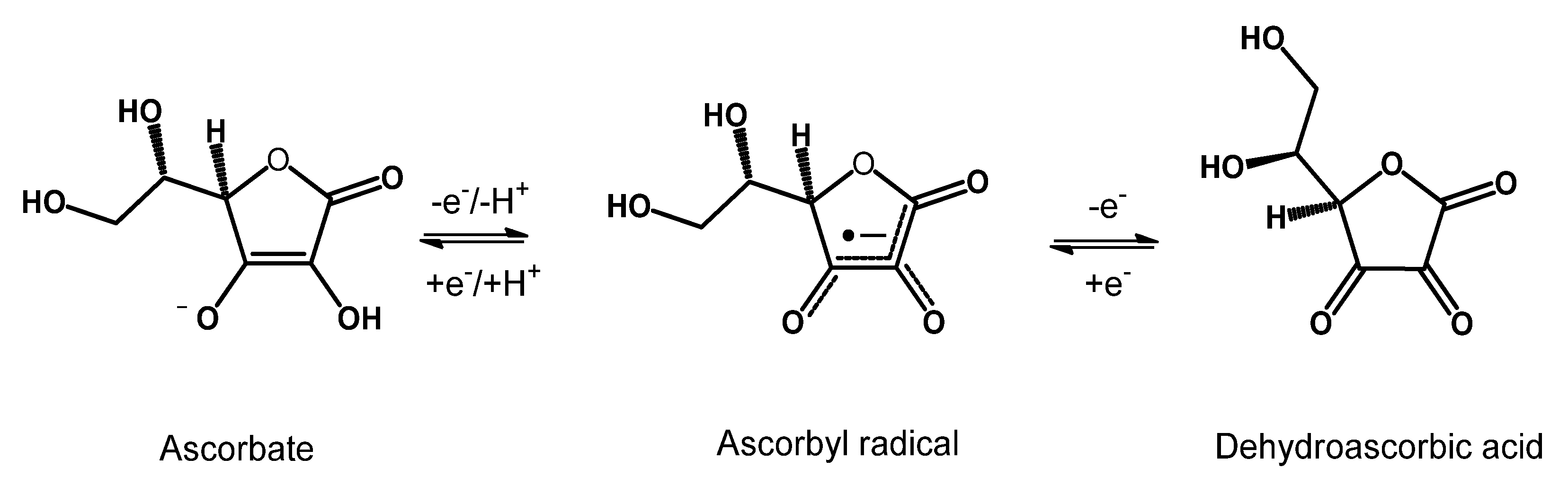
Zinc and selenium are essential micronutrients that must be supplied through the diet. Zinc plays vital roles in the human body, which can be described as catalytic, structural and regulatory functions [113]. Selenium is a fundamental building block of a small cluster of antioxidant proteins, such as thioredoxin reductases, GPXs and iodothyronine deiodinases. These enzymes act as oxidoreductases and depend on selenium availability for proper activity [114]. Selenoproteins P, a selenium transporter found in the tear film responsible for delivering selenium to the corneal epithelium, is reduced in dry eye disease [115]. In contrast, selenium concentration is increased in the tears of diabetic patients [8]. These findings suggest that altered selenium metabolism may play a role in developing or progressing oxidative stress-related eye disorders.
Noteworthy, zinc plays an essential role in the activity of enzymes in the antioxidant defence system, in the regulation of GPXs, and as a co-factor for SOD. Zinc also induces the synthesis of metallothioneins, deputed to reducing hydroxyl radicals and sequestering ROS. In addition, this mineral has been shown to stabilise cell membranes and inhibit the pro-oxidant enzyme NADPH-Oxidase [39,116].
Eye zinc levels were decreased in AMD, suggesting that zinc deficiency may lead to oxidative stress and retinal damage [117].
The AREDS study reported that the group receiving zinc supplementation in combination with antioxidant vitamins showed a bigger decrease in advanced AMD progression (~25%) and the highest prevention of loss of vision (19%) [45,46]. Furthermore, low serum zinc levels are correlated with diabetes, hypertension and microvascular complications, suggesting the idea that it was also correlated with the duration and severity of diabetic retinopathy [118], as confirmed by studies on a rat model of DR [119].

6.2. Ascorbic Acid

Most probably one of the most studied and well-known antioxidant molecules, vitamin C is a water-soluble nutrient of fundamental importance. It is a “universal” antioxidant, present in several aqueous fluids and tissue compartments of the body. It is a reversible reductant and antioxidant which efficiently neutralizes ROS and reactive nitrogen species, as well as peroxynitrite, nitroxide radicals, singlet oxygen and hypochlorite. It also works in synergy with other biological antioxidant such as GSH, vitamin E and flavonoids [120,121,122]. Among mammals, only humans and a few other species (Guinea pigs, some bats and some primates) have lost this ability and must obtain the essential vitamin C from their food [123].
Ascorbic acid is widespread in all eye compartments at high concentrations, suggesting an essential function of vitamin C in eye physiology and in preventing eye disorders [21]. The AREDS study showed that vitamin C supplementation significantly reduces the risk of developing AMD and maintains visual acuity [44].
In 2022, Xiong and colleagues published a cross-sectional study and meta-analysis on the association between vitamin intake and DR. The authors concluded that lower levels of ascorbic acid were significantly associated with the disease [104].
According to the high vitamin C lens content, a large body of literature also investigated the role of vitamin C in cataract prevention, leading to conflicting results [44,124,125,126,127]. This dissension suggests that a simple intervention of vitamin C supplementation might not be effective. Accordingly, plasma and humor aqueous are estimated to be saturated in humans with intakes of <250 mg of vitamins C/day [128].
A recent study found a high correlation between total antioxidant capacity and ascorbic acid concentration in aqueous humor with cataract severity, determined by cumulative dissipated energy during phacoemulsification [129].
Ascorbic acid is known to be relatively unstable as it undergoes oxidation. For this reason, more stable derivatives have been developed, such as ascorbic acid 2-O-alpha-glucoside (AA-2G), and investigated in several applications, including the ophthalmological field. For example, studies on developing chick embryos showed that AA-2G has preventive effects on hydrocortisone-induced cataract formation and inhibits the decrease in GSH lens content and the increase in lipid peroxide lens content, even more than ascorbic acid itself [130,131]. More recently, AA-2G has also been shown to be effective in topical application in solid-in-oil nanodispersions that can promote the wound healing of corneal epithelium [132].

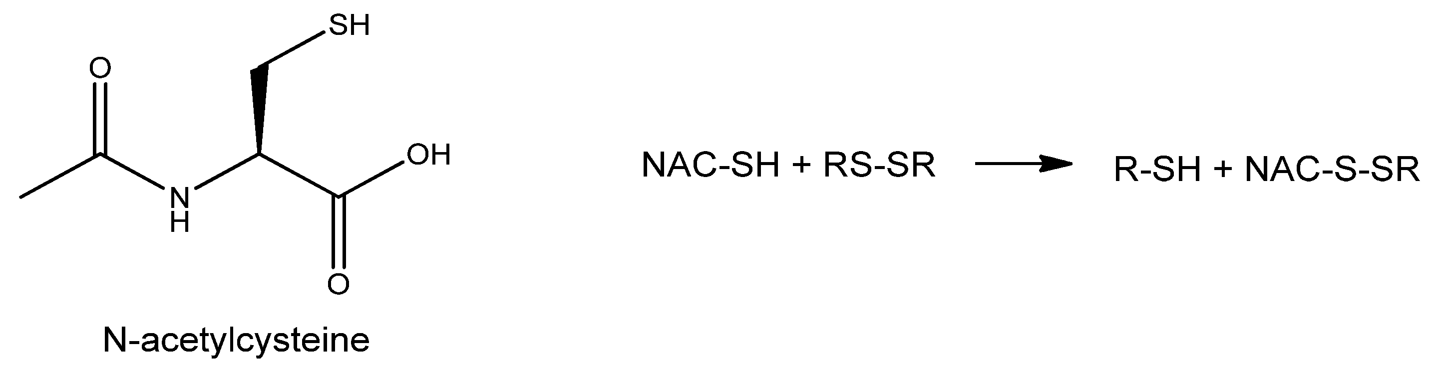
6.3. N-Acetyl Cysteine and Other Cysteine Derivatives

N-Acetyl Cysteine (NAC) is the membrane-permeable acetylated form of the amino acid L-cysteine (Figure 10). It was initially used as a mucolytic agent, as it lowers the viscosity of mucous by reducing the disulphide bonds of mucoproteins.
The presence of the thiol group can donate reducing equivalents such as hydrogen, which can reinstitute losses sustained from exposure to ROS, and this is often referred to as a ‘repair reaction’ [133]. Moreover, NAC also acts as a direct antioxidant, scavenging superoxide and peroxides [134]. However, the slow chemical kinetics of NAC reaction with ROS does not support the direct scavenging narrative. Given that NAC indeed exerts antioxidative (reductive) effects inside cells, it is supposed to do so indirectly [135].
NAC can be hydrolyzed into cysteine, a precursor in the synthesis of GSH. Topical administration of NAC has been reported to improve corneal epithelial healing of different kinds of wounds in animal and human studies, particularly alkaly burns of the ocular surface [136,137]. Additionally, NAC improves the clinical outcome of DED symptoms by having favorable lubricant activity [136]. A 4 times daily administration of either 1% or 0.1% topical NAC solution induced a statistically significant higher cone cell density, as observed by confocal microscopy [136].
The role of NAC was also investigated in cataract development in diabetic rats. NAC topical administration (0.01% and 0.05%) twice daily for 13 weeks resulted in delayed onset of early diabetic cataracts. However, this treatment did not seem to delay or reverse the severity of cataracts at later stages of the disease, as suggested by a study on an in vitro rabbit lens model of hyperoxia-induced cataracts [138]. On the other hand, NAC treatment delayed the progression of cataract onset and increased GSH levels in an ex vivo rabbit model of hyperoxia-induced lens damage [139]. In vivo studies on Wistar rat’s selenite-induced cataracts model assessed the activity of a different modified NAC-related compound, particularly N-acetylcysteine amide (NACA) eye drops reduced the severity of cataract formation along with increasing GSH levels [140].
Additionally, in vitro experiments on human retinas obtained from donors with or without AMD showed NAC to protect retinal pigment epithelium (RPE) cells in the retina from oxidative damage [141].
NAC was also investigated in the prevention of retinitis pigmentosa, a family of inherited diseases having in common a progressive retinal degeneration. A study detected improved retinal cone photoreceptor function in patients with moderately advanced RP after six months of NAC oral administration [142].
Notably, other cysteine derivatives also possess antioxidant properties that have been tested in ophthalmological applications. This is the case with S-allyl-L-cysteine (SAC), an organosulfur constituent of garlic, and S-allylmercapto-N-acetylcysteine (ASSNAC). In vivo studies on rats have demonstrated that SAC protects retinas from various insults, such as intraocular pressure augmentation and kainate excitotoxicity [143,144]. Regarding ASSNAC, in vitro data have shown that it can protect ARPE-19 cells from hydrogen peroxide insult, resulting in a significantly higher increase in GSH compared to cultures treated with NAC. Additionally, pre-incubation with ASSNAC has been shown to inhibit porcine lens hydrogen peroxide-induced opacification [145].

6.4. Riboflavin

Riboflavin (Figure 11) (vitamin B2) is a slightly water-soluble vitamin which acts as an essential cofactor in a wide range of crucial metabolic pathways such as energy production, mitochondrial function, and metabolism of other vitamins, such as B9 and B12.
Although riboflavin has not traditionally been considered an antioxidant molecule, this notion is changing because of an essential number of studies recently summarized by Olfat and colleagues, which suggest that even if indirectly, riboflavin does support the endogenous antioxidant defence system [146]. Indeed, riboflavin-deficient animals showed decreased levels of GSH, SOD, CAT, GR, and higher levels of MDA. Conversely, riboflavin administration significantly reduced MDA levels, with increased GSH production and SOD, CAT, GPXs and GR enzymes expression in different eye compartments. Moreover, riboflavin reinforces the antioxidant effects of vitamins C and E, recovering the antioxidant power of the latter through its role in the GSH redox cycle [146].
In humans, a milestone randomized trial of vitamins and minerals supplementation carried out in the rural communities of Linxian (China) (“Linxian Studies”) observed a lower prevalence of cataracts (follow-up: 5-6 years) in people receiving riboflavin together with niacin (vitamin B3) supplementation [126]. However, one limitation of this study was the lack of lens examinations at the beginning of the trial.
Due to its critical role in energy and glucose metabolism, riboflavin might play a role in DR prevention. Preliminary studies in animal models have shown that riboflavin supplementation protects the retina from oxidative stress and hyperglycaemia and increases glucose uptake and BDNF expression [147]. Moreover, riboflavin well absorbs UV light and is employed in sunlight-protective eye drops [148]. A recent study in an in vivo rabbit model showed a protective effect against UV-B-induced damage of an antioxidant eye drop formulation containing riboflavin and D-α-tocopheryl polyethylene glycol [149].

7. Defense of Cellular Membranes with Lipophilic Antioxidants
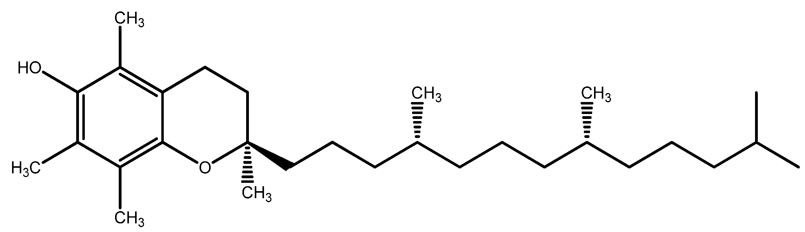
7.1. Vitamin E

Vitamin E (Figure 12) is a group of eight essential lipophilic substances, including tocopherols and tocotrienols, whose most active form is α-tocopherol, that protects cell membranes by breaking the chain reaction of lipid peroxidation by quenching peroxyl radicals [150]. Vitamin E function depends on the activity of other cytosolic antioxidants, such as vitamin C and GSH, as these molecules regenerate the active (reduced) form of vitamin E [120,121,151].
Vitamin E is particularly abundant in the eye in the RPE and photoreceptor cells [23]. Indeed, its deficiency leads to irreversible changes in the retinal structure and function [152].
Vitamin E (400 IU, DL-alpha tocopheryl acetate) belonged to the antioxidant mix used in the AREDS study [44]. Despite failing to counteract lens opacity of cataracts, the antioxidant mix statistically reduced the odds of developing AMD among the trial participants and was effective in preserving visual acuity, too [44,153]. On the other hand, data from other clinical reports indicate that supplementation of vitamin E alone (ranging from 100–600 IU, follow-up duration ranging from 4 to 10 years) does not prevent the development of AMD [154].
Studies investigating vitamin E supplementation for the prevention of cataracts have obtained conflicting results, too [124,154,155], suggesting a role in the pathology of the deficiency of vitamin E rather than a benefit from additional supplementation [156].
A recent study included vitamin E among those micronutrients whose deficiency is associated with DR [104].

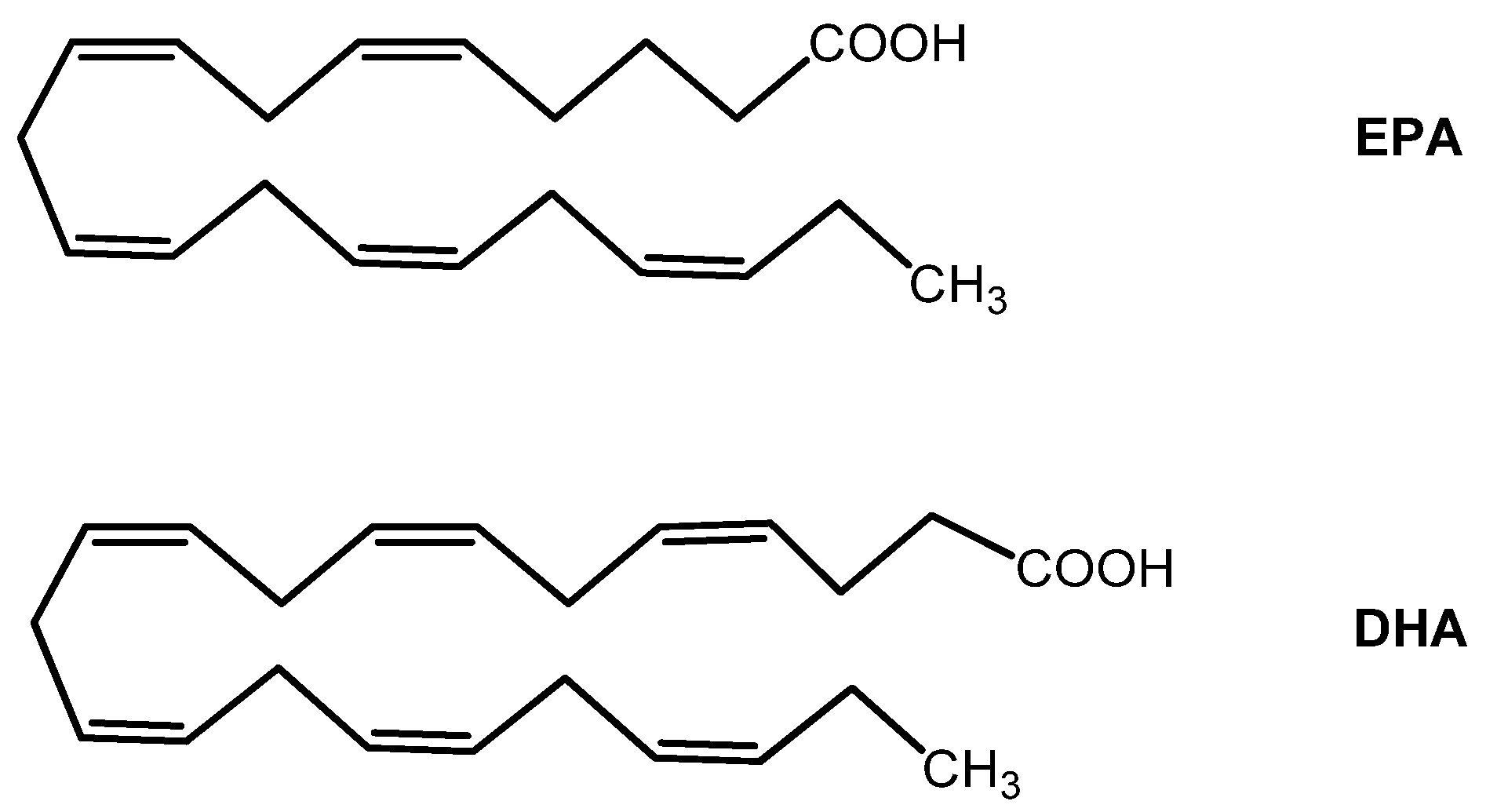
7.2. Omega-3 Fatty Acids

Similarly to vitamin E, the omega-3 (ω-3) fatty acids eicosatetraenoic acid (EPA) and docosahexaenoic acid (DHA) (Figure 13) are essential nutrients for the human body. They must be introduced by nutrition (they are particularly enriched in some fish, such as salmon, mackerel, and anchovies). Once absorbed in the human body, EPA is converted to DHA via an enzymatic series of desaturation reactions.
Many studies regarding their benefits on human health have focused on the role of DHA. Omega-3 fatty acids are well known for their ability to regulate second messengers’ systems resulting in anti-inflammatory activity. In particular, in vitro studies suggested that it increases cellular levels of enzymatic antioxidants, such as SOD and GPXs, and augments GSH levels [157,158]. Data from human studies have also shown that DHA is a crucial element for developing the nervous system and optimal visual acuity. EPA and DHA also exhibit antioxidant properties by counteracting the accumulation of ROS and lipid peroxidation at the cellular membranes [159].
Omega-3 fatty acids are enriched in the ocular tissues, particularly the retina. Therefore, RPE physiology is strictly dependent on EPA and DHA metabolism. The latter have antioxidant effects and protective action toward retinal cells, as shown in a recent in vitro study performed in the ARPE-19 (a human in vitro model of RPE) cell line [24].
Consistently with their importance in the retina, several studies documented the efficacy of dietary supplementation of ω-3 fatty acids in various retinopathies. For example, the PREDIMED (Prevención con Dieta Mediterránea) clinical trial observed that intake of at least 500 mg/day of dietary long-chain ω-3 PUFAs (alpha-linolenic acid— ALA —, EPA, docosapentanoic acid— DPA —, and DHA) was associated with a decreased risk of DR [160].
On the other hand, data on the effects of ω-3 fatty acids on glaucoma are conflicting, and the results are linked to the dosage [161,162]. Therefore, further studies are needed to better shed light on the incidence of ω-3 fatty acids supplementation on glaucoma pathology prevention and/or progression.
The efficacy of ω-3 fatty acids in the DED has recently been reviewed by O’Byrne & O’Keeffe, revealing, despite considerable statistical heterogeneity, an improvement of subjective symptoms in DED patients [163]. On the other hand, a clinical trial (ClinicalTrials.gov Identifier: NCT01880463) was published in the same year (not included in O’Byrne & O’Keeffe meta-analysis) showing no reduction of incidence of DED by long term (median: 5.3 years) supplementation with marine ω-3 fatty acids, 1 g per day [164].
In a recent study, patients with keratoconus received DHA (1000 mg) supplementation for three months, resulting in higher total antioxidant capacity and better outcomes in the astigmatism axis, asphericity coefficient, and intraocular pressure than the placebo group [165].
The use of ω-3 fatty acids in AMD has been investigated in the AREDS2 study, concluding that the addition of DHA (350 mg) and EPA (650 mg) to the original AREDS formulation failed further to reduce the risk of progression to advanced AMD neither had an overall effect on the need for cataract surgery [45]. However, it should be noted that EPA and DHA in the AREDS2 formulation were given to subjects in the ethyl ester form. Esterification is a technological process used to increase the stability and concentration of ω-3 fatty acids from fish oils. Nonetheless, a vast amount of literature developed mainly in the last decade indicates that ω-3 fatty acids in their ethyl ester form have significantly lower bioavailability than triglyceride or monoglyceride forms [166,167,168].
Future evaluations and studies also considering the form of ω-3 fatty acids used in trials are needed better to elucidate the role of these essential nutrients in AMD.

7.3. Coenzyme Q10

Coenzyme Q10 (CoQ10, ubiquinone) (Figure 14) is a crucial element of cells’ electron transport chain and mitochondrial bioenergetics. Its main cellular localization is the mitochondrion, which can also be found in other subcellular fractions and plasma. Unlike other fat-soluble antioxidants, such as vitamin E and ω-3 fatty acids, CoQ10 is synthesized endogenously. Therefore, its content is superior in tissues with higher metabolic activity and oxygen consumption, such as the heart, skeletal muscle, brain, and retina. It directly regulates the redox equilibrium in the mitochondria, acting as a free radical scavenger on site. Its reduced form, ubiquinol (CoQ10H2), inhibits lipid peroxidation and DNA and protein oxidation [169].
Given the crucial role of CoQ10, it is not surprising that its deficiency induces apoptosis and is associated with diseases affecting the retina, such as AMD and glaucoma [170]. In this regard, CoQ10, either topically applied with eye drops or supplemented in the diet alone or in combination with vitamin E, showed evidence of decreasing retinal ganglion cell loss and slowing glaucoma progression [156]. The proposed mechanisms of action involve diminished DNA fragmentation and retinal cell apoptosis, along with a neuroprotective effect exerted by restraining extracellular glutamate accumulation, preventing cytotoxicity and pro-apoptotic factors release, indicating that CoQ10 is beyond a simple free radical scavenger [171].
Data involving CoQ10 potential role in AMD are scarce. However, one study found that CoQ10, in combination with ω-3 fatty acids and acetyl-L-carnitine, significantly improved visual field mean defect (VFMD), visual acuity and foveal sensitivity in early AMD patients [172].
Interestingly, CoQ10 is also finding application in astrobiology as a possible protector of astronauts’ eyes, which often develop neuro-ocular alteration due to the damaging effects of microgravity and cosmic radiation. For example, in an in vitro/in-space study, CoQ10 treatment of ARPE-19 cells transferred for three days on board the International Space Station increased cell resistance to damage from microgravity [173].

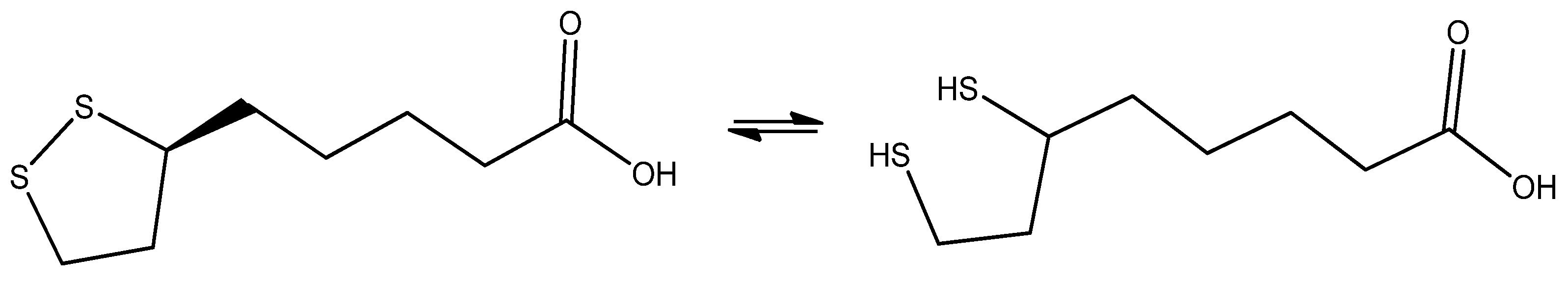
7.4. Alpha Lipoic Acid

α-lipoic acid (LA) (Figure 15) is a natural organosulfur with antioxidant and anti-inflammatory properties, present in animal and plant food and that can be modestly produced by the human body. It acts as a direct antioxidant, inactivating free radicals, and an indirect antioxidant, able to regenerate other cellular antiradical molecules such as GSH, vitamin C and vitamin E [174].
It has been documented that LA can cross the blood-brain barrier and, after ingestion, rapidly reaches the retina and the optic nerve [22]. This and its antioxidant properties make it a promising candidate for treating glaucoma. In a study using the DBA/2J mouse model of glaucoma, supplementation of LA in the diet of animals has been effective in both the prevention and treatment of glaucoma, assessed by reduction of RGC death as well as axon integrity and transport [175]. Despite these promising results, data on humans are scarce and further research is needed on the relationship between LA and glaucoma.
The involvement of LA as a cofactor in many energy-generating metabolic processes suggested exploiting this molecule in the treatment of complications of diabetes, including DR. In a first study, supplementation of LA in diabetic mice limited the reduction of retinal mitochondria number, a DR marker [176]. In contrast, in a second study, treatment with oral LA reduced oxidative stress, NFκB activation and VEGF in the retina of diabetic mice [177]. However, due to conflicting results in the preliminary studies, further research is required to shed light on the effect of LA on DR prevention.
LA activities in the eye have been investigated through topical application, too, despite its limited ocular penetration [178]. In a rabbit model of trabeculectomy (a surgical technique used to treat glaucoma), the instillation of 1% LA eye drops prevents fibrosis of the surgical scar by inhibiting inflammation pathways and accumulation of extracellular matrix [179]. To improve LA corneal penetration, a newly-developed micelle-based formulation Soluplus® (caprolactam-polyvinyl acetate-polyethene glycol copolymer) was able to strongly increase the penetration of LA through the cornea of a bovine model [180].

8. Conclusions and Future Perspectives

In this review, our aim was to gather and present the key scientific findings regarding the antioxidant potential of various anatomical compartments of the eye.
The eye’s physiological processes depend on very high oxygen consumption, which, in turn, implies abundant ROS generation. Moreover, many anatomical compartments are exposed to sunlight, including highly energetic and potentially harmful wavelengths (UV light). The complex balance between radicals’ generation and scavenging is crucial in eye physiology. Losing this delicate equilibrium sets the ground for ageing and a wide range of oxidative stress-related pathologies.
Therefore, several enzymatic and non-enzymatic antioxidant molecules are deputed to safeguard and regulate the antioxidant homeostasis in the eye. From the chemical point of view, this is a heterogeneous group, including metal ions, hydrophilic and lipophilic vitamins, lipids, organic metabolites, peptides, polyphenols, and proteins (enzymes).
These substances are produced autonomously by the human body and/or are introduced from the external environment, mainly through the diet. In the latter case, these molecules are nutritionally defined as essential. Although their chemical structures can be highly diverse, non-enzymatic antioxidants have in common the ability, directly and/or indirectly, alone and/or in synergy with other antioxidants, to inactivate the radicals produced in cells by taking over the highly reactive split electrons with the ability to terminate the radical propagation chain.
From an industrial perspective, non-enzymatic antioxidants are generally easier to produce synthetically or by exploiting natural processes. Additionally, their formulation and stability in food supplements are more easily manageable compared to enzymatic antioxidants… Furthermore, given that it has been established that many eye disorders, such as DR, glaucoma, cataracts, AMD and others, have an aetiology that is rooted in an oxidative stress chronic state, it is not surprising that a large number of studies has investigated the possibility of preventing and/or attenuating these diseases with nutraceutical administration of antioxidants, with a substantial body of literature supporting this hypothesis, and presented in this review.
However, despite some promising results, a considerable number of intervention studies did not demonstrate the real efficacy of one or more oral antioxidants in preventing or attenuating a specific ophthalmic disease. Noteworthily, in many of these studies there was no assessment of the nutritional status of the participating subjects at the beginning of the experiment. The levels of a certain antioxidant at the beginning of supplementation are as essential and decisive for the output of the study as the nutritional intervention itself (dosage, follow-up, time of intervention). Therefore, we believe that studies lacking in evaluating cohorts’ “time zero” nutritional status missed an important parameter crucially contributing to the experimental output. In other words, if, in case of nutritional deficiency, the administration of the missing nutrient could prevent the onset of a disease, it is not necessarily true that the surplus of this nutrient exerts extra benefits.
Moreover, it is also essential to bear in mind that the effectiveness of antioxidants in mitigating oxidative stress also depends on the complex synergistic network with other antioxidant molecules, which can cooperate in complex redox cycles and/or regenerate the activity of each other. This is the case, for example, of glutathione, one of the most important endogenous antioxidants in the human body, which requires the co-presence of other antioxidants, such as vitamins B2, C and E, to carry out its activity and glutathione is synthesized using N-acetyl-cysteine as one of its substrates. Therefore, researchers in the nutraceutical supplementation field will face the challenge of designing future clinical studies that incorporate the aforementioned considerations. This includes evaluating the effects of specific nutrients on health outcomes from a more objective and synergistic perspective.
Another important observation that must be highlighted is that the antioxidant molecules presented in this review have antioxidant actions and diverse and varied biological roles. Some non-exhaustive examples are omega-3 fatty acids, which have an essential anti-inflammatory role and intervene in retinal function via second messenger regulation, CoQ10 and lipoic acid, which are essential cofactors of energy metabolism, as well as the many and diverse cofactor roles of micronutrients (vitamins and minerals) in a vast array of biological pathways. Thus, in considering the efficacy of a molecule with antioxidant properties in modulating the odds of onset and/or progression of a disease, its antioxidant activity plays an important role. However, it is not the only way to explain the molecule’s efficacy.
Finally, an aspect of fundamental importance in the administration (oral or topical) of the molecules mentioned here is their bioavailability, i.e., the amount of the administered molecule that reaches the site of action. We are aware that the bioavailability of many of these molecules can be an issue in their delivery. Indeed, the complex structure of the eye poses a series of anatomical barriers, such as the blood-brain barrier and the fact that the eye’s anterior chamber is partly not vascularised. However, given the importance of this topic, we decided not to address it here in-depth, as it deserves to be approached separately.
In light of the scientific literature produced so far, we believe that the role of antioxidant molecules in eye health is still underestimated and that future studies may highlight their potential and give rise to new applications in terms of nutritional strategies as well as medical devices and pharmaceutical formulations.

Author Contributions

Conceptualization, U.R., C.H., C.G., P.R. and J.D.T.; methodology, U.R., C.H., P.R. and J.D.T.; software, U.R. and C.H.; investigation, U.R. and C.H.; data curation, U.R. and C.H.; writing—original draft preparation, U.R. and C.H.; writing—review and editing, C.G., P.R. and J.D.T.; supervision, P.R. and J.D.T. All authors have read and agreed to the published version of the manuscript.

Funding

This research received no external funding.

Institutional Review Board Statement

Not applicable.

Informed Consent Statement

Not applicable.

Data Availability Statement

Not applicable.

Conflicts of Interest

The authors declare no conflict of interest.

References

  1. Di Meo, S.; Reed, T.T.; Venditti, P.; Victor, V.M. Role of ROS and RNS Sources in Physiological and Pathological Conditions. Oxid. Med. Cell. Longev. 2016, 2016, 1245049. [Google Scholar] [CrossRef] [PubMed]
  2. Sinenko, S.A.; Starkova, T.Y.; Kuzmin, A.A.; Tomilin, A.N. Physiological Signaling Functions of Reactive Oxygen Species in Stem Cells: From Flies to Man. Front. Cell Dev. Biol. 2021, 9, 714370. [Google Scholar] [CrossRef] [PubMed]
  3. Sies, H.; Jones, D.P. Reactive oxygen species (ROS) as pleiotropic physiological signalling agents. Nat. Rev. Mol. Cell Biol. 2020, 21, 363–383. [Google Scholar] [CrossRef] [PubMed]
  4. Ginter, E.; Simko, V.; Panakova, V. Antioxidants in health and disease. Bratisl. Lek. Listy 2014, 115, 603–606. [Google Scholar] [CrossRef]
  5. Birben, E.; Sahiner, U.M.; Sackesen, C.; Erzurum, S.; Kalayci, O. Oxidative stress and antioxidant defense. World Allergy Organ. J. 2012, 5, 9–19. [Google Scholar] [CrossRef]
  6. Willoughby, C.E.; Ponzin, D.; Ferrari, S.; Lobo, A.; Landau, K.; Omidi, Y. Anatomy and physiology of the human eye: Effects of mucopolysaccharidoses disease on structure and function—A review. Clin. Exp. Ophthalmol. 2010, 38, 2–11. [Google Scholar] [CrossRef]
  7. Aragona, P.; Rolando, M. Towards a dynamic customised therapy for ocular surface dysfunctions. Br. J. Ophthalmol. 2013, 97, 955–960. [Google Scholar] [CrossRef]
  8. Cancarini, A.; Fostinelli, J.; Napoli, L.; Gilberti, M.E.; Apostoli, P.; Semeraro, F. Trace elements and diabetes: Assessment of levels in tears and serum. Exp. Eye Res. 2017, 154, 47–52. [Google Scholar] [CrossRef]
  9. Barsouk, A.; Fraiture, B.; Peele, K.; Dalai, M.; Nebes, V.; Stolarski, C.; Kennerdell, J.S.; Jani, B.; Wall, J.R. Levels of manganese superoxide dismutase activity are increased in tears from patients with thyroid-associated ophthalmopathy. Orbit 1996, 15, 205–211. [Google Scholar] [CrossRef]
  10. Chen, Y.; Mehta, G.; Vasiliou, V. Antioxidant defenses in the ocular surface. Ocul. Surf. 2009, 7, 176–185. [Google Scholar] [CrossRef]
  11. Meister, A. Glutathione metabolism. Methods Enzymol. 1995, 251, 3–7. [Google Scholar] [CrossRef] [PubMed]
  12. Bassnett, S.; Shi, Y.; Vrensen, G.F. Biological glass: Structural determinants of eye lens transparency. Philos. Trans. R. Soc. Lond. Ser. B Biol. Sci. 2011, 366, 1250–1264. [Google Scholar] [CrossRef] [PubMed]
  13. Li, B.; Kim, J.-Y.; Martis, R.M.; Donaldson, P.J.; Lim, J.C. Characterisation of Glutathione Export from Human Donor Lenses. Transl. Vis. Sci. Technol. 2020, 9, 37. [Google Scholar] [CrossRef] [PubMed]
  14. Richer, S.P.; Rose, R.C. Water soluble antioxidants in mammalian aqueous humor: Interaction with UV B and hydrogen peroxide. Vis. Res. 1998, 38, 2881–2888. [Google Scholar] [CrossRef]
  15. Behndig, A.; Svensson, B.; Marklund, S.L.; Karlsson, K. Superoxide dismutase isoenzymes in the human eye. Investig. Ophthalmol. Vis. Sci. 1998, 39, 471–475. [Google Scholar]
  16. Sasaki, M.; Yuki, K.; Kurihara, T.; Miyake, S.; Noda, K.; Kobayashi, S.; Ishida, S.; Tsubota, K.; Ozawa, Y. Biological role of lutein in the light-induced retinal degeneration. J. Nutr. Biochem. 2012, 23, 423–429. [Google Scholar] [CrossRef]
  17. Ozawa, Y. Oxidative stress in the light-exposed retina and its implication in age-related macular degeneration. Redox Biol. 2020, 37, 101779. [Google Scholar] [CrossRef]
  18. Yeum, K.J.; Shang, F.M.; Schalch, W.M.; Russell, R.M.; Taylor, A. Fat-soluble nutrient concentrations in different layers of human cataractous lens. Curr. Eye Res. 1999, 19, 502–505. [Google Scholar] [CrossRef]
  19. Johra, F.T.; Bepari, A.K.; Bristy, A.T.; Reza, H.M. A Mechanistic Review of β-Carotene, Lutein, and Zeaxanthin in Eye Health and Disease. Antioxidants 2020, 9, 1046. [Google Scholar] [CrossRef]
  20. Roberts, J.E.; Dennison, J. The Photobiology of Lutein and Zeaxanthin in the Eye. J. Ophthalmol. 2015, 2015, 687173. [Google Scholar] [CrossRef]
  21. Lim, J.C.; Caballero Arredondo, M.; Braakhuis, A.J.; Donaldson, P.J. Vitamin C and the Lens: New Insights into Delaying the Onset of Cataract. Nutrients 2020, 12, 3142. [Google Scholar] [CrossRef]
  22. Smith, A.R.; Shenvi, S.V.; Widlansky, M.; Suh, J.H.; Hagen, T.M. Lipoic acid as a potential therapy for chronic diseases associated with oxidative stress. Curr. Med. Chem. 2004, 11, 1135–1146. [Google Scholar] [CrossRef] [PubMed]
  23. Stephens, R.J.; Negi, D.S.; Short, S.M.; van Kuijk, F.J.; Dratz, E.A.; Thomas, D.W. Vitamin E distribution in ocular tissues following long-term dietary depletion and supplementation as determined by microdissection and gas chromatography-mass spectrometry. Exp. Eye Res. 1988, 47, 237–245. [Google Scholar] [CrossRef] [PubMed]
  24. Saenz de Viteri, M.; Hernandez, M.; Bilbao-Malavé, V.; Fernandez-Robredo, P.; González-Zamora, J.; Garcia-Garcia, L.; Ispizua, N.; Recalde, S.; Garcia-Layana, A. A Higher Proportion of Eicosapentaenoic Acid (EPA) When Combined with Docosahexaenoic Acid (DHA) in Omega-3 Dietary Supplements Provides Higher Antioxidant Effects in Human Retinal Cells. Antioxidants 2020, 9, 828. [Google Scholar] [CrossRef] [PubMed]
  25. Pietras-Baczewska, A.; Nowomiejska, K.; Sztanke, M.; Toro, M.; Rejdak, R. Antioxidants in the retina and vitreous—current state of knowledge. Ophthalmol. J. 2020, 5, 81–86. [Google Scholar] [CrossRef]
  26. Gipson, I.K. The ocular surface: The challenge to enable and protect vision: The Friedenwald lecture. Investig. Ophthalmol. Vis. Sci. 2007, 48, 4390–4391. [Google Scholar] [CrossRef] [PubMed]
  27. Barbazetto, I.A.; Liang, J.; Chang, S.; Zheng, L.; Spector, A.; Dillon, J.P. Oxygen tension in the rabbit lens and vitreous before and after vitrectomy. Exp. Eye Res. 2004, 78, 917–924. [Google Scholar] [CrossRef]
  28. Slingsby, C.; Wistow, G.J.; Clark, A.R. Evolution of crystallins for a role in the vertebrate eye lens. Protein Sci. A Publ. Protein Soc. 2013, 22, 367–380. [Google Scholar] [CrossRef]
  29. Schafheimer, N.; Wang, Z.; Schey, K.; King, J. Tyrosine/cysteine cluster sensitizing human γD-crystallin to ultraviolet radiation-induced photoaggregation in vitro. Biochemistry 2014, 53, 979–990. [Google Scholar] [CrossRef]
  30. Schafheimer, N.; King, J. Tryptophan cluster protects human γD-crystallin from ultraviolet radiation-induced photoaggregation in vitro. Photochem. Photobiol. 2013, 89, 1106–1115. [Google Scholar] [CrossRef]
  31. Chen, J.; Callis, P.R.; King, J. Mechanism of the very efficient quenching of tryptophan fluorescence in human gamma D- and gamma S-crystallins: The gamma-crystallin fold may have evolved to protect tryptophan residues from ultraviolet photodamage. Biochemistry 2009, 48, 3708–3716. [Google Scholar] [CrossRef] [PubMed]
  32. Davies, M.J.; Truscott, R.J.W. Photo-oxidation of proteins and its role in cataractogenesis. J. Photochem. Photobiol. B Biol. 2001, 63, 114–125. [Google Scholar] [CrossRef] [PubMed]
  33. Goel, M.; Picciani, R.G.; Lee, R.K.; Bhattacharya, S.K. Aqueous humor dynamics: A review. Open Ophthalmol. J. 2010, 4, 52–59. [Google Scholar] [CrossRef] [PubMed]
  34. Yu, D.Y.; Cringle, S.J. Oxygen distribution and consumption within the retina in vascularised and avascular retinas and in animal models of retinal disease. Prog. Retin. Eye Res. 2001, 20, 175–208. [Google Scholar] [CrossRef]
  35. Hu, D.N.; Simon, J.D.; Sarna, T. Role of ocular melanin in ophthalmic physiology and pathology. Photochem. Photobiol. 2008, 84, 639–644. [Google Scholar] [CrossRef]
  36. Sparrow, J.R.; Parish, C.A.; Hashimoto, M.; Nakanishi, K. A2E, a lipofuscin fluorophore, in human retinal pigmented epithelial cells in culture. Investig. Ophthalmol. Vis. Sci. 1999, 40, 2988–2995. [Google Scholar]
  37. Tram, N.K.; Swindle-Reilly, K.E. Rheological Properties and Age-Related Changes of the Human Vitreous Humor. Front. Bioeng. Biotechnol. 2018, 6, 199. [Google Scholar] [CrossRef]
  38. Ankamah, E.; Sebag, J.; Ng, E.; Nolan, J.M. Vitreous Antioxidants, Degeneration, and Vitreo-Retinopathy: Exploring the Links. Antioxidants 2019, 9, 7. [Google Scholar] [CrossRef]
  39. Prasad, A.S. Zinc is an Antioxidant and Anti-Inflammatory Agent: Its Role in Human Health. Front. Nutr. 2014, 1, 14. [Google Scholar] [CrossRef]
  40. Goodman, D.; Ness, S. The Role of Oxidative Stress in the Aging Eye. Life 2023, 13, 837. [Google Scholar] [CrossRef]
  41. Dogru, M.; Kojima, T.; Simsek, C.; Tsubota, K. Potential Role of Oxidative Stress in Ocular Surface Inflammation and Dry Eye Disease. Investig. Ophthalmol. Vis. Sci. 2018, 59, Des163–Des168. [Google Scholar] [CrossRef] [PubMed]
  42. Abokyi, S.; To, C.H.; Lam, T.T.; Tse, D.Y. Central Role of Oxidative Stress in Age-Related Macular Degeneration: Evidence from a Review of the Molecular Mechanisms and Animal Models. Oxid. Med. Cell. Longev. 2020, 2020, 7901270. [Google Scholar] [CrossRef] [PubMed]
  43. Li, Q.; Wang, M.; Li, X.; Shao, Y. Aging and diabetic retinopathy: Inherently intertwined pathophysiological processes. Exp. Gerontol. 2023, 175, 112138. [Google Scholar] [CrossRef] [PubMed]
  44. Age-Related Eye Disease Study Research Group. A randomized, placebo-controlled, clinical trial of high-dose supplementation with vitamins C and E, beta carotene, and zinc for age-related macular degeneration and vision loss: AREDS report no. 8. Arch. Ophthalmol. 2001, 119, 1417–1436. [Google Scholar] [CrossRef] [PubMed]
  45. Chew, E.Y.; Clemons, T.E.; Agrón, E.; Sperduto, R.D.; Sangiovanni, J.P.; Kurinij, N.; Davis, M.D. Long-term effects of vitamins C and E, β-carotene, and zinc on age-related macular degeneration: AREDS report no. 35. Ophthalmology 2013, 120, 1604–1611.e1604. [Google Scholar] [CrossRef] [PubMed]
  46. Clemons, T.E.; Kurinij, N.; Sperduto, R.D. Associations of mortality with ocular disorders and an intervention of high-dose antioxidants and zinc in the Age-Related Eye Disease Study: AREDS Report No. 13. Arch. Ophthalmol. 2004, 122, 716–726. [Google Scholar] [CrossRef]
  47. The Age-Related Eye Disease Study 2 (AREDS2) Research Group. Lutein + zeaxanthin and omega-3 fatty acids for age-related macular degeneration: The Age-Related Eye Disease Study 2 (AREDS2) randomized clinical trial. JAMA 2013, 309, 2005–2015. [Google Scholar] [CrossRef]
  48. Chew, E.Y.; Clemons, T.; SanGiovanni, J.P.; Danis, R.; Domalpally, A.; McBee, W.; Sperduto, R.; Ferris, F.L. The Age-Related Eye Disease Study 2 (AREDS2): Study design and baseline characteristics (AREDS2 report number 1). Ophthalmology 2012, 119, 2282–2289. [Google Scholar] [CrossRef]
  49. Pandey, K.B.; Rizvi, S.I. Plant polyphenols as dietary antioxidants in human health and disease. Oxid. Med. Cell. Longev. 2009, 2, 270–278. [Google Scholar] [CrossRef]
  50. Panche, A.N.; Diwan, A.D.; Chandra, S.R. Flavonoids: An overview. J. Nutr. Sci. 2016, 5, e47. [Google Scholar] [CrossRef]
  51. Oh, H.N.; Kim, C.E.; Lee, J.H.; Yang, J.W. Effects of Quercetin in a Mouse Model of Experimental Dry Eye. Cornea 2015, 34, 1130–1136. [Google Scholar] [CrossRef] [PubMed]
  52. Abengózar-Vela, A.; Schaumburg, C.S.; Stern, M.E.; Calonge, M.; Enríquez-de-Salamanca, A.; González-García, M.J. Topical Quercetin and Resveratrol Protect the Ocular Surface in Experimental Dry Eye Disease. Ocul. Immunol. Inflamm. 2019, 27, 1023–1032. [Google Scholar] [CrossRef] [PubMed]
  53. Beyer-Mears, A.; Farnsworth, P.N. Diminished sugar cataractogenesis by quercetin. Exp. Eye Res. 1979, 28, 709–716. [Google Scholar] [CrossRef] [PubMed]
  54. Sanderson, J.; McLauchlan, W.R.; Williamson, G. Quercetin inhibits hydrogen peroxide-induced oxidation of the rat lens. Free Radic. Biol. Med. 1999, 26, 639–645. [Google Scholar] [CrossRef]
  55. Cornish, K.M.; Williamson, G.; Sanderson, J. Quercetin metabolism in the lens: Role in inhibition of hydrogen peroxide induced cataract. Free Radic. Biol. Med. 2002, 33, 63–70. [Google Scholar] [CrossRef]
  56. Lija, Y.; Biju, P.G.; Reeni, A.; Cibin, T.R.; Sahasranamam, V.; Abraham, A. Modulation of selenite cataract by the flavonoid fraction of Emilia sonchifolia in experimental animal models. Phytother. Res. PTR 2006, 20, 1091–1095. [Google Scholar] [CrossRef]
  57. Isai, M.; Sakthivel, M.; Ramesh, E.; Thomas, P.A.; Geraldine, P. Prevention of selenite-induced cataractogenesis by rutin in Wistar rats. Mol. Vis. 2009, 15, 2570–2577. [Google Scholar]
  58. Stefek, M. Natural flavonoids as potential multifunctional agents in prevention of diabetic cataract. Interdiscip. Toxicol. 2011, 4, 69–77. [Google Scholar] [CrossRef]
  59. Tewari, D.; Samoilă, O.; Gocan, D.; Mocan, A.; Moldovan, C.; Devkota, H.P.; Atanasov, A.G.; Zengin, G.; Echeverría, J.; Vodnar, D.; et al. Medicinal Plants and Natural Products Used in Cataract Management. Front. Pharmacol. 2019, 10, 466. [Google Scholar] [CrossRef] [PubMed]
  60. Saccà, S.C.; Izzotti, A.; Vernazza, S.; Tirendi, S.; Scarfì, S.; Gandolfi, S.; Bassi, A.M. Can Polyphenols in Eye Drops Be Useful for Trabecular Protection from Oxidative Damage? J. Clin. Med. 2020, 9, 3584. [Google Scholar] [CrossRef]
  61. Bors, W.; Heller, W.; Michel, C.; Saran, M. Flavonoids as antioxidants: Determination of radical-scavenging efficiencies. Methods Enzymol. 1990, 186, 343–355. [Google Scholar] [CrossRef] [PubMed]
  62. Fraga, C.G.; Galleano, M.; Verstraeten, S.V.; Oteiza, P.I. Basic biochemical mechanisms behind the health benefits of polyphenols. Mol. Asp. Med. 2010, 31, 435–445. [Google Scholar] [CrossRef] [PubMed]
  63. Bernatoniene, J.; Kopustinskiene, D.M. The Role of Catechins in Cellular Responses to Oxidative Stress. Molecules 2018, 23, 965. [Google Scholar] [CrossRef] [PubMed]
  64. Cavet, M.E.; Harrington, K.L.; Vollmer, T.R.; Ward, K.W.; Zhang, J.Z. Anti-inflammatory and anti-oxidative effects of the green tea polyphenol epigallocatechin gallate in human corneal epithelial cells. Mol. Vis. 2011, 17, 533–542. [Google Scholar] [PubMed]
  65. Lee, H.S.; Chauhan, S.K.; Okanobo, A.; Nallasamy, N.; Dana, R. Therapeutic efficacy of topical epigallocatechin gallate in murine dry eye. Cornea 2011, 30, 1465–1472. [Google Scholar] [CrossRef]
  66. Chaudhury, S.; Ghosh, I.; Saha, G.; Dasgupta, S. EGCG prevents tryptophan oxidation of cataractous ocular lens human γ-crystallin in presence of H2O2. Int. J. Biol. Macromol. 2015, 77, 287–292. [Google Scholar] [CrossRef] [PubMed]
  67. Masmali, A.M.; Alanazi, S.A.; Alotaibi, A.G.; Fagehi, R.; Abusharaha, A.; El-Hiti, G.A. The acute effect of a single dose of green tea on the quality and quantity of tears in normal eye subjects. Clin. Ophthalmol. 2019, 13, 605–610. [Google Scholar] [CrossRef]
  68. Nagao, T.; Komine, Y.; Soga, S.; Meguro, S.; Hase, T.; Tanaka, Y.; Tokimitsu, I. Ingestion of a tea rich in catechins leads to a reduction in body fat and malondialdehyde-modified LDL in men. Am. J. Clin. Nutr. 2005, 81, 122–129. [Google Scholar] [CrossRef]
  69. Nejabat, M.; Reza, S.A.; Zadmehr, M.; Yasemi, M.; Sobhani, Z. Efficacy of Green Tea Extract for Treatment of Dry Eye and Meibomian Gland Dysfunction; A Double-blind Randomized Controlled Clinical Trial Study. J. Clin. Diagn. Res. 2017, 11, Nc05–Nc08. [Google Scholar] [CrossRef] [PubMed]
  70. Riva, A.; Togni, S.; Franceschi, F.; Kawada, S.; Inaba, Y.; Eggenhoffner, R.; Giacomelli, L. The effect of a natural, standardized bilberry extract (Mirtoselect®) in dry eye: A randomized, double blinded, placebo-controlled trial. Eur. Rev. Med. Pharmacol. Sci. 2017, 21, 2518–2525. [Google Scholar]
  71. Hitoe, S.; Tanaka, J.; Shimoda, H. MaquiBright™ standardized maqui berry extract significantly increases tear fluid production and ameliorates dry eye-related symptoms in a clinical pilot trial. Panminerva Med. 2014, 56, 1–6. [Google Scholar] [PubMed]
  72. Yamashita, S.I.; Suzuki, N.; Yamamoto, K.; Iio, S.I.; Yamada, T. Effects of MaquiBright® on improving eye dryness and fatigue in humans: A randomized, double-blind, placebo-controlled trial. J. Tradit. Complement. Med. 2019, 9, 172–178. [Google Scholar] [CrossRef] [PubMed]
  73. Prasad, S.; DuBourdieu, D.; Srivastava, A.; Kumar, P.; Lall, R. Metal-Curcumin Complexes in Therapeutics: An Approach to Enhance Pharmacological Effects of Curcumin. Int. J. Mol. Sci. 2021, 22, 7094. [Google Scholar] [CrossRef] [PubMed]
  74. Wang, Y.J.; Pan, M.H.; Cheng, A.L.; Lin, L.I.; Ho, Y.S.; Hsieh, C.Y.; Lin, J.K. Stability of curcumin in buffer solutions and characterization of its degradation products. J. Pharm. Biomed. Anal. 1997, 15, 1867–1876. [Google Scholar] [CrossRef]
  75. Hassaninasab, A.; Hashimoto, Y.; Tomita-Yokotani, K.; Kobayashi, M. Discovery of the curcumin metabolic pathway involving a unique enzyme in an intestinal microorganism. Proc. Natl. Acad. Sci. USA 2011, 108, 6615–6620. [Google Scholar] [CrossRef]
  76. Rodriguez, E.L.; Zhang, C.; Woolfork, A.G.; Li, Z.; Bi, C.; Kaur, H.; Juritsch, A.F.; Moreau, R.; Hage, D.S. Analysis of curcumin and piperine in biological samples by reversed-phase liquid chromatography with multi-wavelength detection. J. Chromatogr. B Anal. Technol. Biomed. Life Sci. 2021, 1162, 122487. [Google Scholar] [CrossRef]
  77. Marchiani, A.; Rozzo, C.; Fadda, A.; Delogu, G.; Ruzza, P. Curcumin and curcumin-like molecules: From spice to drugs. Curr. Med. Chem. 2014, 21, 204–222. [Google Scholar] [CrossRef]
  78. Liu, X.F.; Hao, J.L.; Xie, T.; Mukhtar, N.J.; Zhang, W.; Malik, T.H.; Lu, C.W.; Zhou, D.D. Curcumin, A Potential Therapeutic Candidate for Anterior Segment Eye Diseases: A Review. Front. Pharmacol. 2017, 8, 66. [Google Scholar] [CrossRef]
  79. Guo, C.; Li, M.; Qi, X.; Lin, G.; Cui, F.; Li, F.; Wu, X. Intranasal delivery of nanomicelle curcumin promotes corneal epithelial wound healing in streptozotocin-induced diabetic mice. Sci. Rep. 2016, 6, 29753. [Google Scholar] [CrossRef]
  80. Chung, S.H.; Choi, S.H.; Choi, J.A.; Chuck, R.S.; Joo, C.K. Curcumin suppresses ovalbumin-induced allergic conjunctivitis. Mol. Vis. 2012, 18, 1966–1972. [Google Scholar]
  81. Chen, M.; Hu, D.N.; Pan, Z.; Lu, C.W.; Xue, C.Y.; Aass, I. Curcumin protects against hyperosmoticity-induced IL-1beta elevation in human corneal epithelial cell via MAPK pathways. Exp. Eye Res. 2010, 90, 437–443. [Google Scholar] [CrossRef] [PubMed]
  82. Palsamy, P.; Subramanian, S. Resveratrol protects diabetic kidney by attenuating hyperglycemia-mediated oxidative stress and renal inflammatory cytokines via Nrf2-Keap1 signaling. Biochim. Biophys. Acta 2011, 1812, 719–731. [Google Scholar] [CrossRef] [PubMed]
  83. Chanvitayapongs, S.; Draczynska-Lusiak, B.; Sun, A.Y. Amelioration of oxidative stress by antioxidants and resveratrol in PC12 cells. Neuroreport 1997, 8, 1499–1502. [Google Scholar] [CrossRef] [PubMed]
  84. Murcia, M.A.; Martínez-Tomé, M. Antioxidant activity of resveratrol compared with common food additives. J. Food Prot. 2001, 64, 379–384. [Google Scholar] [CrossRef] [PubMed]
  85. Stojanović, S.; Sprinz, H.; Brede, O. Efficiency and mechanism of the antioxidant action of trans-resveratrol and its analogues in the radical liposome oxidation. Arch. Biochem. Biophys. 2001, 391, 79–89. [Google Scholar] [CrossRef] [PubMed]
  86. Gu, T.; Wang, N.; Wu, T.; Ge, Q.; Chen, L. Antioxidative Stress Mechanisms behind Resveratrol: A Multidimensional Analysis. J. Food Qual. 2021, 2021, 5571733. [Google Scholar] [CrossRef]
  87. De la Lastra, C.A.; Villegas, I. Resveratrol as an antioxidant and pro-oxidant agent: Mechanisms and clinical implications. Biochem. Soc. Trans. 2007, 35, 1156–1160. [Google Scholar] [CrossRef]
  88. Goutham, G.; Manikandan, R.; Beulaja, M.; Thiagarajan, R.; Arulvasu, C.; Arumugam, M.; Setzer, W.N.; Daglia, M.; Nabavi, S.F.; Nabavi, S.M. A focus on resveratrol and ocular problems, especially cataract: From chemistry to medical uses and clinical relevance. Biomed. Pharmacother. 2017, 86, 232–241. [Google Scholar] [CrossRef]
  89. Cordova, A.C.; Jackson, L.S.; Berke-Schlessel, D.W.; Sumpio, B.E. The cardiovascular protective effect of red wine. J. Am. Coll. Surg. 2005, 200, 428–439. [Google Scholar] [CrossRef]
  90. Luna, C.; Li, G.; Liton, P.B.; Qiu, J.; Epstein, D.L.; Challa, P.; Gonzalez, P. Resveratrol prevents the expression of glaucoma markers induced by chronic oxidative stress in trabecular meshwork cells. Food Chem. Toxicol. Int. J. Publ. Br. Ind. Biol. Res. Assoc. 2009, 47, 198–204. [Google Scholar] [CrossRef]
  91. Doganay, S.; Borazan, M.; Iraz, M.; Cigremis, Y. The effect of resveratrol in experimental cataract model formed by sodium selenite. Curr. Eye Res. 2006, 31, 147–153. [Google Scholar] [CrossRef] [PubMed]
  92. Singh, A.; Bodakhe, S.H. Resveratrol delay the cataract formation against naphthalene-induced experimental cataract in the albino rats. J. Biochem. Mol. Toxicol. 2020, 34, e22420. [Google Scholar] [CrossRef] [PubMed]
  93. Yar, A.S.; Menevse, S.; Dogan, I.; Alp, E.; Ergin, V.; Cumaoglu, A.; Aricioglu, A.; Ekmekci, A.; Menevse, A. Investigation of ocular neovascularization-related genes and oxidative stress in diabetic rat eye tissues after resveratrol treatment. J. Med. Food 2012, 15, 391–398. [Google Scholar] [CrossRef] [PubMed]
  94. Kim, Y.H.; Kim, Y.S.; Roh, G.S.; Choi, W.S.; Cho, G.J. Resveratrol blocks diabetes-induced early vascular lesions and vascular endothelial growth factor induction in mouse retinas. Acta Ophthalmol. 2012, 90, e31–e37. [Google Scholar] [CrossRef] [PubMed]
  95. Jomova, K.; Valko, M. Health protective effects of carotenoids and their interactions with other biological antioxidants. Eur. J. Med. Chem. 2013, 70, 102–110. [Google Scholar] [CrossRef]
  96. Ahmed, S.S.; Lott, M.N.; Marcus, D.M. The Macular Xanthophylls. Surv. Ophthalmol. 2005, 50, 183–193. [Google Scholar] [CrossRef]
  97. Bone, R.A.; Landrum, J.T.; Tarsis, S.L. Preliminary identification of the human macular pigment. Vis. Res. 1985, 25, 1531–1535. [Google Scholar] [CrossRef]
  98. Stahl, W.; Sies, H. Antioxidant activity of carotenoids. Mol. Asp. Med. 2003, 24, 345–351. [Google Scholar] [CrossRef]
  99. Galasso, C.; Orefice, I.; Pellone, P.; Cirino, P.; Miele, R.; Ianora, A.; Brunet, C.; Sansone, C. On the Neuroprotective Role of Astaxanthin: New Perspectives? Mar. Drugs 2018, 16, 247. [Google Scholar] [CrossRef]
  100. Johnson, E.J. A possible role for lutein and zeaxanthin in cognitive function in the elderly. Am. J. Clin. Nutr. 2012, 96, 1161s–1165s. [Google Scholar] [CrossRef]
  101. Ma, L.; Liu, R.; Du, J.H.; Liu, T.; Wu, S.S.; Liu, X.H. Lutein, Zeaxanthin and Meso-zeaxanthin Supplementation Associated with Macular Pigment Optical Density. Nutrients 2016, 8, 426. [Google Scholar] [CrossRef] [PubMed]
  102. Wilson, L.M.; Tharmarajah, S.; Jia, Y.; Semba, R.D.; Schaumberg, D.A.; Robinson, K.A. The Effect of Lutein/Zeaxanthin Intake on Human Macular Pigment Optical Density: A Systematic Review and Meta-Analysis. Adv. Nutr. 2021, 12, 2244–2254. [Google Scholar] [CrossRef] [PubMed]
  103. Gao, S.; Qin, T.; Liu, Z.; Caceres, M.A.; Ronchi, C.F.; Chen, C.Y.O.; Yeum, K.-J.; Taylor, A.; Blumberg, J.B.; Liu, Y.; et al. Lutein and zeaxanthin supplementation reduces H2O2-induced oxidative damage in human lens epithelial cells. Mol. Vis. 2011, 17, 3180–3190. [Google Scholar]
  104. Xiong, R.; Yuan, Y.; Zhu, Z.; Wu, Y.; Ha, J.; Han, X.; Wang, W.; He, M. Micronutrients and Diabetic Retinopathy: Evidence From The National Health and Nutrition Examination Survey and a Meta-analysis. Am. J. Ophthalmol. 2022, 238, 141–156. [Google Scholar] [CrossRef] [PubMed]
  105. McNulty, H.P.; Byun, J.; Lockwood, S.F.; Jacob, R.F.; Mason, R.P. Differential effects of carotenoids on lipid peroxidation due to membrane interactions: X-ray diffraction analysis. Biochim. Biophys. Acta 2007, 1768, 167–174. [Google Scholar] [CrossRef] [PubMed]
  106. Bernstein, P.S.; Li, B.; Vachali, P.P.; Gorusupudi, A.; Shyam, R.; Henriksen, B.S.; Nolan, J.M. Lutein, zeaxanthin, and meso-zeaxanthin: The basic and clinical science underlying carotenoid-based nutritional interventions against ocular disease. Prog. Retin. Eye Res. 2016, 50, 34–66. [Google Scholar] [CrossRef] [PubMed]
  107. Yamashita, E. Extensive Bioactivity of Astaxanthin from Haematococcus pluvialis in Human. Adv. Exp. Med. Biol. 2021, 1261, 249–259. [Google Scholar] [CrossRef] [PubMed]
  108. Giannaccare, G.; Pellegrini, M.; Senni, C.; Bernabei, F.; Scorcia, V.; Cicero, A.F.G. Clinical Applications of Astaxanthin in the Treatment of Ocular Diseases: Emerging Insights. Mar. Drugs 2020, 18, 239. [Google Scholar] [CrossRef]
  109. Kikuchi, K.; Dong, Z.; Shinmei, Y.; Murata, M.; Kanda, A.; Noda, K.; Harada, T.; Ishida, S. Cytoprotective Effect of Astaxanthin in a Model of Normal Intraocular Pressure Glaucoma. J. Ophthalmol. 2020, 2020, 9539681. [Google Scholar] [CrossRef]
  110. Tanaka-Gonome, T.; Xie, Y.; Yamauchi, K.; Maeda-Monai, N.; Tanabu, R.; Kudo, T.; Nakazawa, M. The protective effect of astaxanthin on the ganglion cell complex in glutamate/aspartate transporter deficient mice, a model of normal tension glaucoma, analyzed by spectral domain-optical coherence tomography. Biochem. Biophys. Rep. 2020, 23, 100777. [Google Scholar] [CrossRef]
  111. Lennikov, A.; Kitaichi, N.; Fukase, R.; Murata, M.; Noda, K.; Ando, R.; Ohguchi, T.; Kawakita, T.; Ohno, S.; Ishida, S. Amelioration of ultraviolet-induced photokeratitis in mice treated with astaxanthin eye drops. Mol. Vis. 2012, 18, 455–464. [Google Scholar] [PubMed]
  112. Shimokawa, T.; Fukuta, T.; Inagi, T.; Kogure, K. Protective effect of high-affinity liposomes encapsulating astaxanthin against corneal disorder in the in vivo rat dry eye disease model. J. Clin. Biochem. Nutr. 2020, 66, 224–232. [Google Scholar] [CrossRef]
  113. Roohani, N.; Hurrell, R.; Kelishadi, R.; Schulin, R. Zinc and its importance for human health: An integrative review. J. Res. Med. Sci. Off. J. Isfahan Univ. Med. Sci. 2013, 18, 144–157. [Google Scholar]
  114. Higuchi, A.; Inoue, H.; Kawakita, T.; Ogishima, T.; Tsubota, K. Selenium compound protects corneal epithelium against oxidative stress. PLoS ONE 2012, 7, e45612. [Google Scholar] [CrossRef] [PubMed]
  115. Higuchi, A.; Takahashi, K.; Hirashima, M.; Kawakita, T.; Tsubota, K. Selenoprotein P controls oxidative stress in cornea. PLoS ONE 2010, 5, e9911. [Google Scholar] [CrossRef] [PubMed]
  116. Marreiro, D.D.; Cruz, K.J.; Morais, J.B.; Beserra, J.B.; Severo, J.S.; de Oliveira, A.R. Zinc and Oxidative Stress: Current Mechanisms. Antioxidants 2017, 6, 24. [Google Scholar] [CrossRef] [PubMed]
  117. Newsome, D.A.; Miceli, M.V.; Tate, D.J., Jr.; Alcock, N.W.; Oliver, P.D. Zinc content of human retinal pigment epithelium decreases with age and macular degeneration, but superoxide dismutase activity increases. J. Trace Elem. Exp. Med. 1996, 8, 193–199. [Google Scholar] [CrossRef]
  118. Luo, Y.Y.; Zhao, J.; Han, X.Y.; Zhou, X.H.; Wu, J.; Ji, L.N. Relationship Between Serum Zinc Level and Microvascular Complications in Patients with Type 2 Diabetes. Chin. Med. J. 2015, 128, 3276–3282. [Google Scholar] [CrossRef]
  119. Kowluru, R.A.; Kanwar, M.; Chan, P.S.; Zhang, J.P. Inhibition of retinopathy and retinal metabolic abnormalities in diabetic rats with AREDS-based micronutrients. Arch. Ophthalmol. 2008, 126, 1266–1272. [Google Scholar] [CrossRef]
  120. Engin, K.N. Alpha-tocopherol: Looking beyond an antioxidant. Mol. Vis. 2009, 15, 855–860. [Google Scholar]
  121. Traber, M.G.; Stevens, J.F. Vitamins C and E: Beneficial effects from a mechanistic perspective. Free Radic. Biol. Med. 2011, 51, 1000–1013. [Google Scholar] [CrossRef] [PubMed]
  122. Winkler, B.S.; Orselli, S.M.; Rex, T.S. The redox couple between glutathione and ascorbic acid: A chemical and physiological perspective. Free Radic. Biol. Med. 1994, 17, 333–349. [Google Scholar] [CrossRef] [PubMed]
  123. Drouin, G.; Godin, J.R.; Pagé, B. The genetics of vitamin C loss in vertebrates. Curr. Genom. 2011, 12, 371–378. [Google Scholar] [CrossRef] [PubMed]
  124. Mathew, M.C.; Ervin, A.M.; Tao, J.; Davis, R.M. Antioxidant vitamin supplementation for preventing and slowing the progression of age-related cataract. Cochrane Database Syst. Rev. 2012, 6, Cd004567. [Google Scholar] [CrossRef]
  125. Meyer, C.H.; Sekundo, W. Nutritional supplementation to prevent cataract formation. Dev. Ophthalmol. 2005, 38, 103–119. [Google Scholar] [CrossRef]
  126. Sperduto, R.D.; Hu, T.S.; Milton, R.C.; Zhao, J.L.; Everett, D.F.; Cheng, Q.F.; Blot, W.J.; Bing, L.; Taylor, P.R.; Li, J.Y.; et al. The Linxian cataract studies. Two nutrition intervention trials. Arch. Ophthalmol. 1993, 111, 1246–1253. [Google Scholar] [CrossRef]
  127. Williams, D.L. Oxidation, antioxidants and cataract formation: A literature review. Vet. Ophthalmol. 2006, 9, 292–298. [Google Scholar] [CrossRef]
  128. Taylor, A.; Jacques, P.F.; Nowell, T.; Perrone, G.; Blumberg, J.; Handelman, G.; Jozwiak, B.; Nadler, D. Vitamin C in human and guinea pig aqueous, lens and plasma in relation to intake. Curr. Eye Res. 1997, 16, 857–864. [Google Scholar] [CrossRef]
  129. Tsao, Y.T.; Wu, W.C.; Chen, K.J.; Liu, C.F.; Hsueh, Y.J.; Cheng, C.M.; Chen, H.C. An Assessment of Cataract Severity Based on Antioxidant Status and Ascorbic Acid Levels in Aqueous Humor. Antioxidants 2022, 11, 397. [Google Scholar] [CrossRef]
  130. Nagata, M.; Tanioka, H.; Mibu, H.; Hikida, M.; Akiba, M.; Yamamoto, I. Effect of ascorbic acid 2-O-alpha-glucoside on hydrocortisone-induced cataract formation in developing chick embryos: I. Comparison of the preventive effect of ascorbic acid derivatives. J. Ocul. Pharmacol. 1993, 9, 59–68. [Google Scholar] [CrossRef]
  131. Nagata, M.; Hikida, M.; Mibu, H.; Muto, N.; Yamamoto, I. Effect of ascorbic acid 2-O-alpha-glucoside on hydrocortisone-induced cataract formation in developing chick embryos: II. Influence on glutathione and lipid peroxide contents in the lens. J. Ocul. Pharmacol. 1994, 10, 537–542. [Google Scholar] [CrossRef] [PubMed]
  132. Jaber, M.; Jaber, B.; Hamed, S.; AlKhatib, H.S. Preparation and evaluation of ascorbyl glucoside and ascorbic acid solid in oil nanodispersions for corneal epithelial wound healing. Int. J. Pharm. 2022, 627, 122227. [Google Scholar] [CrossRef] [PubMed]
  133. Samuni, Y.; Goldstein, S.; Dean, O.M.; Berk, M. The chemistry and biological activities of N-acetylcysteine. Biochim. Biophys. Acta 2013, 1830, 4117–4129. [Google Scholar] [CrossRef] [PubMed]
  134. Bonanomi, L.; Gazzaniga, A. Toxicological, pharmacokinetic and metabolic studies on acetylcysteine. Eur. J. Respir. Dis. Suppl. 1980, 111, 45–51. [Google Scholar] [PubMed]
  135. Pedre, B.; Barayeu, U.; Ezeriņa, D.; Dick, T.P. The mechanism of action of N-acetylcysteine (NAC): The emerging role of H(2)S and sulfane sulfur species. Pharmacol. Ther. 2021, 228, 107916. [Google Scholar] [CrossRef]
  136. Eghtedari, Y.; Oh, L.J.; Girolamo, N.D.; Watson, S.L. The role of topical N-acetylcysteine in ocular therapeutics. Surv. Ophthalmol. 2022, 67, 608–622. [Google Scholar] [CrossRef]
  137. Singh, P.; Tyagi, M.; Kumar, Y.; Gupta, K.K.; Sharma, P.D. Ocular chemical injuries and their management. Oman J. Ophthalmol. 2013, 6, 83–86. [Google Scholar] [CrossRef]
  138. Messina, M.; Dua, H.S. Early results on the use of chitosan-N-acetylcysteine (Lacrimera(®)) in the management of dry eye disease of varied etiology. Int. Ophthalmol. 2019, 39, 693–696. [Google Scholar] [CrossRef]
  139. Wang, P.; Liu, X.C.; Yan, H.; Li, M.Y. Hyperoxia-induced lens damage in rabbit: Protective effects of N-acetylcysteine. Mol. Vis. 2009, 15, 2945–2952. [Google Scholar]
  140. Maddirala, Y.; Tobwala, S.; Karacal, H.; Ercal, N. Prevention and reversal of selenite-induced cataracts by N-acetylcysteine amide in Wistar rats. BMC Ophthalmol. 2017, 17, 54. [Google Scholar] [CrossRef]
  141. Terluk, M.R.; Ebeling, M.C.; Fisher, C.R.; Kapphahn, R.J.; Yuan, C.; Kartha, R.V.; Montezuma, S.R.; Ferrington, D.A. N-Acetyl-L-cysteine Protects Human Retinal Pigment Epithelial Cells from Oxidative Damage: Implications for Age-Related Macular Degeneration. Oxid. Med. Cell. Longev. 2019, 2019, 5174957. [Google Scholar] [CrossRef] [PubMed]
  142. Campochiaro, P.A.; Iftikhar, M.; Hafiz, G.; Akhlaq, A.; Tsai, G.; Wehling, D.; Lu, L.; Wall, G.M.; Singh, M.S.; Kong, X. Oral N-acetylcysteine improves cone function in retinitis pigmentosa patients in phase I trial. J. Clin. Investig. 2020, 130, 1527–1541. [Google Scholar] [CrossRef] [PubMed]
  143. Chen, Y.Q.; Pan, W.H.; Liu, J.H.; Chen, M.M.; Liu, C.M.; Yeh, M.Y.; Tsai, S.K.; Young, M.S.; Zhang, X.M.; Chao, H.M. The effects and underlying mechanisms of S-allyl l-cysteine treatment of the retina after ischemia/reperfusion. J. Ocul. Pharmacol. Ther. Off. J. Assoc. Ocul. Pharmacol. Ther. 2012, 28, 110–117. [Google Scholar] [CrossRef] [PubMed]
  144. Chao, H.M.; Chen, I.L.; Liu, J.H. S-allyl L-cysteine protects the retina against kainate excitotoxicity in the rat. Am. J. Chin. Med. 2014, 42, 693–708. [Google Scholar] [CrossRef] [PubMed]
  145. Savion, N.; Dahamshi, S.; Morein, M.; Kotev-Emeth, S. S-Allylmercapro-N-Acetylcysteine Attenuates the Oxidation-Induced Lens Opacification and Retinal Pigment Epithelial Cell Death In Vitro. Antioxidants 2019, 8, 25. [Google Scholar] [CrossRef]
  146. Olfat, N.; Ashoori, M.; Saedisomeolia, A. Riboflavin is an antioxidant: A review update. Br. J. Nutr. 2022, 128, 1887–1895. [Google Scholar] [CrossRef]
  147. Shi, C.; Wang, P.; Airen, S.; Brown, C.; Liu, Z.; Townsend, J.H.; Wang, J.; Jiang, H. Nutritional and medical food therapies for diabetic retinopathy. Eye Vis. 2020, 7, 33. [Google Scholar] [CrossRef] [PubMed]
  148. Di Nezza, F.; Caruso, C.; Costagliola, C.; Ambrosone, L. Reaction-diffusion model as framework for understanding the role of riboflavin in “eye defence” formulations. RSC Adv. 2020, 10, 14965–14971. [Google Scholar] [CrossRef]
  149. Bartollino, S.; Palazzo, M.; Semeraro, F.; Parolini, B.; Caruso, C.; Merolla, F.; Guerra, G.; Costagliola, C. Effects of an antioxidant protective topical formulation on retinal tissue of UV-exposed rabbits. Int. Ophthalmol. 2020, 40, 925–933. [Google Scholar] [CrossRef]
  150. Atkinson, J.; Epand, R.F.; Epand, R.M. Tocopherols and tocotrienols in membranes: A critical review. Free Radic. Biol. Med. 2008, 44, 739–764. [Google Scholar] [CrossRef]
  151. Wefers, H.; Sies, H. The protection by ascorbate and glutathione against microsomal lipid peroxidation is dependent on vitamin E. Eur. J. Biochem. 1988, 174, 353–357. [Google Scholar] [CrossRef] [PubMed]
  152. Hayton, S.M.; Kriss, A.; Wade, A.; Muller, D.P.R. The effects of different levels of all-rac- and RRR-α-tocopheryl acetate (vitamin E) on visual function in rats. Clin. Neurophysiol. 2003, 114, 2124–2131. [Google Scholar] [CrossRef] [PubMed]
  153. Edwards, G.; Olson, C.G.; Euritt, C.P.; Koulen, P. Molecular Mechanisms Underlying the Therapeutic Role of Vitamin E in Age-Related Macular Degeneration. Front. Neurosci. 2022, 16, 890021. [Google Scholar] [CrossRef] [PubMed]
  154. Evans, J.R.; Lawrenson, J.G. Antioxidant vitamin and mineral supplements for slowing the progression of age-related macular degeneration. Cochrane Database Syst. Rev. 2017, 7, Cd000254. [Google Scholar] [CrossRef]
  155. Robertson, J.M.; Donner, A.P.; Trevithick, J.R. Vitamin E intake and risk of cataracts in humans. Ann. N. Y. Acad. Sci. 1989, 570, 372–382. [Google Scholar] [CrossRef]
  156. Dziedziak, J.; Kasarełło, K.; Cudnoch-Jędrzejewska, A. Dietary Antioxidants in Age-Related Macular Degeneration and Glaucoma. Antioxidants 2021, 10, 1743. [Google Scholar] [CrossRef]
  157. Leung, H.H.; Galano, J.M.; Crauste, C.; Durand, T.; Lee, J.C. Combination of Lutein and Zeaxanthin, and DHA Regulated Polyunsaturated Fatty Acid Oxidation in H(2)O(2)-Stressed Retinal Cells. Neurochem. Res. 2020, 45, 1007–1019. [Google Scholar] [CrossRef]
  158. Clementi, M.E.; Lazzarino, G.; Sampaolese, B.; Brancato, A.; Tringali, G. DHA protects PC12 cells against oxidative stress and apoptotic signals through the activation of the NFE2L2/HO-1 axis. Int. J. Mol. Med. 2019, 43, 2523–2531. [Google Scholar] [CrossRef]
  159. Powell, N.; Chaudhary, S.; Zaidi, A. It Is Time for an Oil Change: Polyunsaturated Fatty Acids and Human Health. Mo. Med. 2021, 118, 426–430. [Google Scholar]
  160. Sala-Vila, A.; Díaz-López, A.; Valls-Pedret, C.; Cofán, M.; García-Layana, A.; Lamuela-Raventós, R.M.; Castañer, O.; Zanon-Moreno, V.; Martinez-Gonzalez, M.A.; Toledo, E.; et al. Dietary Marine ω-3 Fatty Acids and Incident Sight-Threatening Retinopathy in Middle-Aged and Older Individuals With Type 2 Diabetes: Prospective Investigation From the PREDIMED Trial. JAMA Ophthalmol. 2016, 134, 1142–1149. [Google Scholar] [CrossRef]
  161. Garcia-Medina, J.J.; Garcia-Medina, M.; Garrido-Fernandez, P.; Galvan-Espinosa, J.; Garcia-Maturana, C.; Zanon-Moreno, V.; Pinazo-Duran, M.D. A two-year follow-up of oral antioxidant supplementation in primary open-angle glaucoma: An open-label, randomized, controlled trial. Acta Ophthalmol. 2015, 93, 546–554. [Google Scholar] [CrossRef] [PubMed]
  162. Romeo Villadóniga, S.; Rodríguez García, E.; Sagastagoia Epelde, O.; Álvarez Díaz, M.D.; Domingo Pedrol, J.C. Effects of Oral Supplementation with Docosahexaenoic Acid (DHA) plus Antioxidants in Pseudoexfoliative Glaucoma: A 6-Month Open-Label Randomized Trial. J. Ophthalmol. 2018, 2018, 8259371. [Google Scholar] [CrossRef] [PubMed]
  163. O’Byrne, C.; O’Keeffe, M. Omega-3 fatty acids in the management of dry eye disease-An updated systematic review and meta-analysis. Acta Ophthalmol. 2022, 101, e118–e134. [Google Scholar] [CrossRef]
  164. Christen, W.G.; Cook, N.R.; Manson, J.E.; Buring, J.E.; Lee, I.-M.; Bubes, V.; Friedenberg, G.; Dushkes, R.; Smith, D.; Schaumberg, D.A.; et al. Efficacy of Marine ω-3 Fatty Acid Supplementation vs Placebo in Reducing Incidence of Dry Eye Disease in Healthy US Adults: A Randomized Clinical Trial. JAMA Ophthalmol. 2022, 140, 707–714. [Google Scholar] [CrossRef] [PubMed]
  165. Peris-Martínez, C.; Piá-Ludeña, J.V.; Rog-Revert, M.J.; Fernández-López, E.; Domingo, J.C. Antioxidant and Anti-Inflammatory Effects of Oral Supplementation with a Highly-Concentrated Docosahexaenoic Acid (DHA) Triglyceride in Patients with Keratoconus: A Randomized Controlled Preliminary Study. Nutrients 2023, 15, 1300. [Google Scholar] [CrossRef]
  166. Chevalier, L.; Vachon, A.; Plourde, M. Pharmacokinetics of Supplemental Omega-3 Fatty Acids Esterified in Monoglycerides, Ethyl Esters, or Triglycerides in Adults in a Randomized Crossover Trial. J. Nutr. 2021, 151, 1111–1118. [Google Scholar] [CrossRef]
  167. Dyerberg, J.; Madsen, P.; Møller, J.M.; Aardestrup, I.; Schmidt, E.B. Bioavailability of marine n-3 fatty acid formulations. Prostaglandins Leukot. Essent. Fat. Acids 2010, 83, 137–141. [Google Scholar] [CrossRef]
  168. Offman, E.; Davidson, M.; Abu-Rashid, M.; Chai, P.; Nilsson, C. Systemic Bioavailability and Dose Proportionality of Omega-3 Administered in Free Fatty Acid Form Compared With Ethyl Ester Form: Results of a Phase 1 Study in Healthy Volunteers. Eur. J. Drug Metab. Pharmacokinet. 2017, 42, 815–825. [Google Scholar] [CrossRef]
  169. Littarru, G.P.; Tiano, L. Bioenergetic and antioxidant properties of coenzyme Q10: Recent developments. Mol. Biotechnol. 2007, 37, 31–37. [Google Scholar] [CrossRef]
  170. Manzar, H.; Abdulhussein, D.; Yap, T.E.; Cordeiro, M.F. Cellular Consequences of Coenzyme Q10 Deficiency in Neurodegeneration of the Retina and Brain. Int. J. Mol. Sci. 2020, 21, 9299. [Google Scholar] [CrossRef]
  171. Martucci, A.; Nucci, C. Evidence on neuroprotective properties of coenzyme Q10 in the treatment of glaucoma. Neural Regen. Res. 2019, 14, 197–200. [Google Scholar] [CrossRef]
  172. Zhang, X.; Tohari, A.M.; Marcheggiani, F.; Zhou, X.; Reilly, J.; Tiano, L.; Shu, X. Therapeutic Potential of Co-enzyme Q10 in Retinal Diseases. Curr. Med. Chem. 2017, 24, 4329–4339. [Google Scholar] [CrossRef] [PubMed]
  173. Cialdai, F.; Bolognini, D.; Vignali, L.; Iannotti, N.; Cacchione, S.; Magi, A.; Balsamo, M.; Vukich, M.; Neri, G.; Donati, A.; et al. Effect of space flight on the behavior of human retinal pigment epithelial ARPE-19 cells and evaluation of coenzyme Q10 treatment. Cell. Mol. Life Sci. 2021, 78, 7795–7812. [Google Scholar] [CrossRef] [PubMed]
  174. Biewenga, G.P.; Haenen, G.R.; Bast, A. The pharmacology of the antioxidant lipoic acid. Gen. Pharmacol. 1997, 29, 315–331. [Google Scholar] [CrossRef] [PubMed]
  175. Inman, D.M.; Lambert, W.S.; Calkins, D.J.; Horner, P.J. α-Lipoic acid antioxidant treatment limits glaucoma-related retinal ganglion cell death and dysfunction. PLoS ONE 2013, 8, e65389. [Google Scholar] [CrossRef]
  176. Santos, J.M.; Kowluru, R.A. Role of mitochondria biogenesis in the metabolic memory associated with the continued progression of diabetic retinopathy and its regulation by lipoic acid. Investig. Ophthalmol. Vis. Sci. 2011, 52, 8791–8798. [Google Scholar] [CrossRef]
  177. Gomes, M.B.; Negrato, C.A. Alpha-lipoic acid as a pleiotropic compound with potential therapeutic use in diabetes and other chronic diseases. Diabetol. Metab. Syndr. 2014, 6, 80. [Google Scholar] [CrossRef]
  178. Cagini, C.; Leontiadis, A.; Ricci, M.A.; Bartolini, A.; Dragoni, A.; Pellegrino, R.M. Study of alpha-lipoic acid penetration in the human aqueous after topical administration. Clin. Exp. Ophthalmol. 2010, 38, 572–576. [Google Scholar] [CrossRef]
  179. Ekinci, M.; Cagatay, H.H.; Ceylan, E.; Keles, S.; Koban, Y.; Gokce, G.; Huseyinoğlu, U.; Ozcan, E.; Oba, M.E. Reduction of conjunctival fibrosis after trabeculectomy using topical α-lipoic acid in rabbit eyes. J. Glaucoma 2014, 23, 372–379. [Google Scholar] [CrossRef]
  180. Alvarez-Rivera, F.; Fernández-Villanueva, D.; Concheiro, A.; Alvarez-Lorenzo, C. α-Lipoic Acid in Soluplus® Polymeric Nanomicelles for Ocular Treatment of Diabetes-Associated Corneal Diseases. J. Pharm. Sci. 2016, 105, 2855–2863. [Google Scholar] [CrossRef]
Figure 1. Enzymatic (blue) and non-enzymatic (green) antioxidant molecules are identified in the different compartments of the eye. CAT: catalase; CoQ10: coenzyme Q10; GPXs: glutathione peroxidases; GR: Glutathione Reductase; GSH: Glutathione; SOD: superoxide dismutase. Reference for figure content: [7,8,9,10,11,12,13,14,15,16,17,18,19,20,21,22,23,24,25] created with BioRender.com.
Nutrients 15 02283 g001
Figure 2. Oxidation of ascorbate by two successive one-electron oxidation steps to give the ascorbyl radical and dehydroascorbic acid.
Nutrients 15 02283 g002
Figure 3. Glutathione oxidation/reduction.
Nutrients 15 02283 g003
Figure 4. Basic flavonoid backbone and quercetin structure.
Nutrients 15 02283 g004
Figure 5. Catechin backbone, where stars underline chiral carbons, and epigallocatechin gallate structure.
Nutrients 15 02283 g005
Figure 6. Basic anthocyanin structure.
Nutrients 15 02283 g006
Figure 7. Keto-enol tautomerism of curcumin and metabolism byproducts.
Nutrients 15 02283 g007
Figure 8. Resveratrol and its metabolite dihydroresveratrol.
Nutrients 15 02283 g008
Figure 9. Structure of lutein, zeaxanthin and astaxanthin.
Nutrients 15 02283 g009
Figure 10. N-acetyl cysteine structure and its disulphide bonds reduction reaction.
Nutrients 15 02283 g010
Figure 11. Riboflavin structure.
Nutrients 15 02283 g011
Figure 12. Vitamin E structure.
Nutrients 15 02283 g012
Figure 13. Eicosapentaenoic acid (EPA) and docoahexanoic acid (DHA) structures.
Nutrients 15 02283 g013
Figure 14. Ubiquinone and ubiquinol structure.
Nutrients 15 02283 g014
Figure 15. Alpha-lipoic acid is a cyclic disulfur in equilibrium with the linear reduced form dihydrolipoic acid.
Nutrients 15 02283 g015
Table 1. The primary antioxidants are used in food supplements and ophthalmic medical devices. EPA: eicosapentaenoic acid; DHA: docosahexaenoic acid; GSH: glutathione; ROS: reactive oxygen species; AREDS [44,45,46] and AREDS2 [47,48]: Age-Related Eye Disease Study (1 and 2).
GroupAntioxidantMechanism(s) of Antioxidant ActionNotes
PolyphenolsFlavonoids
Catechins
Anthocyanins
- Radical scavengers
- Metal chelators
- Inhibition of ROS-generation enzymes
- Expression of antioxidant enzymes
- Anti-inflammatory
Curcumin- Radical scavenger
- Metal Chelator
- Anti-inflammatory
- Anti-angiogenetic
Resveratrol- Radical scavenger
- Pro-oxidant effects, beneficial in cancer
- Expression of antioxidant enzymes
- Anti-inflammatory
CarotenoidsBeta-carotene- Cell membrane antioxidant
- Regeneration of other antioxidants
- Vitamin A precursor
- Included in the AREDS study
Lutein- Cell membrane antioxidant
- Regeneration of other antioxidants
- Xanthophylls subgroup
- included in the AREDS2 study
Zeaxanthin- Cell membrane antioxidant
- Regeneration of other antioxidants
- Xanthophylls subgroup
- included in the AREDS2 study
Astaxanthin- Cell membrane antioxidant
- Direct antioxidant
- Anti-inflammatory
- Neuroprotective
- Xanthophylls subgroup
Water-Soluble Promoters of Endogenous Antioxidant SystemZinc- A building block for redox system enzymes- Included in the AREDS study
Selenium- A building block for redox system enzymes
Vitamin C- Direct antioxidant
- Synergy with other antioxidants (Vitamin E, GSH, flavonoids)
- Included in the AREDS study
- The most abundant antioxidant in the eye
N-acetyl
cysteine
- Protection from sulphydryl oxidation
- Scavenger of superoxide and peroxide radicals
- Precursor of GSH
Riboflavin- Indirect antioxidant (supports the endogenous antioxidant system)
- UV light absorption
Lipophilic Antioxidants in cellular membranesVitamin E- Cell membrane antioxidant
- Synergy with other antioxidants (Vitamin C, GSH)
- UV light absorption
- Included in the AREDS study
Omega-3 fatty acids EPA and DHA- Cell membrane antioxidant
- Anti-inflammatory
- Included in the AREDS2 study
- Nervous system and retina development
Coenzyme Q10- Mitochondrial redox equilibrium regulator and antioxidant
- Neuroprotective
- Energy metabolism
Alpha Lipoic Acid- Direct antioxidant
- Regeneration of other antioxidants
- Anti-inflammatory
- Energy metabolism
Disclaimer/Publisher’s Note: The statements, opinions and data contained in all publications are solely those of the individual author(s) and contributor(s) and not of MDPI and/or the editor(s). MDPI and/or the editor(s) disclaim responsibility for any injury to people or property resulting from any ideas, methods, instructions or products referred to in the content.

