Review

Dietary Trace Elements and the Pathogenesis of Neurodegenerative Diseases

by
Masahiro Kawahara
*,
Midori Kato-Negishi
and
Ken-ichiro Tanaka
Department of Bio-Analytical Chemistry, Research Institute of Pharmaceutical Sciences, Faculty of Pharmacy, Musashino University, Tokyo 202-8585, Japan
*
Author to whom correspondence should be addressed.
Nutrients 2023, 15(9), 2067; https://doi.org/10.3390/nu15092067
Submission received: 7 April 2023 / Revised: 21 April 2023 / Accepted: 24 April 2023 / Published: 25 April 2023
(This article belongs to the Section Nutrition and Public Health)

Abstract

:
Trace elements such as iron (Fe), zinc (Zn), copper (Cu), and manganese (Mn) are absorbed from food via the gastrointestinal tract, transported into the brain, and play central roles in normal brain functions. An excess of these trace elements often produces reactive oxygen species and damages the brain. Moreover, increasing evidence suggests that the dyshomeostasis of these metals is involved in the pathogenesis of neurodegenerative diseases, including Alzheimer’s disease, prion diseases, and Lewy body diseases. The disease-related amyloidogenic proteins can regulate metal homeostasis at the synapses, and thus loss of the protective functions of these amyloidogenic proteins causes neurodegeneration. Meanwhile, metal-induced conformational changes of the amyloidogenic proteins contribute to enhancing their neurotoxicity. Moreover, excess Zn and Cu play central roles in the pathogenesis of vascular-type senile dementia. Here, we present an overview of the intake, absorption, and transport of four essential elements (Fe, Zn, Cu, Mn) and one non-essential element (aluminum: Al) in food and their connections with the pathogenesis of neurodegenerative diseases based on metal–protein, and metal–metal cross-talk.

Graphical Abstract

1. Introduction

Several essential trace elements, such as iron (Fe), zinc (Zn), copper (Cu), and manganese (Mn), are distributed in various regions of the brain [1]. These elements, termed “neurometals,” play important roles in various brain functions such as myelination, information processing of neural networks, neurotransmitter synthesis, memory formation, and protection of neural cells. These micronutrients are naturally obtained from food or drinking water and can be absorbed via the gastrointestinal tract. However, their absorption rates are generally low and are influenced by the chemical speciation of metal ions and by coexisting substances in food. Furthermore, the blood–brain barrier (BBB) protects the entrance of trace elements into the brain. Therefore, deficiency of these neurometals tends to occur, which has various adverse effects on normal brain functions, particularly in the developing brain.
An excess of these essential elements is also toxic. The oxidative stress and reactive oxygen species (ROS) cause damage to the brain because it uses about 20% of the total oxygen used in the body, despite its small volume (comprising around 2% of the body) [2]. Excess Fe and Cu, both redox-active metals, produce ROS and damage neurons. Excess Mn also induces oxidative stress and causes neurotoxicity, termed “manganism,” the symptoms of which are similar to Parkinson’s disease [3]. Excess Zn and Cu play key roles in neuronal death after transient global ischemia [4].
It is widely accepted that the disruption of homeostasis of these neurometals (excess as well as deficiency) is involved in the pathogenesis of various neurodegenerative diseases, including Alzheimer’s disease (AD), prion diseases, Lewy body diseases (LBD), and vascular-type senile dementia (VD) [5,6,7,8]. Among these, AD, prion diseases, and LBD share similar pathological hallmarks involving the deposition of disease-related proteins [AßP in AD, prion protein (PrP) in prion diseases, α-synuclein in LBD] in the patient’s brain. Although these proteins have completely different primary sequences, they share the characteristics of being involved in the formation of amyloid fibrils with ß-pleated sheet structures and exhibiting neurotoxicity. Thus, these neurodegenerative diseases are categorized as “conformational diseases” (protein misfolding diseases) [9], and these disease-related proteins are called “amyloidogenic proteins.”
The degenerative processes of these diseases implicate at least two possible pathogenic pathways: the first promotes the loss of normal, protective functions, while the second promotes the gain of toxic functions. Interestingly, trace elements are involved in both pathways. Regarding the first pathway, all of these amyloidogenic proteins commonly possess the ability to bind metals and regulate metal homeostasis [10]. As an additional commonality, they all co-localize at the synapses, which are the sites initially affected in the early stage of these diseases and are enriched in metals. Meanwhile, metal-induced conformational changes of amyloidogenic proteins enhance their neurotoxicity and contribute to the second pathway.
Furthermore, the interactions between metal ions at the synapse may also contribute to the pathogenesis of these neurodegenerative diseases. Different metal ions usually share binding sites of the same protein with different binding constants. For example, Cu2+ and Zn2+ often bind the same metal-binding proteins and interact with one another. Meanwhile, Mn3+, as well as aluminum (Al3+), share similar chemical characteristics with Fe3+ and are transported by binding with transferrin (TF), an iron-binding protein [11,12]. Several types of Zn transporters also transport Fe and Mn in addition to Zn [13]. Therefore, the cross-talk between metal ions at the synapses may also influence the pathogenesis of these neurodegenerative diseases. Moreover, it is known that Zn2+ can enter into neurons via voltage-dependent calcium channels (VDCC) and other Ca2+-permeable channels of glutamate receptors during neuronal depolarization [8]. Thus, the interaction between Ca2+ and other cations is crucial.
In this review, we focus on four essential elements (Fe, Zn, Cu, and Mn) and one toxic element (Al) and present an overview of their biological fates (such as the intake from food, absorption in the gastrointestinal tract, transport in the body, and entry into the brain) and their roles in maintaining normal brain functions. Thereafter, we review the involvement of these trace elements in the pathogenesis of neurodegenerative diseases from the perspectives of metal–protein, and metal–metal cross-talk. We also discuss possible treatments for neurodegenerative diseases based on metallomics studies.

2. Characteristics of Trace Elements and Their Roles in Biological Functions

2.1. Iron

Fe is the most abundant trace element in the brain as well as in the whole body [14]. The human body contains around 3–5 g of Fe, and approximately 70% of the body’s total Fe is contained in heme proteins such as hemoglobin and myoglobin. In the brain, Fe particularly accumulates in the globus pallidus, putamen, and substantia nigra, among others. The highest concentration of Fe in the brain corresponds to the level in the liver. Fe functions as a cofactor or a component of enzymes or functional proteins, including cytochrome C oxidase, catalase, cytochrome P-450, tyrosine hydroxylase, and succinate dehydrogenase. Fe is essential for numerous biological functions such as oxygen transport, mitochondrial respiration, DNA synthesis, oxidative phosphorylation, energy production, metabolism, myelin synthesis, and the synthesis of neurotransmitters (dopamine, catecholamine, serotonin). Therefore, Fe deficiency causes anemia, loss of the ability to exercise, and decreased intellectual ability [15]. Fe supplementation reportedly improves the academic test scores and working memory of adolescent women [16], along with the exercise ability of female athletes [17].
Excess Fe is also toxic. Fe forms ferric iron (Fe3+) and ferrous iron (Fe2+), which in turn produce ROS via the Fenton reaction. The chemical speciation of Fe is regulated by ferroxidases, which oxidize Fe2+ ions to Fe3+, and ferrireductases, reducing Fe3+ ions to Fe2+. Ferroptosis, a non-apoptotic form of programmed cell death caused by excess Fe, is associated with the pathogenesis of various diseases [18,19].
Dietary Fe exists principally as heme Fe and non-heme Fe [20]. Heme Fe is derived predominantly from hemoglobin and myoglobin in meat and is absorbed via transporters such as the heme carrier protein 1 (HCP-1) and a folate transporter with a relatively high absorption rate (10–20%). Conversely, non-heme Fe, largely found as Fe3+, is derived from vegetables and beans and is highly insoluble. Non-heme Fe3+ in such food is reduced to Fe2+ by a ferrireductase such as duodenal cytochrome b or by ascorbic acid in food. It is subsequently absorbed by the divalent metal transporter 1 (DMT1). The absorption rate of non-heme Fe is low (2–5%) and influenced by numerous dietary components. Dietary components commonly found in plants, such as phytate, oxalate, and polyphenols, decrease the absorption of non-heme Fe. Other metal ions, including Zn2+ and Cu2+, also use DMT1 and influence the absorption of Fe.
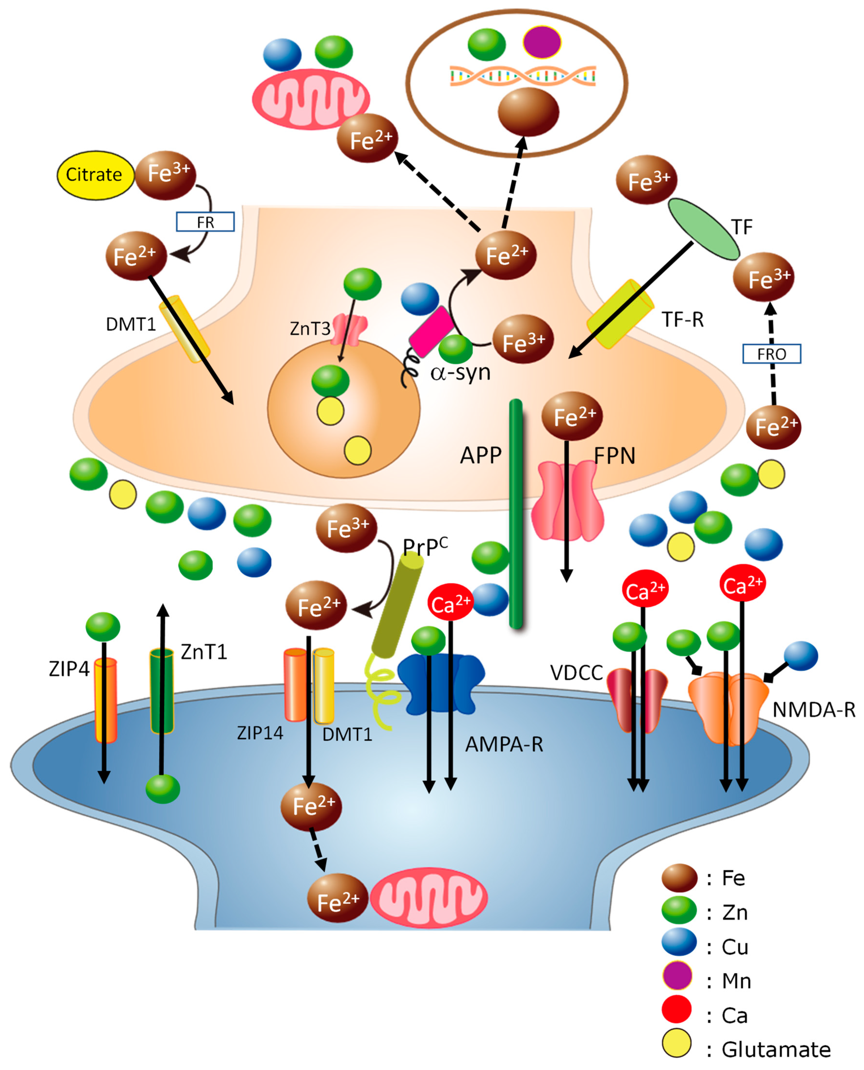
Absorbed Fe2+ is oxidized and stored in ferritin, an iron-storage protein, or is exported to extracellular space via ferroportin (FPN), the Fe2+ exporter protein. Hepcidin, a peptide hormone, controls FPN and contributes to Fe metabolism [21]. The ferroxidases, such as ferritin or ceruloplasmin, convert Fe2+ ions to Fe3+, and the Fe3+ is transported in the blood flow by binding to TF, an iron-binding protein that binds two Fe3+ ions. TF-bound Fe crosses the BBB binding with TF receptors and enters neurons or glial cells. Other Fe [non-transferrin-bound iron (NBTI)] largely forms Fe3+ ions and binds to small molecules such as citrates, after which it is reduced to Fe2+ by ferrireductases, and enters neurons or glial cells via DMT1. DMT1 is also localized in endothelial cells of the BBB and functions in the translocation of Fe2+ through the BBB [22]. Intracellular Fe2+ is transferred to neuronal enzymes in the mitochondria, endoplasmic reticulum (ER), or Golgi apparatus, which require bioactive Fe2+ as a cofactor. FPN exports excess Fe2+ to the extracellular space and regulates its intracellular levels because excess Fe2+ causes ROS and damages neurons. Therefore, the ratio between Fe2+ and Fe3+, as well as the level of Fe, is strictly regulated. The complicated biological fate of Fe is illustrated in Figure 1.
Fe regulates the expression of various Fe-related proteins, such as ferritin, TF, FPN, and DMT1 [23]. The mRNA of these proteins possesses an iron-responsive element (IRE) in the 3′-UTR or 5′-UTR. For example, the mRNA of ferritin has the IRE motif in its 5′-UTR. In Fe-deficient conditions, iron-binding proteins (IRPs; IRP1 and IRP2) bind to the IRE motif and inhibit ferritin expression, which eventually promotes the circulation of Fe in the blood. Under conditions with an excess of Fe, Fe binds to IRPs, promotes their dissociation from ferritin’s mRNA, and enhances the translation of ferritin, which finally decreases the concentration of free Fe. In contrast, the IRE motif at the 3′-UTR of transferrin’s mRNA binds to IRPs in Fe-deficient conditions, protects mRNAs from endonuclease cleavage, and promotes the translation of transferrin and the circulation of Fe. This IRE-IRP signaling pathway controls the level of Fe in the brain as well as other organs. Since the mRNAs encoding amyloidogenic proteins also possess the IRE motif, this IRE-IRP pathway is also implicated in the pathogenesis of neurodegenerative diseases (see Section 3.1).
Owing to the low absorption rate of Fe, Fe deficiency is a common and serious problem for human health, especially in developing countries where meat consumption is insufficient. The World Health Organization (WHO) estimated that approximately 4–5.5 billion people globally have Fe deficiency, while two billion people exhibit Fe-deficient anemia [24]. According to the National Health and Nutrition Survey Japan 2019, the recommended daily intake of Fe is 6.5 mg for women and 7.5 mg for men [25]. However, the estimated average Fe intake is 7 mg per day, and around 13.5% of women and 10.0% of men suffer from Fe deficiency with decreased hemoglobin content. Meanwhile, excess Fe as a result of genetic disorders or the overdose of Fe causes hemochromatosis with the accumulation of Fe [26].

2.2. Zinc

Zn is the second most abundantly present in the brain [27]. A high concentration of Zn is observed in the hippocampus, amygdala, cerebral cortex, thalamus, and olfactory cortex. Zn plays important roles in various physiological functions such as mitotic cell division, protein synthesis, immunological responses, and DNA and RNA synthesis as a cofactor of more than 300 enzymes or metalloproteins. Zn is also responsible for the expression of various genes since many transcription factors possess Zn-finger domains. Zn acts similarly to a signaling molecule to facilitate the transcription of the signaling cascades, similar to Ca. Therefore, Zn is essential for normal growth and development. Prasad et al. reported cases of Zn deficiency in humans for the first time in 1961; this condition is characterized by dwarfism with the retardation of physical and sexual development [28]. Zn deficiency in children is also associated with delayed mental and physical development, immune dysfunction, and learning disabilities, whereas Zn deficiency in adults produces learning, taste, and odor disorders [29,30].
In the brain, a substantial fraction (approximately 10% or more) of Zn is stored as a complex with glutamate in the presynaptic vesicles in excitatory glutamatergic neurons (gluzinergic neurons). Synaptic Zn is secreted into synaptic clefts with glutamate during neuronal excitation as free Zn ions (Zn2+) or is in a loosely bound form [27,31]. The secreted Zn binds to various receptors such as N-methyl-D-aspartate (NMDA)-type glutamate receptors, α-amino-3-hydroxy-5-methylisoxazole-4-propionic acid (AMPA)-type glutamate receptors, γ-aminobutyric acid (GABA) receptors, and glycine receptors, and regulate their functions. Thus, the removal of synaptic Zn by its chelator was reported to induce overexcitation in rat hippocampal neurons. Zn released from mossy fibers in the hippocampus reportedly mediates the spatiotemporal information of neuronal signaling and contributes to synaptic plasticity [32]. Synaptically released Zn is required for the induction of long-term potentiation, a form of synaptic information storage well known as a paradigm of the mechanism by which memory is formed [33].
The maintenance of Zn homeostasis is regulated by two factors: metallothioneins (MTs) and Zn transporters. There are three types of MTs: MT1, MT2, and MT3. MTs are ubiquitous proteins with 20 cysteine residues that can bind seven atoms of Zn, Cu, Cd, and other metals [34]. Among them, MT3 is primarily localized in the central nervous system, while MT1 and MT2 are ubiquitously expressed throughout the body. Meanwhile, there are two types of Zn transporters: ZnT transporters and Zrt-, Irt-like protein (ZIP) transporters that regulate Zn efflux and influx [35]. ZnT1, one of the ZnT transporters that facilitate the efflux of Zn2+, plays central role in Zn2+ efflux in the brain. ZnT3 regulates Zn2+ into synaptic vesicles in membranes of synaptic vesicles. ZIP transporters control Zn influx into cells or subcellular organelles. Genetic dysregulation of these transporters causes Zn deficiency and leads to several diseases, such as Ehlers–Danlos syndrome in humans.
Zn is abundant in food such as meat, oysters, eggs, and liver. Zn in food is absorbed in the gastrointestinal tract via ZIP4 and DMT1, after which it is transported in the blood by binding with albumin and α1-acid glycoprotein. Although Zn is also contained in vegetables, beans, and grains, its absorption rate from these sources is low because phytate in such plants binds to Zn and inhibits its absorption. The recommended daily intake of Zn is around 8 mg for women, 25 mg for pregnant women, and 11 mg for men [25]. The WHO estimated that 2.2 billion people are affected by Zn deficiency and that Zn deficiency is believed to make a major contribution to infant mortality [36]. In Japan, studies have reported that 37.9% of elderly people aged over 60 years and 16.9% of people aged 15–59 years exhibited low serum Zn levels [37].

2.3. Copper

Cu is the third most abundant element in the brain. Cu is a cofactor for numerous enzymes, including cytochrome C, Cu/Zn superoxide dismutase (Cu/Zn-SOD), lysyl oxidase, and tyrosinase. Therefore, Cu is involved in normal brain functions such as neurotransmitter synthesis, myelination, and neuroprotection [38]. Cu is also involved in Fe homeostasis since ceruloplasmin and other ferroxidases, termed ‘multi-copper ferroxidases,’ convert Fe2+ to Fe3+ [39]. Similar to Zn2+, a considerable amount of Cu is stored in synaptic vesicles. The synaptic Cu is released into synaptic clefts during neuronal firings [40]. The released Cu2+ modulates neuronal excitability by binding to NMDA-type glutamate receptors, AMPA-type glutamate receptors, and GABA receptors [41]. The concentrations of Cu and Zn in the synaptic clefts are considered to be higher compared to those in CSF, and Zn as well as Cu will play critical roles in the neurodegenerative processes (see Section 4). Meanwhile, excess free Cu is toxic because Cu exists as oxidized Cu2+ and reduced Cu+ and produces ROS.
Cu homeostasis is regulated by copper-transporting ATPase (ATP7A and ATP7B). A deficiency or excess of Cu caused by genetic disorders associated with these transporters can cause neurodegenerative diseases such as Menkes disease and Wilson’s disease [42]. ATP7A is a Cu efflux protein, delivering Cu to Cu-containing enzymes in the trans-Golgi network. ATP7A is mainly localized in the Golgi apparatus and reportedly plays an essential role in axonal targeting and synaptogenesis [43]. ATP7B has functional similarity to ATP7A and exports Cu into bile for excretion. Excess intracellular Cu can bind to MTs, as well as Zn. Cu transporter 1 (Ctr1), which transports Cu+, and DMT1, also contribute to Cu homeostasis.
Cu is abundant in meat, cereals, vegetables, and seafood such as squid and octopus. Orally ingested Cu is absorbed via DMT1 in the small intestine, mainly in the duodenum, transported to the liver, and distributed by ceruloplasmin in the blood. The daily intake of Cu is estimated to be 0.7 mg. Thus, Cu deficiency is rare in physiological conditions. Meanwhile, excess Cu in drinking water is associated with non-Indian childhood cirrhosis, a form of early childhood liver cirrhosis [44].

2.4. Manganese

Mn acts as a cofactor of various enzymes, such as hydrolases, lyases, and ligases, and is involved in metabolic processes, such as protein glycosylation and lipid synthesis. Mn also participates as a cofactor of superoxide dismutase (Mn-SOD), which detoxifies free radicals [45] and influences neurotransmitter synthesis as a component of glutamine synthetase.
Mn is contained in seaweed, nuts, and other food [46]. Orally administered Mn becomes Mn2+ in the presence of gastric acid, which is then oxidized to Mn3+. This Mn3+ is transported by binding to TF or citrate and can enter the brain via TF receptors, citrate transporters, and other transporters such as DMT1. Some Zn transporters, such as ZnT10, ZIP8, and ZIP14, control the absorption of Mn [13]. The recommended intake of Mn is 4 mg for men and 3.5 mg for women in Japan [23].
Mn deficiency is rare in physiological conditions because Mn is present at adequate levels in food. Mn is also used in fuel additives or fungicides and enters the body via the inhalation of fumes, dust, or air particles [47]. Excess Mn from contamination or occupational exposure is toxic and causes neurotoxicity called “manganism,” which involves symptoms similar to those of Parkinson’s disease (PD) [48]. Mn also reportedly causes oxidative stress and inflammatory responses. Since Mn has similar chemical characteristics to Fe, it influences Fe homeostasis and expressions of Fe-related proteins by affecting the IRE-IRP pathways [49].

2.5. Aluminum

Al is the third most abundant element in the earth’s crust and is widely distributed throughout the environment. Al has been widely used as compounds such as clay, glass, and alum for centuries. However, Al as a silvery metal was first isolated in 1827, and its industrial use began only in 1886.
Despite being widespread in the environment, Al is not essential for life. However, Al is known to inhibit more than 300 biologically significant reactions [50,51]. Al tightly binds to phosphate and inhibits kinases or phosphatases. The ligand-exchange rate for Mg2+ is 105 times faster than that for Al3+, whereas the exchange rate for Al3+ is 108 times slower than that for Ca2+. Therefore, Al3+ cannot participate in Ca2+- or Mg2+-related enzymatic reactions but inhibits numerous Ca2+- or Mg2+-related enzymes. Al3+ has similar characteristics to Fe3+, and thus Al3+ can bind to Fe-binding proteins or chelators. Although Al3+ is not redox-active, Al is known to enhance Fe-induced lipid peroxidation.
Al is naturally contained in several types of food, such as seaweed and tea leaves. Al can also enter the human body through food additives (such as alum and baking powder), contamination from cooking utensils, and drinking water because Al coagulants are widely used in water purification. Al has been used as a pharmacological agent, such as antacids, antiperspirants, and vaccines. Our daily intake of Al is estimated to be approximately 20 mg. However, its rate of absorption from the gastrointestinal tract is low and can be altered by its particular chemical form and substances coexisting in food [52]. For example, Al in tea infusion is poorly absorbed since polyphenol inhibits its absorption. Analysis using a non-radioactive isotope of Al (26Al) revealed that orally administered Al could be absorbed and accumulate in bones, and a considerable amount of Al can enter the brain [53]. Moreover, its biological half-life is quite long, and the level of Al in the brain is not changed after one month of administration [54].
Owing to these adverse characteristics, Al is a neurotoxin. Al causes dialysis encephalopathy in hemodialysis patients since Al is used as a component of hemodialysis solutions or pharmacological compounds. Al is also eluted from the soil by acid rain and is toxic to plants and fish. In addition, numerous studies have suggested its causative links between various neurodegenerative disorders, including AD, amyotrophic lateral sclerosis in the Kii Peninsula and Guam, Gulf War syndrome, and autism spectrum disorder [51]. In particular, the link between Al and AD has been debated for decades but remains controversial.

3. Roles of Trace Elements in the Pathogenesis of Neurodegenerative Diseases

3.1. Alzheimer’s Disease

Growing evidence suggests the involvement of trace elements in the pathogenesis of neurodegenerative diseases such as AD, prion diseases, LBD, and VD.
AD is responsible for approximately half of all cases of senile-type dementia. The hallmarks of senile-type dementia are profound memory loss and an inability to form new memories. The age-dependent increase in its prevalence is a serious problem in the aging world. The number of AD patients in Japan was estimated to be more than 7 million in 2020, a number that is increasing annually [55]. The hallmarks of AD are the loss of neurons and synapses in the hippocampus or cerebral cortex, the extracellular accumulation of senile plaques, and the intraneuronal accumulation of neurofibrillary tangles (NFTs) [56]. The major constituent of senile plaques is AßP, while that of NFTs is phosphorylated tau protein.
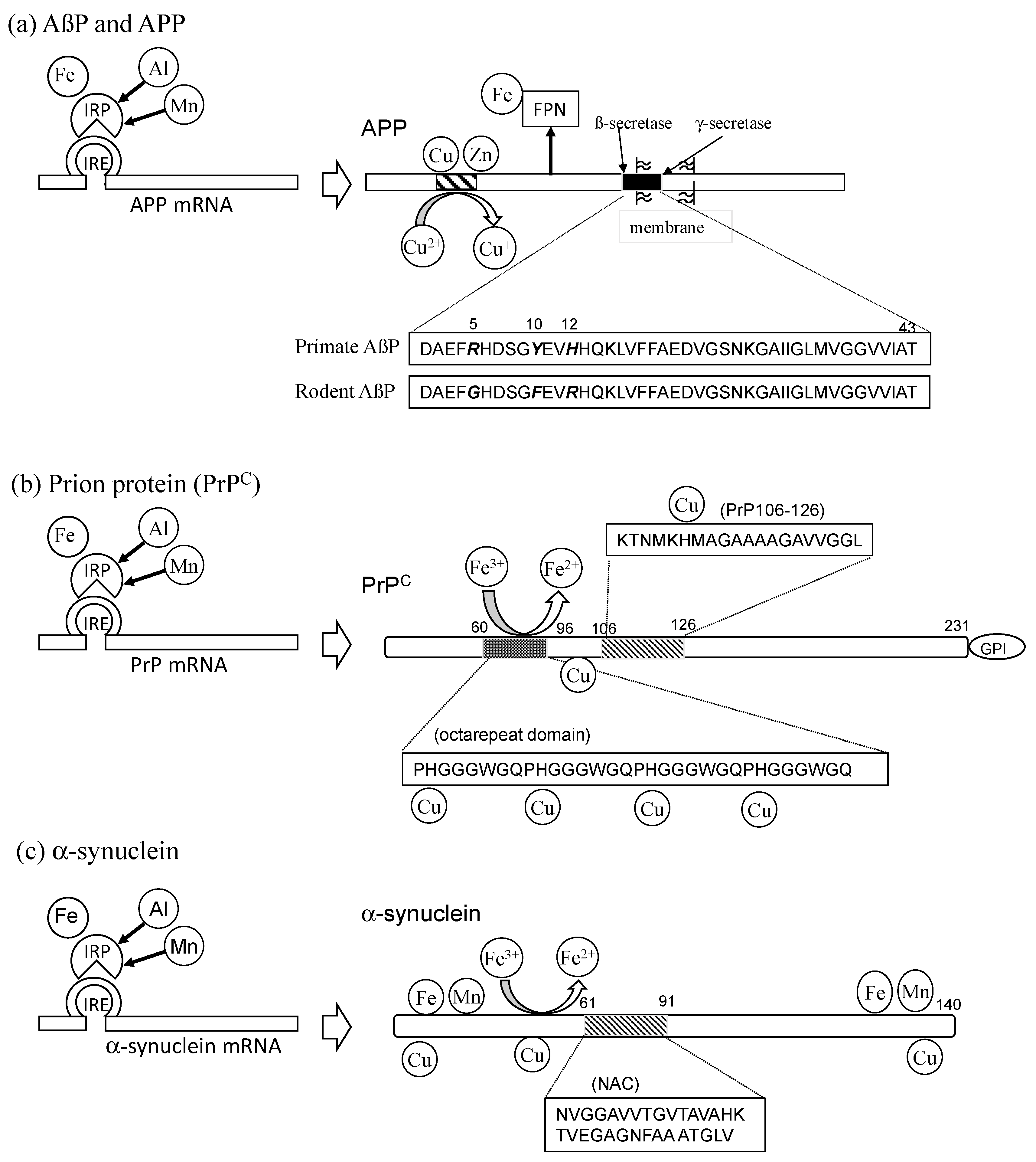
AßP is a small 39–43-amino-acid peptide, which is derived from a large precursor protein (amyloid precursor protein: APP) by the enzymatic cleavage of ß-secretase in its N-terminal and γ-secretase in its C-terminal (Figure 2a). AßP(1–40), comprising the first 40 amino acids of AßP, was found to cause neurotoxicity in vitro as well as in vivo in 1991. After these findings were revealed, the conformational change of AßP was demonstrated to be a crucial determinant of its neurotoxicity [57]. AßP possesses a tendency to form insoluble aggregates (oligomers) with ß-pleated sheet structures in solution. Freshly prepared AßP(1–40), forming an α-helix or random-coiled structure in solution, is non-toxic. However, after an aging process (incubation at 37 °C for several days), the aged AßP(1–40) self-aggregates to form oligomers with ß-sheet structures and was found to cause the death of cultured rat cortical neurons. The proportion of ß-sheet content of AßP was shown to be correlated with its neurotoxicity. Mutations in APP have been revealed to be a risk factor for familiar-type AD. Presenilins (presenilin-1 and presenilin-2), risk factors of early-onset familial AD, are γ-secretases that influence the production of various truncated AßPs. Truncated AßPs, such as AßP(1–42), were revealed to be significant in oligomerization since they act as “seeds” and enhance the oligomerization of AßP(1–40). These biochemical and genetic findings support the “amyloid cascade hypothesis,” which proposes that the conformational changes of oligomerized AßP and the subsequent neurodegeneration are crucial for the pathogenesis of AD [58].
The levels of AßP in the CSF of the elderly without dementia are reportedly similar to those of AD patients, and AßP also exists in the CSF of young individuals [59]. Therefore, acceleratory or inhibitory factors of the oligomerization may be crucial in the pathogenesis of AD [57]. Substances such as rifampicin, transthyretin, curcumin, aspirin, and peptides (such as the ß-sheet breaker peptide) were reported to inhibit oligomerization and are now candidates for drugs to treat AD. The oligomerization of AßP is influenced by its concentration, the composition and pH of solvents, and temperature. The oxidation, mutation, and racemization of AßP accelerate the oligomerization. Among these acceleratory factors, trace elements are of particular interest. Metal ions bind to several amino acid residues, such as histidine (His), arginine (Arg), tyrosine (Tyr), glutamate (Glu), and aspartic acid (Asp) or phosphorylated amino acids. These metals cause conformational changes in various proteins by cross-link amino acid residues. Interestingly, the deposition of senile plaques rarely occurs in rodent brains compared with the level in primate brains, despite primate AßP and rodent AßP differing by only three amino acids (Arg5, Tyr10, and His13) [60]. These three amino acids bind to metal ions, and His residues are particularly essential for the binding of Zn to AßP. Therefore, metals may contribute significantly to the oligomerization of AßP and the deposition of senile plaques. Our and other numerous studies revealed that metal ions, including Al3+, Fe3+, Zn2+, Cu2+, and Mn2+, bind to AßP and enhance its oligomerization [61,62,63,64,65,66]. Moreover, the deposition of Al, Zn, Fe, and Cu was observed in senile plaques [67,68].
APP, the precursor of AßP, is also a metal-binding protein and possesses two Zn and/or Cu binding domains [69]. Both Zn and Cu are implicated in the dimerization of APP, its expression, trafficking, processing, and in the production of AßP [70,71]. APP has the ability to convert oxidized Cu2+ to reduced Cu+ [72]. Moreover, APP reportedly regulates Fe2+ efflux from neurons by stabilizing FPN [73]. Other AD-related proteins, presenilins, are involved in Ca homeostasis in the ER [74] and are implicated in the cellular uptake of Zn and Cu [75]. Meanwhile, Zn and Cu influence the trafficking of presenilins and their γ-secretase activity [76].
Fe regulates the expression of APP. The mRNA of APP contains an IRE domain in its 5′-UTR, similar to ferritin [77]. Therefore, the translation of APP is promoted in the case of excess Fe, and thereafter the export of Fe2+ by FPN can be upregulated to maintain intracellular Fe levels. Indeed, high-Fe diets were reported to cause the cleavage of APP and the accumulation of AßP in the brain, which was inhibited by intranasal exposure to deferoxamine, a chelator of Fe and Al [78].
Numerous other studies have suggested the involvement of metal homeostasis in AD. The genotype of TF C2 is a risk factor for AD [79]. Fe supplementation reportedly has beneficial effects on cognitive functions in AD patients [80]. Lu et al. demonstrated that the expression of DMT1 increases in an age-dependent manner, along with increases in AßP and phosphorylated tau [81]. The brains of AD patients exhibited a decreased level of FPN [82].
We summarize the links between trace elements and AßP/APP from the perspective of metal interactions in Figure 2a. Figure 2 also presents a comparison of other amyloidogenic proteins (PrPC and α-synuclein).

3.2. Prion Diseases

Prion diseases are zoonotic diseases, including bovine spongiform encephalopathy (BSE) in cattle, scrapie in sheep, and chronic wasting disease in elk and deer. In humans, Creutzfeldt–Jakob disease (CJD), Gerstmann–Sträussler–Scheinker syndrome, and kuru are included [83]. They are characterized by the spongiform degeneration of glial cells and neurons and synaptic degeneration. The accumulation of abnormal scrapie-like prion protein (PrPSc) is also observed in the brain. It is widely accepted that the conformational conversion of normal cellular prion protein (PrPC) to pathogenic PrPSc is central to the pathogenesis of prion diseases. PrPC is a 30–35 kDa glycoprotein anchored at the plasma membrane with a glycosylphosphatidylinositol (GPI) domain. It is widely distributed throughout the body, including the liver, heart, and brain. PrPC and PrPSc possess the same chemical characteristics with the same primary sequence, except that PrPSc is protease-resistant, insoluble, and has a high β-sheet content. Prion diseases are also called transmissible spongiform encephalopathy from their infective characteristics. PrPSc has the ability to induce neighboring normal PrPC to misfold and aggregate PrPSc. When PrPSc in food or iatrogenic contamination enters the body via ingestion, the protease-resistant PrPSc invades the brain and converts PrPC to PrPSc.
Metals are involved in both of the neurodegenerative processes in prion diseases: “loss of normal protective functions of PrPC” and “gain of toxic functions of PrPSc.” According to the “loss-of-function neurodegenerative pathway,” several neurological dysfunctions, including the death of Purkinje neurons in the cerebellum, synaptic dysfunction, demyelination, depressive-like behavior, and memory loss in knockout mice of PrPC. In PrP-knockout mice, the decreased Cu levels and the reduced activity of Cu-dependent enzymes were demonstrated [84]. PrPC possesses a highly conserved octarepeat domain composed of multiple tandem copies of an eight-residue sequence (PHGGGWGQ) (residues 66–90) at the N-terminal (Figure 2b). Jackson et al. reported that four Cu atoms bind to the His residues in the octarepeat domain of PrPC, as well as two other Cu atoms to other His residues (His96 and His111) [85]. They also demonstrated that other metals, including Zn2+, Mn2+, and Ni2+, bind to these binding sites with lower affinity than Cu2+. Additionally, PrPC regulates Cu influx into neurons, and Cu-bound PrPC exerts neuroprotective effects by exhibiting Cu/Zn SOD-like activity. Furthermore, PrPC controls the functions of glutamate receptors (MPA-type and/or NMDA-type) in a Cu-dependent manner [86].
According to the “gain of toxic function neurodegenerative pathway,” a fragment peptide of PrP (PrP106–126) has been used as a model peptide of PrPSc because it causes the death of cultured hippocampal neurons, the ability to form fibrils with β-sheet structures, and the ability to bind Cu and Zn [87]. We have demonstrated that Zn and Cu attenuated the neurotoxicity of PrP106–126 and the formation of ß-sheet structures [88].
Cu also affects the cellular trafficking of PrPC and its gene expression. Cu also influences PrPC-induced neurite outgrowth [89]. It is widely accepted that Cu contributes to the conformational conversion of PrPC to PrPSc and the transmission of prion diseases [90]. It is known that the α-helix-rich C-terminal domain of PrPC changes to the β-sheet in the conformational changes of PrPSc. When Cu binds to the octarepeat region in the flexible N-terminal domain of PrPC, Cu crosslinks with His residues in the C-terminal domain and finally causes the conformational change of PrPC to PrPSc. Indeed, the mutation of ATP7A delays the onset of prion disease by disrupting Cu homeostasis in mice [91].
Zn also contributes to the conformational changes of PrPC to PrPSc. Zn2+ binds to the octarepeat domain as well as Cu2+ and affects the conformational changes of the C-terminal domain [92]. Bioinformatic analysis revealed that genes of PrPC have evolutionary similarities to ZIP-type Zn transporters such as ZIP5, ZIP6, and ZIP10 [93]. Watt et al. reported that PrPC acts as a Zn2+ sensor in the binding of synapses with the AMPA-type glutamate receptors and controls the cellular uptake of Zn2+ [94].
PrPC is also involved in Fe homeostasis. Notably, PrPC was revealed to be a ferrireductase that converts Fe3+ to Fe2+ [95]. The reduced Fe2+ is subsequently transported to intracellular space via the complex of DMT1 and ZIP14 and is provided to enzymes requiring bioactive Fe2+ [96]. The octarepeat domain was essential for this ferrireductase activity. Moreover, PrP-knockout mice exhibited altered Fe metabolism and reduced Fe levels in the brain [97]. Meanwhile, the Fe level controls the expression of PrPC because its mRNA possesses an IRE motif as well as the APP gene [98].
Mn has been suggested to facilitate the pathogenesis of prion diseases [99]. Mn reportedly increases the infectivity of PrPSc in mice [100]. The risk of chronic wasting disease in deer was shown to be associated with Mg deficiency and increased Mn concentrations [101]. Scrapie-infected sheep exhibited elevated Mn levels in the blood and brain [102].
Trace elements are also involved in the pathogenesis of human prion diseases. CJD patients exhibited elevated levels of Mn in blood and brains [103]. The epidemiological studies suggested the relationship between the high incidence of CJD in Slovakia and the imbalance of the Mn/Cu ratio in food was suggested [104,105].

3.3. Lewy Body Diseases

The pathological hallmarks of LBD, which include Parkinson’s disease (PD), dementia with Lewy bodies (DLB), and multiple system atrophy, etc., are the abnormal intracellular aggregates termed “Lewy bodies.” The major component of Lewy bodies is α-synuclein [106]. Therefore, these diseases are also called “synucleinopathies.”
α-Synuclein is abundant in the presynaptic terminals, particularly in the membranes of synaptic vesicles. It plays central roles in endocytosis and exocytosis and contributes to synaptic plasticity. The fragment peptide α-synuclein termed the “non-amyloid component” (NAC), co-accumulates with AßP in senile plaques of AD [107]. The oligomerization of α-synuclein contributes to neurotoxicity and the pathogenesis of LBD.
PD patients exhibit damage in Fe-rich regions (such as the substantia nigra). Thus, the roles of trace elements in the pathogenesis of LBD have been suggested. Uverski et al. revealed that Al and other metal ions enhance the oligomerization of α-synuclein [108]. α-Synuclein reportedly binds Cu2+, Zn2+, Mn2+, and other metal ions in its N-terminal and C-terminal domains [109]. Cu affects the binding of α-synuclein to lipid membranes and its functions [110]. The binding of Cu to α-synuclein is supposed to be critical for the pathogenesis because the His50 residue, a mutation that is observed in familiar-type PD, is essential for Cu binding [111].
Interestingly, α-synuclein possesses ferrireductase activity that converts Fe3+ to Fe2+ as well as PrPC [112]. This indicates that α-synuclein controls energy production and neurotransmitter synthesis in the presynaptic domain by providing bioavailable Fe2+ to enzymes requiring Fe2+ in the mitochondria or ER. This ferrireductase activity is related to Cu binding to α-synuclein. Indeed, the levels of Fe3+ were altered in PD [113,114], and the ferrireductase activity was reduced in PD patients [115].
The expression of α-synuclein is controlled by Fe levels because its mRNA possesses an IRE motif in the 5′-UTR similar to APP and PrPC [116]. As noted, the neurotoxicity of Mn shows a similar profile to PD. Mn reportedly induces the overexpression of α-synuclein by affecting the IRE-IRP pathway [117].

3.4. Vascular-Type Senile Dementia

VD is a type of senile dementia that accounts for approximately one-third of the cases. VD is generally caused by a series of small strokes or ischemia [118]. Within three months of the initial stroke, around 30% of the patients exhibit symptoms of dementia. During the interruption of blood flow in transient global ischemia or stroke, the resulting oxygen–glucose deprivation causes long-lasting membrane depolarization of neurons. Hereafter, the excess glutamate is released into the synaptic clefts, which induces overstimulation of glutamate receptors. Finally, the resulting Ca2+ influx triggers delayed neuronal death in the hippocampus or cerebral cortex, which are critical for memory formation.
Under ischemic conditions, excess Zn is secreted into synaptic clefts along with glutamate. The Zn translocation, namely, the increase in intracellular Zn2+ levels in postsynaptic neurons, reportedly occurs in degenerated hippocampal neurons after ischemia [119]. This Zn translocation is controlled by VDCC, NMDA-type glutamate receptor, and Ca2+-permeable AMPA-type glutamate receptor [8]. The administration of Ca-EDTA, a membrane-impermeable chelator of Zn2+, inhibits the translocation of Zn after transient global ischemia, protects hippocampal neurons, and reduces infarct volume [120]. These results strongly suggest that Zn is a key player in delayed neuronal death after transient global ischemia and, finally, in the pathogenesis of VD [121]. We have investigated the molecular mechanism of Zn-induced neurotoxicity and found associations with the ER stress pathway, mitochondrial energy failure, the disruption of Ca homeostasis, the MAP kinase pathway (stress-activated protein kinases/c-Jun amino-terminal kinase (SAPK/JNK) pathway), and the production of ROS [4]. We also found that sublethal concentrations of Cu2+ significantly exacerbated Zn-induced neurotoxicity [122] and demonstrated that the addition of Cu2+ produced ROS, which is known to induce the ER stress pathway and the SAPK/JNK pathway. Several antioxidants were found to attenuate Cu-enhanced Zn-induced neurotoxicity. Therefore, Cu may collaborate with Zn and play key roles in Zn-induced neurotoxicity by producing ROS, finally leading to the pathogenesis of VD [123].

4. Hypothesis: Cross-Talk Occurs between Trace Elements and Amyloidogenic Proteins at Synapses

As described in the previous sections, amyloidogenic proteins share several common characteristics, including the formation of ß-pleated sheet structures, the exhibition of neurotoxicity, and the ability to bind metals. Furthermore, all of these amyloidogenic proteins or related proteins (APP and PSs) reportedly co-localize at synapses. APP primarily exists in the presynaptic membrane [124], while PrPC is located in the postsynaptic membrane, binding to several receptors [125]. α-Synuclein mainly exists in presynaptic cytosol or membranes, particularly in the presynaptic vesicles [106]. PSs are mainly localized in the ER membranes of pre and postsynaptic domains [126]. Considering that synaptic degeneration (synaptotoxicity) occurs in the early stages of these neurodegenerative diseases [127], the roles of amyloidogenic proteins at synapses are of interest.
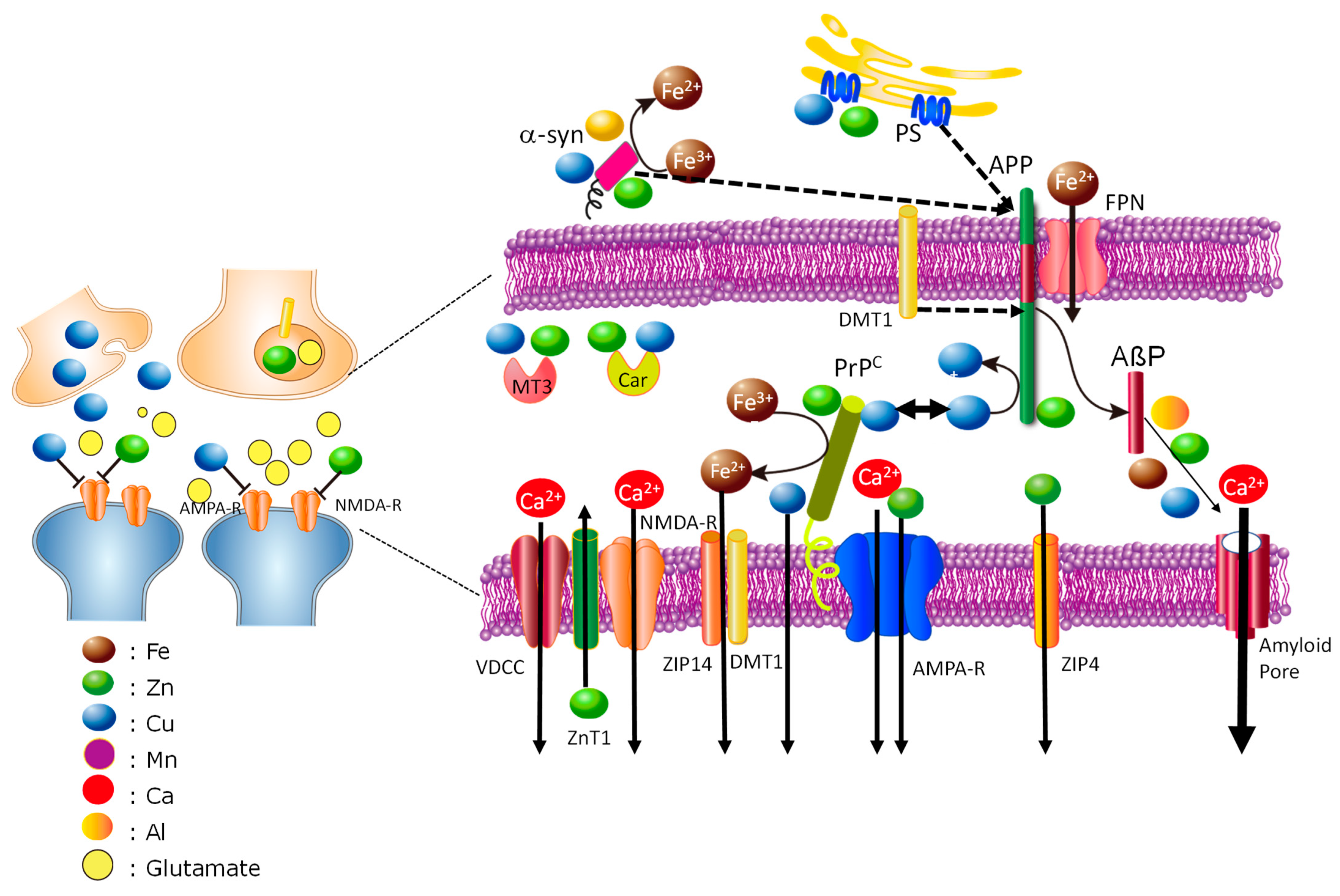
Synapses are metal-rich regions of the brain. The levels of Cu and Zn in the CSF of healthy individuals are less than 1 µM [128]. However, Cu and Zn may be concentrated in the synaptic clefts. The synaptic cleft is a small compartment considered to be a cylinder with a 120 nm radius and height of 20 nm. The total volume of synaptic clefts is estimated to be approximately 1% of the extracellular space of the brain [129]. Vogt et al. estimated that the Zn concentration in synapses is 1–100 μM [130]. Meanwhile, the concentration of Cu released into the synaptic cleft is estimated to be 3–100 μM [131,132]. Considering that synapses are energy-demanding regions in the brain [133] and that Fe is required in energy metabolism in mitochondria, Fe may plausibly be abundant in synapses. Considering these results together, we hypothesize here that amyloidogenic proteins and metals may play concerted roles in maintaining metal homeostasis in the synapse (Figure 3 and Figure 4).
Figure 3 presents overall aspects of the entry of trace elements into neurons and their functions in pre and postsynapses. Orally administered Fe is absorbed via the gastrointestinal tract and transported in the blood by binding to TF or citrate. TF-bound Fe passes the BBB and enters neurons or glial cells via the TF receptor. Intracellular Fe3+ is converted to Fe2+ by α-synuclein in the presynaptic domain and by PrPC in the postsynaptic domain; then, the bioavailable Fe2+ is provided to enzymes that require it in mitochondria or other suborganelles and functions in neurotransmitter synthesis, energy production, and myelination. Fe3+ in NTBI enters neurons or glial cells via DMT1 after being reduced to Fe2+. Excess Fe2+ is exported to extracellular space via FPN and then oxidized to Fe3+ by ferroxidase to enter into circulation. APP stabilizes FPN and plays a key role in Fe export. FPN is reportedly localized in membranes of synaptic vesicles as well as α-synuclein [134] and modulates Fe homeostasis. Since synapses are energy-demanding areas and therefore vulnerable to oxidative stress, the Fe2+ levels there should be strictly regulated by these concerted actions of APP, PrPC, and α-synuclein. Meanwhile, Fe controls the expression of APP, PrPC, and α-synuclein, given that their mRNA possesses the IRE motif in the 5′-UTR. Moreover, DMT1 is involved in the processing of APP and the production of AßP [135].
Both Zn and Cu are absorbed via the gastrointestinal tract, transported in the blood flow, pass through the BBB, and enter neurons or glial cells. Considerable amounts of Zn and Cu accumulate in synaptic vesicles, both of which are released to the synaptic clefts in activity-dependent manner, bind to NMDA-type glutamate receptors or other receptors, and control neuronal excitability under physiological conditions. Synaptic Zn and Cu may play inhibitory roles in translating the information about neuronal firings to neighboring synapses and may contribute to synaptic plasticity. Secreted Zn is translocated by VDCC, NMDA-type glutamate channels, and Ca2+-permeable AMPA-type glutamate channels, as well as Ca2+. Moreover, PrPC acts as a Zn sensor in the synaptic clefts and regulates Zn influx to postsynaptic neurons with AMPA-type glutamate receptors as a ZIP analog. PrPC also plays a key role in the uptake of Cu and Fe. Two major Zn transporters—ZnT1 and ZIP4—are also present in the postsynaptic domain and act to maintain the level of Zn in the synaptic clefts [136,137].
More details on the interactions in the synaptic clefts are shown in Figure 4. As noted, the levels of Zn, Cu, and Fe in the synaptic clefts are strictly regulated by the interactions between amyloidogenic proteins.
The height of the synaptic clefts, namely, the distance from presynaptic membranes to postsynaptic membranes, is as narrow as 20 nm, and therefore these amyloidogenic proteins possibly interact with each other. Indeed, PrPC acts as a toxic receptor of AßP and contributes to its toxicity [138]. α-Synuclein increases AßP secretion by promoting the processing of APP by β/γ-secretase [139]. Presenilins also regulate the secretion of APP by binding to Zn and/or Cu [76]. As noted, ZnT1 and ZIP4 control the level of Zn in the synaptic clefts. ZnT1 also regulates neuronal excitability by binding to NMDA-type glutamate receptors [136] and controls Ca homeostasis by binding to Ca2+ channels [140].
Other factors, such as MT3 and carnosine (ß-alanyl histidine), also control the levels of Zn and Cu in the synapses. MT3, brain-specific metallothionein, is synthesized in neurons or glial cells [141]. Interestingly, the brains of AD patients exhibited a decreased level of MT3. Carnosine is an endogenous dipeptide with various beneficial properties, such as antioxidant, anti-glycation, anti-stress, and anti-cross-link. Carnosine also has the ability to scavenge hydroxyl radicals, maintain the pH balance, and chelate metals, including Zn2+ and Cu2+ ions [142]. Carnosine is reportedly synthesized in oligodendrocytes and secreted into the synaptic cleft [143].
These concerted metal–protein, and metal–metal interactions at synapses are highly complex and vulnerable to perturbation. Therefore, the disorder of these subtle interactions may cause the dyshomeostasis of neurometals, ROS production, and synaptic degeneration, and finally induce neurodegeneration leading to the pathogenesis of the neurodegenerative disease.
For example, high-Fe diets reportedly cause the cleavage of APP and the accumulation of AßP in the brain [78]. The abnormality of APP induced by its mutations may influence FPN stability and the export of Fe2+. A decreased level of FPN, which is reported in AD brains [82], causes the accumulation of Fe2+ and the production of ROS. The invasion of Al from food enters the neurons via TF receptors as well as Fe and influences various Fe-related pathways, such as the IRE-IRP pathway, and thus influences the expression of APP [144]. Al reportedly enhances the expression of APP and the production of AßP [145]. Dietary Cu and its oxidative stress have been linked with AD pathogenesis [146]. Excess Mn from environmental exposure influences the IRE-IRP pathway and causes the accumulation of Fe and the overexpression of α-synuclein [49,117].
Contaminating PrPSc from food or an iatrogenic device, which is resistant to digestion by proteases, passes into the brain, causes the conversion of normal PrPC to PrPSc, leads to the loss of neuroprotective functions of PrPC, and triggers neurotoxic pathways via the accumulation of metals. Mutations of α-synuclein influence its binding to Cu, which affects the membrane binding and ferrireductase activity, causes changes in the Fe2+/Fe3+ ratio, and finally leads to ROS production.
The Cu and Zn levels in the synapses are strictly controlled by several regulatory factors, and secreted Zn and Cu rapidly undergo re-uptake under physiological conditions. However, under pathological conditions such as transient global ischemia, sustained excitation of neurons occurs for prolonged periods in broad areas of the brain, and excess Zn and Cu are simultaneously secreted into the same synaptic clefts for a long period of time. We have demonstrated that a sublethal concentration of Cu (5 μM) significantly exaggerated 30 μM Zn-induced neurotoxicity (molar ratio of Zn:Cu = 6:1) [123]. Therefore, it is possible that the coexistence of Cu triggers the production of ROS and cooperatively enhances Zn-induced neurotoxicity, finally causing vascular-type dementia. Cu has been found to be a risk factor for stroke in several epidemiological studies [147,148].
Trace elements possibly affect the gain of toxic functions of amyloidogenic proteins. Researchers, including ourselves, have demonstrated that AßP forms pore-like structures on the membrane and causes Ca dyshomeostasis, which may be associated with neurotoxicity [149,150,151,152]. PrP106–126 also forms cation-permeable pores in a Cu-sensitive manner as well as AβP [153]. α-Synuclein reportedly forms annular pore-like structures similar to AβP(1–40) [154]. Trace elements can influence neurotoxicity by accelerating the oligomerization and the formation of amyloid pores. However, the roles of metal-induced oligomerization are still controversial and under investigation. The morphologies of AβP oligomers with Al, Cu, Fe, and Zn are quite different [155]. The effects of metal-induced oligomers on the neurotoxicity of AβP are not identical [156,157].
On the basis of this working hypothesis, a metal-based therapeutic pathway may become a good candidate for the treatment/prevention of these neurodegenerative diseases. Clioquinol, a chelator of Zn and Cu, and its analog PBT-2 have been examined for possible treatment of AD [158], although the results remain controversial [159]. Exley et al. demonstrated the efficacy of silicate, which chelates Al, as a possible treatment of AD [160]. Deferoxamine (DFO), a chelator of Fe and Al, exerts beneficial effects on maintaining the daily living skills of AD patients [161,162]. Recent approaches using intranasal DFO treatment attenuated synapse loss in the brains of AD model mice and inhibited Fe-induced APP cleavage [78,163]. DFO and other Fe chelators are focused on possible treatments for PD [164,165]. Moreover, clioquinol reportedly recovers the memory impairment in scrapie [166], and a Cu2+-specific chelator (D-(-)-penicillamine) reportedly inhibits the pathogenesis of prion diseases in vivo [167]. We focused on carnosine as a possible treatment for VD based on its neuroprotective effect against Zn-induced neurotoxicity [168] and its various beneficial characteristics [142]. Carnosine also reportedly inhibits the oligomerization of AβP and attenuates neurodegeneration in AD model mice [169], and protects against ischemia-induced neuronal death in vivo [170]. Carnosine also attenuates PrP106–126-induced neurotoxicity [88]. Based on these beneficial characteristics of carnosine as a neuroprotector, we have granted a patent for carnosine as a possible target for drug treatment of vascular-type senile dementia [171].

5. Conclusions

We have reviewed the enigmatic roles of trace elements in the pathogenesis of various neurodegenerative diseases based on common characteristics of amyloidogenic proteins and the cross-talk between metal ions at synapses. Our hypothesis about the common aspects of neurodegenerative processes may shed light on their pathogenesis. Based on the idea about the link between trace elements and the pathogenesis of neurodegenerative disease will lead to the development of drugs for these neurodegenerative diseases.
However, this hypothesis is still not completed. In particular, the levels of Zn, Cu, and Fe in the synapses are controversial and under investigation. The functions of amyloidogenic proteins and the localizations of metal transporters remain unsolved. However, since we published the prototype of this hypothesis in 2017 [172], great progress has been demonstrated in this research field, particularly in the functions of Fe and in localizations of metal transporters. We hope that future studies about the metals in synapses will evolve the hypothesis and will aid in the treatments for these neurodegenerative diseases.

Author Contributions

Wrote or contributed to the writing of the manuscript: M.K. and M.K.-N.; Participated in research design: M.K., K.-i.T. and M.K.-N.; Conducted experiments: K.-i.T. and M.K.-N. All authors have read and agreed to the published version of the manuscript.

Funding

This work was partially supported by Grants-in-Aid for Scientific Research from the Ministry of Education, Culture, Sports, Science, and Technology of Japan (JSPS Kakenhi Grant numbers JP26460177 and JP21H01786).

Acknowledgments

We thank Mikako Shimoda at Musashino University for their technical assistance. We searched references mainly using the database PUBMED (https://pubmed.ncbi.nlm.nih.gov/) accessed 6 April 2023.

Conflicts of Interest

All of the authors declare that there is no conflict of interest.

References

  1. Becker, J.S.S.; Matusch, A.; Palm, C.; Salber, D.; Morton, K.; Becker, S. Bioimaging of metals in brain tissue by laser ablation inductively coupled plasma mass spectrometry (LA-ICP-MS) and metallomics. Metallomics 2010, 2, 104–111. [Google Scholar] [CrossRef]
  2. Cobley, J.N.; Fiorello, M.L.; Bailey, D.M. 13 reasons why the brain is susceptible to oxidative stress. Redox Biol. 2018, 15, 490–503. [Google Scholar] [CrossRef]
  3. Dirandeh, E.; Palizgir, A.; Kassiri, N. An overview of the relationship between occupational manganese exposure and Parkinsonism. Cureus 2022, 14, e32161. [Google Scholar] [CrossRef]
  4. Kawahara, M.; Tanaka, K.-I.; Kato-Negishi, M. Copper as a collaborative partner of zinc-induced neurotoxicity in the pathogenesis of vascular dementia. Int. J. Mol. Sci. 2021, 22, 7242. [Google Scholar] [CrossRef] [PubMed]
  5. Adlard, P.A.; Bush, A.I. Metals and Alzheimer’s disease: How far have we come in the clinic? J. Alzheimers Dis. 2018, 62, 1369–1379. [Google Scholar] [CrossRef]
  6. Brown, D.R. Metalloproteins and neuronal death. Metallomics 2010, 2, 186–194. [Google Scholar] [CrossRef]
  7. Jiang, H.; Song, N.; Jiao, Q.; Shi, L.; Du, X. Iron pathophysiology in Parkinson diseases. Adv. Exp. Med. Biol. 2019, 1173, 45–66. [Google Scholar] [PubMed]
  8. Weiss, J.H.; Sensi, S.L.; Koh, J.Y. Zn(2+): A novel ionic mediator of neural injury in brain disease. Trends Pharmacol. Sci. 2000, 21, 395–401. [Google Scholar] [CrossRef] [PubMed]
  9. Carrell, R.W.; Lomas, D.A. Conformational disease. Lancet 1997, 350, 134–138. [Google Scholar] [CrossRef]
  10. Kawahara, M.; Kato-Negishi, M.; Tanaka, K.I. Amyloids: Regulators of metal homeostasis in the synapse. Molecules 2020, 25, 1441. [Google Scholar] [CrossRef]
  11. Moshtaghie, A.A.; Ani, M.; Bazrafshan, M.R. Comparative binding study of aluminum and chromium to human transferrin. Biol. Trace Elem. Res. 1992, 32, 39–46. [Google Scholar] [CrossRef]
  12. Nyarko-Danquah, I.; Pajarillo, E.; Digman, A.; Soliman, K.F.A.; Aschner, M.; Lee, E. Manganese accumulation in the brain via various transporters and its neurotoxicity mechanisms. Molecules 2020, 25, 5880. [Google Scholar] [CrossRef]
  13. Fujishiro, H.; Kambe, T. Manganese transport in mammals by zinc transporter family proteins, ZNT and ZIP. J. Pharmacol. Sci. 2022, 148, 125–133. [Google Scholar] [CrossRef]
  14. Thirupathi, A.; Chang, Y.Z. Brain iron metabolism and CNS diseases. Adv. Exp. Med. Biol. 2019, 1173, 1–19. [Google Scholar] [PubMed]
  15. Wang, Y.; Wu, Y.; Li, T.; Wang, X.; Zhu, C. Iron metabolism and brain development in premature infants. Front. Physiol. 2019, 10, 463. [Google Scholar] [CrossRef] [PubMed]
  16. Silitonga, H.T.S.; Salim, L.A.; Nurmala, I.; Wartiningsih, M. Compliance of iron supplementation and determinants among adolescent girls: A systematic review. Iran J. Public Health 2023, 52, 37–48. [Google Scholar] [CrossRef]
  17. Beck, K.L.; von Hurst, P.R.; O’Brien, W.J.; Badenhorst, C.E. Micronutrients and athletic performance: A review. Food Chem. Toxicol. 2021, 158, 112618. [Google Scholar] [CrossRef] [PubMed]
  18. Wang, C.; Liu, H.; Xu, S.; Deng, Y.; Xu, B.; Yang, T.; Liu, W. Ferroptosis and neurodegenerative diseases: Insights into the regulatory roles of SLC7A11. Cell Mol. Neurobiol. 2023; ahead of print. [Google Scholar] [CrossRef]
  19. Pasini, A.M.F.; Stranieri, C.; Busti, F.; Di Leo, E.G.; Girelli, D.; Cominacini, L. New insights into the role of ferroptosis in cardiovascular diseases. Cells 2023, 12, 867. [Google Scholar] [CrossRef] [PubMed]
  20. Collings, R.; Harvey, L.; Hooper, L.; Hurst, R.; Brown, T.; Ansett, J.; King, M.; Fairweather-Tait, S. The absorption of iron from whole diets: A systematic review. Am. J. Clin. Nutr. 2013, 98, 65–81. [Google Scholar] [CrossRef]
  21. Du, F.; Qian, Z.M.; Luo, Q.; Yung, W.H.; Ke, Y. Hepcidin suppresses brain iron accumulation by downregulating iron transport proteins in iron-overloaded rats. Mol. Neurobiol. 2015, 52, 101–114. [Google Scholar] [CrossRef]
  22. Skjørringe, T.; Burkhart, A.; Johnsen, K.B.; Moos, T. Divalent metal transporter 1 (DMT1) in the brain: Implications for a role in iron transport at the blood-brain barrier, and neuronal and glial pathology. Front. Mol. Neurosci. 2015, 8, 19. [Google Scholar]
  23. Zhou, Z.D.; Tan, E.K. Iron regulatory protein (IRP)-iron responsive element (IRE) signaling pathway in human neurodegenerative diseases. Mol. Neurodegener. 2017, 12, 75. [Google Scholar] [CrossRef]
  24. World Health Organization. Assessing the Iron Status of Populations: Including Literature Reviews; World Health Organization: Geneva, Switzerland, 2007; ISBN 9789241596107. [Google Scholar]
  25. Ministry of Health, Labor and Welfare. National Health and Nutrition Survey Japan 2019. Available online: https://www.mhlw.go.jp/stf/seisakunitsuite/bunya/kenkou_iryou/kenkou/eiyou/r1-houkoku_00002.html (accessed on 1 April 2023).
  26. Olynyk, J.K.; Ramm, G.A. Hemochromatosis. N. Engl. J. Med. 2022, 387, 2159–2170. [Google Scholar] [CrossRef]
  27. Frederickson, C.J.; Suh, S.W.; Silva, D.; Frederickson, C.J.; Thompson, R.B. Importance of zinc in the central nervous system: The zinc-containing neuron. J. Nutr. 2000, 130, 1471S–1483S. [Google Scholar] [CrossRef] [PubMed]
  28. Prasad, A.S. Impact of the discovery of human zinc deficiency on health. J. Am. Coll. Nutr. 2009, 28, 257–265. [Google Scholar] [CrossRef] [PubMed]
  29. Hambidge, M. Human zinc deficiency. J. Nutr. 2000, 130, 1344S–1349S. [Google Scholar] [CrossRef]
  30. Takeda, A.; Tamano, H. Subclinical zinc deficiency impairs human brain function. J. Trace Elem. Med. Biol. 2012, 26, 70–73. [Google Scholar]
  31. Takeda, A.; Tamano, H. The impact of synaptic Zn2+ dynamics on cognition and its decline. Int. J. Mol. Sci. 2017, 18, E2411. [Google Scholar] [CrossRef]
  32. Ueno, S.; Tsukamoto, M.; Hirano, T.; Kikuchi, K.; Yamada, K.-M.; Nishiyama, N.; Nagano, N.; Matsuki, N.; Ikegaya, Y. Mossy fiber Zn2+ spillover modulates heterosynaptic N-methyl-D-aspartate receptor activity in hippocampal CA3 circuits. J. Cell Biol. 2002, 158, 215–220. [Google Scholar] [CrossRef] [PubMed]
  33. Vogler, N.W.; Betti, V.M.; Goldberg, J.M.; Tzounopoulos, T. Mechanisms underlying long-term synaptic zinc plasticity at mouse dorsal cochlear nucleus glutamatergic synapses. J. Neurosci. 2020, 40, 4981–4996. [Google Scholar] [CrossRef]
  34. Hamer, D.H. Metallothionein. Annu. Rev. Biochem. 1986, 55, 913–951. [Google Scholar] [CrossRef] [PubMed]
  35. Hara, T.; Yoshigai, E.; Ohashi, T.; Fukada, T. Zinc transporters as potential therapeutic targets: An updated review. J. Pharmacol. Sci. 2022, 148, 221–228. [Google Scholar] [CrossRef] [PubMed]
  36. Joint statement by the World Health Organization, the World Food Programme and the United Nations Children’s Fund. Preventing and Controlling Micronutrient Deficiencies in Populations Affected by an Emergency: Multiple Vitamin and Mineral Supplements for Pregnant and Lactating Women, and for Children Aged 6 to 59 Months. 7 April 2006. Available online: https://www.who.int/publications/m/item/WHO-WFP-UNICEF-statement-micronutrients-deficiencies-emergency (accessed on 1 April 2023).
  37. Kogirima, M.; Kurasawa, R.; Kubori, S.; Sarukura, N.; Nakamori, M.; Okada, S.; Kamioka, S.; Yamamoto, S. Ratio of low serum zinc levels in elderly Japanese people living in the central part of Japan. Eur. J. Clin. Nutr. 2007, 61, 375–381. [Google Scholar] [CrossRef]
  38. An, Y.; Li, S.; Huang, X.; Chen, X.; Shan, H.; Zhang, M. The role of copper homeostasis in brain disease. Int. J. Mol. Sci. 2022, 23, 13850. [Google Scholar] [CrossRef] [PubMed]
  39. Helman, S.L.; Zhou, J.; Fuqua, B.K.; Lu, Y.; Collins, J.F.; Chen, H.; Vulpe, C.D.; Anderson, G.J.; Frazer, D.M. The biology of mammalian multi-copper ferroxidases. Biometals 2023, 36, 263–281. [Google Scholar] [CrossRef]
  40. D’Ambrosi, N.; Rossi, L. Copper at synapse: Release, binding and modulation of neurotransmission. Neurochem. Inter. 2015, 90, 36–45. [Google Scholar] [CrossRef]
  41. Kardos, J.; Héja, L.; Simon, A.; Jablonkai, I.; Kovács, R.; Jemnitz, K. Copper signalling: Causes and consequences. Cell Commun. Signal 2018, 16, 71. [Google Scholar] [CrossRef]
  42. Kodama, H.; Fujisawa, C.; Bhadhprasit, W. Inherited copper transport disorders: Biochemical mechanisms, diagnosis, and treatment. Curr. Drug Metab. 2012, 13, 237–250. [Google Scholar] [CrossRef]
  43. Hartwig, C.; Méndez, G.M.; Bhattacharjee, S.; Vrailas-Mortimer, A.D.; Zlatic, S.A.; Freeman, A.A.H.; Gokhale, A.; Concilli, M.; Werner, E.; Savas, C.S. Golgi-dependent copper homeostasis sustains synaptic development and mitochondrial content. J. Neurosci. 2021, 41, 215–233. [Google Scholar] [CrossRef]
  44. Zietz, B.P.; Dieter, H.H.; Lakomek, M.; Schneider, H.; Kessler-Gaedtke, B.; Dunkelberg, H. Epidemiological investigation on chronic copper toxicity to children exposed via the public drinking water supply. Sci. Total Environ. 2003, 302, 127–144. [Google Scholar] [CrossRef]
  45. Chen, P.; Chakraborty, S.; Mukhopadhyay, S.; Lee, E.; Paoliello, M.M.B.; Bowman, A.B.; Aschner, M. Manganese homeostasis in the nervous system. J. Neurochem. 2015, 134, 601–610. [Google Scholar] [CrossRef]
  46. Martins, A.C.; Krum, B.N.; Queirós, L.; Tinkoval, A.A.; Skalny, A.V.; Bowman, A.B.; Aschner, M. Manganese in the diet: Bioaccessibility, adequate intake, and neurotoxicological effects. J. Agric. Food Chem. 2020, 68, 12893–12903. [Google Scholar] [CrossRef]
  47. Roels, H.A.; Bowler, R.M.; Kim, Y.; Henn, B.C.; Mergler, D.; Hoet, P.; Gocheva, V.V.; Bellinger, D.C.; Wright, R.O.; Harris, M.G.; et al. Manganese exposure and cognitive deficits: A growing concern for manganese neurotoxicity. Neurotoxicology 2012, 33, 872–880. [Google Scholar] [CrossRef] [PubMed]
  48. Peres, T.V.; Schettinger, M.R.C.; Chen, P.; Carvalho, F.; Avila, D.S.; Bowman, A.B.; Aschner, M. Manganese-induced neurotoxicity: A review of its behavioral consequences and neuroprotective strategies. BMC Pharmacol. Toxicol. 2016, 17, 57. [Google Scholar] [CrossRef] [PubMed]
  49. Venkataramani, V.; Doeppner, T.R.; Willkommen, D.; Cahill, C.M.; Xin, Y.; Ye, G.; Liu, Y.; Southon, A.; Aron, A.; Au-Yeung, H.Y.; et al. Manganese causes neurotoxic iron accumulation via translational repression of amyloid precursor protein and H-Ferritin. J. Neurochem. 2018, 147, 831–848. [Google Scholar] [CrossRef] [PubMed]
  50. Kawahara, M.; Kato-Negishi, M. Link between aluminum and the pathogenesis of Alzheimer’s disease: The integration of the aluminum and amyloid cascade hypotheses. Int. J. Alzheimers Dis. 2011, 2011, 276393. [Google Scholar] [CrossRef] [PubMed]
  51. Kawahara, M.; Tanaka, K.-I.; Kato-Negishi, M. Neurotoxicity of Aluminum and its link with neurodegenerative diseases. Metallomics Res. 2021, 1, MR202104. [Google Scholar]
  52. Niu, Q. Overview of the relationship between aluminum exposure and health of human being. Adv. Exp. Med. Biol. 2018, 1091, 1–31. [Google Scholar] [PubMed]
  53. Jouhanneau, P.; Raisbeck, G.M.; Yiou, F.; Lacour, B.; Banide, H.; Drueke, T.B. Gastrointestinal absorption, tissue retention, and urinary excretion of dietary aluminum in rats determined by using 26Al. Clin. Chem. 1997, 43, 1023–1028. [Google Scholar] [CrossRef]
  54. Kobayashi, K.; Yumoto, S.; Nagai, H.; Hosoyama, Y.; Imamura, M.; Masuzawa, S.; Koizumi, Y.; Yamashita, H. 26Al tracer experiment by accelerator mass spectrometry and its application to the studies for amyotrophic lateral sclerosis and Alzheimer’s disease. I. Proc. Japan Acad. Ser. B 1990, 66, 189–192. [Google Scholar] [CrossRef]
  55. Nakahori, N.; Sekine, M.; Yamada, M.; Tatsuse, T.; Kido, H.; Suzuki, M. Future projections of the prevalence of dementia in Japan: Results from the Toyama Dementia Survey. BMC Geriatr. 2021, 21, 602. [Google Scholar] [CrossRef] [PubMed]
  56. Selkoe, D.J. The molecular pathology of Alzheimer’s disease. Neuron 1991, 6, 487–498. [Google Scholar] [CrossRef]
  57. Kawahara, M.; Negishi-Kato, M.; Sadakane, Y. Calcium dyshomeostasis and neurotoxicity of Alzheimer’s beta-amyloid protein. Expert Rev. Neurother. 2009, 9, 681–693. [Google Scholar] [CrossRef] [PubMed]
  58. Wirths, O.; Multhaup, G.; Bayer, T.A. A modified ß-amyloid hypothesis: Intraneuronal accumulation of the beta-amyloid peptide--the first step of a fatal cascade. J. Neurochem. 2004, 91, 513–520. [Google Scholar] [CrossRef] [PubMed]
  59. Fukuyama, R.; Mizuno, T.; Mori, S.; Nakajima, K.; Fushiki, S.; Yanagisawa, K. Age-dependent change in the levels of Aß40 and Aß42 in cerebrospinal fluid from control subjects, and a decrease in the ratio of Aß42 to Aß40 level in cerebrospinal fluid from Alzheimer’s disease patients. Eur. Neurol. 2000, 43, 155–160. [Google Scholar] [CrossRef]
  60. Dyrks, T.; Dyrks, E.; Masters, C.L.; Beyreuther, K. Amyloidogenicity of rodent and human beta A4 sequences. FEBS Lett. 1993, 324, 231–236. [Google Scholar] [CrossRef]
  61. Exley, C.; Price, N.C.; Kelly, S.M.; Birchal, J.D. An interaction of beta-amyloid with aluminium in vitro. FEBS Lett. 1993, 324, 293–295. [Google Scholar] [CrossRef]
  62. Kawahara, M.; Muramoto, K.; Kobayashi, K.; Mori, H.; Kuroda, Y. Aluminum promotes the aggregation of Alzheimer’s amyroid ß-protein in vitro. Biochem. Biophys. Res. Commun. 1994, 198, 531–535. [Google Scholar] [CrossRef]
  63. Mantyh, P.W.; Ghilardi, J.R.; Rogers, S.; DeMaster, E.; Allen, C.J.; Stimson, E.R.; Maggio, J.E. Aluminum, iron, and zinc ions promote aggregation of physiological concentrations of beta-amyloid peptide. J. Neurochem. 1993, 61, 1171–1174. [Google Scholar] [CrossRef]
  64. Bush, A.I.; Pettingell, W.H.; Multhaup, G.; d Paradis, M.; Vonsattel, J.P.; Gusella, J.F.; Beyreuther, K.; Masters, C.L.; Tanzi, R.E. Rapid induction of Alzheimer Aβ amyloid formation by zinc. Science 1994, 265, 1464–1467. [Google Scholar] [CrossRef]
  65. Atwood, C.S.; Moir, R.D.; Huang, X.; Scarpa, R.C.; Bacarra, N.M.; Romano, D.M.; Hartshorn, M.A.; Tanzi, R.E.; Bush, A.I. Dramatic aggregation of Alzheimer aß by Cu(II) is induced by conditions representing physiological acidosis. J. Biol. Chem. 1998, 273, 12817–12826. [Google Scholar] [CrossRef] [PubMed]
  66. Wallin, C.; Kulkarni, Y.S.; Abelein, A.; Jarvet, J.; Liao, Q.; Strodel, B.; Olsson, L.; Luo, J.; Abrahams, J.P.; Sholts, S.B.; et al. Characterization of Mn(II) ion binding to the amyloid-β peptide in Alzheimer’s disease. J. Trace Elem. Med. Biol. 2016, 38, 183–193. [Google Scholar] [CrossRef] [PubMed]
  67. Mold, M.; Linhart, C.; Gómez-Ramírez, J.; Villegas-Lanau, A.; Exley, C. Aluminum and Amyloid-β in Familial Alzheimer’s Disease. J Alzheimers Dis. 2020, 73, 1627–1635. [Google Scholar] [CrossRef]
  68. James, S.A.; Churches, Q.I.; de Jonge, M.D.; Birchall, I.E.; Streltsov, V.; McColl, G.; Adlard, P.A.; Hare, D.J. Iron, copper, and zinc concentration in Aβ Plaques in the APP/PS1 mouse model of Alzheimer’s Disease correlates with metal levels in the surrounding neuropil. ACS Chem. Neurosci. 2017, 8, 629–637. [Google Scholar] [CrossRef] [PubMed]
  69. White, A.R.; Multhaup, G.; Maher, F.; Bellingham, S.; Camakaris, J.; Zheng, H.; Bush, A.I.; Beyreuther, K.; Masters, C.L.; Cappai, R. The Alzheimer’s disease amyloid precursor protein modulates copper-induced toxicity and oxidative stress in primary neuronal cultures. J. Neurosci. 1999, 19, 9170–9179. [Google Scholar] [CrossRef]
  70. Baumkötter, F.; Schmidt, N.; Vargas, C.; Schilling, S.; Weber, R.; Wagner, K.; Fiedler, S.; Klug, W.; Radzimanowski, J.; Nickolaus, S.; et al. Amyloid precursor protein dimerization and synaptogenic function depend on copper binding to the growth factor-like domain. J. Neurosci. 2014, 34, 11159–11172. [Google Scholar] [CrossRef]
  71. Spoerri, L.; Vella, L.J.; Pham, C.L.L.; Barnham, K.J.; Cappai, R. The amyloid precursor protein copper binding domain histidine residues 149 and 151 mediate APP stability and metabolism. J. Biol. Chem. 2012, 287, 26840–26853. [Google Scholar] [CrossRef]
  72. Multhaup, G.; Schlicksupp, A.; Hesse, L.; Beher, D.; Ruppert, T.; Masters, C.K.; Beyreuther, K. The amyloid precursor protein of Alzheimer’s disease in the reduction of copper(II) to copper(I). Science 1996, 271, 1406–1409. [Google Scholar] [CrossRef]
  73. Wong, B.X.; Tsatsanis, A.; Lim, L.Q.; Adlard, P.A.; Bush, A.I.; Duce, J.A. β-Amyloid precursor protein does not possess ferroxidase activity but does stabilize the cell surface ferrous iron exporter ferroportin. PLoS ONE 2014, 9, e114174. [Google Scholar] [CrossRef]
  74. Pizzo, P.; Basso, E.; Filadi, R.; Greotti, E.; Leparulo, A.; Pendin, D.; Redolfi, N.; Rossini, M.; Vajente, N.; Pozzan, T.; et al. Presenilin-2 and calcium handling: Molecules, organelles, cells and brain networks. Cells 2020, 9, 2166. [Google Scholar] [CrossRef]
  75. Greenough, M.A.; Volitakis, I.; Li, Q.X.; Laughton, K.; Evin, G.; Ho, M.; Dalziel, A.H.; Camakaris, J.; Bush, A.I. Presenilins promote the cellular uptake of copper and zinc and maintain copper chaperone of SOD1-dependent copper/zinc superoxide dismutase activity. J. Biol. Chem. 2011, 286, 9776–9786. [Google Scholar] [CrossRef] [PubMed]
  76. Gerber, H.; Wu, F.; Dimitrov, M.; Garcia Osuna, G.M.; Fraering, P.C. Zinc and copper differentially modulate amyloid precursor protein processing by γ-secretase and amyloid-β peptide production. J. Biol. Chem. 2017, 292, 3751–3767. [Google Scholar] [CrossRef] [PubMed]
  77. Rogers, J.T.; Randall, J.D.; Cahill, C.M.; Eder, P.S.; Huang, X.; Gunshin, H.; Leiter, L.; McPhee, J.; Sarang, S.S.; Utsuki, T.; et al. An iron-responsive element type II in the 5′-untranslated region of the Alzheimer’s amyloid precursor protein transcript. J. Biol. Chem. 2002, 277, 45518–45528. [Google Scholar] [CrossRef] [PubMed]
  78. Chen, M.; Zheng, J.; Liu, G.; Zeng, C.; Xu, E.; Zhu, W.; Anderson, G.; Chen, H. High dietary iron disrupts iron homeostasis and induces amyloid-β and phospho-tau expression in the hippocampus of adult wild-type and APP/PS1 transgenic mice. J. Nutr. 2019, 149, 2247–2254. [Google Scholar] [CrossRef] [PubMed]
  79. Namekata, K.; Imagawa, M.; Terashi, A.; Ohta, S.; Oyama, F.; Ihara, Y. Association of transferrin C2 allele with late-onset Alzheimer’s disease. Hum. Genet. 1997, 101, 126–129. [Google Scholar] [CrossRef]
  80. Imagawa, M.; Naruse, S.; Tsuji, S.; Fujioka, A.; Yamaguchi, H. Coenzyme Q10, iron, and vitamin B6 in genetically-confirmed Alzheimer’s disease. Lancet 1992, 340, 671. [Google Scholar] [CrossRef]
  81. Lu, L.N.; Qian, Z.M.; Wu, K.C.; Yung, W.H.; Ke, Y. Expression of iron transporters and pathological hallmarks of Parkinson’s and Alzheimer’s diseases in the brain of young, adult, and aged rats. Mol. Neurobiol. 2017, 54, 5213–5224. [Google Scholar] [CrossRef]
  82. Bao, W.D.; Pang, P.; Zhou, X.T.; Hu, F.; Xiong, W.; Chen, K.; Wang, J.; Wang, F.; Xie, D.; Hu, Y.Z.; et al. Loss of ferroportin induces memory impairment by promoting ferroptosis in Alzheimer’s disease. Cell Death Differ. 2021, 28, 1548–1562. [Google Scholar] [CrossRef]
  83. Prusiner, S.B. Biology and genetics of prions causing neurodegeneration. Annu. Rev. Genet. 2013, 47, 601–623. [Google Scholar] [CrossRef]
  84. Brown, D.R.; Qin, K.; Herms, J.W.; Madlung, A.; Manson, J.; Strome, R.; Fraser, P.E.; Kruck, T.; von Bohlen, A.; Schulz-Schaeffer, W.; et al. The cellular prion protein binds copper in vivo. Nature 1997, 390, 684–687. [Google Scholar] [CrossRef]
  85. Jackson, G.S.; Murray, I.; Hosszu, L.L.; Gibbs, N.; Waltho, J.P.; Clarke, A.R.; Collinge, J. Location and properties of metal-binding sites on the human prion protein. Proc. Natl. Acad. Sci. USA 2001, 98, 8531–8535. [Google Scholar] [CrossRef]
  86. Huang, S.; Chen, L.; Bladen, C.; Stys, P.K.; Zamponi, G.W. Differential modulation of NMDA and AMPA receptors by cellular prion protein and copper ions. Mol. Brain 2018, 11, 62. [Google Scholar] [CrossRef]
  87. Forloni, G.; Chiesa, R.; Bugiani, O.; Salmona, M.; Tagliavini, F. Review: PrP 106-126-25 years after. Neuropathol Appl. Neurobiol. 2019, 45, 430–440. [Google Scholar] [CrossRef] [PubMed]
  88. Kawahara, M.; Koyama, H.; Nagata, T.; Sadakane, Y. Zinc, copper, and carnosine attenuate neurotoxicity of prion fragment PrP106-126. Metallomics 2011, 3, 726–734. [Google Scholar] [CrossRef]
  89. Nguyen, X.T.A.; Tran, T.H.; Cojoc, D.; Legname, G. Copper binding regulates cellular prion protein function. Mol. Neurobiol. 2019, 56, 6121–6133. [Google Scholar] [CrossRef] [PubMed]
  90. Salzano, G.; Giachin, G.; Legname, G. Structural Consequences of Copper Binding to the Prion Protein. Cells 2019, 8, 770. [Google Scholar] [CrossRef] [PubMed]
  91. Siggs, O.M.; Cruite, J.T.; Du, X.; Rutschmann, S.; Masliah, E.; Beutler, B.; Oldstone, M.B.A. Disruption of copper homeostasis due to a mutation of Atp7a delays the onset of prion disease. Proc. Natl. Acad. Sci. USA 2012, 109, 13733–13738. [Google Scholar] [CrossRef]
  92. Spevacek, A.R.; Evans, E.G.B.; Miller, J.L.; Meyer, H.C.; Pelton, J.G.; Millhauser, G.L. Zinc drives a tertiary fold in the prion protein with familial disease mutation sites at the interface. Structure 2013, 21, 236–246. [Google Scholar] [CrossRef]
  93. Schmitt-Ulms, G.; Ehsani, S.; Watts, J.C.; Westaway, D.; Wille, H. Evolutionary descent of prion genes from the ZIP family of metal ion transporters. PLoS ONE 2009, 4, e7208. [Google Scholar] [CrossRef]
  94. Watt, N.T.; Griffiths, H.H.; Hooper, N.M. Neuronal zinc regulation and the prion protein. Prion 2013, 7, 203–208. [Google Scholar] [CrossRef]
  95. Singh, A.; Haldar, S.; Horback, K.; Tom, C.; Zhou, L.; Meyerson, H.; Singh, N. Prion protein regulates iron transport by functioning as a ferrireductase. J. Alzheimers Dis. 2013, 35, 541–552. [Google Scholar] [CrossRef]
  96. Tripathi, A.K.; Haldar, S.; Qian, J.; Beserra, A.; Suda, S.; Singh, A.; Hopfer, U.; Chen, S.G.; Garrick, M.D.; Turner, J.R.; et al. Prion protein functions as a ferrireductase partner for ZIP14 and DMT1. Free Radic. Biol. Med. 2015, 84, 322–330. [Google Scholar] [CrossRef]
  97. Singh, A.; Kong, Q.; Luo, X.; Petersen, R.B.; Meyerson, H.; Singh, N. Prion protein (PrP) knock-out mice show altered iron metabolism: A functional role for PrP in iron uptake and transport. PLoS ONE 2009, 4, e6115. [Google Scholar] [CrossRef]
  98. Rogers, J.T.; Cahill, C.M. Iron-responsive-like elements and neurodegenerative ferroptosis. Learn Mem. 2020, 27, 395–413. [Google Scholar] [CrossRef]
  99. Brown, D.R. Prions and manganese: A maddening beast. Metallomics 2011, 3, 229–238. [Google Scholar] [CrossRef]
  100. Davies, P.; Brown, D.R. Manganese enhances prion protein survival in model soils and increases prion infectivity to cells. PLoS ONE 2009, 4, e7518. [Google Scholar] [CrossRef]
  101. White, S.N.; O’Rourke, K.I.; Gidlewski, T.; VerCauteren, K.C.; Mousel, M.R.; Phillips, G.E.; Spraker, T.R. Increased risk of chronic wasting disease in Rocky Mountain elk associated with decreased magnesium and increased manganese in brain tissue. Can. J. Vet. Res. 2010, 74, 50–53. [Google Scholar]
  102. Hesketh, S.; Sassoon, J.; Knight, R.; Hopkins, J.; Brown, D.R. Elevated manganese levels in blood and central nervous system occur before onset of clinical signs in scrapie and bovine spongiform encephalopathy. J. Anim. Sci. 2007, 85, 1596–1609. [Google Scholar] [CrossRef]
  103. Hesketh, S.; Sassoon, J.; Knight, R.; Brown, D.R. Elevated manganese levels in blood and CNS in human prion disease. Mol. Cell Neurosci. 2008, 37, 590–598. [Google Scholar] [CrossRef]
  104. Slivarichová, D.; Mitrová, E.; Ursínyová, M.; Uhnáková, I.; Koscová, S.; Wsólová, L. Geographic accumulation of Creutzfeldt-Jakob disease in Slovakia--environmental metal imbalance as a possible cofactor. Cent. Eur. J. Public Health 2011, 19, 158–164. [Google Scholar] [CrossRef]
  105. Masánová, V.; Mitrova, E.; Ursinyova, M.; Uhnakova, I.; Slivarichova, D. Manganese and copper imbalance in the food chain constituents in relation to Creutzfeldt-Jakob disease. Int. J. Environ. Health Res. 2007, 17, 419–428. [Google Scholar] [CrossRef]
  106. Bernal-Conde, L.D.; Ramos-Acevedo, R.; Reyes-Hernández, M.A.; Balbuena-Olvera, A.J.; Morales-Moreno, I.D.; Argüero-Sánchez, R.; Schüle, B.; Guerra-Crespo, M. Alpha-synuclein physiology and pathology: A perspective on cellular structures and organelles. Front. Neurosci. 2020, 13, 1399. [Google Scholar] [CrossRef]
  107. Brookes, A.J.; St Clair, D. Synuclein proteins and Alzheimer’s disease. Trends Neurosci. 1994, 14, 404–405. [Google Scholar] [CrossRef]
  108. Uversky, V.N.; Li, J.; Fink, A.L. Metal-triggered structural transformations, aggregation, and fibrillation of human alpha-synuclein. A possible molecular NK between Parkinson’s disease and heavy metal exposure. J. Biol. Chem. 2001, 276, 44284–44296. [Google Scholar] [CrossRef]
  109. Carboni, E.; Lingor, P. Insights on the interaction of alpha-synuclein and metals in the pathophysiology of Parkinson’s disease. Metallomics 2015, 7, 395–404. [Google Scholar] [CrossRef]
  110. Wang, H.; Mörman, C.; Sternke-Hoffmann, R.; Huang, C.Y.; Prota, A.; Ma, P.; Luo, J. Cu2+ ions modulate the interaction between α-synuclein and lipid membranes. J. Inorg. Biochem. 2022, 236, 111945. [Google Scholar] [CrossRef]
  111. Miotto, M.C.; Binolfi, A.; Zweckstetter, M.; Griesinger, C.; Fernández, C.O. Bioinorganic chemistry of synucleinopathies: Deciphering the binding features of Met motifs and His-50 in AS-Cu(I) interactions. J. Inorg. Biochem. 2014, 141, 208–211. [Google Scholar] [CrossRef]
  112. Davies, P.; Moualla, D.; Brown, D.R. Alpha-synuclein is a cellular ferrireductase. PLoS ONE 2011, 6, e15814. [Google Scholar] [CrossRef]
  113. Kienzl, E.; Jellinger, K.; Stachelberger, H.; Linert, W. Iron as catalyst for oxidative stress in the pathogenesis of Parkinson’s disease? Life Sci. 1999, 65, 1973–1976. [Google Scholar] [CrossRef]
  114. Sofic, E.; Riederer, P.; Heinsen, H.; Beckmann, H.; Reynolds, G.P.; Hebenstreit, G.; Youdim, M.B. Increased iron (III) and total iron content in post mortem substantia nigra of parkinsonian brain. J. Neural Transm. 1988, 74, 199–205. [Google Scholar] [CrossRef]
  115. McDowall, J.S.; Ntai, I.; Honeychurch, K.C.; Hart, J.P.; Colin, P.; Schneider, B.L.; Brown, D.R. Alpha-synuclein ferrireductase activity is detectible in vivo, is altered in Parkinson’s disease and increases the neurotoxicity of DOPAL. Mol. Cell. Neurosci. 2017, 85, 1–11. [Google Scholar] [CrossRef]
  116. Cahill, C.M.; Lahiri, D.K.; Huang, X.; Rogers, J.T. Amyloid precursor protein and alpha synuclein translation, implications for iron and inflammation in neurodegenerative diseases. Biochim. Biophys. Acta 2009, 1790, 615–628. [Google Scholar] [CrossRef]
  117. Wang, T.Y.; Ma, Z.; Wang, C.; Liu, C.; Yan, D.Y.; Deng, Y.; Liu, W.; Xu, Z.F.; Xu, B. Manganese-induced alpha-synuclein overexpression impairs synaptic vesicle fusion by disrupting the Rab3 cycle in primary cultured neurons. Toxicol. Lett. 2018, 285, 34–42. [Google Scholar] [CrossRef]
  118. Dugan, L.L.; Choi, D.W. Excitotoxicity, free radicals, and cell membrane changes. Ann. Neurol. 1994, 35 (Suppl. 1), S17–S21. [Google Scholar] [CrossRef]
  119. Koh, J.Y.; Suh, S.W.; Gwag, B.J.; He, Y.Y.; Hsu, C.Y.; Choi, D.W. The role of zinc in selective neuronal death after transient global cerebral ischemia. Science 1996, 272, 1013–1016. [Google Scholar] [CrossRef]
  120. Calderone, A.; Jover, T.; Mashiko, T.; Noh, K.M.; Tanaka, H.; Bennett, M.V.; Zukin, R.S. Late calcium EDTA rescues hippocampal CA1 neurons from global ischemia-induced death. J. Neurosci. 2004, 24, 9903–9913. [Google Scholar] [CrossRef]
  121. Kawahara, M.; Mizuno, D.; Koyama, H.; Konoha, K.; Ohkawara, S.; Sadakane, Y. Disruption of zinc homeostasis and the pathogenesis of senile dementia. Metallomics 2014, 6, 209–219. [Google Scholar] [CrossRef]
  122. Tanaka, K.I.; Kawahara, M. Copper enhances zinc-induced neurotoxicity and the endoplasmic reticulum stress response in a neuronal model of vascular dementia. Front. Neurosci. 2017, 11, 58. [Google Scholar] [CrossRef]
  123. Kawahara, M.; Tanaka, K.I.; Kato-Negishi, M. Crosstalk of copper and zinc in the pathogenesis of vascular dementia. J. Clin. Biochem. Nutr. 2022, 71, 7–15. [Google Scholar] [CrossRef]
  124. Del Prete, D.; Lombino, F.; Liu, X.; D’Adamio, L. APP is cleaved by Bace1 in pre-synaptic vesicles and establishes a pre-synaptic interactome, via its intracellular domain, with molecular complexes that regulate pre-synaptic vesicles functions. PLoS ONE 2014, 9, e108576. [Google Scholar] [CrossRef]
  125. Adle-Biassette, H.; Verney, C.; Peoc’h, K.; Dauge, M.C.; Razavi, F.; Choudat, L.; Gressens, P.; Budka, H.; Henin, D. Immunohistochemical expression of prion protein (PrPC) in the human forebrain during development. J. Neuropathol. Exp. Neurol. 2006, 65, 698–706. [Google Scholar] [CrossRef]
  126. Shen, J. Function and dysfunction of presenilin. Neurodegener. Dis. 2014, 13, 61–63. [Google Scholar] [CrossRef] [PubMed]
  127. Tzioras, M.; McGeachan, R.I.; Durrant, C.S.; Spires-Jones, T.L. Synaptic degeneration in Alzheimer disease. Nat. Rev. Neurol. 2023, 19, 19–38. [Google Scholar] [CrossRef] [PubMed]
  128. Roos, P.M.; Vesterberg, O.; Syversen, T.; Flaten, T.P.; Nordberg, M. Metal concentrations in cerebrospinal fluid and blood plasma from patients with amyotrophic lateral sclerosis. Biol. Trace Elem. Res. 2013, 151, 159–170. [Google Scholar] [CrossRef]
  129. Schikorski, T.; Stevens, C.F. Quantitative ultrastructural analysis of hippocampal excitatory synapses. J. Neurosci. 1997, 17, 5858–5867. [Google Scholar] [CrossRef]
  130. Vogt, K.; Mellor, J.; Tong, G.; Nicoll, R. The actions of synaptically released zinc at hippocampal mossy fiber synapses. Neuron 2000, 26, 187–196. [Google Scholar] [CrossRef] [PubMed]
  131. Kardos, J.; Kovács, I.; Hajós, F.; Kálmán, N.; Simonyi, M. Nerve endings from rat brain tissue release copper upon depolarization. A possible role in regulating neuronal excitability. Neurosci. Lett. 1989, 103, 139–144. [Google Scholar] [CrossRef]
  132. Hopt, A.; Korte, S.; Fink, H.; Panneal, U.; Niessner, R.; Jahn, R.; Kretzschmar, H.; Herms, J. Methods for studying synaptosomal copper release. J. Neurosci. Methods 2003, 128, 159–172. [Google Scholar] [CrossRef]
  133. Faria-Pereira, A.; Morais, V.A. Synapses: The brain’s energy-demanding sites. Int. J. Mol. Sci. 2022, 23, 3627. [Google Scholar] [CrossRef]
  134. Wu, L.J.; Leenders, A.G.M.; Cooperman, S.; Meyron-Holtz, E.; Smith, S.; Land, W.; Tsai, R.Y.L.; Berger, U.V.; Sheng, Z.H.; Rouault, T.A. Expression of the iron transporter ferroportin in synaptic vesicles and the blood-brain barrier. Brain Res. 2004, 1001, 108–117. [Google Scholar] [CrossRef]
  135. Zheng, W.; Xin, N.; Chi, Z.H.; Zhao, B.L.; Zhang, J.; Li, J.X.; Wang, Z.Y. Divalent metal transporter 1 is involved in amyloid precursor protein processing and Abeta generation. FASEB J. 2009, 23, 4207–4217. [Google Scholar] [CrossRef]
  136. Mellone, M.; Pelucchi, S.; Alberti, L.; Genazzani, A.A.; Di Luca, M.; Gardoni, F. Zinc transporter-1: A novel NMDA receptor-binding protein at the postsynaptic density. J. Neurochem. 2015, 132, 159–168. [Google Scholar] [CrossRef] [PubMed]
  137. De Benedictis, C.A.; Haffke, C.; Hagmeyer, S.; Sauer, A.K.; Grabrucker, A.M. Expression analysis of zinc transporters in nervous tissue cells reveals neuronal and synaptic localization of ZIP4. Int. J. Mol. Sci. 2021, 22, 4511. [Google Scholar] [CrossRef]
  138. Purro, S.A.; Nicoll, A.J.; Collinge, J. Prion protein as a toxic acceptor of amyloid ß oligomers. Biol. Psychiatry 2018, 83, 358–368. [Google Scholar] [CrossRef] [PubMed]
  139. Roberts, H.L.; Schneider, B.L.; Brown, D.R. α-Synuclein increases ß-amyloid secretion by promoting β-/γ-secretase processing of APP. PLoS ONE 2017, 12, e0171925. [Google Scholar] [CrossRef] [PubMed]
  140. Segal, D.; Ohana, E.; Besser, L.; Hershfinkel, M.; Moran, A.; Sekler, I. A role for ZnT-1 in regulating cellular cation influx. Biochem. Biophy. Res. Commun. 2004, 323, 1145–1150. [Google Scholar] [CrossRef] [PubMed]
  141. Koh, J.Y.; Lee, S.J. Metallothionein-3 as a multifunctional player in the control of cellular processes and diseases. Mol. Brain 2020, 13, 116. [Google Scholar] [CrossRef] [PubMed]
  142. Boldyrev, A.A.; Aldini, G.; Derave, W. Physiology and pathophysiology of carnosine. Physiol. Rev. 2013, 93, 1803–1845. [Google Scholar] [CrossRef] [PubMed]
  143. Bakardjiev, A. Carnosine and beta-alanine release is stimulated by glutamatergic receptors in cultured rat oligodendrocytes. Glia 1998, 24, 346–351. [Google Scholar]
  144. Wahby, M.M.; Mohammed, D.S.; Newairy, A.A.; Abdou, H.M.; Zaky, A. Aluminum-induced molecular neurodegeneration: The protective role of genistein and chickpea extract. Food Chem. Toxicol. 2017, 107, 57–67. [Google Scholar] [CrossRef]
  145. Praticò, D.; Uryu, K.; Sung, S.; Tang, S.; Trojanowski, J.Q.; Lee, V.M.Y. Aluminum modulates brain amyloidosis through oxidative stress in APP transgenic mice. FASEB J. 2002, 16, 1138–1140. [Google Scholar] [CrossRef]
  146. Brewer, G.J. Copper-2 hypothesis for causation of the current Alzheimer’s disease. Epidemic Together with dietary changes that enhance the epidemic. Chem. Res. Toxicol. 2017, 30, 763–768. [Google Scholar] [CrossRef]
  147. Xu, J.; Xu, G.; Fang, J. Association between serum copper and stroke risk factors in adults: Evidence from the National Health and Nutrition Examination Survey, 2011–2016. Biol. Trace Elem. Res. 2022, 200, 1089–1094. [Google Scholar] [CrossRef]
  148. Zhang, M.; Li, W.; Wang, Y.; Wang, T.; Ma, M.; Tian, C. Association between the change of serum copper and ischemic stroke: A sys tematic review and meta-analysis. J. Mol. Neurosci. 2020, 70, 475–480. [Google Scholar] [CrossRef] [PubMed]
  149. Arispe, N.; Rojas, E.; Pollard, H.B. Alzheimer disease amyloid ß protein forms calcium channels in bilayer membranes: Blockade by tromethamine and aluminum. Proc. Natl. Acad. Sci. USA 1993, 90, 567–571. [Google Scholar] [CrossRef]
  150. Lee, J.; Kim, Y.H.; Arce, F.T.; Gillman, A.G.; Jang, H.; Kagan, B.L.; Nussinov, B.; Yang, J.; Lal, R. Amyloid β ion channels in a membrane comprising brain total lipid extracts. ACS Chem. Neurosci. 2017, 8, 1348–1357. [Google Scholar] [CrossRef] [PubMed]
  151. Kawahara, M.; Ohtsuka, I.; Yokoyama, S.; Kato-Negishi, M.; Sadakane, Y. Membrane incorporation, channel formation, and disruption of calcium homeostasis by Alzheimer’s ß-amyloid protein. Int. J. Alzheimer Dis. 2011, 2011, 304583. [Google Scholar]
  152. Parodi, J.; Ormeño, D.; Ochoa-de la Paz, L.D. Amyloid pore-channel hypothesis: Effect of ethanol on aggregation state using frog oocytes for an Alzheimer’s disease study. BMB Rep. 2015, 48, 13–18. [Google Scholar] [CrossRef] [PubMed]
  153. Kourie, J.I.; Culverson, A. Prion peptide fragment PrP[106-126] forms distinct cation channel types. J. Neurosci. Res. 2000, 62, 120–133. [Google Scholar] [CrossRef]
  154. Lashuel, H.A.; Hartley, D.; Petre, B.M.; Walz, T.; Lansbury, P.T., Jr. Neurodegenerative disease: Amyloid pores from pathogenic mutations. Nature 2002, 418, 291. [Google Scholar] [CrossRef]
  155. Chen, W.T.; Liao, Y.H.; Yu, H.M.; Cheng, I.H.; Chen, Y.R. Distinct effects of Zn2+, Cu2+, Fe3+, and Al3+ on amyloid-beta stability, oligomerization, and aggregation: Amyloid-beta destabilization promotes annular protofibril formation. J. Biol. Chem. 2011, 286, 9646–9656. [Google Scholar] [CrossRef]
  156. Sharma, A.K.; Pavlova, S.T.; Kim, J.; Kim, J.; Mirica, L.M. The effect of Cu(2+) and Zn(2+) on the Aβ42 peptide aggregation and cellular toxicity. Metallomics 2013, 5, 1529–1536. [Google Scholar] [CrossRef]
  157. Bolognin, S.; Zatta, P.; Lorenzetto, E.; Valenti, M.T.; Buffelli, M. β-Amyloid-aluminum complex alters cytoskeletal stability and increases ROS production in cortical neurons. Neurochem. Int. 2013, 62, 566–574. [Google Scholar] [CrossRef] [PubMed]
  158. Faux, N.G.; Ritchie, C.W.; Gunn, A.; Rembach, A.; Tsatsanis, A.; Bedo, J.; Harrison, J.; Lannfelt, L.; Blennow, K.; Zetterberg, H.; et al. PBT2 rapidly improves cognition in Alzheimer’s Disease: Additional phase II analyses. J. Alzheimers Dis. 2010, 20, 509–516. [Google Scholar] [CrossRef] [PubMed]
  159. Sampson, E.L.; Jenagaratnam, L.; McShane, R. Metal protein attenuating compounds for the treatment of Alzheimer’s dementia. Cochrane Database Syst. Rev. 2014, 2, CD005380. [Google Scholar] [CrossRef] [PubMed]
  160. Davenward, S.; Bentham, P.; Wright, J.; Crome, P.; Job, D.; Polwart, A.; Exley, C. Silicon-rich mineral water as a non-invasive test of the ’aluminum hypothesis’ in Alzheimer’s disease. J. Alzheimers Dis. 2013, 33, 423–430. [Google Scholar] [CrossRef]
  161. McLachlan, D.R.C.; Dalton, A.J.; Kruck, T.P.; Bell, M.Y.; Smith, W.L.; Kalow, W.; Andrews, D.F. Intramuscular desferrioxamine in patients with Alzheimer’s disease. Lancet 1991, 337, 1304–1308. [Google Scholar] [CrossRef]
  162. Percy, M.E.; Kruck, T.P.A.; Pogue, A.I.; Lukiw, W.J. Towards the prevention of potential aluminum toxic effects and an effective treatment for Alzheimer’s disease. J. Inorg. Biochem. 2011, 105, 1505–1512. [Google Scholar] [CrossRef] [PubMed]
  163. Farr, A.C.; Xiong, M.P. Challenges and opportunities of deferoxamine delivery for treatment of Alzheimer’s disease, Parkinson’s disease, and intracerebral hemorrhage. Mol. Pharm. 2021, 18, 593–609. [Google Scholar] [CrossRef]
  164. Levi, S.; Volonté, M.A. Iron chelation in early Parkinson’s disease. Lancet Neurol. 2023, 22, 290–291. [Google Scholar] [CrossRef] [PubMed]
  165. Thapa, K.; Khan, H.; Kanojia, N.; Singh, T.G.; Kaur, A.; Kaur, G. Therapeutic insights on ferroptosis in Parkinson’s disease. Eur. J. Pharmacol. 2022, 930, 175133. [Google Scholar] [CrossRef] [PubMed]
  166. Bareggi, S.R.; Braida, D.; Pollera, C.; Bondiolotti, G.; Formentin, E.; Puricelli, M.; Poli, G.; Ponti, W.; Sala, M. Effects of clioquinol on memory impairment and the neurochemical modifications induced by scrapie infection in golden hamsters. Brain Res. 2009, 1280, 195–200. [Google Scholar] [CrossRef] [PubMed]
  167. Sigurdsson, E.M.; Brown, D.R.; Alim, M.A.; Scholtzova, H.; Carp, R.; Meeker, H.C.; Prelli, F.; Frangione, B.; Wisniewski, T. Copper chelation delays the onset of prion disease. J. Biol. Chem. 2003, 278, 46199–46202. [Google Scholar] [CrossRef]
  168. Kawahara, M.; Tanaka, K.-I.; Kato-Negishi, M. Zinc, Carnosine, and Neurodegenerative Diseases. Nutrients 2018, 10, 147. [Google Scholar] [CrossRef]
  169. Corona, C.; Frazzini, V.; Silvestri, E.; Lattanzio, R.; La Sorda, R.; Piantelli, M.; Canzoniero, L.M.; Ciavardelli, D.; Rizzarelli, E.; Sensi, S.L. Effects of dietary supplementation of carnosine on mitochondrial dysfunction, amyloid pathology, and cognitive deficits in 3xTg-AD mice. PLoS ONE 2011, 6, e17971. [Google Scholar] [CrossRef]
  170. Davis, C.K.; Laud, P.J.; Bahor, Z.; Rajanikant, G.K.; Majid, A. Systematic review and stratified meta-analysis of the efficacy of carnosine in animal models of ischemic stroke. J. Cereb. Blood Flow Metab. 2016, 36, 1686–1694. [Google Scholar] [CrossRef] [PubMed]
  171. Kawahara, M.; Konoha, K.; Nagata, T.; Sadakane, Y. Drugs for prevention or treatment of vascular dementia. Patent No. JP5382633, 11 October 2013. [Google Scholar]
  172. Kawahara, M.; Kato-Negishi, M.; Tanaka, K.-I. Cross talk between neurometals and amyloidogenic proteins at the synapse and the pathogenesis of neurodegenerative diseases. Metallomics 2017, 9, 619–633. [Google Scholar] [CrossRef] [PubMed]
Figure 1. Intake, absorption, and transportation of iron in the body. Orally administered heme-Fe is absorbed via heme carrier protein (HCP-1) in the small intestine. Non-heme Fe is primarily in the form of Fe3+ and is reduced to Fe2+ by ferrireductase (FR), such as duodenal cytochrome b, or by ascorbic acid in food. Thereafter, Fe2+ is transported via divalent metal transporter (DMT1). The absorption is influenced by phosphate, phytate, Zn2+, and Cu2+ in food. Some of the absorbed Fe2+ is transported to the liver and stored in ferritin as Fe3+ or exported via ferroportin (FPN). The Fe2+ is oxidized to Fe3+ by ferroxidase (FRO), such as ceruloplasmin, binds to transferrin (TF) or citrate in the blood, and is transported.
Nutrients 15 02067 g001
Figure 2. Characteristics of amyloidogenic proteins and the link with trace elements. (a) Amyloid precursor protein (APP) and ß-amyloid protein (AßP). The expression of APP is regulated by the level of Fe through the iron-responsive element-iron responsive protein (IRE-IRP) pathway. Al3+ and/or Mn3+ can influence the IRE-IRP pathway. APP possesses Cu and/or Zn binding domains and reduces Cu2+ to Cu+. APP also regulates Fe2+ efflux by stabilizing ferroportin (FPN). AßP, which is derived from APP by the enzymatic cleavage of ß-secretase and γ-secretase. The primate AßP possesses the ability to bind metals such as Al, Zn, Cu, Fe, and Mn, although rodent AßP does not. The primary sequences of primate AßP and rodent AßP differ by only three metal-binding amino acids (Arg5, Tyr10, and His13; shown in Italic and Bold form). (b) Prion protein (PrPC). The expression of PrP is regulated by the level of Fe through the IRE-IRP pathway as well as APP. PrPC can bind Cu2+, Zn2+, Mn2+, and other metal ions. Four Cu atoms can bind to the octarepeat domain in PrPC, and two Cu bind to other His residues (His96 and His111). PrP106-126, which is a neurotoxic fragment peptide, can bind to Cu and other metal ions. PrPC possesses the ferrireductase activity that converts Fe3+ to Fe2+. (c) α-Synuclein. The expression of α-synuclein is regulated by the level of Fe through the IRE-IRP pathway as well as APP and PrPC. α-Synuclein reportedly binds Cu2+, Zn2+, Mn2+, and other metal ions in its N-terminal and C-terminal domains. α-Synuclein possesses the ferrireductase activity that converts Fe3+ to Fe2+, similar to PrPC.
Nutrients 15 02067 g002
Figure 3. Hypothetical scheme of the roles of trace elements in synapses: Overview of the entry of trace elements and its roles in synapses. APP is primarily localized to the presynaptic membrane. PrPC is localized to the postsynaptic membrane. α-Synuclein is mainly localized to the presynaptic domain and membranes of synaptic vesicles. Orally administered Fe is absorbed in the gastrointestinal tract and transported in the blood as Fe3+ by binding to transferrin (TF) or citrate. TF-bound Fe3+ can enter neurons or glial cells via its receptors (TF-R). Thereafter, Fe3+ is converted to Fe2+ by ferrireductases (FR). α-Synuclein acts as a ferrireductase in the presynaptic domain and functions to provide bioavailable Fe2+ to enzymes requiring it in mitochondria and other organelles. PrPC acts as a ferrireductase in the postsynaptic domain, and the converted Fe2+ is transported into cells by the complex of ZIP14 and divalent metal transporter 1 (DMT1). Fe3+ binding with citrate is also reduced to Fe2+ by FR and enters cells via DMT1. Excess intracellular Fe2+ is exported via ferroportin (FPN). APP stabilizes FPN and regulates Fe2+ efflux from cells. α-Synuclein and FPN coexist in presynaptic vesicles and control Fe homeostasis in the vesicles. Excess synaptic Fe2+ is oxidized to Fe3+ by ferroxidase (FRO) and turns into circulation. Meanwhile, Fe controls the expressions of these proteins. Mn can influence this IRE-IRP pathway and affect the expressions. Orally administered Zn and Cu enter into the neurons or glial cells, accumulate in synaptic vesicles, and are activity-dependently released to the synaptic clefts. Secreted Zn as well as Cu bind to NMDA-type glutamate receptors or other receptors, and control neuronal excitability. Synaptic Zn is translocated into postsynaptic neurons through voltage-dependent Ca2+ channels (VDCC), NMDA-type glutamate receptor, and Ca2+-permeable APMA receptor as well as Ca2+. PrPC acts as a Zn sensor and transports Zn to postsynaptic neurons. Zn transporters such as ZnT1 and ZIP4 contribute to regulating Zn levels in the synaptic clefts. NMDA-R, NMDA-type glutamate receptor; AMPA-R, AMPA-type glutamate receptor; FPN, ferroportin; TF, transferrin; VDCC; voltage-dependent calcium channel, colored circles represent Zn, Cu, Fe, Mn, Ca, and glutamate.
Nutrients 15 02067 g003
Figure 4. Hypothetical scheme of the roles of trace elements in synapses: Detailed cross-talk of trace elements and amyloidogenic proteins at synaptic clefts. Zn and Cu are secreted to the synaptic clefts during neuronal excitation and transmit the information of neuronal firing to neighboring synapses by binding to NMDA-type glutamate receptors and other receptors. It is suggested that both Zn and Cu contribute to synaptic plasticity. In postsynaptic membranes, PrPC binds to Cu, Zn, and Fe. PrPC acts as a Zn2+ sensor and contributes to the uptake of Zn by AMPA-type glutamate receptors. Additionally, PrPC acts as a ferrireductase and controls Fe influx via ZIP14/DMT1. Other Zn transporters (ZnT1 and ZIP4) also localize at the postsynaptic domain and maintain Zn2+ levels in the synaptic clefts. ZnT1 also regulates NMDA-R and Ca2+ channels. APP binds to Cu and/or Zn in the presynaptic membranes. Considering the narrowness of the synaptic clefts (approximately 20 nm), PrPC may provide Cu2+ to APP, and APP converts Cu2+ to Cu+ and may provide Cu+ to copper transporter 1 (CTR1) for re-uptake into presynaptic vesicles. Zn2+ and Cu2+ influence the secretion of AβP from APP by binding with presenilins (PS), and γ-secretases. Metallotionein3 (MT3) and carnosine (Car) also exist in synaptic clefts and regulate the homeostasis of Zn and Cu. In pathological conditions, AβP and other amyloidogenic proteins form pore-like structures on membranes. The amyloid pores cause Ca2+ dyshomeostasis and trigger various neurodegenerative processes. Trace elements can contribute to the formation of amyloid pores by accelerating the oligomerization of amyloidogenic proteins. NMDA-R, NMDA-type glutamate receptor; AMPA-R, AMPA-type glutamate receptor; FPN, ferroportin; PS, presenilins; VDCC, voltage-dependent calcium channel; colored circles represent Fe, Zn, Cu, Fe, Mn, Al, and glutamate.
Nutrients 15 02067 g004
Disclaimer/Publisher’s Note: The statements, opinions and data contained in all publications are solely those of the individual author(s) and contributor(s) and not of MDPI and/or the editor(s). MDPI and/or the editor(s) disclaim responsibility for any injury to people or property resulting from any ideas, methods, instructions or products referred to in the content.