Article

Investigating Safety and Technological Traits of a Leading Probiotic Species: Lacticaseibacillus paracasei

by
Andrea Colautti
1,
Federica Ginaldi
1,
Lucia Camprini
1,
Giuseppe Comi
1,
Anna Reale
2 and
Lucilla Iacumin
1,*
1
Department of Agrifood, Environmental and Animal Science, University of Udine, Via Sondrio 2/A, 33100 Udine, Italy
2
Institute of Food Science (ISA), National Research Council, Via Roma, 64, 83100 Avellino, Italy
*
Author to whom correspondence should be addressed.
Nutrients 2024, 16(14), 2212; https://doi.org/10.3390/nu16142212
Submission received: 5 June 2024 / Revised: 5 July 2024 / Accepted: 8 July 2024 / Published: 10 July 2024
(This article belongs to the Section Prebiotics and Probiotics)

Abstract

:
Lacticaseibacillus spp. are genetically close lactic acid bacteria species widely used in fermented products for their technological properties as well as their proven beneficial effects on human and animal health. This study, the first to include such a large collection of heterogeneous isolates (121) obtained from international collections belonging to Lacticaseibacillus paracasei, aimed to characterize the safety traits and technological properties of this important probiotic species, also making comparisons with other genetically related species, such as Lacticaseibacillus casei and Lacticaseibacillus zeae. These strains were isolated from a variety of heterogeneous sources, including dairy products, sourdoughs, wine, must, and human body excreta. After a preliminary molecular characterization using repetitive element palindromic PCR (Rep-PCR), Random Amplification of Polymorphic DNA (RAPD), and Sau-PCR, particular attention was paid to safety traits, evaluating antibiotic resistance profiles, biogenic amine (BA) production, the presence of genes related to the production of ethyl carbamate and diaminobenzidine (DAB), and multicopper oxidase activity (MCO). The technological characteristics of the strains, such as the capability to grow at different NaCl and ethanol concentrations and different pH values, were also investigated, as well as the production of bacteriocins. From the obtained results, it was observed that strains isolated from the same type of matrix often shared similar genetic characteristics. However, phenotypic traits were strain-specific. This underscored the vast potential of the different strains to be used for various purposes, from probiotics to bioprotective and starter cultures for food and feed production, highlighting the importance of conducting comprehensive evaluations to identify the most suitable strain for each purpose with the final aim of promoting human health.

1. Introduction

The revised Lactobacillus genus, recently reclassified into 25 genera, comprises 261 bacterial species, including many renowned for their technological traits [1,2]. Among these, the newly categorized Lacticaseibacillus genus is particularly noteworthy, containing two genetically correlated species of significant interest—Lacticaseibacillus paracasei and Lacticaseibacillus casei—from which Lacticaseibacillus zeae was further differentiated [3]. These closely related species were formerly challenging to distinguish taxonomically before the widespread use of Next-Generation Sequencing (NGS) techniques [4]. They are found in diverse environments such as the gastrointestinal tract of animals and humans, as well as various foods and beverages like milk, meat, vegetables, cereals, beer, and wine, and have garnered significant interest [5]. The importance of Lbs. paracasei is particularly notable for its role in modulating the gut microbiota, which is now understood to be far more complex and influential than previously thought [6,7]. Numerous strains of Lbs. casei, Lbs. zeae, and Lbs. paracasei find application in food preservation and serve as starters for fermented dairy, plant-based, and meat products. Furthermore, an expanding range of commercial products leverage the probiotic properties of these lactic acid bacteria, either alone or incorporated into foods, to promote human health by modulating gastrointestinal microbiota and producing beneficial metabolites [8].
Given their increasing relevance, it becomes imperative to assess specific properties of new strains to enhance understanding and explore new applications. The study of Lbs. paracasei opens new avenues in the development of both traditional and innovative probiotics, prebiotics, and postbiotics, enhancing their effectiveness and expanding their use. This includes exploring how Lbs. paracasei can contribute to broader health benefits beyond gut health, such as influencing the gut–brain axis; managing chronic inflammation; and addressing autoimmune disorders, arthritis, obesity, skin disorders, and food allergies. In this regard, it is crucial that these strains withstand the stresses of industrial processes and adapt to the physicochemical characteristics of the matrix into which they are inoculated.
Despite their generally recognized as safe (GRAS) status, all strains used in the food sector must undergo taxonomic identification and genetic and phenotypic characterization, as recommended by EFSA [9,10,11]. Beyond identifying resistance and virulence factors that may pose health risks to vulnerable individuals or genes transferable via horizontal transmission to other microorganisms [12,13], it is essential to investigate the presence of harmful metabolisms such as biogenic amines (BAs) and ethyl carbamate production. BAs, primarily produced by the microbial decarboxylation of amino acids found in many foods, can have toxicological effects if ingested in large amounts, causing allergic reactions, vomiting, hypertension, and headaches [14,15]. However, certain Lacticaseibacillus spp. strains possess enzymes capable of degrading these compounds, offering a potential solution [16].
Therefore, this study aimed to investigate the genetic heterogeneity of a large collection consisting of 121 Lbs. paracasei strains, which were also compared with 6 Lbs. casei and 2 Lbs. zeae strains. The smaller number of Lbs. casei and Lbs. zeae strains in this study is due to the difficulty of isolating them in nature and obtaining them from collections. However, considering their close genetic correlation, their inclusion in the characterization allowed for better defining the borders among the species and aimed to shed light on possible species-specific traits. The microorganisms, previously identified [4] using species-specific PCRs and high-resolution melting analysis (HRM), underwent genetic characterization via Rep-PCR, RAPD, and Sau-PCR. The evaluation also encompassed assessing the presence of arcABC genes related to ethyl carbamate metabolism and BA production, as well as their growth capabilities under various conditions, including different concentrations of NaCl and ethanol, pH levels, antibiotic resistance, production of BAs, BA-degrading activity, and production of antimicrobial compounds. This comprehensive assessment, conducted across a significant number of strains, aimed to broaden our understanding of both the genetic and phenotypic characteristics of these three species, which are of both technological and food safety importance.

2. Materials and Methods

2.1. Bacterial Strains and Culture Conditions

The strains, originating from various sources or obtained from international collections, were gathered by the University of Udine, University of Basilicata, and University of Molise, resulting in a total of 129 strains belonging to the species Lbs. casei (6 strains), Lbs. zeae (2 strains), and Lbs. paracasei (121 strains) (Table 1). Their identification was previously conducted, as documented in a prior study, through species-specific PCR followed by HRM (high-resolution melting) analysis [4]. The strains were cryopreserved at −80 °C in cryovials containing DeMan, Rogosa, and Sharpe broth (MRS, Oxoid, Waltham, MA, USA) supplemented with 20% glycerol. Prior to subsequent analyses, they were streaked twice on MRS Agar and incubated under microaerophilic conditions at 30 °C for 48 h to check their purity.

2.2. DNA Extraction from Pure Colonies

For DNA extraction, pure colonies were cultured for 48 h at 30 °C in MRS broth (Oxoid). Subsequently, 2 mL of the cultures was centrifuged at 10,000× g for 10 min at 4 °C to pellet and harvest the cells. The DNA extraction was performed using the MasterPureComplete™ DNA & RNA Purification Kit (Epicentre Biotechnologies, Madison, USA) following the manufacturer’s instructions.

2.3. Rep-PCR, RAPD, and Sau-PCR Analyses

The molecular characterization was performed according to Iacumin et al. 2020 [17]. A Rep-PCR analysis was performed using the (GTG)5 primer (5′-GTGGTGGTGGTGGTG-3′) [18], using the following reaction mix: 10 mM Tris-HCl (pH 8.3), 50 mM KCl, 1.5 mM MgCl2, 0.2 mM each dNTPs, 1 µM primer (GTG)5, 1.25 U/L Taq-polymerase (Applied Biosystem, Waltham, USA), and 100 ng of DNA, for a total volume of 25 µL. The reactions were carried out using a Euroclone Thermal Cycler (Celbio, Milan, Italy), and the amplification protocol consisted of 31 cycles of denaturation at 94 °C for 3 s, followed by one step at 92 °C for 30 s, annealing at 40 °C for 1 min, and extension at 65 °C for 8 min. The initial denaturation was at 95 °C for 2 min and the final extension at 65 °C for 8 min.
The RAPD analysis was performed using the M13 primer (5′-GAGGGTGGCGGTTCT-3′) [19], using the following reaction mix: 10 mM Tris-HCl (pH 8.3), 50 mM KCl, 1.5 mM MgCl2, 0.2 mM each dNTPs, 1 µM primer M13, 1.25 U/L Taq-polymerase (Applied Biosystem), and 100 ng of the extracted DNA, for a total volume of 25 µL. The reactions were carried out in a Euroclone Thermal Cycler (Celbio) and the amplification protocol consisted of 35 repetitions of 94 °C for 1 min, 38 °C for 1 min, ramp to 72 °C at 0.6 °C/s, and 72 °C for 2 min. At the beginning of the reaction, an initial denaturation step at 94 °C for 5 min was performed and a final extension at 72 °C for 5 min was carried out.
The Sau-PCR analysis was performed using one microliter of Sau3AI restriction endonuclease (10 U/μL) to digest 200 ng of DNA overnight at 37 °C in a final volume of 20 μL. After the enzymatic restriction, the amplification was performed using SAG1 primer (5′-CCGCCGCGATCAG-3′) [20], using the following reaction mix: 10 mM Tris–HCl (pH 8.3), 50 mM KCl, 1.5 mM MgCl2, 0.2 mM dNTPs, 2 µM primer SAG1, 1.25 U/L Taq-polymerase (Applied Biosystem), and 1 µL of the digested DNA, for a total volume of 50 µL. The reactions were carried out using an Euroclone Thermal Cycler (Celbio), using the following protocol: 25 °C for 5 min, ramp to 60 °C at 0.1 °C/s, 60 °C for 30 s, 2 cycles of 95 °C for 1 min, 50 °C for 15 s, ramp to 25 °C at 0.1 °C/s, ramp to 50 °C at 0.1 °C/s, 50 °C for 30 s, 35 cycles of 94 °C for 15 s, 46 °C for 1 min, 65 °C for 2 min, and a final extension at 65 °C for 2 min.
The PCR products obtained from these techniques were separated in a 1.5% (w/v) agarose gel in TBE 0.5× at 120 V, for 6 h for RAPD and Rep-PCR and 4 h for Sau-PCR, and stained for 30 min at the end of the electrophoretic run in TBE 0.5× buffer containing ethidium bromide 0.25 µL/mL (Merck, Darmstadt, Germany). The digital images of the gels were acquired through the BioImaging System GeneGenius imaging software v1.5.7.0 (Syngene, Bangalore, India).
The fingerprints analysis was carried out with the Gel Compare II software Version 4.1 (Applied Maths, Sint-Martens-Latem, Belgium), calculating their similarity using Pearson product-moment correlation coefficients. Dendrograms were obtained by means of the unweighted pair group method using arithmetic average (UPGMA) clustering algorithms [21].

2.4. Growth Capabilities

The resistance of the strains to stressful conditions was assessed by cultivating them in modified MRS broth. Initially, pure colonies were inoculated into MRS broth and incubated at 30 °C for 24 h. Subsequently, the cultures were centrifuged at 1500× g for 5 min, and the resulting pellets were washed twice with a maximum recovery diluent (0.1% w/v bacteriological peptone and 0.85% w/v NaCl at pH 7). The cellular pellets were then used to prepare standardized suspensions with an OD600 of 0.1 (corresponding to 107 cfu/mL), which were inoculated in triplicate for each experiment in the modified media at a final concentration of 102 CFU/mL. The analysis was conducted in the wells of a 96-well microplate with a U-bottom, containing a final volume of 200 μL of MRS broth modified with varying concentrations of NaCl (2%, 4%, and 6.5% w/v) or ethanol (12% and 15% v/v) or adjusted to different pH levels using HCl (pH 3.2, 3.8, 4.2, and 4.6). The microplates were then incubated at 30 °C, and the optical density at 600 nm was measured after 3, 24, and 48 h using a TECAN SUNRISE microplate reader (Tecan, Switzerland). Based on the change in optical density after 48 h of incubation, the strains were classified as sensitive (OD600 from 0 to 0.3), intermediate (OD600 from 0.3 to 0.9), or resistant (OD600 from 0.9 to 1.5) to the tested growth conditions.

2.5. Antibiotic Resistance

The antibiotic resistance profiles of the strains were determined using an antibiotic-susceptible disk diffusion assay (Oxoid) [22]. A panel of antibiotics was employed, including cefoperazone 30 µg (CFP30), cefazolin 30 µg (KZ30), chloramphenicol 10 µg (C10), clindamycin 10 µg (DA10), erythromycin 30 µg (E30), kanamycin 30 µg (K30), ofloxacin 5 µg (OFX5), quinupristin/dalfopristin 15 µg (QD15), rifampicin 30 µg (RD30), streptomycin 25 µg (S25), tetracycline 10 µg (TE10), tobramycin 10 µg (TOB10), and vancomycin 30 µg (VA30). For each trial, bacterial suspensions with an OD600 of 0.1 were prepared from a single colony inoculated in MRS broth and then incubated for 24 h at 37 °C. Subsequently, the antibiotic-susceptible disk diffusion assay was conducted on MRS agar plates that had been previously inoculated with 100 µL of the bacterial suspension. The plates were then incubated under microaerophilic conditions for 24 h at 37 °C. Following incubation, the diameters of the inhibition zones were measured using calipers, and the results were interpreted according to the method outlined by Charteris et al., 1998 [23], classifying each strain as sensitive (S), intermediate (I), or resistant (R) to each tested antibiotic. Staphylococcus aureus ATCC25923, Escherichia coli ATCC35218, Pseudomonas aeruginosa ATCC 27853, Haemophilus influenza ATCC49247, Neisseria gonorrhoeae ATCC49226, Streptococcus pneumoniae ATCC 49619, and Escherichia coli ATCC35218 were used as reference strains.

2.6. Antimicrobial Capabilities (Bacteriocin Production)

The production of antimicrobial peptides was assessed using an agar well diffusion assay [24]. To obtain the cell-free supernatant, cultures incubated overnight in MRS broth at 30 °C were centrifuged for 10 min at 7000× g. Subsequently, the supernatant was filtered through a 0.2 µm pore-size cellulose acetate filter. The filtered supernatant was then divided into three aliquots: one aliquot was used without any modification, one aliquot was pH-adjusted to 6.5 as a control to avoid the antimicrobial effect caused by acidity, and the last aliquot was treated with proteinase K (2 mg/mL, Merck) to deactivate any peptides with potential antimicrobial activity and catalase (1 mg/mL, Merck) to remove the hydrogen peroxide. The effects of these supernatants were tested on Brain Heart Infusion (Oxoid) soft agar plates (0.01 g/mL agar) inoculated with Listeria monocytogenes ATCC7644, Staphylococcus aureus DSM 4910, Escherichia coli DSA 21, or Salmonella enteritidis DSA 11 sourced from the strain collection of the University of Udine. Four wells, each 5 mm in diameter, were made in each plate and filled with 100 µL of the overnight LAB cultures, the filtered supernatant, the pH 6.5 filtered supernatant, or the pK-treated supernatant. Following incubation of the plates for 24 h at 37 °C, the presence of inhibition zones around the wells was observed to determine the antimicrobial activity. Trials were conducted in triplicate.

2.7. Biogenic Amine (BA) Production

Biogenic amine production was evaluated following the method described by Bover-Cid and Holzapfel, 1999 [25]. Briefly, a single bacterial colony was selected and inoculated into MRS broth supplemented with 0.1% of each amino acid precursor (tyrosine free base, histidine monohydrochloride, ornithine monohydrochloride, arginine hydrochloride, and lysine hydrochloride; Merck), along with 0.005% pyridoxal-5-phosphate (Merck), and then incubated overnight at 30 °C. This process was repeated five times, after which the cultures were transferred to Bover-Cid agar and broth mediums and incubated for 48 h at 30 °C. A color change from yellow to purple in both agar and broth indicated a positive result for specific biogenic amine production. Each trial was repeated in triplicate. Additionally, a multiplex PCR assay was performed to detect the presence of hdc, tyrdc, agdi, and odc genes, associated with the production of histamine, tyramine, ornithine, and putrescine, respectively, confirming the target gene by sequencing the PCR amplification products (Eurofins Genomics, Germany) according to the protocols described by Coton et al., 2010 [26]. Enterococcus faecalis EF37 (for tyrdc and agdi), Streptococcus thermophilus PRI60 (for hdc), and Lacticaseibacillus rhamnosus N132 (for odc) sourced from the collection of the University of Udine were used as positive controls.

2.8. arcABC Gene Presence

The strains were screened for the presence of arcABC genes, coding for enzymes involved in the arginine dihydrolase system (ADI) pathway in Lacticaseibacillus spp. To detect the presence of arcABC genes, degenerate primers addressing arcA (arginine deiminase), arcB (ornithine transcarbamylase), and arcC (carbamate kinase) genes that produced amplicons of 266 bp, 181 bp, and 343 bp, respectively, were used as described by Araque et al., 2009 [27]. Lactiplantibacillus plantarum 64 and Lpb. plantarum 70 (strain collection of the Viticulture and Oenology Department, Stellenbosch University, South Africa) were used as positive controls. After PCR, amplicons were resolved by electrophoresis on 1.5% (w/v) agarose gels in 0.5× TBE buffer supplemented with 0.025% (v/v) ethidium bromide (EtBr) and sequenced to confirm the target gene (Eurofins Genomics).

2.9. Diaminobenzidine (DAB) Assay and Multicopper Oxidase (MCO) Detection

The diaminobenzidine (DAB) assay and multicopper oxidase (MCO) detection were conducted according to the protocols outlined by Callejón et al., 2014 [28]. Initially, a single colony was inoculated onto modified MRS agar supplemented with L-cysteine (0.5 g/L) and biogenic amines (putrescine, tyramine, and histamine) at concentrations of 10 mg/L each, followed by overnight incubation at 37 °C. Subsequently, the overnight cultures underwent centrifugation at 10,000× g for 10 min, and the resulting cell pellets were washed twice with 25 mL of a 50 mM solution of sodium phosphate buffer (pH 7.4). After the second wash, the cells were resuspended in 500 µL of the same buffer supplemented with 1 mM phenylmethylsulfonyl fluoride (PMSF). The suspensions were transferred to 1.5 mL tubes containing 1 g of glass beads and subjected to agitation using a Qiagen Shaker for 10 cycles of 40 s each, with the tubes being placed on ice for 5 min after 5 cycles. To recover the cell extract, the tubes were then centrifuged at 10,000× g for 15 min. After, 25 µL of each sample was mixed with gel loading buffer 2× (Merck), loaded in a polyacrylamide gel (stacking 4% and non-denaturing 8%), and electrophoresed with a Miniprotean (BioRad, Hercules, CA, USA) for 1 h at 30 mA in Tris–glycine buffer (25 mM Tris base and 192 mM glycine). After the electrophoretic run, the gel was used to assess DAB and MCO activities. Amine-degrading activity was detected by placing the gel in a sodium phosphate buffer (50 mM, pH 7.4) containing 1 mM histamine, tyramine, and putrescine. After 15 min, the gel was transferred to a sodium phosphate buffer (50 mM, pH 7.4) supplemented with horseradish peroxidase (1000 U/L) and diaminobenzidine (DAB, 0.25 mM). After 3 h, the gels were examined: a brown color on the active band meant the presence of amine-degrading activity. MCO activity was assessed by immersing the gels in a sodium acetate buffer (100 mM, pH 4) containing 10 mM 2,6-dimethoxyphenol (DMP). After 5 min, the gels were transferred to the same buffer supplemented with 1 mM CuSO4. After 10 min, the gels were observed, and the presence of an orange-yellow band indicated MCO activity. Trials were conducted in triplicate for each strain.

3. Results and Discussion

3.1. Rep-PCR, RAPD, and Sau-PCR Molecular Characterization

The fingerprinting analysis revealed the formation of 21 clusters based on the Rep-PCR results, with an 83% similarity coefficient (Figure 1A). Despite the absence of correlations linked to species or geographical origin, three main clusters (V; IX; and, notably, cluster XIV, consisting solely of strains isolated from foods) displayed a notable correlation based on their isolation matrix (Figure 1B).
Similarly, the RAPD-PCR fingerprints were clustered with an 83% similarity threshold. The strains formed 16 clusters and 13 single strains. The results revealed three primary clusters (IV, VI, and VII), along with others (I, II, III, IX, and XV) (Figure 2A), which again showed a strong correlation with food origin (Figure 2B).
In the Sau-PCR analysis, conducted with a more stringent similarity coefficient of 87%, the comparison of fingerprints resulted in the formation of 22 clusters, leaving 18 strains ungrouped (Figure 3A). Despite the increased discriminatory power of this similarity threshold, it was evident that the primary clusters (II and XIII), as well as others (III and XII), exhibited a heterogeneous composition of strains isolated from both food and human matrices (Figure 3B).
The differences in the obtained results, consistent with findings from the literature utilizing Rep-PCR [29,30,31], RAPD [32], and Sau-PCR [20], testified the high genetic variability of the strains, which was differently detected by the three techniques. Interestingly, no distinct grouping based on species was observed; instead, clustering primarily occurred based on the source of isolation. For instance, the RAPD analysis revealed a similarity of over 80% between the Lbs. casei, Lbs. zeae, and Lbs. paracasei strains isolated from both human faeces and dairy products [33,34], thus resulting in non-discriminatory methodologies. Conversely, a recent study suggests that Rep-PCR and RAPD, in conjunction with other techniques, possess the capability to differentiate between the two species [35].

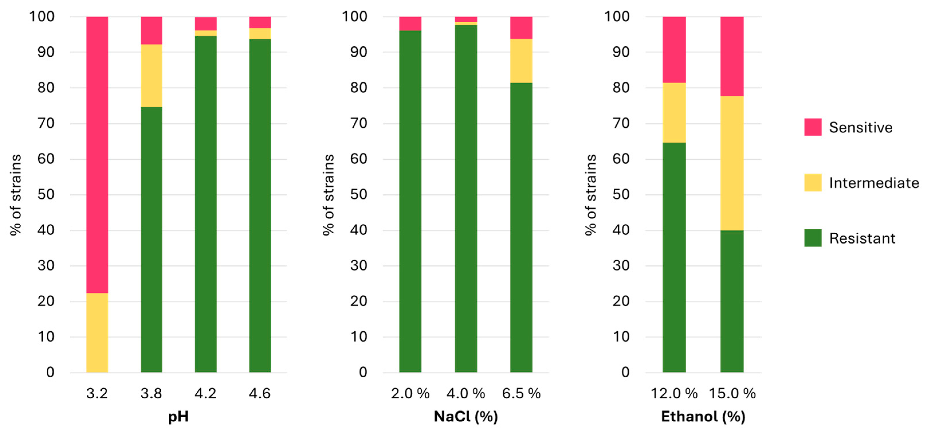
3.2. Stress Resistance

The growth capabilities of various strains may reveal intriguing functional potentials for both probiotic and technological applications. Through the experiments conducted in this study, the capability of the strains to grow at different concentrations of NaCl (2, 4, and 6.5% w/v), ethanol (12–15% v/v), and different pH values (3.2, 3.8, 4.2, 4.6) did not show a correlation with their isolation matrix or geographical origin. Instead, the results indicated that resistance to different stresses was a strain-specific trait. In total, only thirteen strains demonstrated resilience to all tested conditions: two Lbs. casei (DBPZ0571; N2014) and eleven Lbs. paracasei (LMG11961; LMG23518; LMG24101; DBPZ0324; DBPZ0563; DBPZ0579; Q4; M354; I3; DialDan5; DialYak5). As depicted in Figure 4, the majority of the analysed strains exhibited sensitivity to low pH values, results consistent with findings from other studies, in which only few strains managed to thrive at pH values lower than 3.5 [36,37,38,39,40,41], 3.0 [42], or even 2.5 [43], thus confirming the strain-specific nature of these characteristics.
On the other hand, over 96.5% of the strains demonstrated resistance to NaCl at concentrations of 2% and 4%. However, at a concentration of 6.5%, resistance was observed in 81.5% of the strains. These findings are in agreement with several studies documenting the ability of Lacticaseibacillus spp. strains to grow at NaCl concentrations of 6–6.5% [44,45,46], and some strains were also reported to survive above 12% of NaCl [47]. However, other studies have reported either the inability of strains to grow above 4% [48] or, on the contrary, to survive concentrations above 10% [42], emphasizing the strain-specific resistance to this factor, potentially attributable to modifications in cell wall structural properties [49]. Furthermore, 64.6% of the analysed strains exhibited the ability to tolerate ethanol concentrations of 12% v/v, with 40% extending their tolerance up to 15% v/v ethanol. These findings are consistent with other studies [50,51], although significant differences in strain responses are evident across the literature for Lacticaseibacillus spp.

3.3. Antibiotic Resistance

The importance of studying the resistance profiles of Lbs. casei, Lbs. zeae, and Lbs. paracasei lies in their extensive use as microbial starters or probiotics [52], as well as their presence in diverse environments where they may coexist with pathogens without barriers [53]. Lactobacilli are generally recognized as safe (GRAS). However, in certain instances, especially in vulnerable individuals, specific strains have been linked to endocarditis or bacteremia. Therefore, it is imperative to thoroughly assess the resistance factors in strains intended for human use. Intrinsic resistance, such as vancomycin resistance in heterofermentative lactobacilli, is not horizontally transferable, whereas resistance genes acquired via plasmids or transposons can be transferred [12]. For this reason, the European Commission proposed that the safety evaluation of microorganisms used in food production could prioritize the detection of transmissible antibiotic resistance markers [54].
Based on the evidence gathered in this study and consistent with findings from other researchers, it was observed that the majority of the strains (>80%) exhibited resistance to aminoglycosides such as kanamycin [23,44,55], streptomycin [23,44,56], and vancomycin [23,55,57,58], with this resistance being considered intrinsic to the species. Additionally, most strains displayed resistance to tobramycin, although documentation of this resistance in the literature is limited [59], as well as resistance to ofloxacin [60]. However, many strains resulted in being sensitive versus all the other tested antibiotics, as confirmed for Lactobacillus spp. by other studies [23,52,53,61].
Among the results obtained, eight strains with particular resistance profiles were identified (Table 2). Among these, Lbs. paracasei DBPZ0478 exhibited resistance to most of the tested antibiotics, except for clindamycin, quinupristin/dalfopristin, and rifampicin. Some strains isolated from yogurt showed sensitivity to tobramycin but were resistant to erythromycin. Lbs. paracasei DialDan8 emerged as the most sensitive strain, demonstrating kanamycin resistance and intermediate resistance to erythromycin and tobramycin. Surprisingly, Lbs. paracasei M308 and DialDan8 resulted in being sensitive to vancomycin. This finding contradicts established knowledge, which suggests that some LABs, including Lbs. casei and Lbs. rhamnosus, are intrinsically vancomycin resistant [62]. However, in a recent study, also Lbs. paracasei subsp. paracasei strain L1, resulted in being sensitive to vancomycin [63].

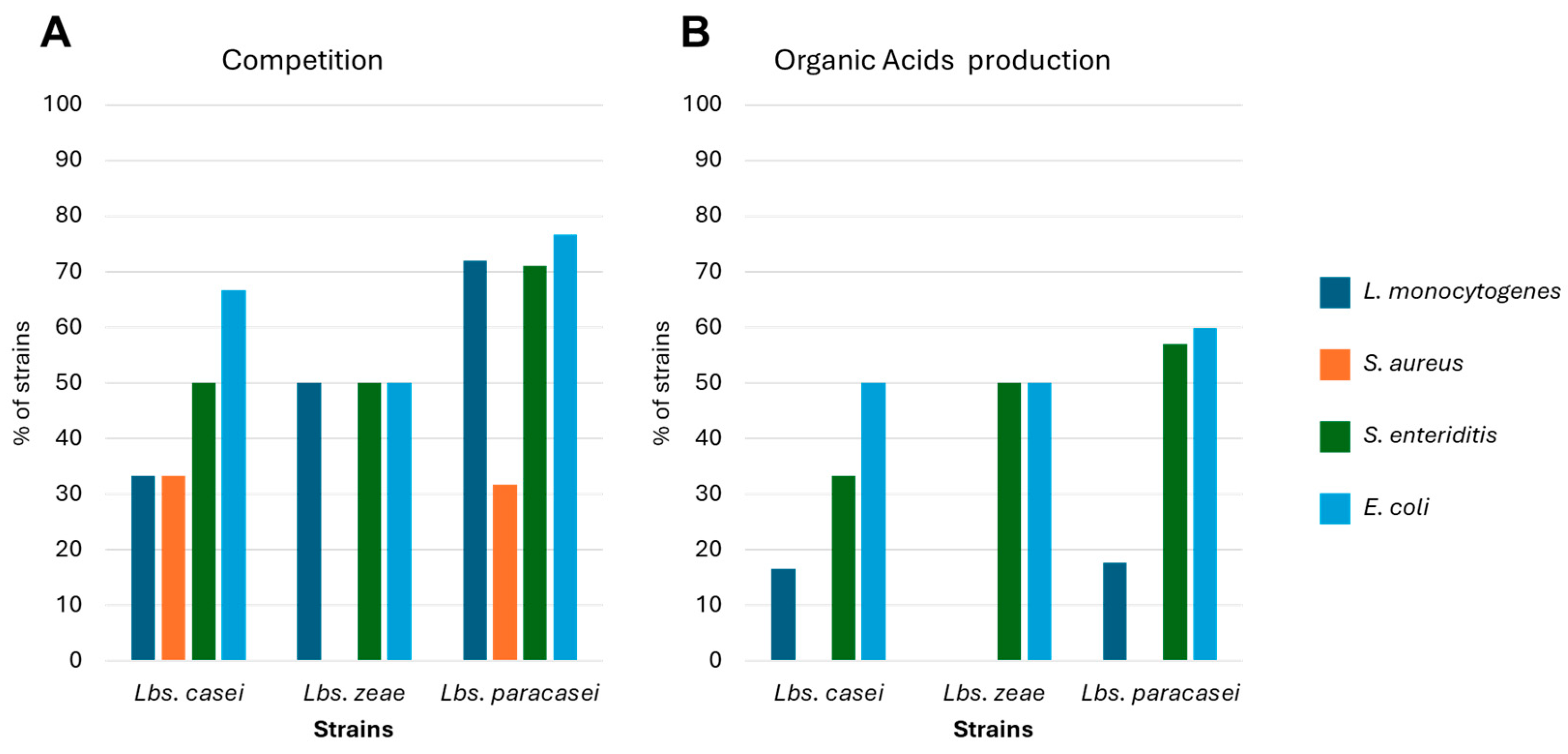
3.4. Antimicrobial Activity and Bacteriocin Production

Antibiotic resistance has escalated in recent decades due to inappropriate antibiotic usage in humans and animals. To address this issue, researchers are exploring alternative substances to treat and prevent bacterial infections. The utilization of bacteriocins could serve as an alternative to antibiotics. Additionally, the well-documented antimicrobial capabilities of lactic acid bacteria (LABs) could be beneficial against foodborne pathogens and in food preservation [64,65,66,67,68]. The production of lactic acid and hydrogen peroxide by LABs can obstruct the growth of pathogens due to their bactericidal effect. The bactericidal/bacteriostatic mechanism of lactic acid likely involves the cytotoxic properties of non-dissociated lactic acid and the insolubility of dissociated lactate, resulting in cytoplasmic acidification and the disruption of proton gradient forces. Moreover, this process may affect the transmembrane pH gradient and reduce the amount of accessible energy for bacteria to grow [69,70].
In this study, the agar well diffusion test was employed to investigate the production of peptidic antimicrobial compounds against Listeria monocytogenes ATCC7644, Staphylococcus aureus DSM 4910, Escherichia coli DSA, and Salmonella enteritidis DSA. However, no strains capable of producing bacteriocins have been identified. On the contrary, several strains were capable of directly inhibiting the growth of the four tested pathogens (Figure 5A). Moreover, thanks to the production of organic acids, several strains inhibited the growth of L. monocytogenes, E. coli, and S. enteritidis. However, no inhibition was observed versus S. aureus (Figure 5B). These properties have previously been reported in the literature, where in several cases, strains of Lbs. casei and Lbs. paracasei have demonstrated the ability to inhibit the growth of various pathogens, including Acinetobacter baumannii, Bacillus cereus, Campylobacter jejuni, Citrobacter freundii, Cronobacter sakazakii, Enterobacter cloacae, Enterococcus faecalis, Enterococcus faecium, E. coli, Klebsiella pneumoniae, L. monocytogenes, Pasteurella multocida, Proteus mirabilis, Pseudomonas aeruginosa, Salmonella enterica, Salmonella typhimurium, Staphylococcus aureus, Staphylococcus epidermidis, Staphylococcus haemolyticus, and Streptococcus mutans [48,58,71,72,73,74]. Furthermore, to contrast the spread of antibiotic resistance, further studies could explore the potential use of these lacticaseibacilli, their supernatants, or cell extracts to counteract the virulence of different pathogens, as reported in several studies [75].

3.5. Biogenic Amine (BA) Production

The decarboxylation of amino acids, particularly in nutrient-scarce environments, can serve as a crucial energy source for microorganisms [76]. Biogenic amines (BAs) are organic bases with aromatic, aliphatic, or heterocyclic structures, commonly found in various beverages and foods. They are primarily generated by the microbial decarboxylation of precursor amino acids [14,77]. Histamine, tyramine, putrescine, and cadaverine are among the BAs typically found in food and beverages. Although their presence often goes undetected due to a lack of associated decline in sensory quality, high consumption of these amines can pose toxicity risks to humans [78,79,80,81,82]. While putrescine and cadaverine are considered non-toxic, they can interfere with histamine and tyramine detoxification processes or potentiate histamine toxicity, respectively. Tyramine ingestion can lead to headaches, vomiting, and hypertension, while histamine can induce breathing difficulties, rash, heart palpitations (allergic-type reactions), low blood pressure, oedema, and vomiting [79,80,83]. The early detection of potential BA-producing bacteria can prevent BA formation and accumulation in food products [78,80] and aid in selecting starter cultures that do not produce BAs [76].
Using the method described by Bover-Cid and Holzapfel, 1999, none of the Lbs. casei and Lbs. zeae strains produced BAs. However, among the Lbs. paracasei strains, strain B195 produced tyramine, strain PSG10 produced agmatine, and strain B169 produced both agmatine and tyramine. A multiplex PCR analysis of the genes involved in BA production revealed that all Lbs. casei and Lbs. zeae strains were negative for all target genes, whereas six strains of Lbs. paracasei were positive for the agdi gene. Additionally, Lbs. paracasei NRRL B-456 tested positive for the tyrdc gene and Lbs. paracasei B350 was positive for the hdc gene. Furthermore, Lbs. paracasei PSG10 tested positive for both the tydc and hdc genes (Table 3). Comparing the results obtained from the two techniques, only one strain yielded concordant results. Despite the identification of these genes through molecular techniques, it was not possible to observe the production of the related BAs. This discrepancy may be attributed to possible gene silencing or very low expression, making it difficult to detect the products using conventional plate growth methods [84,85]. Conversely, according to Buňková et al. (2009), the formation of chemicals with alkaline reactions may have led to colour changes in the pH indicator in the decarboxylase medium, resulting in false-positive reactions [77]. Therefore, PCR analysis could be considered a better and more accurate method for revealing the potential of strains to produce biogenic amines, as it detects strains possessing genes for corresponding enzyme production. However, it should be underlined that the presence of a single gene does not always translate into its expression. Indeed, environmental conditions directly influence microbial metabolism. Therefore, it remains extremely important to test both the genotypic and phenotypic aspects of microorganisms.

3.6. arcABC Gene Presence

In this part of the study, the three genes encoding the main enzymes involved in the arginine deiminase (ADI) pathway were investigated: arcA (arginine deiminase), arcB (ornithine transcarbamylase), and arcC (carbamate kinase). During the ADI pathway, lactic acid bacteria produce carbamoyl phosphate and citrulline, which can naturally react with ethanol to form ethyl carbamate, a potential carcinogenic compound found mainly in wine and various fermented foods [86,87].
Among all the tested strains for the presence of the arcA, arcB, and arcC genes, only five Lbs. paracasei strains (P71; P2P3; B169; B195; DSMZ5622) tested positive for at least one of the genes. Specifically, Lbs. paracasei strains P71 and P2P3 (isolated from cheese and goat milk, respectively) were positive for the arcB gene, while Lbs. paracasei B169, B195, and DSM5622 (isolated from wine, wine, and dairy products, respectively) tested positive for the arcC gene. None of the Lbs. casei and Lbs. zeae strains showed the presence of these genes.
As previously mentioned, one of the main precursors of ethyl carbamate (EC) is citrulline, which could be derived from LAB metabolism. While extensive studies on the presence of these genes in Lacticaseibacillus spp. are lacking, some authors have investigated other facultative or obligate heterofermentative LABs. Specifically, Lentilactobacillus hilgardii, Lpb. plantarum, and Oenococcus oeni have been shown to degrade arginine [86,88,89], while Lentilactobacillus buchneri has been observed to excrete citrulline [90]. Furthermore, strains belonging to Levilactobacillus brevis, Lentilactobacillus hilgardii, Fructilactobacillus florum, Llb. buchneri, Lpb. plantarum, O. oeni, Pediococcus pentosaceus, and Leuconostoc mesenteroides resulted in being positive to the presence of arcABC genes [27,91].

3.7. Biogenic Amine (BA)-Degrading Activity: Diaminobenzidine (DAB) Assay and Multicopper Oxidase (MCO) Detection

In fermented foods, particularly those undergoing spontaneous fermentation, the presence of biogenic amines is frequently observed [92]. Individuals with deficiencies in the natural mechanisms of biogenic amine detoxification are particularly vulnerable to these compounds [79,93]. These metabolites not only affect the sensory attributes of the food but also pose a health hazard to consumers, potentially leading to food poisoning [94]. To mitigate these risks, the food industry utilizes specific starter microorganisms carefully selected for their non-production of the amines. These microorganisms not only exert a bioprotective effect by inhibiting the growth of undesired microorganisms but also possess the capability to degrade existing biogenic amines through specialized enzymes [95]. Various methods are employed to control the formation of biogenic amines in food, including the incorporation of additives and preservatives, supplementation with enzymes such as monoamine oxidase (MAO) and diamine oxidase (DAO), and the utilization of bacteria harbouring these enzymes [28,76,93]. MAO and DAO enzymes catalyze the conversion of amines into non-toxic by-products, which can then be safely eliminated from the body [83]. Hence, the ability to degrade biogenic amines was assessed in the tested strains using the DAB assay. While no extracts exhibited bands indicative of MAO/DAO presence, 16 strains displayed mono-copper oxidase (MCO) activity, as detected by DMP staining (Table 4). This characteristic was evidenced by the appearance of yellow-orange bands at consistent positions on the gels, suggesting the presence of similar enzymes across all positive strains. These findings are corroborated by previous studies where several bacterial strains, including Lbs. paracasei, which tested negative in the DAB assay, yielded positive results in DMP staining. This underscores the ability of certain Lacticaseibacillus spp. strains to utilize histamine, tyramine, putrescine, and DMP as substrates and to catalyze the oxidation of these compounds [28,96]. Given the low number of MCO positive strains, it was not feasible to discern a definitive pattern concerning their origin and isolation source. Nevertheless, six out of the sixteen strains were isolated from dairy products, an ecological niche where strains exhibiting these capabilities have been previously isolated and documented in the literature [97].

3.8. Phenotypic Clustering

Based on all the phenotypic characterization results, we conducted a clustering analysis of the different strains. As can be observed in Figure 6, the most discriminating factors were antibiotic resistance and tolerance to various pH, ethanol, and NaCl conditions. The heatmap colour variation reflects the significance of the correlation among these traits. All strains formed interconnected subgroups, but Lbs. paracasei TMW 1.1444, TMW 1.1259, and V3 (highlighted in blue) emerged as the most distinct strains across all tested characteristics.
Furthermore, the traits under study segregated into two main clusters (I and II). Cluster I exhibited correlations among almost all growth capabilities (pH tolerance and resistance to different NaCl and ethanol concentrations) along with intrinsic resistance to antibiotics (vancomycin and tobramycin). On the other hand, Cluster II encompassed the presence/absence of genes related to biogenic amines (BAs) and ethyl carbamate production, detection of mono-copper oxidase (MCO), and most antibiotic resistance traits. Additionally, a growth capability at pH 3.2 was found within this second group.
Based on these factors, it was possible to gather the analysed strains in four main clusters: the cluster highlighted in red is made up of strains with low resistance to various stress factors and to some antibiotics. On the contrary, it is possible to see how all the other factors analysed were distributed unevenly within the considered strains, underlining once again the strain-specificity of many of the characteristics analysis. Finally, also in this case, it was not possible to observe a clear distinction between the phenotypic characteristics of Lbs. casei, Lbs. zeae, and Lbs. paracasei, with a mixture of the different species among all the different clusters obtained.
However, given the high genotypic and phenotypic heterogeneity of the strains, these clusters do not consist of strains with completely homogeneous characteristics. In this context, representing all tested characteristics through a heatmap can provide a valuable tool following these screening studies. This can facilitate the selection of the best strain based on the desired traits for each potential technological application.

4. Conclusions

In this study, 121 Lbs. paracasei strains isolated from various origins or acquired from international collections were genetically and phenotypically characterized to assess their technological and safety properties, comparing them to 6 Lbs. casei and 2 Lbs. zeae strains. The application of these techniques revealed a significant degree of genetic variability, resulting in numerous clusters from Rep-PCR, RAPD, and Sau-PCR analyses, often associated with the source of isolation but not delineating the different species.
The assessment of safety traits and technological properties uncovered substantial variability in strain behaviours, confirming the strain-specific nature of these attributes. Notably, certain strains exhibited unique characteristics with broad potential applications. For example, Lbs. casei N2014 showed the agdi and hdc genes alongside MCO activity, while Lbs. paracasei B169 and B195 harboured the arcC and agdi genes, with the latter also showcasing MCO activity.
Moreover, the identification of specific strains resistant to a wide spectrum of antibiotics, such as Lbs. paracasei DBPZ0478, underscores the importance of understanding microbial resistance profiles.
The extensive genetic diversity among the strains highlights their vast potential applications. With the increasing prominence of functional foods and probiotics, the availability of diverse strains facilitates tailored product development. For instance, formulations targeting individuals with heightened susceptibility could utilize strains selectively sensitive to antibiotics, while strains with non-transferable resistances could augment certain antibiotic therapies to mitigate adverse effects.
Furthermore, exploring the metabolism of these microorganisms could lead to reduced reliance on food additives. A growing body of research attests to the bioprotective efficacy of these microorganisms, paving the way for their potential substitution or supplementation of antimicrobials and antioxidants, thereby directly benefiting consumer health.
To elucidate the distribution of various traits among strains, the results were consolidated into a unified matrix, accentuating the strain-specificity of the tested attributes. This comprehensive approach unveiled the diverse capabilities of individual strains, providing insights for targeted applications across various domains. Such detailed screening is crucial for characterizing microorganisms with potential probiotic, postbiotic, and gut microbiota-modifying effects, as well as anti-inflammatory potential, opening doors to innovative health interventions and functional food development.

Author Contributions

A.C., F.G. and L.C. contributed equally to the work, sharing first authorship. Conceptualization: L.I.; Data curation: L.I. and A.C.; Formal Analysis: F.G., L.C. and A.C.; Funding acquisition: L.I.; Investigation: G.C. and A.R.; Methodology: L.I. and A.C.; Project administration: L.I.; Resources: L.I., G.C. and A.R.; Software: L.I.; Supervision: L.I.; Validation: L.I. and A.C.; Visualization: A.C.; Writing—original draft: A.C.; Writing—review and editing: L.I. and A.C. All authors have read and agreed to the published version of the manuscript.

Funding

This research was funded by Ministero dell’Istruzione, dell’Università e della Ricerca, Rome, Italy, FIRB n. RBFR107VML.

Institutional Review Board Statement

Not applicable.

Informed Consent Statement

Not applicable.

Data Availability Statement

Further details on the data are available on request.

Conflicts of Interest

The authors declare no conflicts of interest.

References

  1. Zheng, J.; Wittouck, S.; Salvetti, E.; Franz, C.M.A.P.; Harris, H.M.B.; Mattarelli, P.; O’toole, P.W.; Pot, B.; Vandamme, P.; Walter, J.; et al. A Taxonomic Note on the Genus Lactobacillus: Description of 23 Novel Genera, Emended Description of the Genus Lactobacillus Beijerinck 1901, and Union of Lactobacillaceae and Leuconostocaceae. Int. J. Syst. Evol. Microbiol. 2020, 70, 2782–2858. [Google Scholar] [CrossRef] [PubMed]
  2. Qiao, N.; Wittouck, S.; Mattarelli, P.; Zheng, J.; Lebeer, S.; Felis, G.E.; Gänzle, M.G. After the Storm—Perspectives on the Taxonomy of Lactobacillaceae. JDS Commun. 2022, 3, 222–227. [Google Scholar] [CrossRef] [PubMed]
  3. Liu, D.D.; Gu, C.T. Proposal to Reclassify Lactobacillus zhaodongensis, Lactobacillus zeae, Lactobacillus argentoratensis and Lactobacillus buchneri Subsp. silagei as Lacticaseibacillus zhaodongensis Comb. Nov., Lacticaseibacillus zeae Comb. Nov., Lactiplantibacillus argentoratensis comb. nov. and Lentilactobacillus buchneri subsp. silagei comb. nov., respectively and Apilactobacillus kosoi as a later heterotypic synonym of Apilactobacillus micheneri. Int. J. Syst. Evol. Microbiol. 2020, 70, 6414–6417. [Google Scholar] [CrossRef] [PubMed]
  4. Iacumin, L.; Ginaldi, F.; Manzano, M.; Anastasi, V.; Reale, A.; Zotta, T.; Rossi, F.; Coppola, R.; Comi, G. High Resolution Melting Analysis (HRM) as a New Tool for the Identification of Species Belonging to the Lactobacillus Casei Group Andcomparison with Species-Specific PCRs and Multiplex PCR. Food Microbiol. 2015, 46, 357–367. [Google Scholar] [CrossRef] [PubMed]
  5. Coudeyras, S.; Marchandin, H.; Fajon, C.; Forestier, C. Taxonomic and Strain-Specific Identification of the Probiotic Strain Lactobacillus rhamnosus 35 within the Lactobacillus Casei Group. Appl. Environ. Microbiol. 2008, 74, 2679–2689. [Google Scholar] [CrossRef] [PubMed]
  6. Chen, X.; Zhu, Z.; Zhang, X.; Chen, L.; Gu, Q.; Li, P. Lactobacillus paracasei ZFM54 Alters the Metabolomic Profiles of Yogurt and the Co-Fermented Yogurt Improves the Gut Microecology of Human Adults. J. Dairy Sci. 2024. [Google Scholar] [CrossRef] [PubMed]
  7. Ren, S.; Wang, C.; Chen, A.; Bai, Z.; Tian, Y.; Lv, W. Lactobacillus paracasei Influences the Gut-Microbiota-Targeted Metabolic Modulation of the Immune Status of Diarrheal Mice. Food Funct. 2023, 14, 4368–4379. [Google Scholar] [CrossRef] [PubMed]
  8. Giraffa, G.; Chanishvili, N.; Widyastuti, Y. Importance of Lactobacilli in Food and Feed Biotechnology. Res. Microbiol. 2010, 161, 480–487. [Google Scholar] [CrossRef] [PubMed]
  9. EFSA. Introduction of a Qualified Presumption of Safety (QPS) Approach for Assessment of Selected Microorganisms Referred to EFSA—Opinion of the Scientific Committee. EFSA J. 2007, 5, 587. [Google Scholar] [CrossRef]
  10. EFSA. EFSA Statement on the Requirements for Whole Genome Sequence Analysis of Microorganisms Intentionally Used in the Food Chain. EFSA J. 2021, 19, e06506. [Google Scholar] [CrossRef]
  11. EFSA. Guidance on the Characterisation of Microorganisms Used as Feed Additives or as Production Organisms. EFSA J. 2018, 16, e05206. [Google Scholar] [CrossRef]
  12. Colautti, A.; Arnoldi, M.; Comi, G.; Iacumin, L. Antibiotic Resistance and Virulence Factors in Lactobacilli: Something to Carefully Consider. Food Microbiol. 2021, 103, 103934. [Google Scholar] [CrossRef]
  13. Colautti, A.; Rossi, N.; Piazza, C.; Comi, G.; Iacumin, L. Draft Genome Sequences of 14 Lacticaseibacillus Spp. Strains, Representatives of a Collection of 200 Strains. Microbiol. Resour. Announc. 2023, 12, 10–13. [Google Scholar] [CrossRef] [PubMed]
  14. Silla Santos, M.H. Biogenic Amines: Their Importance in Foods. Int. J. Food Microbiol. 1996, 29, 213–231. [Google Scholar] [CrossRef]
  15. Omer, A.K.; Mohammed, R.R.; Mohammed Ameen, P.S.; Abas, Z.A.; Ekici, K. Presence of Biogenic Amines in Food and Their Public Health Implications: A Review. J. Food Prot. 2021, 84, 1539–1548. [Google Scholar] [CrossRef] [PubMed]
  16. Pištěková, H.; Jančová, P.; Berčíková, L.; Buňka, F.; Sokolová, I.; Šopík, T.; Maršálková, K.; de Amaral, O.M.R.P.; Buňková, L. Application of QPCR for Multicopper Oxidase Gene (MCO) in Biogenic Amines Degradation by Lactobacillus casei. Food Microbiol. 2020, 91, 103550. [Google Scholar] [CrossRef]
  17. Iacumin, L.; Osualdini, M.; Bovolenta, S.; Boscolo, D.; Chiesa, L.; Panseri, S.; Comi, G. Microbial, Chemico-Physical and Volatile Aromatic Compounds Characterization of Pitina PGI, a Peculiar Sausage-like Product of North East Italy. Meat Sci. 2020, 163, 108081. [Google Scholar] [CrossRef] [PubMed]
  18. Gevers, D.; Huys, G.; Swings, J. Applicability of rep-PCR fingerprinting for identification of Lactobacillus species. FEMS Microbiol. Lett. 2001, 205, 31–36. [Google Scholar] [CrossRef]
  19. Huey, B.; Hall, J. Hypervariable DNA Fingerprinting in Escherichia coli: Minisatellite Probe from Bacteriophage M13. J. Bacteriol. 1989, 171, 2528–2532. [Google Scholar] [CrossRef]
  20. Corich, V.; Mattiazzi, A.; Soldati, E.; Carraro, A.; Giacomini, A. Sau-PCR, a Novel Amplification Technique for Genetic Fingerprinting of Microorganisms. Appl. Environ. Microbiol. 2005, 71, 6401–6406. [Google Scholar] [CrossRef]
  21. Vauterin, L.; Vauterin, P. Computer-Aided Objective Comparison of Electrophoretic Patterns for Grouping and Identification of Microorganisms. Eur. Microbiol. 1992, 1, 37–41. [Google Scholar]
  22. Salminen, M.K.; Rautelin, H.; Tynkkynen, S.; Poussa, T.; Saxelin, M.; Valtonen, V.; Järvinen, A. Lactobacillus Bacteremia, Species Identification, and Antimicrobial Susceptibility of 85 Blood Isolates. Clin. Infect. Dis. 2006, 42, e35–e44. [Google Scholar] [CrossRef] [PubMed]
  23. Charteris, W.P.; Kelly, P.M.; Morelli, L.; Collins, J.K. Antibiotic Susceptibility of Potentially Probiotic Lactobacillus Species. J. Food Prot. 1998, 61, 1636–1643. [Google Scholar] [CrossRef] [PubMed]
  24. Schillinger, U.; Lucke, F.K. Antimicrobial Activity of Lactobacillus Sake Isolated from Meat. Appl. Environ. Microbiol. 1989, 55, 1901–1906. [Google Scholar] [CrossRef]
  25. Bover-Cid, S.; Holzapfel, W.H. Improved Screening Procedure for Biogenic Amine Production by Lactic Acid Bacteria. Int. J. Food Microbiol. 1999, 53, 33–41. [Google Scholar] [CrossRef]
  26. Coton, M.; Romano, A.; Spano, G.; Ziegler, K.; Vetrana, C.; Desmarais, C.; Lonvaud-Funel, A.; Lucas, P.; Coton, E. Occurrence of Biogenic Amine-Forming Lactic Acid Bacteria in Wine and Cider. Food Microbiol. 2010, 27, 1078–1085. [Google Scholar] [CrossRef]
  27. Araque, I.; Gil, J.; Ramon, C.; Bordons, A.; Reguant, C. Detection of Arc Genes Related with the Ethyl Carbamate Precursors in Wine Lactic Acid Bacteria. J. Agric. Food Chem. 2009, 57, 1841–1847. [Google Scholar] [CrossRef]
  28. Callejón, S.; Sendra, R.; Ferrer, S.; Pardo, I. Identification of a Novel Enzymatic Activity from Lactic Acid Bacteria Able to Degrade Biogenic Amines in Wine. Appl. Microbiol. Biotechnol. 2014, 98, 185–198. [Google Scholar] [CrossRef] [PubMed]
  29. Comi, G.; Urso, R.; Iacumin, L.; Rantsiou, K.; Cattaneo, P.; Cantoni, C.; Cocolin, L. Characterisation of Naturally Fermented Sausages Produced in the North East of Italy. Meat Sci. 2005, 69, 381–392. [Google Scholar] [CrossRef]
  30. Iacumin, L.; Comi, G.; Cantoni, C.; Cocolin, L. Molecular and Technological Characterization of Staphylococcus Xylosus Isolated from Naturally Fermented Italian Sausages by RAPD, Rep-PCR and Sau-PCR Analysis. Meat Sci. 2006, 74, 281–288. [Google Scholar] [CrossRef]
  31. Amadoro, C.; Rossi, F.; Piccirilli, M.; Colavita, G. Features of Lactobacillus sakei Isolated from Italian Sausages: Focus on Strains from Ventricina Del Vastese. Ital. J. Food Saf. 2015, 4, 5449. [Google Scholar] [CrossRef] [PubMed]
  32. Torriani, S.; Zapparoli, G.; Dellaglio, F. Use of PCR-Based Methods for Rapid Differentiation of Lactobacillus Delbrueckii Subsp. Bulgaricus and L. Delbrueckii Subsp. Lactis. Appl. Environ. Microbiol. 1999, 65, 4351–4356. [Google Scholar] [CrossRef] [PubMed]
  33. Turková, K.; Rittich, B.; Španová, A. Identification and Determination of Relatedness of Lactobacilli Using Different DNA Amplification Methods. Chem. Pap. 2012, 66, 842–851. [Google Scholar] [CrossRef]
  34. Rossetti, L.; Giraffa, G. Rapid Identification of Dairy Lactic Acid Bacteria by M13-Generated, RAPD-PCR Fingerprint Databases. J. Microbiol. Methods 2005, 63, 135–144. [Google Scholar] [CrossRef] [PubMed]
  35. Jarocki, P.; Komoń-Janczara, E.; Glibowska, A.; Dworniczak, M.; Pytka, M.; Korzeniowska-Kowal, A.; Wzorek, A.; Kordowska-Wiater, M. Molecular Routes to Specific Identification of the Lactobacillus Casei Group at the Species, Subspecies and Strain Level. Int. J. Mol. Sci. 2020, 21, 2694. [Google Scholar] [CrossRef] [PubMed]
  36. Reale, A.; Di Renzo, T.; Rossi, F.; Zotta, T.; Iacumin, L.; Preziuso, M.; Parente, E.; Sorrentino, E.; Coppola, R. Tolerance of Lactobacillus casei, Lactobacillus paracasei and Lactobacillus rhamnosus Strains to Stress Factors Encountered in Food Processing and in the Gastro-Intestinal Tract. LWT 2015, 60, 721–728. [Google Scholar] [CrossRef]
  37. Wu, C.; He, G.; Zhang, J. Physiological and Proteomic Analysis of Lactobacillus Casei in Response to Acid Adaptation. J. Ind. Microbiol. Biotechnol. 2014, 41, 1533–1540. [Google Scholar] [CrossRef] [PubMed]
  38. Rubio, R.; Jofré, A.; Martín, B.; Aymerich, T.; Garriga, M. Characterization of Lactic Acid Bacteria Isolated from Infant Faeces as Potential Probiotic Starter Cultures for Fermented Sausages. Food Microbiol. 2014, 38, 303–311. [Google Scholar] [CrossRef] [PubMed]
  39. Jacobsen, C.N.; Nielsen, V.R.; Hayford, A.E.; Møller, P.L.; Michaelsen, K.F.; Pærregaard, A.; Sandström, B.; Tvede, M.; Jakobsen, M. Screening of Probiotic Activities of Forty-Seven Strains of Lactobacillus Spp. by in Vitro Techniques and Evaluation of the Colonization Ability of Five Selected Strains in Humans. Appl. Environ. Microbiol. 1999, 65, 4949–4956. [Google Scholar] [CrossRef]
  40. Federici, S.; Ciarrocchi, F.; Campana, R.; Ciandrini, E.; Blasi, G.; Baffone, W. Identification and Functional Traits of Lactic Acid Bacteria Isolated from Ciauscolo Salami Produced in Central Italy. Meat Sci. 2014, 98, 575–584. [Google Scholar] [CrossRef]
  41. Kou, X.; Chen, Q.; Ju, X.; Xue, Z.; Chen, W.; Xue, Z. A Tolerant Lactic Acid Bacteria, Lactobacillus paracasei, and Its Immunoregulatory Function. Can. J. Microbiol. 2014, 60, 729–736. [Google Scholar] [CrossRef] [PubMed]
  42. Qureshi, N.; Gu, Q.; Li, P. Whole Genome Sequence Analysis and in Vitro Probiotic Characteristics of a Lactobacillus Strain Lactobacillus paracasei ZFM54. J. Appl. Microbiol. 2020, 129, 422–433. [Google Scholar] [CrossRef] [PubMed]
  43. Wang, Y.; Zhou, J.; Xia, X.; Zhao, Y.; Shao, W. Probiotic Potential of Lactobacillus paracasei FM-LP-4 Isolated from Xinjiang Camel Milk Yoghurt. Int. Dairy J. 2016, 62, 28–34. [Google Scholar] [CrossRef]
  44. Radulović, Z.; Petrović, T.; Nedović, V.; Dimitrijević, S.; Mirković, N.; Petrušić, M.; Paunović, D. Characterization of Autochthonous Lactobacillus paracasei Strains on Potential Probiotic Ability. Mljekarstvo 2010, 60, 86–93. [Google Scholar]
  45. Muna, M.A.; Adel, M.M. Isolation of Lactobacillus Strains with Probiotic Potential from Camels Milk. Afr. J. Microbiol. Res. 2014, 8, 1645–1655. [Google Scholar] [CrossRef]
  46. De Albuquerque, T.M.R.; Garcia, E.F.; De Oliveira Araújo, A.; Magnani, M.; Saarela, M.; De Souza, E.L. In Vitro Characterization of Lactobacillus Strains Isolated from Fruit Processing By-Products as Potential Probiotics. Probiotics Antimicrob. Proteins 2018, 10, 704–716. [Google Scholar] [CrossRef]
  47. Dishan, A.; Gönülalan, Z. Lacticaseibacillus paracasei AD22 Stress Response in Brined White Cheese Matrix: In Vitro Probiotic Profiles and Molecular Characterization. Probiotics Antimicrob. Proteins 2024. [Google Scholar] [CrossRef] [PubMed]
  48. Ahmed, Z.; Vohra, M.S.; Khan, M.N.; Ahmed, A.; Khan, T.A. Antimicrobial Role of Lactobacillus Species as Potential Probiotics against Enteropathogenic Bacteria in Chickens. J. Infect. Dev. Ctries. 2019, 13, 130–136. [Google Scholar] [CrossRef]
  49. Piuri, M.; Sanchez-Rivas, C.; Ruzal, S.M. Cell Wall Modifications during Osmotic Stress in Lactobacillus Casei. J. Appl. Microbiol. 2005, 98, 84–95. [Google Scholar] [CrossRef]
  50. Pittet, V.; Morrow, K.; Ziola, B. Ethanol Tolerance of Lactic Acid Bacteria, Including Relevance of the Exopolysaccharide Gene Gtf. J. Am. Soc. Brew. Chem. 2011, 69, 57–61. [Google Scholar] [CrossRef]
  51. Lee, H.I.; Kim, M.H.; Kim, K.Y.; So, J.S. Screening and Selection of Stress Resistant Lactobacillus Spp. Isolated from the Marine Oyster (Crassostrea gigas). Anaerobe 2010, 16, 522–526. [Google Scholar] [CrossRef]
  52. Sharma, P.; Tomar, S.K.; Goswami, P.; Sangwan, V.; Singh, R. Antibiotic Resistance among Commercially Available Probiotics. Food Res. Int. 2014, 57, 176–195. [Google Scholar] [CrossRef]
  53. Belletti, N.; Gatti, M.; Bottari, B.; Neviani, E.; Tabanelli, G.; Gardini, F. Antibiotic Resistance of Lactobacilli Isolated from Two Italian Hard Cheeses. J. Food Prot. 2009, 72, 2162–2169. [Google Scholar] [CrossRef]
  54. European Commission—HEALTH & CONSUMER PROTECTION DIRECTORATE-GENERAL—Directorate C—Scientific Opinions C2—Management of Scientific Committees; Scientific Co-Operation and Networks. Position Paper of the Scientific Committee on Animal Nutrition on Safety Assessment and Regulatory Aspects of Micro-organisms in Feed and Food Applications. Available online: https://food.ec.europa.eu/document/download/b2beaff9-5fb3-45eb-9e63-d5396ec85154_en?filename=sci-com_scan-old_report_out85.pdf (accessed on 7 July 2024).
  55. Coton, M.; Lebreton, M.; Leyva Salas, M.; Garnier, L.; Navarri, M.; Pawtowski, A.; Le Blay, G.; Valence, F.; Coton, E.; Mounier, J. Biogenic Amine and Antibiotic Resistance Profiles Determined for Lactic Acid Bacteria and a Propionibacterium Prior to Use as Antifungal Bioprotective Cultures. Int. Dairy J. 2018, 85, 21–26. [Google Scholar] [CrossRef]
  56. Huys, G.; D’Haene, K.; Danielsen, M.; Mättö, J.; Egervärn, M.; Vandamme, P. Phenotypic and Molecular Assessment of Antimicrobial Resistance in Lactobacillus paracasei Strains of Food Origin. J. Food Prot. 2008, 71, 339–344. [Google Scholar] [CrossRef] [PubMed]
  57. Gómez, N.C.; Ramiro, J.M.P.; Quecan, B.X.V.; de Melo Franco, B.D.G. Use of Potential Probiotic Lactic Acid Bacteria (LAB) Biofilms for the Control of Listeria monocytogenes, Salmonella typhimurium, and Escherichia coli O157: H7 Biofilms Formation. Front. Microbiol. 2016, 7, 863. [Google Scholar] [CrossRef] [PubMed]
  58. Wei, X.; Zhang, Y.; Zhou, H.; Tian, F.; Ni, Y. Antimicrobial Activities and in Vitro Properties of Cold-Adapted Lactobacillus Strains Isolated from the Intestinal Tract of Cold Water Fishes of High Latitude Water Areas in Xinjiang, China. BMC Microbiol. 2019, 19, 247. [Google Scholar] [CrossRef] [PubMed]
  59. Sharma, P.; Tomar, S.K.; Sangwan, V.; Goswami, P.; Singh, R. Antibiotic Resistance of Lactobacillus Sp. Isolated from Commercial Probiotic Preparations. J. Food Saf. 2016, 36, 38–51. [Google Scholar] [CrossRef]
  60. Todorov, S.D.; Perin, L.M.; Carneiro, B.M.; Rahal, P.; Holzapfel, W.; Nero, L.A. Safety of Lactobacillus plantarum ST8Sh and Its Bacteriocin. Probiotics Antimicrob. Proteins 2017, 9, 334–344. [Google Scholar] [CrossRef]
  61. Ammor, M.S.; Belén Flórez, A.; Mayo, B. Antibiotic Resistance in Non-Enterococcal Lactic Acid Bacteria and Bifidobacteria. Food Microbiol. 2007, 24, 559–570. [Google Scholar] [CrossRef]
  62. Goldstein, E.J.C.; Tyrrell, K.L.; Citron, D.M. Lactobacillus Species: Taxonomic Complexity and Controversial Susceptibilities. Clin. Infect. Dis. 2015, 60, S98–S107. [Google Scholar] [CrossRef] [PubMed]
  63. Xu, Y.; Tian, Y.; Cao, Y.; Li, J.; Guo, H.; Su, Y.; Tian, Y.; Wang, C.; Wang, T.; Zhang, L. Probiotic Properties of Lactobacillus paracasei Subsp. Paracasei L1 and Its Growth Performance-Promotion in Chicken by Improving the Intestinal Microflora. Front. Physiol. 2019, 10, 937. [Google Scholar] [CrossRef] [PubMed]
  64. Lozo, J.; Jovcic, B.; Kojic, M.; Dalgalarrondo, M.; Chobert, J.M.; Haertlé, T.; Topisirovic, L. Molecular Characterization of a Novel Bacteriocin and an Unusually Large Aggregation Factor of Lactobacillus paracasei Subsp. Paracasei BGSJ2-8, a Natural Isolate from Homemade Cheese. Curr. Microbiol. 2007, 55, 266–271. [Google Scholar] [CrossRef] [PubMed]
  65. Verdenelli, M.C.; Ghelfi, F.; Silvi, S.; Orpianesi, C.; Cecchini, C.; Cresci, A. Probiotic Properties of Lactobacillus rhamnosus and Lactobacillus paracasei Isolated from Human Faeces. Eur. J. Nutr. 2009, 48, 355–363. [Google Scholar] [CrossRef] [PubMed]
  66. Georgieva, R.; Yocheva, L.; Tserovska, L.; Zhelezova, G.; Stefanova, N.; Atanasova, A.; Danguleva, A.; Ivanova, G.; Karapetkov, N.; Rumyan, N.; et al. Antimicrobial Activity and Antibiotic Susceptibility of Lactobacillus and Bifidobacterium Spp. Intended for Use as Starter and Probiotic Cultures. Biotechnol. Biotechnol. Equip. 2015, 29, 84–91. [Google Scholar] [CrossRef]
  67. Peng, M.; Tabashsum, Z.; Patel, P.; Bernhardt, C.; Biswas, D. Linoleic Acids Overproducing Lactobacillus Casei Limits Growth, Survival, and Virulence of Salmonella Typhimurium and Enterohaemorrhagic Escherichia coli. Front. Microbiol. 2018, 9, 2663. [Google Scholar] [CrossRef] [PubMed]
  68. Iacumin, L.; Cappellari, G.; Colautti, A.; Comi, G. Listeria Monocytogenes Survey in Cubed Cooked Ham Packaged in Modified Atmosphere and Bioprotective Effect of Selected Lactic Acid Bacteria. Microorganisms 2020, 8, 898. [Google Scholar] [CrossRef]
  69. Reis, J.A.; Paula, A.T.; Casarotti, S.N.; Penna, A.L.B. Lactic Acid Bacteria Antimicrobial Compounds: Characteristics and Applications. Food Eng. Rev. 2012, 4, 124–140. [Google Scholar] [CrossRef]
  70. Capra, M.L.; Tibaldo, M.M.; Vinderola, G.; Reinheimer, J.A.; Quiberoni, A. Technological and Probiotic Characterisation of Lactobacillus Casei/Paracasei Strains and Their Phage-Resistant Mutants. Int. Dairy J. 2014, 37, 39–47. [Google Scholar] [CrossRef]
  71. Campana, R.; Van Hemert, S.; Baffone, W. Strain-Specific Probiotic Properties of Lactic Acid Bacteria and Their Interference with Human Intestinal Pathogens Invasion. Gut Pathog. 2017, 9, 12. [Google Scholar] [CrossRef]
  72. Muyyarikkandy, M.S.; Amalaradjou, M.A. Lactobacillus bulgaricus, Lactobacillus rhamnosus and Lactobacillus paracasei Attenuate Salmonella Enteritidis, Salmonella Heidelberg and Salmonella Typhimurium Colonization and Virulence Gene Expression in Vitro. Int. J. Mol. Sci. 2017, 18, 2381. [Google Scholar] [CrossRef]
  73. Bartkiene, E.; Lele, V.; Ruzauskas, M.; Domig, K.J.; Starkute, V.; Zavistanaviciute, P.; Bartkevics, V.; Pugajeva, I.; Klupsaite, D.; Juodeikiene, G.; et al. Lactic Acid Bacteria Isolation from Spontaneous Sourdough and Their Characterization Including Antimicrobial and Antifungal Properties Evaluation. Microorganisms 2020, 8, 64. [Google Scholar] [CrossRef]
  74. Coman, M.M.; Verdenelli, M.C.; Cecchini, C.; Silvi, S.; Orpianesi, C.; Boyko, N.; Cresci, A. In Vitro Evaluation of Antimicrobial Activity of Lactobacillus rhamnosus IMC 501®, Lactobacillus paracasei IMC 502® and SYNBIO® against Pathogens. J. Appl. Microbiol. 2014, 117, 518–527. [Google Scholar] [CrossRef]
  75. Colautti, A.; Orecchia, E.; Comi, G.; Iacumin, L. Lactobacilli, a Weapon to Counteract Pathogens through the Inhibition of Their Virulence Factors. J. Bacteriol. 2022, 204, e00272-22. [Google Scholar] [CrossRef] [PubMed]
  76. Suzzi, G.; Gardini, F. Biogenic Amines in Dry Fermented Sausages: A Review. Int. J. Food Microbiol. 2003, 88, 41–54. [Google Scholar] [CrossRef]
  77. Buňková, L.; Buňka, F.; Hlobilová, M.; Vaňátková, Z.; Nováková, D.; Dráb, V. Tyramine Production of Technological Important Strains of Lactobacillus, Lactococcus and Streptococcus. Eur. Food Res. Technol. 2009, 229, 533–538. [Google Scholar] [CrossRef]
  78. Coton, E.; Coton, M. Multiplex PCR for Colony Direct Detection of Gram-Positive Histamine- and Tyramine-Producing Bacteria. J. Microbiol. Methods 2005, 63, 296–304. [Google Scholar] [CrossRef]
  79. Landete, J.M.; de las Rivas, B.; Marcobal, A.; Muñoz, R. Molecular Methods for the Detection of Biogenic Amine-Producing Bacteria on Foods. Int. J. Food Microbiol. 2007, 117, 258–269. [Google Scholar] [CrossRef] [PubMed]
  80. Landete, J.M.; Ferrer, S.; Pardo, I. Biogenic Amine Production by Lactic Acid Bacteria, Acetic Bacteria and Yeast Isolated from Wine. Food Control 2007, 18, 1569–1574. [Google Scholar] [CrossRef]
  81. Ladero, V.; Coton, M.; Fernández, M.; Buron, N.; Martín, M.C.; Guichard, H.; Coton, E.; Alvarez, M.A. Biogenic Amines Content in Spanish and French Natural Ciders: Application of QPCR for Quantitative Detection of Biogenic Amine-Producers. Food Microbiol. 2011, 28, 554–561. [Google Scholar] [CrossRef]
  82. Costantini, A.; Pietroniro, R.; Doria, F.; Pessione, E.; Garcia-Moruno, E. Putrescine Production from Different Amino Acid Precursors by Lactic Acid Bacteria from Wine and Cider. Int. J. Food Microbiol. 2013, 165, 11–17. [Google Scholar] [CrossRef] [PubMed]
  83. García-Ruiz, A.; González-Rompinelli, E.M.; Bartolomé, B.; Moreno-Arribas, M.V. Potential of Wine-Associated Lactic Acid Bacteria to Degrade Biogenic Amines. Int. J. Food Microbiol. 2011, 148, 115–120. [Google Scholar] [CrossRef] [PubMed]
  84. Landete, J.M.; Ferrer, S.; Pardo, I. Which Lactic Acid Bacteria Are Responsible for Histamine Production in Wine? J. Appl. Microbiol. 2005, 99, 580–586. [Google Scholar] [CrossRef] [PubMed]
  85. Postupolski, J.; Stasiak, M.; Maćkiw, E.; Kowalska, J.; Kucharek, K. Silent Genes: Antimicrobial Resistance and Antibiotic Production. Pol. J. Microbiol. 2021, 70, 421–429. [Google Scholar] [CrossRef]
  86. Tonon, T.; Lonvaud-Funel, A. Arginine Metabolism by Wine Lactobacilli Isolated from Wine. Food Microbiol. 2002, 19, 451–461. [Google Scholar] [CrossRef]
  87. Fang, R.S.; Dong, Y.C.; Xu, T.Y.; He, G.Q.; Chen, Q.H. Ethyl Carbamate Formation Regulated by Ornithine Transcarbamylase and Urea Metabolism in the Processing of Chinese Yellow Rice Wine. Int. J. Food Sci. Technol. 2013, 48, 2551–2556. [Google Scholar] [CrossRef]
  88. Arena, M.E.; Saguir, F.M.; Manca De Nadra, M.C. Arginine, Citrulline and Ornithine Metabolism by Lactic Acid Bacteria from Wine. Int. J. Food Microbiol. 1999, 52, 155–161. [Google Scholar] [CrossRef] [PubMed]
  89. du Toit, M.; Engelbrecht, L.; Lerm, E.; Krieger-Weber, S. Lactobacillus: The Next Generation of Malolactic Fermentation Starter Cultures-an Overview. Food Bioprocess Technol. 2011, 4, 876–906. [Google Scholar] [CrossRef]
  90. Mira De Orduña, R.; Liu, S.Q.; Patchett, M.L.; Pilone, G.J. Ethyl Carbamate Precursor Citrulline Formation from Arginine Degradation by Malolactic Wine Lactic Acid Bacteria. FEMS Microbiol. Lett. 2000, 183, 31–35. [Google Scholar] [CrossRef]
  91. Mtshali, P.S.; Divol, B.; Du Toit, M. Identification and Characterization of Lactobacillus Florum Strains Isolated from South African Grape and Wine Samples. Int. J. Food Microbiol. 2012, 153, 106–113. [Google Scholar] [CrossRef]
  92. Li, B.; Lu, S. The Importance of Amine-Degrading Enzymes on the Biogenic Amine Degradation in Fermented Foods: A Review. Process Biochem. 2020, 99, 331–339. [Google Scholar] [CrossRef]
  93. Naila, A.; Flint, S.; Fletcher, G.; Bremer, P.; Meerdink, G. Control of Biogenic Amines in Food—Existing and Emerging Approaches. J. Food Sci. 2010, 75, R139–R150. [Google Scholar] [CrossRef] [PubMed]
  94. Tabanelli, G. Biogenic Amines and Food Quality: Emerging Challenges and Public Health Concerns. Foods 2020, 9, 7–10. [Google Scholar] [CrossRef] [PubMed]
  95. Kongkiattikajorn, J. Potential of Starter Culture to Reduce Biogenic Amines Accumulation in Som-Fug, a Thai Traditional Fermented Fish Sausage. J. Ethn. Foods 2015, 2, 186–194. [Google Scholar] [CrossRef]
  96. Fadda, S.; Vignolo, G.; Oliver, G. Tyramine Degradation and Tyramine/Histamine Production by Lactic Acid Bacteria and Kocuria Strains. Biotechnol. Lett. 2001, 23, 2015–2019. [Google Scholar] [CrossRef]
  97. Guarcello, R.; de Angelis, M.; Settanni, L.; Formiglio, S.; Gaglio, R.; Minervini, F.; Moschetti, G.; Gobbetti, M. Selection of Amine-Oxidizing Dairy Lactic Acid Bacteria and Identification of the Enzyme and Gene Involved in the Decrease of Biogenic Amines. Appl. Environ. Microbiol. 2016, 82, 6870–6880. [Google Scholar] [CrossRef]
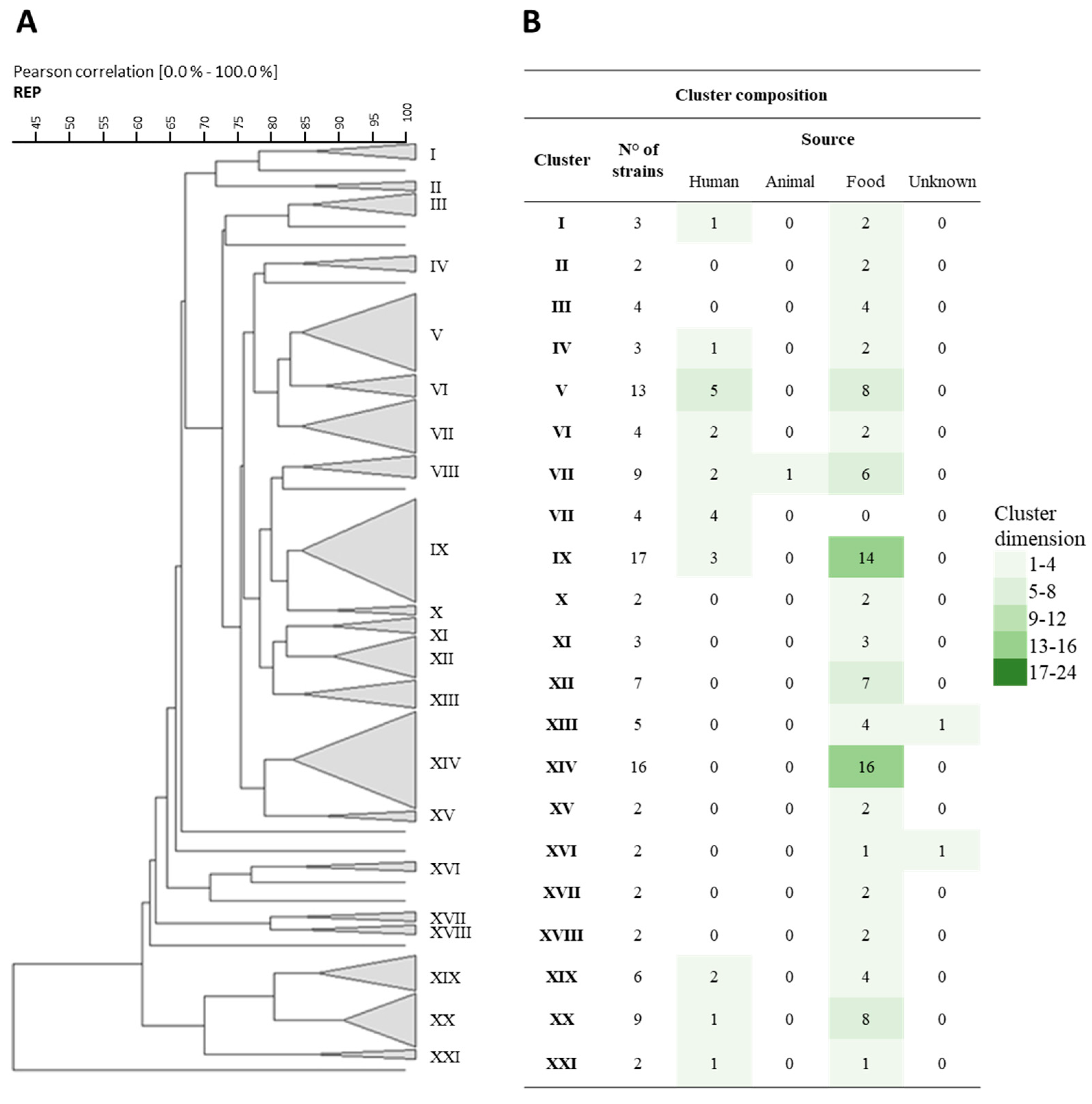
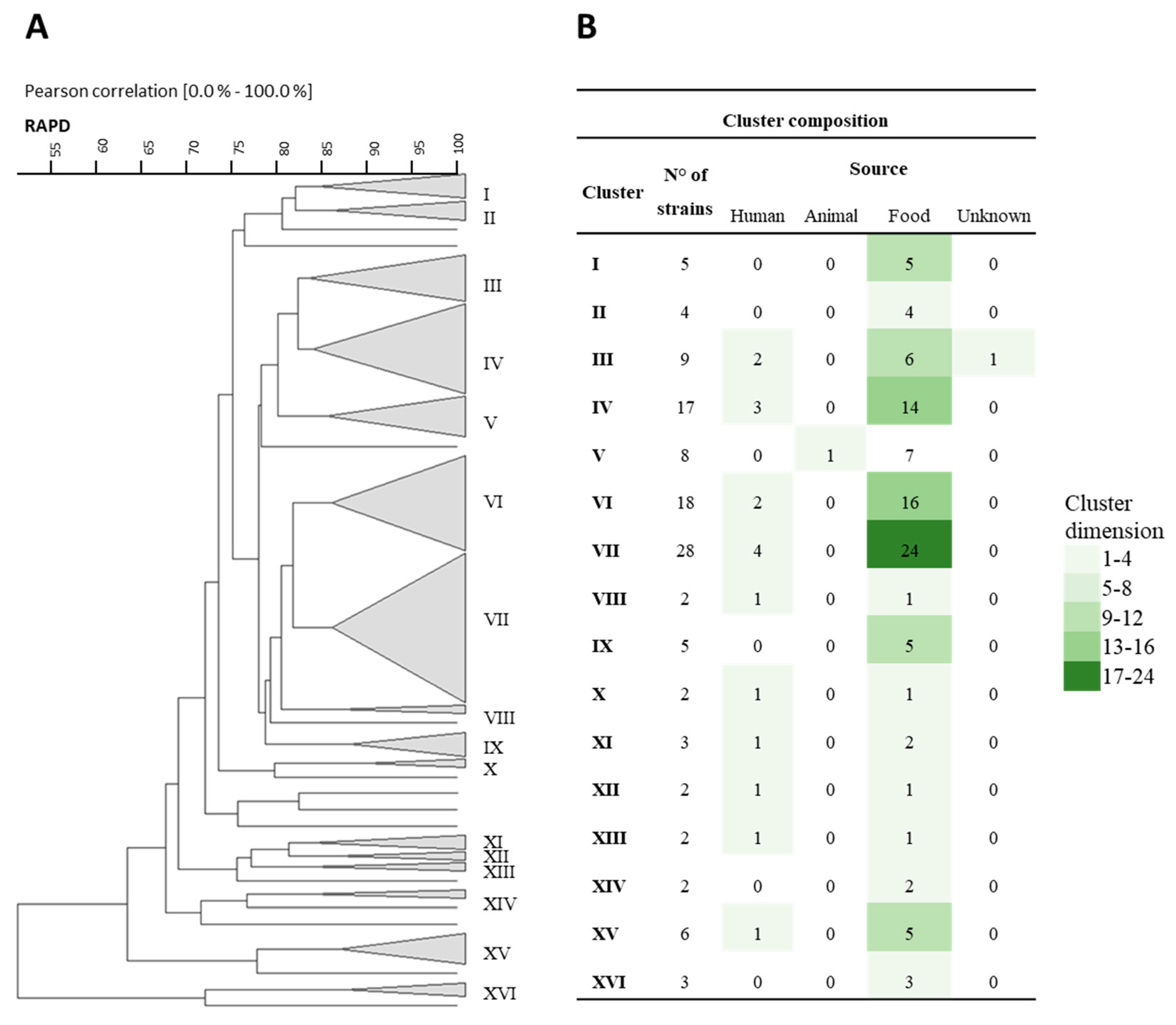
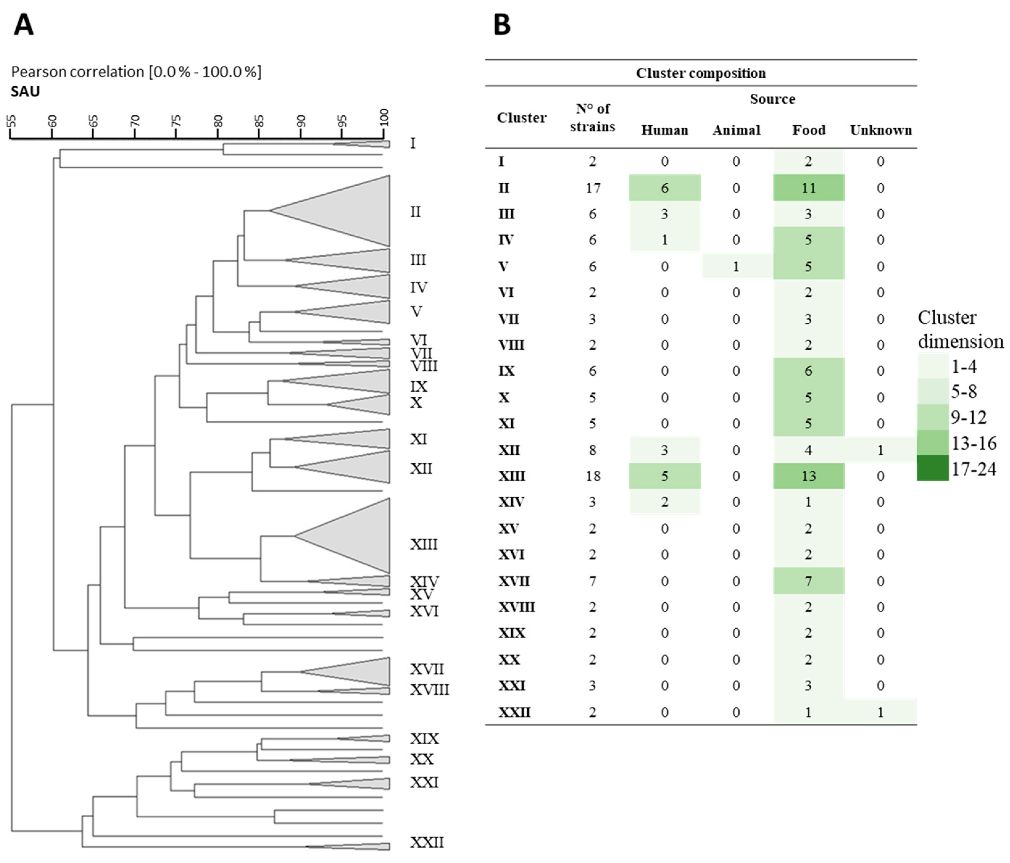
Figure 1. (A) Cluster analysis using Pearson product-moment correlation coefficients and unweighted pair group method using an arithmetic average (UPGMA) of the profiles obtained by Rep-PCR analysis of the different tested strains. A similarity coefficient of 83% was arbitrarily chosen. The identified clusters are indicated with Roman numerals. Strain DBPZ0420 did not provide a comparable Rep profile to the other strains, so it was not included in this figure. (B) The table within the figure shows the composition of the clusters, also in relation to their source of isolation.
Nutrients 16 02212 g001
Figure 2. (A) Cluster analysis using Pearson product-moment correlation coefficients and unweighted pair group method using an arithmetic average (UPGMA) of the profiles obtained by RAPD-PCR analysis of the different tested strains. A similarity coefficient of 83% was arbitrarily chosen. The identified clusters are indicated with Roman numerals. (B) The table within the figure shows the composition of the clusters, also in relation to their source of isolation.
Nutrients 16 02212 g002
Figure 3. (A) Cluster analysis using Pearson product-moment correlation coefficients and unweighted pair group method using an arithmetic average (UPGMA) of the profiles obtained by the SAU-PCR analysis of the different tested strains. A similarity coefficient of 87% was arbitrarily chosen. The identified clusters are indicated with Roman numerals. (B) The table within the figure shows the composition of the clusters, also in relation to their source of isolation.
Nutrients 16 02212 g003
Figure 4. Stress resistance results for pH, NaCl (%), and ethanol (%) expressed as relative percentage of resistant, intermediate, or sensitive strains.
Nutrients 16 02212 g004
Figure 5. Percentage of Lbs. casei and Lbs. paracasei strains that showed competition (A) against tested pathogens and production of organic acids (B).
Nutrients 16 02212 g005
Figure 6. Heatmap of strains obtained by the comparison of the several characteristic studied. Each number indicates a characteristic: 1 to 13, antibiotic resistance (cefoperazone 30 µg, cefazolin 30 µg, chloramphenicol 10 µg, clindamycin 10 µg, erythromycin 30 µg, kanamycin 30 µg, ofloxacin 5 µg, quinupristin/dalfopristin 15 µg, rifampicin 30 µg, streptomycin 25 µg, tetracycline 10 µg, tobramycin 10 µg, and vancomycin 30 µg); 14 to 22, growth abilities (pH 3.2, 3.8, 4.2, and 4.6; NaCl 2%, 4%, and 6.5%; and ethanol 12% and 15%); 23, MCO production; 24 to 27, BA production evaluated using the Bover-Cid et al., 1999, method (histamine, tyramine, putrescine or agmatine, and cadaverine); 28 to 31, genes involved in BA production (the tyrdc, odc, agdi, and hdc genes); and 32 to 34, genes involved in ethyl carbamate production (arcA, arcB, and arcC genes). Strain DBPZ0420 did not provide a comparable Rep profile to the other strains, so it was not included in this analysis. The different colours in the left side underline the different clusters A (Nutrients 16 02212 i001), B (Nutrients 16 02212 i002), C (Nutrients 16 02212 i003), and D (Nutrients 16 02212 i004).
Nutrients 16 02212 g006
Table 1. List of strains studied in this work, with their isolation source.
OriginGiven Identification
Raw and heat-treated milk, yogurt, milking machinesLbs. paracasei: LMG9192 1, DSM5622 2, P1E6 3, P2P3 3, Mo4 14, DialYac1 14, DialYac2 14, DialYac3 14, DialYac4 14, DialYac5 14, DialYac6 14, DialYac7 14, DialYac8 14, DialDan1 14, DialDan2 14, DialDan3 14, DialDan4 14, DialDan5 14, DialDan6 14, DialDan7 14, DialDan8 14
Lbs. paracasei subsp. tolerans: LMG9191 1, DSM20258 2
Green, creamy, seasoned cheeses
(Italian cheeses: Scamorza, Parmigiano Reggiano, Grana Padano, Spressa, Asiago, Montasio, Canestrato di Moliterno, Morlacco, Bellunese, Pecorino, Caciocavallo, Provolone, Emmenthal, Raclette de Savoie; Chinese and Tunisian cheeses)
Lbs. zeae: CI4368 11
Lbs. paracasei: LMG 6904 1, LMG25880 1, LMG25883 1, LMG12164 1, DBPZ0420 7, DBPZ0421 7, DBPZ0422 7, DBPZ0424 7, DBPZ0434 7, DBPZ0435 7, DBPZ0450 7, DBPZ0451 7, DBPZ0472 7, DBPZ0475 7, DBPZ0476 7, DBPZ0477 7, DBPZ0478 7, DBPZ0635 7, DBPZ0733 7, DBPZ0734 7, M266 7, M268 7, M299 7, M308 7, M348 7, M354 7, M359 7, S1 7, S3 7, V3 7, W11 7, DSG03 7, DSG05 7, DSG07 7, ESG10 7, HSG09 7, PSG06 7, PSG09 7, PSG10 7, P71 8, TH1229 8, SP57 8, L24 8, TH406 8, FSL346 9, FSL451 9, DBPZ0436 7, TMW1.1444 5, TMW1.1259 5, LACcas7 6, Cst7 10, 3LC 10, DBPZ0718 7, CF143 11, R61 12, F17 12, N24 14, H12 12
Fermented sausagesLbs. paracasei: CTC1675 13
SourdoughsLbs. casei: DBPZ0571 7
Lbs. paracasei: DBPZ0561 7, DBPZ0572 7, Q2 7, Q4 7, I1 4, I2 15, DBPZ0563 7, DBPZ0564 7, DBPZ0579 7, I3 15
Wine, must, cellar equipmentLbs. paracasei: LMG11961 1, LMG11963 1, LMG13717 1, LMG13731 1, B061 16, B161 16, B169 16, B171 16, B195 16, B196 16, B350 16
Beer, maltLbs. paracasei: LACcas25 6, LACcas29 6, TMW 1.300 5
Corn steep liquorLbs. zeae: DSM20178 2
Humans
(saliva, dental caries, blood, urethra, faeces of infants and adults)
Lbs. casei: LMG23516 1, N87 15, N811 15, DSM4905 2, N2014 15
Lbs. paracasei: DSM20020 2, LMG9438 1, LMG11459 1, LMG23511 1, LMG23518 1, LMG23523 1, LMG23538 1, LMG23543 1, LMG24098 1, LMG24101 1, LMG24132 1, DBTA34 17, N161 15, N42 15, N44 15, N76 15,
UnknownLbs. paracasei: NRRL B-456 18
1 LMG: BCCM/LMG, Belgian Co-ordinated Collections of Micro-organisms (BCCM™), Ghent, Belgium. 2 DSM: DSM, Deutsche Sämmlung von Mikroorganismen und Zellkülturen, Braunschweig, Germany. 3 Dipartimento di Agraria, Università degli Studi di Sassari, Sassari, Italy. 4 Harmonium International Inc., Mirabel, QC, Canada. 5 Lehrstuhl für Technische Mikrobiologie, Technische Universität München, Freising, Germany. 6 Dipartimento di Scienze e Tecnologie Alimentari e Microbiologiche, Università degli Studi di Milano, Italy. 7 Scuola di Scienze Agrarie, Alimentari e Ambientali, Università degli Studi della Basilicata, Potenza, Italy. 8 Università degli Studi di Verona, Dipartimento di Biotecnologie, Strada le Grazie 15, Verona, Italy. 9 Istituto Zooprofilattico Sperimentale della Sardegna, Sassari, Italy. 10 Istituto sperimentale Lattiero Caseario—I.L.C., Lodi, Italy. 11 Dipartimento di Scienze e Tecnologie Agro-Alimentari, Unversità degli Studi di Bologna, Bologna, Italy. 12 Dipartimento di Scienze delle Produzioni Agrarie e Agroalimentari, Università degli Studi di Catania, Catania, Italy. 13 Institut de Recerca I Technologia Agroalimentaries (IRTA), Lleida, Spain. 14 Dipartimento di Scienze degli Alimenti, Università degli studi di Udine, Udine, Italy. 15 Dipartimento di Agricoltura, Ambiente e Alimenti, Unversità degli Studi del Molise, Campobasso, Italy. 16 Institute for Wine Biotechnology Department of Viticulture and Oenology, Stellenbosh University, South Africa. 17 Dipartimento di Biotecnologie, Università degli Studi di Verona, Verona, Italy. 18 ARS Culture (NRRL) Collection, United States Department of Agriculture, USA.
Table 2. Differences in antibiotic resistance traits using Lbs. casei LMG23516 as reference strain.
StrainOriginAntibiotics
CFP30KZ30C10DA10E30K30OFX5QD15RD30S25TE10TOB10VA30
Lbs. casei LMG 23516Human faecesSSSSSRRSSRSRR
Lbs. paracasei DBPZ0478CaciocavalloRRRSRRRSSRRRR
Lbs. paracasei M308Canestrato MoliternoSSSISRRISISRS
Lbs. paracasei DialDan8YogurtSSSSIRSSSSSIS
Lbs. paracasei DialYak4YogurtSSSSRRISSRSSR
Lbs. paracasei DialYak5YogurtSSSSRRISSRSSR
Lbs. paracasei DialYak6YogurtSSSSSRISSRSRR
Lbs. paracasei DialYak7YogurtSSSSRRISSRSSR
Lbs. paracasei DialYak8YogurtSSSSRRISSRSSR
Legend: CFP30, Cefoperazone 30 µg; KZ30, Cefazolin 30 µg; C10, Chloramphenicol 10 µg; DA10, Clindamycin 10 µg; E30, Erythromycin 30 µg; K30, Kanamycin 30 µg; OFX5, Ofloxacin 5 µg; QD15, Quinupristin/Dalfopristin 15 µg; RD30, Rifampicin 30 µg; S25, Streptomycin 25 µg; TE10, Tetracycline 10 µg; TOB10, Tobramycin 10 µg; VA30, Vancomycin 30 µg. S—red colour, sensitive; R—green colour, resistant; I—yellow colour, intermediate resistance.
Table 3. Differences between detected genes and positive reaction in decarboxylase medium in Lbs. paracasei strains. Legend: +, positive; −, negative.
GeneBiogenic Amine
StraintyrdcodcagdihdcTymAgm or PutrPutrCad
NRRL B-456+
B085+
B161+
B169+++
B195++
LacCas7+
B350+
PSG10+++
N2014++
Table 4. Strains positive for multicopper oxidase activity.
StrainSourceStrainSource
Lbs. zeae DSM20178Corn steep liquorLbs. paracasei R61Pecorino cheese
Lbs. casei N87Body excretaLbs. paracasei F17Pecorino cheese
Lbs. casei N2014Body excretaLbs. paracasei H12Pecorino cheese
Lbs. paracasei NRRL B-456UnknownLbs. paracasei N76Body excreta
Lbs. paracasei DBPZ0564Altamura sourdoughLbs. paracasei B195Wine
Lbs. paracasei DBPZ0572Altamura sourdoughLbs. paracasei LACcas25Elixir
Lbs. paracasei V3Canestrato MoliternoLbs. paracasei TMW 1.1444Pecorino cheese
Lbs. paracasei ESG10Parmigiano ReggianoLbs. paracasei TMW 1.1259Parmesan cheese
Disclaimer/Publisher’s Note: The statements, opinions and data contained in all publications are solely those of the individual author(s) and contributor(s) and not of MDPI and/or the editor(s). MDPI and/or the editor(s) disclaim responsibility for any injury to people or property resulting from any ideas, methods, instructions or products referred to in the content.

