Review

Role of Gut Microbial Metabolites in the Pathogenesis of Primary Liver Cancers

by
Maria Pallozzi
1,
Valeria De Gaetano
1,
Natalia Di Tommaso
1,
Lucia Cerrito
1,
Francesco Santopaolo
1,
Leonardo Stella
1,
Antonio Gasbarrini
1,2 and
Francesca Romana Ponziani
1,2,*
1
Liver Unit, Centro Malattie dell’Apparato Digerente (CEMAD), Medicina Interna e Gastroenterologia, Fondazione Policlinico Universitario Gemelli Istituto di Ricovero e Cura a Carattere Scientifico, IRCCS, 00168 Rome, Italy
2
Dipartimento di Medicina e Chirurgia Traslazionale, Università Cattolica del Sacro Cuore, 00168 Rome, Italy
*
Author to whom correspondence should be addressed.
Nutrients 2024, 16(14), 2372; https://doi.org/10.3390/nu16142372
Submission received: 29 May 2024 / Revised: 8 July 2024 / Accepted: 10 July 2024 / Published: 22 July 2024
(This article belongs to the Special Issue Diet, Gut Microbiota, and Gastrointestinal Disease)

Abstract

:
Hepatobiliary malignancies, which include hepatocellular carcinoma (HCC) and cholangiocarcinoma (CCA), are the sixth most common cancers and the third leading cause of cancer-related death worldwide. Hepatic carcinogenesis is highly stimulated by chronic inflammation, defined as fibrosis deposition, and an aberrant imbalance between liver necrosis and nodular regeneration. In this context, the gut–liver axis and gut microbiota have demonstrated a critical role in the pathogenesis of HCC, as dysbiosis and altered intestinal permeability promote bacterial translocation, leading to chronic liver inflammation and tumorigenesis through several pathways. A few data exist on the role of the gut microbiota or bacteria resident in the biliary tract in the pathogenesis of CCA, and some microbial metabolites, such as choline and bile acids, seem to show an association. In this review, we analyze the impact of the gut microbiota and its metabolites on HCC and CCA development and the role of gut dysbiosis as a biomarker of hepatobiliary cancer risk and of response during anti-tumor therapy. We also discuss the future application of gut microbiota in hepatobiliary cancer management.

1. Introduction

Worldwide, hepatobiliary malignancies are the sixth most common cancer and the third leading cause of cancer death [1]. Hepatobiliary cancers include hepatocellular carcinoma (HCC), which is the fourth leading cause of cancer-related death and the main cause of death in patients with compensated chronic liver disease [2]. Cholangiocarcinoma (CCA) is the second most frequent primary liver cancer following HCC, and it accounts for up to 15% of primary hepatic tumors [3]. The gut microbiota is a key regulator of host homeostasis. In recent years, the disruption of the gut–liver axis and gut dysbiosis have been shown to play a critical role in promoting liver diseases.
The gut microbiota is made by the complex collection of various microorganisms (bacteria, archaea, eukarya, and viruses) that colonize the intestine and cooperate in multiple functions, such as host nutrient metabolism, xenobiotic and drug metabolism, immune modulation, and protection against pathogens through the maintenance of the structural integrity of the intestinal barrier [4]. The advances in anaerobic culture techniques and the development of culture-independent approaches (i.e., gene sequencing) have led to a better understanding of the composition and function of the “healthy” gut microbiota [5], which is mainly composed of seven phyla (Firmicutes, Bacteroidetes, Proteobacteria, Actinobacteria, Verrucomicrobia, Fusobacteria, and Cyanobacteria, with a predominance of Firmicutes and Proteobacteria) [6,7]. In homeostatic conditions, Firmicutes and Bacteroidetes represent 90% of the bacteria resident in the gastrointestinal tract, followed by Actinobacteria. From the oral cavity to the rectum, the number, density and diversity of microbiota varies. This balance is highly influenced by multiple factors, and it is disrupted during inflammation, infection or metabolic-associated conditions, such as alcohol consumption, unhealthy diet, obesity, and diabetes [8]. A high grade of gut dysbiosis has also been reported in patients with cancer, suggesting a causative role for gut dysbiosis in oncogenesis through multiple pathways [9,10]. A strict association between Helicobacter pylori infection and gastric cancer has been extensively demonstrated [11], and an increase in the abundance of Bacteroidetes, Firmicutes and Proteobacteria has been associated with esophageal cancer development [12]. Colorectal cancer occurrence is also influenced by the overgrowth of Bacteroides fragilis, Clostridium septicum, Escherichia coli [13], Enterococcus faecalis [14], Fusobacterium spp. and Streptococcus bovis, as described by many studies [15,16]. The gut microbiota can also influence tumorigenesis through the production of metabolites that may originate directly from bacteria or the bacterial transformation of dietary compounds or host-produced molecules; these include short-chain fatty acids (SCFAs), branched-chain amino acids (BCAAs), trimethylamine N-oxide (TMAO) and bile acids (BAs) [17]. In this review, we describe the role of the gut microbiota and its metabolites in the pathogenesis of HCC and CCA, and we also report the evidence on the predictive and prognostic importance of gut dysbiosis as a novel biomarker.

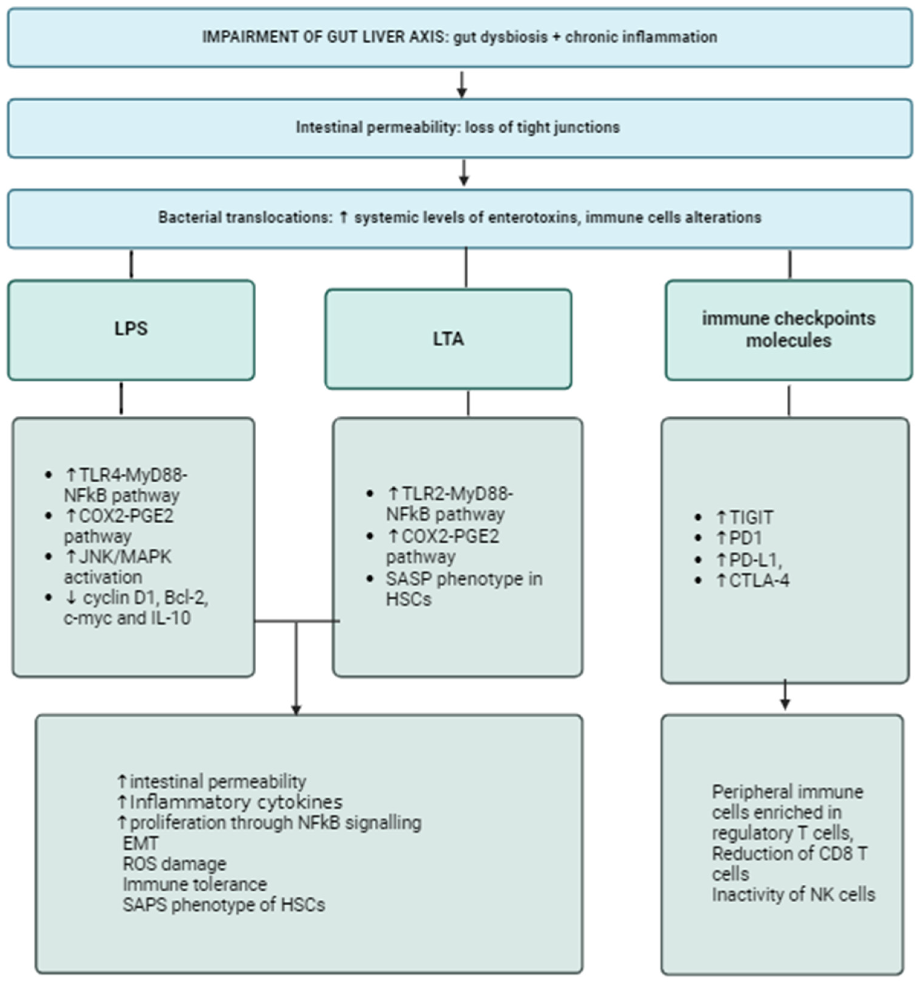
1.1. The Gut-Biliary-Liver Axis: Between Inflammation and Immunosuppression

One first role of gut microbiota in promoting HCC is linked to impaired functioning of the gut–liver axis. The gut and the liver are functionally and anatomically linked by the gut–liver axis, which includes the intestinal barrier and consists of vascular and lymphatic structures, as well as molecular and immune pathways that are deeply influenced by the gut microbiota [18,19]. The intestinal barrier is made up of multiple layers (the epithelium, the lamina propria, and the endothelium) that influence each other and present specific functions for the preservation of host homeostasis [20]. In healthy conditions, the intestinal barrier prevents the translocation of the gut bacteria and their products into systemic circulation. Any perturbation that produces an imbalance between the intestinal barrier and the gut microbiota affects the gut–liver axis and acts as a trigger for several liver diseases, including liver cirrhosis and HCC [21]. It is not surprising that gut dysbiosis represents the main driver in the gut–liver axis derangement, as it remarkably influences the intestinal barrier integrity and, as a consequence, pathological bacterial translocation (BT) is favored [22]. The presence of dysbiosis increases the concentration of enterotoxins in the gut, which stimulates the inflammatory response and increases intestinal barrier leakiness through the disruption of epithelial tight junctions (TJs). It is reported that elevated levels of zonula occludens 1 (ZO-1) are associated with increased intestinal permeability and disease severity in HCC [23]. Bacterial products such as the endotoxin lipopolysaccharides (LPSs) are also released in systemic circulation, leading to low-grade chronic inflammation [24,25]. LPSs are involved in several pathways that converge on HCC; interestingly, patients with liver cirrhosis and those with HCC show higher levels of circulating LPSs, according to animal models of carcinogen-induced hepatocarcinogenesis [26]. LPSs directly damage TJs through the activation of the Toll-Like Receptor (TLR) 4-Myeloid differentiation primary response 88 (MyD88) pathway, which activates myosin light chain kinase and enhances intestinal permeability. Moreover, through this same pathway, LPS acts as an inflammatory trigger for the liver, stimulating Kupffer cells to release pro-inflammatory cytokines such as interleukin (IL)-1 beta, IL-6, and tumor necrosis factor (TNF) alpha via the activation of the nuclear factor kappa B (NF-kB) cascade [27,28,29]. Also, Gram-positive bacteria influence systemic inflammation and carcinogenesis through the release of lipoteichoic acid (LTA), which acts on TLR-2 and converges eventually on Myd88. Indeed, in an obesity-driven murine model of HCC, the activation of TLR-2-Cyclooxygenase-2 (COX-2) [30] by LTA produced by Gram-positive bacteria was proven to favor hepatocarcinogenesis through two different mechanisms: the senescence of hepatic stellate cells (HSCs) and the modification of bile acid composition [31,32,33]. Moreover, gut dysbiosis and gut-derived products stimulate HSCs to express the senescence-associated secretory phenotype (SASP), reprogramming their transcriptome towards the production of inflammatory cytokines, such as IL-1 beta, as observed in mice models of non-fibrotic HCC after the establishment of diet-induced gut dysbiosis [32]. To analyze the possible contribution of HSC senescence and HCC development, Yoshimoto and colleagues used a mice model knock-out for the gene of IL-1 beta, a cytokine associated with both cancerogenesis and tumor suppression since it acts on the tumor microenvironment (TME) [32,34,35], and observed a parallel reduction in activated HSCs and the number of liver tumors [32,35]. Concerning the LTA/TLR-2/COX-2 pathway, it promotes the activation of prostaglandin E2 (PGE2) in HSCs, which is associated with a reduced anti-tumor response in the TME [36]. Moreover, TLR-4 triggers proliferative and antiapoptotic signals in resident hepatocytes of non-myeloid origin that are involved in cell growth and angiogenesis through the activation of STAT3 and its downstream genes cyclin D1, Bcl-2, c-myc, and IL-10 [25,37]. The activation of janus kinase/mitogen-activated protein kinase (JNK/MAPK) signaling by LPSs in HCC cells via TLR-4 results in enhanced invasive capacity and an epithelial–mesenchymal transition [38]. The interplay between low-grade chronic inflammation and gut dysbiosis also impacts immune system efficacy [39,40,41]. This has been observed in colorectal cancer, where gut dysbiosis, represented by a higher abundance of Fusobacterium nucleatum, has been associated with the impairment of natural killer (NK) cell expression and activity and a reduction in cluster differentiation (CD)-3+ T cells. These changes resulted from the direct interaction between F. nucleatum and the immune checkpoint “T cell immunoglobulin and immunoreceptor tyrosine-based inhibitory motif” (TIGIT), which is expressed on the surfaces of immune cells to downregulate their activity [42]. Bacterial translocation is a key player in this mechanism, as high circulating levels of bacterial extracts from patients with HCC injected in healthy patients induce a peculiar pattern of lymphocytes in the peripheral blood mononuclear cells, which are enriched in regulatory T cells and poor in cytotoxic CD-8 T cells [37,43,44]. How dysbiosis influences the anti-tumor response is not completely defined. Bacteria can stimulate immune cells differently. Many examples of the regulatory effect of the gut microbiota on immune cells through the upregulation of immune checkpoint molecules are reported in HCC patients: the overexpression of Programmed Death 1 (PD-1), Programmed Death Ligand 1 (PD-L1) and Cytotoxic T-Lymphocyte Antigen 4 (CTLA-4) leads to an increase in regulatory T cells, while CD 4+ T cells, CD 8+ T cells and NK cells are downregulated [45]. In patients with HCC, Bacteroidetes and E. coli were associated with higher levels of these antagonists of the immune response, resulting in a reduced efficacy of immunotherapy [45,46,47]. Gut-derived metabolites may have a role in this interaction; in particular, SCFAs inhibit the histone deacetylase, which limits the expression of PD-1 and results in its upregulation, but their role has not been clarified yet [48,49,50,51]. Also, primary bile acids promote the accumulation of CXC receptor 6+ NK T cells in the liver, which favors tumor inhibition, whereas secondary bile acids have opposite effects [52]. Changes in the gut–liver axis and gut dysbiosis are considered key players in the development of chronic disease of the biliary tract. Preclinical studies demonstrated that in healthy conditions, bile ducts are sterile; intestinal barrier disruption causes the translocation of bacteria and LPSs in the portal circulation and in the biliary tract. In this condition, gut microbiota and its products are able to activate TLRs exposed by cholangiocytes. In response to chronic TLR activation, cholangiocytes produce a variety of inflammatory cytokines, such as IL-1, IL-6, IL-8, TNF, transforming growth factor (TGF), and interferon gamma (IFN gamma), leading to an aberrant reaction that results in fibrosis, bile cell proliferation, and the generation of an immunosuppressive milieu [53,54]. In particular, the upregulation of myeloid-derived suppressor cells (MDSCs) causes the inhibition of cytotoxic T-lymphocytes and influences tumor replication, intravascular invasion, and metastasis. Hepatocytes contribute to this process by secreting CXC motif chemokine ligand 1 (CXCL1), which interacts with the CXC chemokine receptor on the aforementioned myeloid cells [55]. Thus, the chronic activation of TLR-4 and the upregulation of its gene correlates with CCA occurrence and progression [56,57]. Another possible mechanism involved in cholangiocarcinogenesis is molecular mimicry, observed in autoimmune conditions such as primary biliary cholangitis; this consists of the aberrant production of autoantibodies that are very similar, have bacterial and virus epitopes, and are able to activate effector T cells. According to this hypothesis, autoantibodies participate in the promotion and maintenance of liver inflammation and tumorigenesis [58,59,60].

1.2. Gut Dysbiosis and Hepatobiliary Carcinogenesis

Liver cirrhosis is characterized by a profound derangement of the gut microbiota composition. In particular, while bacterial alpha diversity and the abundance of Lactobacilli and Bifidobacteria is reduced, a marked increase in Enterobacteriaceae, Enterococci, Bacteroides, and Ruminococcaceae is observed [61,62,63]. Aerobic Gram-negative bacteria, specifically E. coli and other Enterobacteriaceae, have been identified as the most frequently involved in bacterial translocation and the major source of serum endotoxin [64]. On the other hand, anaerobic bacteria such as Bifidobacteria and Lactobacilli limit the overgrowth of potentially invasive microorganisms and exert ant-inflammatory and homeostatic properties [65]. In patients with HCC, an abundance of E. coli [66], Actinobacteria and other bacteria that produce LPSs has been observed with a reduction in butyrate-producing ones [62]; moreover, stools from patients with non-viral HCC harbored more pro-inflammatory bacteria, such as Escherichia, Shigella and Enterococcus, and less Faecalibacterium, Ruminococcus, and Ruminoclostridium spp., which instead produce anti-inflammatory compounds such as SCFAs [67]. Gut dysbiosis is a key player in the development of HCC in patients with NAFLD. In overweight patients, bacterial overgrowth syndrome is observed, and this is associated with higher levels of LPS released, with consequent hyperstimulation of inflammation. Inflammation also stimulates cell proliferation and influences immune cell downregulation. Intestinal permeability is another key regulator of this process, with a high release of LPSs into circulation, which is associated with the progression from NAFLD to NASH [32,68]. Modifications in gut microbiota have been associated with NAFLD, with significant changes occurring due to the evolution of liver fibrosis. In particular, it is observed that there is a loss of bacterial diversity associated with the depletion of Lactobacillus and Bifidobacterium spp. in association with the increase in Ruminococcus and Escherichia spp. when fibrosis is detected. In this condition, changes in gut-derived metabolites are mandatory, in particular, BA changes are crucial in the development of obesity-associated HCC [69,70]. In obesity, higher levels of DCA are observed, and this stimulates the expression of the SASP in hepatic stellate cells, which release inflammatory cytokines that raise the risk of HCC. Similar effects are observed in HCC that occurred in patients with non-alcoholic steatohepatitis. The activation of TLR in NASH models by LPS or bacterial DNA also activates HSCs, leading to fibrosis [69,70]. In patients with non-alcoholic steatohepatitis (NASH)-related cirrhosis and HCC, Bacteroides and Ruminococcaceae are increased, with a relative reduction in Bifidobacterium and Akkermansia [63]. Ren et al. analyzed fecal samples of 75 patients with cirrhosis and early HCC, 40 cirrhotic patients and 75 healthy controls from a heterogeneous Chinese population to identify possible genomic biomarkers of HCC. Patients with early HCC showed a greater microbial diversity and an increase in Actinobacteria phylum and Gemmiger, Parabacteroides and Paraprevotella genera in contrast to cirrhotic controls; moreover, butyrate-producing bacteria were decreased while LPS-producing bacteria were increased [62]. Also, an increased fecal abundance of Gram-negative E. coli has been found in the stool of cirrhotic patients with HCC, and this may lead to enhanced intestinal and hepatic inflammation mediated by the LPS-TLR4-NF-kB pathway. This was recapitulated in a mouse model of diethylnitrosamine (DEN) plus carbon tetrachloride (CCl4)-induced HCC, as animals with nonfunctional TLR-4 (TLR4-mut) had a significant reduction in tumor number and size but no difference in tumor incidence when compared to TLR-4 wild-type mice exposed to the same oncogenic toxins [37]. Gut sterilization obtained by the administration of antibiotics decreased LPS serum levels and was associated with an effect on tumor size and number similar to TLR-4 mutation, further confirming that gut microbiota-driven inflammation favors HCC progression [37]. Recent studies have also focused on the role of intratumor microbiota [65,66]. Li et al. [67] analyzed tissues derived from 29 patients with HBV-related HCC using next-generation sequencing (NGS) and discovered a lower bacterial diversity in tumor samples compared to non-tumor ones. HCC tissues were also divided into two subtypes: one with bacteria predominance (Bacteria-T) and the other with virus predominance (Virus-T). In Bacteria-T, E. coli, Shigella dysenteriae, Babesia bigemina and Pannonibacter phragmitetus were enriched, while HBV was prevalent in Virus-T. Bacteria-T HCC was characterized by larger size and capsular invasion and a higher prevalence of M2 macrophages in the TME compared to Virus-T [71,72]. Compared to patients with HCC or liver cirrhosis, the gut microbiota of patients affected by intrahepatic CCA (iCCA) is enriched in four genera (Lactobacillus, Actinomyces, Peptostreptococcaceae and Alloscardovia), and Ruminococcaceae abundance was positively associated with vascular invasion [72]. In vitro experiments showed that the most described intratumor microbiome was composed of Burkholderiales, Pseudomonadales, Xanthomonadales, Bacillales and Clostridiales. Moreover, Paraburkholderia fungorum was associated with CA19-9 levels and located in the paracancerous tissues [73]. Other bacteria involved in cholangiocarcinogenesis are H. pylori, H. hepaticus and Opisthorchis viverrini, and the most abundant genera identified in the biliary microbiota were Enterococcus, Streptococcus, Bacteroides, Klebsiella and Pyramidobacter spp. [74]. Figure 1 summarizes the relationship between the impairment of gut–liver axis homeostasis and hepatic tumorigenesis. Table 1 synthesizes the main features of gut microbiota composition in primary liver cancers.

2. Gut Metabolome

While the “direct” effect of bacterial products on inflammation and tumorigenesis has been well established, understanding the role of gut metabolites in the pathogenesis of primary hepatobiliary cancers is a page yet to be written. The gut microbiota acts as a bioreactor of metabolic functions through the enzymatic transformation of endogenous and exogenous substances into its metabolites, influencing host homeostasis. Gut dysbiosis alters the levels of metabolites, including those involved in carcinogenesis, causing dramatic consequences [75,76]. Data on the role of gut-derived metabolites in carcinogenesis are accumulating; here, we describe the main results on the role of BAs, SCFAs, ethanol, BCAAs and indole-derived products.

2.1. Bile Acids

BAs are steroid derivatives of amphipathic molecules produced in the liver and are transformed by the gut microbiota into signaling agents. Despite the fact that the production of primary BAs is a prerogative of the host, secondary BAs are derived from an active transformation mediated by the gut microbiota through a process involving the deconjugation, dehydroxylation and dehydrogenation of primary BAs mediated by colonic bacteria with bile salt hydroxylate activity [77]. As a consequence, BAs may be assimilated into gut-derived metabolites with significant signaling activity. In healthy conditions, primary BAs are transported with the bile in the intestine, where they are converted into secondary BAs. BAs participate in several functions, such as the elimination of cholesterol and catabolites, the emulsification of fat-soluble vitamins, the regulation of intestinal motility, and the maintenance of balance in the small intestinal and biliary tract microbiota [31,33,77]. In the colon, the gut microbiota participates in debinding water from BAs as well as in their deconjugation, dihydroxylation, and dehydrogenation. Regarding deconjugation, Bacteroides, Clostridium, Lactobacillus, Bifidobacterium, Listeria, and Escherichia spp. are involved in this process, showing bile salt hydrolase (BSH) activity [78], and unconjugated primary BAs are further dehydrogenated and 7α-dehydroxylated mainly by Clostridium spp. forming secondary BAs. In the presence of chronic inflammation, gut dysbiosis leads to an imbalance in the intrahepatic levels of BAs since it negatively influences farnesoid X receptor (FXR) expression, a key regulator of BA metabolism and signaling, resulting in hepatotoxicity [78] through the modulation of hepatic transporters. This mechanism negatively influences farnesoid X receptor (FXR) expression, leading to hepatotoxicity. Indeed, excessive levels of BAs damage the plasma membrane, activating the protein kinase C/MAPK/NFkB pathway that upregulates inflammatory cytokines such as TNF alpha, IL-1 beta, and IL-6. These cytokines counteract the apoptotic processes through the JAK-STAT3 and phosphatidylinositol 3-kinase (PI3K) pathways, favoring cell immortalization [79]. Hydrophobic secondary BAs, including cholic acid (CA), glycocholic acid (GCA), lithocholic acid (LCA), chenodeoxycholic acid (CDCA), and deoxycholic acid (DCA), are the main ones responsible for hepatocyte death [80]. Their action appears to be mediated by the activation of TNF-related apoptosis-inducing ligand receptor (TRAILR) and Fas death receptor signaling pathways [81]. Another mechanism that leads to tumorigenesis is the exaggerated activation of apoptotic pathways directly or indirectly mediated by BAs; indeed, BAs mediate the transfer of Fas-containing vesicles to hepatocyte membranes. The interaction between Fas and a death-inducing signaling complex composed of the Fas-associated death domain and procaspases 8 and 10 activates apoptosis; specifically, procaspases are activated into caspases 8 and 10, and Bax is translocated into mitochondria. Other mechanisms include cytochrome c release and caspase 9 activation [82]. Moreover, the release of cytochrome c can occur through the direct action of BAs in Bax translocation and through BA-induced reactive oxygen species (ROS) production [79]. Finally, BAs (DCA and G-CDCA) induce endoplasmic reticulum (ER) stress, resulting in the release of Ca2+ in the cytoplasm and the further promotion of oxidative stress [83]. The stimulation of proliferative pathways through the activation of an inflammatory cascade that leads to the upregulation of gene signaling is another common mechanism involved in tumorigenesis mediated by BAs: BAs upregulate Early growth response-1 (Egr-1), which is required for the development of liver inflammation during cholestasis, via MAPK signaling [80] directly or via previous activation of FXR, which heterodimerizes with RXR and modulates gene expression [84]. Furthermore, CDCA and DCA activate epithelial growth factor receptor (EGFR) and upregulate Egr-1, causing the production of vascular endothelial cell adhesion molecule-1 (VCAM-1), IL-1β, and IL-10 in hepatocytes [85]. Moreover, damage to the plasmatic membrane by toxic BAs triggers protein kinase C (PKC), which, in turn, activates the MAPK cascade, leading to the activation of NF-kB and the production of IL-6, IL-1 beta, and TNF alpha [79,86]. FXR is the most important nuclear receptor for BAs involved in the process of hepatocarcinogenesis. Another mechanism of hepatocarcinogenesis influenced by BAs is related to the inhibition of FXR signaling. FXR inhibits HCC development through the modulation of several metabolic pathways involving BAs, glucose, and lipids, the suppression of liver inflammation, the promotion of tissue repair after liver injury, the expression of partial tumor suppressor genes, and the inhibition of transcription of multiple oncogenes [87,88]. When binding to caspase 8, FXR prevents the activation and conduction of apoptotic signals in a ligand-independent pathway. During liver injury, in response to high levels of TRAIL, TNFα, and Fas ligand, FXR expression reduces, suggesting that the decrease in FXR in hepatocytes precedes apoptosis [81]. BAs can downregulate FXR through the reduction of sirtuin 1 (SIRT1), a transcriptional regulator of FXR expression in hepatocytes [89]. This results in sustained activation of the Wnt/β-catenin pathway and increased risk of hepatocarcinogenesis [90]. A diet enriched in lipids increases the levels of the gut microbiota-derived DCA, which is known to cause DNA damage. In hepatic stellate cells, DCA induces the SASP, turning in the secretion of inflammatory and tumor-promoting factors in the liver [32] and the overexpression of COX-2 and its downstream inflammatory cascade, together with TLR-2-mediated signaling [6,25,52]. BAs also influence the immune response and tolerance against tumors. Commensal bacteria are important regulators of anti-tumor immunity, and gut microbiome-mediated primary-to-secondary BA conversion can regulate the accumulation of NK T cells by CXCL16 expression in liver sinusoidal endothelial cells [91]. Primary BAs such as CDCA and toxic CA increase CXCL16 expression, which binds to its receptor CXCR6 on NKT, thereby increasing IFN gamma production and inhibiting tumor growth [6]. Lithocholic acid was also reported to influence the balance between Th17 and T regulatory cells by acting on the retinoic acid-associated orphan receptors gamma T [91]. As gut-derived metabolites may modulate the risk of HCC development acting on modulatory signaling pathways, several drugs, such as FXR agonists, are under evaluation. The inhibition of FXR functions is associated with impaired cell proliferation and DNA damage, while its activation exerts a protective role on tumorigenesis [92,93]; in a mouse model, the administration of FXR agonists (GSK2324) was able to inhibit lipogenesis, the intestinal uptake of lipids and their intrahepatic accumulation, modulating the levels of monounsaturated fatty acids, polyunsaturated fatty acids and BAs [94]. It is not known whether BA qualitative changes observed in CCA patients are a cause or a consequence of the presence of CCA since this tumor uses BA receptors and may influence BA metabolism. Preliminary evidence reports that in CCA patients, there is an increase in GCA, a decrease in secondary Bas, and changes in the ratio between glycoconjugated and tauroconjugated BAs. The tumor-associated production of IL-6 influences the expression of FXR and G Protein-coupled BA Receptor 1, known as TGR5 receptors. Patients with CCA and vascular invasion show higher levels of glycochenodeoxycholic acid (GCDCA), taurocholic acid (TCA), glycodeoxycholic acid (GDCA), taurodeoxycholic acid (TDCA) and tauroursodeoxycholic acid (TUDCA). On the other hand, in this study, the levels of IL-6, as well as those of CDCA, were lower in the group with vascular invasion than in patients without vascular invasion, which is discordant with previous observations on the association between BA accumulation, the increase in IL-6 and downregulation of FXR and cytochrome (CYP) 7A1, the key enzyme for BA synthesis. The existence of interrelationships among gut microbiota, BA profile, serum levels of cytokines, and iCCA patients’ outcomes deserves to be explored in larger studies [94]. Thus, BAs may damage the liver, stimulating carcinogenesis through the intrahepatic accumulation or the modulation of the main receptors of BAs, which leads to chronic inflammation, insufficient apoptotic processes, and aberrant cell proliferation.

2.2. Choline and TMAO

Choline is a product derived from the catabolism of dietary compounds such as meat and egg yolk. Gut microbiota promotes its metabolism into trimethylamine (TMA), mainly bacteria belonging to Firmicutes and Proteobacteria phyla (Anaerococcus hydrogenalis, Clostridium asparagiforme, C. hathewayi, C. sporogenes, E. fergusonii, Proteus penneri, Providencia rettgeri and Edwardsiella tarda) [95,96,97]. TMA obtained from this process reaches the portal circulation, and it is converted in the liver by flavin monooxygenase 3 into trimethylamine-N-oxide (TMAO). TMAO is associated with an increased risk of cardiovascular disease, as it promotes the synthesis of proinflammatory cytokines and the release of ROS from endothelial cells [98,99,100,101]. The role of choline in carcinogenesis is controversial. Dietary choline intake positively correlates with the occurrence of colorectal and prostatic cancers [102,103], even if a meta-analysis reported a protective role of this metabolite against tumorigenesis [104]. Moreover, in a recent study, high levels of dietary choline were able to reduce the risk of steatotic liver disease in humans; conversely, lower levels led to the accumulation of intrahepatic triglycerides, resulting in a higher risk of metabolic-associated disease, including HCC. [105,106,107]. Liu et al. demonstrated in a large Chinese population that patients with high plasma levels of TMAO and low choline showed a higher risk of developing liver cancers compared to individuals with high choline and low TMAO plasma levels, but the underlying mechanism has not been elucidated yet [108]. TMAO may stimulate the activation of mammalian target of rapamycin (mTOR) signaling through the upregulation of the periostin (POSTN) gene, as observed in a model of Hepa 1–6 cells and Huh7 cells exposed to TNF [109,110]. POSTN is a gene involved in tumorigenesis, the modulation of the tumor microenvironment, intravascular tumor dissemination and metastases [111]. Another trial conducted on 40 healthy men documented that fish consumption induced high TMAO production among individuals with low gut microbiota diversity and higher Firmicutes to Bacteroidetes ratio, recapitulating the essential contribution of gut microbiota in determining plasma levels of TMA and TMAO metabolites [112]. An interesting field of research is the functional interplay between BA metabolism and TMAO. The combination of TMAO and a high-fat diet (HFD) seems to have a synergistic effect on NAFLD development since TMAO could influence BA metabolism towards the increased synthesis of FXR-antagonists [113]; as we mentioned above [90], FXR inhibition is associated with spontaneous HCC development through the Wnt/β-catenin pathway. More studies are needed to understand whether dietary restrictions or gut microbiota manipulation toward lower TMA production could prevent HCC development and progression, especially in NAFLD-NASH-HCC. Preclinical studies showed that a choline-depleting diet may also stimulate CCA development. Rats treated with 2 acetylaminofluorene fed with a choline-devoid diet showed the occurrence of CCA after 6 months in 38% of cases, while a diet enriched with choline prevented tumorigenesis. Similar results were obtained using other tumor-inducing substances in association with a low-choline diet [114]. A low-choline and high-methionine diet for 6–12 months in mice caused the occurrence of primary liver tumors (both HCC and CCA). In all cases, osteopontin was upregulated and able to activate the beta-catenin signaling pathway and cause a decrease in endothelial cadherin [114]. No data about the effects of TMAO in CCA are reported in the literature. To summarize, low levels of choline and higher levels of TMAO are associated with a higher risk of liver cancer occurrence since choline influences intrahepatic fat deposition, leading to hepatotoxicity, and TMAO is involved in the stimulation of abnormal cell proliferation.

2.3. Indoles

Indoles are gut microbiota products derived from tryptophan metabolism that are produced by many bacterial species, mainly by E. coli. Indoles may cross the intestinal barrier and reach the liver, where they are converted in a two-step process into 3-hydroxyindole (indoxyl) and conjugated with sulfate in indoxyl sulfate. Moreover, molecules of indoxyl may dimerize to form the blue dye indigo [115,116]. These products act as aryl hydrocarbon nuclear receptors (AhRs). In the presence of indole derivatives (indoles), intestinal cells express AhRs, promoting immune cell development and intestinal barrier permeability. In the presence of inflammatory conditions such as chronic liver diseases, indole levels are reduced [117]. In a study by Krishnan et al., exposure to a high-fat diet significantly inhibited indole-3-acetate intestinal levels, resulting in the disruption of the intestinal barrier, overexpression of inflammatory cytokines, and inhibition of immune cells [118]. The anti-inflammatory effects of indoles may be due to the direct action on the host intestinal barrier and are partially attributed to changes in gut microbiota metabolism with no influence on its composition. Indoles also reduce fatty acid oxidation [119]. The oral supplementation of indoles reverts inflammatory processes [118] and may be evaluated in patients with chronic inflammatory diseases, including primary hepatobiliary cancers.

2.4. Short-Chain Fatty Acids

SCFAs are produced by the intestinal microbiota from the fermentation of indigestible carbohydrates. They are involved in the homeostasis of the intestinal epithelial barrier, and contribute to the maintenance of gut microbiota diversity [120,121,122,123,124]. Acetate, propionate, and butyrate are the main SCFAs produced in healthy individuals, with a molar ratio in the human colon of 60:20:20. Acetate derives from anaerobic bacteria such as A. muciniphila and Bacteroides spp; in normal conditions, it is involved in lipid synthesis, glucose metabolism, and immunity regulation [125]. Propionate is obtained during glycolysis through the succinate pathway and is commonly produced by the enzyme methylmanolyl-Coenzyme A (CoA) decarboxylase of Gram-negative bacteria such as Akkermansia muciniphila and Roseburia inulinivorans; it is involved in lipid metabolism, inhibits lipogenesis and favors liver gluconeogenesis [125,126]. Butyrate derives from Clostridium clusters IV and XIVa metabolism, and it is produced via glycolysis from the combination of two acetyl CoA molecules to form acetoacetyl CoA, which is reduced in butyryl CoA. Butyrate is finally obtained from the butyryl CoA acetate CoA transferase pathway or through the phospho butyrate and butyrate kinase pathway [125,127]. Butyrate is involved in the regulation of intestinal barrier permeability and intestinal epithelial cell metabolism, and it acts as an inhibitor of carcinogenesis in the colon [128]. SCFAs modulate human health via several pathways. First, they interact with GPR41, GPR43, and GPR109A, which activate the phospholipase C beta, the protein kinase A (PKA), and protein kinase C (PKC), leading to ERK activation. SCFAs also modulate the immune system, enhancing the production of macrophages, dendritic cells, and CD 4+ T cells. Moreover, butyrate inhibits inflammation in the colon, enhancing the production of T-regulatory cells and IL-10 through the GPR109A receptor [125,129]. In HCC patients, stool analysis revealed that SCFA-producing bacteria such as butyrate-producing Lachnospira, Ruminococcus, and Butyricicoccus are less represented [130]. Other studies showed an association of SCFAs with hepatocarcinogenesis [46]. Mice with elevated BAs and hyperbilirubinemia, when fed with inulin to induce SCFA production, showed higher liver inflammation, neutrophil influx and risk of developing HCC [131]. Enrichment in beneficial bacteria such as Blautia and Lactobacillus spp. may increase sodium butyrate levels with the aim of reducing inflammation and bile acids such as DCA [132,133]. SCFAs could also contribute to the development of an immunosuppressive microenvironment and HCC progression by promoting the production of IL-10 by microbiota antigen-specific T helper (Th) 1 cells or suppressing inflammatory macrophages in the lamina propria [134]. Indeed, some studies showed increased circulating levels of IL-10 and T-regulatory cells (T regs) after exposure to butyrate [135,136]. In a mouse model of hepatitis B virus-induced HCC, it was noted that SCFA administration reduced the number of dysplastic nodules and HCC in HBx transgenic mice, downregulating some genes critically involved in tumor progression, such as several growth factors, PI3K, Wnt, Vascular Endothelial Growth Factor (VEGF) and Ras [134]. It is recognized that SCFAs interfere with the cancer cell cycle through DNA epigenetic modifications [137] and histone deacetylase activity (HDAC) inhibition, which is a critical regulator of gene transcription that is often found to be dysregulated in cancer [138]. A recent study showed that Lactobacillus reuteri administration in HCC-recipient mice was positively associated with increased acetate plasma levels and contrasts tumor growth [139]. Also, L. reuteri could inhibit type 3 innate lymphoid cells (ILC3s), a subtype of tissue-resident lymphocytes, to produce IL-17A, a cytokine with a well-known role in favoring HCC development [139,140,141]. Researchers showed that acetate itself is able to downregulate ILC3 functions by blocking HDAC activity; thus, they documented that the combination of acetate plus anti-PD-1 therapy could enhance the immune response in HCC mice models receiving ICIs, decreasing IL-17A mRNA levels [139]. Moreover, supplementation with probiotics can be used for the same purpose; some bacteria, such as Bifidobacterium pseudolongum, produce acetate, which, through the interaction with GPR43 on hepatocytes, inhibits cell proliferation and induces apoptosis in a model of NAFLD-HCC mice [142]. Finally, SCFAs can activate the CD-8 T-cell response in patients receiving anti-PD-1 therapy by the release of IL-17 [143] and exert anti-tumor activity that inhibits CTLA-4 on dendritic cells and T cells. Butyrate, on the contrary, may stimulate an anergic phenotype that enhances T-regulatory cells. Butyrate metabolism is highly activated in patients with HCC compared with healthy individuals, resulting in reduced plasma levels of butyrate. Butyrate supplementation or inhibition of its catabolism significantly reduced liver tumorigenesis in a process that influences intracellular calcium homeostasis and the production of ROS. Moreover, higher levels of butyrate were associated with higher efficacy of tyrosine kinase inhibitor-based therapy; the administration of encapsulated pegylated butyrate and sorafenib nanoparticles prolonged drug retention time and optimized the anti-tumor response [144]. In vitro studies also demonstrated that CCA cells treated with butyrate showed a positive effect on cilia formation and acetylated tubulin levels. In HuCCT1 cell lines, migration, and mitosis were inhibited by the administration of butyrate. No effects were observed in non-tumor mature cells. Butyrate stimulates the effects of HDAC inhibition and reduces the levels of cyclin D1, vimentin, and other proteins involved in cell proliferation [145,146]. Thus, the interplay between SCFAs and HCC is complex and depends on multiple factors, including the degree of dysbiosis, the presence of other metabolites, such as BAs, and the conditioning of TME and the anti-tumor immune response [135].

2.5. Ethanol

In physiologic conditions, small amounts of endogenous ethanol are produced after the intake of alcohol-free food as metabolic intermediaries or products [147,148]. In the case of intestinal dysbiosis, the abundance of high alcohol producers such as Klebsiella pneumoniae increases, contributing to the pathogenesis of disorders such as the autobrewery syndrome or alcohol-mediated liver damage [148,149,150,151]. Also, other members of the gut microbiota, such as Proteobacteria, more specifically E. coli [149], and yeasts, such as Saccaromyces cerevisiae and Candida spp., play an important role in endogenous ethanol production [152]. The mechanisms of ethanol-induced carcinogenesis are closely related to its metabolism. Ethanol is converted into acetaldehyde by three pathways: (1) alcohol dehydrogenase (ADH), which leads to the formation of acetaldehyde and reduced nicotinamide adenine dinucleotide (NADH); (2) cytochrome P450 2E1 (CYP2E1), with the formation of reactive oxygen species; (3) catalase, but to a much lesser extent. Once formed, acetaldehyde is metabolized into acetate by acetaldehyde dehydrogenase (ALDH) [153,154]. Acetaldehyde is a well-known carcinogen since it interferes with DNA synthesis and repair [155,156,157]. Moreover, it causes structural and functional alterations to proteins to which it binds [158]. Levels of acetaldehyde do not always correlate with alcohol consumption, as the activity of ADH and ALDH is primarily responsible for the amount of acetaldehyde generated. The expression and activity of these enzymes depend on genetic factors [155]; however, the oral microbiome can also convert ethanol to acetaldehyde by oxidation, while further conversion in acetate is limited [159,160]. In the oral cavity, mainly Gram-positive aerobic bacteria and yeasts appeared associated with higher acetaldehyde production, with an increased risk of developing oral cavity malignancy [161]; moreover, compared to germ-free rats, conventional animals showed higher levels of intracolic acetaldehyde and a more pronounced mucosal injury and cellular hyper-regeneration [162]. Acetaldehyde does not appear to play a crucial role in hepatocarcinogenesis due to its effective hepatic metabolism and, consequently, low intrahepatic levels of this compound. Liver damage related to ethanol is rather driven by ROS production. As stated above, ethanol-mediated ROS formation involves CYP2E1; the chronic consumption of ethanol can induce and make this pathway predominant [163]. Moreover, NADH derived from the ADH pathway shuttles into mitochondria and generates electron leakage from the mitochondrial respiratory chain, forming ROS. ROS interact with lipids, generating lipid peroxidation products, such as trans-4-hydroxy-2-nonenal (4HNE), which can be converted to 2,3-epoxy-4-hydroxynonenal. This compound reacts with deoxyadenosine or deoxycytidine to form exocyclic etheno-DNA adducts such as 1,N6-ethenoadenine or 3,N4-ethenocytosine, causing DNA damage. To note, acetaldehyde and nitric oxide derived from inducible nitric oxide synthase induced by ethanol inhibit the DNA repair system; furthermore, acetaldehyde also increases the burden of ROS indirectly by injuring mitochondria, resulting in an inadequate reoxidation of the large quantities of nicotinamide adenine dinucleotide (NADH) that are produced through the alcohol dehydrogenase (ADH) reaction. As mitochondrial damage occurs, this cascade of events leading to apoptosis is initiated [164,165,166]. Thus, ethanol induces liver damage indirectly through the exaggerated production of ROS, which causes mitochondrial dysfunction or directly acting on DNA stability.

2.6. Branched-Chain Amino Acids

BCAAs are essential amino acids with an aliphatic chain that are involved in multiple anabolic processes, such as gluconeogenesis. They are degraded by the branched-chain aminotransferases (BCATs) 1 and 2 into branched-chain alpha ketoacids, which are fundamental for the skeletal muscle and the nervous system [167,168]. From the muscle, BCAAs may be converted by the branched-chain alpha ketoacid dehydrogenase to be released and utilized for gluconeogenesis. Free BCAAs stimulate the release of insulin and its receptor, the insulin receptor substrate (IRS1), leading to the activation of the PI3K/protein kinase B (AKT)/mTOR complex 1 (mTORC1); BCAAs may also stimulate the direct activation of the mTORC1 pathway, which is involved in cell proliferation, angiogenesis and apoptosis [167,168,169,170,171,172]. In the presence of gut dysbiosis, such as in HCC, BCAA levels increased not only in plasma but even in the tumor tissue and the surrounding area; interestingly, the downregulation of the catabolic enzymes of BCAAs is reported in tumor cells, leading to BCAA accumulation and mTORC1 activation. BCAA levels were also associated with tumor size and tumor number in a mice model of HCC. Another mechanism of HCC progression related to BCAAs is the production of mitochondrial distress through the accumulation of branched-chain ketoacids. The administration of BCAAs with diet seems to inhibit this process, suggesting that oral supplementation may enhance the anti-tumor response through the modulation of the gut microbiota; BCAAs may alter the gut microbiota composition, reducing serum levels. BCAA supplementation with diet increases the abundance of Ruminococcus flavefaciens and Bifidobacterium strains while reducing Proteobacteria, which are involved in several dysmetabolic conditions [164,165,166,167,168,169]. Intratumor degradation of valine, leucine, and isoleucine is observed in CCA due to the upregulation of BCAT 1 and BCAT 2 receptors. The levels of these amino acids are negatively associated with tumor prognosis [173]. In conclusion, dietary BCAAs may reduce the risk of tumorigenesis; on the contrary, gut dysbiosis and chronic inflammation through the modulation of enzymes involved in BCAA metabolism may cause intratumor accumulation of BCAAs, resulting in cell survival and the promotion of angiogenesis.
Table 2 and Figure 2 summarize the main gut-derived metabolites and their role in hepatocarcinogenesis.

3. Discussion and Future Perspectives

Gut-derived metabolites are gatekeepers of liver homeostasis, inflammation, and tumorigenesis. Their quantification for the diagnosis or their use for the treatment of primary liver tumors has gained attention, thanks to the progress of metagenomics and proteomics techniques [6,174]. Liquid biopsy is based on these premises, employing the detection of a combination of products in biological material (stool, blood, urine) to identify the presence of a tumor and describe its metabolic features. A well-known application of liquid biopsy has been reported in colorectal cancer. Through the assessment of a panel of gut microbiome-associated serum metabolites derived from blood and fecal samples, a specific signature in colorectal cancer has been demonstrated, as well as the possibility of discriminating adenomas from tumors. This panel was superior to the carcinoembryonic antigen marker, with a high accuracy in detecting early-stage colorectal cancer [175]. Regarding gut-derived metabolites, the catabolism of BCAAs has been reported in several types of malignancies, leading to low levels in blood or saliva, and blood tryptophan levels were altered in patients with lung cancer and colorectal and pancreatic cancers [176]. Unfortunately, no significant similar studies are reported concerning HCC, except for low levels of butyrate in patients with early HCC. Similarly, the analysis of intratumor gut microbiota and its metabolites is another field of research for both HCC and CCA, promising to give novel information on the aggressiveness and resistance of tumor cells and the likelihood of response during systemic treatment [177]. Another promising field of the application of metabolomics regards HCC systemic therapy, specifically in the evaluation of the anti-tumor response [178]. Many studies are focused on the role of the gut microbiota as a key player in immunomodulation and as a biomarker of treatment response [179]. Changes in the gut microbiota have been associated with the development of early HCC. A high Firmicutes/Bacteroidetes ratio has been associated with a lower rate of response to immunotherapy, while the Prevotella/Bacteroides ratio has shown a positive association with the response in patients receiving Nivolumab. How gut microbiota may influence the anti-tumor response is not completely defined, and it is reported that different bacteria may stimulate immune cells differently; indeed, Bacteroides fragilis is able to activate CD-4 T cells to produce IFN gamma, while Bifidobacterium enhances the response to anti-PD-L1 and anti-PD-1, thus activating CD-8 T cells [180,181,182,183,184]. Therefore, the prevalence of these species that cause the activation of the immune system may elicit anti-tumor immunity in patients who respond to immunotherapy. As a confirmation, studies also suggest that the administration of Bacteroides, the sensitization of T cells to LPS, and the modulation of the gut microbiota may revert the resistance to immunotherapy and ameliorate the anti-tumor response [185,186,187]. As reported, gut-derived metabolites such as SCFAs show similar effects and may act on other pathways linked to inflammation through the modulation of intestinal permeability, cell proliferation, apoptosis, and angiogenesis [143,145]. Similarly, the oral administration of indoles may downregulate the inflammatory pathways involved in hepatocarcinogenesis. In CCA, Jin et al. observed that gut microbiota composition may be a prognostic marker of tumor progression, as patients who rapidly progressed showed a higher abundance of phylum Pseudomonadota compared with patients with slower tumor progression [186]. In this context, fecal microbiota transplantation (FMT), correcting gut dysbiosis and providing a community of bacteria producing anti-inflammatory substances may restore the integrity of the gut barrier, stimulate the immune system, and rebalance gut microbiota-derived metabolites, favoring the engraftment of Faecalibacterium and Akkermansia spp. [188,189]. Despite these reports, few studies have analyzed the possible role of FMT on HCC promotion and evolution. However, ongoing studies are evaluating the effects of FMT on HCC therapies. Two clinical trials are evaluating whether FMT from patients with HCC sensitive to Atezolizumab/Bevacizumab may revert a phenotype of resistance to therapy in other patients affected by HCC [190]. A clinical trial of FMT has also been performed in 10 patients with primary sclerosing cholangitis, resulting in the amelioration of intestinal bacterial diversity with a sustained clinical response in the following 24 weeks, with no adverse events being reported. FMT in this contest may reduce the risk of developing CCA, but further data are needed to confirm this hypothesis [191]. Moreover, the use of intestinal microbial strains engineered to metabolize substances such as ammonia or to produce BSH represents a novel strategy to influence liver homeostasis and prevent HCC. These strains are superior compared to wild-type microbes, as they can be directed specifically to the pathogenic molecule or metabolic pathways of interest, sparing the remaining microbial community [192]. Concerning CCA prevention, another strategy may target specific gut bacteria using bacteriophages in patients with PSC, a strategy with reported efficacy in reducing bacterial translocation to bile ducts in mice models [193,194]. All these results are limited by the absence of large randomized clinical trials or meta-analyses on patients. Moreover, the current knowledge of gut microbiota and its involvement in hepatocarcinogenesis is prevalently based on animal models or on samples of fecal microbiota from patients. These models are bidimensional and limited, as they lack the continuous internal and external stimuli that, in real life, influence the metabolism and functions of gut microbiota and its production of metabolites. In the future, the progression of gene sequencing and machine learning-based data analysis will guarantee more reproducible gut microbiota signatures that potentially will also be used as biomarkers.

4. Conclusions

In conclusion, evidence suggests a promising role for the gut metabolome as a critical biomarker that must be included in patient evaluation to guarantee the best personalized therapy in the oncological setting. Despite these radiant visions, high costs and the need for multiple platforms and technologies for gut metabolome analysis still limit the diffusion of this analysis in clinical practice. The lack of large randomized clinical trials aimed at the specific analysis of gut-derived metabolites during anti-cancer therapy is another gap in knowledge that should be considered.

Author Contributions

Conceptualization, investigation, writing: M.P., V.D.G. and N.D.T.; writing—review and editing, L.C., F.S., L.S., F.R.P. and A.G.; supervision, F.R.P. and A.G. All authors have read and agreed to the published version of the manuscript.

Funding

This research received no external funding.

Acknowledgments

Thanks to the Fondazione Roma for the invaluable support for scientific research—FR-CEMAD 21–25.

Conflicts of Interest

The authors declare no conflicts of interest.

Abbreviations

Acetaldehyde dehydrogenase (ALDH), alcohol dehydrogenase (ADH), bacterial translocation (BT), bile acids (BAs), bile salt hydrolase (BSH), branched-chain amino acids (BCAAs), carbon tetrachloride (CCl4), cholangiocarcinoma (CCA), chenodeoxycholic acid (CDCA), Coenzyme A (CoA), cholic acid (CA), cluster of differentiation (CD), Cyclooxygenase-2 (COX-2), CXC motif chemokine ligand 1 (CXCL1), Cytotoxic T-Lymphocyte Antigen 4 (CTLA-4), cytochrome (CYP), deoxycholic acid (DCA), diethylnitrosamine (DEN), Early growth response -1 (Egr-1), endoplasmic reticulum (ER), farnesoid X receptor (FXR), faecal microbiota transplantation (FMT), glycocholic acid (GCA), Glycochenodeoxycholic acid (GCDA), Glycodeoxycholic acid (GDCA), G Protein-coupled BA Receptor 1 (TGR5), hepatic stellate cells (HSCs), hepatocellular carcinoma (HCC), histone deacetylase activity (HDAC), type 3 innate lymphoid cells (ILC3s), interleukin (IL), Interferon gamma (IFN gamma), intrahepatic CCA (iCCA), Janus Kinase (JNK), lipopolysaccharides (LPSs), lipoteichoic acid (LTA), lithocholic acid (LCA), mammalian target of rapamycin (mTOR), mTOR complex 1 (mTORC1); mitogen-activated protein kinase (MAPK), myeloid-derived suppressor cells (MDSCs), Myeloid differentiation primary response 88 (MyD88), natural killer (NK), next-generation sequencing (NGS), nicotinamide adenine dinucleotide (NADH), non-alcoholic steatohepatitis (NASH), nuclear factor kappa B (NF-kB), periostin (POSTN), phosphatidylinositol 3-kinase (PI3K), Programmed Death 1 (PD-1), Programmed Death Ligand 1 (PD-L1), prostaglandin E2 (PGE2), protein kinase A (PKA), protein kinase C (PKC), reactive oxygen species (ROS), senescence-associated secretory phenotype (SASP), short-chain fatty acids (SCFAs), sirtuin 1 (SIRT1), T helper (Th), T-regulatory cells (Tregs), taurocholic acid (TCA), taurodeoxycholic acid (TDCA), tauroursodeoxycholic acid (TUDCA), tight junctions (TJs), “T cell immunoglobulin and immunoreceptor tyrosine-based inhibitory motif” (TIGIT), TNF-related apoptosis-inducing ligand receptor (TRAILR), Toll-like receptor (TLR), Transforming Growth Factor (TGF), trans-4- hydroxy-2-nonenal (4HNE), trimethylamine (TMA), trimethylamine N-oxide (TMAO), tumor microenvironment (TME), tumor necrosis factor (TNF), vascular endothelial cell adhesion molecule-1 (VCAM-1), Vascular Endothelial Growth Factor (VEGF), zonula occludens 1 (ZO-1).

References

  1. Sung, H.; Ferlay, J.; Siegel, R.L.; Laversanne, M.; Soerjomataram, I.; Jemal, A.; Bray, F. Global Cancer Statistics 2020: GLOBOCAN Estimates of Incidence and Mortality Worldwide for 36 Cancers in 185 Countries. CA Cancer J. Clin. 2021, 71, 209–249. [Google Scholar] [CrossRef]
  2. Rich, N.E. Changing Epidemiology of Hepatocellular Carcinoma Within the United States and Worldwide. Surg. Oncol. Clin. N. Am. 2024, 33, 1–12. [Google Scholar] [CrossRef] [PubMed]
  3. Pascale, A.; Rosmorduc, O.; Duclos-Vallée, J.-C. New epidemiologic trends in cholangiocarcinoma. Clin. Res. Hepatol. Gastroenterol. 2023, 47, 102223. [Google Scholar] [CrossRef] [PubMed]
  4. Jandhyala, S.M.; Talukdar, R.; Subramanyam, C.; Vuyyuru, H.; Sasikala, M.; Nageshwar Reddy, D. Role of the normal gut microbiota. World J. Gastroenterol. 2015, 21, 8787–8803. [Google Scholar] [CrossRef] [PubMed]
  5. Yu, Z.; Morrison, M. Improved extraction of PCR-quality community DNA from digesta and fecal samples. Biotechniques 2004, 36, 808–812. [Google Scholar] [CrossRef] [PubMed]
  6. Luo, W.; Guo, S.; Zhou, Y.; Zhao, J.; Wang, M.; Sang, L.; Chang, B.; Wang, B. Hepatocellular Carcinoma: How the Gut Microbiota Contributes to Pathogenesis, Diagnosis, and Therapy. Front. Microbiol. 2022, 13, 873160. [Google Scholar] [CrossRef] [PubMed]
  7. Arumugam, M.; Raes, J.; Pelletier, E.; Le Paslier, D.; Yamada, T.; Mende, D.R.; Fernandes, G.R.; Tap, J.; Bruls, T.; Batto, J.M.; et al. Enterotypes of the human gut microbiome. Nature 2011, 473, 174–180. [Google Scholar] [CrossRef] [PubMed]
  8. Nardone, G.; Compare, D. The human gastric microbiota: Is it time to rethink the pathogenesis of stomach diseases? United Eur. Gastroenterol. J. 2015, 3, 255–260. [Google Scholar] [CrossRef]
  9. Osman, M.-A.; Neoh, H.-M.; Ab Mutalib, N.-S.; Chin, S.-F.; Jamal, R. 16S rRNA Gene Sequencing for Deciphering the Colorectal Cancer Gut Microbiome: Current Protocols and Workflows. Front. Microbiol. 2018, 9, 767. [Google Scholar] [CrossRef]
  10. Wei, L.Q.; Cheong, I.H.; Yang, G.H.; Li, X.G.; Kozlakidis, Z.; Ding, L.; Liu, N.N.; Wang, H. The Application of High-Throughput Technologies for the Study of Microbiome and Cancer. Front. Genet. 2021, 12, 699793. [Google Scholar] [CrossRef]
  11. Zhou, C.; Bisseling, T.M.; van der Post, R.S.; Boleij, A. The influence of Helicobacter pylori, proton pump inhibitor, and obesity on the gastric microbiome in relation to gastric cancer development. Comput. Struct. Biotechnol. J. 2023, 23, 186–198. [Google Scholar] [CrossRef] [PubMed]
  12. Meng, Y.; Duan, Q.; Jiao, K.; Xue, J. A screened predictive model for esophageal squamous cell carcinoma based on salivary flora data. Math. Biosci. Eng. MBE 2023, 20, 18368–18385. [Google Scholar] [CrossRef] [PubMed]
  13. Pleguezuelos-Manzano, C.; Puschhof, J.; Rosendahl Huber, A.; Van Hoeck, A.; Wood, H.M.; Nomburg, J.; Gurjao, C.; Manders, F.; Dalmasso, G.; Stege, P.B.; et al. Mutational signature in colorectal cancer caused by genotoxic pks+ E. coli. Nature 2020, 580, 269–273. [Google Scholar] [CrossRef] [PubMed]
  14. de Almeida, C.V.; Taddei, A.; Amedei, A. The controversial role of Enterococcus faecalis in colorectal cancer. Ther. Adv. Gastroenterol. 2018, 11, 1756284818783606. [Google Scholar] [CrossRef] [PubMed]
  15. Wong, C.C.; Yu, J. Gut microbiota in colorectal cancer development and therapy. Nat. Rev. Clin. Oncol. 2023, 20, 429–452. [Google Scholar] [CrossRef] [PubMed]
  16. Ayuningtyas, N.F.; Mahdani, F.Y.; Pasaribu, T.A.S.; Chalim, M.; Ayna, V.K.P.; Santosh, A.B.R.; Santacroce, L.; Surboyo, M.D.C. Role of Candida albicans in Oral Carcinogenesis. Pathophysiol. Off. J. Int. Soc. Pathophysiol. 2022, 29, 650–662. [Google Scholar] [CrossRef]
  17. Liu, J.; Tan, Y.; Cheng, H.; Zhang, D.; Feng, W.; Peng, C. Functions of Gut Microbiota Metabolites, Current Status and Future Perspectives. Aging Dis. 2022, 13, 1106–1126. [Google Scholar] [CrossRef] [PubMed]
  18. Albillos, A.; de Gottardi, A.; Rescigno, M. The gut-liver axis in liver disease: Pathophysiological basis for therapy. J. Hepatol. 2020, 72, 558–577. [Google Scholar] [CrossRef]
  19. Juneja, P.; Tripathi, D.M.; Kaur, S. Revisiting the gut-liver axis: Gut lymphatic system in liver cirrhosis and portal hypertension. Am. J. Physiol. Liver Physiol. 2022, 322, G473–G479. [Google Scholar] [CrossRef]
  20. Brescia, P.; Rescigno, M. The gut vascular barrier: A new player in the gut–liver–brain axis. Trends Mol. Med. 2021, 27, 844–855. [Google Scholar] [CrossRef]
  21. Milosevic, I.; Vujovic, A.; Barac, A.; Djelic, M.; Korac, M.; Radovanovic Spurnic, A.; Gmizic, I.; Stevanovic, O.; Djordjevic, V.; Lekic, N.; et al. Gut-Liver Axis, Gut Microbiota, and Its Modulation in the Management of Liver Diseases: A Review of the Literature. Int. J. Mol. Sci. 2019, 20, 395. [Google Scholar] [CrossRef] [PubMed]
  22. Song, Q.; Zhang, X. The Role of Gut–Liver Axis in Gut Microbiome Dysbiosis Associated NAFLD and NAFLD-HCC. Biomedicines 2022, 10, 524. [Google Scholar] [CrossRef] [PubMed]
  23. Ram, A.K.; Pottakat, B.; Vairappan, B. Increased systemic zonula occludens 1 associated with inflammation and independent biomarker in patients with hepatocellular carcinoma. BMC Cancer 2018, 18, 572. [Google Scholar] [CrossRef] [PubMed] [PubMed Central]
  24. Trivedi, Y.; Bolgarina, Z.; Desai, H.N.; Senaratne, M.; Swami, S.S.; Aye, S.L.; Mohammed, L. The Role of Gut Microbiome in Hepatocellular Carcinoma: A Systematic Review. Cureus 2023, 15, e43862. [Google Scholar] [CrossRef] [PubMed]
  25. Schwabe, R.F.; Greten, T.F. Gut microbiome in HCC—Mechanisms, diagnosis and therapy. J. Hepatol. 2020, 72, 230–238. [Google Scholar] [CrossRef] [PubMed]
  26. De Muynck, K.; Vanderborght, B.; Van Vlierberghe, H.; Devisscher, L. The Gut–Liver Axis in Chronic Liver Disease: A Macrophage Perspective. Cells 2021, 10, 2959. [Google Scholar] [CrossRef]
  27. Guo, J.; Friedman, S.L. Toll-like receptor 4 signaling in liver injury and hepatic fibrogenesis. Fibrogenesis Tissue Repair 2010, 3, 21. [Google Scholar] [CrossRef] [PubMed]
  28. Manilla, V.; Di Tommaso, N.; Santopaolo, F.; Gasbarrini, A.; Ponziani, F.R. Endotoxemia and Gastrointestinal Cancers: Insight into the Mechanisms Underlying a Dangerous Relationship. Microorganisms 2023, 11, 267. [Google Scholar] [CrossRef] [PubMed]
  29. Khan, H.U.; Aamir, K.; Jusuf, P.R.; Sethi, G.; Sisinthy, S.P.; Ghildyal, R.; Arya, A. Lauric acid ameliorates lipopolysaccharide (LPS)-induced liver inflammation by mediating TLR4/MyD88 pathway in Sprague Dawley (SD) rats. Life Sci. 2021, 265, 118750. [Google Scholar] [CrossRef]
  30. Getachew, A.; Hussain, M.; Huang, X.; Li, Y. Toll-like receptor 2 signaling in liver pathophysiology. Life Sci. 2021, 284, 119941. [Google Scholar] [CrossRef]
  31. Ramírez-Pérez, O.; Cruz-Ramón, V.; Chinchilla-López, P.; Méndez-Sánchez, N. The Role of the Gut Microbiota in Bile Acid Metabolism. Ann. Hepatol. 2017, 16 Suppl. 1: s3-105, s15–s20. [Google Scholar] [CrossRef] [PubMed]
  32. Yoshimoto, S.; Loo, T.M.; Atarashi, K.; Kanda, H.; Sato, S.; Oyadomari, S.; Iwakura, Y.; Oshima, K.; Morita, H.; Hattori, M.; et al. Obesity-induced gut microbial metabolite promotes liver cancer through senescence secretome. Nature 2013, 499, 97–101. [Google Scholar] [CrossRef]
  33. Larabi, A.B.; Masson, H.L.P.; Bäumler, A.J. Bile acids as modulators of gut microbiota composition and function. Gut Microbes 2023, 15, 2172671. [Google Scholar] [CrossRef]
  34. Litmanovich, A.; Khazim, K.; Cohen, I. The Role of Interleukin-1 in the Pathogenesis of Cancer and its Potential as a Therapeutic Target in Clinical Practice. Oncol. Ther. 2018, 6, 109–127. [Google Scholar] [CrossRef]
  35. Garlanda, C.; Mantovani, A. Interleukin-1 in tumor progression, therapy, and prevention. Cancer Cell 2021, 39, 1023–1027. [Google Scholar] [CrossRef]
  36. Loo, T.M.; Kamachi, F.; Watanabe, Y.; Yoshimoto, S.; Kanda, H.; Arai, Y.; Nakajima-Takagi, Y.; Iwama, A.; Koga, T.; Sugimoto, Y.; et al. Gut Microbiota Promotes Obesity-Associated Liver Cancer through PGE2-Mediated Suppression of Antitumor Immunity. Cancer Discov. 2017, 7, 522–538. [Google Scholar] [CrossRef]
  37. Dapito, D.H.; Mencin, A.; Gwak, G.-Y.; Pradere, J.-P.; Jang, M.-K.; Mederacke, I.; Caviglia, J.M.; Khiabanian, H.; Adeyemi, A.; Bataller, R.; et al. Promotion of Hepatocellular Carcinoma by the Intestinal Microbiota and TLR4. Cancer Cell 2012, 21, 504–516. [Google Scholar] [CrossRef] [PubMed]
  38. Li, H.; Li, Y.; Liu, D.; Liu, J. LPS promotes epithelial–mesenchymal transition and activation of TLR4/JNK signaling. Tumour Biol. J. Int. Soc. Oncodevelopmental Biol. Med. 2014, 35, 10429–10435. [Google Scholar] [CrossRef]
  39. Rooks, M.G.; Garrett, W.S. Gut microbiota, metabolites and host immunity. Nat. Rev. Immunol. 2016, 16, 341–352. [Google Scholar] [CrossRef]
  40. Kiriyama, Y.; Nochi, H. The Role of Gut Microbiota-Derived Lithocholic Acid, Deoxycholic Acid and Their Derivatives on the Function and Differentiation of Immune Cells. Microorganisms 2023, 11, 2730. [Google Scholar] [CrossRef]
  41. Schroeder, B.O.; Bäckhed, F. Signals from the gut microbiota to distant organs in physiology and disease. Nat. Med. 2016, 22, 1079–1089. [Google Scholar] [CrossRef]
  42. Gur, C.; Ibrahim, Y.; Isaacson, B.; Yamin, R.; Abed, J.; Gamliel, M.; Enk, J.; Bar-On, Y.; Stanietsky-Kaynan, N.; Coppenhagen-Glazer, S.; et al. Binding of the Fap2 Protein of Fusobacterium nucleatum to Human Inhibitory Receptor TIGIT Protects Tumors from Immune Cell Attack. Immunity 2015, 42, 344–355. [Google Scholar] [CrossRef]
  43. Geissmann, F.; Cameron, T.O.; Sidobre, S.; Manlongat, N.; Kronenberg, M.; Briskin, M.J.; Dustin, M.L.; Littman, D.R. Intravascular Immune Surveillance by CXCR6+ NKT Cells Patrolling Liver Sinusoids. PLoS Biol. 2005, 3, e113. [Google Scholar] [CrossRef]
  44. Albillos, A.; Lario, M.; Álvarez-Mon, M. Cirrhosis-associated immune dysfunction: Distinctive features and clinical relevance. J. Hepatol. 2014, 61, 1385–1396. [Google Scholar] [CrossRef]
  45. Zheng, Y.; Wang, T.; Tu, X.; Huang, Y.; Zhang, H.; Tan, D.; Jiang, W.; Cai, S.; Zhao, P.; Song, R.; et al. Gut microbiome affects the response to anti-PD-1 immunotherapy in patients with hepatocellular carcinoma. J. Immunother. Cancer 2019, 7, 193. [Google Scholar] [CrossRef]
  46. Behary, J.; Amorim, N.; Jiang, X.-T.; Raposo, A.; Gong, L.; McGovern, E.; Ibrahim, R.; Chu, F.; Stephens, C.; Jebeili, H.; et al. Gut microbiota impact on the peripheral immune response in non-alcoholic fatty liver disease related hepatocellular carcinoma. Nat. Commun. 2021, 12, 187. [Google Scholar] [CrossRef] [PubMed]
  47. Pfister, D.; Núñez, N.G.; Pinyol, R.; Govaere, O.; Pinter, M.; Szydlowska, M.; Gupta, R.; Qiu, M.; Deczkowska, A.; Weiner, A.; et al. NASH limits anti-tumour surveillance in immunotherapy-treated HCC. Nature 2021, 592, 450–456. [Google Scholar] [CrossRef] [PubMed]
  48. Nomura, M.; Nagatomo, R.; Doi, K.; Shimizu, J.; Baba, K.; Saito, T.; Matsumoto, S.; Inoue, K.; Muto, M. Association of Short-Chain Fatty Acids in the Gut Microbiome With Clinical Response to Treatment With Nivolumab or Pembrolizumab in Patients With Solid Cancer Tumors. JAMA Netw. Open 2020, 3, e202895. [Google Scholar] [CrossRef]
  49. Li, M.; van Esch, B.C.A.M.; Henricks, P.A.J.; Folkerts, G.; Garssen, J. The Anti-inflammatory Effects of Short Chain Fatty Acids on Lipopolysaccharide- or Tumor Necrosis Factor α-Stimulated Endothelial Cells via Activation of GPR41/43 and Inhibition of HDACs. Front. Pharmacol. 2018, 9, 533. [Google Scholar] [CrossRef]
  50. Woods, D.M.; Sodré, A.L.; Villagra, A.; Sarnaik, A.A.; Sotomayor, E.M.; Weber, J. HDAC Inhibition Upregulates PD-1 Ligands in Melanoma and Augments Immunotherapy with PD-1 Blockade. Cancer Immunol. Res. 2015, 3, 1375–1385. [Google Scholar] [CrossRef]
  51. Coutzac, C.; Jouniaux, J.-M.; Paci, A.; Schmidt, J.; Mallardo, D.; Seck, A.; Asvatourian, V.; Cassard, L.; Saulnier, P.; Lacroix, L.; et al. Systemic short chain fatty acids limit antitumor effect of CTLA-4 blockade in hosts with cancer. Nat. Commun. 2020, 11, 2168. [Google Scholar] [CrossRef] [PubMed]
  52. Ma, C.; Han, M.; Heinrich, B.; Fu, Q.; Zhang, Q.; Sandhu, M.; Agdashian, D.; Terabe, M.; Berzofsky, J.A.; Fako, V.; et al. Gut microbiome–mediated bile acid metabolism regulates liver cancer via NKT cells. Science 2018, 360, eaan5931. [Google Scholar] [CrossRef] [PubMed]
  53. Elvevi, A.; Laffusa, A.; Gallo, C.; Invernizzi, P.; Massironi, S. Any Role for Microbiota in Cholangiocarcinoma? A Comprehensive Review. Cells 2023, 12, 370. [Google Scholar] [CrossRef] [PubMed]
  54. Wheatley, R.C.; Kilgour, E.; Jacobs, T.; Lamarca, A.; Hubner, R.A.; Valle, J.W.; McNamara, M.G. Potential influence of the microbiome environment in patients with biliary tract cancer and implications for therapy. Br. J. Cancer 2022, 126, 693–705. [Google Scholar] [CrossRef] [PubMed]
  55. Chagani, S.; Kwong, L.N. Cholangiocarcinoma Risk Factors Open the Floodgates for Gut Microbes and Immunosuppressive Myeloid Cells. Cancer Discov. 2021, 11, 1014–1015. [Google Scholar] [CrossRef] [PubMed]
  56. Jusakul, A.; Cutcutache, I.; Yong, C.H.; Lim, J.Q.; Huang, M.N.; Padmanabhan, N.; Nellore, V.; Kongpetch, S.; Ng, A.W.T.; Ng, L.M.; et al. Whole-Genome and Epigenomic Landscapes of Etiologically Distinct Subtypes of Cholangiocarcinoma. Cancer Discov. 2017, 7, 1116–1135. [Google Scholar] [CrossRef] [PubMed]
  57. Chaisaingmongkol, J.; Budhu, A.; Dang, H.; Rabibhadana, S.; Pupacdi, B.; Kwon, S.M.; Forgues, M.; Pomyen, Y.; Bhudhisawasdi, V.; Lertprasertsuke, N.; et al. Common Molecular Subtypes Among Asian Hepatocellular Carcinoma and Cholangiocarcinoma. Cancer Cell 2017, 32, 57–70.e3. [Google Scholar] [CrossRef] [PubMed]
  58. Zhang, L.; Yang, L.; Chu, H. Targeting Gut Microbiota for the Treatment of Primary Biliary Cholangitis: From Bench to Bedside. J. Clin. Transl. Hepatol. 2023, 11, 958–966. [Google Scholar] [CrossRef]
  59. Bogdanos, D.-P.; Baum, H.; Grasso, A.; Okamoto, M.; Butler, P.; Ma, Y.; Rigopoulou, E.; Montalto, P.; Davies, E.T.; Burroughs, A.K.; et al. Microbial mimics are major targets of crossreactivity with human pyruvate dehydrogenase in primary biliary cirrhosis. J. Hepatol. 2004, 40, 31–39. [Google Scholar] [CrossRef]
  60. Bogdanos, D.; Choudhuri, K.; Vergani, D. Molecular mimicry and autoimmune liver disease: Virtuous intentions, malign consequences. Liver Int. 2001, 21, 225–232. [Google Scholar] [CrossRef]
  61. Huang, L.; Yu, Q.; Peng, H.; Zhen, Z. Alterations of gut microbiome and effects of probiotic therapy in patients with liver cirrhosis: A systematic review and meta-analysis. Medicine 2022, 101, e32335. [Google Scholar] [CrossRef] [PubMed] [PubMed Central]
  62. Ren, Z.; Li, A.; Jiang, J.; Zhou, L.; Yu, Z.; Lu, H.; Xie, H.; Chen, X.; Shao, L.; Zhang, R.; et al. Gut microbiome analysis as a tool towards targeted non-invasive biomarkers for early hepatocellular carcinoma. Gut 2019, 68, 1014–1023. [Google Scholar] [CrossRef] [PubMed] [PubMed Central]
  63. Ponziani, F.R.; Bhoori, S.; Castelli, C.; Putignani, L.; Rivoltini, L.; Del Chierico, F.; Sanguinetti, M.; Morelli, D.; Sterbini, F.P.; Petito, V.; et al. Hepatocellular Carcinoma Is Associated With Gut Microbiota Profile and Inflammation in Nonalcoholic Fatty Liver Disease. Hepatology 2019, 69, 107–120. [Google Scholar] [CrossRef]
  64. Bendixen, S.M.; Jakobsgaard, P.R.; Hansen, D.; Hejn, K.H.; Terkelsen, M.K.; Bjerre, F.A.; Thulesen, A.P.; Eriksen, N.G.; Hallenborg, P.; Geng, Y.; et al. Single cell-resolved study of advanced murine MASH reveals a homeostatic pericyte signaling module. J. Hepatol. 2024, 80, 467–481. [Google Scholar] [CrossRef]
  65. Wiest, R.; Garcia-Tsao, G. Bacterial translocation (BT) in cirrhosis. Hepatology 2005, 41, 422–433. [Google Scholar] [CrossRef]
  66. Grąt, M.; Wronka, K.; Krasnodębski, M.; Masior, Ł.; Lewandowski, Z.; Kosińska, I.; Grąt, K.; Stypułkowski, J.; Rejowski, S.; Wasilewicz, M.; et al. Profile of Gut Microbiota Associated With the Presence of Hepatocellular Cancer in Patients With Liver Cirrhosis. Transplant. Proc. 2016, 48, 1687–1691. [Google Scholar] [CrossRef]
  67. Liu, Q.; Li, F.; Zhuang, Y.; Xu, J.; Wang, J.; Mao, X.; Zhang, Y.; Liu, X. Alteration in gut microbiota associated with hepatitis B and non-hepatitis virus related hepatocellular carcinoma. Gut Pathog. 2019, 11, 1. [Google Scholar] [CrossRef]
  68. Zhang, X.; Coker, O.O.; Chu, E.S.; Fu, K.; Lau, H.C.H.; Wang, Y.-X.; Chan, A.W.H.; Wei, H.; Yang, X.; Sung, J.J.Y.; et al. Dietary cholesterol drives fatty liver-associated liver cancer by modulating gut microbiota and metabolites. Gut 2020, 70, 761–774. [Google Scholar] [CrossRef] [PubMed] [PubMed Central]
  69. Xie, G.; Wang, X.; Liu, P.; Wei, R.; Chen, W.; Rajani, C.; Hernandez, B.Y.; Alegado, R.; Dong, B.; Li, D.; et al. Distinctly altered gut microbiota in the progression of liver disease. Oncotarget 2016, 7, 19355–19366. [Google Scholar] [CrossRef]
  70. Long, C.; Zhou, X.; Xia, F.; Zhou, B. Intestinal Barrier Dysfunction and Gut Microbiota in Non-Alcoholic Fatty Liver Disease: Assessment, Mechanisms, and Therapeutic Considerations. Biology 2024, 13, 243. [Google Scholar] [CrossRef]
  71. Nejman, D.; Livyatan, I.; Fuks, G.; Gavert, N.; Zwang, Y.; Geller, L.T.; Rotter-Maskowitz, A.; Weiser, R.; Mallel, G.; Gigi, E.; et al. The human tumor microbiome is composed of tumor type–specific intracellular bacteria. Science 2020, 368, 973–980. [Google Scholar] [CrossRef]
  72. Poore, G.D.; Kopylova, E.; Zhu, Q.; Carpenter, C.; Fraraccio, S.; Wandro, S.; Kosciolek, T.; Janssen, S.; Metcalf, J.; Song, S.J.; et al. Microbiome analyses of blood and tissues suggest cancer diagnostic approach. Nature 2020, 579, 567–574. [Google Scholar] [CrossRef]
  73. Li, S.; Xia, H.; Wang, Z.; Zhang, X.; Song, T.; Li, J.; Xu, L.; Zhang, N.; Fan, S.; Li, Q.; et al. Intratumoral microbial heterogeneity affected tumor immune microenvironment and determined clinical outcome of HBV-related HCC. Hepatology 2023, 78, 1079–1091. [Google Scholar] [CrossRef] [PubMed] [PubMed Central]
  74. Effenberger, M.; Waschina, S.; Bronowski, C.; Sturm, G.; Tassiello, O.; Sommer, F.; Zollner, A.; Watschinger, C.; Grabherr, F.; Gstir, R.; et al. A gut bacterial signature in blood and liver tissue characterizes cirrhosis and hepatocellular carcinoma. Hepatol. Commun. 2023, 7, e00182. [Google Scholar] [CrossRef]
  75. Purohit, H.J. Gut-Bioreactor and Human Health in Future. Indian J. Microbiol. 2018, 58, 3–7. [Google Scholar] [CrossRef] [PubMed] [PubMed Central]
  76. Blanchet, M.; Brunel, J.M. Bile Acid Derivatives: From Old Molecules to a New Potent Therapeutic Use: An Overview. Curr. Med. Chem. 2018, 25, 3613–3636. [Google Scholar] [CrossRef]
  77. Winston, J.A.; Theriot, C.M. Diversification of host bile acids by members of the gut microbiota. Gut Microbes 2020, 11, 158–171. [Google Scholar] [CrossRef]
  78. Jia, W.; Xie, G.; Jia, W. Bile acid–microbiota crosstalk in gastrointestinal inflammation and carcinogenesis. Nat. Rev. Gastroenterol. Hepatol. 2018, 15, 111–128. [Google Scholar] [CrossRef] [PubMed] [PubMed Central]
  79. Thomas, C.; Pellicciari, R.; Pruzanski, M.; Auwerx, J.; Schoonjans, K. Targeting bile-acid signalling for metabolic diseases. Nat. Rev. Drug Discov. 2008, 7, 678–693. [Google Scholar] [CrossRef]
  80. Wei, S.; Ma, X.; Zhao, Y. Mechanism of Hydrophobic Bile Acid-Induced Hepatocyte Injury and Drug Discovery. Front. Pharmacol. 2020, 11, 1084. [Google Scholar] [CrossRef] [PubMed] [PubMed Central]
  81. Meng, X.; Chang, Z.; Che, N.; Wu, J.; Dang, T.; Chai, J. Acid/bile exposure triggers TRAIL-mediated apoptosis in esophageal cancer cells by suppressing the decoy receptors and c-FLIPR. Int. J. Biochem. Cell Biol. 2020, 122, 105736. [Google Scholar] [CrossRef]
  82. Higuchi, H.; Bronk, S.F.; Taniai, M.; Canbay, A.; Gores, G.J. Cholestasis Increases Tumor Necrosis Factor-Related Apoptotis-Inducing Ligand (TRAIL)-R2/DR5 Expression and Sensitizes the Liver to TRAIL-Mediated Cytotoxicity. J. Pharmacol. Exp. Ther. 2002, 303, 461–467. [Google Scholar] [CrossRef]
  83. Maresca, M.; Yahi, N.; Younès-Sakr, L.; Boyron, M.; Caporiccio, B.; Fantini, J. Both direct and indirect effects account for the pro-inflammatory activity of enteropathogenic mycotoxins on the human intestinal epithelium: Stimulation of interleukin-8 secretion, potentiation of interleukin-1β effect and increase in the transepithelial passage of commensal bacteria. Toxicol. Appl. Pharmacol. 2008, 228, 84–92. [Google Scholar] [CrossRef]
  84. Yan, S.-F.; Fujita, T.; Lu, J.; Okada, K.; Zou, Y.S.; Mackman, N.; Pinsky, D.J.; Stern, D.M. Egr-1, a master switch coordinating upregulation of divergent gene families underlying ischemic stress. Nat. Med. 2000, 6, 1355–1361. [Google Scholar] [CrossRef]
  85. Anwer, M.S. Intracellular Signaling by Bile Acids. J. Bio-Sci. 2012, 20, 1–23. [Google Scholar] [CrossRef]
  86. Huang, X.-F.; Zhao, W.-Y.; Huang, W.-D. FXR and liver carcinogenesis. Acta Pharmacol. Sin. 2015, 36, 37–43. [Google Scholar] [CrossRef]
  87. Fiorucci, S.; Baldelli, F. Farnesoid X receptor agonists in biliary tract disease. Curr. Opin. Gastroenterol. 2009, 25, 252–259. [Google Scholar] [CrossRef]
  88. Zhao, Q.; Liu, F.; Cheng, Y.; Xiao, X.-R.; Hu, D.-D.; Tang, Y.-M.; Bao, W.-M.; Yang, J.-H.; Jiang, T.; Hu, J.-P.; et al. Celastrol Protects from Cholestatic Liver Injury Through Modulation of SIRT1-FXR Signaling. Mol. Cell. Proteom. MCP 2019, 18, 520–533. [Google Scholar] [CrossRef]
  89. Wolfe, A.; Thomas, A.; Edwards, G.; Jaseja, R.; Guo, G.L.; Apte, U. Increased activation of the Wnt/β-catenin pathway in spontaneous hepatocellular carcinoma observed in farnesoid X receptor knockout mice. J. Pharmacol. Exp. Ther. 2011, 338, 12–21. [Google Scholar] [CrossRef]
  90. Said, I.; Ahad, H.; Said, A. Gut microbiome in non-alcoholic fatty liver disease associated hepatocellular carcinoma: Current knowledge and potential for therapeutics. World J. Gastrointest. Oncol. 2022, 14, 947–958. [Google Scholar] [CrossRef]
  91. Fu, T.; Coulter, S.; Yoshihara, E.; Oh, T.G.; Fang, S.; Cayabyab, F.; Zhu, Q.; Zhang, T.; Leblanc, M.; Liu, S.; et al. FXR Regulates Intestinal Cancer Stem Cell Proliferation. Cell 2019, 176, 1098–1112.e18. [Google Scholar] [CrossRef]
  92. Sun, L.; Cai, J.; Gonzalez, F.J. The role of farnesoid X receptor in metabolic diseases, and gastrointestinal and liver cancer. Nat. Rev. Gastroenterol. Hepatol. 2021, 18, 335–347. [Google Scholar] [CrossRef] [PubMed]
  93. Yang, M.; Khoukaz, L.; Qi, X.; Kimchi, E.T.; Staveley-O’carroll, K.F.; Li, G. Diet and Gut Microbiota Interaction-Derived Metabolites and Intrahepatic Immune Response in NAFLD Development and Treatment. Biomedicines 2021, 9, 1893. [Google Scholar] [CrossRef] [PubMed] [PubMed Central]
  94. Herraez, E.; Romero, M.R.; Macias, R.I.R.; Monte, M.J.; Marin, J.J.G. Clinical relevance of the relationship between changes in gut microbiota and bile acid metabolism in patients with intrahepatic cholangiocarcinoma. Hepatobiliary Surg. Nutr. 2020, 9, 211–214. [Google Scholar] [CrossRef]
  95. Zhen, J.; Zhou, Z.; He, M.; Han, H.-X.; Lv, E.-H.; Wen, P.-B.; Liu, X.; Wang, Y.-T.; Cai, X.-C.; Tian, J.-Q.; et al. The gut microbial metabolite trimethylamine N-oxide and cardiovascular diseases. Front. Endocrinol. 2023, 14, 1085041. [Google Scholar] [CrossRef] [PubMed]
  96. Canyelles, M.; Borràs, C.; Rotllan, N.; Tondo, M.; Escolà-Gil, J.C.; Blanco-Vaca, F. Gut Microbiota-Derived TMAO: A Causal Factor Promoting Atherosclerotic Cardiovascular Disease? Int. J. Mol. Sci. 2023, 24, 1940. [Google Scholar] [CrossRef] [PubMed] [PubMed Central]
  97. Zheng, Y.; He, J.-Q. Pathogenic Mechanisms of Trimethylamine N-Oxide-induced Atherosclerosis and Cardiomyopathy. Curr. Vasc. Pharmacol. 2022, 20, 29–36. [Google Scholar] [CrossRef] [PubMed] [PubMed Central]
  98. Seldin, M.M.; Meng, Y.; Qi, H.; Zhu, W.; Wang, Z.; Hazen, S.L.; Lusis, A.J.; Shih, D.M. Trimethylamine N-Oxide Promotes Vascular Inflammation Through Signaling of Mitogen-Activated Protein Kinase and Nuclear Factor-κB. J. Am. Heart Assoc. 2016, 5, e002767. [Google Scholar] [CrossRef] [PubMed] [PubMed Central]
  99. Matthews, D.R.; Li, H.; Zhou, J.; Li, Q.; Glaser, S.; Francis, H.; Alpini, G.; Wu, C. Methionine- and Choline-Deficient Diet–Induced Nonalcoholic Steatohepatitis Is Associated with Increased Intestinal Inflammation. Am. J. Pathol. 2021, 191, 1743–1753. [Google Scholar] [CrossRef] [PubMed] [PubMed Central]
  100. Shanmugham, M.; Bellanger, S.; Leo, C.H. Gut-Derived Metabolite, Trimethylamine-N-oxide (TMAO) in Cardio-Metabolic Diseases: Detection, Mechanism, and Potential Therapeutics. Pharmaceuticals 2023, 16, 504. [Google Scholar] [CrossRef]
  101. Seyyedsalehi, M.S.; Rossi, M.; Hadji, M.; Rashidian, H.; Marzban, M.; Parpinel, M.; Fiori, F.; Naghibzadeh-Tahami, A.; Hannun, Y.A.; Luberto, C.; et al. Dietary Choline and Betaine Intake and Risk of Colorectal Cancer in an Iranian Population. Cancers 2023, 15, 2557. [Google Scholar] [CrossRef] [PubMed] [PubMed Central]
  102. Richman, E.L.; Kenfield, S.A.; Stampfer, M.J.; Giovannucci, E.L.; Zeisel, S.H.; Willett, W.C.; Chan, J.M. Choline intake and risk of lethal prostate cancer: Incidence and survival. Am. J. Clin. Nutr. 2012, 96, 855–863. [Google Scholar] [CrossRef] [PubMed] [PubMed Central]
  103. Sun, S.; Li, X.; Ren, A.; Du, M.; Du, H.; Shu, Y.; Zhu, L.; Wang, W. Choline and betaine consumption lowers cancer risk: A meta-analysis of epidemiologic studies. Sci. Rep. 2016, 6, 35547. [Google Scholar] [CrossRef]
  104. Chai, C.; Chen, L.; Deng, M.-G.; Liang, Y.; Liu, F.; Nie, J.-Q. Dietary choline intake and non-alcoholic fatty liver disease (NAFLD) in U.S. adults: National Health and Nutrition Examination Survey (NHANES) 2017–2018. Eur. J. Clin. Nutr. 2023, 77, 1160–1166. [Google Scholar] [CrossRef]
  105. Caballero, F.; Fernández, A.; Matías, N.; Martínez, L.; Fucho, R.; Elena, M.; Caballeria, J.; Morales, A.; Fernández-Checa, J.C.; García-Ruiz, C. Specific contribution of methionine and choline in nutritional nonalcoholic steatohepatitis: Impact on mitochondrial S-adenosyl-L-methionine and glutathione. J. Biol. Chem. 2010, 285, 18528–18536. [Google Scholar] [CrossRef] [PubMed]
  106. Ikawa-Yoshida, A.; Matsuo, S.; Kato, A.; Ohmori, Y.; Higashida, A.; Kaneko, E.; Matsumoto, M. Hepatocellular carcinoma in a mouse model fed a choline-deficient, L-amino acid-defined, high-fat diet. Int. J. Exp. Pathol. 2017, 98, 221–233. [Google Scholar] [CrossRef]
  107. Liu, Z.-Y.; Tan, X.-Y.; Li, Q.-J.; Liao, G.-C.; Fang, A.-P.; Zhang, D.-M.; Chen, P.-Y.; Wang, X.-Y.; Luo, Y.; Long, J.-A.; et al. Trimethylamine N-oxide, a gut microbiota-dependent metabolite of choline, is positively associated with the risk of primary liver cancer: A case-control study. Nutr. Metab. 2018, 15, 81. [Google Scholar] [CrossRef] [PubMed]
  108. Wu, Y.; Rong, X.; Pan, M.; Wang, T.; Yang, H.; Chen, X.; Xiao, Z.; Zhao, C. Integrated Analysis Reveals the Gut Microbial Metabolite TMAO Promotes Inflammatory Hepatocellular Carcinoma by Upregulating POSTN. Front. Cell Dev. Biol. 2022, 10, 840171. [Google Scholar] [CrossRef]
  109. González-González, L.; Alonso, J. Periostin: A Matricellular Protein With Multiple Functions in Cancer Development and Progression. Front. Oncol. 2018, 8, 225. [Google Scholar] [CrossRef] [PubMed] [PubMed Central]
  110. Chen, L.; Tian, X.; Gong, W.; Sun, B.; Li, G.; Liu, D.; Guo, P.; He, Y.; Chen, Z.; Xia, Y.; et al. Periostin mediates epithelial-mesenchymal transition through the MAPK/ERK pathway in hepatoblastoma. Cancer Biol. Med. 2019, 16, 89–100. [Google Scholar] [CrossRef]
  111. Cho, C.E.; Taesuwan, S.; Malysheva, O.V.; Bender, E.; Tulchinsky, N.F.; Yan, J.; Sutter, J.L.; Caudill, M.A. Trimethylamine-N-oxide (TMAO) response to animal source foods varies among healthy young men and is influenced by their gut microbiota composition: A randomized controlled trial. Mol. Nutr. Food Res. 2017, 61, 1600324. [Google Scholar] [CrossRef]
  112. Tan, X.; Liu, Y.; Long, J.; Chen, S.; Liao, G.; Wu, S.; Li, C.; Wang, L.; Ling, W.; Zhu, H. Trimethylamine N-Oxide Aggravates Liver Steatosis through Modulation of Bile Acid Metabolism and Inhibition of Farnesoid X Receptor Signaling in Nonalcoholic Fatty Liver Disease. Mol. Nutr. Food Res. 2019, 63, e1900257. [Google Scholar] [CrossRef]
  113. Lombardi, B.; Shinozuka, H. Enhancement of 2-acetylaminofluorene liver carcinogenesis in rats fed a choline-devoid diet. Int. J. Cancer 1979, 23, 565–570. [Google Scholar] [CrossRef] [PubMed]
  114. Liu, Y.; Cao, L.; Chen, R.; Zhou, X.; Fan, X.; Liang, Y.; Jia, R.; Wang, H.; Liu, G.; Guo, Y.; et al. Osteopontin Promotes Hepatic Progenitor Cell Expansion and Tumorigenicity via Activation of β-Catenin in Mice. Stem Cells 2015, 33, 3569–3580. [Google Scholar] [CrossRef]
  115. Li, X.; Zhang, B.; Hu, Y.; Zhao, Y. New Insights Into Gut-Bacteria-Derived Indole and Its Derivatives in Intestinal and Liver Diseases. Front. Pharmacol. 2021, 12, 769501. [Google Scholar] [CrossRef]
  116. Gillam, E.M.J.; Notley, L.M.; Cai, H.; De Voss, J.J.; Guengerich, F.P. Oxidation of Indole by Cytochrome P450 Enzymes. Biochemistry 2000, 39, 13817–13824. [Google Scholar] [CrossRef]
  117. Liu, Y.; Hou, Y.; Wang, G.; Zheng, X.; Hao, H. Gut Microbial Metabolites of Aromatic Amino Acids as Signals in Host–Microbe Interplay. Trends Endocrinol. Metab. TEM 2020, 31, 818–834. [Google Scholar] [CrossRef] [PubMed]
  118. Ding, Y.; Yanagi, K.; Yang, F.; Callaway, E.; Cheng, C.; Hensel, M.E.; Menon, R.; Alaniz, R.C.; Lee, K.; Jayaraman, A. Oral supplementation of gut microbial metabolite indole-3-acetate alleviates diet-induced steatosis and inflammation in mice. eLife 2024, 12, RP87458. [Google Scholar] [CrossRef]
  119. Nasta, T.Z.; Tabandeh, M.R.; Amini, K.; Abbasi, A.; Dayer, D.; Jalili, C. The influence of indole propionic acid on molecular markers of steroidogenesis, ER stress, and apoptosis in rat granulosa cells exposed to high glucose conditions. J. Steroid Biochem. Mol. Biol. 2024, 240, 106509. [Google Scholar] [CrossRef] [PubMed]
  120. Den Besten, G.; van Eunen, K.; Groen, A.K.; Venema, K.; Reijngoud, D.-J.; Bakker, B.M. The role of short-chain fatty acids in the interplay between diet, gut microbiota, and host energy metabolism. J. Lipid Res. 2013, 54, 2325–2340. [Google Scholar] [CrossRef]
  121. Visekruna, A.; Luu, M. The Role of Short-Chain Fatty Acids and Bile Acids in Intestinal and Liver Function, Inflammation, and Carcinogenesis. Front. Cell Dev. Biol. 2021, 9, 703218. [Google Scholar] [CrossRef] [PubMed]
  122. Pérez-Reytor, D.; Puebla, C.; Karahanian, E.; García, K. Use of Short-Chain Fatty Acids for the Recovery of the Intestinal Epithelial Barrier Affected by Bacterial Toxins. Front. Physiol. 2021, 12, 650313. [Google Scholar] [CrossRef] [PubMed]
  123. Willemsen, L.E.M.; Koetsier, M.A.; van Deventer, S.J.H.; van Tol, E.A.F. Short chain fatty acids stimulate epithelial mucin 2 expression through differential effects on prostaglandin E(1) and E(2) production by intestinal myofibroblasts. Gut 2003, 52, 1442–1447. [Google Scholar] [CrossRef] [PubMed]
  124. Singh, N.; Gurav, A.; Sivaprakasam, S.; Brady, E.; Padia, R.; Shi, H.; Thangaraju, M.; Prasad, P.D.; Manicassamy, S.; Munn, D.H.; et al. Activation of Gpr109a, receptor for niacin and the commensal metabolite butyrate, suppresses colonic inflammation and carcinogenesis. Immunity 2014, 40, 128–139. [Google Scholar] [CrossRef] [PubMed]
  125. Zhang, D.; Jian, Y.-P.; Zhang, Y.-N.; Li, Y.; Gu, L.-T.; Sun, H.-H.; Liu, M.-D.; Zhou, H.-L.; Wang, Y.-S.; Xu, Z.-X. Short-chain fatty acids in diseases. Cell Commun. Signal. CCS 2023, 21, 212. [Google Scholar] [CrossRef] [PubMed]
  126. Brütting, C.; Bisch, M.L.; Brandsch, C.; Hirche, F.; Stangl, G.I. Impact of dietary propionate on fructose-induced changes in lipid metabolism, gut microbiota and short-chain fatty acids in mice. Int. J. Food Sci. Nutr. 2021, 72, 160–173. [Google Scholar] [CrossRef] [PubMed]
  127. Singh, V.; Lee, G.; Son, H.; Koh, H.; Kim, E.S.; Unno, T.; Shin, J.-H. Butyrate producers, “The Sentinel of Gut”: Their intestinal significance with and beyond butyrate, and prospective use as microbial therapeutics. Front. Microbiol. 2023, 13, 1103836. [Google Scholar] [CrossRef] [PubMed]
  128. van der Hee, B.; Wells, J.M. Microbial Regulation of Host Physiology by Short-chain Fatty Acids. Trends Microbiol. 2021, 29, 700–712. [Google Scholar] [CrossRef]
  129. Kim, M.H.; Kang, S.G.; Park, J.H.; Yanagisawa, M.; Kim, C.H. Short-chain fatty acids activate GPR41 and GPR43 on intestinal epithelial cells to promote inflammatory responses in mice. Gastroenterology 2013, 145, 396–406.e10. [Google Scholar] [CrossRef]
  130. Lapidot, Y.; Amir, A.; Nosenko, R.; Uzan-Yulzari, A.; Veitsman, E.; Cohen-Ezra, O.; Davidov, Y.; Weiss, P.; Bradichevski, T.; Segev, S.; et al. Alterations in the Gut Microbiome in the Progression of Cirrhosis to Hepatocellular Carcinoma. mSystems 2020, 5, e00153-20. [Google Scholar] [CrossRef]
  131. Singh, V.; San Yeoh, B.; Chassaing, B.; Xiao, X.; Saha, P.; Olvera, R.A.; Lapek, J.D., Jr.; Zhang, L.; Wang, W.B.; Hao, S.; et al. Dysregulated Microbial Fermentation of Soluble Fiber Induces Cholestatic Liver Cancer. Cell 2018, 175, 679–694.e22. [Google Scholar] [CrossRef] [PubMed]
  132. Zhou, D.; Pan, Q.; Xin, F.-Z.; Zhang, R.-N.; He, C.-X.; Chen, G.-Y.; Liu, C.; Chen, Y.-W.; Fan, J.-G. Sodium butyrate attenuates high-fat diet-induced steatohepatitis in mice by improving gut microbiota and gastrointestinal barrier. World J. Gastroenterol. 2017, 23, 60–75. [Google Scholar] [CrossRef] [PubMed]
  133. Mollica, M.P.; Mattace Raso, G.; Cavaliere, G.; Trinchese, G.; De Filippo, C.; Aceto, S.; Prisco, M.; Pirozzi, C.; Di Guida, F.; Lama, A.; et al. Butyrate Regulates Liver Mitochondrial Function, Efficiency, and Dynamics in Insulin-Resistant Obese Mice. Diabetes 2017, 66, 1405–1418. [Google Scholar] [CrossRef] [PubMed]
  134. Rajapakse, J.; Khatiwada, S.; Akon, A.C.; Yu, K.L.; Shen, S.; Zekry, A. Unveiling the complex relationship between gut microbiota and liver cancer: Opportunities for novel therapeutic interventions. Gut Microbes 2023, 15, 2240031. [Google Scholar] [CrossRef] [PubMed]
  135. Lasitschka, F.; Giese, T.; Paparella, M.; Kurzhals, S.R.; Wabnitz, G.; Jacob, K.; Gras, J.; Bode, K.A.; Heninger, A.; Sziskzai, T.; et al. Human monocytes downregulate innate response receptors following exposure to the microbial metabolite n-butyrate. Immunity, Inflamm. Dis. 2017, 5, 480–492. [Google Scholar] [CrossRef] [PubMed]
  136. Chang, P.V.; Hao, L.; Offermanns, S.; Medzhitov, R. The microbial metabolite butyrate regulates intestinal macrophage function via histone deacetylase inhibition. Proc. Natl. Acad. Sci. USA 2014, 111, 2247–2252. [Google Scholar] [CrossRef] [PubMed]
  137. McBrearty, N.; Arzumanyan, A.; Bichenkov, E.; Merali, S.; Merali, C.; Feitelson, M. Short chain fatty acids delay the development of hepatocellular carcinoma in HBx transgenic mice. Neoplasia 2021, 23, 529–538. [Google Scholar] [CrossRef] [PubMed]
  138. Son, M.-Y.; Cho, H.-S. Anticancer Effects of Gut Microbiota-Derived Short-Chain Fatty Acids in Cancers. J. Microbiol. Biotechnol. 2023, 33, 849–856. [Google Scholar] [CrossRef]
  139. Ropero, S.; Esteller, M. The role of histone deacetylases (HDACs) in human cancer. Mol. Oncol. 2007, 1, 19–25. [Google Scholar] [CrossRef] [PubMed] [PubMed Central]
  140. Hu, C.; Xu, B.; Wang, X.; Wan, W.; Lu, J.; Kong, D.; Jin, Y.; You, W.; Sun, H.; Mu, X.; et al. Gut microbiota–derived short-chain fatty acids regulate group 3 innate lymphoid cells in HCC. Hepatology 2023, 77, 48–64. [Google Scholar] [CrossRef] [PubMed] [PubMed Central]
  141. Ma, H.-Y.; Yamamoto, G.; Xu, J.; Liu, X.; Karin, D.; Kim, J.Y.; Alexandrov, L.B.; Koyama, Y.; Nishio, T.; Benner, C.; et al. IL-17 signaling in steatotic hepatocytes and macrophages promotes hepatocellular carcinoma in alcohol-related liver disease. J. Hepatol. 2020, 72, 946–959. [Google Scholar] [CrossRef]
  142. Vivier, E.; Artis, D.; Colonna, M.; Diefenbach, A.; Di Santo, J.P.; Eberl, G.; Koyasu, S.; Locksley, R.M.; McKenzie, A.N.J.; Mebius, R.E.; et al. Innate Lymphoid Cells: 10 Years On. Cell 2018, 174, 1054–1066. [Google Scholar] [CrossRef] [PubMed]
  143. Song, Q.; Zhang, X.; Liu, W.; Wei, H.; Liang, W.; Zhou, Y.; Ding, Y.; Ji, F.; Cheung, A.H.-K.; Wong, N.; et al. Bifidobacterium pseudolongum-generated acetate suppresses non-alcoholic fatty liver disease-associated hepatocellular carcinoma. J. Hepatol. 2023, 79, 1352–1365. [Google Scholar] [CrossRef]
  144. Zhu, X.; Li, K.; Liu, G.; Wu, R.; Zhang, Y.; Wang, S.; Xu, M.; Lu, L.; Li, P. Microbial metabolite butyrate promotes anti-PD-1 antitumor efficacy by modulating T cell receptor signaling of cytotoxic CD8 T cell. Gut Microbes 2023, 15, 2249143. [Google Scholar] [CrossRef]
  145. Che, Y.; Chen, G.; Guo, Q.; Duan, Y.; Feng, H.; Xia, Q. Gut microbial metabolite butyrate improves anticancer therapy by regulating intracellular calcium homeostasis. Hepatology 2023, 78, 88–102. [Google Scholar] [CrossRef] [PubMed]
  146. Pant, K.; Richard, S.; Gradilone, S.A. Short-Chain Fatty Acid Butyrate Induces Cilia Formation and Potentiates the Effects of HDAC6 Inhibitors in Cholangiocarcinoma Cells. Front. Cell Dev. Biol. 2022, 9, 809382. [Google Scholar] [CrossRef] [PubMed]
  147. Wang, G.; Santerre, J.; Labow, R. High-performance liquid chromatographic separation and tandem mass spectrometric identification of breakdown products associated with the biological hydrolysis of a biomedical polyurethane. J. Chromatogr. B Biomed. Sci. Appl. 1997, 698, 69–80. [Google Scholar] [CrossRef]
  148. Xue, G.; Feng, J.; Zhang, R.; Du, B.; Sun, Y.; Liu, S.; Yan, C.; Liu, X.; Du, S.; Feng, Y.; et al. Three Klebsiella species as potential pathobionts generating endogenous ethanol in a clinical cohort of patients with auto-brewery syndrome: A case control study. EBioMedicine 2023, 91, 104560. [Google Scholar] [CrossRef]
  149. Chen, X.; Zhang, Z.; Li, H.; Zhao, J.; Wei, X.; Lin, W.; Zhao, X.; Jiang, A.; Yuan, J. Endogenous ethanol produced by intestinal bacteria induces mitochondrial dysfunction in non-alcoholic fatty liver disease. J. Gastroenterol. Hepatol. 2020, 35, 2009–2019. [Google Scholar] [CrossRef]
  150. Chen, J.; Vitetta, L. Gut Microbiota Metabolites in NAFLD Pathogenesis and Therapeutic Implications. Int. J. Mol. Sci. 2020, 21, 5214. [Google Scholar] [CrossRef]
  151. Yuan, J.; Chen, C.; Cui, J.; Lu, J.; Yan, C.; Wei, X.; Zhao, X.; Li, N.; Li, S.; Xue, G.; et al. Fatty Liver Disease Caused by High-Alcohol-Producing Klebsiella pneumoniae. Cell Metab. 2019, 30, 675–688.e7. [Google Scholar] [CrossRef] [PubMed]
  152. Bayoumy, A.B.; Mulder, C.J.J.; Mol, J.J.; Tushuizen, M.E. Gut fermentation syndrome: A systematic review of case reports. United Eur. Gastroenterol. J. 2021, 9, 332–342. [Google Scholar] [CrossRef] [PubMed]
  153. Zakhari, S. Overview: How is alcohol metabolized by the body? Alcohol Res. Health J. Natl. Inst. Alcohol Abus. Alcohol. 2006, 29, 245–254. [Google Scholar]
  154. Seitz, H.K.; Stickel, F. Molecular mechanisms of alcohol-mediated carcinogenesis. Nat. Rev. Cancer 2007, 7, 599–612. [Google Scholar] [CrossRef] [PubMed]
  155. Seitz, H.K.; Stickel, F. Acetaldehyde as an underestimated risk factor for cancer development: Role of genetics in ethanol metabolism. Genes Nutr. 2010, 5, 121–128. [Google Scholar] [CrossRef] [PubMed] [PubMed Central]
  156. Helander, A.; Lindahl-Kiessling, K. Increased frequency of acetaldehyde-induced sister-chromatid exchanges in human lymphocytes treated with an aldehyde dehydrogenase inhibitor. Mutat. Res. Lett. 1991, 264, 103–107. [Google Scholar] [CrossRef] [PubMed]
  157. Dellarco, V.L. A mutagenicity assessment of acetaldehyde. Mutat. Res. Genet. Toxicol. 1988, 195, 1–20. [Google Scholar] [CrossRef]
  158. Roos, W.P.; Thomas, A.D.; Kaina, B. DNA damage and the balance between survival and death in cancer biology. Nat. Rev. Cancer 2016, 16, 20–33. [Google Scholar] [CrossRef]
  159. Matsuo, K.; Hamajima, N.; Shinoda, M.; Hatooka, S.; Inoue, M.; Takezaki, T.; Tajima, K. Gene-environment interaction between an aldehyde dehydrogenase-2 (ALDH2) polymorphism and alcohol consumption for the risk of esophageal cancer. Carcinog. 2001, 22, 913–916. [Google Scholar] [CrossRef]
  160. Mbaye, B.; Wasfy, R.M.; Alou, M.T.; Borentain, P.; Gerolami, R.; Dufour, J.-C.; Million, M. A catalog of ethanol-producing microbes in humans. Futur. Microbiol. 2024, 19, 697–714. [Google Scholar] [CrossRef]
  161. Kocaelli, H.; Apaydin, A.; Aydil, B.; Ayhan, M.; Karadeniz, A.; Ozel, S.; Yılmaz, E.; Akgün, B.; Eren, B. Evaluation of potential salivary acetaldehyde production from ethanol in oral cancer patients and healthy subjects. Hippokratia 2014, 18, 269–274. [Google Scholar]
  162. Seitz, H.K.; Simanowski, U.A.; Garzon, F.T.; Rideout, J.M.; Peters, T.J.; Koch, A.; Berger, M.R.; Einecke, H.; Maiwald, M. Possible role of acetaldehyde in ethanol-related rectal cocarcinogenesis in the rat. Gastroenterology 1990, 98, 406–413. [Google Scholar] [CrossRef] [PubMed]
  163. Cao, L.; Shao, N.; Du, J.; Zhu, H.; Gao, J.; Li, Q.; Sun, Y.; Hu, J.; Yin, G.; Xu, G. Involvement of reactive oxygen species (ROS) in the hepatopancreatic cytotoxicity, oxidative stress, and apoptosis induced by microcystin-LR in Eriocheir sinensis. Comp. Biochem. Physiol. Part C Toxicol. Pharmacol. 2024, 276, 109801. [Google Scholar] [CrossRef] [PubMed]
  164. Blomstrand, R. Observations on the formation of ethanol in the intestinal tract in man. Life Sci. Pt. 2 Biochem. Gen. Mol. Biol. 1971, 10, 575–582. [Google Scholar] [CrossRef]
  165. Zhu, L.; Baker, S.S.; Gill, C.; Liu, W.; Alkhouri, R.; Baker, R.D.; Gill, S.R. Characterization of gut microbiomes in nonalcoholic steatohepatitis (NASH) patients: A connection between endogenous alcohol and NASH. Hepatology 2013, 57, 601–609. [Google Scholar] [CrossRef] [PubMed]
  166. Engstler, A.J.; Aumiller, T.; Degen, C.; Dürr, M.; Weiss, E.; Maier, I.B.; Schattenberg, J.M.; Jin, C.J.; Sellmann, C.; Bergheim, I. Insulin resistance alters hepatic ethanol metabolism: Studies in mice and children with non-alcoholic fatty liver disease. Gut 2016, 65, 1564–1571. [Google Scholar] [CrossRef]
  167. Biswas, D.; Duffley, L.; Pulinilkunnil, T. Role of branched-chain amino acid–catabolizing enzymes in intertissue signaling, metabolic remodeling, and energy homeostasis. FASEB J. Off. Publ. Fed. Am. Soc. Exp. Biol. 2019, 33, 8711–8731. [Google Scholar] [CrossRef] [PubMed]
  168. Zhang, S.; Zeng, X.; Ren, M.; Mao, X.; Qiao, S. Novel metabolic and physiological functions of branched chain amino acids: A review. J. Anim. Sci. Biotechnol. 2017, 8, 10. [Google Scholar] [CrossRef] [PubMed] [PubMed Central]
  169. She, P.; Reid, T.M.; Bronson, S.K.; Vary, T.C.; Hajnal, A.; Lynch, C.J.; Hutson, S.M. Disruption of BCATm in mice leads to increased energy expenditure associated with the activation of a futile protein turnover cycle. Cell Metab. 2007, 6, 181–194. [Google Scholar] [CrossRef]
  170. Sun, E.J.; Wankell, M.; Palamuthusingam, P.; McFarlane, C.; Hebbard, L. Targeting the PI3K/Akt/mTOR Pathway in Hepatocellular Carcinoma. Biomedicines 2021, 9, 1639. [Google Scholar] [CrossRef]
  171. Moberg, M.; Apró, W.; Ekblom, B.; van Hall, G.; Holmberg, H.-C.; Blomstrand, E. Activation of mTORC1 by leucine is potentiated by branched-chain amino acids and even more so by essential amino acids following resistance exercise. Am. J. Physiol. Physiol. 2016, 310, C874–C884. [Google Scholar] [CrossRef] [PubMed]
  172. Lo, E.K.K.; Felicianna; Xu, J.-H.; Zhan, Q.; Zeng, Z.; El-Nezami, H. The Emerging Role of Branched-Chain Amino Acids in Liver Diseases. Biomedicines 2022, 10, 1444. [Google Scholar] [CrossRef]
  173. Kitagawa, A.; Osawa, T.; Noda, M.; Kobayashi, Y.; Aki, S.; Nakano, Y.; Saito, T.; Shimizu, D.; Komatsu, H.; Sugaya, M.; et al. Convergent genomic diversity and novel BCAA metabolism in intrahepatic cholangiocarcinoma. Br. J. Cancer 2023, 128, 2206–2217. [Google Scholar] [CrossRef]
  174. Bi, C.; Xiao, G.; Liu, C.; Yan, J.; Chen, J.; Si, W.; Zhang, J.; Liu, Z. Molecular Immune Mechanism of Intestinal Microbiota and Their Metabolites in the Occurrence and Development of Liver Cancer. Front. Cell Dev. Biol. 2021, 9, 702414. [Google Scholar] [CrossRef] [PubMed]
  175. Chen, F.; Dai, X.; Zhou, C.-C.; Li, K.-X.; Zhang, Y.-J.; Lou, X.-Y.; Zhu, Y.-M.; Sun, Y.-L.; Peng, B.-X.; Cui, W. Integrated analysis of the faecal metagenome and serum metabolome reveals the role of gut microbiome-associated metabolites in the detection of colorectal cancer and adenoma. Gut 2022, 71, 1315–1325. [Google Scholar] [CrossRef]
  176. Wang, W.; Zhen, S.; Ping, Y.; Wang, L.; Zhang, Y. Metabolomic biomarkers in liquid biopsy: Accurate cancer diagnosis and prognosis monitoring. Front. Oncol. 2024, 14, 1331215. [Google Scholar] [CrossRef]
  177. Xin, H.-Y.; Zou, J.-X.; Sun, R.-Q.; Hu, Z.-Q.; Chen, Z.; Luo, C.-B.; Zhou, Z.-J.; Wang, P.-C.; Li, J.; Yu, S.-Y.; et al. Characterization of tumor microbiome and associations with prognosis in intrahepatic cholangiocarcinoma. J. Gastroenterol. 2024, 59, 411–423. [Google Scholar] [CrossRef]
  178. Ponziani, F.R.; Nicoletti, A.; Gasbarrini, A.; Pompili, M. Diagnostic and therapeutic potential of the gut microbiota in patients with early hepatocellular carcinoma. Ther. Adv. Med. Oncol. 2019, 11, 1758835919848184. [Google Scholar] [CrossRef]
  179. Yavuz, B.G.; Datar, S.; Chamseddine, S.; Mohamed, Y.I.; LaPelusa, M.; Lee, S.S.; Hu, Z.I.; Koay, E.J.; Cao, H.S.T.; Jalal, P.K.; et al. The Gut Microbiome as a Biomarker and Therapeutic Target in Hepatocellular Carcinoma. Cancers 2023, 15, 4875. [Google Scholar] [CrossRef] [PubMed] [PubMed Central]
  180. Lee, P.-C.; Wu, C.-J.; Hung, Y.-W.; Lee, C.J.; Chi, C.-T.; Lee, I.-C.; Yu-Lun, K.; Chou, S.-H.; Luo, J.-C.; Hou, M.-C.; et al. Gut microbiota and metabolites associate with outcomes of immune checkpoint inhibitor–treated unresectable hepatocellular carcinoma. J. Immunother. Cancer 2022, 10, e004779. [Google Scholar] [CrossRef]
  181. Gopalakrishnan, V.; Spencer, C.N.; Nezi, L.; Reuben, A.; Andrews, M.C.; Karpinets, T.V.; Prieto, P.A.; Vicente, D.; Hoffman, K.; Wei, S.C.; et al. Gut microbiome modulates response to anti–PD-1 immunotherapy in melanoma patients. Science 2018, 359, 97–103. [Google Scholar] [CrossRef] [PubMed]
  182. Matson, V.; Fessler, J.; Bao, R.; Chongsuwat, T.; Zha, Y.; Alegre, M.-L.; Luke, J.J.; Gajewski, T.F. The commensal microbiome is associated with anti–PD-1 efficacy in metastatic melanoma patients. Science 2018, 359, 104–108. [Google Scholar] [CrossRef] [PubMed]
  183. Routy, B.; le Chatelier, E.; DeRosa, L.; Duong, C.P.M.; Alou, M.T.; Daillère, R.; Fluckiger, A.; Messaoudene, M.; Rauber, C.; Roberti, M.P.; et al. Gut microbiome influences efficacy of PD-1–based immunotherapy against epithelial tumors. Science 2018, 359, 91–97. [Google Scholar] [CrossRef] [PubMed]
  184. Pallozzi, M.; Di Tommaso, N.; Maccauro, V.; Santopaolo, F.; Gasbarrini, A.; Ponziani, F.R.; Pompili, M. Non-Invasive Biomarkers for Immunotherapy in Patients with Hepatocellular Carcinoma: Current Knowledge and Future Perspectives. Cancers 2022, 14, 4631. [Google Scholar] [CrossRef] [PubMed]
  185. Derosa, L.; Hellmann, M.D.; Spaziano, M.; Halpenny, D.; Fidelle, M.; Rizvi, H.; Long, N.; Plodkowski, A.J.; Arbour, K.C.; Chaft, J.E.; et al. Negative association of antibiotics on clinical activity of immune checkpoint inhibitors in patients with advanced renal cell and non-small-cell lung cancer. Ann. Oncol. Off. J. Eur. Soc. Med. Oncol. 2018, 29, 1437–1444. [Google Scholar] [CrossRef] [PubMed]
  186. Tinsley, N.; Zhou, C.; Tan, G.; Rack, S.; Lorigan, P.; Blackhall, F.; Krebs, M.; Carter, L.; Thistlethwaite, F.; Graham, D.; et al. Cumulative Antibiotic Use Significantly Decreases Efficacy of Checkpoint Inhibitors in Patients with Advanced Cancer. Oncologist 2020, 25, 55–63. [Google Scholar] [CrossRef] [PubMed]
  187. Binda, C.; Gibiino, G.; Coluccio, C.; Sbrancia, M.; Dajti, E.; Sinagra, E.; Capurso, G.; Sambri, V.; Cucchetti, A.; Ercolani, G.; et al. Biliary Diseases from the Microbiome Perspective: How Microorganisms Could Change the Approach to Benign and Malignant Diseases. Microorganisms 2022, 10, 312. [Google Scholar] [CrossRef] [PubMed] [PubMed Central]
  188. Allegretti, J.R.; Kassam, Z.; Mullish, B.H.; Chiang, A.; Carrellas, M.; Hurtado, J.; Marchesi, J.R.; McDonald, J.A.K.; Pechlivanis, A.; Barker, G.F.; et al. Effects of Fecal Microbiota Transplantation with Oral Capsules in Obese Patients. Clin. Gastroenterol. Hepatol. Off. Clin. Pract. J. Am. Gastroenterol. Assoc. 2020, 18, 855–863.e2. [Google Scholar] [CrossRef] [PubMed]
  189. Dao, M.C.; Everard, A.; Aron-Wisnewsky, J.; Sokolovska, N.; Prifti, E.; Verger, E.O.; Kayser, B.D.; Levenez, F.; Chilloux, J.; Hoyles, L.; et al. Akkermansia muciniphila and improved metabolic health during a dietary intervention in obesity: Relationship with gut microbiome richness and ecology. Gut 2016, 65, 426–436. [Google Scholar] [CrossRef]
  190. Study Details|FMT in IT-refractory HCC—FAB-HCC Pilot Study|ClinicalTrials.gov. Available online: https://clinicaltrials.gov/study/NCT05750030 (accessed on 9 July 2024).
  191. Allegretti, J.R.; Kassam, Z.; Carrellas, M.; Mullish, B.H.; Marchesi, J.R.; Pechlivanis, A.; Smith, M.; Gerardin, Y.; Timberlake, S.; Pratt, D.S.; et al. Fecal Microbiota Transplantation in Patients with Primary Sclerosing Cholangitis: A Pilot Clinical Trial. Am. J. Gastroenterol. 2019, 114, 1071–1079. [Google Scholar] [CrossRef]
  192. Agus, A.; Clément, K.; Sokol, H. Gut microbiota-derived metabolites as central regulators in metabolic disorders. Gut 2021, 70, 1174–1182. [Google Scholar] [CrossRef] [PubMed]
  193. Meng, X.; Li, S.; Li, Y.; Gan, R.-Y.; Li, H.-B. Gut Microbiota’s Relationship with Liver Disease and Role in Hepatoprotection by Dietary Natural Products and Probiotics. Nutrients 2018, 10, 1457. [Google Scholar] [CrossRef] [PubMed]
  194. Ichikawa, M.; Nakamoto, N.; Kredo-Russo, S.; Weinstock, E.; Weiner, I.N.; Khabra, E.; Ben-Ishai, N.; Inbar, D.; Kowalsman, N.; Mordoch, R.; et al. Bacteriophage therapy against pathological Klebsiella pneumoniae ameliorates the course of primary sclerosing cholangitis. Nat. Commun. 2023, 14, 3261. [Google Scholar] [CrossRef] [PubMed]
Figure 1. Gut dysbiosis and chronic inflammation leads to increased intestinal permeability, enhancing translocation of enterotoxins, bacteria and their fragments into the bloodstream. LPS and LTA share similar mechanisms, such as stimulating the TLR-MyD88-NFkB pathway, which is a well-known driver of inflammation and cell proliferation, and the activation of the COX2-PGE2 pathway, which causes DNA damage through the production of ROS. LPS also inhibits apoptosis in hepatocytes, enhancing cell replication and favoring the epithelial-to-mesenchymal cell transition. LTA also influences the phenotype of HSCs, leading to their senescence. BT also stimulates the upregulation of immune checkpoint molecules, resulting in increased levels of peripheral anergic cells, such as Tregs, and the loss of CD-8 T cells and NK cells. Abbreviations: LPS, lipopolysaccharide; LTA, lipoteichoic acid; TLR, Toll-like receptor; NFkB, nuclear factor k beta; COX2-PGE2, cyclooxygenase prostaglandin 2; JNK, Janus Kinase; IL, interleukin; SASP, senescence-associated secretory phenotype; HSCs, hepatic stellate cells; TIGIT, T-cell immunoglobulin and immunoreceptor tyrosine-based inhibitory motif; PD1, Programmed Death 1; PD-L1, Programmed Death Ligand 1; CTLA-4, Cytotoxic T-Lymphocyte Antigen 4; ETM, epithelial-to-mesenchymal transition; ROS, reactive oxygen species; Tregs, regulatory T-cells; CD, cluster of differentiation; NK, natural killer.
Nutrients 16 02372 g001
Figure 2. Gut dysbiosis leads to increased intestinal permeability and alterations of the gut–liver axis, leading to liver inflammation. The reiteration of this mechanism promotes an immunosuppressive microenvironment that favors the occurrence of primary liver cancers, including HCC and CCA. Moreover, during dysbiosis, a shift in gut-derived metabolites occurs, deeply influencing tumorigenesis. Dysbiosis is associated with BA retention and increased levels of secondary BAs, especially deoxycholic acid, which drives liver damage, aberrant liver cell proliferation and HCC promotion by the downregulation of FXR expression and the upregulation of the Wnt/β-catenin pathway. In CCA, a decrease in secondary BAs and changes in the glycoconjugated to tauroconjugated BAs ratio is described. High TMAO levels deriving from bacterial choline metabolism are associated with liver cancers, whereas high-choline diets exert a protective effect. Indole derivatives are important for immune cell development and intestinal barrier protection; a reduction in indole levels is typical of chronic liver diseases and high-fat diets. Among SCFAs, butyrate seems to have contradictory effects on HCC, sustaining cancer progression in some studies, and increasing immunotherapy efficacy in others, whereas butyrate seems to reduce cell mitosis in CCA. COnversely, acetate can contrast HCC progression by ILC3 inhibition and interfering with HDAC activity. Ethanol is a well-recognized carcinogenic agent; other than diet, increased ethanol plasma levels can derive from ethanol-producing bacteria, such as Klebsiella pneumoniae, which is over-represented in gut dysbiosis. Finally, high BCAA tumor levels and high BCAA intratumor metabolism have been associated with HCC and CCA. Differently, BCAA administration with diet seems to inhibit this process, suggesting a reciprocal relationship between dysbiosis and BCAA metabolism. Abbreviations: HCC: hepatocellular carcinoma; CCA: cholangiocarcinoma; FXR: farnesoid X receptor; TMAO: trimethylamine N-oxide; SCFAs: short-chain fatty acids; HDACs: histone deacetylases; BCAAs: branched-chain amino acids; ILC3s: type 3 innate lymphoid cells; BAs: bile acids. Created with Biorender.com.
Nutrients 16 02372 g002
Table 1. Microbiota composition in primary liver cancer.
Study ModelTumorMicrobiota CompositionOther Features
Observational, fecal sample
(healthy vs. cirrhosis vs. early HCC) [61]
early HCC↑ Actinobacteria, Klebsiella and Haemophilus (producing LPS)

↓ Ruminococcus, Oscillibacter, Faecalibacterium, Clostridium IV, Coprococcus (butyrate-producing bacteria families)
Fecal microbial diversity was decreased from healthy controls to cirrhosis, but it was increased from cirrhosis to early HCC with cirrhosis
Observational,
HBV-related HCC (B-HCC) vs. non-HBV and non-HCV-related HCC (NBNC-HCC) [67]
HCC↑Escherichia,
Shigella,
Enterococcus

↓ Faecalibacterium, Ruminococcus, Ruminoclostridium
Higher species richness of fecal microbiota of B-HCC vs. others
Observational
NAFLD-related cirrhosis and HCC vs. NAFLD-related cirrhosis without HCC vs. healthy controls [63]
HCCBacteroides and Ruminococcaceae

↓ Bifidobacterium, Akkermansia
Akkermansia and Bifidobacterium were inversely correlated with calprotectin concentration, which, in turn, was associated with humoral and cellular inflammatory markers
Case–control
HBV-related HCC tissues vs. chronic hepatitis [69]
HCC↑ E. coli
S. dysenteriae
↓ Intratumoral microbial heterogeneity of HCC tissues decreased compared with that of nontumor tissues
Observational
CCA vs. HCC vs. liver cirrhosis vs. healthy [71]
CCA↑ Lactobacillus, Actinomyces,
Peptostreptococcaceae, and Alloscardovia
↑ α-diversities and β-diversities compared to other groups
Observational, in vitro
tumor tissue vs. paracancerous tumor [72]
CCA↑ Burkholderiales, Pseudomonadales, Xanthomonadales, Bacillales and ClostridialesP. fungorum higher in the paracancerous tissues and negatively correlated with CA19.9
Abbreviations: CCA, cholangiocarcinoma; HBV, hepatitis B virus; HCV, hepatitis C virus; HCC, hepatocellular carcinoma; NAFLD, non-alcoholic fatty liver disease.
Table 2. Gut derived metabolites in hepatocarcinogenesis.
Gut MetaboliteMechanism of ActionEffectsReference


Bile Acids

DCA induces SASP phenotype in HSCs

DCA and G-CDCA induce endoplasmic reticulum (ER) stress with Ca2+ release and promotion of ROS

CA, GCA, LCA and CDCA interact with TRAIL and Fas > PKC/MAPK/NFkB and JAk-STAT3 and the PI3-K


CDCA and DCA act on EGFR, on Erg-1/MAPK signaling [80] and PKC/MAPK/NFkB

BAs reduce FXR activity through SIRT1 in hepatocytes via the Wnt/β-catenin pathway

fibrogenesis

chronic inflammation


chronic inflammation, fibrogenesis

cell proliferation

cell proliferation


[32]

[6,25,83]


[81,82]


[79,80,85]


[90]


Choline and TMAo
↑ ROS

↓ intrahepatic triglycerides, resulting in a higher risk of metabolic-associated disease, including HCC [101,102,103]

activation of mTOR signaling
upregulation POSTN gene
↑ f FXR-antagonists
↑ Wnt/β-catenin pathway
DNA damage


cell necrosis


cell proliferation
[98,99,100,101]

[105,106,107]


[109,110,111]

[90,114]


SCFAs
GPR41, GPR43 or GPR109/PKC/ERK, PKA/ERK activation

DNA epigenetic modifications and HDAC inhibition

Butyrate ↑ regulatory T cells

↑ IL-10 by microbiota antigen-specific Th 1 cells ↓macrophages in the lamina propria
cell proliferation

DNA damage

immune suppression


[125,129]

[137]

[139]

[134]

Ethanol

↑ ADH, NADH, CYP2E1
DNA synthesis and repair
mucosal injury and cellular
DNA instability
cell necrosis
[153,154,162,164]


BCAA
↑ IRS1/PI3K/AKT/mTORC1

↑ catabolic enzymes of BCAAs in tumor cells,

↑ accumulation of branched-chain ketoacids

cell proliferation, angiogenesis and apoptosis


[167,168,169,170,171,172]


Indoles

↓ indole-3-acetate intestinal levels in HFD

↓ fatty acid oxidation
disruption of the intestinal barrier, overexpression of inflammatory cytokines and inhibition of immune cells
[117,118,119]
Abbreviations: alcohol dehydrogenase (ADH), bile acids (BAs), branched-chain amino acids (BCAAs), chenodeoxycholic acid (CDCA), deoxycholic acid (DCA), endoplasmic reticulum (ER), farnesoid X receptor (FXR), glycocholic acid (GCA), hepatic stellate cells (HSCs), histone deacetylase activity (HDAC), lithocholic acid (LCA), mammalian target of rapamycin (mTOR), nicotinamide adenine dinucleotide (NADH), periostin (POSTN), prostaglandin E2 (PGE2), reactive oxygen species (ROS), senescence-associated secretory phenotype (SASP), short-chain fatty acids (SCFAs), TNF related apoptosis inducing ligand (TRAIL), trimethylamine-N-oxide (TMAo).
Disclaimer/Publisher’s Note: The statements, opinions and data contained in all publications are solely those of the individual author(s) and contributor(s) and not of MDPI and/or the editor(s). MDPI and/or the editor(s) disclaim responsibility for any injury to people or property resulting from any ideas, methods, instructions or products referred to in the content.

