Article

Lactiplantibacillus plantarum P470 Isolated from Fermented Chinese Chives Has the Potential to Improve In Vitro the Intestinal Microbiota and Biological Activity in Feces of Coronary Heart Disease (CHD) Patients

1
College of Food Science, South China Agricultural University, Guangzhou 510642, China
2
National Health Commission Science and Technology Innovation Platform for Nutrition and Safety of Microbial Food, Guangdong Provincial Key Laboratory of Microbial Safety and Health, State Key Laboratory of Applied Microbiology Southern China, Institute of Microbiology, Guangdong Academy of Sciences, Guangzhou 510070, China
3
The First Affiliated Hospital, School of Clinical Medicine of Guangdong Pharmaceutical University, Guangzhou 510060, China
*
Authors to whom correspondence should be addressed.
These authors contributed equally to this work.
Nutrients 2024, 16(17), 2945; https://doi.org/10.3390/nu16172945
Submission received: 26 July 2024 / Revised: 23 August 2024 / Accepted: 31 August 2024 / Published: 2 September 2024
(This article belongs to the Section Prebiotics and Probiotics)

Abstract

:
Traditional fermented foods are known to offer cardiovascular health benefits. However, the potential of fermented Chinese chives (FCC) in reducing coronary heart disease (CHD) remains unclear. This study employed anaerobic fermentation to investigate Lactiplantibacillus plantarum (L. plantarum) P470 from FCC. The results indicated that L. plantarum P470 enhanced hydroxyl radical scavenging and exhibited anti-inflammatory effects on RAW264.7 macrophages in the fecal fermentation supernatant of CHD patients. These effects were attributed to the modulation of gut microbiota and metabolites, including short-chain fatty acids (SCFAs). Specifically, L. plantarum P470 increased the abundance of Bacteroides and Lactobacillus while decreasing Escherichia-Shigella, Enterobacter, Veillonella, Eggerthella, and Helicobacter in CHD patient fecal samples. Furthermore, L. plantarum P470 regulated the biosynthesis of unsaturated fatty acids and linoleic acid metabolism. These findings suggest that L. plantarum P470 from FCC can improve the fecal physiological status in patients with CHD by modulating intestinal microbiota, promoting SCFA production, and regulating lipid metabolism.

1. Introduction

Coronary heart disease (CHD) with atherosclerosis is a persistent inflammatory and oxidative process that remains a major contributor to illness and death on a global scale [1]. Atherosclerosis leads to the accumulation of cholesterol carried by low-density lipoproteins (LDLs) within the arterial wall. The oxidative modification of LDL particles in the blood vessels is a key mechanism that initiates an inflammatory response, damaging the inner lining of the vessels [2]. In response to this inflammation, macrophages are attracted to the inflammatory sites and engulf the modified LDL particles, forming lipid-rich foam cells. These foam cells exacerbate local inflammation by releasing pro-inflammatory mediators such as cytokines like interleukin (IL)-1β and tumor necrosis factor (TNF)-α. These primary pro-inflammatory cytokines are released at every stage of atherosclerotic plaque formation and trigger the production of secondary signaling cytokines such as IL-6 [3]. Lipopolysaccharide (LPS) is a compound found in the cell walls of Gram-negative bacteria that plays a crucial role in inducing inflammatory responses and contributing to various inflammatory diseases. It can drive macrophages to polarize into the M1 phenotype, exacerbating inflammation and ultimately leading to the worsening of atherosclerosis (AS) [4].
Traditionally, CHD with atherosclerosis has been treated with medications, such as statins [5], anticoagulants (NOAC) [6], and adrenoceptor-blocking agents [6]. However, these treatments may cause adverse effects, including gastrointestinal issues, fatigue, headaches, scalp tingling, skin rashes, urinary retention, and erectile dysfunction [7,8]. Therefore, exploring novel strategies and therapies for the prevention and treatment of CHD is necessary. Patients with atherosclerosis often exhibit intestinal dysbiosis, suggesting that modulating the intestinal microbiota could offer a novel method for improving CHD management. Karlsson (2012) et al. proposed that, in patients with atherosclerosis, Collinsella levels in the gut microbiota are significantly higher, whereas levels of Roseburia and Eubacterium are markedly lower. Additionally, the composition of the intestinal metagenome is closely associated with the host’s inflammatory condition [9]. In a separate study, the prevalence of Prevotella was notably higher, while levels of Clostridium and Faecalibacterium were significantly reduced in male patients with atherosclerosis in Poland [10]. In Chinese male subjects, the presence of Streptococcus and Escherichia was markedly elevated, while Bacteroides and Prevotella were notably diminished [10]. This underscores the potential importance of exploring new treatment approaches that involve regulating the gut microbiota to prevent or treat CHD.
Recently, numerous studies have highlighted the beneficial effects of traditional fermented foods on overall health. These foods have been shown to reduce the risk of cardiovascular disease, improve blood sugar and lipid profiles, alleviate constipation and diabetes symptoms, boost the immune system, and exhibit anti-cancer properties [11,12,13]. During fermentation, nutrients become more bioavailable, and probiotics play a crucial role in this process, further promoting health [14]. One such traditional fermented food is Chinese chives, which contain a dominant strain of Lactiplantibacillus plantarum (L. plantarum) [15]. L. plantarum has been found to offer potential functions, including reducing serum cholesterol and providing anti-oxidation effects that help lower the risk of cardiovascular disease [16,17]. Additionally, L. plantarum strains have been demonstrated to regulate the gut microbiota by producing bacteriocins that target various harmful bacteria, including Staphylococcus aureus, Listeria monocytogenes (Gram-positive), Escherichia coli, Salmonella spp., and Shigella spp. [18]. L. plantarum has also been shown to influence conditions such as obesity, ulcerative colitis, depression, and cardiovascular disease by regulating gut microbes [19,20,21,22]. These findings suggest that L. plantarum has the potential to ameliorate disease by modulating the gut microbiota.
However, it is still unknown whether L. plantarum P470 isolated from fermented Chinese chives could modulate gut microbiota and metabolic status in feces obtained from CHD patients. In this study, we investigated the effects of L. plantarum P470 on antioxidant and anti-inflammatory activities, as well as its impact on gut microbiota and metabolic profiles in fecal samples from CHD patients, using an in vitro anaerobic fermentation model. The objective is to explore how beneficial bacteria from traditional fermented foods can modulate the fecal microbiota in individuals with cardiovascular disease.

2. Materials and Methods

2.1. Materials

The L. plantarum P470 strain, derived from fermented Chinese chives in Xinjiang, was obtained from the Institute of Microbiology, Guangdong Academy of Sciences (Guangzhou, China). The strain was cultured in De Man, Rogosa, and Sharpe (MRS) broth at 37 °C for 18 h. Escherichia coli O111:B4 lipopolysaccharide (LPS) and standards for short-chain fatty acids (SCFAs) were purchased from Sigma-Aldrich Chemical Co., Ltd. (St. Louis, MO, USA). The RAW264.7 mouse monocyte macrophage cell line was also sourced from the Institute of Microbiology, Guangdong Academy of Sciences (Guangzhou, China). Dulbecco’s Modified Eagle Medium (DMEM), streptomycin, penicillin, trypsin-EDTA, and fetal calf serum (FCS) were acquired from Gibco/Invitrogen (Carlsbad, CA, USA).

2.2. In Vitro Colonic Fermentation Models

Stool samples were obtained from both a healthy population (HP) and patients with CHD who had no history of gastrointestinal disorders and had not consumed probiotics or antibiotics in the preceding four weeks. The fecal samples from CHD patients were obtained from the First Affiliated Hospital of Guangdong Pharmaceutical University, and informed consent was obtained from all participating volunteers. The fermentation of L. plantarum P470 in vitro was conducted following the procedures described by Chen et al. [23], we made several detailed adjustments. The base nutrient medium was prepared by dissolving 0.20 g of peptone, 0.40 g of yeast extract, 0.01 g of sodium chloride, 4 mg of K2HPO4, 4.00 mg of KH2PO4, 1 mg of MgSO4, 1 mg of CaCl2, 20 mg of chlorhematin, 46 mg of cysteine hydrochloride, 50 mg of bile salts, 0.10 mg of resazurin, 0.20 mL of Tween 80, and 1.00 μL of vitamin K1 in 100 mL of distilled water. The pH of the nutrient medium was adjusted to 7 with 0.50 M NaHCO3, and the medium was sterilized by autoclaving. Fecal samples were collected from four healthy volunteers and four patients with CHD (one female, three males, 60 to 75 years old). Fecal samples from HP and CHD patients were combined in equal volumes with sterilized saline at a 1:10 ratio (w:v) and centrifuged at 500× g for 5 min at 4 °C. Then, 1.00 mL of the supernatant from each group was added to 4 mL of autoclaved basal nutrient medium to form the HP-48 and CHD-48 groups. Next, 0.10 mL of L. plantarum P470 (109 CFU) was added to 4 mL of the same medium with 1.0 mL of fecal supernatant, creating the HP-48-LP and CHD-48-LP groups. The fermentation groups, including HP-48, CHD-48, HP-48-LP, and CHD-48-LP groups, were incubated in an Anaero Pack System (Mitsubishi Gas Chemical Co., Inc., Tokyo, Japan) at 37 °C for 48 h. After centrifugation at 7000× g for 5 min, the supernatant and pellet were collected for further experiments.

2.3. Determination of Antioxidant Activity before and after Fermentation

The antioxidant activity of the fermentation supernatants from the HP-48, CHD-48, HP-48-LP, and CHD-48-LP groups was evaluated using both the 1,1-diphenyl-2-picrylhydrazyl (DPPH) free radical scavenging assay and the hydroxyl radical scavenging assay. The DPPH free radical scavenging assay was conducted with modifications to the method described by [24]. In a 96-well plate, 100 μL of each sample was combined with 100 μL of 0.20 mM DPPH solution (Yuanye Biology, Shanghai, China). The mixture was incubated in the dark at room temperature for 30 min. After incubation, the absorbance at 517 nm (A517) was measured and recorded as Ai. The DPPH scavenging effect was determined using the following formula:
Scavenging   effect   ( % ) = [ Ac Aj Ac ] × 100
where Aj is A517 of DPPH with the basal nutrient medium sample, and Ac is A517 with distilled water and DPPH.
The hydroxyl radical scavenging assay was conducted following the methodology outlined in the referenced publication [25]. Briefly, a mixture was prepared comprising 30 μL of 0.75 mM phenanthrene solution, 60 μL of 0.20 M phosphate buffer solution (PBS) with a pH of 7.40, 30 μL of 0.75 mM FeSO4, 30 μL of the sample solution, and 30 μL of 0.01% (v/v) H2O2. The samples were then incubated in a water bath at 37 °C for 60 min, after which the absorbance was measured at 536 nm.
Hydroxyl   radical   scavenging   activity   ( % ) = [ ( Aa A 0 ) ( Ab A 0 ) ] × 100
where Aa is 30 μL of sample solution and 30 μL of 0.01% (v/v) H2O2; Ab is 30 μL distilled water and 30 μL sample; A0 is 30 μL distilled water and 30 μL of 0.01% (v/v) H2O2.

2.4. In Vitro Assessment of Anti-Inflammatory Effects

2.4.1. Measurement of Cell Counting Kit-8 (CCK8) in RAW264.7 Cells

RAW264.7 is the most commonly used inflammatory cell model. It is difficult to establish inflammatory models on colon cell lines as there is little literature, so RAW264.7 was selected as the object of inflammation research. The RAW264.7 cells were cultured in DMEM with 10% FCS, 100 μg/mL of penicillin, and 100 μg/mL of streptomycin at 37 °C with 5% CO2. Initially, 104 cells in 100 μL of suspension were plated into each well of a 96-well plate and incubated for 24 h. The following day, the supernatant was discarded, and the cells were rinsed twice with PBS. Then, 90 μL of DMEM culture medium and 10 μL of sterile fermentation broth at various concentrations were added to the wells. After 24 h incubation, 10 μL of Cell Counting Kit-8 (CCK-8) solution was introduced to each well. The plate was incubated for a further 4 h, after which absorbance at 450 nm was measured. The cell survival rate was determined using the formula:
Survival   rate   ( % ) = [ ( Ba B 0 ) ( Bb B 0 ) ]   ×   100
where Ba is 90 μL of DMEM with varying concentrations of the sample solution and 10 μL of CCK8; Bb is 90 μL of DMEM with 10 μL of CCK8; B0 is 90 μL of DMEM with 10 μL of CCK8 but without cells.

2.4.2. In Vitro Evaluation of Anti-Inflammatory Effects

The 5 × 10⁵ RAW264.7 cells were seeded into 12-well culture plates and subsequently incubated at 37 °C with 5% CO2. On the second day, the old medium was removed after 24 h of incubation, and fresh DMEM minus any concentration causing cell damage (survival rate > 95%) was added to each well. Following a 1 h incubation period, LPS (at a final concentration of 1 μg/mL) was added to each group, excluding the blank control group. The cells were incubated for an additional 24 h. Subsequently, RNA was extracted using the RNA Easy Fast Animal Tissue/Cell Total RNA Extraction Kit (Magen Biotech Co., Ltd., Guangzhou, China). The expression levels of inflammatory factors (TNF-α, IL-6, and IL-10) were then measured by fluorescent quantitative PCR (qPCR) using the LightCycler® 480 SYBR Green I Master system (Roche, Basel, Switzerland). The primer sequences for the relevant genes used in the experiment are listed in Table 1. The relative mRNA levels of TNF-α, IL-6, and IL-10 were determined using the 2−ΔΔCt method.

2.5. DNA Extraction and Gut Microbiota Analysis

Centrifuge the fermentation solution at 10,000× g for 5 min to collect the pellet. Extract bacterial DNA from the pellet using a commercial kit (QiAamp DNA Stool Mini Kit, Hilden, Germany). After the DNA samples pass amplification testing, the PCR products are mixed and purified. Subsequently, end repair, A-tailing, adapter ligation, and further purification are performed to complete the library preparation. The hypervariable regions V3-V4 (341F: CCTAYGGGRBGCASCAG, 806R: GGACTACNNGGGTATCTAAT) of the bacterial 16S rRNA gene were sequenced by Novogene Co., Ltd. (Beijing, China) using the Illumina NovaSeq PE250 (California, USA) strategy for double-ended sequencing. The open-source platform QIIME 2 (202202) was used to process and filter the raw data to obtain clean data. The DADA2 method was then employed to de-noise the clean data and filter sequences with an abundance of less than 5, resulting in the final ASV table. Subsequently, species annotations and phylogenetic trees were generated from the ASV table, followed by alpha diversity analysis and diversity analysis. The observed_otus index was used to plot sparse curves, Chao1 and Shannon indexes were used for alpha diversity analysis, and the PCoA plot (unweighted UniFrac distance) was used to depict beta diversity indices.

2.6. Gas Chromatography for SCFA Analysis

First, feces samples were treated with 0.001% sulfuric acid and centrifuged for 13,000× g for 25 min, and the supernatant was collected. Then, the concentration of SCFAs in the supernatant was measured using an Agilent 7890A gas chromatograph (Agilent Technologies, Santa Clara, CA, USA), with chromatography performed using a Thermo TG-624S1MS (30 m × 320 µm i.d., 1.8 µm). The experiment utilized specific parameters, including a pressure of 7.5649 pounds per square inch (psi), a desolvation gas flow rate of 10 milliliters per minute (mL/min), a cone gas flow rate 3 mL/min, and a cylinder temperature ranging from 60 to 180 °C. Additionally, an ion flame detector was used with an inlet temperature set to 250 °C. Nitrogen was employed as the carrier gas. Short-chain fatty acids in the samples were determined by an external standard method. Retention times and peak areas of the samples were determined through the analysis of a standard mixture [26].

2.7. UPLC-MS/MS for Untargeted Metabolomics

Firstly, 100 μL of fermentation supernatant was centrifuged at 13,000× g for 25 min. The resulting pellet was dissolved in 250 μL of a pre-cooled mixture of methanol, acetonitrile, and water (4:2:2; v:v:v). Subsequently, the mixture was subjected to ultrasonication in a water bath for 10 min, followed by refrigeration at −20 °C for 1 h and centrifugation at 12,000× g at 4 °C for 10 min. The supernatant was collected and evaporated to dryness at room temperature using a vacuum drying oven. The dried sample was reconstituted in 200 μL of chilled acetonitrile and water (1:1, v:v) and then centrifuged at 12,000 rpm at 4 °C for 15 min. Both the supernatant and quality control (QC) samples were analyzed with the same loading volume [27]. The raw data from the UPLC UltiMate3000 system were processed using Thermo Compound Discoverer™ 3.1 software. Metabolite structures were identified by consulting the HMDB (http://www.hmdb.ca/), KEGG (http://www.kegg.jp/), and ChemSpider (http://www.chemspider.com/) databases, accessed on 1 February 2023. The raw data were then normalized using summation normalization.

2.8. Statistic Analysis

The data are expressed as mean ± standard deviation (SD). Statistical analyses were performed using SPSS 25 software (IBM), with either a t-test or Mann–Whitney test applied based on the data distribution. A significance level of (p < 0.05) was considered statistically significant. Using the RStudio to create plots related to gut microbiota. Additional metabolite analyses were conducted using GraphPad Prism 8 software (GraphPad Software, La Jolla, CA, USA) and MetaboAnalyst (https://www.metaboanalyst.ca/), accessed on 20 February 2023. Compounds with (p)-values less than 0.05 and fold changes greater than 2 or less than 0.5 were classified as differential metabolites.

3. Results

3.1. L. plantarum P470 Exerts an Antioxidant Activity in Feces from CHD Patients

To investigate whether the fermentation of human feces by L. plantarum P470 can enhance the antioxidant activity of the supernatant, we evaluated the fermentation supernatant’s DPPH and hydroxyl radical antioxidant activities. As shown in Figure 1, after 48 h of fermentation, no significant difference was observed in DPPH antioxidant activity among the groups. However, the level of hydroxyl radical antioxidant activity was significantly increased after L. plantarum P470 fermentation of feces from patients with CHD (CHD-48-LP group, 92.68 ± 11.04%) compared to the CHD-48 group (78.06 ± 8.80%). No significant difference was observed in fermentations using feces from healthy individuals. This discrepancy is likely due to differences in the fecal environment between healthy and diseased populations. Based on these results, we conclude that L. plantarum P470 has the ability to enhance the hydroxyl radical antioxidant capacity in the feces of CHD patients.

3.2. L. plantarum P470 Seems to Exert Some Anti-Inflammatory Activity in Feces of CHD Patients

The exact role of L. plantarum P470 in mediating the anti-inflammatory effects observed in human feces remains uncertain. Hence, we conducted experiments using RAW264.7 cells to explore whether L. plantarum P470 has the potential to inhibit the inflammatory responses induced by LPS. As depicted in Figure 2, exposure to LPS resulted in a significant elevation in TNF-α and IL-6 expression, along with a decrease in IL-10 expression compared to the control group. However, in the CHD-48-LP group, there was a noteworthy decrease in TNF-α and IL-6 expression (1.27 ± 0.89, 1.69 ± 0.51, respectively) compared to the CHD-48 group (p < 0.05, 4.38 ± 0.83, 4.27 ± 1.89, respectively). In addition, the expression levels of TNF-α and IL-6 were also found to be lower in the HP-48-LP group compared to the HP-48 group. Although the expression level of IL-10 in HP-48-LP was increased, there was no significant difference compared to the HP-48 group. While the levels of TNF-α and IL-6 in the HP-48-LP group (2.46 ± 1.86, 1.46 ± 0.50, respectively) were decreased compared to the HP-48 group (6.27 ± 4.77, 3.19 ± 1.66, respectively), our findings indicate that L. plantarum P470 may enhance the anti-inflammatory capacity of human feces, particularly in individuals with CHD.

3.3. L. plantarum P470 Exerts Restricted Efficacy in Feces from CHD Patients In Vitro

The impact of L. plantarum P470 on gut microbiota was examined using high-throughput sequencing of 16S rRNA genes. As the number of sequences increased, the observed OTUs reached a plateau (Figure 3A), indicating that the data were sufficient for further analysis. The effects of L. plantarum P470 on microbial composition and diversity were assessed through analyses of alpha-diversity (Chao1, Shannon) and beta-diversity (PCA) (Figure 3B–D). The analysis revealed no significant differences between CHD-48 and CHD-48-LP or between HP-48 and HP-48-LP in these indices. Our findings suggest that L. plantarum P470 has limited efficacy in altering the composition and diversity of gut microbiota.
However, the taxonomic profile data revealed distinct variations in the gut microbiota structure among the groups. At the phylum level, Proteobacteria, Bacillota, Bacteroidota, and Actinobacteriota, were the dominant gates among the four groups after 48 h of fermentation. Although there were no notable differences between the groups, the ratio of Bacillota to Bacteroidota decreased from 2.35 to 2.00 in the HP group and from 1.73 to 1.49 in the CHD group after treatment with L. plantarum P470 in vitro. The results suggested that L. plantarum P470 may enhance the abundance of Bacteroidota (Figure 3E, Table S1). At the family level, the gut microbiota in patients with CHD showed a higher proportion of Enterobacteriaceae, Clostridiaceae, Eggerthellaceae, Veillonellaceae, Tannerellaceae, Erysipelotrichaceae, and a lower relative abundance of Bacteroidaceae, Lachnospiraceae, Prevotellaceae, Lactobacillaceae compared to the HP groups (Figure 3F, Table S2). Additionally, L. plantarum was observed to enhance the gut microbiota in patients with CHD by reducing the relative prevalence of Enterobacteriaceae, Eggerthellaceae, Veillonellaceae, Tannerellaceae, and Erysipelotrichaceae, while augmenting the presence of Bacteroidaceae, Prevotellaceae, Lactobacillaceae. The results indicated the L. plantarum P470 could alter the abundance of some of the family, notably decreasing levels of Eggerthellaceae and Erysipelotrichaceae, and increasing the levels of Prevotellaceae, Lactobacillaceae in the CHD-48-LP group compared to CHD-48 group. In terms of genus level, the relative abundance of Escherichia-Shigella, Clostridium_sensu_stricto_1, Enterobacter, Eggerthella, Veillonella, Erysipelotrichaceae_UCG-003, Enterococcus, Subdoligranulum, Holdemanella was increased, while the abundance of Lactobacillus, Bacteroides, Prevotella, Ralstonia was decreased in CHD-48 group, compare to HP-48 group. However, L. plantarum P470 significantly reduced the abundance of Enterobacter, Eggerthella, Subdoligranulum, Holdemanella, while increasing the proportion of Lactobacillus, Ralstonia in the CHD-48-LP group compared with CHD-48 group (Figure 3G,H, Table S3, p < 0.05). Notably, Escherichia-Shigella, Bacteroides, and Veillonella were found to be the most significantly impacted bacteria in the gut microbiota of patients with CHD. Specifically, supplementation with L. plantarum P470 led to a rise in the relative presence of Bacteroides, increasing from 8.71 ± 8.88% (CHD-48 group) to 13.32 ± 15.81% (CHD-48-LP group). Additionally, it resulted in a reduction in the relative abundance of Escherichia-Shigella from 42.04 ± 13.93% (CHD-48 group) to 27.91 ± 12.53% (CHD-48-LP group), as well as a decrease in the relative abundance of Veillonella from 3.8 ± 7.52% (CHD-48 group) to 1.11 ± 2.21% (CHD-48-LP group), although these differences did not reach statistical significance. Furthermore, the levels of Enterobacter, Veillonella, Eggerthella, and Streptococcus were significantly decreased, and the relative abundance of Lactobacillus was significantly elevated in HP-48-LP compared to HP-48 (Figure 3G, Table S3, p < 0.05), primarily modulating the abundance of genera such as Escherichia-Shigella, Enterobacter, Veillonella, Eggerthella, Bacteroides, and Lactobacillus, thereby improving the gut microbiota in the feces of CHD patients.
The researchers utilized the PICRUSt algorithm to predict the functional contributions of genomes based on 16S rRNA sequence data. The top 30 KEGG orthologs (KOs) at level 3, as determined by the functional annotation of differentially expressed genes across the four study groups, are presented in Figure 4. Compared to the HP-48 group, the CHD-48 group exhibited lower levels of arginine and proline metabolism, bacterial motility proteins, oxidative phosphorylation, and amino acid-related enzymes. In contrast, the CHD-48 group showed higher levels of energy metabolism, fructose and mannose metabolism, and carbon fixation pathways in prokaryotes. Importantly, L. plantarum P470 was found to enhance the pathways related to arginine and proline metabolism, glycolysis/gluconeogenesis, bacterial motility proteins, and the secretion system in both the CHD and HP groups. Additionally, L. plantarum P470 increased the levels of amino sugar and nucleotide sugar metabolism, Starch and sucrose metabolism, peptidases, and amino acid-related enzymes in the CHD-48-LP group compared to the CHD-48 group. Thus, the main effect of L. plantarum P470 appears to be the modulation of gut microbiota in the feces of CHD patients, leading to alterations in microbiota function, particularly in amino acid-related enzymes, peptidases, arginine and proline metabolism, and glycolysis/gluconeogenesis.

3.4. L. plantarum P470 Enhances Fecal SCFAs Content of CHD Patients In Vitro

Abundant evidence underscores the crucial role of short-chain fatty acids (SCFAs) in maintaining gut and metabolic health. This explains why modifications in gut microbiota can affect disease pathophysiology [28]. To further investigate the potential health-promoting functions of L. plantarum P470, we measured SCFAs using gas chromatography (GC). As shown in Figure 5, we quantified the levels of acetic acid, propionic acid, isobutyric acid, and butyric acid, which are primary fermentation metabolites. Our results indicated that L. plantarum P470 significantly increased the levels of acetic acid and propionic acid in the CHD-48-LP group compared to the CHD-48 group (13.35 ± 2.76 mmol/L vs. 8.71 ± 1.50 mmol/L, 4.12 ± 0.92 mmol/L vs. 2.15 ± 0.74 mmol/L, respectively; p < 0.05). Additionally, L. plantarum P470 significantly elevated the levels of propionic acid and butyric acid in the HP-48-LP group compared to the HP-48 group (3.39 ± 0.68 mmol/L vs. 2.47 ± 1.00 mmol/L, 1.54 ± 0.80 mmol/L vs. 0.64 ± 0.19 mmol/L, respectively; p < 0.05). Although no significant difference in isobutyric acid levels was observed among the groups, an upward trend in fermentation was noted with L. plantarum P470 in both the CHD-48-LP and HP-48-LP groups. Therefore, our findings suggest that L. plantarum P470 can enhance the production of SCFAs in human feces, potentially contributing to its health-promoting benefits.

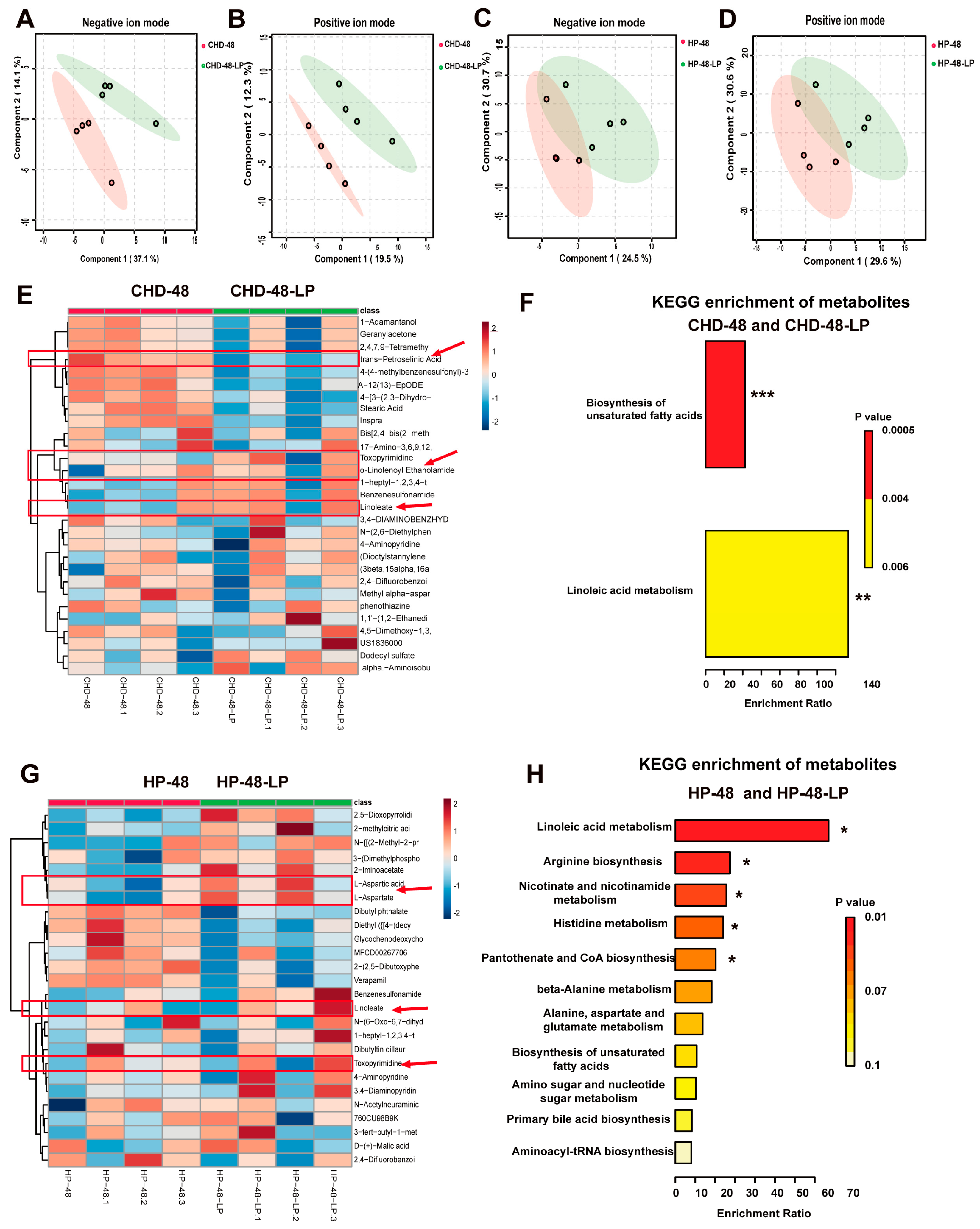
3.5. L. plantarum P470 Regulated Metabolism in Feces of CHD Patients In Vitro

To assess the influence of L. plantarum P470 treatment on the metabolite profile, we conducted an untargeted metabolomic analysis on the fermentation supernatant collected from both healthy individuals and those with CHD. We utilized partial least squares—discriminant analysis (PLS-DA) to visualize the distribution of positive and negative ion scanning data across the two groups. The PLS-DA plot exhibited a clear clustering of metabolites from each group, demonstrating distinct separation (Figure 6A–D). Differential metabolites were defined as those with p < 0.05 and fold change of >2 or <0.5. We conducted cluster analysis on the distinct metabolites found in the fermentation supernatant obtained from both the healthy and CHD groups. Our results showed that the levels of α-linolenoyl ethanolamide, linoleate, and toxopyrimidine were higher, while the content of trans-petroselinic acid, A-12(13)-EpODE, and stearic acid were lower in the CHD-48-LP group compared to the CHD-48 group (Figure 6E, Table 2). Moreover, differential metabolite enrichment pathways were analyzed using the Kyoto Encylopaedia of Genes and Genomes (KEGG). These differential metabolites were significantly enriched in the pathways for Biosynthesis of unsaturated fatty acids and linoleic acid metabolism (Figure 6F). In the HP-48-LP group, the levels of L-aspartic acid, L-aspartate, linoleate, and toxopyrimidine were significantly increased, while the level of glycochenodeoxycholic acid was lower when compared to the HP-48 group (Figure 6F, Table 2). These differential metabolites were mainly enriched in the Linoleic acid metabolism, arginine biosynthesis, nicotinate and nicotinamide metabolism, histidine metabolism, pantothenate and CoA biosynthesis, beta-alanine metabolism, alanine, aspartate and glutamate metabolism, and biosynthesis of unsaturated fatty acids pathways. Notably, the content of linoleate and toxopyrimidine was significantly increased following treatment with L. plantarum P470 in fermentation supernatant when compared to the HP-48 and CHD-48 groups. Our findings suggest that L. plantarum P470 mainly regulates the biosynthesis of unsaturated fatty acids and linoleic acid metabolism in fermentation metabolites in vitro.

4. Discussion

Cardiovascular disease (CVD) is a leading global cause of mortality, characterized by the rising prevalence of CHD and atherosclerosis [29]. Research has shown that the structure and function of the gut microbiota are related to CHD [30]. Probiotics and prebiotics have gained attention in recent years due to their potential for disease prevention by modulating the gut microbiota [31]. Fermented Chinese chives, a traditional food in China, have been found to offer potential benefits for human health [32], with the dominant strain L. plantarum possibly contributing to these effects. In this study, L. plantarum P470, isolated from fermented Chinese chives, was used to investigate its effects on anti-oxidation, anti-inflammatory responses, gut microbiota, and metabolic status in the feces of CHD patients using an anaerobic fermentation model in vitro.
The antioxidant activity against hydroxyl free radicals in the fermentation broth may be related to the fermentation metabolites [33], which further supports the notion that L. plantarum P470 ferments human feces to alter the metabolic state of the fecal microenvironment, often with beneficial effects. The results of this study demonstrated that L. plantarum P470 improved the scavenging ability of hydroxyl radicals in the feces of CHD patients but showed no significant difference in the feces of healthy individuals. This suggests that different metabolic states exhibit varying antioxidant activities, warranting further sample verification. TNF-α and IL-6 are key inflammatory factors in cardiovascular diseases [34]. During inflammation, macrophages are recruited to the inflamed area and engulf modified LDL particles, leading to the formation of lipid-laden foam cells. These foam cells exacerbate local inflammation by secreting pro-inflammatory mediators, including cytokines like TNF-α, and trigger the production of secondary signaling cytokines such as IL-6 [3]. The results of the anti-inflammatory experiment in the fermentation supernatant indicated that L. plantarum P470 significantly reduced the levels of TNF-α and IL-6 in the stool of CHD patients, while these levels showed a downward trend in healthy individuals. This finding suggests that L. plantarum P470 improves the anti-inflammatory state during fecal fermentation, particularly in CHD feces.
After 48 h of fermentation, L. plantarum P470 was found to modulate the gut microbiota in the feces of patients with CHD. At the phylum level, the ratio of Bacillota to Bacteroidota (F/B) was reduced following treatment with L. plantarum P470 in CHD gut microbiota in vitro. Stool samples obtained from individuals with atherosclerosis demonstrated elevated levels of the Bacillota phylum and reduced abundance of the Bacteroidetes phylum compared to samples from healthy controls [35]. Emoto et al. proposed that a decreased occurrence of the Bacteroidota phylum and an elevated F/B in the gastrointestinal tract might be associated with the development of coronary artery disease [36]. Bacteroidota were found to be enriched in various carbohydrate metabolism pathways, while Bacillota bins showed a higher enrichment in transport systems [37]. The study also found that L. plantarum P470 could increase the bacterial function of several metabolic pathways, including Arginine_and proline_metabolism, glycolysis/gluconeogenesis, bacterial_motility proteins, amino_sugar and_nucleotide sugar metabolism, Starch and sucrose metabolism using PICRUSt algorithm, compared with CHD-48 group. This increase in bacterial function may be related to the increased abundance of Bacteroidetes in CHD patient feces.
Imbalanced ratios of Bacillota to Bacteroidetes are a major cause of intestinal microbial imbalance, and adjusting this ratio has received extensive attention. Moreover, Veillonellaceae, a family within the Bacillota phylum, has been observed to have a favorable association with metabolic disorders triggered by high-fat diets [38], while Eggerthella lenta (Acfinobacteria phylum) has been observed to stimulate intestinal inflammation by stimulating the expression of Rorc and genes associated with Th17 [39,40,41]. Additionally, in functional studies, Eggerthella lenta has been implicated in the conversion of L-carnitine to TMA [42]. Our findings revealed the presence of Veillonella and Eggerthella were decreased following treatment with L. plantarum P470 in feces of CHD gut microbiota in vitro.
Proteobacteria is a phylum that contains more pathogenic microorganisms, including Escherichia/Shigella, Helicobacter, and Campylobacter, which are found in higher abundance in CHD patients with atherosclerosis [43]. Previous research has shown that Proteobacteria was positively correlated with adipose inflammatory factors [44], suggesting a potential link between this phylum and CVD risk factors. TMA-producing bacteria, such as Escherichia-Shigella, have been found to aggravate the progression of atherosclerosis [45], further emphasizing the importance of regulating microbial composition in relation to CHD. Additionally, Helicobacter pylori infection has been suggested as a risk factor for CHD, especially in individuals under 60 years old and without cardiovascular risk factors [46]. In this study, the abundance of Proteobacteria, specifically Escherichia-Shigella, Enterobacter, and Helicobacter, was found to be higher in the CHD-48 group but decreased following the addition of L. plantarum P470. These findings suggest that L. plantarum P470 isolated from fermented Chinese Chives may have the potential to improve fecal physiological status in patients with CHD by regulating the microbiota and reducing the abundance of pathogenic microorganisms such as Escherichia-Shigella, Enterobacter, and Helicobacter.
Short-chain fatty acids (SCFAs), including acetate, propionate, and butyrate, are essential for maintaining intestinal homeostasis. During atherosclerosis development, SCFAs are recognized as beneficial gut microbial products with anti-atherosclerotic effects, such as inhibiting systemic inflammation, promoting endothelial cell function, and maintaining intestinal barrier integrity [47,48]. Previous research has shown that under lipopolysaccharide (LPS) stimulation, SCFAs at concentrations ranging from 0.20 to 20 mmol/L can decrease the production of TNF-α and monocyte chemotactic protein-1 (MCP-1) [49]. Furthermore, butyrate treatment of Ea.hy926 cells has been found to reduce ox-LDL uptake, CD36, VCAM-1, TNF-α, and interleukin-1β/6 production while increasing IL-10 production, suggesting its potential as a treatment for atherosclerosis [50]. In this study, it was observed that L. plantarum P470 significantly elevated the levels of acetic acid and propionic acid in the feces of CHD patients and increased propionic acid and butyric acid levels in the group with Helicobacter pylori infection (HP) (p < 0.05). These findings suggest that L. plantarum P470 holds promise as a potential strategy for enhancing host health and as a treatment approach for CHD patients. Additionally, the fermentation solution administered to the CHD-48-LP group resulted in significant reductions in TNF-α and IL-6 levels (p < 0.05) while increasing IL-10 content in the HP-48-LP group, indicating that L. plantarum P470 may regulate the intestinal microbiota to produce SCFAs, thereby playing an anti-inflammatory role.
Linoleic acid (LA, ω-6 18:2) is highly susceptible to oxidation in the presence of lipoxygenase, cytochrome P450, metal ions, and free radicals. LA can be converted to the epoxide epoxyeicosatetraenoic acid (EPODE), one of the isoforms known to cause pulmonary edema in pathophysiological studies [51,52]. Several studies employing mixed supplementation strategies with conjugated linoleic acid (CLA) have demonstrated a reduction in the size of atherosclerotic lesions in the aorta, accompanied by decreased accumulation of macrophages and a decrease in the expression of pro-inflammatory genes [53,54]. Both 9,11-CLA and 10,12-CLA have been observed to inhibit the expression of crucial monocyte adhesion molecules found on endothelial cells, namely vascular cell adhesion molecule-1 (VCAM-1) and intercellular adhesion molecule-1 (ICAM-1) [55]. In this work, we found that L. plantarum P470 can significantly increase the levels of LA in the CHD patient group and HP group. In this study, we found that L. plantarum P470 significantly increased the levels of LA in both the CHD patient group and the HP group. Additionally, L. plantarum P470 significantly reduced the level of A-12(13)-EpODE and increased hydroxyl radical scavenging ability in the CHD patient group, suggesting an improvement in antioxidant capacity in feces and a reduction in the oxidation of linoleic acid. Trans-petroselinic acid, the trans isomer of petroselinic acid and an isomer of oleic acid, was shown in a study to increase cellular levels of triacylglycerols (TG) and cholesterol (TC) esters at a concentration of 100 μM. This also up-regulated the transcription of genes involved in fatty acid synthesis, including sterol regulatory element binding protein-1c (SREBP-1c), acetyl-CoA carboxylase alpha (ACACA), fatty acid synthase (FASN), stearoyl-CoA desaturase-1 (SCD1), 3-hydroxy-3-methylglutaryl-CoA reductase (HMGCR), 3-hydroxy-3-methylglutaryl-CoA synthase-1 (HMGCS1), farnesyl-diphosphate farnesyltransferase 1 (FDFT1), and SREBP-2. These effects were observed in human hepatoma HepG2 cells [56]. Our results indicate that L. plantarum P470 significantly decreased the level of trans-petroselinic acid, likely due to reduced fatty acid oxidation. These findings suggest that L. plantarum P470 can modulate lipid metabolism and generate short-chain fatty acids or other metabolites, leading to enhanced antioxidant and anti-inflammatory properties in fecal samples. This effect is primarily attributed to the regulation of the composition of intestinal microbiota.
However, our study has several limitations: significant individual differences, a small sample size leading to considerable intra-group variability, and suboptimal statistical analysis. Additionally, the fecal fermentation supernatant from healthy individuals did not exhibit significant antioxidant or anti-inflammatory activity, although there was a trend toward improvement in the overall fermentation supernatant. This suggests differences in fecal microecology and metabolic status between healthy participants and patients with CHD, which warrant further investigation with larger sample sizes. Factors such as patient gender, age, and geographical location should be considered to enhance the applicability of the results. Future research should explore a range of variables, including different diets, lifestyles, stages of disease, dosages, and timing of interventions, and should include larger, population-based cohort studies to confirm these findings.

5. Conclusions

To summarize, we examined the antioxidant and anti-inflammatory properties of L. plantarum P470, isolated from fermented Chinese Chives, utilizing an in vitro anaerobic fermentation model. Our findings suggest that L. plantarum P470 can increase hydroxyl radical scavenging activity and lower the expression of TNF-α and IL-6 in CHD feces. Furthermore, the L. plantarum P470 could be metabolized into SCFAs, which can regulate lipid metabolism by CHD gut microbiota. L. plantarum P470 was also found to decrease the F/B, as well as the abundance of other harmful bacteria such as Escherichia-Shigella, Enterobacter, Veillonella, Eggerthella, and Helicobacter, while increasing the abundance of beneficial genera such as Bacteroides, Lactobacillus, in the gut microbiota of CHD patient feces. This suggests that traditional fermented food such as chives can be used as a good strategy to regulate the intestinal microbiota to combat oxidation, inflammation, and slow down the progression of disease, especially by increasing the production of SCFAs. Overall, it is necessary to develop and utilize the effective components of traditional fermented food prebiotics and probiotics, which have significant potential in healthcare research.

Supplementary Materials

The following supporting information can be downloaded at: https://www.mdpi.com/article/10.3390/nu16172945/s1, Table S1: The relative abundance at the phylum level; Table S2: The relative abundance at the family level; Table S3: The relative abundance at the genus level.

Author Contributions

L.Y. was responsible for conceptualizing the article, conducting experiments, and writing the manuscript. Y.W. handled the experimental operations and statistical analysis. J.Y. was responsible for sample collection at the hospital and statistical analysis. Q.W., X.X. and Y.L. contributed to this work by performing statistical analyses. X.Z., T.L., L.L., H.Z., T.Z., T.J. and J.Z. collected crucial background information. Y.L. wrote the initial draft of the manuscript. All authors reviewed and approved the final version of the manuscript.

Funding

This study was funded by the National Key Research and Development Project (2021YFA0910200), the Key Laboratory of Guangdong Province (2020B121201009), the GDAS Special Project for Capacity Building in Innovation-driven Development (2020GDASYL-20200103026), and the Department of Science and Technology of Guangdong Province (2019QN01N107).

Institutional Review Board Statement

The study was conducted in accordance with the Declaration of Helsinki, and approved by the Ethics Committee of the First Affiliated Hospital of Guangdong Pharmaceutical University (Medical Review [2021] No. (13)).

Informed Consent Statement

Informed consent was obtained from all subjects involved in the study.

Data Availability Statement

All data and materials are available from the corresponding author upon reasonable request.

Conflicts of Interest

The authors state that the study was conducted with no commercial advertising or a financial relationship that may be interpreted as potential conflicts of interest.

References

  1. Mendis, S.; Davis, S.; Norrving, B. Organizational update: The world health organization global status report on noncommunicable diseases 2014; one more landmark step in the combat against stroke and vascular disease. Stroke 2015, 46, e121–e122. [Google Scholar] [CrossRef] [PubMed]
  2. Hansson, G.K.; Hermansson, A. The immune system in atherosclerosis. Nat. Immunol. 2011, 12, 204–212. [Google Scholar] [CrossRef]
  3. Ridker, P.M. From C-Reactive Protein to Interleukin-6 to Interleukin-1: Moving Upstream to Identify Novel Targets for Atheroprotection. Circ. Res. 2016, 118, 145–156. [Google Scholar] [CrossRef]
  4. Liu, H.; Wang, H.; Li, Q.; Wang, Y.W.; He, Y.; Li, X.J.; Sun, C.Y.; Onder, E.; Can, F.S.; Pang, Z.Q.; et al. LPS adsorption and inflammation alleviation by polymyxin B-modified liposomes for atherosclerosis treatment. Acta Pharm. Sin. B 2023, 13, 3817–3833. [Google Scholar] [CrossRef] [PubMed]
  5. Almeida, S.O.; Budoff, M. Effect of statins on atherosclerotic plaque. Trends Cardiovasc. Med. 2019, 29, 451–455. [Google Scholar] [CrossRef]
  6. Saiyitijiang, A.; Aizezi, M.; Zhao, Y.; Gao, Y. Efficacy and safety of new oral anticoagulants combined with antiplatelet drugs in the treatment of coronary heart disease: Systematic evaluation and meta-analysis. Ann. Noninvasive Electrocardiol. 2022, 27, e12977. [Google Scholar] [CrossRef] [PubMed]
  7. Chen, S.J.; Tsui, P.F.; Chuang, Y.P.; Chiang, D.M.; Chen, L.W.; Liu, S.T.; Lin, F.Y.; Huang, S.M.; Lin, S.H.; Wu, W.L.; et al. Carvedilol Ameliorates Experimental Atherosclerosis by Regulating Cholesterol Efflux and Exosome Functions. Int. J. Mol. Sci. 2019, 20, 5202. [Google Scholar] [CrossRef]
  8. Padam, P.; Barton, L.; Wilson, S.; David, A.; Walji, S.; de Lorenzo, F.; Ray, K.K.; Jones, B.; Cegla, J. Lipid lowering with inclisiran: A real-world single-centre experience. Open Heart 2022, 9, e002184. [Google Scholar] [CrossRef]
  9. Karlsson, F.H.; Fak, F.; Nookaew, I.; Tremaroli, V.; Fagerberg, B.; Petranovic, D.; Backhed, F.; Nielsen, J. Symptomatic atherosclerosis is associated with an altered gut metagenome. Nat. Commun. 2012, 3, 1245. [Google Scholar] [CrossRef]
  10. Jin, L.; Shi, X.; Yang, J.; Zhao, Y.; Xue, L.; Xu, L.; Cai, J. Gut microbes in cardiovascular diseases and their potential therapeutic applications. Protein Cell 2021, 12, 346–359. [Google Scholar] [CrossRef]
  11. Ditano-Vazquez, P.; Torres-Pena, J.D.; Galeano-Valle, F.; Perez-Caballero, A.I.; Demelo-Rodriguez, P.; Lopez-Miranda, J.; Katsiki, N.; Delgado-Lista, J.; Alvarez-Sala-Walther, L.A. The Fluid Aspect of the Mediterranean Diet in the Prevention and Management of Cardiovascular Disease and Diabetes: The Role of Polyphenol Content in Moderate Consumption of Wine and Olive Oil. Nutrients 2019, 11, 2833. [Google Scholar] [CrossRef] [PubMed]
  12. Yang, H.Y.; Yang, S.C.; Chao, J.C.; Chen, J.R. Beneficial effects of catechin-rich green tea and inulin on the body composition of overweight adults. Br. J. Nutr. 2012, 107, 749–754. [Google Scholar] [CrossRef] [PubMed]
  13. Mohamadshahi, M.; Veissi, M.; Haidari, F.; Shahbazian, H.; Kaydani, G.A.; Mohammadi, F. Effects of probiotic yogurt consumption on inflammatory biomarkers in patients with type 2 diabetes. Bioimpacts 2014, 4, 83–88. [Google Scholar] [PubMed]
  14. Mokoena, M.P.; Mutanda, T.; Olaniran, A.O. Perspectives on the probiotic potential of lactic acid bacteria from African traditional fermented foods and beverages. Food Nutr. Res. 2016, 60, 29630. [Google Scholar] [CrossRef] [PubMed]
  15. Yang, J.; Ji, Y.; Park, H.; Lee, J.; Park, S.; Yeo, S.; Shin, H.; Holzapfel, W.H. Selection of functional lactic acid bacteria as starter cultures for the fermentation of Korean leek (Allium tuberosum Rottler ex Sprengel.). Int. J. Food Microbiol. 2014, 191, 164–171. [Google Scholar] [CrossRef]
  16. Kumar, R.; Grover, S.; Batish, V.K. Hypocholesterolaemic effect of dietary inclusion of two putative probiotic bile salt hydrolase-producing Lactobacillus plantarum strains in Sprague-Dawley rats. Br. J. Nutr. 2011, 105, 561–573. [Google Scholar] [CrossRef]
  17. Jeun, J.; Kim, S.; Cho, S.Y.; Jun, H.J.; Park, H.J.; Seo, J.G.; Chung, M.J.; Lee, S.J. Hypocholesterolemic effects of Lactobacillus plantarum KCTC3928 by increased bile acid excretion in C57BL/6 mice. Nutrition 2010, 26, 321–330. [Google Scholar]
  18. Song, D.F.; Zhu, M.Y.; Gu, Q. Purification and characterization of Plantaricin ZJ5, a new bacteriocin produced by Lactobacillus plantarum ZJ5. PLoS ONE 2014, 9, e105549. [Google Scholar] [CrossRef]
  19. Mo, S.J.; Lee, K.; Hong, H.J.; Hong, D.K.; Jung, S.H.; Park, S.D.; Shim, J.J.; Lee, J.L. Effects of Lactobacillus curvatus HY7601 and Lactobacillus plantarum KY1032 on Overweight and the Gut Microbiota in Humans: Randomized, Double-Blinded, Placebo-Controlled Clinical Trial. Nutrients 2022, 14, 2484. [Google Scholar] [CrossRef]
  20. Hao, H.; Zhang, X.; Tong, L.; Liu, Q.; Liang, X.; Bu, Y.; Gong, P.; Liu, T.; Zhang, L.; Xia, Y.; et al. Effect of Extracellular Vesicles Derived From Lactobacillus plantarum Q7 on Gut Microbiota and Ulcerative Colitis in Mice. Front. Immunol. 2021, 12, 777147. [Google Scholar] [CrossRef]
  21. Zhu, R.; Fang, Y.; Li, H.; Liu, Y.; Wei, J.; Zhang, S.; Wang, L.; Fan, R.; Wang, L.; Li, S.; et al. Psychobiotic Lactobacillus plantarum JYLP-326 relieves anxiety, depression, and insomnia symptoms in test anxious college via modulating the gut microbiota and its metabolism. Front. Immunol. 2023, 14, 1158137. [Google Scholar] [CrossRef] [PubMed]
  22. Qiu, L.; Tao, X.; Xiong, H.; Yu, J.; Wei, H. Lactobacillus plantarum ZDY04 exhibits a strain-specific property of lowering TMAO via the modulation of gut microbiota in mice. Food Funct. 2018, 9, 4299–4309. [Google Scholar] [CrossRef] [PubMed]
  23. Chen, G.; Wang, M.; Zeng, Z.; Xie, M.; Xu, W.; Peng, Y.; Zhou, W.; Sun, Y.; Zeng, X.; Liu, Z. Fuzhuan brick tea polysaccharides serve as a promising candidate for remodeling the gut microbiota from colitis subjects in vitro: Fermentation characteristic and anti-inflammatory activity. Food Chem. 2022, 391, 133203. [Google Scholar] [CrossRef] [PubMed]
  24. Hara, K.; Someya, T.; Sano, K.; Sagane, Y.; Watanabe, T.; Wijesekara, R.G.S. Antioxidant activities of traditional plants in Sri Lanka by DPPH free radical-scavenging assay. Data Brief 2018, 17, 870–875. [Google Scholar] [CrossRef]
  25. Chen, H.; Li, Y.; Xie, X.; Chen, M.; Xue, L.; Wang, J.; Ye, Q.; Wu, S.; Yang, R.; Zhao, H.; et al. Exploration of the Molecular Mechanisms Underlying the Anti-Photoaging Effect of Limosilactobacillus fermentum XJC60. Front. Cell Infect. Microbiol. 2022, 12, 838060. [Google Scholar] [CrossRef] [PubMed]
  26. Yang, L.; Xie, X.; Li, Y.; Wu, L.; Fan, C.; Liang, T.; Xi, Y.; Yang, S.; Li, H.; Zhang, J.; et al. Evaluation of the Cholesterol-Lowering Mechanism of Enterococcus faecium Strain 132 and Lactobacillus paracasei Strain 201 in Hypercholesterolemia Rats. Nutrients 2021, 13, 1982. [Google Scholar] [CrossRef]
  27. Li, J.; Cao, Y.; Lu, R.; Li, H.; Pang, Y.; Fu, H.; Fang, G.; Chen, Q.; Liu, B.; Wu, J.; et al. Integrated Fecal Microbiome and Serum Metabolomics Analysis Reveals Abnormal Changes in Rats with Immunoglobulin A Nephropathy and the Intervention Effect of Zhen Wu Tang. Front. Pharmacol. 2020, 11, 606689. [Google Scholar]
  28. Blaak, E.E.; Canfora, E.E.; Theis, S.; Frost, G.; Groen, A.K.; Mithieux, G.; Nauta, A.; Scott, K.; Stahl, B.; van Harsselaar, J.; et al. Short chain fatty acids in human gut and metabolic health. Benef. Microbes 2020, 11, 411–455. [Google Scholar] [CrossRef]
  29. Low Wang, C.C.; Hess, C.N.; Hiatt, W.R.; Goldfine, A.B. Clinical Update: Cardiovascular Disease in Diabetes Mellitus: Atherosclerotic Cardiovascular Disease and Heart Failure in Type 2 Diabetes Mellitus-Mechanisms, Management, and Clinical Considerations. Circulation 2016, 133, 2459–2502. [Google Scholar] [CrossRef]
  30. Brown, J.M.; Hazen, S.L. The gut microbial endocrine organ: Bacterially derived signals driving cardiometabolic diseases. Annu. Rev. Med. 2015, 66, 343–359. [Google Scholar] [CrossRef]
  31. Sanders, M.E.; Merenstein, D.J.; Reid, G.; Gibson, G.R.; Rastall, R.A. Probiotics and prebiotics in intestinal health and disease: From biology to the clinic. Nat. Rev. Gastroenterol. Hepatol. 2019, 16, 605–616. [Google Scholar] [CrossRef] [PubMed]
  32. Leeuwendaal, N.K.; Stanton, C.; O’Toole, P.W.; Beresford, T.P. Fermented Foods, Health and the Gut Microbiome. Nutrients 2022, 14, 1527. [Google Scholar] [CrossRef] [PubMed]
  33. Xu, R.; Shang, N.; Li, P. In vitro and in vivo antioxidant activity of exopolysaccharide fractions from Bifidobacterium animalis RH. Anaerobe 2011, 17, 226–231. [Google Scholar] [CrossRef] [PubMed]
  34. Linton, M.F.; Moslehi, J.J.; Babaev, V.R. Akt Signaling in Macrophage Polarization, Survival, and Atherosclerosis. Int. J. Mol. Sci. 2019, 20, 2703. [Google Scholar] [CrossRef]
  35. Liu, S.; Zhao, W.; Liu, X.; Cheng, L. Metagenomic analysis of the gut microbiome in atherosclerosis patients identify cross-cohort microbial signatures and potential therapeutic target. FASEB J. 2020, 34, 14166–14181. [Google Scholar] [CrossRef]
  36. Emoto, T.; Yamashita, T.; Sasaki, N.; Hirota, Y.; Hayashi, T.; So, A.; Kasahara, K.; Yodoi, K.; Matsumoto, T.; Mizoguchi, T.; et al. Analysis of Gut Microbiota in Coronary Artery Disease Patients: A Possible Link between Gut Microbiota and Coronary Artery Disease. J. Atheroscler. Thromb. 2016, 23, 908–921. [Google Scholar] [CrossRef]
  37. Manco, M.; Putignani, L.; Bottazzo, G.F. Gut microbiota, lipopolysaccharides, and innate immunity in the pathogenesis of obesity and cardiovascular risk. Endocr. Rev. 2010, 31, 817–844. [Google Scholar] [CrossRef]
  38. Alard, J.; Lehrter, V.; Rhimi, M.; Mangin, I.; Peucelle, V.; Abraham, A.L.; Mariadassou, M.; Maguin, E.; Waligora-Dupriet, A.J.; Pot, B.; et al. Beneficial metabolic effects of selected probiotics on diet-induced obesity and insulin resistance in mice are associated with improvement of dysbiotic gut microbiota. Environ. Microbiol. 2016, 18, 1484–1497. [Google Scholar] [CrossRef]
  39. Boo, T.W.; Cryan, B.; O’Donnell, A.; Fahy, G. Prosthetic valve endocarditis caused by Veillonella parvula. J. Infect. 2005, 50, 81–83. [Google Scholar] [CrossRef]
  40. Alexander, M.; Ang, Q.Y.; Nayak, R.R.; Bustion, A.E.; Sandy, M.; Zhang, B.; Upadhyay, V.; Pollard, K.S.; Lynch, S.V.; Turnbaugh, P.J. Human gut bacterial metabolism drives Th17 activation and colitis. Cell Host Microbe 2022, 30, 17–30.e19. [Google Scholar] [CrossRef]
  41. Eicher, T.P.; Mohajeri, M.H. Overlapping Mechanisms of Action of Brain-Active Bacteria and Bacterial Metabolites in the Pathogenesis of Common Brain Diseases. Nutrients 2022, 14, 2661. [Google Scholar] [CrossRef] [PubMed]
  42. Li, J.; Li, Y.; Ivey, K.L.; Wang, D.D.; Wilkinson, J.E.; Franke, A.; Lee, K.H.; Chan, A.; Huttenhower, C.; Hu, F.B.; et al. Interplay between diet and gut microbiome, and circulating concentrations of trimethylamine N-oxide: Findings from a longitudinal cohort of US men. Gut 2022, 71, 724–733. [Google Scholar] [CrossRef] [PubMed]
  43. Jie, Z.; Xia, H.; Zhong, S.L.; Feng, Q.; Li, S.; Liang, S.; Zhong, H.; Liu, Z.; Gao, Y.; Zhao, H.; et al. The gut microbiome in atherosclerotic cardiovascular disease. Nat. Commun. 2017, 8, 845. [Google Scholar] [CrossRef]
  44. Zhang, L.; Shi, M.; Ji, J.; Hu, X.; Chen, F. Gut microbiota determines the prevention effects of Luffa cylindrica (L.) Roem supplementation against obesity and associated metabolic disorders induced by high-fat diet. FASEB J. 2019, 33, 10339–10352. [Google Scholar] [CrossRef]
  45. Romano, K.A.; Vivas, E.I.; Amador-Noguez, D.; Rey, F.E. Intestinal microbiota composition modulates choline bioavailability from diet and accumulation of the proatherogenic metabolite trimethylamine-N-oxide. mBio 2015, 6, e02481. [Google Scholar] [CrossRef]
  46. Shi, H.; Li, Y.; Dong, C.; Si, G.; Xu, Y.; Peng, M.; Li, Y. Helicobacter pylori infection and the progression of atherosclerosis: A systematic review and meta-analysis. Helicobacter 2022, 27, e12865. [Google Scholar] [CrossRef]
  47. Morrison, D.J.; Preston, T. Formation of short chain fatty acids by the gut microbiota and their impact on human metabolism. Gut Microbes 2016, 7, 189–200. [Google Scholar] [CrossRef]
  48. Lin, L.; Zhang, J. Role of intestinal microbiota and metabolites on gut homeostasis and human diseases. BMC Immunol. 2017, 18, 2. [Google Scholar] [CrossRef] [PubMed]
  49. Cox, M.A.; Jackson, J.; Stanton, M.; Rojas-Triana, A.; Bober, L.; Laverty, M.; Yang, X.; Zhu, F.; Liu, J.; Wang, S.; et al. Short-chain fatty acids act as antiinflammatory mediators by regulating prostaglandin E2 and cytokines. World J. Gastroenterol. 2009, 15, 5549–5557. [Google Scholar] [CrossRef]
  50. Aguilar, E.C.; dos Santos, L.C.; Leonel, A.J.; de Oliveira, J.S.; Santos, E.A.; Navia-Pelaez, J.M.; da Silva, J.F.; Mendes, B.P.; Capettini, L.S.A.; Teixeira, L.G.; et al. Oral butyrate reduces oxidative stress in atherosclerotic lesion sites by a mechanism involving NADPH oxidase down-regulation in endothelial cells. J. Nutr. Biochem. 2016, 34, 99–105. [Google Scholar] [CrossRef]
  51. Hu, J.N.; Taki, F.; Sugiyama, S.; Asai, J.; Izawa, Y.; Satake, T.; Ozawa, T. Neutrophil-derived epoxide, 9,10-epoxy-12-octadecenoate, induces pulmonary edema. Lung 1988, 166, 327–337. [Google Scholar] [PubMed]
  52. Sugiyama, S.; Hayakawa, M.; Nagai, S.; Ajioka, M.; Ozawa, T. Leukotoxin, 9, 10-epoxy-12-octadecenoate, causes cardiac failure in dogs. Life Sci. 1987, 40, 225–231. [Google Scholar] [CrossRef] [PubMed]
  53. Toomey, S.; Harhen, B.; Roche, H.M.; Fitzgerald, D.; Belton, O. Profound resolution of early atherosclerosis with conjugated linoleic acid. Atherosclerosis 2006, 187, 40–49. [Google Scholar] [CrossRef]
  54. Toomey, S.; Roche, H.; Fitzgerald, D.; Belton, O. Regression of pre-established atherosclerosis in the apoE−/− mouse by conjugated linoleic acid. Biochem. Soc. Trans. 2003, 31, 1075–1079. [Google Scholar] [CrossRef] [PubMed]
  55. Stachowska, E.; Siennicka, A.; Baskiewcz-Halasa, M.; Bober, J.; Machalinski, B.; Chlubek, D. Conjugated linoleic acid isomers may diminish human macrophages adhesion to endothelial surface. Int. J. Food Sci. Nutr. 2012, 63, 30–35. [Google Scholar] [CrossRef]
  56. Vahmani, P.; Meadus, W.J.; Duff, P.; Rolland, D.C.; Dugan, M.E.R. Comparing the lipogenic and cholesterolgenic effects of individual trans-18:1 isomers in liver cells. Eur. J. Lipid Sci. Technol. 2016, 119, 1600162. [Google Scholar] [CrossRef]
Figure 1. The effects of L. plantarum P470 on antioxidant ability. (A), 1,1-Diphenyl-2-picrylhydrazyl radical (DPPH) free radical scavenging assay, (B), hydroxyl radical scavenging assay. Significant differences between the CHD-48-LP group and the CHD-48 group, as well as between the HP-48-LP group and the HP-48 group, were assessed using either a t-test or Mann–Whitney test (* p < 0.05). The results are presented as means from the data (n = 4).
Nutrients 16 02945 g001
Figure 2. The effect of fermentation solution on the expression of TNF- α (A), IL-6 (B), and IL-10 (C) in RAW264.7 macrophage cells. Significant differences between the CHD-48-LP group and the CHD-48 group, or the HP-48-LP group and the HP-48 group, were analyzed using a t-test or Mann–Whitney test (* p < 0.05, ** p < 0.01). Results are reported as means of the data (n = 4).
Nutrients 16 02945 g002
Figure 3. The effects of L. plantarum P470 on gut microbial structure and relative abundance. (A) observed_otus rarefaction curve with sample; (B) Chao1 alpha-diversity index; (C) Shannon alpha-diversity index; (D) beta-diversity of PCoA; (E) the relative abundance at phylum level; (F) the relative abundance at family level; (G) the relative abundance at genus level; (H) correlation heatmap at genus level, between the CHD-48-LP and CHD-48 groups, and between the HP-48-LP and HP-48 groups, were assessed using a t-test or Mann–Whitney test. Results are presented as means of the data (n = 4).
Nutrients 16 02945 g003
Figure 4. PICRUSt function prediction based on Level 3.
Nutrients 16 02945 g004
Figure 5. The contents of major short-chain fatty acids during 48 h of fermentation with L. plantarum P470: (A) acetic acid; (B) propionic acid; (C) isobutyric acid; (D) butyric acid. Significant differences between the CHD-48-LP and CHD-48 groups or between the HP-48-LP and HP-48 groups (* p < 0.05, ** p < 0.01) were analyzed using the t-test or Mann–Whitney test. Results are presented as means (n = 4).
Nutrients 16 02945 g005
Figure 6. The impact of L. plantarum P470 on metabolites in fermentation supernatants, as assessed by untargeted metabolomic analysis using UPLC-MS/MS. (A) Partial least squares discriminant analysis (PLS-DA) of negative models comparing the CHD-48-LP group to the CHD-48 group; (B) PLS-DA of positive models comparing the CHD-48-LP group to the CHD-48 group; (C) PLS-DA of negative models comparing the HP-48-LP group to the HP-48 group; (D) PLS-DA of positive models comparing the HP-48-LP group to the HP-48 group; (E) correlation heat map of differential metabolites for the CHD-48-LP group versus the CHD-48 group; (F) KEGG enrichment analysis of differential metabolites for the CHD-48-LP group versus the CHD-48 group (** p < 0.01, *** p < 0.001); (G) correlation heat map of differential metabolites for the HP-48-LP group versus the HP-48 group; (H) KEGG enrichment analysis of differential metabolites for the HP-48-LP group versus the HP-48 group (* p < 0.05).
Nutrients 16 02945 g006
Table 1. Inflammatory factor-related primer sequence.
GeneOligonucleotide Sequence (5′–3′)
TNF-αGGACTAGCCAGGAGGGAGAA
CGCGGATCATGCTTTCTGTG
IL-6TGGAGTACCATAGCTACCTGGA
TCTCTCTGAAGGACTCTGGCT
IL-10GGGCGAGTGTAACAAGACCT
ATTTGCTGGGTTCCCACACT
β-actinAGGGAAATCGTGCGTGACAT
GGAAAAGAGCCTCAGGGCAT
Table 2. Different metabolite information of each group.
CHD-48 vs. CHD-48-LP
CompoundFormulaMolecular WeightRT (min)HMDBPubChemKEGGp-ValueFold Changelog2(FC)
Stearic acidC18H36O2284.2718616.997HMDB00008275281C015300.0448162.2771.1871
ToxopyrimidineC6H9N3O139.0747419.621METPA0166-C012790.0144440.15151−2.7225
GeranylacetoneC13H22O194.1670117.101HMDB00318461713001C132970.0120860.49681−1.0092
LinoleateC18H32O2280.2401716.121HMDB00006735280450C015950.00812580.14277−2.8082
InspraC24H30O6414.2041410.885HMDB0014838443872C125120.0145934.04972.0178
A-12(13)-EpODEC18H30O3294.2194116.398HMDB001020016061061 0.0263165.26342.396
α-linolenoyl EthanolamideC20H35NO2321.266816.105NANANA0.0344820.24847−2.0089
HP-48 vs. HP-48-LP
CompoundFormulaMolecular WeightRT (min)HMDBPubChemKEGGp-valueFold Changelog2(FC)
L-aspartic acidC4H7N O4133.036690.772HMDB00001915960C000490.037590.077347−3.6925
Dibutyl phthalateC16H22O4278.1518117.236HMDB00332443026C142140.0299042.65411.4082
D-(+)-malic acidC4H6O5134.020820.783HMDB003151892824C004970.0364640.21635−2.2086
2-methylcitric acidC7H10O7206.043450.847HMDB00003795290C022250.0465530.059527−4.0703
Linoleic acidC18H32O2280.2404718.049HMDB00006735280450C015950.0124850.17687−2.4992
L-aspartic acidC4H7NO4133.036690.772HMDB00001915960C000490.0385650.034422−4.8605
Chenodeoxycholic acidC24H40O4438.298657.765HMDB000063722833540C054660.03171226.8124.7448
N-acetylneuraminic acidC11H19NO9309.105841.004HMDB0000230445063C199100.0499370.24727−2.0159
VerapamilC27H38N2O4454.283166.051HMDB00018502520C071880.0150413.17451.6665
Disclaimer/Publisher’s Note: The statements, opinions and data contained in all publications are solely those of the individual author(s) and contributor(s) and not of MDPI and/or the editor(s). MDPI and/or the editor(s) disclaim responsibility for any injury to people or property resulting from any ideas, methods, instructions or products referred to in the content.

