
Potential Neuroprotective Effects of Alpinia officinarum Hance (Galangal): A Review
 , , , , , and
Abstract
1. Introduction
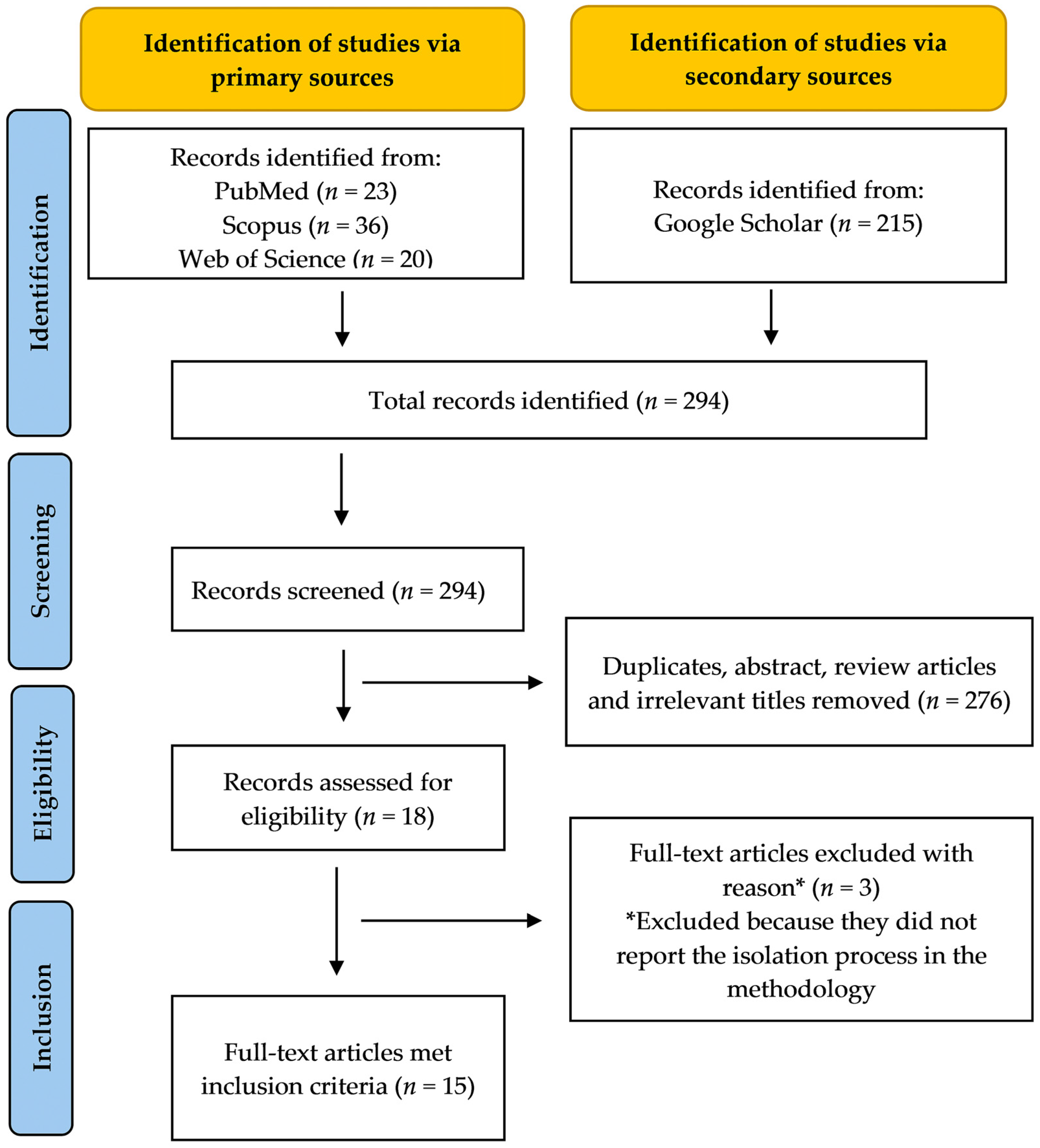
2. Methodology
3. Phytochemistry and Constituents of Alpinia officinarum Hance (Galangal)
4. Effects of Alpinia officinarum Hance (Galangal) on Central Nervous System (CNS)
Effects of A. officinarum on Enhancement and Regeneration of Neuronal Cells
5. Effects of A. officinarum on Neurological Disorders
5.1. Effect of A. officinarum on Alzheimer’s Disease
5.2. Effect of A. officinarum on Parkinson’s Disease

| Study Design | Plant Extract/Bioactive Compound | Treatment Dosage | Duration of Study | Findings | References |
|---|---|---|---|---|---|
| In vitro studies | |||||
| Primary cortical neuron induced with MPP+ (1-methyl-4-phenylpyridinium) for 20 h. | Diarylheptanoids: Alpininoids A [(+)-1] | 4, 8, 16 and 32 μM | 12 h | ↑ cell viability. | [84] |
| α-synuclein aggregation assay. | Diarylheptanoids: Alpinin A and Alpinin B | 10 µM | - | Inhibits α-synuclein aggregation. | [88] |
5.3. Effect of A. officinarum on Ischaemia-Reperfusion Injury
5.4. Effect of A. officinarum on Depression
5.5. Effect of A. officinarum on Epilepsy and Seizure
| Study Design | Plant Extract/Bioactive Compound | Treatment Dosage | Duration of Study | Findings | References |
|---|---|---|---|---|---|
| In vivo studies | |||||
| Male albino mice induced with PTZ (60 mg/kg, i.p.) | A. officinarum extract (hydroalcoholic) | 200, 400 and 600 mg/kg, i.p. (30 min before PTZ induction) | - | ↑ the onset of seizure. ↓ the duration of seizure. Flumazenil and naloxone pretreatments inhibited the anticonvulsant activity of A. officinarum. | [123] |
| Male Wistar rats induced with PTZ (35 mg/kg i.p.) daily every 48 h for 9 days and PTZ (60 mg/kg i.p.) on the 10th day. | A. officinarum extract (hydroalcoholic) | 50, 100 and 150 mg/kg, i.p. (30 min before PTZ induction) | 10 days | ↑ survival rate. ↑ onset of seizure. ↓ duration of seizure. ↓ total frequency of entire body seizures. ↓ total frequency of repeated spinning and jumping. ↓ immobility time in TST. Improved passive avoidance memory test. ↑ serum antioxidant capacity. ↓ serum and brain MDA level. Flumazenil pretreatment inhibited the anticonvulsant activity of A. officinarum. | [129] |
5.6. Effect of A. officinarum on Nociceptive Pain
6. Conclusions and Future Direction
Author Contributions
Funding
Conflicts of Interest
References
- Alasmary, F.A.; Assirey, E.A.; El-Meligy, R.M.; Awaad, A.S.; El-Sawaf, L.A.; Allah, M.M.; Alqasoumi, S.I. Analysis of Alpina officinarum Hance, chemically and biologically. Saudi Pharm. J. 2019, 27, 1107–1112. [Google Scholar] [CrossRef] [PubMed]
- Bitari, A.; Oualdi, I.; Touzani, R.; Elachouri, M.; Legssyer, A. Alpinia officinarum Hance: A mini review. Mater. Today Proc. 2023, 72, 3869–3874. [Google Scholar] [CrossRef]
- Ramanunny, A.K.; Wadhwa, S.; Gulati, M.; Vishwas, S.; Khursheed, R.; Paudel, K.R.; Gupta, S.; Porwal, O.; Alshahrani, S.M.; Jha, N.K.; et al. Journey of Alpinia galanga from kitchen spice to nutraceutical to folk medicine to nanomedicine. J. Ethnopharmacol. 2022, 291, 115144. [Google Scholar] [CrossRef] [PubMed]
- Yuandani; Jantan, I.; Haque, M.A.; Rohani, A.S.; Nugraha, S.E.; Salim, E.; Septama, A.W.; Juwita, N.A.; Khairunnisa, N.A.; Nasution, H.R.; et al. Immunomodulatory effects and mechanisms of the extracts and secondary compounds of Zingiber and Alpinia species: A review. Front. Pharmacol. 2023, 14, 1222195. [Google Scholar] [CrossRef]
- Khairullah, A.R.; Solikhah, T.I.; Ansori, A.N.M.; Fadholly, A.; Ramandinianto, S.C.; Ansharieta, R.; Widodo, A.; Riwu, K.H.P.; Putri, N.; Proboningrat, A. A review of an important medicinal plant: Alpinia galanga (L.) willd. Syst. Rev. Pharm. 2020, 11, 387–395. [Google Scholar]
- Zhou, Y.Q.; Liu, H.; He, M.X.; Wang, R.; Zeng, Q.Q.; Wang, Y.; Ye, W.C.; Zhang, Q.W. A review of the botany, phytochemical, and pharmacological properties of galangal. In Natural and Artificial Flavoring Agents and Food Dyes; Elsevier: Amsterdam, The Netherlands, 2018; pp. 351–396. [Google Scholar]
- Lim, T.K. Alpinia officinarum. In Edible Medicinal and Non-Medicinal Plants: Modified Stems, Roots, Bulbs; Springer International Publishing: Cham, Switzerland, 2016; Volume 12. [Google Scholar]
- Basri, A.M.; Taha, H.; Ahmad, N. A Review on the Pharmacological Activities and Phytochemicals of Alpinia officinarum (Galangal) Extracts Derived from Bioassay-Guided Fractionation and Isolation. Pharmacogn. Rev. 2017, 11, 43–56. [Google Scholar] [CrossRef]
- Ding, P.; Yang, L.; Feng, C.; Xian, J.C. Research and application of Alpinia officinarum in medicinal field. Chin. Herb. Med. 2019, 11, 132–140. [Google Scholar] [CrossRef]
- Akbar, S. (Ed.) Alpinia officinarum Hance. (Zingiberaceae). In Handbook of 200 Medicinal Plants: A Comprehensive Review of Their Traditional Medical Uses and Scientific Justifications; Springer International Publishing: Cham, Switzerland, 2020; pp. 217–224. [Google Scholar] [CrossRef]
- Hamad, A.; Djalil, A.; Hartanti, D. Galangal and ginger essential oils exerted microbial growth inhibitory activity and preservation potential on tofu. Food Res. 2023, 7, 27–34. [Google Scholar] [CrossRef]
- Liu, J.K. Natural products in cosmetics. Nat. Prod. Bioprospect. 2022, 12, 40. [Google Scholar] [CrossRef]
- Khan, M.S.; Adnan, Q.; Akhtar, N. Profiling of phytochemicals using LC-ESI-MS2, in vitro, in vivo characterization and cosmeceutical effects of Alpinia galanga (wild) extract loaded emulgel. J. Cosmet. Dermatol. 2023, 22, 1628–1641. [Google Scholar] [CrossRef]
- Rehman, B.; Umer, A.; Amanc, M.K.; Hamid, H.A.; Iqbal, D.; Khan, S.I. Phytochemical and Pharmacological Investigation of Lespedesea gerardiana. Glob. Drug Des. Dev. Rev. 2023, VIII, 23–33. [Google Scholar] [CrossRef]
- Parvin, S.; Reza, A.; Das, S.; Miah, M.M.; Karim, S. Potential Role and International Trade of Medicinal and Aromatic Plants in the World. Eur. J. Agric. Food Sci. 2023, 5, 89–99. [Google Scholar] [CrossRef]
- Nath, R.A.; Kityania, S.I.; Nath, D.E.; Talkudar, A.D.; Sarma, G.A. An extensive review on medicinal plants in the special context of economic importance. Asian J. Pharm. Clin. Res. 2023, 16, 6–11. [Google Scholar] [CrossRef]
- Laksmitawati, D.R.; Pratami, D.K.; Widowati, W.; Kusuma, H.S.W.; Wijayanti, C.R.; Rizal, R. The Potency of Alpinia galanga as Natural Antioxidant. Maj. Obat Tradis. 2022, 27, 165–171. [Google Scholar] [CrossRef]
- Hassanein, E.H.M.; El-Maksoud, M.S.A.; Ibrahim, I.M.; Abd-Alhameed, E.K.; Althagafy, H.S.; Mohamed, N.M.; Ross, S.A. The molecular mechanisms underlying anti-inflammatory effects of galangin in different diseases. Phytother. Res. 2023, 37, 3161–3181. [Google Scholar] [CrossRef]
- Lin, K.; Fu, D.; Wang, Z.; Zhang, X.; Zhu, C. Analgesic and anti-inflammatory effects of galangin: A potential pathway to inhibit transient receptor potential vanilloid 1 receptor activation. Korean J. Pain 2024, 37, 151–163. [Google Scholar] [CrossRef]
- Tian, Y.; Jia, X.; Wang, Q.; Lu, T.; Deng, G.; Tian, M.; Zhou, Y. Antioxidant, Antibacterial, Enzyme Inhibitory, and Anticancer Activities and Chemical Composition of Alpinia galanga Flower Essential Oil. Pharmaceuticals 2022, 15, 1069. [Google Scholar] [CrossRef]
- Fu, J.; Wang, Y.; Sun, M.; Xu, Y.; Chen, L. Antibacterial Activity and Components of the Methanol-Phase Extract from Rhizomes of Pharmacophagous Plant Alpinia officinarum Hance. Molecules 2022, 27, 4308. [Google Scholar] [CrossRef]
- Panigrahi, R.; Parida, R. Preliminary Screening of in vitro Kaempferia galanga Oil for Anti-Proliferative Activities against HeLa and MCF-7 Cell. Pharmacogn. Mag. 2023, 19, 660–666. [Google Scholar] [CrossRef]
- Li, S.; Nguyen, I.P.; Urbanczyk, K. Common infectious diseases of the central nervous system-clinical features and imaging characteristics. Quant. Imaging Med. Surg. 2020, 10, 2227–2259. [Google Scholar] [CrossRef]
- Brochet, B. (Ed.) The Spectrum of Demyelinating Inflammatory Diseases of the Central Nervous System. In Neuropsychiatric Symptoms of Inflammatory Demyelinating Diseases; Springer International Publishing: Cham, Switzerland, 2015; pp. 3–15. [Google Scholar] [CrossRef]
- Garofalo, M.; Pandini, C.; Bordoni, M.; Pansarasa, O.; Rey, F.; Costa, A.; Minafra, B.; Diamanti, L.; Zucca, S.; Carelli, S.; et al. Alzheimer’s, Parkinson’s Disease and Amyotrophic Lateral Sclerosis Gene Expression Patterns Divergence Reveals Different Grade of RNA Metabolism Involvement. Int. J. Mol. Sci. 2020, 21, 9500. [Google Scholar] [CrossRef] [PubMed]
- Flanagan, M.; Sonnen, J.A.; Keene, C.D.; Hevner, R.F.; Montine, T.J. Chapter 29—Molecular Basis of Diseases of the Nervous System. In Molecular Pathology, 2nd ed.; Coleman, W.B., Tsongalis, G.J., Eds.; Academic Press: Cambridge, MA, USA, 2018; pp. 651–690. [Google Scholar] [CrossRef]
- Ostrom, Q.T.; Francis, S.S.; Barnholtz-Sloan, J.S. Epidemiology of Brain and Other CNS Tumors. Curr. Neurol. Neurosci. Rep. 2021, 21, 68. [Google Scholar] [CrossRef]
- Gerstl, J.V.E.; Yearley, A.G.; Kilgallon, J.L.; Lassarén, P.; Robertson, F.C.; Herdell, V.; Wang, A.Y.; Segar, D.J.; Bernstock, J.D.; Laws, E.R.; et al. A national stratification of the global macroeconomic burden of central nervous system cancer. J. Neurosurg. 2023, 138, 1522–1530. [Google Scholar] [CrossRef] [PubMed]
- Feigin, V.L.; Vos, T.; Nichols, E.; Owolabi, M.O.; Carroll, W.M.; Dichgans, M.; Deuschl, G.; Parmar, P.; Brainin, M.; Murray, C. The global burden of neurological disorders: Translating evidence into policy. Lancet Neurol. 2020, 19, 255–265. [Google Scholar] [CrossRef] [PubMed]
- Gulyamova, S.T.; Abdul Aziz, S.F.; Omar, N.H.; Mohd, R.H. Workplace-Related Socioeconomic Issues Associated with Job Performance and Productivity among Employees with Various Impairments: A Systematic Literature Review. Soc. Sci. 2023, 12, 275. [Google Scholar] [CrossRef]
- Vuong, T.D.; Wei, F.; Beverly, C.J. Absenteeism due to Functional Limitations Caused by Seven Common Chronic Diseases in US Workers. J. Occup. Environ. Med. 2015, 57, 779–784. [Google Scholar] [CrossRef]
- Huntoon, K.; Lonser, R.R.; Elder, J.B. Chapter 19—Drug- and Disease-Specific Paradigms for Drug Delivery to the Central Nervous System. In Nervous System Drug Delivery; Lonser, R.R., Sarntinoranont, M., Bankiewicz, K., Eds.; Academic Press: Cambridge, MA, USA, 2019; pp. 379–403. [Google Scholar] [CrossRef]
- Doke, R.R.; Naik, T.S.; Lamkhade, D.L.; Bhise, T.S.; Khokrale, V.N.; Gosavi, Y.B. Novel therapeutic delivery for neurodegenerative diseases: Strategies to overcome CNS barriers. J. Pharm. Biol. Sci. 2023, 11, 1–8. [Google Scholar] [CrossRef]
- Vila, J.; Bosch, J.; Muñoz-Almagro, C. Molecular diagnosis of the central nervous system (CNS) infections. Enferm. Infecc. Microbiol. Clin. 2021, 39, 403–410. [Google Scholar] [CrossRef]
- Wexler, B.E. Returning to basic principles to develop more effective treatments for central nervous system disorders. Exp. Biol. Med. 2022, 247, 856–867. [Google Scholar] [CrossRef]
- Abubakar, I.B.; Malami, I.; Yahaya, Y.; Sule, S.M. A review on the ethnomedicinal uses, phytochemistry and pharmacology of Alpinia officinarum Hance. J. Ethnopharmacol. 2018, 224, 45–62. [Google Scholar] [CrossRef]
- Pillai, M.K.; Young, D.J.; Bin Hj Abdul Majid, H.M. Therapeutic Potential of Alpinia officinarum. Mini Rev. Med. Chem. 2018, 18, 1220–1232. [Google Scholar] [CrossRef] [PubMed]
- Ahmed, A.; Riaz, S.; Farooq, R.; Ahmed, M.; Hussain, N. Alpinia officinarum (galangal): A beneficial plant. J. Med. Public Health 2023, 4, 1057. [Google Scholar]
- Lei, X.; Wang, J.; Zuo, K.; Xia, T.; Zhang, J.; Xu, X.; Liu, Q.; Li, X. Alpinia officinarum Hance: A comprehensive review of traditional uses, phytochemistry, pharmacokinetic and pharmacology. Front. Pharmacol. 2024, 15, 1414635. [Google Scholar] [CrossRef] [PubMed]
- Van, H.T.; Thang, T.D.; Luu, T.N.; Doan, V.D. An overview of the chemical composition and biological activities of essential oils from Alpinia genus (Zingiberaceae). RSC Adv. 2021, 11, 37767–37783. [Google Scholar] [CrossRef] [PubMed]
- Mukherjee, A.; Chouhan, G.K.; Singh, S.; Chatterjee, K.; Kumar, A.; Gaurav, A.K.; Jaiswal, D.K.; Verma, J.P. Alpinia officinarum. In Naturally Occurring Chemicals against Alzheimer’s Disease; Academic Press: Cambridge, MA, USA, 2021; pp. 453–461. [Google Scholar]
- Mohd Sairazi, N.S.; Sirajudeen, K.N.S. Natural Products and Their Bioactive Compounds: Neuroprotective Potentials against Neurodegenerative Diseases. Evid. Based Complement. Altern. Med. 2020, 2020, 6565396. [Google Scholar] [CrossRef]
- Ly, T.N.; Shimoyamada, M.; Kato, K.; Yamauchi, R. Isolation and characterization of some antioxidative compounds from the rhizomes of smaller galanga (Alpinia officinarum Hance). J. Agric. Food Chem. 2003, 51, 4924–4929. [Google Scholar] [CrossRef]
- Zheng, X.; Zhang, Y.; Tan, Y.; Li, Y.; Xue, Q.; Li, H.; Zhang, X.; Pan, Y.; Xu, J.; Zhang, J. Alpinia officinarum Hance extract ameliorates diabetic gastroparesis by regulating SCF/c-kit signaling pathway and rebalancing gut microbiota. Fitoterapia 2024, 172, 105730. [Google Scholar] [CrossRef]
- Al Garni, H.A.; El-Halawany, A.M.; Koshak, A.E.; Malebari, A.M.; Alzain, A.A.; Mohamed, G.A.; Ibrahim, S.R.M.; El-Sayed, N.S.; Abdallah, H.M. Potential antioxidant, α-glucosidase, butyrylcholinesterase and acetylcholinesterase inhibitory activities of major constituents isolated from Alpinia officinarum hance rhizomes: Computational studies and in vitro validation. SAR QSAR Environ. Res. 2024, 35, 391–410. [Google Scholar] [CrossRef]
- An, N.; Xu, L.-Z.; Zou, Z.-M.; Yang, S.-L. Diarylheptanoids from Alpinia officinarum. J. Asian Nat. Prod. Res. 2006, 8, 637–641. [Google Scholar] [CrossRef]
- Fan, G.J.; Kang, Y.H.; Han, Y.N.; Han, B.H. Platelet-activating factor (PAF) receptor binding antagonists from Alpinia officinarum. Bioorg. Med. Chem. Lett. 2007, 17, 6720–6722. [Google Scholar] [CrossRef]
- Liu, Z.; Sang, S.; Hartman, T.G.; Ho, C.T.; Rosen, R.T. Determination of diarylheptanoids from Alpinia officinarum (Lesser Galangal) by HPLC with photodiode array and electrochemical detection. Phytochem. Anal. 2005, 16, 252–256. [Google Scholar] [CrossRef] [PubMed]
- Dong, G.-Z.; Lee, S.Y.; Zhao, H.-Y.; Lee, Y.-I.; Jeong, J.H.; Jeon, R.; Lee, H.J.; Ryu, J.-H. Diarylheptanoids from lesser galangal suppress human colon cancer cell growth through modulating Wnt/β-catenin pathway. J. Funct. Foods 2015, 18, 47–57. [Google Scholar] [CrossRef]
- An, N.; Zou, Z.M.; Tian, Z.; Luo, X.Z.; Yang, S.L.; Xu, L.Z. Diarylheptanoids from the rhizomes of Alpinia officinarum and their anticancer activity. Fitoterapia 2008, 79, 27–31. [Google Scholar] [CrossRef] [PubMed]
- Sun, Y.; Tabata, K.; Matsubara, H.; Kitanaka, S.; Suzuki, T.; Yasukawa, K. New cytotoxic diarylheptanoids from the rhizomes of Alpinia officinarum. Planta Med. 2008, 74, 427–431. [Google Scholar] [CrossRef] [PubMed]
- Sun, Y.; Matsubara, H.; Kitanaka, S.; Yasukawa, K. Diarylheptanoids from the Rhizomes of Alpinia officinarum. Helv. Chim. Acta 2008, 91, 118–123. [Google Scholar] [CrossRef]
- Shin, D.; Kinoshita, K.; Koyama, K.; Takahashi, K. Antiemetic principles of Alpinia officinarum. J. Nat. Prod. 2002, 65, 1315–1318. [Google Scholar] [CrossRef]
- An, N.; Zhang, H.-W.; Xu, L.-Z.; Yang, S.-L.; Zou, Z.-M. New diarylheptanoids from the rhizome of Alpinia officinarum Hance. Food Chem. 2010, 119, 513–517. [Google Scholar] [CrossRef]
- Tang, G.; Dong, X.; Huang, X.; Huang, X.J.; Liu, H.; Wang, Y.; Ye, W.C.; Shi, L. A natural diarylheptanoid promotes neuronal differentiation via activating ERK and PI3K-Akt dependent pathways. Neuroscience 2015, 303, 389–401. [Google Scholar] [CrossRef]
- Lee, J.S.; Kim, J.H.; Han, Y.K.; Ma, J.Y.; Kim, Y.H.; Li, W.; Yang, S.Y. Cholinesterases inhibition studies of biological active compounds from the rhizomes of Alpinia officinarum Hance and in silico molecular dynamics. Int. J. Biol. Macromol. 2018, 120, 2442–2447. [Google Scholar] [CrossRef]
- Liu, D.; Qu, W.; Zhao, L.; Guan, F.Q.; Liang, J.Y. A new dimeric diarylheptanoid from the rhizomes of Alpinia officinarum. Chin. J. Nat. Med. 2014, 12, 139–141. [Google Scholar] [CrossRef]
- Zhao, L.; Liang, J.Y.; Qu, W. A novel dimeric diarylheptanoid from the rhizomes of Alpinia officinarum. Chem. Nat. Compd. 2012, 48, 836–838. [Google Scholar] [CrossRef]
- Xu, S.-M.; Huang, X.-J.; Wang, Y.; Ye, W.-C. A new cadinane sesquiterpene from the rhizomes of Alpinia officinarum. Chin. J. Nat. Med. 2012, 10, 374–377. [Google Scholar] [CrossRef]
- Zou, Q.Y.; Wu, H.F.; Tang, Y.L.; Chen, D.Z. A new labdane diterpene from the rhizomes of Alpinia officinarum. Nat. Prod. Res. 2016, 30, 1–6. [Google Scholar] [CrossRef] [PubMed]
- Zhang, H.; Xu, L.X.; Wu, P.; Wei, X.Y. Flavonoids from the Aerial Parts of Alpinia officinarum. J. Trop. Subtrop. Bot. 2014, 22, 89–92. [Google Scholar] [CrossRef]
- Zhang, J.Q.; Wang, Y.; Li, H.L.; Wen, Q.; Yin, H.; Zeng, N.K.; Lai, W.Y.; Wei, N.; Cheng, S.Q.; Kang, S.L.; et al. Simultaneous quantification of seventeen bioactive components in rhizome and aerial parts of Alpinia officinarum Hance using LC-MS/MS. Anal. Methods 2015, 7, 4919. [Google Scholar] [CrossRef]
- Honmore, V.S.; Kandhare, A.D.; Kadam, P.P.; Khedkar, V.M.; Sarkar, D.; Bodhankar, S.L.; Zanwar, A.A.; Rojatkar, S.R.; Natu, A.D. Isolates of Alpinia officinarum Hance as COX-2 inhibitors: Evidence from anti-inflammatory, antioxidant and molecular docking studies. Int. Immunopharmacol. 2016, 33, 8–17. [Google Scholar] [CrossRef]
- Matsuda, H.; Ando, S.; Kato, T.; Morikawa, T.; Yoshikawa, M. Inhibitors from the rhizomes of Alpinia officinarum on production of nitric oxide in lipopolysaccharide-activated macrophages and the structural requirements of diarylheptanoids for the activity. Bioorg. Med. Chem. 2006, 14, 138–142. [Google Scholar] [CrossRef]
- Matsuda, H.; Nakashima, S.; Oda, Y.; Nakamura, S.; Yoshikawa, M. Melanogenesis inhibitors from the rhizomes of Alpinia officinarum in B16 melanoma cells. Bioorg. Med. Chem. 2009, 17, 6048–6053. [Google Scholar] [CrossRef]
- Liu, D.; Liu, Y.W.; Guan, F.Q.; Liang, J.Y. New cytotoxic diarylheptanoids from the rhizomes of Alpinia officinarum Hance. Fitoterapia 2014, 96, 76–80. [Google Scholar] [CrossRef]
- Zhang, W.X.; Chao, I.C.; Hu, D.J.; Shakerian, F.; Ge, L.; Liang, X.; Wang, Y.; Zhao, J.; Li, S.P. Comparison of Antioxidant Activity and Main Active Compounds among Different Parts of Alpinia officinarum Hance Using High-Performance Thin Layer Chromatography-Bioautography. J. AOAC Int. 2019, 102, 726–733. [Google Scholar] [CrossRef]
- Tabata, K.; Yamazaki, Y.; Okada, M.; Fukumura, K.; Shimada, A.; Sun, Y.; Yasukawa, K.; Suzuki, T. Diarylheptanoids derived from Alpinia officinarum induce apoptosis, S-phase arrest and differentiation in human neuroblastoma cells. Anticancer Res. 2009, 29, 4981–4988. [Google Scholar] [PubMed]
- Tian, Z.; An, N.; Zhou, B.; Xiao, P.; Kohane, I.S.; Wu, E. Cytotoxic diarylheptanoid induces cell cycle arrest and apoptosis via increasing ATF3 and stabilizing p53 in SH-SY5Y cells. Cancer Chemother. Pharmacol. 2009, 63, 1131–1139. [Google Scholar] [CrossRef]
- Bekris, L.M.; Yu, C.E.; Bird, T.D.; Tsuang, D.W. Genetics of Alzheimer disease. J. Geriatr. Psychiatry Neurol. 2010, 23, 213–227. [Google Scholar] [CrossRef] [PubMed]
- Cerejeira, J.; Lagarto, L.; Mukaetova-Ladinska, E.B. Behavioral and psychological symptoms of dementia. Front. Neurol. 2012, 3, 73. [Google Scholar] [CrossRef] [PubMed]
- Silva, M.V.F.; Loures, C.M.G.; Alves, L.C.V.; de Souza, L.C.; Borges, K.B.G.; Carvalho, M.D.G. Alzheimer’s disease: Risk factors and potentially protective measures. J. Biomed. Sci. 2019, 26, 33. [Google Scholar] [CrossRef] [PubMed]
- Choi, S.B.; Kwon, S.; Kim, J.H.; Ahn, N.H.; Lee, J.H.; Yang, S.H. The Molecular Mechanisms of Neuroinflammation in Alzheimer’s Disease, the Consequence of Neural Cell Death. Int. J. Mol. Sci. 2023, 24, 11757. [Google Scholar] [CrossRef] [PubMed]
- Thammasart, S.; Namchaiw, P.; Pasuwat, K.; Tonsomboon, K.; Khantachawana, A. Attenuation Aβ1-42-induced neurotoxicity in neuronal cell by 660 nm and 810 nm LED light irradiation. PLoS ONE 2023, 18, e0283976. [Google Scholar] [CrossRef]
- Huang, X.; Tang, G.; Liao, Y.; Zhuang, X.; Dong, X.; Liu, H.; Huang, X.J.; Ye, W.C.; Wang, Y.; Shi, L. 7-(4-Hydroxyphenyl)-1-phenyl-4E-hepten-3-one, a Diarylheptanoid from Alpinia officinarum, Protects Neurons against Amyloid-β Induced Toxicity. Biol. Pharm. Bull. 2016, 39, 1961–1967. [Google Scholar] [CrossRef]
- Xiao, H.; Zhang, Q.; Peng, Y.; Tang, G.; Liao, Y.; Zhuang, X.; Ye, W.C.; Wang, Y.; Shi, L. 7-(4-Hydroxy-3-methoxyphenyl)-1-phenyl-4E-hepten-3-one alleviates Aβ(1-42) induced cytotoxicity through PI3K-mTOR pathways. Biochem. Biophys. Res. Commun. 2017, 484, 365–371. [Google Scholar] [CrossRef]
- Campanari, M.L.; Navarrete, F.; Ginsberg, S.D.; Manzanares, J.; Sáez-Valero, J.; García-Ayllón, M.S. Increased Expression of Readthrough Acetylcholinesterase Variants in the Brains of Alzheimer’s Disease Patients. J. Alzheimers Dis. 2016, 53, 831–841. [Google Scholar] [CrossRef]
- Köse, L.P.; Gülçin, İ.; Gören, A.C.; Namiesnik, J.; Martinez-Ayala, A.L.; Gorinstein, S. LC–MS/MS analysis, antioxidant and anticholinergic properties of galanga (Alpinia officinarum Hance) rhizomes. Ind. Crops Prod. 2015, 74, 712–721. [Google Scholar] [CrossRef]
- Mu, L.; Wang, J.; Zhou, T.; Qiao, W.; Hu, W.; Zhang, R.; Chen, X. Diarylheptanoids with neuroprotective effects from Alpinia officinarum rhizomes. Fitoterapia 2024, 175, 105980. [Google Scholar] [CrossRef] [PubMed]
- DeMaagd, G.; Philip, A. Parkinson’s Disease and Its Management: Part 1: Disease Entity, Risk Factors, Pathophysiology, Clinical Presentation, and Diagnosis. Pharm. Ther. 2015, 40, 504–532. [Google Scholar]
- Chinta, S.J.; Andersen, J.K. Dopaminergic neurons. Int. J. Biochem. Cell Biol. 2005, 37, 942–946. [Google Scholar] [CrossRef] [PubMed]
- Kamath, T.; Abdulraouf, A.; Burris, S.J.; Langlieb, J.; Gazestani, V.; Nadaf, N.M.; Balderrama, K.; Vanderburg, C.; Macosko, E.Z. Single-cell genomic profiling of human dopamine neurons identifies a population that selectively degenerates in Parkinson’s disease. Nat. Neurosci. 2022, 25, 588–595. [Google Scholar] [CrossRef]
- Kawahata, I.; Finkelstein, D.I.; Fukunaga, K. Dopamine D1–D5 Receptors in Brain Nuclei: Implications for Health and Disease. Receptors 2024, 3, 155–181. [Google Scholar] [CrossRef]
- Liu, H.; Wu, Z.L.; Huang, X.J.; Peng, Y.; Huang, X.; Shi, L.; Wang, Y.; Ye, W.C. Evaluation of Diarylheptanoid-Terpene Adduct Enantiomers from Alpinia officinarum for Neuroprotective Activities. J. Nat. Prod. 2018, 81, 162–170. [Google Scholar] [CrossRef]
- Javed, H.; Nagoor Meeran, M.F.; Azimullah, S.; Adem, A.; Sadek, B.; Ojha, S.K. Plant Extracts and Phytochemicals Targeting α-Synuclein Aggregation in Parkinson’s Disease Models. Front. Pharmacol. 2018, 9, 1555. [Google Scholar] [CrossRef]
- Power, J.H.; Barnes, O.L.; Chegini, F. Lewy Bodies and the Mechanisms of Neuronal Cell Death in Parkinson’s Disease and Dementia with Lewy Bodies. Brain Pathol. 2017, 27, 3–12. [Google Scholar] [CrossRef]
- Koeglsperger, T.; Rumpf, S.L.; Schließer, P.; Struebing, F.L.; Brendel, M.; Levin, J.; Trenkwalder, C.; Höglinger, G.U.; Herms, J. Neuropathology of incidental Lewy body & prodromal Parkinson’s disease. Mol. Neurodegener. 2023, 18, 32. [Google Scholar] [CrossRef]
- Fu, G.; Zhang, W.; Du, D.; Ng, Y.P.; Ip, F.C.F.; Tong, R.; Ip, N.Y. Diarylheptanoids from Rhizomes of Alpinia officinarum Inhibit Aggregation of α-Synuclein. J. Agric. Food Chem. 2017, 65, 6608–6614. [Google Scholar] [CrossRef] [PubMed]
- Li, M.; Tang, H.; Li, Z.; Tang, W. Emerging Treatment Strategies for Cerebral Ischemia—Reperfusion Injury. Neuroscience 2022, 507, 112–124. [Google Scholar] [CrossRef] [PubMed]
- Zhang, M.; Liu, Q.; Meng, H.; Duan, H.; Liu, X.; Wu, J.; Gao, F.; Wang, S.; Tan, R.; Yuan, J. Ischemia-reperfusion injury: Molecular mechanisms and therapeutic targets. Signal Transduct. Target. Ther. 2024, 9, 12. [Google Scholar] [CrossRef] [PubMed]
- Hayashi, T.; Noshita, N.; Sugawara, T.; Chan, P.H. Temporal profile of angiogenesis and expression of related genes in the brain after ischemia. J. Cereb. Blood Flow Metab. 2003, 23, 166–180. [Google Scholar] [CrossRef] [PubMed]
- Wen, Y.; Yang, S.; Liu, R.; Simpkins, J.W. Cell-cycle regulators are involved in transient cerebral ischemia induced neuronal apoptosis in female rats. FEBS Lett. 2005, 579, 4591–4599. [Google Scholar] [CrossRef]
- Strbian, D.; Durukan, A.; Pitkonen, M.; Marinkovic, I.; Tatlisumak, E.; Pedrono, E.; Abo-Ramadan, U.; Tatlisumak, T. The blood-brain barrier is continuously open for several weeks following transient focal cerebral ischemia. Neuroscience 2008, 153, 175–181. [Google Scholar] [CrossRef]
- Witt, K.A.; Mark, K.S.; Sandoval, K.E.; Davis, T.P. Reoxygenation stress on blood-brain barrier paracellular permeability and edema in the rat. Microvasc. Res. 2008, 75, 91–96. [Google Scholar] [CrossRef]
- Kishimoto, M.; Suenaga, J.; Takase, H.; Araki, K.; Yao, T.; Fujimura, T.; Murayama, K.; Okumura, K.; Ueno, R.; Shimizu, N.; et al. Oxidative stress-responsive apoptosis inducing protein (ORAIP) plays a critical role in cerebral ischemia/reperfusion injury. Sci. Rep. 2019, 9, 13512. [Google Scholar] [CrossRef]
- Ravikumar, B.; Sarkar, S.; Davies, J.E.; Futter, M.; Garcia-Arencibia, M.; Green-Thompson, Z.W.; Jimenez-Sanchez, M.; Korolchuk, V.I.; Lichtenberg, M.; Luo, S.; et al. Regulation of mammalian autophagy in physiology and pathophysiology. Physiol. Rev. 2010, 90, 1383–1435. [Google Scholar] [CrossRef]
- Shi, R.; Weng, J.; Zhao, L.; Li, X.M.; Gao, T.M.; Kong, J. Excessive autophagy contributes to neuron death in cerebral ischemia. CNS Neurosci. Ther. 2012, 18, 250–260. [Google Scholar] [CrossRef]
- Su, J.; Zhang, T.; Wang, K.; Zhu, T.; Li, X. Autophagy activation contributes to the neuroprotection of remote ischemic perconditioning against focal cerebral ischemia in rats. Neurochem. Res. 2014, 39, 2068–2077. [Google Scholar] [CrossRef]
- Sun, D.; Wang, W.; Wang, X.; Wang, Y.; Xu, X.; Ping, F.; Du, Y.; Jiang, W.; Cui, D. bFGF plays a neuroprotective role by suppressing excessive autophagy and apoptosis after transient global cerebral ischemia in rats. Cell Death Dis. 2018, 9, 172. [Google Scholar] [CrossRef] [PubMed]
- Shao, Z.Q.; Dou, S.S.; Zhu, J.G.; Wang, H.Q.; Wang, C.M.; Cheng, B.H.; Bai, B. Apelin-13 inhibits apoptosis and excessive autophagy in cerebral ischemia/reperfusion injury. Neural Regen. Res. 2021, 16, 1044–1051. [Google Scholar] [CrossRef] [PubMed]
- Gao, X.; Chen, W.; Li, J.; Shen, C.; Zhou, P.; Che, X.; Li, X.; Xie, R. The protective effect of alpha-lipoic acid against brain ischemia and reperfusion injury via mTOR signaling pathway in rats. Neurosci. Lett. 2018, 671, 108–113. [Google Scholar] [CrossRef] [PubMed]
- Yang, B.; Li, Y.; Ma, Y.; Zhang, X.; Yang, L.; Shen, X.; Zhang, J.; Jing, L. Selenium attenuates ischemia/reperfusion injury-induced damage to the blood-brain barrier in hyperglycemia through PI3K/AKT/mTOR pathway-mediated autophagy inhibition. Int. J. Mol. Med. 2021, 48, 178. [Google Scholar] [CrossRef] [PubMed]
- Juntunen, M.; Hagman, S.; Moisan, A.; Narkilahti, S.; Miettinen, S. In Vitro Oxygen-Glucose Deprivation-Induced Stroke Models with Human Neuroblastoma Cell- and Induced Pluripotent Stem Cell-Derived Neurons. Stem Cells Int. 2020, 2020, 8841026. [Google Scholar] [CrossRef] [PubMed]
- Shi, Q.; Zhang, Q.; Peng, Y.; Zhang, X.; Wang, Y.; Shi, L. A natural diarylheptanoid protects cortical neurons against oxygen-glucose deprivation-induced autophagy and apoptosis. J. Pharm. Pharmacol. 2019, 71, 1110–1118. [Google Scholar] [CrossRef]
- Liu, H.; Wang, X.; Shi, Q.; Li, L.; Zhang, Q.; Wu, Z.L.; Huang, X.J.; Zhang, Q.W.; Ye, W.C.; Wang, Y.; et al. Dimeric Diarylheptanoids with Neuroprotective Activities from Rhizomes of Alpinia officinarum. ACS Omega 2020, 5, 10167–10175. [Google Scholar] [CrossRef]
- Kanter, J.W.; Busch, A.M.; Weeks, C.E.; Landes, S.J. The nature of clinical depression: Symptoms, syndromes, and behavior analysis. Behav. Anal. 2008, 31, 1–21. [Google Scholar] [CrossRef]
- Zakaria, F.H.; Samhani, I.; Mustafa, M.Z.; Shafin, N. Pathophysiology of Depression: Stingless Bee Honey Promising as an Antidepressant. Molecules 2022, 27, 5091. [Google Scholar] [CrossRef]
- Liu, B.; Liu, J.; Wang, M.; Zhang, Y.; Li, L. From Serotonin to Neuroplasticity: Evolvement of Theories for Major Depressive Disorder. Front. Cell. Neurosci. 2017, 11, 305. [Google Scholar] [CrossRef] [PubMed]
- Boku, S.; Nakagawa, S.; Toda, H.; Hishimoto, A. Neural basis of major depressive disorder: Beyond monoamine hypothesis. Psychiatry Clin. Neurosci. 2018, 72, 3–12. [Google Scholar] [CrossRef] [PubMed]
- Andrade, C.; Rao, N.S. How antidepressant drugs act: A primer on neuroplasticity as the eventual mediator of antidepressant efficacy. Indian J. Psychiatry 2010, 52, 378–386. [Google Scholar] [CrossRef] [PubMed]
- Iob, E.; Kirschbaum, C.; Steptoe, A. Persistent depressive symptoms, HPA-axis hyperactivity, and inflammation: The role of cognitive-affective and somatic symptoms. Mol. Psychiatry 2020, 25, 1130–1140. [Google Scholar] [CrossRef] [PubMed]
- Smith, S.M.; Vale, W.W. The role of the hypothalamic-pituitary-adrenal axis in neuroendocrine responses to stress. Dialogues Clin. Neurosci. 2006, 8, 383–395. [Google Scholar] [CrossRef]
- Dziurkowska, E.; Wesolowski, M. Cortisol as a Biomarker of Mental Disorder Severity. J. Clin. Med. 2021, 10, 5204. [Google Scholar] [CrossRef]
- Awari, D.M.; Somani, R.S. Evaluation of the Ameliorative Effect of Alpinia Officinarum Methanol Extract in an Experimental Model of Depression. Int. J. Pharm. Sci. Res. 2019, 10, 1160–1171. [Google Scholar] [CrossRef]
- Yankelevitch-Yahav, R.; Franko, M.; Huly, A.; Doron, R. The forced swim test as a model of depressive-like behavior. J. Vis. Exp. 2015, 2015, e52587. [Google Scholar] [CrossRef]
- Ueno, H.; Takahashi, Y.; Murakami, S.; Wani, K.; Matsumoto, Y.; Okamoto, M.; Ishihara, T. Effect of simultaneous testing of two mice in the tail suspension test and forced swim test. Sci. Rep. 2022, 12, 9224. [Google Scholar] [CrossRef]
- Salehi, A.; Rabiei, Z.; Setorki, M. Antidepressant effects of hydroalcoholic extract of Alpinia officinarum rhizome on chronic unpredictable stress induced depression in BALB/c mice. J. Med. Plants 2020, 1, 170–179. [Google Scholar] [CrossRef]
- Stafstrom, C.E. Epilepsy: A review of selected clinical syndromes and advances in basic science. J. Cereb. Blood Flow Metab. 2006, 26, 983–1004. [Google Scholar] [CrossRef] [PubMed]
- Anwar, H.; Khan, Q.U.; Nadeem, N.; Pervaiz, I.; Ali, M.; Cheema, F.F. Epileptic seizures. Discoveries 2020, 8, e110. [Google Scholar] [CrossRef] [PubMed]
- Boleti, A.P.A.; Cardoso, P.H.O.; Frihling, B.E.F.; de Moraes, L.; Nunes, E.A.C.; Mukoyama, L.T.H.; Nunes, E.A.C.; Carvalho, C.M.E.; Macedo, M.L.R.; Migliolo, L. Pathophysiology to Risk Factor and Therapeutics to Treatment Strategies on Epilepsy. Brain Sci. 2024, 14, 71. [Google Scholar] [CrossRef] [PubMed]
- Attia, G.M.; Elmansy, R.A.; Elsaed, W.M. Neuroprotective effect of nilotinib on pentylenetetrazol-induced epilepsy in adult rat hippocampus: Involvement of oxidative stress, autophagy, inflammation, and apoptosis. Folia Neuropathol. 2019, 57, 146–160. [Google Scholar] [CrossRef]
- Perucca, E.; Bialer, M.; White, H.S. New GABA-Targeting Therapies for the Treatment of Seizures and Epilepsy: I. Role of GABA as a Modulator of Seizure Activity and Recently Approved Medications Acting on the GABA System. CNS Drugs 2023, 37, 755–779. [Google Scholar] [CrossRef]
- Nejad, S.R.; Motevalian, M.; Fatemi, I.; Shojaii, A. Anticonvulsant Effects of the Hydroalcoholic Extract of Alpinia officinarum Rhizomesin Mice: Involvement of Benzodiazepine and Opioid Receptors. J. Epilepsy Res. 2017, 7, 33–38. [Google Scholar] [CrossRef]
- Novak, A.; Vizjak, K.; Rakusa, M. Cognitive Impairment in People with Epilepsy. J. Clin. Med. 2022, 11, 267. [Google Scholar] [CrossRef]
- Vacca, M.; Fernandes, M.; Spanetta, M.; Placidi, F.; Izzi, F.; Lombardo, C.; Mercuri, N.B.; Liguori, C. Depressive symptoms in patients with epilepsy and clinically associated features in a single tertiary center. Neurol. Sci. 2022, 43, 1965–1974. [Google Scholar] [CrossRef]
- Brandt, C.; Glien, M.; Potschka, H.; Volk, H.; Löscher, W. Epileptogenesis and neuropathology after different types of status epilepticus induced by prolonged electrical stimulation of the basolateral amygdala in rats. Epilepsy Res. 2003, 55, 83–103. [Google Scholar] [CrossRef]
- Dingledine, R.; Varvel, N.H.; Dudek, F.E. When and how do seizures kill neurons, and is cell death relevant to epileptogenesis? Adv. Exp. Med. Biol. 2014, 813, 109–122. [Google Scholar] [CrossRef]
- Asgharzade, S.; Rabiei, Z.; Rabiei, S.; Bijad, E.; Rafieian-Kopaei, M. Therapeutic Effects of Oleuropein in Improving Seizure, Oxidative Stress and Cognitive Disorder in Pentylenetetrazole Kindling Model of Epilepsy in Mice. Iran. J. Pharm. Res. 2020, 19, 98–110. [Google Scholar] [CrossRef] [PubMed]
- Solati, K.; Rabiei, Z.; Asgharzade, S.; Amini-Khoei, H.; Hassanpour, A.; Abbasiyan, Z.; Anjomshoa, M.; Rafieian-Kopaei, M. The effect of pretreatment with hydroalcoholic extract of Alpinia officinarum rhizome on seizure severity and memory impairment in pentylenetetrazol-induced kindling model of seizure in rat. AIMS Neurosci. 2019, 6, 128–145. [Google Scholar] [CrossRef] [PubMed]
- Bashkatova, V.; Narkevich, V.; Vitskova, G.; Vanin, A. The influence of anticonvulsant and antioxidant drugs on nitric oxide level and lipid peroxidation in the rat brain during penthylenetetrazole-induced epileptiform model seizures. Prog. Neuropsychopharmacol. Biol. Psychiatry 2003, 27, 487–492. [Google Scholar] [CrossRef] [PubMed]
- Guo, Q.; Wang, Y.; Xu, D.; Nossent, J.; Pavlos, N.J.; Xu, J. Rheumatoid arthritis: Pathological mechanisms and modern pharmacologic therapies. Bone Res. 2018, 6, 15. [Google Scholar] [CrossRef]
- Schaible, H.G.; Richter, F. Pathophysiology of pain. Langenbecks Arch. Surg. 2004, 389, 237–243. [Google Scholar] [CrossRef]
- Walsh, D.A.; McWilliams, D.F. Mechanisms, impact and management of pain in rheumatoid arthritis. Nat. Rev. Rheumatol. 2014, 10, 581–592. [Google Scholar] [CrossRef]
- Steeds, C.E. The anatomy and physiology of pain. Surgery 2016, 34, 55–59. [Google Scholar] [CrossRef]
- Yam, M.F.; Loh, Y.C.; Tan, C.S.; Khadijah Adam, S.; Abdul Manan, N.; Basir, R. General Pathways of Pain Sensation and the Major Neurotransmitters Involved in Pain Regulation. Int. J. Mol. Sci. 2018, 19, 2164. [Google Scholar] [CrossRef]
- Bullock, J.; Rizvi, S.A.A.; Saleh, A.M.; Ahmed, S.S.; Do, D.P.; Ansari, R.A.; Ahmed, J. Rheumatoid Arthritis: A Brief Overview of the Treatment. Med. Princ. Pract. 2018, 27, 501–507. [Google Scholar] [CrossRef]
- Lee, J.; Kim, K.A.; Jeong, S.; Lee, S.; Park, H.J.; Kim, N.J.; Lim, S. Anti-inflammatory, anti-nociceptive, and anti-psychiatric effects by the rhizomes of Alpinia officinarum on complete Freund’s adjuvant-induced arthritis in rats. J. Ethnopharmacol. 2009, 126, 258–264. [Google Scholar] [CrossRef]
- Vasic, V.; Schmidt, M.H.H. Resilience and Vulnerability to Pain and Inflammation in the Hippocampus. Int. J. Mol. Sci. 2017, 18, 739. [Google Scholar] [CrossRef] [PubMed]
- Duric, V.; McCarson, K.E. Persistent pain produces stress-like alterations in hippocampal neurogenesis and gene expression. J. Pain 2006, 7, 544–555. [Google Scholar] [CrossRef] [PubMed]
- Mutso, A.A.; Radzicki, D.; Baliki, M.N.; Huang, L.; Banisadr, G.; Centeno, M.V.; Radulovic, J.; Martina, M.; Miller, R.J.; Apkarian, A.V. Abnormalities in hippocampal functioning with persistent pain. J. Neurosci. 2012, 32, 5747–5756. [Google Scholar] [CrossRef] [PubMed]
- Lwin, M.N.; Serhal, L.; Holroyd, C.; Edwards, C.J. Rheumatoid Arthritis: The Impact of Mental Health on Disease: A Narrative Review. Rheumatol. Ther. 2020, 7, 457–471. [Google Scholar] [CrossRef] [PubMed]


| Type | Compound | Reference |
|---|---|---|
| Phenylpropanoid | (4E)-1,5-bis(4-hydroxyphenyl)-1-methoxy-2-(methoxymethyl)-4-pentene; (4E)-1,5bis(4-hydroxyphenyl)-2-(methoxymethyl)-4-penten-1-ol; (4E)-1,5-bis(4-hydroxyphenyl)-1-ethoxy-2-(methoxymethyl)-4-pentene; (4E)-1,5bis(4-hydroxyphenyl)-2-(hydroxymethyl)-4-penten-1-ol; (4E)-1,5-bis(4-hydroxyphenyl)-1-[(2E)-3-(4-acetoxyphenyl)-2-propenoxy]-2-(methoxymethyl)-4-pentene; (E)-p-coumaryl alcohol-O-methylether; trans-p-Coumaryl alcohol | [43] |
| Flavanoids | Flavonols | |
| Galangin; galangin-3-methylether; kaempferol; kaempferide; quercetin | [46] | |
| Apigenin | [47] | |
| Flavanonols | ||
| Pinocembrin; pinobaksin; epicatechin | [46] | |
| Diarylheptanoid | Linear | |
| (4E)-1,7-diphenyl-4-en-3-heptanone; 7-(4-hydroxylphenyl)-1-phenyl-4-en-3-heptanone; 7-(4-hydroxyl-3-methoxyphenyl)-1-phenyl-4-en-3-heptanone or 7-(4-Hydroxy-3-methoxyphenyl)-1-phenyl-4E-hepten-3-one | [48] | |
| (4E)-1,7-diphenylhept-4-en-3-one; (4E)-7-(4-hydroxy-3-methoxyphenyl)-1-phenylhept-4-en-3-one;5-hydroxy-1,7-diphenylheptan-3-one; 5-hydroxy-7-(4-hydroxy-3-methoxyphenyl)-1-phenylheptan-3-one | [49] | |
| (4E)-7-(3,4-dihydroxylphenyl)-1-(4-hydroxyl-3-methoxyphenyl)-4-en-3-heptanone; (5R)-1-(3,4-dihydroxyphenyl)-5-hydroxy-7-(4-hydroxy-3-methoxy-phenyl)-3-heptanone; (5S)-7-(3,4-dihydroxyphenyl)-5-hydroxy-1-phenyl-3-heptanone | [50] | |
| (E)-7-(4-hydroxy-3-methoxyphenyl)-1-(hydroxyphenyl) hept-4-en-3-one; (R)-5-hydroxy-7-(4-hydroxy-3-methoxyphenyl)-1-phenyl-3-heptanone; (S)-5-hydroxy-7-(4-hydroxyphenyl)-1-phenylheptan-3-one; (5S)-1-(4-hydroxyphenyl)-5-hydroxy-7-(4-hydroxy-3 -methoxy-phenyl)-3-heptanone; (5S)-1,7-diphenyl-5-methoxy-3-heptanone; (S)-7-(4-hydroxyphenyl)-5-methoxy-1-phenylheptan-3-one; (R)-7-(4-hydroxy-3-methoxy phenyl)-5-methoxy-1-phenylheptan-3-one; (S)-7-(4-hydroxy-3-methoxyphenyl)-1-(4-hydroxyphenyl)-5-methoxyheptan-3-one; (3R,5R)-1-(4-hydroxy-3-methoxyphenyl)-7-phenyl-3,5-heptanediol; (S,E)-2-hydroxy-1,7-diphenylhept-4-en-3-one | [51] | |
| (4E, 6R)-6-hydroxy-7-(4-hydroxy-3-methoxyphenyl)-1-phenyl-4-en-3-heptanone; (4E, 6R)-6-hydroxy-1,7-diphenyl-4-en-3-heptanone | [52] | |
| (5R)-1-(4-hydroxy-3-methoxy-phenyl)-5-hydroxy-7-(4-hydroxy phenyl)-3-heptanone; (5R)-1-(4-hydroxyphenyl)-5-hydroxy-7-(4-hydroxy-3-methoxy-phenyl)-3-heptanone; 1-(4-hydroxy-3-methoxyphenyl)-7-phenyl-3,5-heptanediol; (3S,5S)-1-(4-hydroxyphenyl)-7-phenyl-3,5-heptanediol | [53] | |
| 5(S)-acetoxy-7-(4-dihydroxyphenyl)-1-phenyl-3-heptanone rhizomes | [54] | |
| (4Z,6E)-5-hydroxy-1-(4-hydroxy-3-methoxyphenyl)-7-phenylhepta-4,6-dien-3-one | [46] | |
| (5R)-5-hydroxy-1,7-diphenylheptan-3-one; (5R)-7-(4-hydroxy-3-methoxyphenyl)-5-methoxy-1-phenylheptan-3-one; (5R)-5-ethoxy-7-(4-hydroxy-3-methoxyphenyl)-1-phenylheptan-3-one | [51,55] | |
| (4E)-7-(4-hydroxyphenyl)-1-phenyl-4-hepten-3-one; (4E)-7-(4-hydroxy-3-methoxyphenyl)-1-phenyl-hept-4-en-3-one; (5R)-7-(4-hydroxy-3-methoxyphenyl)-5-methoxy-1-phenyl-3-heptanone | [56] | |
| Cyclic | ||
| Alpinoid A, B, C, D; 3,6-furan-1,7-diphenylheptane | [51] | |
| Dimeric-diarylheptanoids | ||
| Alpinin A, B, C, D | [57,58] | |
| Novel | ||
| Officinaruminane B | [54] | |
| Glycosides | (1R,3S, 4S)-trans-3-hydroxy-1,8-cineole β-d-glucopyranoside; 3-methyl-but-2-en-1-yl β-d-glucopyranoside; benzyl β-d-glucopyranoside, chavicol β-glycoside; chavicol β-rutinoside; 1-hydroxy-2-O-β-d-glucopyranosyl-4-allylbenzene; demethyleugenol β-dglucopyranoside; demethyleugenol β-rutinoside; chavicol β-rutinoside; 1,2-di-O-β-d-glucopyranosyl-4-allylbenzene | [43] |
| alpinoside A; n-butyl-β-d-fructofuranoside | [46] | |
| Sesquiterpenes and Diterpenes | Alpiniaterpene A | [59] |
| (Z)-12, 14-labdadien-15(16)-olide-17-oic acid; 4-isopropyl-6-methyl-1-naphthalenemethanol | [60] |
| Plant Part | Extraction/Isolation Method | Major Phytochemical Compounds | Potential Biological Activity | References |
|---|---|---|---|---|
| Aerial parts | Not mentioned | Galangin, 3-O-Methylgalangin, Pinocembrin, Pinobanksin, Kaempferide | Not mentioned. | [61] |
| Aerial parts and rhizome | Maceration (methanol) and ultrasonic extraction | Nootkatone, Diarylheptanoids, Chrysin, Pinocembrin, Tectochrysin, Apigenin, Galangin, Acacetin, Quercetin, Isorhamnetin, Hannokinol, Izalpinin, Rutin, Yakuchinone A, Hexahydrocurcumin, Luteolin, Kaempferol, Kaempferide | Not mentioned. | [62] |
| Rhizome | Maceration (methanol) and solvent partition | 1,7-diphenylhept-4-en-3-one, 5-hydroxy-1,7-diphenyl-3-heptanone, Galangin, Kaempferide, 5-hydroxy-7-(4″-hydroxy-3″-methoxyphenyl)-1-phenyl-3-heptanone | Galangin and 5-hydroxy-7-(4″-hydroxy-3″-methoxyphenyl)-1-phenyl-3-heptanone exhibit anti-inflammatory and antioxidant activities. | [63] |
| Rhizome | Solvent partition (80% acetone extract) | 5-hydroxy-1,7-diphenyl-3-heptanone, 7-(4″-hydroxy-3″-methoxyphenyl)-1-phenylhept-4-en-3-one, 5-hydroxy-7-(4″-hydroxy-3″-methoxyphenyl)-1-phenyl-3-heptanone, 3,5-dihydroxy-1,7-diphenylheptane, Kaempferide, Galangin | Anti-proliferative inhibition of nitric oxide production enzyme and transcription factor inhibitor. | [64,65] |
| Rhizome | Solvent partition (ethanol extract) | Alpinin B, 1,7-diphenyl-3,5-heptanedione, (4E)-1,7-diphenylhept-4-en-3-one, (4E)-7-(4-hydroxyphenyl)-1-phenylhept-4-en-3-one, Alpinin C, Alpinin D | Anticancer | [57,66] |
| Taproot, aerial and fibril | Maceration (methanol) and ultrasonic extraction | 5R-hydroxy-7-(4-hydroxy3-methpxyphenyl)-1-phenyl-3-heptanone), Kaempferide, Galangin | Antioxidant | [67] |
| Study Design | Plant Extract/Bioactive Compound | Treatment Dosage | Duration of Study | Findings | References |
|---|---|---|---|---|---|
| In vitro studies | |||||
| Human neuroblastoma cells, NB-39 cells (undifferentiated neurons). | Diarylheptanoids: 7-(4-hydroxy-3-methoxyphenyl)-1-phenyl-4E-hepten-3-one (5R)-5-methoxy-7-(4-hydroxy-3-methoxyphenyl)-1-phenyl-3-heptanone | 10−8 M | 48 h | ↑ neurite outgrowth and branching in neurons. | [68] |
| Mouse neuroblastoma cells, Neuro-2a cells and primary hippocampal cells. | Diarylheptanoids: 7-(4-hydroxy-3-methoxyphenyl)-1-phenyl-4E-hepten-3-one 7-(4-hydroxy-3-methoxyphenyl)-1-phenyl-4E-hepten-3-one (5S)-5-hydroxy7-(3,4-dihydroxyphenyl)-1-phenyl-3-heptanone | 2–4 μM | 24 h | ↑ neurite length in Neuro-2a and hippocampal cells. ↑ differentiation rate in Neuro-2a. ↑ percentage of axon-bearing cells in hippocampal cells. ↑ expression of neurofilament-M. ↑ phosphorylation of Thr308 and Ser473 residues. No effect on JNK signalling pathway. | [55] |
| In vivo study | |||||
| Healthy male C57BL/6 mice. | Diarylheptanoids: 7-(4-hydroxy-3-methoxyphenyl)-1-phenyl-4E-hepten-3-one 7-(4-hydroxy-3-methoxyphenyl)-1-phenyl-4E-hepten-3-one (5S)-5-hydroxy7-(3,4-dihydroxyphenyl)-1-phenyl-3-heptanone | 28 mg/kg/daily i.p. | 14 days | No effect on the proliferation of progenitor cells. ↑ neuN-positive cells in the adult dentate gyrus. ↓ DCX-positive cells in the adult dentate gyrus. ↑ p-ERK and p-Akt levels. | [55] |
| Study Design | Plant Extract/Bioactive Compound | Treatment Dosage | Duration of Study | Findings | References |
|---|---|---|---|---|---|
| In vitro studies | |||||
| Anticholinergic assay | A. officinarum extract (Aqueous, ethanol, Aqueous-ethanol extracts) | - | - | ↑ AChE inhibition activity. ↑ Fe3+ and Cu2+ reducing capability. ↑ DPPH radical scavenging activity. | [78] |
| Primary hippocampal cells induced with Aβ42 peptides (1 µM) for 24 h. | Diarylheptanoids: 7-(4-hydroxyphenyl)-1-phenyl-4E-hepten-3-one (AO-1) | 0.5 µM | 1 h | ↓ dendritic impairment. ↓ number of apoptotic cells. ↓ caspase-3 activity. ↓ ROS level. Activates PI3K-mTOR signalling pathway. No effect on MEK activity. | [75] |
| PC12 cells and primary hippocampal cells induced with Aβ42 peptides (1 µM) for 24 h. | Diarylheptanoids: 7-(4-Hydroxy-3-methoxyphenyl)-1-phenyl-4E-hepten-3-one (AO-2) | 0.5–4 µsM | 2 h | ↑ PC12 cell viability. ↓ apoptosis and necrosis in PC12 and hippocampal cells. ↓ caspase-3 activity. ↓ ROS level. ↓ dendritic impairment. Activates PI3K-mTOR signalling pathway. | [76] |
| AChE and BChE assays. | Diarylheptanoids: (-)-alpininoid B, (4E)-1,7-diphenyl-4-hepten-3-one (4E)-7-(4-hydroxyphenyl)-1-phenyl-4-hepten-3-one (4E)-7-(4-hydroxy-3-methoxyphenyl)-1-phenyl-hept-4-en-3-one Dihydroyashsbushiketol (5R)-7-(4-hydroxy-3-methoxyphenyl)-5-methoxy-1-phenyl-3-heptanone Flavonoids: Kaempferide Galangin | - | - | ↑ AChE inhibition activity. Weak to none BChE activity. | [56] |
| Human neuroblastoma (Sh-SY5Y) cells induced with H2O2 for 24 h. | Several diarylheptanoid compounds from A. officinarum (compounds 7, 10, 12, 20, 22, 25, 28, 33, 35, 37 and 42) | 5, 10 or 20 μM | ↑ cell viability. ↓ MDA and NO production. ↓ ROS level. | [79] | |
| Study Design | Plant Extract/Bioactive Compound | Treatment Dosage | Duration of Study | Findings | References |
|---|---|---|---|---|---|
| In vitro studies | |||||
| Primary cortical neurons exposed to 4 h of oxygen–glucose deprivation and 24 h of reoxygenation (OGD/R model). | Diarylheptanoids: 7-(4-Hydroxy-3-methoxyphenyl)-1-phenyl-4E-hepten-3-one (AO-2) | 0.25, 0.5 and 1 µM | 2 h | ↑ cell viability. ↓ cell apoptosis. ↓ LC3-II and cleaved saspase-3 activities. ↑ activation of AKT/mTOR signalling pathway. | [104] |
| Primary cortical neurons exposed to 4 h of oxygen–glucose deprivation and 24 h of reoxygenation (OGD/R model). | Diarylheptanoids: Alpinidinoids A [(+)-1] | 0.5, 1 and 5 µM | 4 h | ↑ cell viability. ↓ cell apoptosis. ↓ cleaved caspase-3 activity. ↑ activation of PI3K/AKT/mTOR signalling pathway. | [105] |
| Study Design | Plant Extract/Bioactive Compound | Treatment Dosage | Duration of Study | Findings | References |
|---|---|---|---|---|---|
| In vivo studies | |||||
| Male Swiss albino mice subjected to tail suspension (TST) and forced swimming (FST) tests to induce depression-like behavior. | A. officinarum extract (methanol) | 100, 200 and 400 mg/kg/day p.o. | 15 days | ↓ immobility time in TST and FST. ↑ Na+, K+-ATPase level in brain in TST. ↓ plasma corticosterone level. ↑ monoamine (NE, DA and 5HT) level in brain in FST. ↑ gamma-amino-butyric acid (GABA) level in brain in FST. | [114] |
| Male BALB/c mice exposed to chronic unpredictable stressors (CUS) for 3 weeks. | A. officinarum extract (hydroalcoholic) | 50, 100 and 150 mg/kg/day i.p. | 21 days | ↓ immobility time in TST and FST. No effect on rotarod test. ↓ MDA level in brain and serum. ↑ FRAP level in brain and serum. | [117] |
| Study Design | Plant Extract/Bioactive Compound | Treatment Dosage | Duration of Study | Findings | References |
|---|---|---|---|---|---|
| In vivo study | |||||
| Male Sprague–Dawley (SD) rats induced with complete Freund’s adjuvant with Mycobacterium butyricum 1% suspension in mineral oil (S.C.) | A. officinarum extract (ethanol) | 200 and 500 mg/kg day p.o. | 21 days | ↑ paw withdrawal latency (PWL). ↓ ankles flexion scores. ↑ c-Fos in brain hippocampus. Dentate gyrus showed highest c-Fos expression compared to C1, C2 and C3 regions. | [137] |
Disclaimer/Publisher’s Note: The statements, opinions and data contained in all publications are solely those of the individual author(s) and contributor(s) and not of MDPI and/or the editor(s). MDPI and/or the editor(s) disclaim responsibility for any injury to people or property resulting from any ideas, methods, instructions or products referred to in the content. |
© 2024 by the authors. Licensee MDPI, Basel, Switzerland. This article is an open access article distributed under the terms and conditions of the Creative Commons Attribution (CC BY) license (https://creativecommons.org/licenses/by/4.0/).
Share and Cite
Abd Rahman, I.Z.; Adam, S.H.; Hamid, A.A.; Mokhtar, M.H.; Mustafar, R.; Kashim, M.I.A.M.; Febriza, A.; Mansor, N.I. Potential Neuroprotective Effects of Alpinia officinarum Hance (Galangal): A Review. Nutrients 2024, 16, 3378. https://doi.org/10.3390/nu16193378
Abd Rahman IZ, Adam SH, Hamid AA, Mokhtar MH, Mustafar R, Kashim MIAM, Febriza A, Mansor NI. Potential Neuroprotective Effects of Alpinia officinarum Hance (Galangal): A Review. Nutrients. 2024; 16(19):3378. https://doi.org/10.3390/nu16193378
Chicago/Turabian StyleAbd Rahman, Izzat Zulhilmi, Siti Hajar Adam, Adila A. Hamid, Mohd Helmy Mokhtar, Ruslinda Mustafar, Mohd Izhar Ariff Mohd Kashim, Ami Febriza, and Nur Izzati Mansor. 2024. "Potential Neuroprotective Effects of Alpinia officinarum Hance (Galangal): A Review" Nutrients 16, no. 19: 3378. https://doi.org/10.3390/nu16193378
APA StyleAbd Rahman, I. Z., Adam, S. H., Hamid, A. A., Mokhtar, M. H., Mustafar, R., Kashim, M. I. A. M., Febriza, A., & Mansor, N. I. (2024). Potential Neuroprotective Effects of Alpinia officinarum Hance (Galangal): A Review. Nutrients, 16(19), 3378. https://doi.org/10.3390/nu16193378