Article

Sodium Benzoate Induces Fat Accumulation and Reduces Lifespan via the SKN-1/Nrf2 Signaling Pathway: Evidence from the Caenorhabditis elegans Model

Department of Biology, East Carolina University, Greenville, NC 27858, USA
*
Author to whom correspondence should be addressed.
Nutrients 2024, 16(21), 3753; https://doi.org/10.3390/nu16213753
Submission received: 17 September 2024 / Revised: 23 October 2024 / Accepted: 29 October 2024 / Published: 31 October 2024
(This article belongs to the Section Nutrition and Metabolism)

Abstract

:
Background: Sodium benzoate (SB) is widely used in food products, cosmetics, and medical solutions due to its antimicrobial properties. While it is generally considered safe and has potential neuroprotective benefits, SB has also been linked to adverse effects, including hepatic oxidative stress and inflammation. However, the potential effects of SB on obesity and lifespan remain poorly understood. Objectives: In this study, we investigated the effects of SB on fat accumulation and lifespan using the nematode Caenorhabditis elegans (C. elegans) as a model system. Methods: Wild-type worms were exposed to various SB concentrations (0%, 0.0004%, 0.0008%, 0.004%, and 0.1%) and 0.016% glucose as a positive control for 72 h in liquid or on NGM agar plates. Result: Fat accumulation was assessed through the Oil Red O staining, which revealed that SB induced more fat accumulation compared to vehicle control, even at low concentrations, including the dosage of 0.0004%. Lifespan analysis also demonstrated that SB significantly reduced lifespan in wild-type worms, even at low concentrations. Further investigations found that SKN-1 (an Nrf2 homolog) is necessary for SB-induced fat accumulation and lifespan reduction. Moreover, SB inhibited the nuclear localization of SKN-1 under oxidative stress conditions. Conclusion: These findings suggest that SB may induce fat accumulation and reduce lifespan by inhibiting the oxidative stress-mediated SKN-1 signaling pathway.

1. Introduction

Preservatives are substances added to food products like processed foods, meats, dairy, and beverages, as well as non-food items like cosmetics and medications, to prevent spoilage and extend shelf life [1]. They accomplish this by inhibiting the growth of microorganisms such as bacteria and fungi and slowing the oxidation process [1]. There are two types of preservatives—natural and artificial. Natural preservatives, such as ascorbic acid, aloe vera, and citric acid, are found in food products and cosmetics. However, artificial preservatives, including sodium benzoate and sodium nitrite, are often used instead due to their effectiveness, longer shelf life, and lower cost [1]. While artificial preservatives can be beneficial by protecting nutrients from oxidation and spoilage, they may also degrade nutrients, react with them, and disrupt the healthy microbiome, potentially leading to metabolic diseases such as obesity, diabetes, and gut disease [1,2,3,4].
Sodium benzoate (SB) (Figure 1A) is one of the commonly used preservatives and is currently classified as “Generally Regarded as Safe” (GRAS) by the US Food and Drug Administration (FDA) [5]. The permissible limit of its consumption is up to 5 mg/kg of body weight or 0.1% in food products [5,6]. Recent studies have reported both positive and negative effects of SB on animal development and behaviors. For instance, in mice, SB has been found to improve cognitive functions following cortical impact injury [7] and stimulate dopamine production [8] but increased brain oxidative stress [9]. Additionally, SB exposure delays development in fly larvae by altering endocrine hormone levels and commensal microbiota and shortens their lifespan by inducing oxidative stress [10,11]. SB also causes developmental defects, oxidative stress, and anxiety-like behaviors in zebrafish [12]. However, despite increasing studies on the toxicity of SB, few have explored its potential obesogenic effects [13].
As obesity rates continue to rise, there is an increasing need to identify environmental obesogens and establish preventative measures to address this significant health concern. According to the Centers for Disease Control and Prevention National Center for Health Statistics (CDC NCHS), in the United States (US), 42.4% of adults (ages ≥ 20) were diagnosed as obese in 2017–2018 [14], and 18.5% of youth (ages 2–19) were estimated to be obese [15]. According to the World Health Organization (WHO) (Available from: https://www.who.int/news-room/fact-sheets/detail/obesity-and-overweight, accessed on 1 March 2024), the obesity rate nearly tripled in 2020 compared to 1975. In 2016, approximately 1.9 billion (39%) adults were overweight, with 650 million (13%) considered to be obese. Individuals classified as either overweight or obese are at higher risk of developing diabetes, sleep apnea, stroke, hypertension, coronary heart disease, and various types of cancers (NHLBI Obesity Education Initiative Expert Panel on the Identification, Evaluation, and Treatment of Obesity in Adults (US). Clinical Guidelines on the Identification, Evaluation, and Treatment of Overweight and Obesity in Adults: The Evidence Report. Bethesda (MD): National Heart, Lung, and Blood Institute; September 1998. Available from: https://www.ncbi.nlm.nih.gov/books/NBK2003/, accessed on 1 September 1998). Despite ongoing efforts to curb the rise in obesity rates, the availability and consumption of supposedly ‘safe’ food today exerts a significant impact on human health and nutrition. Given that processed foods comprise roughly 75% of the diet in Western societies, the utilization of preservatives is a familiar practice. While numerous preservatives have been extensively researched for their obesogenic effects [16], some others, such as SB, have still not been adequately investigated [16]. Therefore, investigating the obesogenic effects of SB could be crucial in addressing the issue of globally increasing obesity rates, particularly concerning the large-scale utilization of food preservatives.
The nematode C. elegans is a well-established model organism that has been used in a variety of studies due to its substantial genetic overlap with humans (60–80% homology) and conservation in biochemical and cellular pathways. It also features short generation and life cycles, self-fertilization, and transparency [17]. Although C. elegans lacks the leptin hormone and designated fat tissues, it shares many aspects of similarities with higher organisms including humans, making it a valuable model organism for studying fat metabolism and obesity. For example, they store fat in the form of triglycerides in the intestines, oocytes, and hypodermis. They also share numerous conserved metabolic pathways regulating fat metabolism, such as the SKN-1 (an Nrf2 homolog) pathway [18].
In this research, we investigated the effect of SB on fat accumulation and lifespan reduction, as these two are highly associated with each other [19]. Wild-type worms were treated with various SB concentrations (0%, 0.0004%, 0.0008%, 0.004%, and 0.1%) and glucose (0.016%) as a positive control. Even at concentrations notably lower than the FDA’s permissible limit (0.1%), such as 0.0004%, SB induced significantly more fat accumulation and reduced lifespan in wild-type worms as compared to control. Subsequent analysis using mutant worms revealed that SB-induced fat accumulation and lifespan reduction are driven through the oxidative stress-mediated SKN-1 signaling pathway. Therefore, our findings suggest the potential risks of SB consumption with obesity and lifespan reduction, along with an underlying mechanism, and shed light on future studies in other organisms, including humans.

2. Materials and Methods

2.1. C. elegans Culture and Strains

C. elegans were maintained on nematode growth medium (NGM) agar plates seeded with Escherichia coli OP50 strain at 20 °C [20]. The wild-type Bristol strain (N2) as well as mutant strains, including QV225 [skn-1(zj15)] [21] and LD1 [idIs7 skn-1b/c::GFP + rol-6(su1006)] [21], were provided by the Caenorhabditis Genetic Center (CGC).

2.2. SB Treatment

SB was purchased from Millipore Sigma (Cat#: PHR1231, Burlington, MA, USA). To prepare a 5% stock solution, 2.5 g SB powder was dissolved in 50 mL distilled water. For the liquid SB treatment, the 5% SB stock solution was diluted with K-media (2.36 g KCl, 3 g NaCl, in 1 L ddH2O) to make final concentrations of 0%, 0.0004%, 0.0008%, 0.004%, and 0.1% SB in 24-well plates. For SB treatment on NGM agar plates, 5% SB stock solution was diluted into autoclaved NGM agar media (~55 °C) to make final concentrations of 0%, 0.0004%, 0.0008%, 0.004%, and 0.1% SB before being poured into 6 cm petri dish plates. L1-synchronized worms were cultured for 72 h at 20 °C in OP50-added K-media and on OP50-seeded NGM agar plates with various concentrations of SB and 0.016% glucose as a positive control. For starvation conditions, L4-synchronized worms were cultured for 72 h at 20 °C in K-media without OP50 E. coli but with various concentrations of SB and 0.016% glucose. For prolonged SB treatment, the first larval stage (L1)-synchronized worms were cultured for 120 h at 20 °C on OP50-seeded NGM agar plates with various concentrations of SB and 0.016% glucose.

2.3. Oil Red O (ORO) Staining

ORO staining was conducted as previously reported [22]. The ORO stock solution was prepared by dissolving 500 mg of ORO powder (Cat#: O0625, Burlington, MA, USA) in 100 mL of 100% isopropanol. To prepare the ORO working solution, the ORO stock solution was diluted in water (3:2) to make a final concentration of 60% isopropanol. The ORO working solution was filtered using a 0.2 μm cellulose acetate sterile syringe filter. SB-treated worms were collected with 1 × PBST (1 × PBS plus 0.1% Tween 20) solution and washed three times with 1 × PBST solution. After centrifugation, the supernatant was removed except for 100 μL, and 600 μL of 40% isopropanol was added to each worm pellet, followed by rocking at 20 °C for 3 min. After centrifugation, the supernatant was removed except for 100 μL, and 600 μL of the ORO working solution was added to each sample. The samples were shaken in the dark for 2 h at 20 °C. All samples were washed six times for 1 h with a 10-min interval to remove excess ORO stain. Images of ORO-stained worms were captured using a Leica differential interference contrast (DIC) microscope equipped with a color camera.

2.4. SKN-1::GFP Expression Analysis

To analyze the effect of SB on the localization of SKN-1::GFP in response to oxidative stress, L1-synchronized skn-1b/c::gfp transgenic worms were cultured for 48 h at 20 °C on OP50-seeded NGM agar plates with varying SB concentrations and 0.016% glucose. skn-1b/c::gfp transgenic worms were then collected using K-media and treated with 5 mM sodium arsenite for 30 min at 20 °C to induce oxidative stress. To visualize the localization of SKN-1::GFP, skn-1b/c::gfp transgenic worms were dissected, fixed with 4% paraformaldehyde/1 × PBS, and then post-fixed in cold methanol. The fixed intestines were incubated for 30 min with 1 × PBST/0.5% BSA blocking solution, which consisted of 1 × phosphate-buffered saline (PBS), 0.1% Tween 20, and 0.5% bovine serum albumin (BSA). After washing three times, the dissected intestines were incubated for 2 h at 25 °C in antibodies against GFP (Abcam, Waltham, MA, USA; Cat#: ab6556) and nuclear pore complex (NPC) proteins (mAb414, Abcam, Waltham, MA, USA; Cat#: ab24609) diluted (1:500) in 1 × PBST/0.5% BSA solution. After washing with 1 × PBST/0.5% BSA solution three times, the dissected intestines were incubated for 2 h at 25 °C with secondary antibodies (ThermoFisher Scientific, Waltham, MA, USA). The nuclei were stained with 4,6-diamidino-2-phenylindole (DAPI).

2.5. Image Analysis

The ORO intensities were measured using ImageJ software (Version 1.54), with the intensity of each image subtracted from the background. The intensity of each sample was then normalized to the average intensity of 0% SB-treated worms per experiment. SKN-1::GFP intensities in the cytoplasm and nucleus were also measured using ImageJ software. The ratio of nuclear to cytoplasmic SKN-1::GFP proteins was calculated by dividing the nuclear SKN-1::GFP intensity by the cytoplasmic SKN-1::GFP intensity for each individual gonad.

2.6. Lifespan Analysis

L1-synchronized worms were cultured at 20 °C on NGM agar plates with various concentrations of SB and 0.016% glucose, seeded with OP50 E. coli. All tested worms were transferred to fresh plates every three days, and their survival rates were scored daily.

2.7. Pharyngeal Pumping Assay

Dosed wild-type worms were recorded with Leica LAZ ES software 3.0 to observe the pharyngeal pumping rate under a Leica color microscope. Video recordings of the pharyngeal pumping were recorded in 30-s intervals. The footage was visually slowed to about 50% of the original speed to count the pharyngeal pumping events over one minute, with the final rate calculated by multiplying the observed counts by two.

2.8. Statistical Analysis

Statistical significance was analyzed using one-way analysis of variance (ANOVA) or the two-tailed Student’s t-test. The error bars represent the respective standard deviation (SD) values. The significance levels are denoted as follows: not significant (ns), p > 0.05; *, 0.01 < p ≤ 0.05; **, 0.001 < p ≤ 0.01; ***, 0.0001 < p ≤ 0.001; **** p ≤ 0.0001.

3. Results

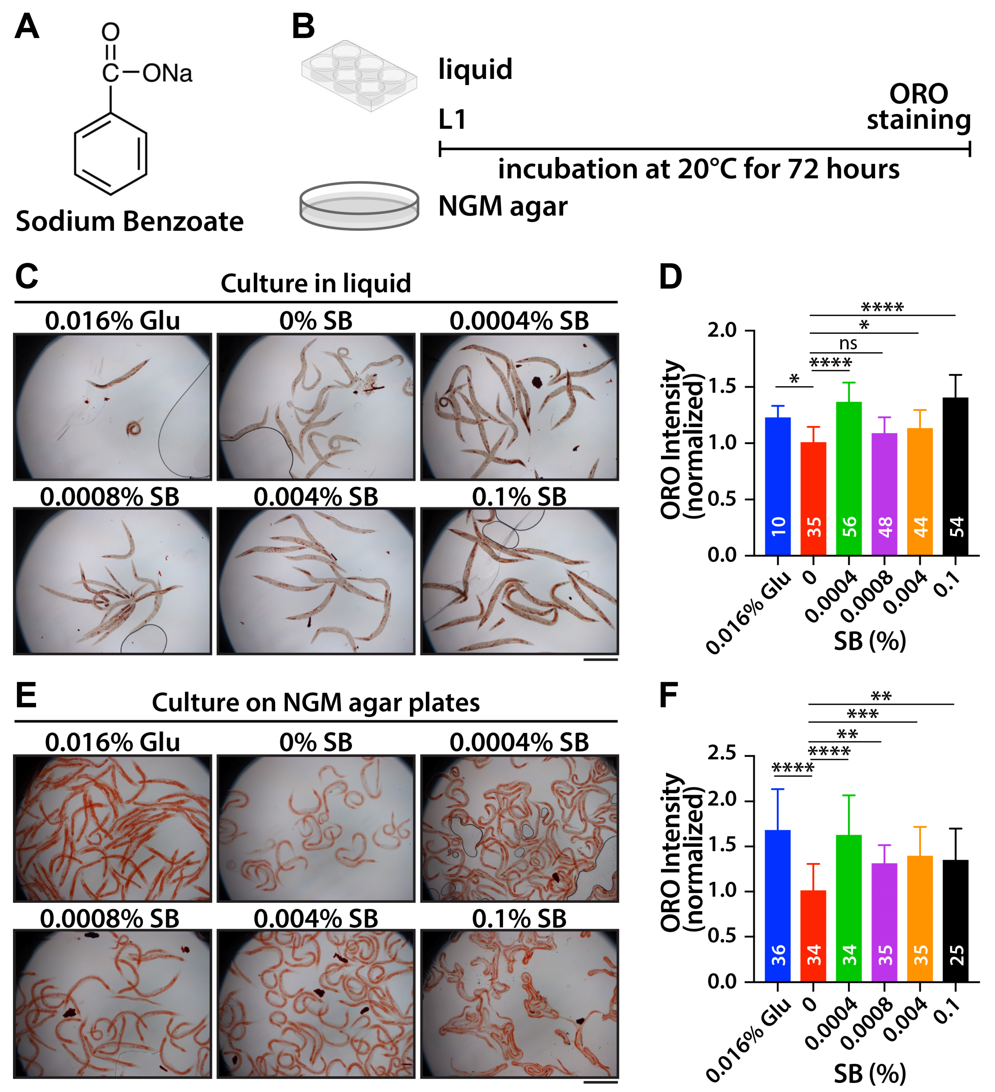
3.1. SB Increases Fat Accumulation in Wild-Type Worms

To evaluate whether SB exhibits an obesogenic effect, L1-synchronized wild-type worms were exposed to various concentrations of SB (0%, 0.0004%, 0.0008%, 0.004%, and 0.1%) and 0.016% glucose as a positive control for 72 h in liquid (K-media) conditions. Fat accumulation was visualized by staining collected worms using Oil Red O (ORO) dye, as previously reported [22] (Figure 1B). The fat accumulation was primarily observed in the intestines, oocytes, and hypodermis. Fat accumulation levels in the entire body were quantified using ImageJ software and normalized by the ORO intensity of 0% SB-treated worms. A significant increase in fat accumulation was observed across the tested SB concentrations, except for 0.0008% of SB. Notably, higher fat accumulation was detected at 0.0004% and 0.1% SB (Figure 1C,D). To confirm this result, L1-synchronized wild-type worms were cultured on OP50-seeded NGM agar plates with varying SB concentrations and 0.016% glucose for 72 h (Figure 1B). Fat accumulation levels were measured as described above. Similar patterns of fat accumulation were observed under NGM agar culture conditions (Figure 1E,F). Interestingly, 0.0004% SB-treated worms had a higher fat accumulation than 0.0008% SB-treated worms. Our results suggest that SB increases fat accumulation in wild-type C. elegans worms even at much lower concentrations than the FDA’s permissible concentration (0.1%).

3.2. SB May Increase Fat Accumulation Through Food Metabolism Pathways

To explore the potential impact of SB on fat accumulation during starvation, fourth larval stage (L4)-synchronized wild-type worms were cultured in K-media with various concentrations of SB and 0.016% glucose in the absence of OP50 E. coli food. After 72 h, the collected wild-type worms were stained with ORO dye. The results indicated no significant increase in fat accumulation (Figure 2A). Intriguingly, fat accumulation was even decreased at high SB concentrations (0.004% and 0.1%) (Figure 2B). Next, to investigate whether SB treatment enhances fat accumulation over an extended time, L1-synchronized wild-type worms were cultured for 120 h on OP50-seeded NGM agar plates with varying SB concentrations and 0.016% glucose. It is known that aged worms typically exhibit an increasing deposit of fat. In this study, we also observed that most aged adult worms (120 h past L1) displayed more fat accumulation than young adult worms (72 h past L1) (Compare Figure 2C to Figure 1E). Notably, prolonged SB treatment did not further increase fat accumulation compared to worms treated with 0% SB (Figure 2C,D). Therefore, these results indicate that SB treatment may enhance fat accumulation through food metabolic pathways, which is also related to lifespan.

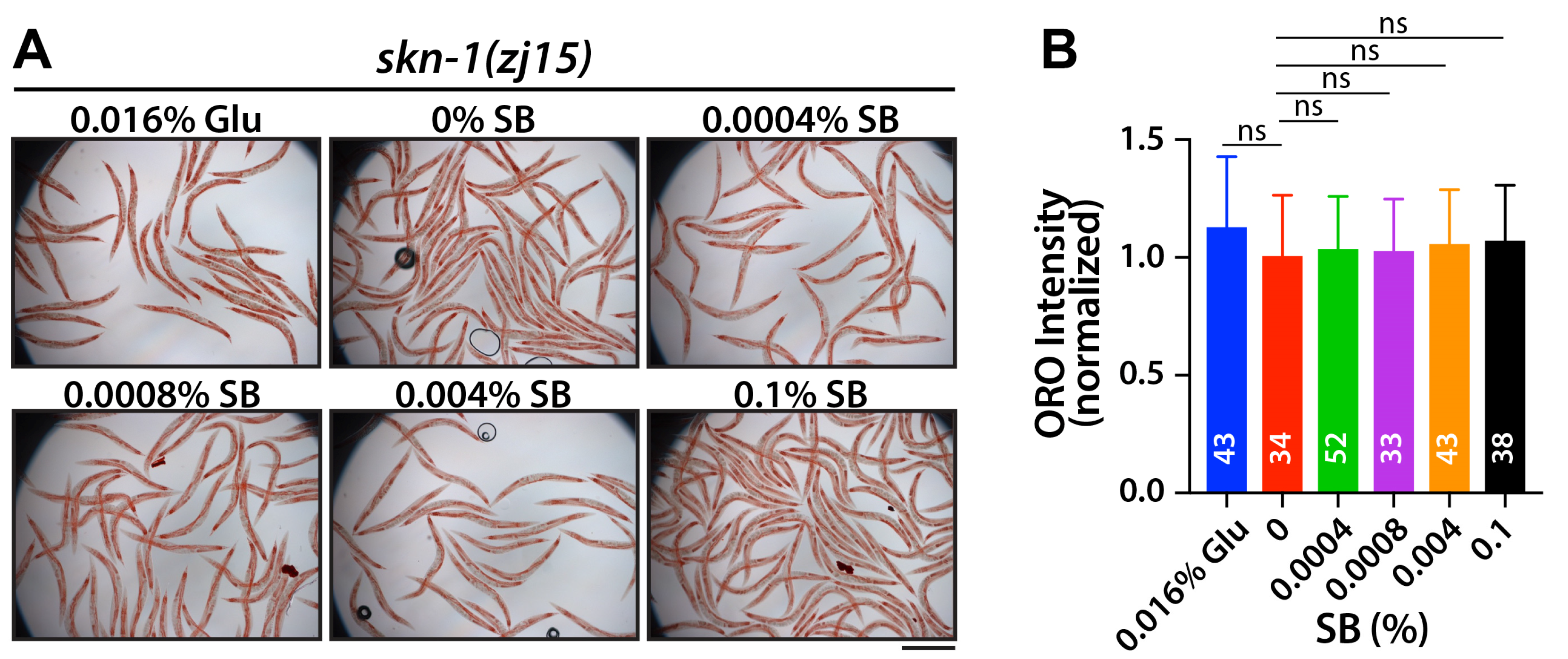
3.3. SB Increases Fat Accumulation, at Least in Part, Through the SKN-1/Nrf2 Signaling Pathway

SKN-1 (an Nrf2 homolog) is a well-known pathway associated with lipid metabolism [18,23]. SKN-1 activates diverse lipid metabolism genes and reduces fat storage [18,23]. To assess whether SB treatment increases fat accumulation through SKN-1, skn-1(zj15) loss-of-function mutant worms were used. L1-synchronized skn-1(zj15) mutant worms were cultured for 72 h on NGM agar plates with various concentrations of SB and 0.016% glucose. Fat accumulation was measured by ORO staining. While the overall fat accumulation levels in skn-1(zj15) mutant worms were significantly higher than those in wild-type worms [raw ORO intensity: wild-type (24.6 ± 7.5) vs. skn-1(zj15) (39.7 ± 8.8)] [18,23], no significant changes in fat accumulation due to SB and glucose treatment were observed in the skn-1(zj15) mutants as compared to vehicle control (Figure 3A,B). This result indicates that SKN-1 may be involved in SB-induced fat accumulation in wild-type worms.

3.4. SB Treatment Reduces Lifespan Primarily Through the SKN-1/Nrf2 Signaling Pathway

The SKN-1 signaling pathway is highly linked to longevity [24,25]. Under stress conditions, SKN-1 proteins translocate into the nucleus and activate the expression of their target genes, which are essential for detoxification or stress responses [24,25]. To test whether SB treatment affects the lifespan of C. elegans, L1-synchronized wild-type worms were cultured on OP50-seeded NGM agar plates with various concentrations of SB and 0.016% glucose. Survival rates were recorded daily, and surviving worms were transferred to fresh NGM plates every three days. The average lifespan of wild-type worms was approximately 16.23 ± 3.18 days (Figure 4A,B; Table 1). However, treatments with 0.016% glucose and SB significantly reduced the average lifespans of wild-type worms; 15.01 ± 3.69 days for 0.016% glucose, 16.23 ± 3.18 days for 0% SB, 13.27 ± 2.57 days for 0.0004% SB, 15.74 ± 3.75 days for 0.0008% SB, 14.92 ± 3.12 days for 0.004% SB, and 12.98 ± 3.23 days for 0.1% SB (Figure 4B; Table 1). Next, to investigate whether SB treatment decreases the lifespan of wild-type worms through the SKN-1 signaling pathway, L1-synchronized skn-1(zj15) mutant worms were cultured on OP50-seeded NGM agar plates with various concentrations of SB and 0.016% glucose, and their survival rates were recorded daily as described above. Unlike wild-type worms, skn-1(zj15) mutant worms did not show a significant reduction in average lifespan at 0.0004% and 0.0008% SB concentrations. Their average lifespans were only slightly decreased with higher SB treatments (0.004% and 0.1%) (Figure 4C,D; Table 1). Specifically, the average lifespans of skn-1(zj15) mutant worms were 9.93 ± 2.29 days for 0.016% glucose, 10.21 ± 2.51 days for 0% SB, 10.03 ± 2.35 days for 0.0004% SB, 9.79 ± 2.38 days for 0.0008% SB, 9.23 ± 2.22 days for 0.004% SB, and 9.23 ± 1.67 days for 0.1% SB (Figure 4D and Table 1). These results indicate that SB treatment reduces the average lifespan of wild-type worms, at least in part, through the SKN-1 signaling pathway.

3.5. SB Inhibits SKN-1 Nuclear Localization

SB has been known to induce oxidative stress in rodents [9] and zebrafish [12]. The major regulator of the oxidative stress response is SKN-1. Under oxidative stress conditions, SKN-1 proteins translocate into the nucleus and increase the expression of detoxification and antioxidant-related genes, thereby reducing cellular oxidative stress [26]. To test whether SB could inhibit the nuclear localization of the SKN-1 protein, transgenic worms expressing a skn-1(b/c)::gfp gene were employed. SKN-1 proteins are predominantly localized in the cytoplasm under normal conditions (Figure 5A weak), but they translocate to the nucleus in the intestine (Figure 5A strong) in response to oxidative stress from various sources, such as sodium arsenite (mitochondrial toxin). The skn-1(b/c)::gfp transgenic worms were cultured on OP50-seeded NGM agar plates with various concentrations of SB and 0.016% glucose at 20 °C. 48 h later, the transgenic worms were collected from the plates with M9 buffer and exposed to 5 mM sodium arsenite for 30 min. The nuclear localization of SKN-1::GFP proteins in the intestines was analyzed by staining using antibodies against GFP and nuclear pore complex (NPC) proteins. The ratio of SKN-1::GFP proteins in the nucleus to the cytoplasm was measured using ImageJ software. While SKN-1::GFP proteins exhibited dramatic translocation into the nucleus upon treatment with 5 mM sodium arsenite in the absence of SB (Figure 5A strong; Figure 5B), however, SKN-1 nuclear localization was significantly inhibited by 0.016% glucose treatment as reported [27] and in the presence of SB even at lower concentrations, including 0.0004% (Figure 4A moderate or weak; Figure 4B). Additionally, the inhibition of SKN-1 nuclear localization by SB treatment was observed even in the absence of sodium arsenite (Supplementary Figure S2). These results indicate that SB may inhibit the nuclear localization of SKN-1 in C. elegans intestinal cells.

4. Discussion

In this study, we aimed to explore the effect of SB on obesity and lifespan using the nematode C. elegans as a model system. Our results reveal that SB treatment increases fat accumulation and reduces lifespan, at least in part, through the SKN-1 signaling pathway (Figure 6). These findings suggest potential risks associated with SB consumption in the context of obesity and lifespan.
SB has been known to inhibit the growth of bacteria, fungi, and yeast. A recent study demonstrated that SB slows the larvae’s developmental timing and shortens the adult lifespan in Drosophila melanogaster by affecting endocrine hormone levels and commensal microbiota [10,11]. SB has also been associated with reduced reproductive capacity, developmental defects, oxidative stress, and anxiety-like behavior [12,28]. In zebrafish, SB treatment induces delayed hatching, morphological defects, oxidative stress, and abnormal behaviors in developing larvae [12]. However, in C. elegans, SB treatment did not lead to significant developmental defects or reproductive problems, even at a high concentration (0.1%) of SB. Questions persist regarding the potential occurrence of these abnormalities in C. elegans over multiple generations with long-term SB exposure.
We investigated the effect of SB on fat accumulation and lifespan reduction in wild-type worms using five different concentrations (0%, 0.0004%, 0.0008%, 0.004%, and 0.1%). The effect of SB on fat accumulation and lifespan reduction was high at 0.0004% and 0.1% SB. However, at 0.0008% and 0.004%, the effects were lower compared to 0.0004%. It is interesting that SB induced more significant fat accumulation in relatively low dosages (“low-dose effect”), which is consistent with what was observed in mice [29]. While a 0.0125% SB diet significantly increased body weight and food intake, higher SB concentrations (0.025% and 0.05%) did not alter body weight compared to control groups [29]. It was proposed that SB may enhance food palatability at a 0.0125% concentration [29], but higher concentrations could potentially reduce food intake [30]. To explore the potential impact of SB on food intake in C. elegans, we evaluated the pharyngeal pumping rate. Our findings revealed a significant increase in pharyngeal pumping rate at 0.016% glucose and only higher concentrations (≥0.004%) of SB (Supplementary Figure S1), indicating that food intake was not a major factor of the more pronounced fat accumulation and lifespan reduction effects observed in the 0.0004% dosage group.
tBHQ (tert-butylhydroquinone) is also used as a food preservative [31]. Unlike SB, tBHQ reduces oxidative stress by activating the Nrf2 signaling pathway. In the cytoplasm, KEAP1 (Kelch-like ECH-associated protein 1) inhibits this pathway by destabilizing Nrf2 proteins. tBHQ binds to the cysteine residues on the keap1 protein, enabling the nuclear localization of stabilized Nrf2 and promoting the expression of antioxidant genes [32]. How does SB inhibit the Nrf2 signaling pathway? We do not know but suggest that SB may activate KEAP1-mediated Nrf2 destabilization, leading to increased oxidative stress. In rats, SB treatment also increases oxidative stress by inhibiting the activity of glutathione peroxidase (GPx), catalase (CAT), glutathione-s-transferase (GST), glutathione reductase (GR) and superoxide dismutase (SOD), which are Nrf2 targets [33]. Therefore, understanding the action mechanism of SB is important for comprehending its potential effects on oxidative stress-mediated diseases in mammals, including humans.
SKN-1 transcription factor plays a crucial role in the cellular response to oxidative stress [34]. In the nucleus, SKN-1 activates the expression of antioxidant and detoxification genes. Thus, disrupting the SKN-1-mediated stress response mechanism increases oxidative stress, which reduces lifespan (Figure 6) [34]. Oxidative stress is closely linked to liver diseases such as drug-induced liver injury, viral hepatitis, and alcoholic hepatitis [35]. SB exposure affects the lipid profile and parameters of liver and kidney functions in rats [6]. Furthermore, SB has been shown to induce damage to kidney structure and function [36]. Specifically, SB elevates hepatic oxidative stress and inflammation in liver injury [37]. Nrf2 plays a critical role in various liver diseases, including metabolic dysfunction-associated fatty liver disease [35], and is considered a potential therapeutic target for non-alcoholic steatohepatitis [38]. Nrf2 also contributes to protecting the kidney from oxidative damage [37]. Thereby, SB may affect the functions of the liver and kidney and their associated diseases, most likely through the modulation of the Nrf2 signaling pathway.
In C. elegans, lipid metabolism and longevity are also regulated through an insulin signaling pathway, including DAF-2 (an IGF homolog) and DAF-16 (a FOXO homolog) [39]. In C. elegans, DAF-2 has been known to inhibit both DAF-16 and SKN-1 through AKT-1/2 kinases in response to external stress [40,41,42]. Both DAF-16 and SKN-1 are involved in the oxidative stress response and longevity [24,25]. However, DAF-16 and SKN-1 exhibit opposite functions in lipid metabolism—DAF-16 promotes fat accumulation, whereas SKN-1 inhibits it [18,43]. A recent study also demonstrated that SKN-1 functions as a negative regulator of DAF-16 [44]. Therefore, we propose that more complicated regulatory networks, including SKN-1 and insulin signaling pathways, may be involved in SB-induced fat accumulation and lifespan reduction. Although the detailed mechanism by which SB affects the insulin signaling pathway remains unclear, future research addressing this aspect will contribute to a better understanding of SB-induced obesity and lifespan reduction, thereby significantly advancing our knowledge in this field. Additionally, further studies are needed to explore the long-term impact of low concentrations of SB in mammals and disease model systems. This underscores the necessity for a safety reevaluation of SB in mammals, including humans.

5. Conclusions

Our study on the effects of SB on fat accumulation and lifespan in C. elegant raises significant safety concerns, particularly at low concentrations. We found that SB exposure induces fat accumulation and reduces lifespan, at least in part, by inhibiting the SKN-1/Nrf2 signaling pathway, which is essential for oxidative stress response. Since SB is commonly used in various foods and consumer products, our findings highlight the need for a comprehensive safety reevaluation of SB, particularly regarding its potential contributions to obesity, lifespan reduction, and oxidative stress-mediated diseases in mammals, including humans.

Supplementary Materials

The following supporting information can be downloaded at: https://www.mdpi.com/article/10.3390/nu16213753/s1, Figure S1: Average pharyngeal pumping rate of wild-type (N2) worms after 72 h of sodium benzoate treatment; Figure S2: SB inhibits SKN-1 nuclear localization under normal conditions.

Author Contributions

Conceptualization, J.D.L., J.V. and X.P.; Methodology, J.D.L., J.L., J.V. and X.P.; Validation, J.D.L. and J.L.; Formal analysis, J.D.L., J.L. and J.V.; Investigation, J.D.L., J.L. and J.V.; Resources, X.P.; Data curation, J.L.; Writing—original draft, J.D.L. and J.L.; Writing—review & editing, X.P.; Supervision, X.P.; Funding acquisition, X.P. All authors have read and agreed to the published version of the manuscript.

Funding

Partially supported by NIEHS P30ES025128.

Institutional Review Board Statement

Not applicable.

Informed Consent Statement

Not applicable.

Data Availability Statement

The original contributions presented in the study are included in the article, further inquiries can be directed to the corresponding author.

Acknowledgments

We are grateful to Lee at ECU for C. elegans strains and antibodies against GFP and NPC and for helpful discussions during our work. QV225 [skn-1(zj15)] and LD1 [idIs7 skn-1b/c::GFP + rol-6(su1006)] strains were obtained from the Caenorhabditis Genetic Center (CGC), which is supported by the NIH-National Center for Research Resources.

Conflicts of Interest

The authors declare no conflict of interest.

References

  1. Bensid, A.; El Abed, N.; Houicher, A.; Regenstein, J.M.; Ozogul, F. Antioxidant and antimicrobial preservatives: Properties, mechanism of action and applications in food—A review. Crit. Rev. Food Sci. Nutr. 2022, 62, 2985–3001. [Google Scholar] [CrossRef] [PubMed]
  2. Hrncirova, L.; Machova, V.; Trckova, E.; Krejsek, J.; Hrncir, T. Food Preservatives Induce Proteobacteria Dysbiosis in Human-Microbiota Associated Nod2-Deficient Mice. Microorganisms 2019, 7, 383. [Google Scholar] [CrossRef] [PubMed]
  3. Stapleton, M.; Daly, N.; O’Kelly, R.; Turner, M.J. Time and temperature affect glycolysis in blood samples regardless of fluoride-based preservatives: A potential underestimation of diabetes. Ann. Clin. Biochem. 2017, 54, 671–676. [Google Scholar] [CrossRef] [PubMed]
  4. Ruiz-Rico, M.; Renwick, S.; Vancuren, S.J.; Robinson, A.V.; Gianetto-Hill, C.; Allen-Vercoe, E.; Barat, J.M. Impact of food preservatives based on immobilized phenolic compounds on an in vitro model of human gut microbiota. Food Chem. 2023, 403, 134363. [Google Scholar] [CrossRef]
  5. Lennerz, B.S.; Vafai, S.B.; Delaney, N.F.; Clish, C.B.; Deik, A.A.; Pierce, K.A.; Ludwig, D.S.; Mootha, V.K. Effects of sodium benzoate, a widely used food preservative, on glucose homeostasis and metabolic profiles in humans. Mol. Genet. Metab. 2015, 114, 73–79. [Google Scholar] [CrossRef]
  6. Walczak-Nowicka, L.J.; Herbet, M. Sodium Benzoate-Harmfulness and Potential Use in Therapies for Disorders Related to the Nervous System: A Review. Nutrients 2022, 14, 1497. [Google Scholar] [CrossRef]
  7. Rangasamy, S.B.; Raha, S.; Dasarathy, S.; Pahan, K. Sodium Benzoate, a Metabolite of Cinnamon and a Food Additive, Improves Cognitive Functions in Mice after Controlled Cortical Impact Injury. Int. J. Mol. Sci. 2021, 23, 192. [Google Scholar] [CrossRef]
  8. Rangasamy, S.B.; Dasarathi, S.; Nutakki, A.; Mukherjee, S.; Nellivalasa, R.; Pahan, K. Stimulation of Dopamine Production by Sodium Benzoate, a Metabolite of Cinnamon and a Food Additive. J. Alzheimer’s Dis. Rep. 2021, 5, 295–310. [Google Scholar] [CrossRef]
  9. Khoshnoud, M.J.; Siavashpour, A.; Bakhshizadeh, M.; Rashedinia, M. Effects of sodium benzoate, a commonly used food preservative, on learning, memory, and oxidative stress in brain of mice. J. Biochem. Mol. Toxicol. 2018, 32, e22022. [Google Scholar] [CrossRef]
  10. Asejeje, F.O.; Alade, T.F.; Oyibo, A.; Abolaji, A.O. Toxicological assessment of sodium benzoate in Drosophila melanogaster. J. Biochem. Mol. Toxicol. 2023, 38, e23586. [Google Scholar] [CrossRef]
  11. Dong, Y.; Ding, Z.; Song, L.; Zhang, D.; Xie, C.; Zhang, S.; Feng, L.; Liu, H.; Pang, Q. Sodium Benzoate Delays the Development of Drosophila melanogaster Larvae and Alters Commensal Microbiota in Adult Flies. Front. Microbiol. 2022, 13, 911928. [Google Scholar] [CrossRef] [PubMed]
  12. Gaur, H.; Purushothaman, S.; Pullaguri, N.; Bhargava, Y.; Bhargava, A. Sodium benzoate induced developmental defects, oxidative stress and anxiety-like behaviour in zebrafish larva. Biochem. Biophys. Res. Commun. 2018, 502, 364–369. [Google Scholar] [CrossRef] [PubMed]
  13. Ciardi, C.; Jenny, M.; Tschoner, A.; Ueberall, F.; Patsch, J.; Pedrini, M.; Ebenbichler, C.; Fuchs, D. Food additives such as sodium sulphite, sodium benzoate and curcumin inhibit leptin release in lipopolysaccharide-treated murine adipocytes in vitro. Br. J. Nutr. 2012, 107, 826–833. [Google Scholar] [CrossRef] [PubMed]
  14. Hales, C.M.; Carroll, M.D.; Fryar, C.D.; Ogden, C.L. Prevalence of Obesity and Severe Obesity Among Adults: United States, 2017–2018; NCHS Data Brief; National Center for Health Statistics: Hyattsville, MD, USA, 2020; pp. 1–8. [Google Scholar]
  15. Hales, C.M.; Carroll, M.D.; Fryar, C.D.; Ogden, C.L. Prevalence of Obesity Among Adults and Youth: United States, 2015–2016; NCHS Data Brief; National Center for Health Statistics: Hyattsville, MD, USA, 2017; pp. 1–8. [Google Scholar]
  16. Simmons, A.L.; Schlezinger, J.J.; Corkey, B.E. What Are We Putting in Our Food That Is Making Us Fat? Food Additives, Contaminants, and Other Putative Contributors to Obesity. Curr. Obes. Rep. 2014, 3, 273–285. [Google Scholar] [CrossRef]
  17. Kaletta, T.; Hengartner, M.O. Finding function in novel targets: C. elegans as a model organism. Nat. Rev. Drug Discov. 2006, 5, 387–398. [Google Scholar] [CrossRef]
  18. Pang, S.; Lynn, D.A.; Lo, J.Y.; Paek, J.; Curran, S.P. SKN-1 and Nrf2 couples proline catabolism with lipid metabolism during nutrient deprivation. Nat. Commun. 2014, 5, 5048. [Google Scholar] [CrossRef]
  19. Shen, P.; Yue, Y.; Park, Y. A living model for obesity and aging research: Caenorhabditis elegans. Crit. Rev. Food Sci. Nutr. 2018, 58, 741–754. [Google Scholar] [CrossRef]
  20. Brenner, S. The genetics of Caenorhabditis elegans. Genetics 1974, 77, 71–94. [Google Scholar] [CrossRef]
  21. An, J.H.; Blackwell, T.K. SKN-1 links C. elegans mesendodermal specification to a conserved oxidative stress response. Genes Dev. 2003, 17, 1882–1893. [Google Scholar] [CrossRef]
  22. Escorcia, W.; Ruter, D.L.; Nhan, J.; Curran, S.P. Quantification of Lipid Abundance and Evaluation of Lipid Distribution in Caenorhabditis elegans by Nile Red and Oil Red O Staining. J. Vis. Exp. JoVE 2018, 133, 57352. [Google Scholar] [CrossRef]
  23. Steinbaugh, M.J.; Narasimhan, S.D.; Robida-Stubbs, S.; Moronetti Mazzeo, L.E.; Dreyfuss, J.M.; Hourihan, J.M.; Raghavan, P.; Operana, T.N.; Esmaillie, R.; Blackwell, T.K. Lipid-mediated regulation of SKN-1/Nrf in response to germ cell absence. eLife 2015, 4, e07836. [Google Scholar] [CrossRef] [PubMed]
  24. Ogg, S.; Paradis, S.; Gottlieb, S.; Patterson, G.I.; Lee, L.; Tissenbaum, H.A.; Ruvkun, G. The Fork head transcription factor DAF-16 transduces insulin-like metabolic and longevity signals in C. elegans. Nature 1997, 389, 994–999. [Google Scholar] [CrossRef] [PubMed]
  25. Tullet, J.M.; Hertweck, M.; An, J.H.; Baker, J.; Hwang, J.Y.; Liu, S.; Oliveira, R.P.; Baumeister, R.; Blackwell, T.K. Direct inhibition of the longevity-promoting factor SKN-1 by insulin-like signaling in C. elegans. Cell 2008, 132, 1025–1038. [Google Scholar] [CrossRef] [PubMed]
  26. Inoue, H.; Hisamoto, N.; An, J.H.; Oliveira, R.P.; Nishida, E.; Blackwell, T.K.; Matsumoto, K. The C. elegans p38 MAPK pathway regulates nuclear localization of the transcription factor SKN-1 in oxidative stress response. Genes Dev. 2005, 19, 2278–2283. [Google Scholar] [CrossRef]
  27. Li, L.; Chen, Y.; Chenzhao, C.; Fu, S.; Xu, Q.; Zhao, J. Glucose negatively affects Nrf2/SKN-1-mediated innate immunity in C. elegans. Aging 2018, 10, 3089–3103. [Google Scholar] [CrossRef]
  28. El-Shennawy, L.; Kamel, M.A.E.; Khalaf, A.H.Y.; Yousef, M.I. Dose-dependent reproductive toxicity of sodium benzoate in male rats: Inflammation, oxidative stress and apoptosis. Reprod. Toxicol. 2020, 98, 92–98. [Google Scholar] [CrossRef]
  29. Olofinnade, A.T.; Onaolapo, A.Y.; Onaolapo, O.J.; Olowe, O.A. The potential toxicity of food-added sodium benzoate in mice is concentration-dependent. Toxicol. Res. 2021, 10, 561–569. [Google Scholar] [CrossRef]
  30. Nair, B. Final report on the safety assessment of Benzyl Alcohol, Benzoic Acid, and Sodium Benzoate. Int. J. Toxicol. 2001, 20 (Suppl. S3), 23–50. [Google Scholar] [CrossRef]
  31. Leonard, E.R.; Marques, E.S.; Roy, M.A.; Conlin, S.M.; Ranjan, R.; Timme-Laragy, A.R. Dietary exposure to the food preservative tert-Butylhydroquinone (tBHQ) impairs zebrafish (Danio rerio) survival, growth, organ development, and gene expression in Nrf2a-dependent and independent ways. Food Chem. Toxicol. Int. J. Publ. Br. Ind. Biol. Res. Assoc. 2023, 176, 113788. [Google Scholar] [CrossRef]
  32. Bryan, H.K.; Olayanju, A.; Goldring, C.E.; Park, B.K. The Nrf2 cell defence pathway: Keap1-dependent and -independent mechanisms of regulation. Biochem. Pharmacol. 2013, 85, 705–717. [Google Scholar] [CrossRef]
  33. Khan, I.S.; Dar, K.B.; Ganie, S.A.; Ali, M.N. Toxicological impact of sodium benzoate on inflammatory cytokines, oxidative stress and biochemical markers in male Wistar rats. Drug Chem. Toxicol. 2022, 45, 1345–1354. [Google Scholar] [CrossRef] [PubMed]
  34. Blackwell, T.K.; Steinbaugh, M.J.; Hourihan, J.M.; Ewald, C.Y.; Isik, M. SKN-1/Nrf, stress responses, and aging in Caenorhabditis elegans. Free Radic. Biol. Med. 2015, 88, 290–301. [Google Scholar] [CrossRef] [PubMed]
  35. Zhou, J.; Zheng, Q.; Chen, Z. The Nrf2 Pathway in Liver Diseases. Front. Cell Dev. Biol. 2022, 10, 826204. [Google Scholar] [CrossRef] [PubMed]
  36. Zeghib, K.; Boutlelis, D.A. Food Additive (Sodium benzoate)-induced Damage on Renal Function and Glomerular Cells in Rats; Modulating Effect of Aqueous Extract of Atriplex halimus L. Iran. J. Pharm. Res. IJPRjwan 2021, 20, 296–306. [Google Scholar] [CrossRef]
  37. Nezu, M.; Suzuki, N. Roles of Nrf2 in Protecting the Kidney from Oxidative Damage. Int. J. Mol. Sci. 2020, 21, 2951. [Google Scholar] [CrossRef]
  38. Galicia-Moreno, M.; Lucano-Landeros, S.; Monroy-Ramirez, H.C.; Silva-Gomez, J.; Gutierrez-Cuevas, J.; Santos, A.; Armendariz-Borunda, J. Roles of Nrf2 in Liver Diseases: Molecular, Pharmacological, and Epigenetic Aspects. Antioxidants 2020, 9, 980. [Google Scholar] [CrossRef]
  39. Hellerer, T.; Axang, C.; Brackmann, C.; Hillertz, P.; Pilon, M.; Enejder, A. Monitoring of lipid storage in Caenorhabditis elegans using coherent anti-Stokes Raman scattering (CARS) microscopy. Proc. Natl. Acad. Sci. USA 2007, 104, 14658–14663. [Google Scholar] [CrossRef]
  40. Cahill, C.M.; Tzivion, G.; Nasrin, N.; Ogg, S.; Dore, J.; Ruvkun, G.; Alexander-Bridges, M. Phosphatidylinositol 3-kinase signaling inhibits DAF-16 DNA binding and function via 14-3-3-dependent and 14-3-3-independent pathways. J. Biol. Chem. 2001, 276, 13402–13410. [Google Scholar] [CrossRef]
  41. Hertweck, M.; Gobel, C.; Baumeister, R. C. elegans SGK-1 is the critical component in the Akt/PKB kinase complex to control stress response and life span. Dev. Cell 2004, 6, 577–588. [Google Scholar] [CrossRef]
  42. Lin, K.; Hsin, H.; Libina, N.; Kenyon, C. Regulation of the Caenorhabditis elegans longevity protein DAF-16 by insulin/IGF-1 and germline signaling. Nat. Genet. 2001, 28, 139–145. [Google Scholar] [CrossRef]
  43. Xu, A.; Zhang, Z.; Ko, S.H.; Fisher, A.L.; Liu, Z.; Chen, L. Microtubule regulators act in the nervous system to modulate fat metabolism and longevity through DAF-16 in C. elegans. Aging Cell 2019, 18, e12884. [Google Scholar] [CrossRef]
  44. Deng, J.; Dai, Y.; Tang, H.; Pang, S. SKN-1 Is a Negative Regulator of DAF-16 and Somatic Stress Resistance in Caenorhabditis elegans. G3 2020, 10, 1707–1712. [Google Scholar] [CrossRef]
Figure 1. SB treatment increases fat accumulation in wild-type worms. (A) Chemical structure of sodium benzoate. (B) Strategy for SB treatment in liquid or on NGM agar plate. (C) ORO staining with SB-treated worms in liquid. (D) Normalized ORO intensity. (E) ORO staining with SB-treated worms on NGM agar plates. (F) Normalized ORO intensity. Not significant (ns), p > 0.05; *, 0.01 < p ≤ 0.05, **, 0.001 < p ≤ 0.01; ***, 0.0001 < p ≤ 0.001; **** p ≤ 0.0001. Scale bars: 500 μm.
Nutrients 16 03753 g001
Figure 2. SB-induced fat accumulation may require metabolic pathways. (A) ORO staining with starved worms. (B) Normalized ORO intensity. (C) ORO staining with prolonged SB-treated worms. (D) Normalized ORO intensity. Not significant (ns), p > 0.05; *, 0.01 < p ≤ 0.05, **, 0.001 < p ≤ 0.01. Scale bars: 500 μm.
Nutrients 16 03753 g002
Figure 3. SB-induced fat accumulation may require the SKN-1 signaling pathway. (A) ORO staining with skn-1(zj15) mutant worms. (B) Normalized ORO intensity. Not significant (ns), p > 0.05. Scale bar: 500 μm.
Nutrients 16 03753 g003
Figure 4. SB shortens the average lifespan through the SKN-1 signaling pathway. (A,C) Lifespan curves of wild-type and skn-1(zh15) mutant worms. (B,D) Individual days of survival of wild-type and skn-1(zh15) mutant worms. Not significant (ns), p > 0.05; *, 0.01 < p ≤ 0.05; **** p ≤ 0.0001. See Table 1 for average lifespans and errors.
Nutrients 16 03753 g004
Figure 5. SB inhibits SKN-1 nuclear localization under oxidative stress conditions. (A) Immunostaining. Dissected intestines were stained with antibodies against GFP and the nuclear pore complex (NPC) proteins. Representative images of SKN-1::GFP nuclear localization are categorized as follows: weak, no significant difference in SKN-1::GFP levels between the nucleus and cytoplasm (ratio ~1.0 or less); moderate, SKN-1::GFP levels are notably higher in the nucleus than in the cytoplasm (ratio ~1.5); strong, most SKN-1GFP is translocated into the nucleus (ratio ~2 or higher). Arrowheads indicate intestinal nuclei. All images were acquired using the same setting and magnification. Scale bar: 20 μm. (B) The ratio of GFP intensities in the nucleus to the cytoplasm. Below are the images of SKN-1::GFP (green) dynamics and NPC proteins (red). Not significant (ns), p > 0.05; **** p ≤ 0.0001.
Nutrients 16 03753 g005
Figure 6. A potential action model of SB in fat accumulation and lifespan reduction.
Nutrients 16 03753 g006
Table 1. Average lifespans.
StrainTreatmentAverage Lifespan ± (StDv) p-Value (t-Test)
wild-type0.016% Glu15.01 ± 3.69 days0.0420
0% SB16.23 ± 3.18 days-
0.0004% SB13.27 ± 2.57 days<0.0001
0.0008% SB15.74 ± 3.75 days0.3802
0.004% SB14.92 ± 3.12 days0.0167
0.1% SB12.98 ± 3.23 days<0.0001
skn-1(zj15)0.016% Glu 9.93 ± 2.29 days0.1667
0% SB10.21 ± 2.50 days-
0.0004% SB10.03 ± 2.35 days0.6873
0.0008% SB9.79 ± 2.38 days0.3673
0.004% SB9.23 ± 2.22 days0.0338
0.1% SB9.23 ± 1.67 days0.0138
Disclaimer/Publisher’s Note: The statements, opinions and data contained in all publications are solely those of the individual author(s) and contributor(s) and not of MDPI and/or the editor(s). MDPI and/or the editor(s) disclaim responsibility for any injury to people or property resulting from any ideas, methods, instructions or products referred to in the content.

