Review

Beneficial Effects of Omega-3 Fatty Acids on Obesity and Related Metabolic and Chronic Inflammatory Diseases

by
Donia Jerab
1,2,
Ferdinand Blangero
1,
Paulo César Trindade da Costa
3,
José Luiz de Brito Alves
3,
Rym Kefi
2,
Henda Jamoussi
4,
Beatrice Morio
1 and
Assia Eljaafari
1,5,*
1
CarMeN Laboratory, Institut National de Recherche pour l’ Agriculture, l’ Alimentation et l’Environnement, UMR1397, Institut National de la Santé et de la Recherche Médicale, U 1060, Université Claude Bernard Lyon I, 69310 Pierre-Bénite, France
2
Laboratory of Biomedical Genomics and Oncogenetics, Institut Pasteur de Tunis, Tunis 1002, Tunisia
3
Department of Nutrition, Health Sciences Center, Federal University of Paraíba, João Pessoa 58051-900, Brazil
4
Research Unit “Obesity: Etiopathology and Treatment, UR18ES01”, Faculty of Medicine, Tunis El Manar University, Tunis 2092, Tunisia
5
Department of Clinical Research, Hospices Civils de Lyon, 69002 Lyon, France
*
Author to whom correspondence should be addressed.
Nutrients 2025, 17(7), 1253; https://doi.org/10.3390/nu17071253
Submission received: 27 February 2025 / Revised: 28 March 2025 / Accepted: 30 March 2025 / Published: 3 April 2025
(This article belongs to the Section Nutrition and Obesity)

Abstract

:
Omega-3 polyunsaturated fatty acids (n-3 PUFAs) are known to help resolve inflammation through generation of anti-inflammatory eicosanoids and specialized pro-resolving mediators, including resolvins, protectins, and maresins. Through binding to the GPR120/FFAR4 receptor, their beneficial effects result from phospholipid membrane remodeling, impairment of inflammatory signaling molecules clustering, subsequent inhibition of NF-κB and inflammasome activation, and a reduction in oxidative stress. Obesity, a chronic inflammatory disease that contributes to metabolic disorders, is alleviated by n-3 PUFAs. In the adipose tissue (AT) of individuals with obesity, n-3 PUFAs counteract hypoxia, inhibit immune cell infiltration and AT inflammation, improve insulin sensitivity, and reduce fat mass. Beyond AT, n-3 PUFAs also alleviate other metabolic disorders such as metabolic-associated steatotic liver disease (MASLD), gut dysbiosis, and/or renal dysfunction. In cardiovascular disease (CVD), they are mainly recommended as a secondary prevention for patients with coronary heart disease risks. This review provides an in-depth analysis of the benefits of n-3 PUFAs in obesity and related metabolic diseases, examining both the mechanistic and clinical aspects. Additionally, it also explores the effects of n-3 PUFAs in obesity-related chronic inflammatory conditions, including inflammatory bowel disease, psoriasis, rheumatoid arthritis, osteoarthritis, and multiple sclerosis, by targeting specific pathophysiological mechanisms. Clinical applications and limitations of n-3 PUFAs are discussed based on findings from human clinical trials.

1. Introduction

Obesity is a chronic inflammatory disease that often leads to co-morbidities such as type 2 diabetes (T2D), metabolic-associated steatotic liver disease (MASLD), and cardiovascular diseases (CVD) [1]. In 2022, the prevalence of obesity reached 890 million people worldwide, underlining its status as a major public health concern [2]. The onset of obesity is usually driven by sedentary lifestyle and hypercaloric diet, leading to energy imbalance and subsequent metabolic disorders. Excess calories are stored as fat in adipose tissue (AT), leading to hyperplasia, hypertrophy, AT remodeling, homeostasis disruption, and the switch of AT environment from an anti-inflammatory and insulin-sensitive state to a pro-inflammatory, hypoxic, and insulin-resistant state. Following this, ectopic deposition of fatty acids (FAs) in other organs and propagation of inflammation to the periphery contribute to metabolic disorders [3]. Beyond metabolic disorders, obesity-mediated inflammation exacerbates various auto-immune and inflammatory diseases, such as inflammatory bowel diseases (IBD), psoriasis, rheumatic diseases, multiple sclerosis, and cancers, as previously reviewed [4,5].
Current approaches to mitigate obesity focus on rebalancing energy expenditure by increasing physical activity and reducing excess calorie intake. Besides pharmacological [6] and bariatric surgical strategies [7], adjunction of dietary interventions that emphasize the consumption of fish and fruits and vegetables rich in bioactive compounds, such as the Mediterranean diet, is often required to promote better metabolic health [8]. In the Mediterranean diet, dietary components such as polyphenols are known to exert their effects through antioxidant activity, epigenetic modulation, and microbiota-dependent pathways [9]. Dietary fibers act by modulating gut microbiota, producing short-chain fatty acids, and improving gut barrier integrity [10,11], while n-3 PUFAs act by directly resolving inflammation, modulating membrane composition, and improving lipid metabolism. In addition to their direct anti-inflammatory properties, n-3 PUFAs also exert their effects by competing with n-6 PUFAs for enzymatic conversion. Therefore, by inhibiting the synthesis of pro-inflammatory prostaglandins derived from n-6 PUFAs, which are abundant in many dietary sources, such as PGE2, n-3 PUFAs help shift the balance toward a more anti-inflammatory state [12].
Beyond their potent anti- inflammatory and adipogenic effects within adipose tissues [13], as well as their systemic beneficial effects, n-3 PUFAs may thus help reduce obesity and related metabolic disorders and chronic inflammatory diseases. The aim of this review is to provide a comprehensive analysis of the benefits of n-3 PUFAs with respect to these diseases.

2. Results

2.1. Beneficial Effects of Omega-3 Fatty Acids on Obesity

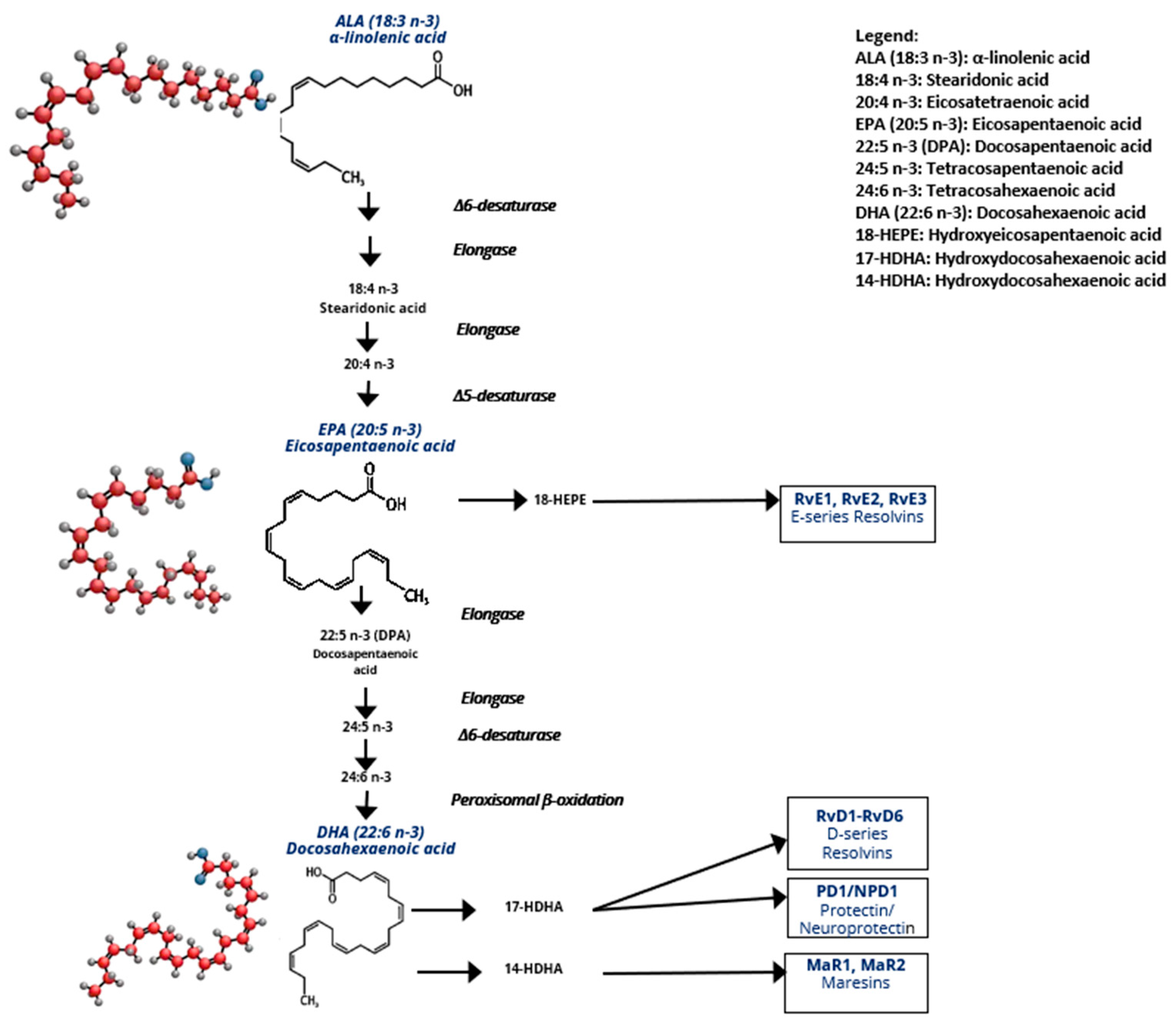
2.1.1. ω-3/ω-6 Eicosanoid Generation

FAs are identified by the length of their carbon chain and the number and position of unsaturated sites on carbons. FAs are thus classified as saturated FAs (SFAs, 0 double bonds), monounsaturated fatty acids (MUFAs, 1 double bond), or polyunsaturated fatty acids (PUFAs, ≥2 double bonds). Among PUFAs, n-3 and n-6 PUFAs differ in the position of the first carbon–carbon double bond relative to the terminal methyl group. Thus, alpha-linolenic acid (ALA) is referred to as an 18:3 n-3 PUFA, indicating that it is composed of 18 carbons and three double bonds, with the first unsaturated site occurring at the third carbon from the terminal methyl group. Linoleic acid (LA), which is designated as an 18:2 n-6 PUFA, contains 18 carbons and two double bonds, with the first double bond occurring at the sixth carbon from the methyl end group. ALA and its derivatives, eicosapentaenoic acid (EPA, 20:5 n-3 PUFA) and docosahexaenoic acid (DHA, 22:6 n-3 PUFA), are the predominant n-3 PUFAs [14].
Once incorporated into membranes, LA is metabolized into arachidonic acid (AA 20:4 n-6), a key precursor of several pro-inflammatory lipid mediators. In response to stimuli, AA and LA, which are stored in the phospholipid membranes, can be released through the action of phospholipase A2 (PLA2) [15]. In the cytoplasm, AA is metabolized by constitutive cyclooxygenases (COX-1) and inflammation-inducible cyclooxygenase (COX-2), as well as by lipoxygenase (LOX) and other enzymes, into lipid derivatives known as eicosanoids, such as PGD2, PGE2, TXA2, and LTB4 mediators. These eicosanoids are pro-inflammatory and play crucial roles in metabolic and chronic inflammation diseases. By replacing LA or AA within the phospholipids, dietary supplementation with n-3 PUFAs reduces the ω-6/ω-3 ratio within the membranes. When released from membranes, n-3 PUFAs also compete with n-6 PUFAs for the LOX and COX enzymes involved in eicosanoid synthesis. ALA-derived metabolites such as EPA or DHA are substrates for COX, LOX, and cytochrome P450, which contribute to the synthesis of prostaglandin E3 (PGE3), prostaglandin D3 (PGD3), thromboxane A3 (TXA3), and leukotriene B5 (LTB5), which are less pro-inflammatory than AA-derived mediators [14]. In addition, EPA and DHA are able to be converted into specialized pro-resolving mediators (SPMs) via the COX pathway for resolvins and protectins or the LOX pathway for maresins. Indeed, resolvins D 1–6, protectin 1, and maresins 1–2 are derived from DHA, while resolvins 1–3 are derived from EPA, as shown in Figure 1. All of these PMs are known to contribute to the resolution of inflammation, either by (i) limiting neutrophil infiltration, or trans-endothelial migration of leukocytes, (ii) promoting macrophage phagocytosis, or (iii) inhibiting TNFα and/or IL1β [16,17].

2.1.2. Dietary Sources of n-3 PUFAs

ALA is an essential PUFA, but EPA and DHA are considered conditionally essential because their endogenous synthesis from ALA is extremely limited. Therefore, they must be provided by the diet. ALA is mainly obtained from plant sources, such as flaxseeds, rapeseeds, and chia seeds, or from dried fruits such as nuts or walnuts, while EPA and DHA are mainly provided by marine sources, such as oily fish [18]. EPA and DHA can also be generated from ALA but at very low levels, since in humans, the conversion rate of ALA to EPA or DHA is less than 5%, and is further influenced by the n-6/n-3 PUFA ratio [19]. Thus, a high intake of n-6 PUFAs limits the conversion of ALA into EPA or DHA. The recommended ω-6/ω-3 ratio for good health is 5:1. Such a ratio is met and even exceeded in the Japanese population, where the ratio averages 4:1, while this ratio averages 10:1 in Europe and even 20:1 in the United States [20]. Western diets are thus characterized by a high ω-6/ω-3 ratio, and are associated with increased risks of obesity and metabolic disorders [21]. In the African continent, in particular in Tunisia, a recent cross-sectional study comparing obese patients with and without metabolic syndrome showed that those without metabolic disorders were more likely to adhere to a Mediterranean-style diet, with a higher intake of oily fish [22]. Moreover, in another study, the average ω-6/ω-3 ratio reached 17:1 in the Tunisian T2D population, with none of these patients achieving the recommended dietary intake of n-3 PUFAs [23].
Recent dietary guidelines recommend eating at least two servings of fish per week, equivalent to 500 mg of EPA + DHA [24].

2.1.3. Mechanisms of the Anti-Inflammatory Effects of n-3 PUFAs

In addition to their ability to compete with n-6 PUFAs in the phospholipid membranes for enzymes needed for eicosanoid synthesis, n-3 PUFAs may act as agonists for several free FA receptors expressed on a wide range of cells. Notably, EPA and DHA have been shown to activate FFAR4, also known as GPR120, a G protein-coupled receptor [25,26]. This leads to the recruitment of β-arrestin 2 to the plasma membrane. The GPR120/β-arrestin 2 complex is then internalized into the cytoplasmic compartment, where it binds to the TAK1-binding protein (TAB1) and blocks its association with TAK1. This results in the inhibition of the IKK-β/NF-κB and JNK/AP-1 signaling pathways and a reduction in TNF-α and IL-6 transcription [27]. B-arrestin2 has also been found to act downstream of GPR120 and GPR40 to inhibit NLRP3 inflammasome activation, which results in the inhibition of caspase-1 activation and a reduction in the subsequent secretion of IL-1β and IL-18 pro-inflammatory cytokines. n-3 PUFA supplementation in high-fat diet (HFD) mice has thus been demonstrated to prevent NLRP3 inflammasome-dependent insulin resistance in vivo [28].
Another mechanism by which n-3 PUFA may inhibit AT inflammation is through their ability to bind to peroxisome proliferator-activated receptor (PPAR) nuclear receptors, particularly PPAR-α and PPAR-γ. Once activated by ligand binding, these PPAR isoforms form heterodimers with the retinoid X receptor (RXR) and bind to peroxisome proliferator-activated receptor response elements (PPREs) to trans-repress the activity of transcription factors such as NF-κB. PPAR-α and PPAR-γ are also involved in the regulation of lipid metabolism and adipocyte differentiation, [29,30]. n-3 PUFAs have thus been shown to modulate inflammation in adipocytes and immune cells, improve adipogenesis and lipogenesis, and enhance insulin sensitivity through binding to PPAR-α and PPAR-γ [31,32,33].
n-3 PUFA action may also result from their incorporation into plasma membranes at the expense of n-6 PUFAs, leading to the disruption of lipid rafts. Lipid rafts are regions rich in cholesterol, phospholipids, and sphingolipids that form rigid membrane domains able to anchor receptors and signaling molecules, including those involved in the immunological synapse. Incorporation of n-3 PUFAs into these rafts disrupts their structure, thereby inhibiting immune response [34]. Lipid rafts are also involved in the dimerization of toll like receptor 4 (TLR-4) and recruitment of MYD88, leading to NF-κB activation and subsequent inflammation [35]. Of interest, in obesity, reduced intestinal barrier integrity due to gut dysbiosis leads to lipopolysaccharide (LPS) release and endotoxemia [36]. Inflammation may then occur via the binding of LPS to TLR4. However, disruption of lipid rafts by n-3 PUFAs reduces the translocation of TLR4 and myeloid differentiation primary response (MYD88) to the membrane, and thus inhibits NF-kB activation [32,35]. A study by Bidu et al. supports the role of n-3 PUFAs in reducing gut dysbiosis and inflammation. Using the Fat-1 model in which mice constitutively produce ALA this study demonstrated that mice did not develop gut dysbiosis and displayed reduced metabolic endotoxemia and AT inflammation when fed an obesogenic diet. Moreover, they also demonstrated that transplantation of their gut microbiota to wild-type mice prevented obesity and glucose intolerance, therefore suggesting that n-3 PUFAs may also reduce fat mass and insulin resistance by modulating the gut microbiota [37].

2.1.4. Beneficial Effects of n-3 PUFAs on AT Inflammation and Adipose Metabolism in Obesity

In addition to their anti-inflammatory properties, n-3 PUFAs may improve metabolic dysfunction. Indeed, in AT, n-3 PUFAs are known to improve insulin sensitivity by increasing adiponectin secretion [38,39,40], enhancing glucose uptake through an increase in GLUT-4 content [41] and reducing leptin and ObR levels [42]. In addition, n-3 PUFAs have been shown to enhance adipogenesis and VEGFα release through PPARγ activation, which helps counteract hypoxia and reduce AT inflammation [43,44]. Accordingly, supplementation with EPA (1% of total energy intake) has been shown to preserve glucose homeostasis and reduce fat mass accumulation in mice fed an obesogenic diet, with a particular reduction in visceral AT mass [45,46]. However, the effects of n-3 PUFAs on weight loss are likely weak, as shown in a mouse model of diet-induced obesity, where no significant effects were observed, although accelerated mammary tumor growth was prevented [47].
Whereas transgenerational supplementation with EPA in mice has been shown to reduce whole body weight and metabolic disorders in HFD mice [48,49], clinical evidence that n-3 PUFAs may benefit weight loss or reduced fat mass in humans is less consistent, except when they are combined with calorie restriction [50].
A systematic review of randomized clinical trials found an association between n-3 PUFA intake and reduced plasma biomarkers of inflammation and endothelial activation, such as C-reactive protein, pro-inflammatory cytokines (IL-6, TNF-α), adhesion molecules (ICAM-1, VCAM-1), and eicosanoids, and suggested that n-3 PUFA supplementation may alleviate obesity-related inflammation [51]. Accordingly, a higher proportion of n-3 PUFAs in red blood cell (RBC) membranes has been associated with a lower fat mass index, reduced leptin levels, and decreased inflammatory markers such as C-reactive protein (CRP) and triglycerides [52,53,54].
Therefore, despite mixed findings on body weight, n-3 PUFAs may provide benefits with respect to inflammation and metabolic disorders in patients with unhealthy obesity [55].

2.2. Effects of n-3 PUFAs on Obesity-Related Metabolic Diseases

2.2.1. Cardiovascular Disease

Cardiovascular disease (CVD) is one of the leading causes of death and morbidity worldwide. Obesity associated with chronic low-grade inflammation is an important risk factor for the development of metabolic CVD [56]. Consumption of n-3 PUFAs may confer benefits in the prevention of cardiovascular risk, such as dyslipidemia, inflammation, and blood pressure associated with obesity, but this is less evident at a therapeutic level. Indeed, through their antioxidant and anti-inflammatory effects, n-3 PUFAs have been shown to reduce autonomic sympathetic nerve activity in the hearts of rats, leading to the lowering of cardiovascular risks [57]. Supporting these results, DHA administration in rats has been shown to reduce heart rate and blood pressure, and thus cardiovascular risks, through activation of the FFA receptor in cardiac-projecting nucleus ambiguus neurons, which provide parasympathetic control of heart rate [58].
But, in humans, an extensive meta-analysis study of 86 clinical trials and 162,796 participants concluded that n-3 PUFAs, such as EPA or DHA, have little or no effect on reducing cardiovascular mortality and cardiovascular events, but slight effects in terms of reducing serum triglyceride, coronary heart disease (CHD) mortality, and CHD events. However, increasing ALA dietary intake from dried fruits or enriched margarine resulted in slightly reducing the risks of cardiovascular events and arrhythmia [59]. Overall, numerous prospective and meta-analytic studies agree that n-3 PUFA supplementation may substantially reduce the risks of CHD from 15 to 25%, depending on the study [60], with the exception of the ASCEND study, which was carried out on patients with T2D but without existing CVD at baseline. Indeed, the authors did not find any benefit in consuming 1 g/d of n-3 PUFAs as compared to olive oil, as there was no reduction in the primary end points of serious vascular events, such as ischemic attack, nonfatal myocardial infection, stroke, or vascular death. This was supported by the prospective VITAL study, which also used 1 g/d of fish oil capsule and found no benefit in humans without CVD at baseline [61]. At present, recommendations are in favor of a 1 g/d intake of n-3 PUFAs, but only in persons with prevalent CHD or heart failure [62].
Interventional studies in patients with or at risk of CVD showed less consistent effects when n-3 PUFAs were used as adjunctive therapy to statins. In support of the beneficial effects of n-3 PUFAs in reducing CVD outcomes is the REDUCE-IT clinical trial, which was carried out on patients with established cardiovascular disease, diabetes, or other CVD risk factors, such as hypertriglyceridemia, and who had been receiving statin therapy. They were treated with 3.6 g/d of EPA ethyl ester compound, which resulted in reducing the risks of the primary composite end points of cardiovascular death, nonfatal myocardial infarction, nonfatal stroke, coronary revascularization, or unstable angina by 25%, as compared to the placebo group who received mineral oil [63]. In the EVAPORATE study, by using the same treatment, at the same dose, in a high cardiovascular risk population treated with statins and with persistently high triglyceride levels, a beneficial effect was again found, as the progression of total (calcified and non-calcified) coronary plaque was slowed by 42% [64]. Disagreeing with these studies, in the STRENGTH study, in which both EPA and DHA were given (2.2 g and 0.8 g, respectively) to statin-treated patients with high cardiovascular risk, high triglycerides, and low HDL cholesterol levels, no effect on major cardiovascular events was observed, as compared to corn oil in the placebo group [65]. Moreover, such intake resulted in an increase in low-density lipoprotein (LDL) cholesterol levels even though high-density lipoprotein (HDL) cholesterol increased. This contributed to the decision to discontinue the trial, as a higher risk of atrial fibrillation was reported to occur when n-3 PUFA supplementation exceeded 1 g/day [66].
The reason for these discrepant results was attributed to the n-3 PUFA molecules used in these studies (EPA versus EPA + DHA), since EPA alone resulted in decreasing LDL cholesterol by 6.6%, whereas EPA + DHA increased it by 3%. The different placebos used (mineral oil versus corn oil) were also proposed as a possible explanation, as discussed by Djuricic et al. [60]. It seems also likely that environmental factors, such as lifestyle, dietary pattern, physical activity, and disease management, may have interfered with the efficiency of n-3 PUFAs with respect to CVD [67]. Moreover, there is evidence that statins may also exert antagonistic effects on n-3 PUFAs [68]. Indeed, while n-3 PUFAs are known to limit ischemia–reperfusion injury through improvement of mitochondrial function, one of the antagonistic effects of statins is the impairment of mitochondrial respiration [69]. n-3 PUFAs are also known to modulate excitability of myocytes, thus reducing arrythmias, but while these effects require the proper functioning of the cytochrome P450 (CYP) enzyme, statins may affect the production of eicosanoids through interactions with CYP [70,71]. Thus, the doses and the different conditionings of statins may also have played a role in these discrepant results.
In conclusion, the beneficial effects of n-3 PUFAs on CVD appear to be mainly observed at low doses of ≤1 g/day in the secondary prevention of CHD in patients with or at risk of CHD. Increasing the doses of n-3 PUFAs to more than 1 g/d, from an adjunctive therapeutic perspective, carries arisk of atrial fibrillation. Robust clinical trials are still needed to take into consideration the type (dietary or supplemental), dose, and duration of n-3 PUFA intervention, as well as lifestyle factors (sedentary or exercise) and possible synergy with other compounds that may interfere with n-3 PUFAs.

2.2.2. Type 2 Diabetes (T2D)

T2D represents a major metabolic disorder most often related to obesity-mediated inflammation. Consumption of n-3 PUFAs may confer several benefits in terms of both the prevention and treatment of T2D, primarily through modulation of inflammation and insulin sensitivity [72]. n-3 PUFAs act on glucose metabolism through multiple interconnected pathways in T2D. At the cellular level, these PUFAs demonstrate remarkable influence on pancreatic β-cell function, enhancing their insulin secretion capacities while providing protection against oxidative damages. These effects are particularly achieved through improvement of the mitochondrial function and stimulation of insulin release through action on calcium channels [73]. Accordingly, a recent study has highlighted the role of n-3 PUFAs in improving mitochondrial biogenesis and function, particularly in insulin-sensitive tissues, resulting in better glucose utilization and reduced insulin resistance [74]. Upregulation of the nuclear factor erythroid 2-related factor 2 (Nrf2), a cellular antioxidant, by n-3 PUFAs also helps to alleviate T2D, as oxidative stress is a major contributor to β-cell dysfunction and insulin resistance [75]. Moreover, the influence of n-3 PUFAs extends beyond the pancreatic function to encompass broader metabolic regulation. A significant aspect of their action involves the modulation of glucose transport and insulin signaling in peripheral tissues. Notably, EPA has been shown to enhance glucose uptake through activation of the AMP-activated protein kinase (AMPK) pathway in muscles, leading to improved insulin-stimulated glucose disposal [76]. Of particular importance in T2D is the ability of n-3 PUFAs to modulate the incretin function, as demonstrated by their capacity to enhance glucagon-like peptide-1 (GLP-1) secretion and sensitivity, thereby improving both insulin secretion and glycemic control. This mechanism represents a distinct pathway by which n-3 PUFAs may influence glucose homeostasis, and suggests their use as an adjunct to other therapies in the management of T2D [77].
Numerous intervention studies have provided clinical validation of reductions in fasting blood glucose and insulin resistance, which was confirmed by a meta-analysis [78]. The therapeutic potential of n-3 PUFAs in T2D has been demonstrated in a comprehensive meta-analysis of 45 randomized controlled trials, in which n-3 PUFA supplementation has been shown to significantly induce hypolipidemic effects, together with a reduction in pro-inflammatory mediator and HbA1c levels [72].

2.2.3. Metabolic Dysfunction-Associated Steatotic Liver Disease (MASLD)

MASLD, characterized by excessive fat accumulation and inflammation in the liver, is a major complication of obesity. As in the case of CVD and T2D, n-3 PUFAs have emerged as a possible adjunct therapy in the management of MASLD [79]. Molecular understanding of their effects in MASLD comes from the discovery of their ability to modulate liver inflammation. Indeed, Jump et al. demonstrated that n-3 PUFAs inhibit the NF-κB pathway in the liver, leading to decreased transcription of pro-inflammatory cytokines and reduced liver inflammation [80]. n-3 PUFAs have also demonstrated liver protective effects by reducing neutrophil infiltration and enhancing the clearance of apoptotic cells, a process particularly crucial in the resolution of liver inflammation [81]. In addition, n-3 PUFAs have also demonstrated a potent ability to modulate Toll-like receptor signaling in liver cells, with DHA reducing liver expression of the TLR2 and TLR4 receptors, as well as their co-receptor CD14, thus contributing to the anti-inflammatory effects of n-3 PUFAs in liver [82]. DHA has also been shown to inhibit NLRP3 inflammasome activation in the liver, thereby reducing inflammation and preventing the progression from simple steatosis to steatohepatitis [83]. Recent advances in this area have revealed significant effects of n-3 PUFAs on liver mitophagy, as DHA induce mitophagy of damaged mitochondria through activation of the GPR120/ERK pathway, thus leading to a reduction in mitochondrial dysfunction and oxidative stress [84]. The improved mitochondrial function in hepatocytes by n-3 PUFAs has been supported by a study in which DHA supplementation significantly reduced the levels of markers of oxidative stress, liver damage (plasma alanine [ALT] and aspartate [AST] amino-transferase), and inflammation [85,86]. Finally, maresin 1, a DHA-derived pro-resolving mediator, has been shown to act as a ligand for the nuclear receptor RORα, which is involved in the protection of liver inflammation through M2 polarization and in the attenuation of MASLD progression [87]. n-3 PUFAs also affect liver lipid metabolism by (i) down-regulating SREBP-1 transcription, thereby reducing de novo lipogenesis [88,89]; (ii) modifying the composition of very low-density lipoproteins (VLDL) with less palmitic, palmitoleic, and oleic acids; and (iii) reducing triglyceride secretion [90].
In support to these finding, the clinical validation of the n-3 PUFA effects on MASLD has been particularly well documented through several key interventional studies. The landmark WELCOME study (Wessex Evaluation of fatty Liver and Cardiovascular markers in NAFLD with OMacor thErapy), a randomized, placebo-controlled study, provided substantial evidence of the beneficial effects of n-3 PUFAs by showing a significant reduction in liver steatosis, which was positively correlated with DHA enrichment in red blood cells [91]. Moreover, recent long-term observational data from the UK Biobank have provided compelling evidence for the broader protective effects of n-3 PUFAs in liver disease. This comprehensive cohort study showed that a regular n-3 PUFA intake was associated with a 28% reduced risk of liver inflammatory disease, including alcoholic or non-alcoholic liver disease, and liver failure, while this was not the case with multivitamins, vitamin C, or vitamin B12 [92].
These studies have been further validated by recent meta-analyses. Thus, an umbrella review of 6561 participants demonstrated significant improvements in liver enzymes, with reductions in ALT (−6.72 IU/L), AST (−3.73 IU/L), and GGT levels (−4.20 IU/L), as well as liver fat content (−5.16%) [93]. A systematic review of studies from 2018–2023 confirmed these benefits, showing significant decreases in liver enzymes and improvements in serum lipid profiles [94]. These data were supported by a meta-analysis study which indicated an association of n-3 PUFA intake with improvement in metabolic parameters, liver enzyme levels, and steatosis score [95]. The integration of n-3 PUFAs into MASLD management strategies remains an active area of research, with emerging evidence in favor of their potential role in the prevention and treatment of obesity-related MASLD.

2.2.4. Chronic Kidney Disease (CKD)

CKD represents another comorbidity of obesity-mediated inflammation, with significant morbidity and mortality worldwide. A significant advancement in understanding the mechanisms involved in the beneficial effects of n-3 PUFAs comes from the identification of specialized pro-resolving mediators (SPMs). Indeed, in the context of CKD, these compounds showed protective mechanisms able to mitigate renal inflammation and dysfunction, as assessed by a randomized controlled trial, in which n-3 PUFA supplementation increased the production of pro-resolving mediators, such as RvE1 and RvD5, which was correlated with improved inflammatory markers and renal function [96]. n-3 PUFAs may also improve renal hemodynamics, as shown by their ability to reduce the plasma levels of 20-hydroxyeicosatetraenoic acid (20-HETE), a potent vasoconstrictor, in patients with advanced stages of kidney disease. This was also associated with improved blood pressure control [97]. In addition, n-3 PUFAs have also shown significant antioxidant properties by reducing the levels of LDL and reactive-oxygen species (ROS) in end-stage renal disease hemodialysis patients [98].
Clinical evidence for the efficacy of n-3 PUFAs in CKD has been provided by several multiple intervention studies. A comprehensive meta-analysis of 17 clinical trials showed that n-3 PUFAs significantly reduced proteinuria in patients with CKD [99]. Moreover, PUFAs may also significantly reduce the serum levels of pro-inflammatory cytokines, such as TNF-α and IL-6, in hemodialysis patients [100]. Regarding cardiovascular outcomes, which are particularly important given the high cardiovascular burden in CKD patients, Svensson et al. found that n-3 PUFAs significantly reduced the risk of myocardial infarction in chronic hemodialysis patients [101]. A comprehensive meta-analysis also reported that PUFAs significantly reduced triglyceride levels and increased HDL cholesterol in hemodialysis patients [102].
In summary, n-3 PUFAs are likely to exert beneficial effects, from early CKD to hemodialysis patients.

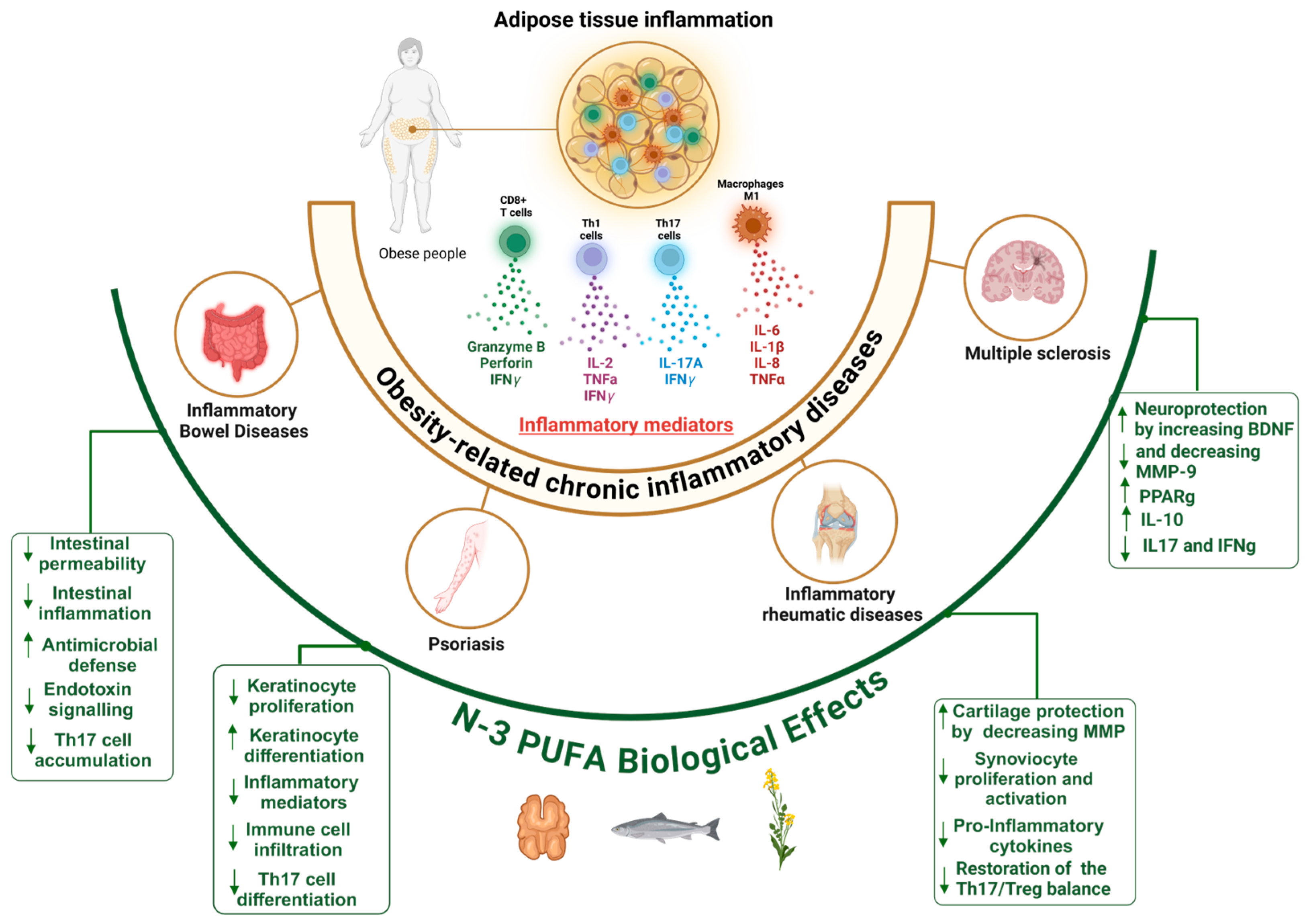
2.3. n-3 PUFAs in Obesity Related-Chronic Inflammatory Immune Diseases

2.3.1. Mechanisms of AT Low-Grade Inflammation in Obesity

Under physiological conditions, AT is infiltrated by anti-inflammatory immune cells, including M2 macrophages, regulatory T cells that secrete IL-10, and type 2 innate lymphoid cells. However, during obesity development, adipocyte hypertrophy limits oxygen supply to adipose tissue, inducing hypoxia [103]. This hypoxic state leads to the overexpression of hypoxia-inducible factor 1-alpha (HIF1α) by mature adipocytes and adipocyte precursors, promoting neovascularization [104]. Additionally, hypoxia induced secretion of monocyte chemoattractant protein-1 (MCP-1) and CCL5 by adipose tissue-derived stem cells (ASCs), contributing to the recruitment of immune cells, including circulating lymphocytes and monocytes [105,106,107]. AT is then infiltrated by pro-inflammatory immune cells, such as CD4+ (Th1) T cells, CD8+ T cells, and M1 macrophages. CD8+ T cells, which are the first cells to infiltrate AT, play a role in the recruitment and activation of type 1 macrophages. Indeed, gene depletion of CD8+ T cells has been shown to reduce macrophage recruitment and AT inflammation [108]. These processes lead to the development of a pro-inflammatory environment within obese AT, characterized by the presence of pro-inflammatory cytokines such as TNFα [109,110], IL1β, IL8, and IL-6, increased levels of leptin, and decreased levels of adiponectin [111], which are implicated in the disruption of insulin signaling. In addition, we have demonstrated the leading role of ASCs in promoting Th17 cell polarization and reducing insulin sensitivity in adipocytes [112,113]. Accordingly, obesity has been shown to promote expansion of Th17 cells in AT or periphery in a diet-induced obesity (DIO) murine model [114] as well as in humans. Indeed, infiltrating IL-17+-secreting cells have been found to be more abundant in obese AT than lean AT, and in visceral rather than subcutaneous AT [115]. Moreover, significant increases in circulating IL-17 and IL-23 cytokines were observed in obese individuals as compared to lean individuals [116]. Finally, Dalmas et al. have shown that several sub-populations of CD4+ cells secreting IL17, together or not with IL-22, were highly induced in the AT of obese subjects as compared to non-obese subjects [116]. Through binding to its ubiquitous receptors, IL-17 plays a crucial role in the propagation of inflammation, leading to the development of a low-grade chronic inflammation within AT and contributing to chronic inflammatory diseases associated with obesity.

2.3.2. Shared Mechanisms of Inflammation in Obesity and Related Chronic Inflammatory Diseases

Chronic inflammation within AT promotes an inflammatory microenvironment with increased expression of soluble factors such as adipokines, cytokines, metalloproteinases, pro-angiogenic factors, and ROS [117]. Through propagation of sustained inflammation, obesity thus exacerbates several chronic inflammatory diseases [118]. Indeed, increased adiposity is associated with a worse outcome in terms of autoimmune diseases such as psoriasis, inflammatory bowel disease (IBD), and multiple sclerosis (MS) [119]. Obesity-induced inflammation also aggravates rheumatic diseases through enhanced production of pro-inflammatory mediators and altered immune responses [120,121,122]. Additionally, recent evidence suggests that obesity-related inflammation may accelerate the progression of neurodegenerative diseases, including Alzheimer and Parkinson’s diseases [116]. Furthermore, obesity has been associated with increased cancer risk and poorer prognosis, in part due to the pro-inflammatory environment that promotes tumor growth and progression, and to the contribution of ASCs in upregulating immune check point expression in adipocytes and neighboring cancer cells [4,123,124].
In the past, a subset of Th17 lymphocytes, the Th17.1 lymphocytes, has been identified in several inflammatory and auto-immune diseases such as inflammatory bowel disease (IBD), multiple sclerosis (MS), psoriasis, and rheumatoid arthritis (RA), which are all aggravated by obesity. The Th17.1 lymphocyte subset presents the particularity of secreting both IL-17A and IFN-γ (a signature cytokine for Th1 lymphocytes) [125,126]. One of the key drivers leading to Th17.1 generation is the cytokine IL-23, as IL-6 + TGFβ without IL-23 leads to the generation of physiological Th17 cells, whereas the addition of IL-23 leads to Th17.1 cell generation [127,128]. In addition to pathogenic Th17.1 cell driving, IL-23 restrains the activity of regulatory T cells [129]. Interestingly, a dysregulation of the Th17/Treg balance has been observed in obesity [130]. Furthermore, we have shown that Th17.1 cells are promoted by ASCs in AT from obese, but not lean, individuals [112]. Moreover, we have also reported the ability of n-3 PUFAs to decrease ASC-mediated Th17-cell activation by reducing ICAM-1 expression [131]. In agreement with this, n-3 PUFAs have been shown to reduce membrane anchoring of IL-6 receptors on naïve lymphocytes, decreasing their polarization into Th17 lymphocytes [132]. Taken together, these studies suggest that n-3 PUFAs may alleviate obesity-related chronic inflammatory diseases through multiple mechanisms, including a reduction in IL-17 A secretion and subsequent propagation of inflammation, as shown in Figure 2.
These mechanisms highlight the broad impact of obesity-mediated AT inflammation on related inflammatory diseases. Their effects will be discussed herein, with the exception of cancer.

2.3.3. Inflammatory Bowel Diseases (IBDs)

Inflammatory Bowel Diseases (IBDs), including Crohn’s disease and ulcerative colitis, are chronic inflammatory diseases closely linked to obesity-mediated inflammation. The chronic low-grade inflammation associated with obesity creates a fertile ground for intestinal inflammatory processes, where immune dysregulation and metabolic disruptions converge [133]. Epidemiological links have been reported between obesity and IBD [134]. Excess AT generates pro-inflammatory cytokines that can exacerbate intestinal inflammation, while simultaneously compromising the gut microbiome’s delicate balance [135]. Therapeutic approaches targeting inflammatory pathways have gained increasing attention. Among these, n-3 PUFAs have emerged as promising agents, offering multiple mechanisms to address both metabolic and inflammatory aspects of IBD [136]. Using a mouse model of HFD + trinitrobenzene sulfonic acid (TNBS)-induced colitis, dietary n-3 PUFAs have been shown to reduce colitis-associated disease severity and colonic mRNA expression of cytokines (IL-6, IL-17A, IL-17F, IL-21, IL-23, and IFNγ) versus corn oil in control HFD mice. Concomitantly, adipose tissue mRNA expression of inflammatory cytokines (MCP-1, IFNγ, IL-6, IL17F, and IL-21) and macrophage infiltration were reduced [137]. This was supported using the Fat-1 mouse model, which constitutively produces n-3 PUFAs and in which chronic colitis was induced by dextran–sodium–sulfate (DSS) during 3 cycles of 5 days. Indeed, colon histological scores were improved, together with reduced spleen and colonic mucosal IL-17 and CCR6 mRNA levels, compared to WT mice [138]. In addition to the ability of n-3 PUFAs to modulate inflammatory pathways, Willemsen et al., who investigated the effects of PUFAs on epithelial barrier integrity, reported that DHA, AA, dihomo-gamma-linolenic acid (DGLA), and EPA increase basal transepithelial resistance and significantly reduce IL-4-mediated permeability in human intestinal epithelial cells [139]. In support to this study, DHA limited the effect of inflammatory stimulus on occludin, zona occludins-1, and barrier function when Caco-2 cells were exposed for 24 h to a cocktail of pro-inflammatory cytokines (IL-1β/TNF-α/IFNγ) at their basolateral side and to LPS at both sides [140]. Interestingly, even without cell injury, the addition of EPA enhanced permeability and transepithelial resistance in Caco-2 confluent cells, as compared to the addition of AA or LA, or even oleic acid (OA) [141]. These observations suggest that n-3 PUFAs may maintain the intestinal barrier function and combat inflammation-mediated permeability. Beyond these direct effects on the intestinal barrier function, SPMs have been reported to be a promising innovative approach to the treatment of IBD, as they contribute to the resolution of inflammation in IBD through binding to G protein coupled receptors [142]. They have also been shown to reduce neutrophil infiltration, stimulate phagocytosis and efferocytosis, enhance antimicrobial defense, attenuate endotoxin signaling, reduce mucosal pro-inflammatory cytokine production, and improve wound repair [142,143].
However, the clinical evidence for the use of n-3 PUFAs in the treatment of IBD is mixed. In ulcerative colitis disease (UC), a meta-analysis was inconclusive on the benefits of n-3 PUFA supplementation with respect to increasing disease remission [144], which was confirmed in a more recent review [145]. Nevertheless, a modest reduction in the need for corticosteroids in patients with active disease and a delay of early relapse via n-3 fatty acids have been reported in UC [146,147]. However, gastrointestinal side effects such as diarrhea and upper digestive symptoms have been reported in patients receiving n-3 PUFA supplementation [144]. In Crohn’s disease, statistically significant benefits have been reported, especially in children where a randomized controlled trial showed significantly lower relapse rates at one year when receiving enteric-coated n-3 PUFA supplementation compared to a placebo (61% vs. 95%, p = 0.0016) [148]. Finally, a recent study reported that a Mediterranean diet may reduce active disease and inflammatory markers in both UC and Crohn’s disease due to its high content of biologically active foods, including nuts and fish, which are rich in n-3 PUFAs [149].
While adult studies have shown limited efficacy and side effects, pediatric data with enteric-coated formulations have demonstrated more encouraging results. This divergence suggests that factors such as age or formulation may be important determinants of success. However, until further clinical trials are concluded, a Mediterranean diet should be proposed as an additive strategy in the treatment of IBD.

2.3.4. Psoriasis

Psoriasis represents a chronic inflammatory skin disease which is impacted by obesity-mediated inflammation. Like other obesity-related disorders, psoriasis demonstrates complex interactions between metabolic dysfunction and immune dysregulation. Recent epidemiological studies have established strong associations between obesity and psoriasis onset or severity, as elevated BMI is positively correlated with increased disease burden [150]. Thus, obesity acts as an independent risk factor for the development and worsening of psoriasis [151]. Obesity and psoriasis exhibit similar systemic inflammatory profiles, with elevated serum levels of cytokines such as TNF-α, IL-6, and IL-12 [152]. Interestingly, weight loss has been shown to have a beneficial effect on psoriasis severity and treatment response [153]. Tissue-engineered skin models used to investigate the impact of n-3 PUFAs on psoriasis demonstrated that supplementation in a culture medium with 10 μM ALA results in the incorporation of its EPA metabolite into the epidermal phospholipid fraction, inhibition of keratinocyte proliferation, and restoration of keratinocyte differentiation capacity, and thus a decrease in the pathological phenotype of psoriatic skin substitutes [154]. Likewise, DHA supplementation mitigates psoriatic characteristics by normalizing epidermal cell differentiation and proliferation and by decreasing the levels of lipid mediators derived from n-6 PUFAs, mainly prostaglandin E2 (PGE2) and 12-hydroxyeicosatetraenoic acid (12-HETE), in a 3D tissue-engineered skin model [155].
As mentioned above, psoriasis is a chronic inflammatory skin disease induced via the IL-23/IL-17 axis. While SFAs can exacerbate psoriatic dermatitis through activation of the inflammasome in keratinocytes and induction of IL-17-producing cells such as Th17 and IL-17-producing γδ T cells in the skin, n-3 PUFAs may, on the contrary, suppress Th17 differentiation [156]. Accordingly, in a 3D psoriatic skin model, EPA normalized the proliferation of psoriatic keratinocytes and diminished the levels of IL-17 [157]. Supporting these results, activation of the GPR120/FFAR4 receptor with agonists in a psoriasis model resulted in the attenuation of skin lesions, a decrease in inflammatory cytokine levels, and inhibition of Th17 cell differentiation [158]. Accordingly, topical treatment with 30 μM DHA in rats was shown to accelerate wound healing and reduce inflammatory mediator production through GPR120 activation in skin [159].
Significant progress has also been made on the identification of SPMs derived from n-3 PUFAs in psoriatic skin. Indeed, resolvin E1 (RvE1) has been demonstrated to reduce inflammatory cell infiltration and epidermal hyperplasia in a murine model of psoriasis and to decrease IL-23 expression and production [160]. Similarly, resolvin D1 (RvD1) and resolvin D5 (RvD5) have been shown to exert anti-inflammatory effects by reducing the expression of interleukin-24 and S100A12 in human keratinocytes [161]. In dietary interventions using a mouse model of psoriasis, DHA showed more pronounced effects than EPA in reducing inflammation by increasing skin RvD5, protectin D, and maresin 2 levels [162].
The clinical validation of n-3 PUFAs in the treatment of psoriasis is provided by the analysis of multiple interventional studies. In a systematic review of 18 randomized controlled trials, it was concluded that supplementation with n-3 PUFAs, but only when they were combined with conventional treatments, decreases several inflammatory mediators and reduces psoriasis severity, as quantified by the Psoriasis Area Severity Index (PASI) score and lesion area. In addition, n-3 PUFAs were also found to reduce the risks of obesity and cardiovascular and metabolic diseases [163]. Another meta-analysis provided robust evidence supporting the efficacy of n-3 PUFA supplementation in the amelioration of several psoriasis parameters. Specifically, a dose-dependent improvement in clinical manifestations, including erythema, scaling, and pruritus, was seen with n-3 PUFA supplementation [164]. Thus, n-3 PUFAs and their derivatives are likely to become a promising adjunct approach for the treatment of psoriasis.

2.3.5. Inflammatory Rheumatic Diseases

Osteoarthritis

Rheumatic diseases, particularly osteoarthritis (OA) and, to a lesser extent, rheumatoid arthritis (RA), are associated with obesity. The relationship between obesity and OA extends beyond mechanical stress, with obesity being identified as one of the main risk factors contributing to both incidence and progression [165,166], with a critical role in terms of systemic and chronic low-grade inflammation [167]. AT acts as an endocrine organ through the release of soluble mediators, including cytokines, adipokines, and ROS, which induce detrimental effects on joint tissues [168]. Most adipokines (leptin, visfatin, and resistin) promote the activation of an inflammatory circuit, known as meta-inflammation, leading to the synthesis of degradative enzymes, ROS, and prostaglandins [117,168]. But IL-1β is one of the most active cytokines that leads to cartilage destruction, due to its ability to activate MMP 1 and MMP 13, which results in the loss of sulphated glycosaminoglycans (sGAG) and type II collagen [169]. Using an in vitro model of equine IL-1β-synoviocyte culture, Caron et al. demonstrated that the addition of DHA to cultures resulted in the incorporation of DHA in synoviocytes, which was associated with a reduction in MMPs, IL1β, IL-6, and Cox-2 expression. Moreover, DHA-derived SPMs, RvD1, RvD2, Maresin1, and Protectin DX were also able to abrogate MMP-1, MMP-13, and IL-6 gene expression in IL-1β-stimulated synoviocytes [170]. Supporting the beneficial effects of n-3 PUFAs on IL-1β-mediated cartilage degradation, Wann et al. demonstrated that the addition of EPA or DHA at a 0.1 or 1 μM concentration resulted in less IL-1β-mediated sGAG loss [169]. Moreover, in a high-fat-diet mouse model where cartilage degradation was surgically induced, n-3 PUFA, but not n6 PUFA or SFA, supplementation resulted in the inhibition of TLR4/NF-κB and NLRP3/caspase-1 expression and signaling, which led to a reduction in inflammatory response and IL-1β-mediated pyroptosis in chondrocytes [171]. Accordingly, another study, also using a high-fat-diet mouse model, demonstrated that supplementation with n-3 PUFAs, but not with n-6 PUFAs, attenuated injury-induced OA, decreased leptin and resistin levels, and significantly enhanced wound repair [172].
In humans, several clinical trials have demonstrated the positive effects of n-3 PUFAs in terms of managing OA. A comprehensive meta-analysis of nine randomized controlled trials involving 2070 patients with OA concluded that n-3 PUFA supplementation significantly alleviates arthritis pain and improves joint function compared to a placebo [173]. However, another study, using genome-wide association study (GWAS) and Mendelian randomization (MR) methods, concluded that PUFA may not be helpful in OA [174]. But, n-3 PUFAs, coupled with emerging optimized delivery methods, particularly through liposomal formulations, should offer new perspectives for improving their therapeutic efficacy, as demonstrated by the success of liposome-encapsulated RvD1 delivery in the joints of obese mice with OA [175].

Rheumatoid Arthritis

In rheumatoid arthritis (RA), the link with obesity is not as high as in OA. Indeed, the odds ratio of RA risk is 1.32 in patients with obesity [176], while it reaches 4.55 for knee OA [177], as calculated in a meta-analysis, and approximately 2 for hip OA [178]. However, obesity appears to be a clear risk factor for a poor response to treatment, regardless of whether a biologic or non-biologic therapy is being used [120,179,180].
Th17 cells play a central role in RA by inducing synoviocyte inflammation and neutrophil and macrophage infiltration [181]. In this context, we have previously shown the preponderant role of mesenchymal stem cells and synoviocytes in promoting Th17 cell polarization [182], a mechanism that is closely similar to the one we have demonstrated in obesity with ASCs and adipocytes [112,113]. Using the Fat-1 mouse model, which endogenously produces n-3 PUFAs, increased n-3 PUFA levels have been shown to attenuate inflammatory arthritis. This effect was associated with reduced IL-17 production and increased FoxP3 expression, therefore promoting Treg differentiation [183]. A similar finding was reported in a model of collagen-induced arthritis, where activation of the GPR120/FFAR resulted in restoring the Th1/Th17 and Treg balance [184]. n-3 PUFAs have also been reported to decrease antigen presentation via MHC II, inflammatory cytokine production by monocyte/macrophages, and ROS production by various leukocytes in RA [185].
In clinical intervention studies, a systematic review has found an inverse association between RA risk and oily fish consumption, even though this association was not linear [186]. Supporting these results, combining n-3 PUFA supplementation with an anti-inflammatory dietary intervention that provides an arachidonic acid intake of less than 90 mg/day has demonstrated significant reductions in CRP, TNF-α, number of tender joints, LTB4 levels, and the dose of corticosteroids used [187]. In a meta-analysis study, consumption of n-3 PUFAs has also been found to significantly improve disease-activity-related markers and reduce blood leukotriene B4 and triacylglycerol levels [188]. Finally, the benefits of n-3 PUFAs in terms of reducing (i) duration of morning stiffness, (ii) number of tender or swollen joints, (iii) time to fatigue, (iv) pain or disease activity, and (v) use of NSAIDs and increasing grip strength have been reported in RA [185].
In conclusion, these findings collectively support the therapeutic potential of n-3 PUFAs for RA, while highlighting the importance of limiting saturated and n-6 PUFA intake.

2.3.6. Multiple Sclerosis

Multiple sclerosis (MS) is an autoimmune disease characterized by inflammation and damage to axons leading to demyelination and formation of plaques in the central nervous system (CNS) [183]. Obesity is known to influence the progression and severity of this disease [189].
The effects of n-3 PUFAs on MS have been linked to their ability to regulate inflammation in the periphery and the central nervous system as well. DHA and EPA have indeed been found to cross the blood–brain barrier and directly impair neuroinflammatory processes [190]. Brain phospholipids enrichment in DHA more than EPA affects cell signaling by modifying the composition of lipid rafts [191]. n-3 PUFAs may also decrease the activity and production of matrix metalloproteinase-9, which is implicated in blood–brain barrier breakdown in MS, thereby preserving blood–brain barrier integrity [192]. In addition, n-3 PUFAs, especially DHA, demonstrate neuroprotective properties by promoting neuronal survival and neurite outgrowth [191]. Thus, n-3 PUFAs, such as DHA and EPA, have been shown to upregulate the expression of brain-derived neurotrophic factor (BDNF), which plays a role in promoting neuronal survival and synaptic plasticity [193,194]. In addition, SPMs derived from DHA, more particularly RvD1 and neuroprotectin D1, have also demonstrated neuroprotective properties [195].
Th17 cells, especially pathogenic Th17.1 cells, have been involved in MS physiopathology using the Experimental Autoimmune Encephalomyelitis model [196,197]. Of interest, n-3 PUFAs have demonstrated an ability to inhibit both IL-17 and IFNγ production in MS pathology, together with activation of PPARγ, but whether this results in inhibiting Th17.1 cell activation has not been demonstrated [198].
Clinical evidence on the efficacy of n-3 PUFA supplementation in the treatment of MS is mixed. Indeed, some studies have shown promising results, with a significant reduction in the mean annual exacerbation rate and the mean Expanded Disability Status Scale (EDSS) in newly diagnosed MS patients supplemented with fish oil, vitamins, and dietary advice [199], or a reduction in relapse rates and inflammatory markers and an improved quality of life in MS patients [200]. But this was not supported by other studies, although in one study n-3 PUFA supplementation (4 g/d, 12 months) resulted in a reduction in serum pro-inflammatory cytokines such as TNF-α, IL-1β, and IL-6 while increasing anti-inflammatory IL-10 levels in treated MS patients [201]. In another study, supplementation involved capsules with 1350 mg of EPA and 850 mg of DHA daily for 24 months, but no significant effect from n-3 PUFAs on disease activity or relapse rates was observed [202]. However, to reconcile these studies, Farinotti et al. reported that although polyunsaturated n-3 PUFAs do not appear to have a major effect on the main clinical outcome in MS, namely disease progression, they may tend to reduce the frequency of relapses over two years [203].
Thus, in light of these conflicting findings, it seems that more research is needed to fully understand the potential benefits of n-3 PUFA supplementation in the management of MS. Clinical trials combining neuroimaging biomarkers with cerebrospinal fluid analysis could help determine whether n-3 PUFAs exert direct neuroprotective effects beyond their anti-inflammatory action, potentially establishing them as adjunctive therapy for MS patients.

3. Conclusions

Our comprehensive review highlights the potential of n-3 PUFAs to contribute to the resolution of inflammation in obesity and related metabolic and chronic inflammatory diseases. These beneficial effects occur through several interrelated pathways, including activation of the GPR120/FFAR4 and PPARγ receptors and generation of anti-inflammatory eicosanoids and SPMs. In addition, n-3 PUFAs also demonstrate significant effects on T cell activation, specifically inhibition of Th1 and Th17 cell differentiation or activation, but promotion of regulatory T cell responses [132,183,184,204]. This immunomodulatory capacity extends to macrophages, as they can switch their balance from M1 to M2 macrophages and modulate their antigen-presenting cell function [205,206].
However, clinical evidence shows variable efficacy in the diseases that we have mentioned herein.

3.1. Limitations of Current Evidence

A comprehensive examination of n-3 PUFA research across obesity-related disorders highlights several significant limitations. Firstly, there exists an imbalance between preclinical and clinical evidence, with excessive reliance on murine models and insufficient translation to human applications. While mouse studies demonstrate robust effects of n-3 PUFA supplementation on fat mass prevention in high-fat-diet conditions, human trials show more modest outcomes, typically requiring a combination with caloric restriction to achieve significant weight loss.
In CVD research, inconsistent findings from major clinical trials raise methodological concerns. The REDUCE-IT study demonstrated cardiovascular benefits with high-dose EPA, whereas the STRENGTH trial using combined EPA + DHA showed no significant improvements. Such discrepancies may stem from differences in n-3 PUFA formulations, dosages, or control substances used for comparison.
Regarding T2D, despite promising mechanistic findings on insulin sensitivity and beta-cell function, clinical studies reveal variable glycemic improvements that do not consistently translate to meaningful HbA1c reductions across different patient populations. Similarly, in MASLD investigations, the WELCOME trial and subsequent studies demonstrate improvements in hepatic steatosis but provide limited data on histological outcomes or disease progression markers.
For inflammatory conditions, notable inconsistencies emerge across disease states. In inflammatory bowel disease, enteric-coated n-3 PUFA formulations show promising results in pediatric populations but limited efficacy in adults. Psoriasis studies similarly demonstrate considerable variability in clinical response, potentially attributable to differences in disease severity or concurrent conventional therapies.
Furthermore, methodological heterogeneity across studies complicates interpretation, including variations in n-3 PUFA sources, dosages (ranging from <1 g/day to >4 g/day), treatment duration, and administration routes. Moreover, the relationship between n-3 PUFAs and gender has rarely been investigated, despite their potential relevance, particularly in autoimmune conditions with known gender disparities.
Bioavailability issues represent another limitation, as conventional n-3 PUFA formulations may yield suboptimal tissue incorporation compared to emerging delivery systems such as liposomal preparations, which have shown superior results in preclinical models of rheumatic diseases. Additionally, while growing evidence suggests that gut microbiota mediates some n-3 PUFA effects, few studies adequately assess baseline microbiome composition or monitor changes throughout the intervention period.
These limitations underscore the need for well-designed clinical trials with appropriate patient stratification, extended follow-up periods, standardized dosing protocols, and clinically relevant endpoints to better define optimal therapeutic strategies for n-3 PUFA interventions in obesity-related inflammatory conditions.

3.2. Perspectives

However, there is evidence to suggest that earlier intervention may offer greater benefit in terms of preventing rather than treating the disease. SPMs derived from n-3 PUFAs represent a particularly promising area of investigation, especially when encapsulated into liposomes, which may offer a new therapeutic approach, notably in the field of obesity and its inflammatory comorbidities. The identification of new agonists, mimetics, and improved SPM formulations represents a promising approach to specifically target deficient resolution processes [207]. Moreover, the ability of SPMs to modulate adaptive immune responses, particularly on B and T lymphocyte populations, underscores their potential in treating autoimmune and chronic inflammatory diseases associated with obesity [208]. These therapeutic innovations, combined with a better understanding of the specific mechanisms of action of different SPMs, could enable more targeted and personalized approaches for patients suffering from obesity and its inflammatory complications.

Author Contributions

D.J. supervised the project, wrote the initial draft of multiple sections, including omega-3 metabolism and its effects on inflammatory pathways, conducted a comprehensive literature search on all pathologies, designed and conceptualized the figures, and contributed substantially to the final revision; A.E. supervised the project, contributed substantially to the sections on autoimmune diseases (psoriasis, multiple sclerosis) and inflammatory bowel diseases, revised and edited the entire manuscript, and coordinated the collaborative work; F.B. created the graphical figures, conducted an extensive literature research, wrote the sections on omega-3 fatty acids and obesity mechanisms, and contributed significantly to multiple sections; P.C.T.d.C. and J.L.d.B.A. contributed to the cardiovascular disease section and performed critical analysis of clinical trials related to cardiovascular outcomes; R.K. wrote and reviewed the sections on hepatic diseases and MASLD; H.J. contributed to the sections on type 2 diabetes, metabolic syndrome, and insulin resistance; and B.M. wrote the sections on omega-3 mechanisms of action, renal diseases, and rheumatic diseases (rheumatoid arthritis and osteoarthritis). All authors have read and agreed to the published version of the manuscript.

Funding

We would like to thank the Groupe Lipides et Nutrition (GLN) for the prize they have awarded us for our work on omega-3 fatty acids and the EDISS doctoral school for the funding of F. Blangero’s fellowship. And we would thank the MobiDoc 941 managed by the ANPR under the EU SWAFY project for D. Jerab’s fellowship.

Acknowledgments

Figures were created in https://BioRender.com.

Conflicts of Interest

The authors declare no conflicts of interest.

Abbreviations

The following abbreviations are used in this manuscript:
AAArachidonic acidMSMultiple Sclerosis
ALAAlpha-linolenic acidMUFAMonounsaturated Fatty Acid
ALTAlanine amino-transferaseMyD88Myeloid Differentiation primary response 88
AMPKAMP-activated protein kinasen-3Omega-3
AP-1Activator Protein 1n-6Omega-6
ASCAdipose-derived stem cellsNF-κBNuclear factor kappa B
ASTAspartate amino-transferaseNLRP3NOD-like receptor family pyrin domain containing 3
ATAdipose tissueNrf2Nuclear factor erythroid 2-related factor 2
BDNFBrain-derived neurotrophic factorOAOsteoarthritis
BMIBody mass indexObOAObesity-associated osteoarthritis
CKDChronic kidney diseasePASIPsoriasis Area Severity Index
COXCyclooxygenasePD-L1Programmed Death-Ligand 1
CRPC-reactive proteinPDXProtectin DX
CVDCardiovascular diseasePGProstaglandin
CYPCytochrome P450PLA2Phospholipase A2
DHADocosahexaenoic acidPPARPeroxisome proliferator-activated receptor
DGLADihomo-gamma-linolenic acidPPREPeroxisome proliferator-activated receptor response element
EDSSExpanded Disability Status ScalePUFAPolyunsaturated fatty acid
EPAEicosapentaenoic acidRARheumatoid arthritis
ERKExtracellular signal-regulated kinaseRAGEReceptor for advanced glycation end-products
ESRDEnd-ctage renal diseaseRBCRed blood cell
FAFatty acidRORαRAR-related Orphan Receptor alpha
FFAR4Free Fatty Acid Receptor 4ROSReactive Oxygen Species
GLP-1Glucagon-like peptide-1RvResolvin
GLUT-4Glucose Transporter Type 4RXRRetinoid X receptor
GM-CSFGranulocyte-Macrophage Colony-Stimulating FactorSFASaturated fatty acid
GPR120G protein-coupled receptor 120SIRT1Sirtuin 1
GSDMDGasdermin DSPMSpecialized pro-resolving mediator
HbA1cHemoglobin A1cSPMsSpecialized pro-resolving mediators
HDLHigh-density lipoproteinSREBP-1Sterol Regulatory Element-Binding Protein-1
HIF1αHypoxia-inducible factor 1-alphaT2DType 2 diabetes
HMGB1High Mobility Group Box 1TAB1TAK1-binding protein 1
IBDInflammatory bowel diseaseTAK1Transforming growth factor beta-activated kinase 1
ICAM-1Intercellular Adhesion Molecule 1TGFβTransforming growth factor beta
IFNInterferonTh1T helper 1
IKK-βInhibitor of Nuclear Factor Kappa-B Th17T helper 17
IL-Interleukin-TLRToll-like receptor
JNKc-Jun N-terminal kinaseTregRegulatory T cell
LALinoleic acidTXAThromboxane A
LDLLow-density lipoproteinUCUlcerative colitis
LOXLipoxygenaseVEGFVascular Endothelial Growth Factor
LPSLipopolysaccharideVLDLVery low-density lipoproteins
LTB4Leukotriene B4
LTB5Leukotriene B5
MASLDMetabolic dysfunction-associated steatotic liver disease
MCP-1Monocyte chemoattractant protein-1
MMPMatrix metalloproteinase
MRIMagnetic resonance imaging

References

  1. Deng, T.; Lyon, C.J.; Bergin, S.; Caligiuri, M.A.; Hsueh, W.A. Obesity, Inflammation, and Cancer. Annu. Rev. Pathol. 2016, 11, 421–449. [Google Scholar] [CrossRef] [PubMed]
  2. NCD Risk Factor Collaboration (NCD-RisC). Worldwide trends in underweight and obesity from 1990 to 2022: A pooled analysis of 3663 population-representative studies with 222 million children, adolescents, and adults. Lancet 2024, 403, 1027–1050. [Google Scholar]
  3. Sakers, A.; De Siqueira, M.K.; Seale, P.; Villanueva, C.J. Adipose-tissue plasticity in health and disease. Cell 2022, 185, 419–446. [Google Scholar] [PubMed]
  4. Pestel, J.; Blangero, F.; Eljaafari, A. Pathogenic Role of Adipose Tissue-Derived Mesenchymal Stem Cells in Obesity and Obesity-Related Inflammatory Diseases. Cells 2023, 12, 348. [Google Scholar] [CrossRef] [PubMed]
  5. Chehimi, M.; Vidal, H.; Eljaafari, A. Pathogenic Role of IL-17-Producing Immune Cells in Obesity, and Related Inflammatory Diseases. J. Clin. Med. 2017, 6, 68. [Google Scholar] [CrossRef] [PubMed]
  6. Chakhtoura, M.; Haber, R.; Ghezzawi, M.; Rhayem, C.; Tcheroyan, R.; Mantzoros, C.S. Pharmacotherapy of obesity: An update on the available medications and drugs under investigation. eClinicalMedicine 2023, 58, 101882. [Google Scholar] [CrossRef]
  7. Cummings, D.E.; Cohen, R.V. Bariatric/Metabolic Surgery to Treat Type 2 Diabetes in Patients with a BMI < 35 kg/m2. Diabetes Care 2016, 39, 924–933. [Google Scholar]
  8. Negi, A.; Asokkumar, R.; Ravi, R.; Lopez-Nava, G.; Bautista-Castaño, I. Nutritional Management and Role of Multidisciplinary Follow-Up after Endoscopic Bariatric Treatment for Obesity. Nutrients 2022, 14, 3450. [Google Scholar] [CrossRef]
  9. Singh, M.; Thrimawithana, T.; Shukla, R.; Adhikari, B. Managing obesity through natural polyphenols: A review. Future Foods 2020, 1–2, 100002. [Google Scholar]
  10. Dong, L.; Qin, C.; Li, Y.; Wu, Z.; Liu, L. Oat phenolic compounds regulate metabolic syndrome in high fat diet-fed mice via gut microbiota. Food Biosci. 2022, 50, 101946. [Google Scholar]
  11. Li, Y.; Qin, C.; Dong, L.; Zhang, X.; Wu, Z.; Liu, L.; Yang, J.; Liu, L. Whole grain benefit: Synergistic effect of oat phenolic compounds and β-glucan on hyperlipidemia via gut microbiota in high-fat-diet mice. Food Funct. 2022, 13, 12686–12696. [Google Scholar] [CrossRef] [PubMed]
  12. Araujo, P.; Belghit, I.; Aarsæther, N.; Espe, M.; Lucena, E.; Holen, E. The Effect of Omega-3 and Omega-6 Polyunsaturated Fatty Acids on the Production of Cyclooxygenase and Lipoxygenase Metabolites by Human Umbilical Vein Endothelial Cells. Nutrients 2019, 11, 966. [Google Scholar] [CrossRef] [PubMed]
  13. Liddle, D.M.; Hutchinson, A.L.; Wellings, H.R.; Power, K.A.; Robinson, L.E.; Monk, J.M. Integrated Immunomodulatory Mechanisms through which Long-Chain n-3 Polyunsaturated Fatty Acids Attenuate Obese Adipose Tissue Dysfunction. Nutrients 2017, 9, 1289. [Google Scholar] [CrossRef] [PubMed]
  14. Calder, P.C. n-3 PUFA and inflammation: From membrane to nucleus and from bench to bedside. Proc. Nutr. Soc. 2020, 79, 404–416. [Google Scholar] [CrossRef] [PubMed]
  15. Norris, P.C.; Gosselin, D.; Reichart, D.; Glass, C.K.; Dennis, E.A. Phospholipase A2 regulates eicosanoid class switching during inflammasome activation. Proc. Natl. Acad. Sci. USA 2014, 111, 12746–12751. [Google Scholar] [CrossRef] [PubMed]
  16. Spite, M.; Clària, J.; Serhan, C.N. Resolvins, specialized proresolving lipid mediators, and their potential roles in metabolic diseases. Cell Metab. 2014, 19, 21–36. [Google Scholar] [CrossRef] [PubMed]
  17. Serhan, C.N.; Yang, R.; Martinod, K.; Kasuga, K.; Pillai, P.S.; Porter, T.F.; Oh, S.F.; Spite, M. Maresins: Novel macrophage mediators with potent antiinflammatory and proresolving actions. J. Exp. Med. 2009, 206, 15–23. [Google Scholar] [CrossRef]
  18. Abedi, E.; Sahari, M.A. Long-chain polyunsaturated fatty acid sources and evaluation of their nutritional and functional properties. Food Sci. Nutr. 2014, 2, 443–463. [Google Scholar] [CrossRef] [PubMed]
  19. Baker, E.J.; Miles, E.A.; Burdge, G.C.; Yaqoob, P.; Calder, P.C. Metabolism and functional effects of plant-derived omega-3 fatty acids in humans. Prog. Lipid Res. 2016, 64, 30–56. [Google Scholar] [CrossRef]
  20. Simopoulos, A.P. The omega-6/omega-3 fatty acid ratio: Health implications. Oléagineux Corps Gras Lipides 2010, 17, 267–275. [Google Scholar] [CrossRef]
  21. Simopoulos, A.P. An Increase in the Omega-6/Omega-3 Fatty Acid Ratio Increases the Risk for Obesity. Nutrients 2016, 8, 128. [Google Scholar] [CrossRef] [PubMed]
  22. Ben Othman, R.; Berriche, O.; Gamoudi, A.; Mizouri, R.; Jerab, D.; Ben Amor, N.; Mahjoub, F.; Jamoussi, H. Cross sectional study about nutritional risk factors of metabolically unhealthy obesity. Rom. J. Intern. Med. 2023, 61, 53–62. [Google Scholar] [CrossRef] [PubMed]
  23. Jamoussi, H.; Chaabouni, S.; Gammoudi, A.; Mahjoub, F.; Ounaissa, K.; Berriche, O.; Amrouche, C.; Blouza, S. Apports spontanés en acides gras oméga 3 chez des diabétiques de type 2 tunisiens. Oléagineux Corps Gras Lipides 2014, 21, A501. [Google Scholar] [CrossRef]
  24. Snetselaar, L.G.; de Jesus, J.M.; DeSilva, D.M.; Stoody, E.E. Dietary Guidelines for Americans, 2020–2025: Understanding the Scientific Process, Guidelines, and Key Recommendations. Nutr. Today 2021, 56, 287–295. [Google Scholar] [CrossRef]
  25. Hara, T.; Kashihara, D.; Ichimura, A.; Kimura, I.; Tsujimoto, G.; Hirasawa, A. Role of free fatty acid receptors in the regulation of energy metabolism. Biochim. Biophys. Acta 2014, 1841, 1292–1300. [Google Scholar] [CrossRef] [PubMed]
  26. Han, L.; Song, S.; Niu, Y.; Meng, M.; Wang, C. Eicosapentaenoic Acid (EPA) Induced Macrophages Activation through GPR120-Mediated Raf-ERK1/2-IKKβ-NF-κB p65 Signaling Pathways. Nutrients 2017, 9, 937. [Google Scholar] [CrossRef] [PubMed]
  27. Oh, D.Y.; Talukdar, S.; Bae, E.J.; Imamura, T.; Morinaga, H.; Fan, W.; Li, P.; Lu, W.J.; Watkins, S.M.; Olefsky, J.M. GPR120 is an omega-3 fatty acid receptor mediating potent anti-inflammatory and insulin-sensitizing effects. Cell 2010, 142, 687–698. [Google Scholar] [CrossRef] [PubMed]
  28. Yan, Y.; Jiang, W.; Spinetti, T.; Tardivel, A.; Castillo, R.; Bourquin, C.; Guarda, G.; Tian, Z.; Tschopp, J.; Zhou, R. Omega-3 fatty acids prevent inflammation and metabolic disorder through inhibition of NLRP3 inflammasome activation. Immunity 2013, 38, 1154–1163. [Google Scholar] [CrossRef]
  29. Li, A.C.; Glass, C.K. PPAR- and LXR-dependent pathways controlling lipid metabolism and the development of atherosclerosis. J. Lipid Res. 2004, 45, 2161–2173. [Google Scholar] [CrossRef]
  30. Varga, T.; Czimmerer, Z.; Nagy, L. PPARs are a unique set of fatty acid regulated transcription factors controlling both lipid metabolism and inflammation. Biochim. Biophys. Acta (BBA)—Mol. Basis Dis. 2011, 1812, 1007–1022. [Google Scholar] [CrossRef]
  31. Li, H.; Ruan, X.Z.; Powis, S.H.; Fernando, R.; Mon, W.Y.; Wheeler, D.C.; Moorhead, J.F.; Varghese, Z. EPA and DHA reduce LPS-induced inflammation responses in HK-2 cells: Evidence for a PPAR-gamma-dependent mechanism. Kidney Int. 2005, 67, 867–874. [Google Scholar] [CrossRef] [PubMed]
  32. Zapata-Gonzalez, F.; Rueda, F.; Petriz, J.; Domingo, P.; Villarroya, F.; Diaz-Delfin, J.; de Madariaga, M.A.; Domingo, J.C. Human dendritic cell activities are modulated by the omega-3 fatty acid, docosahexaenoic acid, mainly through PPAR(gamma):RXR heterodimers: Comparison with other polyunsaturated fatty acids. J. Leukoc. Biol. 2008, 84, 1172–1182. [Google Scholar] [CrossRef] [PubMed]
  33. Martínez-Fernández, L.; Laiglesia, L.M.; Huerta, A.E.; Martínez, J.A.; Moreno-Aliaga, M.J. Omega-3 fatty acids and adipose tissue function in obesity and metabolic syndrome. Prostaglandins Other Lipid Mediat. 2015, 121, 24–41. [Google Scholar] [CrossRef] [PubMed]
  34. Hou, T.Y.; McMurray, D.N.; Chapkin, R.S. Omega-3 fatty acids, lipid rafts, and T cell signaling. Eur. J. Pharmacol. 2016, 785, 2–9. [Google Scholar] [CrossRef]
  35. Hwang, D.H.; Kim, J.-A.; Lee, J.Y. Mechanisms for the activation of Toll-like receptor 2/4 by saturated fatty acids and inhibition by docosahexaenoic acid. Eur. J. Pharmacol. 2016, 785, 24–35. [Google Scholar] [CrossRef]
  36. Cani, P.D.; Amar, J.; Iglesias, M.A.; Poggi, M.; Knauf, C.; Bastelica, D.; Neyrinck, A.M.; Fava, F.; Tuohy, K.M.; Chabo, C.; et al. Metabolic endotoxemia initiates obesity and insulin resistance. Diabetes 2007, 56, 1761–1772. [Google Scholar] [CrossRef] [PubMed]
  37. Bidu, C.; Escoula, Q.; Bellenger, S.; Spor, A.; Galan, M.; Geissler, A.; Bouchot, A.; Dardevet, D.; Morio, B.; Cani, P.D.; et al. The Transplantation of ω3 PUFA-Altered Gut Microbiota of fat-1 Mice to Wild-Type Littermates Prevents Obesity and Associated Metabolic Disorders. Diabetes 2018, 67, 1512–1523. [Google Scholar] [CrossRef] [PubMed]
  38. Flachs, P.; Mohamed-Ali, V.; Horakova, O.; Rossmeisl, M.; Hosseinzadeh-Attar, M.J.; Hensler, M.; Ruzickova, J.; Kopecky, J. Polyunsaturated fatty acids of marine origin induce adiponectin in mice fed a high-fat diet. Diabetologia 2006, 49, 394–397. [Google Scholar] [CrossRef] [PubMed]
  39. Itoh, M.; Suganami, T.; Satoh, N.; Tanimoto-Koyama, K.; Yuan, X.; Tanaka, M.; Kawano, H.; Yano, T.; Aoe, S.; Takeya, M.; et al. Increased adiponectin secretion by highly purified eicosapentaenoic acid in rodent models of obesity and human obese subjects. Arterioscler. Thromb. Vasc. Biol. 2007, 27, 1918–1925. [Google Scholar] [CrossRef] [PubMed]
  40. da Cunha de Sá, R.D.C.; Simão, J.d.J.; Silva, V.S.d.; Farias, T.M.d.; Cruz, M.M.; Antraco, V.J.; Armelin-Correa, L.; Alonso-Vale, M.I. Fish Oil Enriched in EPA, but Not in DHA, Reverses the Metabolic Syndrome and Adipocyte Dysfunction Induced by a High-Fat Diet. Nutrients 2021, 13, 754. [Google Scholar] [CrossRef] [PubMed]
  41. Figueras, M.; Olivan, M.; Busquets, S.; López-Soriano, F.J.; Argilés, J.M. Effects of eicosapentaenoic acid (EPA) treatment on insulin sensitivity in an animal model of diabetes: Improvement of the inflammatory status. Obesity 2011, 19, 362–369. [Google Scholar] [CrossRef] [PubMed]
  42. Fan, C.; Liu, X.; Shen, W.; Deckelbaum, R.J.; Qi, K. The Regulation of Leptin, Leptin Receptor and Pro-opiomelanocortin Expression by N-3 PUFAs in Diet-Induced Obese Mice Is Not Related to the Methylation of Their Promoters. Nutr. Metab. 2011, 8, 31. [Google Scholar] [CrossRef]
  43. Yang, X.; Li, X.; Hu, M.; Huang, J.; Yu, S.; Zeng, H.; Mao, L. EPA and DHA differentially improve insulin resistance by reducing adipose tissue inflammation-targeting GPR120/PPARγ pathway. J. Nutr. Biochem. 2024, 130, 109648. [Google Scholar] [CrossRef] [PubMed]
  44. Hasan, A.U.; Ohmori, K.; Konishi, K.; Igarashi, J.; Hashimoto, T.; Kamitori, K.; Yamaguchi, F.; Tsukamoto, I.; Uyama, T.; Ishihara, Y.; et al. Eicosapentaenoic acid upregulates VEGF-A through both GPR120 and PPARγ mediated pathways in 3T3-L1 adipocytes. Mol. Cell. Endocrinol. 2015, 406, 10–18. [Google Scholar] [CrossRef]
  45. Pinel, A.; Pitois, E.; Rigaudiere, J.-P.; Jouve, C.; De Saint-Vincent, S.; Laillet, B.; Montaurier, C.; Huertas, A.; Morio, B.; Capel, F. EPA prevents fat mass expansion and metabolic disturbances in mice fed with a Western diet. J. Lipid Res. 2016, 57, 1382–1397. [Google Scholar] [CrossRef] [PubMed]
  46. Ruzickova, J.; Rossmeisl, M.; Prazak, T.; Flachs, P.; Sponarova, J.; Veck, M.; Tvrzicka, E.; Bryhn, M.; Kopecky, J. Omega-3 PUFA of marine origin limit diet-induced obesity in mice by reducing cellularity of adipose tissue. Lipids 2004, 39, 1177–1185. [Google Scholar] [CrossRef] [PubMed]
  47. Liu, L.; Jin, R.; Hao, J.; Zeng, J.; Yin, D.; Yi, Y.; Zhu, M.; Mandal, A.; Hua, Y.; Ng, C.K.; et al. Consumption of the Fish Oil High-Fat Diet Uncouples Obesity and Mammary Tumor Growth through Induction of Reactive Oxygen Species in Protumor Macrophages. Cancer Res. 2020, 80, 2564–2574. [Google Scholar] [CrossRef] [PubMed]
  48. Pinel, A.; Rigaudière, J.P.; Jouve, C.; Montaurier, C.; Jousse, C.; LHomme, M.; Morio, B.; Capel, F. Transgenerational supplementation with eicosapentaenoic acid reduced the metabolic consequences on the whole body and skeletal muscle in mice receiving an obesogenic diet. Eur. J. Nutr. 2021, 60, 3143–3157. [Google Scholar] [CrossRef]
  49. Pinel, A.; Rigaudière, J.-P.; Morio, B.; Capel, F. Adipose Tissue Dysfunctions in Response to an Obesogenic Diet Are Reduced in Mice after Transgenerational Supplementation with Omega 3 Fatty Acids. Metabolites 2021, 11, 838. [Google Scholar] [CrossRef] [PubMed]
  50. Delpino, F.M.; Figueiredo, L.M.; da Silva, B.G.C. Effects of omega-3 supplementation on body weight and body fat mass: A systematic review. Clin. Nutr. ESPEN 2021, 44, 122–129. [Google Scholar] [CrossRef] [PubMed]
  51. Rangel-Huerta, O.D.; Aguilera, C.M.; Mesa, M.D.; Gil, A. Omega-3 long-chain polyunsaturated fatty acids supplementation on inflammatory biomakers: A systematic review of randomised clinical trials. Br. J. Nutr. 2012, 107 (Suppl. S2), S159–S170. [Google Scholar] [CrossRef] [PubMed]
  52. Billingsley, H.; Carbone, S.; Canada, J.; Buckley, L.; Dixon, D.; Kadariya, D.; Wohlford, G.; Trankle, C.; Tassell, B.V.; Abbate, A. Omega-3 Red Blood Cell Content Is Associated with Fat Mass Index and Leptin in Subjects with Obesity and Heart Failure with Preserved Ejection Fraction (P21-001-19). Curr. Dev. Nutr. 2019, 3, nzz041.P21-001-19. [Google Scholar] [CrossRef]
  53. Skulas-Ray, A.C.; Flock, M.R.; Richter, C.K.; Harris, W.S.; West, S.G.; Kris-Etherton, P.M. Red Blood Cell Docosapentaenoic Acid (DPA n-3) is Inversely Associated with Triglycerides and C-reactive Protein (CRP) in Healthy Adults and Dose-Dependently Increases Following n-3 Fatty Acid Supplementation. Nutrients 2015, 7, 6390–6404. [Google Scholar] [CrossRef] [PubMed]
  54. Fontes, J.a.D.; Rahman, F.; Lacey, S.; Larson, M.G.; Vasan, R.S.; Benjamin, E.J.; Harris, W.S.; Robins, S.J. Red blood cell fatty acids and biomarkers of inflammation: A cross-sectional study in a community-based cohort. Atherosclerosis 2015, 240, 431–436. [Google Scholar] [CrossRef] [PubMed]
  55. Banaszak, M.; Dobrzyńska, M.; Kawka, A.; Górna, I.; Woźniak, D.; Przysławski, J.; Drzymała-Czyż, S. Role of Omega-3 fatty acids eicosapentaenoic (EPA) and docosahexaenoic (DHA) as modulatory and anti-inflammatory agents in noncommunicable diet-related diseases—Reports from the last 10 years. Clin. Nutr. ESPEN 2024, 63, 240–258. [Google Scholar] [CrossRef] [PubMed]
  56. Hariharan, R.; Odjidja, E.N.; Scott, D.; Shivappa, N.; Hébert, J.R.; Hodge, A.; de Courten, B. The dietary inflammatory index, obesity, type 2 diabetes, and cardiovascular risk factors and diseases. Obes. Rev. 2022, 23, e13349. [Google Scholar] [CrossRef]
  57. Wei, S.-G.; Yu, Y.; Zhang, Z.-H.; Felder, R.B. Proinflammatory cytokines upregulate sympathoexcitatory mechanisms in the subfornical organ of the rat. Hypertension 2015, 65, 1126–1133. [Google Scholar] [CrossRef]
  58. Barr, J.L.; Lindenau, K.L.; Brailoiu, E.; Brailoiu, G.C. Direct evidence of bradycardic effect of omega-3 fatty acids acting on nucleus ambiguus. Neurosci. Lett. 2020, 735, 135196. [Google Scholar]
  59. Abdelhamid, A.S.; Brown, T.J.; Brainard, J.S.; Biswas, P.; Thorpe, G.C.; Moore, H.J.; Deane, K.H.; Summerbell, C.D.; Worthington, H.V.; Song, F.; et al. Omega-3 fatty acids for the primary and secondary prevention of cardiovascular disease. Cochrane Database Syst. Rev. 2020, 3, CD003177. [Google Scholar] [CrossRef]
  60. Djuricic, I.; Calder, P.C. Beneficial Outcomes of Omega-6 and Omega-3 Polyunsaturated Fatty Acids on Human Health: An Update for 2021. Nutrients 2021, 13, 2421. [Google Scholar] [CrossRef]
  61. Manson, J.E.; Cook, N.R.; Lee, I.-M.; Christen, W.; Bassuk, S.S.; Mora, S.; Gibson, H.; Albert, C.M.; Gordon, D.; Copeland, T.; et al. Marine n−3 Fatty Acids and Prevention of Cardiovascular Disease and Cancer. N. Engl. J. Med. 2019, 380, 23–32. [Google Scholar] [CrossRef] [PubMed]
  62. Siscovick, D.S.; Barringer, T.A.; Fretts, A.M.; Wu, J.H.Y.; Lichtenstein, A.H.; Costello, R.B.; Kris-Etherton, P.M.; Jacobson, T.A.; Engler, M.B.; Alger, H.M.; et al. Omega-3 Polyunsaturated Fatty Acid (Fish Oil) Supplementation and the Prevention of Clinical Cardiovascular Disease. Circulation 2017, 135, e867–e884. [Google Scholar] [CrossRef] [PubMed]
  63. Bhatt, D.L.; Steg, P.G.; Miller, M.; Brinton, E.A.; Jacobson, T.A.; Ketchum, S.B.; Doyle, R.T.; Juliano, R.A.; Jiao, L.; Granowitz, C.; et al. Cardiovascular Risk Reduction with Icosapent Ethyl for Hypertriglyceridemia. N. Engl. J. Med. 2019, 380, 11–22. [Google Scholar] [CrossRef] [PubMed]
  64. Budoff, M.J.; Muhlestein, J.B.; Bhatt, D.L.; Le Pa, V.T.; May, H.T.; Shaikh, K.; Shekar, C.; Kinninger, A.; Lakshmanan, S.; Roy, S.K.; et al. Effect of icosapent ethyl on progression of coronary atherosclerosis in patients with elevated triglycerides on statin therapy: A prospective, placebo-controlled randomized trial (EVAPORATE): Interim results. Cardiovasc. Res. 2021, 117, 1070–1077. [Google Scholar] [CrossRef] [PubMed]
  65. Nicholls, S.J.; Lincoff, A.M.; Garcia, M.; Bash, D.; Ballantyne, C.M.; Barter, P.J.; Davidson, M.H.; Kastelein, J.J.P.; Koenig, W.; McGuire, D.K.; et al. Effect of High-Dose Omega-3 Fatty Acids vs Corn Oil on Major Adverse Cardiovascular Events in Patients at High Cardiovascular Risk: The STRENGTH Randomized Clinical Trial. JAMA 2020, 324, 2268–2280. [Google Scholar] [CrossRef] [PubMed]
  66. Gencer, B.; Djousse, L.; Al-Ramady, O.T.; Cook, N.R.; Manson, J.E.; Albert, C.M. Effect of Long-Term Marine ɷ-3 Fatty Acids Supplementation on the Risk of Atrial Fibrillation in Randomized Controlled Trials of Cardiovascular Outcomes: A Systematic Review and Meta-Analysis. Circulation 2021, 144, 1981–1990. [Google Scholar] [CrossRef] [PubMed]
  67. Tutor, A.; O’Keefe, E.L.; Lavie, C.J.; Elagizi, A.; Milani, R.; O’Keefe, J. Omega-3 fatty acids in primary and secondary prevention of cardiovascular diseases. Prog. Cardiovasc. Dis. 2024, 84, 19–26. [Google Scholar]
  68. Neuvonen, P.J. Drug interactions with HMG-CoA reductase inhibitors (statins): The importance of CYP enzymes, transporters and pharmacogenetics. Curr. Opin. Investig. Drugs 2010, 11, 323–332. [Google Scholar]
  69. de Lorgeril, M.; Salen, P.; Defaye, P.; Rabaeus, M. Recent findings on the health effects of omega-3 fatty acids and statins, and their interactions: Do statins inhibit omega-3? BMC Med. 2013, 11, 5. [Google Scholar] [CrossRef]
  70. Arnold, C.; Konkel, A.; Fischer, R.; Schunck, W.-H. Cytochrome P450-dependent metabolism of omega-6 and omega-3 long-chain polyunsaturated fatty acids. Pharmacol. Rep. 2010, 62, 536–547. [Google Scholar] [CrossRef]
  71. Bird, J.K.; Calder, P.C.; Eggersdorfer, M. The Role of n-3 Long Chain Polyunsaturated Fatty Acids in Cardiovascular Disease Prevention, and Interactions with Statins. Nutrients 2018, 10, 775. [Google Scholar] [CrossRef] [PubMed]
  72. O’Mahoney, L.L.; Matu, J.; Price, O.J.; Birch, K.M.; Ajjan, R.A.; Farrar, D.; Tapp, R.; West, D.J.; Deighton, K.; Campbell, M.D. Omega-3 polyunsaturated fatty acids favourably modulate cardiometabolic biomarkers in type 2 diabetes: A meta-analysis and meta-regression of randomized controlled trials. Cardiovasc. Diabetol. 2018, 17, 98. [Google Scholar] [CrossRef] [PubMed]
  73. Acosta-Montaño, P.; García-González, V. Effects of Dietary Fatty Acids in Pancreatic Beta Cell Metabolism, Implications in Homeostasis. Nutrients 2018, 10, 393. [Google Scholar] [CrossRef] [PubMed]
  74. Lepretti, M.; Martucciello, S.; Burgos Aceves, M.A.; Putti, R.; Lionetti, L. Omega-3 Fatty Acids and Insulin Resistance: Focus on the Regulation of Mitochondria and Endoplasmic Reticulum Stress. Nutrients 2018, 10, 350. [Google Scholar] [CrossRef]
  75. Golpour, P.; Nourbakhsh, M.; Mazaherioun, M.; Janani, L.; Nourbakhsh, M.; Yaghmaei, P. Improvement of NRF2 gene expression and antioxidant status in patients with type 2 diabetes mellitus after supplementation with omega-3 polyunsaturated fatty acids: A double-blind randomised placebo-controlled clinical trial. Diabetes Res. Clin. Pract. 2020, 162, 108120. [Google Scholar] [CrossRef]
  76. Aas, V.; Rokling-Andersen, M.H.; Kase, E.T.; Thoresen, G.H.; Rustan, A.C. Eicosapentaenoic acid (20:5 n-3) increases fatty acid and glucose uptake in cultured human skeletal muscle cells. J. Lipid Res. 2006, 47, 366–374. [Google Scholar] [CrossRef] [PubMed]
  77. Bhaswant, M.; Poudyal, H.; Brown, L. Mechanisms of enhanced insulin secretion and sensitivity with n-3 unsaturated fatty acids. J. Nutr. Biochem. 2015, 26, 571–584. [Google Scholar] [CrossRef] [PubMed]
  78. Delpino, F.M.; Figueiredo, L.M.; da Silva, B.G.C.; da Silva, T.G.; Mintem, G.C.; Bielemann, R.M.; Gigante, D.P. Omega-3 supplementation and diabetes: A systematic review and meta-analysis. Crit. Rev. Food Sci. Nutr. 2022, 62, 4435–4448. [Google Scholar] [CrossRef] [PubMed]
  79. He, X.-X.; Wu, X.-L.; Chen, R.-P.; Chen, C.; Liu, X.-G.; Wu, B.-J.; Huang, Z.-M. Effectiveness of Omega-3 Polyunsaturated Fatty Acids in Non-Alcoholic Fatty Liver Disease: A Meta-Analysis of Randomized Controlled Trials. PLoS ONE 2016, 11, e0162368. [Google Scholar] [CrossRef]
  80. Jump, D.B.; Tripathy, S.; Depner, C.M. Fatty acid-regulated transcription factors in the liver. Annu. Rev. Nutr. 2013, 33, 249–269. [Google Scholar] [CrossRef] [PubMed]
  81. Herrera Vielma, F.; Valenzuela, R.; Videla, L.A.; Zúñiga-Hernández, J. N-3 Polyunsaturated Fatty Acids and Their Lipid Mediators as A Potential Immune-Nutritional Intervention: A Molecular and Clinical View in Hepatic Disease and Other Non-Communicable Illnesses. Nutrients 2021, 13, 3384. [Google Scholar] [CrossRef] [PubMed]
  82. Lytle, K.A.; Wong, C.P.; Jump, D.B. Docosahexaenoic acid blocks progression of western diet-induced nonalcoholic steatohepatitis in obese Ldlr-/- mice. PLoS ONE 2017, 12, e0173376. [Google Scholar] [CrossRef]
  83. Yang, W.; Tao, K.; Zhang, P.; Chen, X.; Sun, X.; Li, R. Maresin 1 protects against lipopolysaccharide/d-galactosamine-induced acute liver injury by inhibiting macrophage pyroptosis and inflammatory response. Biochem. Pharmacol. 2022, 195, 114863. [Google Scholar] [CrossRef] [PubMed]
  84. Chen, J.; Wang, D.; Zong, Y.; Yang, X. DHA Protects Hepatocytes from Oxidative Injury through GPR120/ERK-Mediated Mitophagy. Int. J. Mol. Sci. 2021, 22, 5675. [Google Scholar] [CrossRef] [PubMed]
  85. Depner, C.M.; Traber, M.G.; Bobe, G.; Kensicki, E.; Bohren, K.M.; Milne, G.; Jump, D.B. A metabolomic analysis of omega-3 fatty acid-mediated attenuation of western diet-induced nonalcoholic steatohepatitis in LDLR-/- mice. PLoS ONE 2013, 8, e83756. [Google Scholar] [CrossRef] [PubMed]
  86. Depner, C.M.; Philbrick, K.A.; Jump, D.B. Docosahexaenoic acid attenuates hepatic inflammation, oxidative stress, and fibrosis without decreasing hepatosteatosis in a Ldlr(-/-) mouse model of western diet-induced nonalcoholic steatohepatitis. J. Nutr. 2013, 143, 315–323. [Google Scholar] [CrossRef]
  87. Han, Y.-H.; Shin, K.-O.; Kim, J.-Y.; Khadka, D.B.; Kim, H.-J.; Lee, Y.-M.; Cho, W.-J.; Cha, J.-Y.; Lee, B.-J.; Lee, M.-O. A maresin 1/RORα/12-lipoxygenase autoregulatory circuit prevents inflammation and progression of nonalcoholic steatohepatitis. J. Clin. Investig. 2019, 129, 1684–1698. [Google Scholar] [CrossRef]
  88. Botolin, D.; Wang, Y.; Christian, B.; Jump, D.B. Docosahexaneoic acid (22:6,n-3) regulates rat hepatocyte SREBP-1 nuclear abundance by Erk- and 26S proteasome-dependent pathways. J. Lipid Res. 2006, 47, 181–192. [Google Scholar] [CrossRef]
  89. Kim, H.J.; Takahashi, M.; Ezaki, O. Fish oil feeding decreases mature sterol regulatory element-binding protein 1 (SREBP-1) by down-regulation of SREBP-1c mRNA in mouse liver. A possible mechanism for down-regulation of lipogenic enzyme mRNAs. J. Biol. Chem. 1999, 274, 25892–25898. [Google Scholar] [CrossRef]
  90. Sato, A.; Kawano, H.; Notsu, T.; Ohta, M.; Nakakuki, M.; Mizuguchi, K.; Itoh, M.; Suganami, T.; Ogawa, Y. Antiobesity effect of eicosapentaenoic acid in high-fat/high-sucrose diet-induced obesity: Importance of hepatic lipogenesis. Diabetes 2010, 59, 2495–2504. [Google Scholar] [CrossRef] [PubMed]
  91. Scorletti, E.; Bhatia, L.; McCormick, K.G.; Clough, G.F.; Nash, K.; Hodson, L.; Moyses, H.E.; Calder, P.C.; Byrne, C.D. WELCOME Study Effects of purified eicosapentaenoic and docosahexaenoic acids in nonalcoholic fatty liver disease: Results from the WELCOME* study. Hepatology 2014, 60, 1211–1221. [Google Scholar] [CrossRef] [PubMed]
  92. Vell, M.S.; Creasy, K.T.; Scorletti, E.; Seeling, K.S.; Hehl, L.; Rendel, M.D.; Schneider, K.M.; Schneider, C.V. Omega-3 intake is associated with liver disease protection. Front. Public Health 2023, 11, 1192099. [Google Scholar] [CrossRef] [PubMed]
  93. Musazadeh, V.; Karimi, A.; Malekahmadi, M.; Ahrabi, S.S.; Dehghan, P. Omega-3 polyunsaturated fatty acids in the treatment of non-alcoholic fatty liver disease: An umbrella systematic review and meta-analysis. Clin. Exp. Pharmacol. Physiol. 2023, 50, 327–334. [Google Scholar] [CrossRef]
  94. Aziz, T.; Niraj, M.K.; Kumar, S.; Kumar, R.; Parveen, H. Effectiveness of Omega-3 Polyunsaturated Fatty Acids in Non-alcoholic Fatty Liver Disease: A Systematic Review and Meta-Analysis. Cureus 2024, 16, e68002. [Google Scholar] [CrossRef]
  95. Musa-Veloso, K.; Venditti, C.; Lee, H.Y.; Darch, M.; Floyd, S.; West, S.; Simon, R. Systematic review and meta-analysis of controlled intervention studies on the effectiveness of long-chain omega-3 fatty acids in patients with nonalcoholic fatty liver disease. Nutr. Rev. 2018, 76, 581–602. [Google Scholar] [CrossRef] [PubMed]
  96. Mas, E.; Barden, A.; Burke, V.; Beilin, L.J.; Watts, G.F.; Huang, R.-C.; Puddey, I.B.; Irish, A.B.; Mori, T.A. A randomized controlled trial of the effects of n-3 fatty acids on resolvins in chronic kidney disease. Clin. Nutr. 2016, 35, 331–336. [Google Scholar] [CrossRef] [PubMed]
  97. Barden, A.E.; Burke, V.; Mas, E.; Beilin, L.J.; Puddey, I.B.; Watts, G.F.; Irish, A.B.; Mori, T.A. n-3 fatty acids reduce plasma 20-hydroxyeicosatetraenoic acid and blood pressure in patients with chronic kidney disease. J. Hypertens. 2015, 33, 1947–1953. [Google Scholar] [CrossRef] [PubMed]
  98. Taccone-Gallucci, M.; Manca-di-Villahermosa, S.; Battistini, L.; Stuffler, R.G.; Tedesco, M.; Maccarrone, M. N-3 PUFAs reduce oxidative stress in ESRD patients on maintenance HD by inhibiting 5-lipoxygenase activity. Kidney Int. 2006, 69, 1450–1454. [Google Scholar] [CrossRef] [PubMed]
  99. Miller, E.R.; Juraschek, S.P.; Appel, L.J.; Madala, M.; Anderson, C.A.M.; Bleys, J.; Guallar, E. The effect of n-3 long-chain polyunsaturated fatty acid supplementation on urine protein excretion and kidney function: Meta-analysis of clinical trials. Am. J. Clin. Nutr. 2009, 89, 1937–1945. [Google Scholar] [CrossRef]
  100. Gharekhani, A.; Khatami, M.-R.; Dashti-Khavidaki, S.; Razeghi, E.; Abdollahi, A.; Hashemi-Nazari, S.-S.; Mansournia, M.-A. Effects of oral supplementation with omega-3 fatty acids on nutritional state and inflammatory markers in maintenance hemodialysis patients. J. Ren. Nutr. 2014, 24, 177–185. [Google Scholar] [CrossRef] [PubMed]
  101. Svensson, M.; Schmidt, E.B.; Jørgensen, K.A.; Christensen, J.H.; OPACH Study Group. N-3 fatty acids as secondary prevention against cardiovascular events in patients who undergo chronic hemodialysis: A randomized, placebo-controlled intervention trial. Clin. J. Am. Soc. Nephrol. 2006, 1, 780–786. [Google Scholar] [PubMed]
  102. Zhu, W.; Dong, C.; Du, H.; Zhang, H.; Chen, J.; Hu, X.; Hu, F. Effects of fish oil on serum lipid profile in dialysis patients: A systematic review and meta-analysis of randomized controlled trials. Lipids Health Dis. 2014, 13, 127. [Google Scholar] [PubMed]
  103. Hosogai, N.; Fukuhara, A.; Oshima, K.; Miyata, Y.; Tanaka, S.; Segawa, K.; Furukawa, S.; Tochino, Y.; Komuro, R.; Matsuda, M.; et al. Adipose tissue hypoxia in obesity and its impact on adipocytokine dysregulation. Diabetes 2007, 56, 901–911. [Google Scholar] [PubMed]
  104. He, Q.; Gao, Z.; Yin, J.; Zhang, J.; Yun, Z.; Ye, J. Regulation of HIF-1{alpha} activity in adipose tissue by obesity-associated factors: Adipogenesis, insulin, and hypoxia. Am. J. Physiol. Endocrinol. Metab. 2011, 300, E877–E885. [Google Scholar] [CrossRef]
  105. Lumeng, C.N.; Bodzin, J.L.; Saltiel, A.R. Obesity induces a phenotypic switch in adipose tissue macrophage polarization. J. Clin. Investig. 2007, 117, 175–184. [Google Scholar] [CrossRef] [PubMed]
  106. Liao, X.; Zeng, Q.; Xie, L.; Zhang, H.; Hu, W.; Xiao, L.; Zhou, H.; Wang, F.; Xie, W.; Song, J.; et al. Adipose stem cells control obesity-induced T cell infiltration into adipose tissue. Cell Rep. 2024, 43, 113963. [Google Scholar] [CrossRef] [PubMed]
  107. Balcerczyk, A.; Eljaafari, A.; Pirola, L. Adipose stem cells drive T cell infiltration in obesity. Trends Endocrinol. Metab. 2024, 35, 931–933. [Google Scholar] [CrossRef] [PubMed]
  108. Nishimura, S.; Manabe, I.; Nagasaki, M.; Eto, K.; Yamashita, H.; Ohsugi, M.; Otsu, M.; Hara, K.; Ueki, K.; Sugiura, S.; et al. CD8+ effector T cells contribute to macrophage recruitment and adipose tissue inflammation in obesity. Nat. Med. 2009, 15, 914–920. [Google Scholar] [CrossRef] [PubMed]
  109. Hotamisligil, G.S.; Shargill, N.S.; Spiegelman, B.M. Adipose expression of tumor necrosis factor-alpha: Direct role in obesity-linked insulin resistance. Science 1993, 259, 87–91. [Google Scholar] [CrossRef] [PubMed]
  110. Hotamisligil, G.S.; Arner, P.; Caro, J.F.; Atkinson, R.L.; Spiegelman, B.M. Increased adipose tissue expression of tumor necrosis factor-alpha in human obesity and insulin resistance. J. Clin. Investig. 1995, 95, 2409–2415. [Google Scholar] [CrossRef]
  111. Sabio, G.; Das, M.; Mora, A.; Zhang, Z.; Jun, J.Y.; Ko, H.J.; Barrett, T.; Kim, J.K.; Davis, R.J. A stress signaling pathway in adipose tissue regulates hepatic insulin resistance. Science 2008, 322, 1539–1543. [Google Scholar] [CrossRef] [PubMed]
  112. Eljaafari, A.; Robert, M.; Chehimi, M.; Chanon, S.; Durand, C.; Vial, G.; Bendridi, N.; Madec, A.-M.; Disse, E.; Laville, M.; et al. Adipose Tissue-Derived Stem Cells From Obese Subjects Contribute to Inflammation and Reduced Insulin Response in Adipocytes Through Differential Regulation of the Th1/Th17 Balance and Monocyte Activation. Diabetes 2015, 64, 2477–2488. [Google Scholar] [CrossRef] [PubMed]
  113. Chehimi, M.; Robert, M.; Bechwaty, M.E.; Vial, G.; Rieusset, J.; Vidal, H.; Pirola, L.; Eljaafari, A. Adipocytes, like their progenitors, contribute to inflammation of adipose tissues through promotion of Th-17 cells and activation of monocytes, in obese subjects. Adipocyte 2016, 5, 275–282. [Google Scholar] [CrossRef] [PubMed]
  114. Winer, S.; Chan, Y.; Paltser, G.; Truong, D.; Tsui, H.; Bahrami, J.; Dorfman, R.; Wang, Y.; Zielenski, J.; Mastronardi, F.; et al. Normalization of obesity-associated insulin resistance through immunotherapy. Nat. Med. 2009, 15, 921–929. [Google Scholar] [CrossRef]
  115. Fabbrini, E.; Cella, M.; McCartney, S.A.; Fuchs, A.; Abumrad, N.A.; Pietka, T.A.; Chen, Z.; Finck, B.N.; Han, D.H.; Magkos, F.; et al. Association between specific adipose tissue CD4+ T-cell populations and insulin resistance in obese individuals. Gastroenterology 2013, 145, 366–374.e3. [Google Scholar] [CrossRef] [PubMed]
  116. Dalmas, E.; Venteclef, N.; Caer, C.; Poitou, C.; Cremer, I.; Aron-Wisnewsky, J.; Lacroix-Desmazes, S.; Bayry, J.; Kaveri, S.V.; Clément, K.; et al. T cell-derived IL-22 amplifies IL-1β-driven inflammation in human adipose tissue: Relevance to obesity and type 2 diabetes. Diabetes 2014, 63, 1966–1977. [Google Scholar] [CrossRef] [PubMed]
  117. Pestel, J.; Blangero, F.; Watson, J.; Pirola, L.; Eljaafari, A. Adipokines in obesity and metabolic-related-diseases. Biochimie 2023, 212, 48–59. [Google Scholar] [CrossRef]
  118. Artemniak-Wojtowicz, D.; Kucharska, A.M.; Pyrżak, B. Obesity and chronic inflammation crosslinking. Cent. Eur. J. Immunol. 2020, 45, 461–468. [Google Scholar] [CrossRef]
  119. Matarese, G. The link between obesity and autoimmunity. Science 2023, 379, 1298–1300. [Google Scholar] [CrossRef]
  120. Gremese, E.; Tolusso, B.; Gigante, M.R.; Ferraccioli, G. Obesity as a Risk and Severity Factor in Rheumatic Diseases (Autoimmune Chronic Inflammatory Diseases). Front. Immunol. 2014, 5, 576. [Google Scholar]
  121. Grzechnik, K.; Targońska-Stępniak, B. Metabolic Syndrome and Rheumatoid Arthritis Activity: An Analysis of Clinical, Laboratory, and Ultrasound Parameters. Nutrients 2023, 15, 4756. [Google Scholar] [CrossRef] [PubMed]
  122. Azevedo, S.; Santos-Faria, D.; Leite Silva, J.; Ramos Rodrigues, J.; Sousa Neves, J.; Peixoto, D.; Tavares-Costa, J.; Alcino, S.; Afonso, C.; Teixeira, F. Obesity, metabolic syndrome and other comorbidities in rheumatoid arthritis and psoriatic arthritis: Influence on disease activity and quality of life. Acta Reumatol. Port. 2019, 44, 322–324. [Google Scholar] [PubMed]
  123. De Pergola, G.; Silvestris, F. Obesity as a major risk factor for cancer. J. Obes. 2013, 2013, 291546. [Google Scholar] [CrossRef] [PubMed]
  124. Blangero, F.; Robert, M.; Andraud, T.; Dumontet, C.; Vidal, H.; Eljaafari, A. Contribution of Mesenchymal Stem Cells from Obese Adipose Tissue to PD-L1 Over-Expression and Breast Cancer Progression through Pathogenic Th17 Cell Activation. Cancers 2023, 15, 2963. [Google Scholar] [CrossRef] [PubMed]
  125. Kumar, R.; Theiss, A.L.; Venuprasad, K. RORγt protein modifications and IL-17-mediated inflammation. Trends Immunol. 2021, 42, 1037–1050. [Google Scholar]
  126. Stockinger, B.; Omenetti, S. The dichotomous nature of T helper 17 cells. Nat. Rev. Immunol. 2017, 17, 535–544. [Google Scholar] [PubMed]
  127. Lee, Y.; Awasthi, A.; Yosef, N.; Quintana, F.J.; Xiao, S.; Peters, A.; Wu, C.; Kleinewietfeld, M.; Kunder, S.; Hafler, D.A.; et al. Induction and molecular signature of pathogenic TH17 cells. Nat. Immunol. 2012, 13, 991–999. [Google Scholar] [CrossRef] [PubMed]
  128. Meyer Zu Horste, G.; Wu, C.; Wang, C.; Cong, L.; Pawlak, M.; Lee, Y.; Elyaman, W.; Xiao, S.; Regev, A.; Kuchroo, V.K. RBPJ Controls Development of Pathogenic Th17 Cells by Regulating IL-23 Receptor Expression. Cell Rep. 2016, 16, 392–404. [Google Scholar] [CrossRef] [PubMed]
  129. Ahern, P.P.; Izcue, A.; Maloy, K.J.; Powrie, F. The interleukin-23 axis in intestinal inflammation. Immunol. Rev. 2008, 226, 147–159. [Google Scholar] [CrossRef]
  130. Zhang, S.; Gang, X.; Yang, S.; Cui, M.; Sun, L.; Li, Z.; Wang, G. The Alterations in and the Role of the Th17/Treg Balance in Metabolic Diseases. Front. Immunol. 2021, 12, 678355. [Google Scholar]
  131. Chehimi, M.; Ward, R.; Pestel, J.; Robert, M.; Pesenti, S.; Bendridi, N.; Michalski, M.-C.; Laville, M.; Vidal, H.; Eljaafari, A. Omega-3 Polyunsaturated Fatty Acids Inhibit IL-17A Secretion through Decreased ICAM-1 Expression in T Cells Co-Cultured with Adipose-Derived Stem Cells Harvested from Adipose Tissues of Obese Subjects. Mol. Nutr. Food Res. 2019, 63, e1801148. [Google Scholar] [CrossRef] [PubMed]
  132. Allen, M.J.; Fan, Y.-Y.; Monk, J.M.; Hou, T.Y.; Barhoumi, R.; McMurray, D.N.; Chapkin, R.S. n-3 PUFAs reduce T-helper 17 cell differentiation by decreasing responsiveness to interleukin-6 in isolated mouse splenic CD4+ T cells. J. Nutr. 2014, 144, 1306–1313. [Google Scholar] [CrossRef] [PubMed]
  133. Szilagyi, A. Relationship(s) between obesity and inflammatory bowel diseases: Possible intertwined pathogenic mechanisms. Clin. J. Gastroenterol. 2020, 13, 139–152. [Google Scholar] [CrossRef] [PubMed]
  134. Harper, J.W.; Zisman, T.L. Interaction of obesity and inflammatory bowel disease. World J. Gastroenterol. 2016, 22, 7868–7881. [Google Scholar] [CrossRef] [PubMed]
  135. Bilski, J.; Mazur-Bialy, A.; Wojcik, D.; Surmiak, M.; Magierowski, M.; Sliwowski, Z.; Pajdo, R.; Kwiecien, S.; Danielak, A.; Ptak-Belowska, A.; et al. Role of Obesity, Mesenteric Adipose Tissue, and Adipokines in Inflammatory Bowel Diseases. Biomolecules 2019, 9, 780. [Google Scholar] [CrossRef]
  136. Scaioli, E.; Liverani, E.; Belluzzi, A. The Imbalance between n-6/n-3 Polyunsaturated Fatty Acids and Inflammatory Bowel Disease: A Comprehensive Review and Future Therapeutic Perspectives. Int. J. Mol. Sci. 2017, 18, 2619. [Google Scholar] [CrossRef]
  137. Monk, J.M.; Hou, T.Y.; Turk, H.F.; Weeks, B.; Wu, C.; McMurray, D.N.; Chapkin, R.S. Dietary n-3 polyunsaturated fatty acids (PUFA) decrease obesity-associated Th17 cell-mediated inflammation during colitis. PLoS ONE 2012, 7, e49739. [Google Scholar] [CrossRef]
  138. Monk, J.M.; Jia, Q.; Callaway, E.; Weeks, B.; Alaniz, R.C.; McMurray, D.N.; Chapkin, R.S. Th17 Cell Accumulation Is Decreased during Chronic Experimental Colitis by (n-3) PUFA in Fat-1 Mice1–23. J. Nutr. 2012, 142, 117–124. [Google Scholar] [CrossRef] [PubMed]
  139. Willemsen, L.E.M.; Koetsier, M.A.; Balvers, M.; Beermann, C.; Stahl, B.; van Tol, E.A.F. Polyunsaturated fatty acids support epithelial barrier integrity and reduce IL-4 mediated permeability in vitro. Eur. J. Nutr. 2008, 47, 183–191. [Google Scholar] [CrossRef] [PubMed]
  140. Beguin, P.; Errachid, A.; Larondelle, Y.; Schneider, Y.-J. Effect of polyunsaturated fatty acids on tight junctions in a model of the human intestinal epithelium under normal and inflammatory conditions. Food Funct. 2013, 4, 923–931. [Google Scholar] [CrossRef] [PubMed]
  141. Usami, M.; Muraki, K.; Iwamoto, M.; Ohata, A.; Matsushita, E.; Miki, A. Effect of eicosapentaenoic acid (EPA) on tight junction permeability in intestinal monolayer cells. Clin. Nutr. 2001, 20, 351–359. [Google Scholar] [CrossRef] [PubMed]
  142. Quiros, M.; Nusrat, A. Saving Problematic Mucosae: SPMs in Intestinal Mucosal Inflammation and Repair. Trends Mol. Med. 2019, 25, 124–135. [Google Scholar] [CrossRef] [PubMed]
  143. Wang, R.X.; Colgan, S.P. Special pro-resolving mediator (SPM) actions in regulating gastro-intestinal inflammation and gut mucosal immune responses. Mol. Aspects Med. 2017, 58, 93–101. [Google Scholar] [CrossRef] [PubMed]
  144. Turner, D.; Shah, P.S.; Steinhart, A.H.; Zlotkin, S.; Griffiths, A.M. Maintenance of remission in inflammatory bowel disease using omega-3 fatty acids (fish oil): A systematic review and meta-analyses. Inflamm. Bowel Dis. 2011, 17, 336–345. [Google Scholar] [CrossRef] [PubMed]
  145. Radziszewska, M.; Smarkusz-Zarzecka, J.; Ostrowska, L.; Pogodziński, D. Nutrition and Supplementation in Ulcerative Colitis. Nutrients 2022, 14, 2469. [Google Scholar] [CrossRef] [PubMed]
  146. Hawthorne, A.B.; Daneshmend, T.K.; Hawkey, C.J.; Belluzzi, A.; Everitt, S.J.; Holmes, G.K.; Malkinson, C.; Shaheen, M.Z.; Willars, J.E. Treatment of ulcerative colitis with fish oil supplementation: A prospective 12 month randomised controlled trial. Gut 1992, 33, 922–928. [Google Scholar] [PubMed]
  147. Loeschke, K.; Ueberschaer, B.; Pietsch, A.; Gruber, E.; Ewe, K.; Wiebecke, B.; Heldwein, W.; Lorenz, R. n-3 fatty acids only delay early relapse of ulcerative colitis in remission. Dig. Dis. Sci. 1996, 41, 2087–2094. [Google Scholar] [CrossRef]
  148. Romano, C.; Cucchiara, S.; Barabino, A.; Annese, V.; Sferlazzas, C.; Diseases, S.I.S.G. of P.I.B. Usefulness of ω-3 fatty acid supplementation in addition to mesalazine in maintaining remission in pediatric Crohn’s disease: A double-blind, randomized, placebo-controlled study. World J. Gastroenterol. 2005, 11, 7118–7121. [Google Scholar]
  149. Chicco, F.; Magrì, S.; Cingolani, A.; Paduano, D.; Pesenti, M.; Zara, F.; Tumbarello, F.; Urru, E.; Melis, A.; Casula, L.; et al. Multidimensional Impact of Mediterranean Diet on IBD Patients. Inflamm. Bowel Dis. 2020, 27, 1–9. [Google Scholar]
  150. Fleming, P.; Kraft, J.; Gulliver, W.P.; Lynde, C. The Relationship of Obesity With the Severity of Psoriasis: A Systematic Review. J. Cutan. Med. Surg. 2015, 19, 450–456. [Google Scholar] [CrossRef] [PubMed]
  151. Azfar, R.S.; Gelfand, J.M. Psoriasis and metabolic disease: Epidemiology and pathophysiology. Curr. Opin. Rheumatol. 2008, 20, 416–422. [Google Scholar] [CrossRef] [PubMed]
  152. Liakou, A.I.; Zouboulis, C.C. Links and risks associated with psoriasis and metabolic syndrome. Psoriasis (Auckl.) 2015, 5, 125–128. [Google Scholar] [CrossRef] [PubMed]
  153. Correia, B.; Torres, T. Obesity: A key component of psoriasis. Acta Biomed. 2015, 86, 121–129. [Google Scholar] [PubMed]
  154. Simard, M.; Rioux, G.; Morin, S.; Martin, C.; Guérin, S.L.; Flamand, N.; Julien, P.; Fradette, J.; Pouliot, R. Investigation of Omega-3 Polyunsaturated Fatty Acid Biological Activity in a Tissue-Engineered Skin Model Involving Psoriatic Cells. J. Investig. Dermatol. 2021, 141, 2391–2401.e13. [Google Scholar] [CrossRef] [PubMed]
  155. Morin, S.; Simard, M.; Flamand, N.; Pouliot, R. Biological action of docosahexaenoic acid in a 3D tissue-engineered psoriatic skin model: Focus on the PPAR signaling pathway. Biochim. Biophys. Acta Mol. Cell Biol. Lipids 2021, 1866, 159032. [Google Scholar] [CrossRef]
  156. Honda, T.; Kabashima, K. Current understanding of the role of dietary lipids in the pathophysiology of psoriasis. J. Dermatol. Sci. 2019, 94, 314–320. [Google Scholar] [CrossRef]
  157. Morin, S.; Bélanger, S.; Cortez Ghio, S.; Pouliot, R. Eicosapentaenoic acid reduces the proportion of IL-17A-producing T cells in a 3D psoriatic skin model. J. Lipid Res. 2023, 64, 100428. [Google Scholar] [CrossRef]
  158. Son, S.-E.; Koh, J.-M.; Im, D.-S. Free Fatty Acid Receptor 4 (FFA4) Activation Ameliorates Imiquimod-Induced Psoriasis in Mice. Int. J. Mol. Sci. 2022, 23, 4482. [Google Scholar] [CrossRef] [PubMed]
  159. Arantes, E.L.; Dragano, N.; Ramalho, A.; Vitorino, D.; de-Souza, G.F.; Lima, M.H.M.; Velloso, L.A.; Araújo, E.P. Topical Docosahexaenoic Acid (DHA) Accelerates Skin Wound Healing in Rats and Activates GPR120. Biol. Res. Nurs. 2016, 18, 411–419. [Google Scholar] [CrossRef] [PubMed]
  160. Sawada, Y.; Honda, T.; Nakamizo, S.; Otsuka, A.; Ogawa, N.; Kobayashi, Y.; Nakamura, M.; Kabashima, K. Resolvin E1 attenuates murine psoriatic dermatitis. Sci. Rep. 2018, 8, 11873. [Google Scholar] [CrossRef]
  161. Sorokin, A.V.; Norris, P.C.; English, J.T.; Dey, A.K.; Chaturvedi, A.; Baumer, Y.; Silverman, J.; Playford, M.P.; Serhan, C.N.; Mehta, N.N. Identification of proresolving and inflammatory lipid mediators in human psoriasis. J. Clin. Lipidol. 2018, 12, 1047–1060. [Google Scholar] [CrossRef] [PubMed]
  162. Sorokin, A.V.; Arnardottir, H.; Svirydava, M.; Ng, Q.; Baumer, Y.; Berg, A.; Pantoja, C.J.; Florida, E.M.; Teague, H.L.; Yang, Z.-H.; et al. Comparison of the dietary omega-3 fatty acids impact on murine psoriasis-like skin inflammation and associated lipid dysfunction. J. Nutr. Biochem. 2023, 117, 109348. [Google Scholar] [CrossRef]
  163. Chen, X.; Hong, S.; Sun, X.; Xu, W.; Li, H.; Ma, T.; Zheng, Q.; Zhao, H.; Zhou, Y.; Qiang, Y.; et al. Efficacy of fish oil and its components in the management of psoriasis: A systematic review of 18 randomized controlled trials. Nutr. Rev. 2020, 78, 827–840. [Google Scholar] [CrossRef] [PubMed]
  164. Clark, C.C.T.; Taghizadeh, M.; Nahavandi, M.; Jafarnejad, S. Efficacy of ω-3 supplementation in patients with psoriasis: A meta-analysis of randomized controlled trials. Clin. Rheumatol. 2019, 38, 977–988. [Google Scholar] [CrossRef] [PubMed]
  165. Visser, A.W.; de Mutsert, R.; le Cessie, S.; den Heijer, M.; Rosendaal, F.R.; Kloppenburg, M.; NEO Study Group. The relative contribution of mechanical stress and systemic processes in different types of osteoarthritis: The NEO study. Ann. Rheum. Dis. 2015, 74, 1842–1847. [Google Scholar] [CrossRef]
  166. Wang, X.; Hunter, D.; Xu, J.; Ding, C. Metabolic triggered inflammation in osteoarthritis. Osteoarthr. Cartil. 2015, 23, 22–30. [Google Scholar] [CrossRef]
  167. Thijssen, E.; van Caam, A.; van der Kraan, P.M. Obesity and osteoarthritis, more than just wear and tear: Pivotal roles for inflamed adipose tissue and dyslipidaemia in obesity-induced osteoarthritis. Rheumatology 2015, 54, 588–600. [Google Scholar] [CrossRef]
  168. Scotece, M.; Conde, J.; López, V.; Lago, F.; Pino, J.; Gómez-Reino, J.J.; Gualillo, O. Adiponectin and leptin: New targets in inflammation. Basic Clin. Pharmacol. Toxicol. 2014, 114, 97–102. [Google Scholar] [CrossRef] [PubMed]
  169. Wann, A.K.T.; Mistry, J.; Blain, E.J.; Michael-Titus, A.T.; Knight, M.M. Eicosapentaenoic acid and docosahexaenoic acid reduce interleukin-1β-mediated cartilage degradation. Arthritis Res. Ther. 2010, 12, R207. [Google Scholar] [CrossRef] [PubMed]
  170. Caron, J.P.; Gandy, J.C.; Brown, J.L.; Sordillo, L.M. Omega-3 fatty acids and docosahexaenoic acid oxymetabolites modulate the inflammatory response of equine recombinant interleukin1β-stimulated equine synoviocytes. Prostaglandins Other Lipid Mediat. 2019, 142, 1–8. [Google Scholar] [PubMed]
  171. Jin, X.; Dong, X.; Sun, Y.; Liu, Z.; Liu, L.; Gu, H. Dietary Fatty Acid Regulation of the NLRP3 Inflammasome via the TLR4/NF-κB Signaling Pathway Affects Chondrocyte Pyroptosis. Oxid. Med. Cell. Longev. 2022, 2022, 3711371. [Google Scholar] [CrossRef] [PubMed]
  172. Wu, C.-L.; Jain, D.; McNeill, J.N.; Little, D.; Anderson, J.A.; Huebner, J.L.; Kraus, V.B.; Rodriguiz, R.M.; Wetsel, W.C.; Guilak, F. Dietary fatty acid content regulates wound repair and the pathogenesis of osteoarthritis following joint injury. Ann. Rheum. Dis. 2015, 74, 2076–2083. [Google Scholar] [CrossRef] [PubMed]
  173. Deng, W.; Yi, Z.; Yin, E.; Lu, R.; You, H.; Yuan, X. Effect of omega-3 polyunsaturated fatty acids supplementation for patients with osteoarthritis: A meta-analysis. J. Orthop. Surg. Res. 2023, 18, 381. [Google Scholar] [CrossRef]
  174. Shi, W.; Xu, C.; Xu, Q.; Zhang, H.; Li, Z.; Li, H. Polyunsaturated fatty acids may not be helpful for people with osteoarthritis: A two-sample Mendelian randomization analysis. Sci. Rep. 2025, 15, 6065. [Google Scholar] [CrossRef]
  175. Dravid, A.A.; Dhanabalan, K.M.; Naskar, S.; Vashistha, A.; Agarwal, S.; Padhan, B.; Dewani, M.; Agarwal, R. Sustained release resolvin D1 liposomes are effective in the treatment of osteoarthritis in obese mice. J. Biomed. Mater. Res. A 2023, 111, 765–777. [Google Scholar] [CrossRef] [PubMed]
  176. Zhou, Y.; Sun, M. A meta-analysis of the relationship between body mass index and risk of rheumatoid arthritis. EXCLI J. 2018, 17, 1079–1089. [Google Scholar] [PubMed]
  177. Zheng, H.; Chen, C. Body mass index and risk of knee osteoarthritis: Systematic review and meta-analysis of prospective studies. BMJ Open 2015, 5, e007568. [Google Scholar] [CrossRef]
  178. Lievense, A.M. Influence of obesity on the development of osteoarthritis of the hip: A systematic review. Rheumatology 2002, 41, 1155–1162. [Google Scholar] [CrossRef]
  179. Dubovyk, V.; Vasileiadis, G.K.; Fatima, T.; Zhang, Y.; Kapetanovic, M.C.; Kastbom, A.; Rizk, M.; Söderbergh, A.; Zhao, S.S.; van Vollenhoven, R.F.; et al. Obesity is a risk factor for poor response to treatment in early rheumatoid arthritis: A NORD-STAR study. RMD Open 2024, 10, e004227. [Google Scholar] [CrossRef]
  180. George, M.D.; Baker, J.F. The Obesity Epidemic and Consequences for Rheumatoid Arthritis Care. Curr. Rheumatol. Rep. 2016, 18, 6. [Google Scholar] [CrossRef]
  181. van den Berg, W.B.; Miossec, P. IL-17 as a future therapeutic target for rheumatoid arthritis. Nat. Rev. Rheumatol. 2009, 5, 549–553. [Google Scholar] [CrossRef] [PubMed]
  182. Eljaafari, A.; Tartelin, M.-L.; Aissaoui, H.; Chevrel, G.; Osta, B.; Lavocat, F.; Miossec, P. Bone marrow-derived and synovium-derived mesenchymal cells promote Th17 cell expansion and activation through caspase 1 activation: Contribution to the chronicity of rheumatoid arthritis. Arthritis Rheum. 2012, 64, 2147–2157. [Google Scholar] [CrossRef] [PubMed]
  183. Kim, J.Y.; Lim, K.; Kim, K.H.; Kim, J.H.; Choi, J.S.; Shim, S.-C. N-3 polyunsaturated fatty acids restore Th17 and Treg balance in collagen antibody-induced arthritis. PLoS ONE 2018, 13, e0194331. [Google Scholar] [CrossRef] [PubMed]
  184. Lee, J.-E.; Lee, J.-H.; Koh, J.-M.; Im, D.-S. Free Fatty Acid 4 Receptor Activation Attenuates Collagen-Induced Arthritis by Rebalancing Th1/Th17 and Treg Cells. Int. J. Mol. Sci. 2024, 25, 5866. [Google Scholar] [CrossRef] [PubMed]
  185. Calder, P.C. Marine omega-3 fatty acids and inflammatory processes: Effects, mechanisms and clinical relevance. Biochim. Biophys. Acta 2015, 1851, 469–484. [Google Scholar] [CrossRef] [PubMed]
  186. Dong, Y.; Greenwood, D.C.; Webster, J.; Uzokwe, C.; Tao, J.; Hardie, L.J.; Cade, J.E. Dose-Response Associations Between Diet and Risk of Rheumatoid Arthritis: A Meta-Analysis of Prospective Cohort Studies. Nutrients 2024, 16, 4050. [Google Scholar] [CrossRef]
  187. Adam, O.; Beringer, C.; Kless, T.; Lemmen, C.; Adam, A.; Wiseman, M.; Adam, P.; Klimmek, R.; Forth, W. Anti-inflammatory effects of a low arachidonic acid diet and fish oil in patients with rheumatoid arthritis. Rheumatol. Int. 2003, 23, 27–36. [Google Scholar] [CrossRef] [PubMed]
  188. Gioxari, A.; Kaliora, A.C.; Marantidou, F.; Panagiotakos, D.P. Intake of ω-3 polyunsaturated fatty acids in patients with rheumatoid arthritis: A systematic review and meta-analysis. Nutrition 2018, 45, 114–124.e4. [Google Scholar] [CrossRef] [PubMed]
  189. Versini, M.; Jeandel, P.-Y.; Rosenthal, E.; Shoenfeld, Y. Obesity in autoimmune diseases: Not a passive bystander. Autoimmun. Rev. 2014, 13, 981–1000. [Google Scholar] [CrossRef] [PubMed]
  190. Ouellet, M.; Emond, V.; Chen, C.T.; Julien, C.; Bourasset, F.; Oddo, S.; LaFerla, F.; Bazinet, R.P.; Calon, F. Diffusion of docosahexaenoic and eicosapentaenoic acids through the blood-brain barrier: An in situ cerebral perfusion study. Neurochem. Int. 2009, 55, 476–482. [Google Scholar] [CrossRef]
  191. Dyall, S.C. Long-chain omega-3 fatty acids and the brain: A review of the independent and shared effects of EPA, DPA and DHA. Front. Aging Neurosci. 2015, 7, 52. [Google Scholar] [CrossRef] [PubMed]
  192. Shinto, L.; Marracci, G.; Baldauf-Wagner, S.; Strehlow, A.; Yadav, V.; Stuber, L.; Bourdette, D. Omega-3 fatty acid supplementation decreases matrix metalloproteinase-9 production in relapsing-remitting multiple sclerosis. Prostaglandins Leukot. Essent. Fatty Acids 2009, 80, 131–136. [Google Scholar] [PubMed]
  193. Ceccarini, M.R.; Ceccarelli, V.; Codini, M.; Fettucciari, K.; Calvitti, M.; Cataldi, S.; Albi, E.; Vecchini, A.; Beccari, T. The Polyunsaturated Fatty Acid EPA, but Not DHA, Enhances Neurotrophic Factor Expression through Epigenetic Mechanisms and Protects against Parkinsonian Neuronal Cell Death. Int. J. Mol. Sci. 2022, 23, 16176. [Google Scholar] [CrossRef] [PubMed]
  194. Majou, D.; Dermenghem, A.-L. DHA (omega-3 fatty acid) increases the action of brain-derived neurotrophic factor (BDNF). OCL 2024, 31, 1. [Google Scholar] [CrossRef]
  195. Serhan, C.N. Novel Pro-Resolving Lipid Mediators in Inflammation Are Leads for Resolution Physiology. Nature 2014, 510, 92–101. [Google Scholar] [CrossRef] [PubMed]
  196. Nylander, A.; Hafler, D.A. Multiple sclerosis. J. Clin. Investig. 2012, 122, 1180–1188. [Google Scholar] [CrossRef] [PubMed]
  197. Langrish, C.L.; Chen, Y.; Blumenschein, W.M.; Mattson, J.; Basham, B.; Sedgwick, J.D.; McClanahan, T.; Kastelein, R.A.; Cua, D.J. IL-23 drives a pathogenic T cell population that induces autoimmune inflammation. J. Exp. Med. 2005, 201, 233–240. [Google Scholar] [CrossRef] [PubMed]
  198. Unoda, K.; Doi, Y.; Nakajima, H.; Yamane, K.; Hosokawa, T.; Ishida, S.; Kimura, F.; Hanafusa, T. Eicosapentaenoic acid (EPA) induces peroxisome proliferator-activated receptors and ameliorates experimental autoimmune encephalomyelitis. J. Neuroimmunol. 2013, 256, 7–12. [Google Scholar] [CrossRef]
  199. Nordvik, I.; Myhr, K.M.; Nyland, H.; Bjerve, K.S. Effect of dietary advice and n-3 supplementation in newly diagnosed MS patients. Acta Neurol. Scand. 2000, 102, 143–149. [Google Scholar] [CrossRef]
  200. AlAmmar, W.A.; Albeesh, F.H.; Ibrahim, L.M.; Algindan, Y.Y.; Yamani, L.Z.; Khattab, R.Y. Effect of omega-3 fatty acids and fish oil supplementation on multiple sclerosis: A systematic review. Nutr. Neurosci. 2021, 24, 569–579. [Google Scholar] [CrossRef]
  201. Ramirez-Ramirez, V.; Macias-Islas, M.A.; Ortiz, G.G.; Pacheco-Moises, F.; Torres-Sanchez, E.D.; Sorto-Gomez, T.E.; Cruz-Ramos, J.A.; Orozco-Aviña, G.; Celis de la Rosa, A.J. Efficacy of fish oil on serum of TNFα, IL-1β, and IL-6 oxidative stress markers in multiple sclerosis treated with interferon beta-1b. Oxid. Med. Cell. Longev. 2013, 2013, 709493. [Google Scholar] [CrossRef] [PubMed]
  202. Torkildsen, O.; Wergeland, S.; Bakke, S.; Beiske, A.G.; Bjerve, K.S.; Hovdal, H.; Midgard, R.; Lilleås, F.; Pedersen, T.; Bjørnarå, B.; et al. ω-3 fatty acid treatment in multiple sclerosis (OFAMS Study): A randomized, double-blind, placebo-controlled trial. Arch. Neurol. 2012, 69, 1044–1051. [Google Scholar] [CrossRef] [PubMed]
  203. Farinotti, M.; Vacchi, L.; Simi, S.; Di Pietrantonj, C.; Brait, L.; Filippini, G. Dietary interventions for multiple sclerosis. Cochrane Database Syst. Rev. 2012, 12, CD004192. [Google Scholar] [CrossRef] [PubMed]
  204. Jin, S.; Chen, H.; Li, Y.; Zhong, H.; Sun, W.; Wang, J.; Zhang, T.; Ma, J.; Yan, S.; Zhang, J.; et al. Maresin 1 improves the Treg/Th17 imbalance in rheumatoid arthritis through miR-21. Ann. Rheum. Dis. 2018, 77, 1644–1652. [Google Scholar] [CrossRef] [PubMed]
  205. Hughes, D.A.; Pinder, A.C. N-3 polyunsaturated fatty acids modulate the expression of functionally associated molecules on human monocytes and inhibit antigen-presentation in vitro. Clin. Exp. Immunol. 1997, 110, 516–523. [Google Scholar] [CrossRef] [PubMed]
  206. Clària, J.; González-Périz, A.; López-Vicario, C.; Rius, B.; Titos, E. New insights into the role of macrophages in adipose tissue inflammation and Fatty liver disease: Modulation by endogenous omega-3 Fatty Acid-derived lipid mediators. Front. Immunol. 2011, 2, 49. [Google Scholar] [CrossRef]
  207. Peh, H.Y.; Chen, J. Pro-resolving lipid mediators and therapeutic innovations in resolution of inflammation. Pharmacol. Ther. 2025, 265, 108753. [Google Scholar] [CrossRef] [PubMed]
  208. Julliard, W.A.; Myo, Y.P.A.; Perelas, A.; Jackson, P.D.; Thatcher, T.H.; Sime, P.J. Specialized pro-resolving mediators as modulators of immune responses. Semin. Immunol. 2022, 59, 101605. [Google Scholar] [CrossRef]
Figure 1. Metabolism of n-3 PUFAs. Conversion of ALA into EPA and DHA metabolites and EPA and DHA into resolvins, maresins, and protectins. Abbreviations: HEPE: hydroxyperoxyeicosapentaenoic acid; HDHA: hydroxyperoxydocosahexaenoic acid.
Nutrients 17 01253 g001
Figure 2. Biological effects of n-3 PUFAs on chronic inflammatory diseases related to obesity. Obesity exacerbates several chronic inflammatory immune diseases due to its low-grade inflammatory state. n-3 PUFAs contribute to the resolution of such inflammatory diseases though multiple pathways. Up and down arrows correspond to enhancing or inhibiting effects, respectively.
Nutrients 17 01253 g002
Disclaimer/Publisher’s Note: The statements, opinions and data contained in all publications are solely those of the individual author(s) and contributor(s) and not of MDPI and/or the editor(s). MDPI and/or the editor(s) disclaim responsibility for any injury to people or property resulting from any ideas, methods, instructions or products referred to in the content.

