Comparison of Abdominal Visceral Adipose Tissue Area Measured by Computed Tomography with That Estimated by Bioelectrical Impedance Analysis Method in Korean Subjects

 , ,
, ,
Abstract
:1. Introduction
2. Materials and Methods
2.1. Subjects
2.2. Anthropometrics
2.3. Biochemical Tests
2.4. Abdominal VFA Estimation by BIA
2.5. Abdominal VFA Measurement Using CT
2.6. Statistical Analysis
3. Results
3.1. Baseline Clinical Characteristics of the Study Populations

| All | Men | Women | *p | |
|---|---|---|---|---|
| (n = 1006) | (n = 492) | (n = 514) | ||
| Age (years) | 55.2 ± 11.8 | 53.7 ± 11.9 | 56.7 ± 11.5 | <0.001 |
| Height (cm) | 163.4 ± 8.7 | 169.6 ± 6.1 | 157.4 ± 6.2 | <0.001 |
| Weight (kg) | 69.0 ± 12.4 | 74.8 ± 11.9 | 63.5 ± 10.1 | <0.001 |
| BMI (kg/m2) | 26.0 ± 3.5 | 26.1 ± 3.4 | 25.8 ± 3.6 | 0.193 |
| WC (cm) | 89.2 ± 9.5 | 91.8 ± 8.7 | 86.7 ± 9.5 | <0.001 |
| SBP (mmHg) | 129.6 ± 15.2 | 130.5 ± 16.0 | 128.7 ± 14.4 | 0.059 |
| DBP (mmHg) | 78.1 ± 10.8 | 79.5 ± 10.9 | 76.7 ± 10.6 | <0.001 |
| Laboratory findings | ||||
| FPG (mg/dL) | 138.1 ± 48.9 | 143.6 ± 49.4 | 132.2 ± 47.8 | 0.001 |
| A1c (%) | 7.1 ± 1.8 | 7.3 ± 1.8 | 7.0 ± 1.8 | 0.018 |
| WBC (103/μL) | 6.65 ± 2.13 | 7.02 ± 2.28 | 6.32 ± 1.93 | <0.001 |
| Hemoglobin (g/dL) | 14.2 ± 1.7 | 15.1 ± 1.5 | 13.4 ± 1.4 | <0.001 |
| Hematocrit (%) | 42.7 ± 4.5 | 45.1 ± 4.0 | 40.6 ± 3.7 | <0.001 |
| Platelet (103/μL) | 241.1 ± 58.2 | 227.6 ± 57.0 | 253.1 ± 56.7 | <0.001 |
| Total cholesterol (mg/dL) | 200.9 ± 41.6 | 197.5 ± 40.8 | 204.2 ± 42.0 | 0.011 |
| Triglycerides (mg/dL) | 156.2 ± 94.7 | 165.5 ± 97.3 | 147.7 ± 91.5 | 0.005 |
| HDL-cholesterol (mg/dL) | 52.5 ± 14.4 | 49.9 ± 15.5 | 55.0 ± 12.9 | <0.001 |
| LDL-cholesterol (mg/dL) | 109.2 ± 31.1 | 110.4 ± 31.0 | 108.2 ± 31.3 | 0.281 |
| BUN (mg/dL) | 14.7 ± 4.6 | 15.1 ± 4.4 | 14.3 ± 4.7 | 0.003 |
| Cr (mg/dL) | 0.97 ± 0.21 | 1.07 ± 0.19 | 0.88 ± 0.18 | <0.001 |
| eGFR (mL/min/1.73 m2) | 76.0 ± 16.0 | 78.9 ± 16.3 | 73.2 ± 15.2 | <0.001 |
| AST (IU/L) | 27.3 ± 17.3 | 29.7 ± 21.2 | 25.0 ± 12.0 | <0.001 |
| ALT (IU/L) | 32.8 ± 28.7 | 38.3 ± 35.2 | 27.4 ± 19.1 | <0.001 |
| Fat area at umbilicus level measured by CT | ||||
| VFA by CT (cm2) | 131.9 ± 57.3 | 145.1 ± 60.4 | 119.3 ± 51.1 | <0.001 |
| SFA by CT (cm2) | 182.2 ± 83.9 | 148.2 ± 73.4 | 214.7 ± 80.5 | <0.001 |
| Body composition by BIA | ||||
| Total body water (L) | 35.6 ± 7.1 | 41.0 ± 5.1 | 30.4 ± 4.3 | <0.001 |
| Lean body mass (kg) | 45.7 ± 9.2 | 52.7 ± 6.6 | 39.0 ± 5.8 | <0.001 |
| Whole body fat mass (kg) | 21.2 ± 7.5 | 19.8 ± 7.7 | 22.5 ± 7.0 | <0.001 |
| Whole body fat percent (%) | 30.3 ± 8.1 | 25.6 ± 6.4 | 34.8 ± 6.8 | <0.001 |
| VFA by BIA (cm2) | 110.5 ± 33.9 | 106.9 ± 34.9 | 113.9 ± 32.6 | 0.001 |
| Lifestyles, % | ||||
| Smoking | ||||
| non/ex-/current | 61.8/18.7/19.5 | 27.3/35.3/37.4 | 95.1/2.8/2.1 | <0.001 |
| Alcohol | ||||
| non/light to moderate/heavy | 60.3/31.2/8.5 | 35.8/49.9/14.3 | 84.3/12.9/2.8 | <0.001 |
| Exercise | ||||
| Regular/irregular or Non | 57.3/42.7 | 59.7/40.3 | 54.9/45.1 | 0.002 |
| Comorbidity, n (%) | ||||
| Diabetes mellitus | 664 (66.0) | 358 (72.8) | 306 (59.5) | <0.001 |
| Hypertension | 408 (40.8) | 199 (40.7) | 209 (40.8) | 0.968 |
| Dyslipidemia | 441 (45.1) | 212 (44.4) | 229 (45.8) | 0.649 |
| Medications, n (%) | ||||
| Diuretics | 122 (12.1) | 59 (12.0) | 63 (12.3) | 0.898 |
| Thiazolidinedione | 47 (4.7) | 26 (5.3) | 21 (4.1) | 0.368 |
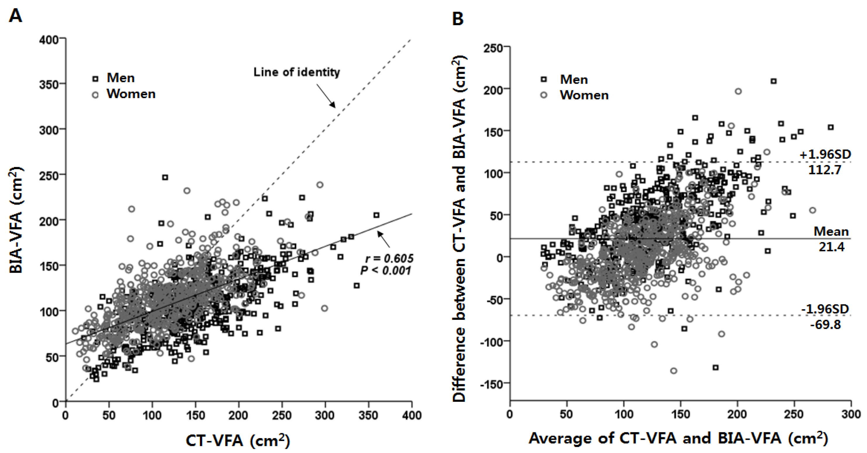
3.2. Associations between VFAs Measured by CT and BIA

| N | CT-VFA (cm2) | BIA-VFA (cm2) | CT-VFA-BIA-VFA (cm2) | *p | †p | ICC | |
|---|---|---|---|---|---|---|---|
| Total | 1006 | 131.9 ± 57.3 | 110.5 ± 33.9 | 21.4 ± 45.6 | <0.001 | 0.481 | |
| Gender | <0.001 | ||||||
| Men | 492 | 145.1 ± 60.4 | 106.9 ± 34.9 | 38.2 ± 45.9 | <0.001 | 0.438 | |
| Women | 512 | 119.3 ± 51.1 | 113.9 ± 32.6 | 5.4 ± 39.2 | 0.002 | 0.577 | |
| BMI (kg/m2) | <0.001 | ||||||
| <20 | 23 | 53.5 ± 24.1 | 67.6 ± 16.4 | −14.1 ± 29.2 | 0.031 | −0.008 | |
| 20–22.9 | 161 | 81.1 ± 37.2 | 77.8 ± 22.9 | 3.2 ± 33.3 | 0.218 | 0.417 | |
| 23–24.9 | 232 | 113.3 ± 42.7 | 98.3 ± 23.5 | 15.0 ± 39.3 | <0.001 | 0.319 | |
| 25–26.9 | 248 | 138.6 ± 44.3 | 108.9 ± 19.4 | 29.7 ± 43.5 | <0.001 | 0.140 | |
| 27–29.9 | 223 | 158.0 ± 52.0 | 125.3 ± 22.8 | 32.8 ± 48.8 | <0.001 | 0.196 | |
| ≥30 | 119 | 189.3 ± 55.9 | 162.1 ± 31.4 | 27.2 ± 58.4 | <0.001 | 0.145 | |
| Age (years) | 0.050 | ||||||
| 19–39 | 95 | 107.8 ± 64.8 | 99.3 ± 42.8 | 8.5 ± 52.2 | 0.115 | 0.544 | |
| 40–49 | 205 | 123.5 ± 50.4 | 101.8 ± 33.8 | 21.7 ± 40.4 | <0.001 | 0.496 | |
| 50–59 | 314 | 131.3 ± 55.1 | 106.8 ± 30.3 | 24.4 ± 44.7 | <0.001 | 0.430 | |
| 60–69 | 291 | 138.1 ± 55.4 | 116.9 ± 30.5 | 21.2 ± 45.3 | <0.001 | 0.438 | |
| ≥70 | 101 | 155.8 ± 64.0 | 131.2 ± 32.9 | 24.6 ± 51.4 | <0.001 | 0.441 | |
| n | r | *p | †p | ||
|---|---|---|---|---|---|
| Anemia | Hb ≥ 12 g/dL | 662 | 0.652 | <0.001 | 0.219 |
| Hb < 12 g/dL | 37 | 0.510 | 0.001 | ||
| Kidney Function | eGFR ≥ 60 mL/min/1.73 m2 | 740 | 0.630 | <0.001 | 0.327 |
| eGFR < 60 mL/min/1.73 m2 | 103 | 0.563 | <0.001 | ||
| Liver Function | ALT ≥ 40 IU/L | 227 | 0.607 | <0.001 | 0.569 |
| ALT < 40 IU/L | 771 | 0.579 | <0.001 | ||
| AST ≥ 40 IU/L | 111 | 0.503 | <0.001 | 0.144 | |
| AST < 40 IU/L | 887 | 0.606 | <0.001 | ||
| Diabetes Mellitus | DM (−) | 342 | 0.625 | <0.001 | 0.516 |
| DM (+) | 664 | 0.598 | <0.001 | ||
| HbA1c < 8% | 428 | 0.574 | <0.001 | 0.223 | |
| HbA1c ≥ 8% | 236 | 0.637 | <0.001 | ||
| Medications | Diuretics (−) and Thiazolidinedione (−) | 823 | 0.608 | <0.001 | |
| Thiazolidinedione (+) | 47 | 0.659 | <0.001 | 0.484 | |
| Diuretics (+) | 122 | 0.564 | <0.001 | 0.430 |
3.3. Agreement Levels between VFAs by CT and BIA According to Gender, BMI, and Age

3.4. Subgroup Comparison of CT-VFAs and BIA-VFAs According to Age and BMI Categories by Gender
| n | CT-VFA (cm2) | BIA-VFA (cm2) | CT-VFA–BIA-VFA (cm2) | p | ICC | |
|---|---|---|---|---|---|---|
| Men | ||||||
| BMI (kg/m2) | ||||||
| <25 | 188 | 105.4 ± 45.0 | 84.2 ± 28.0 | 21.2 ± 37.8 | <0.001 | 0.424 |
| ≥ 25 | 304 | 169.6 ± 55.6 | 120.9 ± 31.2 | 48.8 ± 47.3 | <0.001 | 0.285 |
| Age (years) | ||||||
| <50 | 175 | 132.4 ± 56.7 | 98.9 ± 36.9 | 33.5 ± 43.4 | <0.001 | 0.472 |
| ≥50 | 317 | 152.1 ± 61.4 | 111.3 ± 33.0 | 40.8 ± 47.0 | <0.001 | 0.407 |
| Women | ||||||
| BMI (kg/m2) | ||||||
| <25 | 228 | 91.0 ± 42.0 | 92.4 ± 22.4 | −1.4 ± 33.8 | 0.539 | 0.496 |
| ≥25 | 286 | 141.8 ± 46.3 | 131.1 ± 29.1 | 10.7 ± 42.3 | <0.001 | 0.387 |
| Age (years) | ||||||
| <50 | 125 | 99.1 ± 48.3 | 104.0 ± 36.8 | −4.9 ± 36.4 | 0.135 | 0.638 |
| ≥50 | 389 | 125.8 ± 50.3 | 117.1 ± 30.5 | 8.7 ± 39.6 | <0.001 | 0.537 |
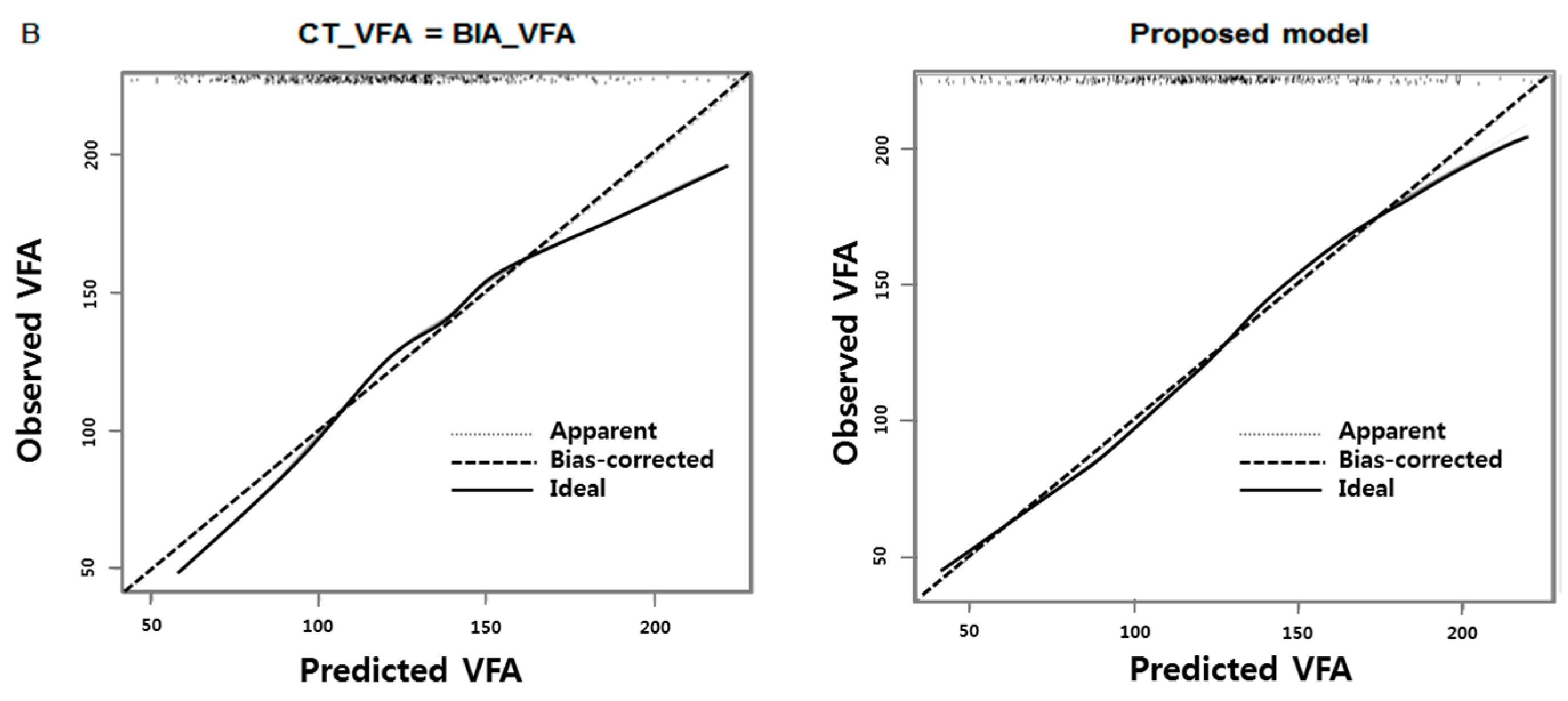
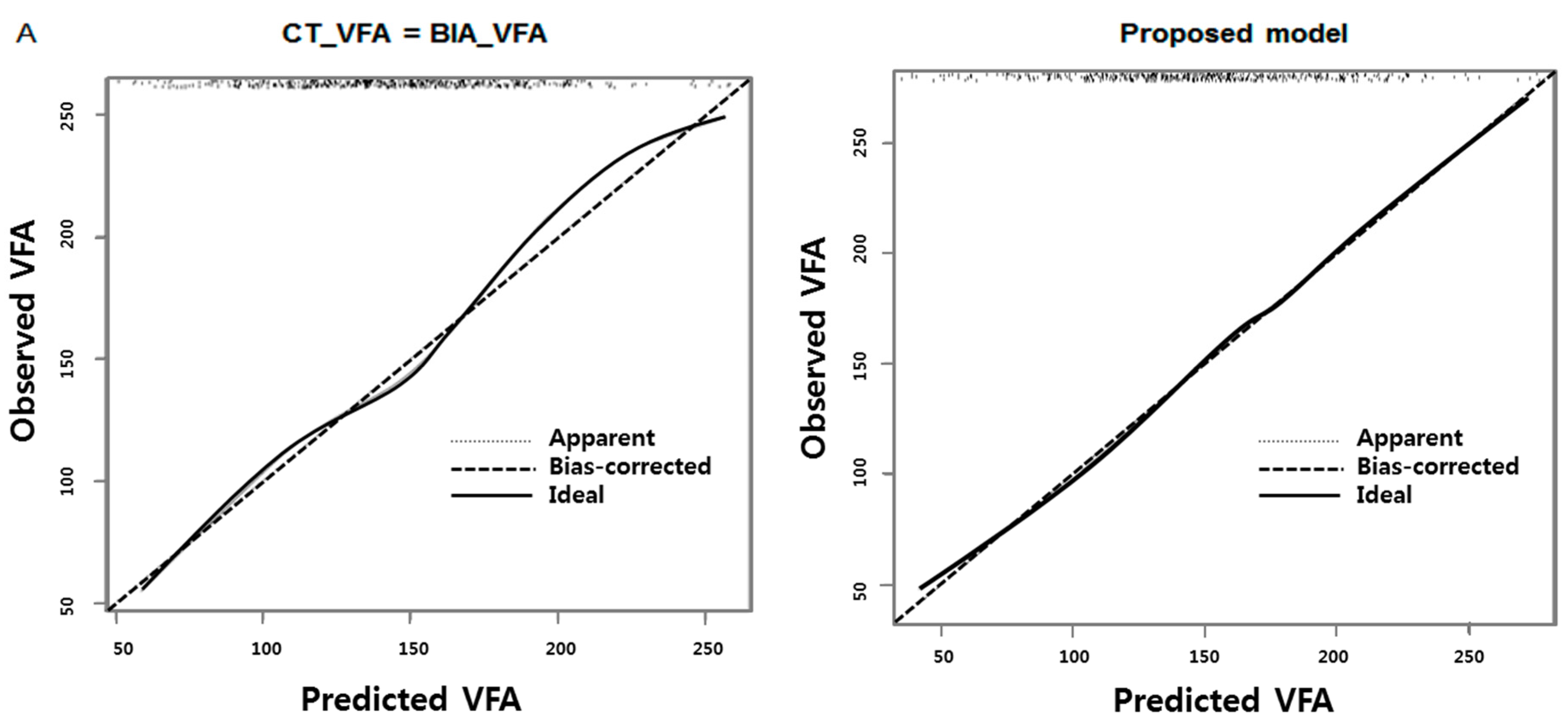
3.5. New Formula to Predict CT-VFA Using BIA-VFA Data

| Men | Women | |||||
|---|---|---|---|---|---|---|
| Coefficient | 95% CI | Coefficient | 95% CI | |||
| Lower | Upper | Lower | Upper | |||
| Intercept | −184.51 | −323.06 | −45.95 | −142.77 | −277.24 | −8.30 |
| BIA-VFA | 1.11 | 0.46 | 1.76 | 1.40 | 0.72 | 2.08 |
| Age | −1.49 | −3.61 | 0.63 | −1.29 | −3.68 | 1.10 |
| BMI | 2.10 | −3.28 | 7.47 | −0.98 | −6.49 | 4.54 |
| WC | 2.03 | 1.33 | 2.72 | 2.14 | 1.55 | 2.73 |
| BIA-VFA*BMI | −0.02 | −0.04 | 0.01 | −0.03 | −0.05 | −0.01 |
| Age*BMI | 0.08 | −0.01 | 0.16 | 0.07 | −0.02 | 0.17 |
4. Discussion

5. Conclusions
Acknowledgments
Author Contributions
Conflicts of Interest
References
- Ogden, C.L.; Carroll, M.D.; Curtin, L.R.; McDowell, M.A.; Tabak, C.J.; Flegal, K.M. Prevalence of overweight and obesity in the united states, 1999–2004. J. Am. Med. Assoc. 2006, 295, 1549–1555. [Google Scholar] [CrossRef] [PubMed]
- Matsuzawa, Y.; Funahashi, T.; Nakamura, T. The concept of metabolic syndrome: Contribution of visceral fat accumulation and its molecular mechanism. J. Atheroscler. Thromb. 2011, 18, 629–639. [Google Scholar] [CrossRef] [PubMed]
- Berker, D.; Koparal, S.; Isik, S.; Pasaoglu, L.; Aydin, Y.; Erol, K.; Delibasi, T.; Guler, S. Compatibility of different methods for the measurement of visceral fat in different body mass index strata. Diagn. Interv. Radiol. 2010, 16, 99–105. [Google Scholar] [CrossRef] [PubMed]
- Kaess, B.M.; Pedley, A.; Massaro, J.M.; Murabito, J.; Hoffmann, U.; Fox, C.S. The ratio of visceral to subcutaneous fat, a metric of body fat distribution, is a unique correlate of cardiometabolic risk. Diabetologia 2012, 55, 2622–2630. [Google Scholar] [CrossRef] [PubMed]
- Shah, R.V.; Murthy, V.L.; Abbasi, S.A.; Blankstein, R.; Kwong, R.Y.; Goldfine, A.B.; Jerosch-Herold, M.; Lima, J.A.; Ding, J.; Allison, M.A. Visceral adiposity and the risk of metabolic syndrome across body mass index: The mesa study. JACC Cardiovasc. Imaging 2014, 7, 1221–1235. [Google Scholar] [CrossRef] [PubMed]
- Montague, C.T.; O’Rahilly, S. The perils of portliness: Causes and consequences of visceral adiposity. Diabetes 2000, 49, 883–888. [Google Scholar] [CrossRef] [PubMed]
- Kahn, B.B.; Flier, J.S. Obesity and insulin resistance. J. Clin. Investig. 2000, 106, 473–481. [Google Scholar] [CrossRef] [PubMed]
- Yoshizumi, T.; Nakamura, T.; Yamane, M.; Islam, A.H.; Menju, M.; Yamasaki, K.; Arai, T.; Kotani, K.; Funahashi, T.; Yamashita, S.; et al. Abdominal fat: Standardized technique for measurement at CT. Radiology 1999, 211, 283–286. [Google Scholar] [CrossRef] [PubMed]
- Ribeiro-Filho, F.F.; Faria, A.N.; Azjen, S.; Zanella, M.T.; Ferreira, S.R. Methods of estimation of visceral fat: Advantages of ultrasonography. Obes. Res. 2003, 11, 1488–1494. [Google Scholar] [CrossRef] [PubMed]
- Ryo, M.; Maeda, K.; Onda, T.; Katashima, M.; Okumiya, A.; Nishida, M.; Yamaguchi, T.; Funahashi, T.; Matsuzawa, Y.; Nakamura, T.; et al. A new simple method for the measurement of visceral fat accumulation by bioelectrical impedance. Diabet. Care 2005, 28, 451–453. [Google Scholar] [CrossRef]
- Wajchenberg, B.L. Subcutaneous and visceral adipose tissue: Their relation to the metabolic syndrome. Endocr. Rev. 2000, 21, 697–738. [Google Scholar] [CrossRef] [PubMed]
- Sobol, W.; Rossner, S.; Hinson, B.; Hiltbrandt, E.; Karstaedt, N.; Santago, P.; Wolfman, N.; Hagaman, A.; Crouse, J.R., 3rd. Evaluation of a new magnetic resonance imaging method for quantitating adipose tissue areas. Int. J. Obes. 1991, 15, 589–599. [Google Scholar] [PubMed]
- Van der Kooy, K.; Seidell, J.C. Techniques for the measurement of visceral fat: A practical guide. Int. J. Obes. Relat. Metab. Disord. 1993, 17, 187–196. [Google Scholar] [PubMed]
- Nagai, M.; Komiya, H.; Mori, Y.; Ohta, T.; Kasahara, Y.; Ikeda, Y. Development of a new method for estimating visceral fat area with multi-frequency bioelectrical impedance. Tohoku J. Exp. Med. 2008, 214, 105–112. [Google Scholar] [CrossRef] [PubMed]
- Unno, M.; Furusyo, N.; Mukae, H.; Koga, T.; Eiraku, K.; Hayashi, J. The utility of visceral fat level by bioelectrical impedance analysis in the screening of metabolic syndrome—The results of the kyushu and okinawa population study (KOPS). J. Atheroscler. Thromb. 2012, 19, 462–470. [Google Scholar] [CrossRef] [PubMed]
- Nagai, M.; Komiya, H.; Mori, Y.; Ohta, T.; Kasahara, Y.; Ikeda, Y. Estimating visceral fat area by multifrequency bioelectrical impedance. Diabet. Care 2010, 33, 1077–1079. [Google Scholar] [CrossRef] [PubMed]
- Shoji, K.; Maeda, K.; Nakamura, T.; Funahashi, T.; Matsuzawa, Y.; Shimomura, I. Measurement of visceral fat by abdominal bioelectrical impedance analysis is beneficial in medical checkup. Obes. Res. Clin. Pract. 2008, 2. [Google Scholar] [CrossRef] [PubMed]
- Shil Hong, E.; Khang, A.R.; Roh, E.; Jeong Ku, E.; An Kim, Y.; Min Kim, K.; Hoon Moon, J.; Hee Choi, S.; Soo Park, K.; Woong Kim, K.; et al. Counterintuitive relationship between visceral fat and all-cause mortality in an elderly asian population. Obesity 2015, 23, 220–227. [Google Scholar] [CrossRef] [PubMed]
- Faria, S.L.; Faria, O.P.; Menezes, C.S.; de Gouvea, H.R.; de Almeida Cardeal, M. Metabolic profile of clinically severe obese patients. Obes. Surg. 2012, 22, 1257–1262. [Google Scholar] [CrossRef] [PubMed]
- Poggio, E.D.; Nef, P.C.; Wang, X.; Greene, T.; van Lente, F.; Dennis, V.W.; Hall, P.M. Performance of the cockcroft-gault and modification of diet in renal disease equations in estimating GFR in ill hospitalized patients. Am. J. Kidney Dis. 2005, 46, 242–252. [Google Scholar] [CrossRef] [PubMed]
- Posada, D.; Buckley, T.R. Model selection and model averaging in phylogenetics: Advantages of akaike information criterion and bayesian approaches over likelihood ratio tests. Syst. Boil. 2004, 53, 793–808. [Google Scholar] [CrossRef] [PubMed]
- Winter, J.E.; MacInnis, R.J.; Wattanapenpaiboon, N.; Nowson, C.A. Bmi and all-cause mortality in older adults: A meta-analysis. Am. J. Clin. Nutr. 2014, 99, 875–890. [Google Scholar] [CrossRef] [PubMed]
- Matsushita, Y.; Nakagawa, T.; Shinohara, M.; Yamamoto, S.; Takahashi, Y.; Mizoue, T.; Yokoyama, T.; Noda, M. How can waist circumference predict the body composition? Diabetol. Metab. Syndr. 2014, 6. [Google Scholar] [CrossRef] [PubMed]
- Ashwell, M.; Gunn, P.; Gibson, S. Waist-to-height ratio is a better screening tool than waist circumference and BMI for adult cardiometabolic risk factors: Systematic review and meta-analysis. Obes. Rev. 2012, 13, 275–286. [Google Scholar] [CrossRef] [PubMed]
- Yamakage, H.; Ito, R.; Tochiya, M.; Muranaka, K.; Tanaka, M.; Matsuo, Y.; Odori, S.; Kono, S.; Shimatsu, A.; Satoh-Asahara, N. The utility of dual bioelectrical impedance analysis in detecting intra-abdominal fat area in obese patients during weight reduction therapy in comparison with waist circumference and abdominal ct. Endocr. J. 2014, 61, 807–819. [Google Scholar] [CrossRef] [PubMed]
- Dhaliwal, S.S.; Welborn, T.A. Measurement error and ethnic comparisons of measures of abdominal obesity. Prev. Med. 2009, 49, 148–152. [Google Scholar] [CrossRef] [PubMed]
- Suh, Y.S.; Kim, D.H.; Lee, I.K. Usefulness of lumbar AP spine DXA for measuring the percentage of perilumbar regional fat and predicting visceral fat in obese postmenopausal women. Nutrition 2002, 18, 84–85. [Google Scholar] [CrossRef]
- Bonora, E.; Micciolo, R.; Ghiatas, A.A.; Lancaster, J.L.; Alyassin, A.; Muggeo, M.; DeFronzo, R.A. Is it possible to derive a reliable estimate of human visceral and subcutaneous abdominal adipose tissue from simple anthropometric measurements? Metabolism 1995, 44, 1617–1625. [Google Scholar] [CrossRef]
- Rossner, S.; Bo, W.J.; Hiltbrandt, E.; Hinson, W.; Karstaedt, N.; Santago, P.; Sobol, W.T.; Crouse, J.R. Adipose tissue determinations in cadavers—A comparison between cross-sectional planimetry and computed tomography. Int. J. Obes. 1990, 14, 893–902. [Google Scholar] [PubMed]
- Rosito, G.A.; Massaro, J.M.; Hoffmann, U.; Ruberg, F.L.; Mahabadi, A.A.; Vasan, R.S.; O’Donnell, C.J.; Fox, C.S. Pericardial fat, visceral abdominal fat, cardiovascular disease risk factors, and vascular calcification in a community-based sample: The framingham heart study. Circulation 2008, 117, 605–613. [Google Scholar] [CrossRef] [PubMed]
- Liu, J.; Fox, C.S.; Hickson, D.A.; May, W.D.; Hairston, K.G.; Carr, J.J.; Taylor, H.A. Impact of abdominal visceral and subcutaneous adipose tissue on cardiometabolic risk factors: The jackson heart study. J. Clin. Endocrinol. Metab. 2010, 95, 5419–5426. [Google Scholar] [CrossRef] [PubMed]
- Hanley, A.J.; Wagenknecht, L.E.; Norris, J.M.; Bryer-Ash, M.; Chen, Y.I.; Anderson, A.M.; Bergman, R.; Haffner, S.M. Insulin resistance, beta cell dysfunction and visceral adiposity as predictors of incident diabetes: The insulin resistance atherosclerosis study (IRAS) family study. Diabetologia 2009, 52, 2079–2086. [Google Scholar] [CrossRef] [PubMed]
- Matsuzawa, Y. Metabolic syndrome—Definition and diagnostic criteria in Japan. J. Atheroscler. Thromb. 2005, 12, 301. [Google Scholar] [CrossRef] [PubMed]
- Kyle, U.G.; Bosaeus, I.; de Lorenzo, A.D.; Deurenberg, P.; Elia, M.; Gomez, J.M.; Heitmann, B.L.; Kent-Smith, L.; Melchior, J.C.; Pirlich, M.; et al. Bioelectrical impedance analysis—Part I: Review of principles and methods. Clin. Nutr. 2004, 23, 1226–1243. [Google Scholar] [CrossRef] [PubMed]
- Kyle, U.G.; Bosaeus, I.; de Lorenzo, A.D.; Deurenberg, P.; Elia, M.; Manuel Gomez, J.; Lilienthal Heitmann, B.; Kent-Smith, L.; Melchior, J.C.; Pirlich, M.; et al. Bioelectrical impedance analysis—Part II: Utilization in clinical practice. Clin. Nutr. 2004, 23, 1430–1453. [Google Scholar] [CrossRef] [PubMed]
- Volgyi, E.; Tylavsky, F.A.; Lyytikainen, A.; Suominen, H.; Alen, M.; Cheng, S. Assessing body composition with DXA and bioimpedance: Effects of obesity, physical activity and age. Obesity 2008, 16, 700–705. [Google Scholar] [CrossRef] [PubMed]
- Ida, M.; Hirata, M.; Odori, S.; Mori, E.; Kondo, E.; Fujikura, J.; Kusakabe, T.; Ebihara, K.; Hosoda, K.; Nakao, K. Early changes of abdominal adiposity detected with weekly dual bioelectrical impedance analysis during calorie restriction. Obesity 2013, 21. [Google Scholar] [CrossRef] [PubMed] [Green Version]
- Pietilainen, K.H.; Kaye, S.; Karmi, A.; Suojanen, L.; Rissanen, A.; Virtanen, K.A. Agreement of bioelectrical impedance with dual-energy X-ray absorptiometry and MRI to estimate changes in body fat, skeletal muscle and visceral fat during a 12-month weight loss intervention. Br. J. Nutr. 2013, 109, 1910–1916. [Google Scholar] [CrossRef] [PubMed]
© 2015 by the authors; licensee MDPI, Basel, Switzerland. This article is an open access article distributed under the terms and conditions of the Creative Commons by Attribution (CC-BY) license (http://creativecommons.org/licenses/by/4.0/).
Share and Cite
Lee, D.-H.; Park, K.S.; Ahn, S.; Ku, E.J.; Jung, K.Y.; Kim, Y.J.; Kim, K.M.; Moon, J.H.; Choi, S.H.; Park, K.S.; et al. Comparison of Abdominal Visceral Adipose Tissue Area Measured by Computed Tomography with That Estimated by Bioelectrical Impedance Analysis Method in Korean Subjects. Nutrients 2015, 7, 10513-10524. https://doi.org/10.3390/nu7125548
Lee D-H, Park KS, Ahn S, Ku EJ, Jung KY, Kim YJ, Kim KM, Moon JH, Choi SH, Park KS, et al. Comparison of Abdominal Visceral Adipose Tissue Area Measured by Computed Tomography with That Estimated by Bioelectrical Impedance Analysis Method in Korean Subjects. Nutrients. 2015; 7(12):10513-10524. https://doi.org/10.3390/nu7125548
Chicago/Turabian StyleLee, Dong-Hwa, Kyeong Seon Park, Soyeon Ahn, Eu Jeong Ku, Kyong Yeun Jung, Yoon Ji Kim, Kyoung Min Kim, Jae Hoon Moon, Sung Hee Choi, Kyong Soo Park, and et al. 2015. "Comparison of Abdominal Visceral Adipose Tissue Area Measured by Computed Tomography with That Estimated by Bioelectrical Impedance Analysis Method in Korean Subjects" Nutrients 7, no. 12: 10513-10524. https://doi.org/10.3390/nu7125548