Review

Inflammatory Effects of Bothrops Phospholipases A2: Mechanisms Involved in Biosynthesis of Lipid Mediators and Lipid Accumulation

by
Vanessa Moreira
1,†,
Elbio Leiguez
2,†,
Priscila Motta Janovits
2,
Rodrigo Maia-Marques
2,
Cristina Maria Fernandes
2 and
Catarina Teixeira
2,*
1
Departamento de Farmacologia, Escola Paulista de Medicina, Universidade Federal de Sao Paulo, Sao Paulo 04044-020, Brazil
2
Laboratório de Farmacologia, Instituto Butantan, Sao Paulo 05503-900, Brazil
*
Author to whom correspondence should be addressed.
These authors contributed with equal importance.
Toxins 2021, 13(12), 868; https://doi.org/10.3390/toxins13120868
Submission received: 3 November 2021 / Revised: 19 November 2021 / Accepted: 30 November 2021 / Published: 4 December 2021

Abstract

:
Phospholipases A2s (PLA2s) constitute one of the major protein groups present in the venoms of viperid and crotalid snakes. Snake venom PLA2s (svPLA2s) exhibit a remarkable functional diversity, as they have been described to induce a myriad of toxic effects. Local inflammation is an important characteristic of snakebite envenomation inflicted by viperid and crotalid species and diverse svPLA2s have been studied for their proinflammatory properties. Moreover, based on their molecular, structural, and functional properties, the viperid svPLA2s are classified into the group IIA secreted PLA2s, which encompasses mammalian inflammatory sPLA2s. Thus, research on svPLA2s has attained paramount importance for better understanding the role of this class of enzymes in snake envenomation and the participation of GIIA sPLA2s in pathophysiological conditions and for the development of new therapeutic agents. In this review, we highlight studies that have identified the inflammatory activities of svPLA2s, in particular, those from Bothrops genus snakes, which are major medically important snakes in Latin America, and we describe recent advances in our collective understanding of the mechanisms underlying their inflammatory effects. We also discuss studies that dissect the action of these venom enzymes in inflammatory cells focusing on molecular mechanisms and signaling pathways involved in the biosynthesis of lipid mediators and lipid accumulation in immunocompetent cells.
Key Contribution: This review provides an overview and recent advances in the understanding of inflammatory mechanisms triggered by svPLA2s with a focus on their actions on lipid mediator biosynthetic pathways and lipid accumulation in immunocompetent cells.

1. Introduction

Bothrops spp. snakes are responsible for the majority of snakebites in Latin America. Envenomation by these snakes induces severe pathological alterations at the site of venom injection, characterized by an intense local inflammatory reaction associated with myonecrosis, pain, and hemorrhage, potentially leading to permanent tissue damage and disability [1,2,3,4,5,6]. The local inflammatory response to bothropic envenomation involves a set of events including an increase in vascular permeability, edema formation, hyperalgesia, the activation and infiltration of immunocompetent cells, and a complex network of inflammatory mediators driving the inflammatory response [2]. These events are caused by the activation of critical host defense mechanisms of the victims by direct and indirect actions of the toxins present in the snake venom. Proteomic studies of Bothrops snake venoms revealed that the phospholipases A2 (PLA2s) are ubiquitous components of these venoms and play an important role in the pathophysiology of envenoming by these snakes [7,8,9,10,11]. During envenomation, in addition to aiding prey digestion, these toxins have been described to display myotoxic, cytotoxic, hemolytic, hypotensive, anticoagulant, platelet aggregation inhibition/activation, and proinflammatory effects [12,13,14]. Regarding inflammation, in addition to exerting direct effects on cell membranes, these lipolytic enzymes can recruit mammalian PLA2s analogs of similar activity and trigger endogenous signaling systems that display and amplify the cell injury and host defense mechanisms triggered by the whole venom. This amplification is responsible for many acutely important consequences of Bothrops envenoming. Yet, as discussed below, studies with in vitro and in vivo models aiming to understand the inflammatory action of isolated venom PLA2s can contribute to the knowledge of the local inflammatory mechanisms induced by Bothrops snake venoms and those from the Viperidae family [3,15,16,17]. These studies might lead to the discovery of new therapeutic targets for a more efficient treatment of envenoming by viperid snakes, since the currently available antivenoms have low effectiveness to neutralize the local events promoted by their venoms [18]. Finally, due to the structural and functional similarities to mammalian group (G) IIA PLA2, the Bothrops PLA2s can constitute useful tools for studies on the roles of human GIIA PLA2s in inflammatory diseases. In this regard, the effectiveness of varespladib, an inhibitor of svPLA2s [19] in attenuating inflammatory events caused by Viperidae snake venoms has been demonstrated in mice experimental model [20].

1.1. Phospholipases A2

Phospholipases A2 constitute a group of enzymes with diverse biological functions, ranging from homeostasis and membrane remodeling to the generation of metabolites and second messengers involved in biological processes and signal transduction. These enzymes hydrolyze the acyl ester bond at the sn-2 position of phospholipids, generating free fatty acids, such as arachidonic acid (AA) and oleic acid, and lysophospholipids, such as lyso-PAF [21,22]. Currently, PLA2s are classified into close families, including secretory PLA2 (sPLA2), cytosolic phospholipase (cPLA2), and Ca2+-independent PLA2 (iPLA2); platelet activating factor acetylhydrolase (PAF); and lysosomal PLA2 (LPLA2), PLA/acyltransferase (PLAAT), α/β hydrolase (ABHD), adipose-PLA2 (AdPLA), and glycosylphosphatidylinositol (GPI)-specific PLA2 [23,24,25]. This classification of PLA2s is based on the cell location, amino acid sequence, molecular weight, presence of intramolecular disulfide bridges, calcium requirement, and catalytic activity, including the hydrolysis at the sn-2 position of glycerophospholipids [22,26]. The sPLA2 family is largely distributed in nature, and its components are classified into I, II, III, V, IX, X, XI, XII, XIII, and XIV groups. They are present in high concentrations in snake, bee, and wasp venoms and in several mammalian organs, cells, and pancreatic juice [21,27]. Among them, we highlight group I PLA2s, comprising the secreted enzymes found in the snake venoms of the Elapidae and Hydrophiidae families and in the pancreas secretion of mammals. Meanwhile, group II consists of sPLA2s found in snake venoms of the Viperidae family and in mammalian tissues and are expressed in inflammatory processes [13,28].
For a long time, the pathophysiological activities of sPLA2s were related exclusively to their enzymatic activity, capable of providing a substrate for the synthesis of second messengers and inflammatory mediators. However, alternative mechanisms of action have been associated with the ability of sPLA2 to interact with receptors or even specific domains present on cell membrane surfaces, such as heparan sulfate proteoglycans, which have already been described to be important for triggering the activation of other sPLA2s in the target cells [29,30,31,32]. The identification of sPLA2s binding proteins was initially achieved by Lambeau et al. (1989) [33], who used the sPLA2 from the Oxyuranus scutelatus snake venom, called OS2. Due to its prevalence in brain tissue, this protein was denominated an N (neuronal) type receptor. Then, through screening in various tissues and cells, for studies of other OS2 binding proteins, a second type of PLA2 receptor was described, present in rabbit skeletal muscles, called an M (muscle) type receptor [34]. The presence of this latter receptor was identified in several tissues and in neutrophils, monocytes, and human alveolar macrophages [35,36], but not in murine peritoneal macrophages [37]. The M-type phospholipase A2 receptor has a high degree of homology (~30%) to mannose receptors, a member of the lectin receptor family, constitutively expressed in macrophages [38,39]. Mannose receptors are involved in phagocytosis, antigen processing [29,38,40], and the production of inflammatory cytokines by macrophages [40,41,42]. Additionally, this receptor has been demonstrated to play a role in cell proliferation and AA release, via MAPKs (protein kinase activated by the mitogens family), induced by the group IB sPLA2s [43,44,45]. Furthermore, it has been revealed that group IIA sPLA2s can bind to mannose receptors and promote the release of IL-6 by human alveolar macrophages [46].
Investigations into the biological role of mammalian group IIA PLA2, also known as inflammatory PLA2, in the development of several pathologies of inflammatory and immunological origin have been described. Several studies have revealed that group IIA sPLA2s are present in high levels in rheumatoid arthritis [47,48,49,50], acute pancreatitis [51,52,53], septic shock [54,55], Crohn’s disease and ulcerative colitis [56,57,58], respiratory distress syndrome [59,60,61], bronchial asthma and allergic rhinitis [59,62], atherosclerosis [63,64], autoimmune diseases [65], and cancer [66,67,68,69]. These observations imply that both local and systemic inflammation are associated with the release of sPLA2s in vivo, thus raising the unclear question of the role of these PLA2s in inflammatory reactions. Additionally, it was found that proinflammatory cytokines, such as interleukin (IL)-1β, IL-6, and tumor necrosis factor alpha (TNF-α), induce, in a variety of tissues, the gene transcription of sPLA2s and the subsequent increase in their secretion, thus, supporting the hypothesis of the involvement of sPLA2s in inflammation [70,71,72,73]. In addition, sPLA2s activate intracellular signaling events in cells that participate in inflammatory processes, caused by the generation of second messengers, and the phosphorylation of kinases as of MAPK [74,75,76,77]. Thus, sPLA2s represent an important target for investigations regarding the mechanisms of inflammatory events.

1.2. Inflammation—General Concepts and Signaling Pathways

Inflammation is a response of body tissues to noxious conditions for restoring homeostasis, setting the stage for the healing and reconstitution of injured tissue. The acute inflammatory response to injury involves functional alterations of microvessels that occur early after injury and develop at varying rates. The major features of these alterations include transient vasoconstriction, followed by vasodilation, then, leakage of protein-rich fluid from the microcirculation leading to edema formation, and movement of phagocytic leukocytes into the site of injury followed by local pain [78]. Immunocompetent cells, such as neutrophils and monocytes, found in blood circulation are capable of rapidly infiltrating tissues; macrophages and dendritic cells reside within tissue and play key roles in tissue surveillance and antigen presentation [79,80]. The vascular and cellular reactions are triggered and highly regulated by chemical factors, called inflammatory mediators, which include cytokines, chemokines, vasoactive amines, and eicosanoids and are produced by plasma components or are released in close proximity to the injury by endothelial cells, tissue-resident leukocytes such as mast cells, and macrophages at the early stages, followed by infiltrated leukocytes. The effects of inflammatory mediators involve the engagement of specific receptors, which then display signaling pathways responsible for the immune response [81,82,83]. Parallel to changes in blood flow, the margination of leukocytes begins, and leukocytes adhere to the microvascular endothelium through rolling and firm adhesion, and then moving through the vascular wall into the interstitial tissue. The various steps in the leukocyte migration are regulated by different subsets of cell-adhesion molecules expressed by both leukocyte and endothelial cells [84,85]. Thereafter, the phagocytosis of offending agents by migrated leukocytes occurs, followed by a release of lysosomal enzymes and an increase in the oxidative metabolism in leukocytes, known as respiratory burst, resulting in the production of microbicidal agents, such as superoxide anion (O2) and hydrogen peroxide (H2O2) [86,87]. Four major classes of receptor-mediated phagocytosis exist: receptors of complement that recognize complement-coated particles; Fcg receptors, which are constitutively active for phagocytosis of IgG-coated particles; mannose receptors that recognize mannose and fucose on the surface of pathogens; and β-glucan receptors that recognize β-glucans-bearing ligands [87]. Furthermore, receptors for the Fc portion of immunoglobulin G and mannose/fucose residues lead to the release of proinflammatory mediators and reactive oxygen [88].
Despite the stereotyped features associated with an inflammatory response, the signal pathways involved in this response are determined by the nature of the inflammatory trigger, the sensors that detect them, the inflammatory mediators released, and the tissue affected. A number of surface and cytosolic receptors expressed in innate immune cells can sense pathogen-associated molecular patterns (PAMPs), damage-associated molecular patterns (DAMPs), or venom-associated molecular patterns (VAMPs) with high sensitivity and specificity. The recognition of these molecules is achieved by interacting with pattern recognition receptors (PRRs) [89]. These sensing receptors include Toll-like receptors (TLRs) [90,91], C-type lectin receptors (CLRS), RIG-I-like receptors (RLRs), and nucleotide-binding domain leucine-rich repeat (NLRs) or nucleotide-binding and oligomerisation domain (NOD)-like receptors [92,93]. Among them, TLRs (TLR1-TLR10) are highly conserved transmembrane proteins that play an important role in recognizing microbial pathogens and endogenous damage molecules, thereby triggering the generation of signals for the production of proinflammatory proteins and cytokines, via cooperation of adaptor proteins (MyD88, TIRAP, TRIF, and TRAM) [94,95]. The recognition of specific molecular patterns by NLRs induces the oligomerisation of proteins in the cytosol, generating platforms called inflammasomes [96]. The inflammasome is a high molecular weight protein complex that elicits the activation of inflammatory caspases and the processing of pro-interleukin-1β (pro-IL-1β) and pro-IL-18, generating the mature biologically active cytokines and a rapid inflammatory form of cell death termed pyroptosis [97]. Several distinct inflammasomes have been identified, including NLR and the pyrin domain containing receptor 1 (NLRP1), NLRP3, and NLR; the caspase recruitment domain containing receptor 4 (NLRC4); and the AIM2-like receptors (ALR) family [98,99]. These receptors positively regulate genes related to inflammatory mediators, including cytokines and key enzymes in the biosynthetic pathways of lipid mediators known as eicosanoids [100].
The early induction of most inflammatory transcripts depends on networks of transcription factors whose activation is coupled to pathways of signal transduction. The nuclear factor-kappa B (NF-κB) is a major and the best-studied transcription factor of inflammatory response [101,102]. The binding of activated NF-κB to the nuclear promoter region of diverse inflammatory factors leads to the transcriptional activation and expression of inflammatory mediators and enzymes. Currently, the major signaling pathways involved in inflammation are mediated by the cascade phosphorylation of protein kinases, such as the mitogen activated kinases (MAPKs) encompassing ERK1/2, SAPK, c-Jun NH2-terminal or JNK and p38MAPKs, phosphatidylinositol 3 kinase (PI3K), protein kinase C (PKC), and protein tyrosine kinase (PTK). These kinases mediate distinct intracellular signaling pathways associated with cytokines production, cytokine receptors, growth factors, mobilization of intracellular Ca2+, and regulate a variety of functions of immunocompetent cells, including cell migration, phagocytosis, degranulation, respiratory burst, and programmed cell death [103,104,105,106,107,108,109,110,111,112].
An effective acute inflammatory response results in the removal of noxious factors followed by the resolution and repair stages [113]. The shift in inflammatory markers, including the switch from proinflammatory mediators to anti-inflammatory, resolution-inducing mediators (lipoxins, maresins, protectins, and resolvins), is vital for the change from inflammation to resolution [114]. This switch drives the transition from neutrophil to monocyte recruitment into the affected sites, resulting in the clearance of dead cells and other debris, assisted by the lymphatic system, and the initiation of tissue repair at the damaged site [115]. However, if the acute events are not properly controlled, the inflammatory response becomes detrimental to the host. Yet, if the acute response does not succeed in neutralizing the injurious stimulus, the resolution phase might not be appropriately induced, and a chronic inflammatory state may ensue, leading to several inflammatory-mediated diseases [116,117].

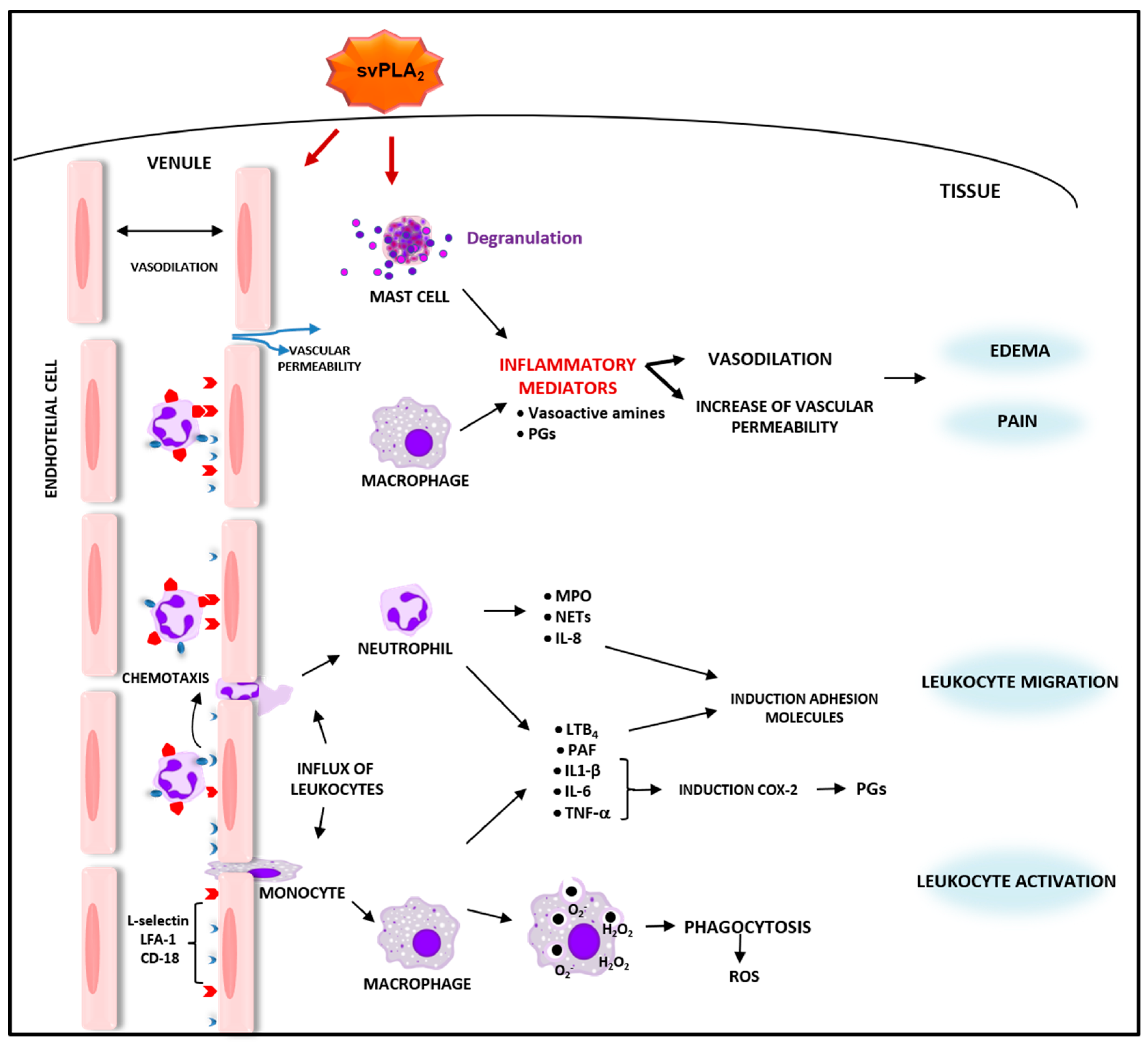
2. Inflammatory Effects of sPLA2 from Bothrops spp. Venoms

2.1. Bothrops svPLA2s Induce Inflammatory Events and Activate Defense Functions in Leukocytes

Phospholipases A2 of GIIA are major components of Bothrops spp. snake venoms and play important roles in the pathophysiology of envenoming by these snakes, including the inflammatory response. Although these enzymes conserve a chemistry and catalytic structure, the natural evolution of viperid venoms introduced alterations in their primary amino acid residues, generating various other biological and toxicological effects [118]. In general, the GIIA sPLA2s found in viperid snake venoms are classified as sPLA2s, known as ‘classic’, containing an amino acid aspartate at position 49 (Asp49) and catalyzing the hydrolysis of the ester bond at position sn-2 of glycerophospholipids in a Ca2+ dependent manner. Meanwhile, the other type of sPLA2s is described as ‘variant’ and contains a lysine at the same position 49 (Lys49), with or without low catalytic activity [119]. Such a substitution affects the ability of these proteins to bind to Ca2+, an essential cofactor for the stabilization of tetrahedral intermediate, which occurs in the catalytic reaction performed by the Asp49-sPLA2s [120]. Despite the lack of enzymatic activity, sPLA2s-Lys49 homologues maintain their damaging capacity in membranes through a mechanism that is not completely understood and independent of Ca2+ [12,121,122].
It has long been demonstrated that viperid sPLA2s are potent inductors of inflammation. Although they present differences in their catalytic activity, both viperid Asp49 and Lys49 PLA2 homologues are capable of inducing local inflammation in diverse experimental models [123,124,125]. As such, this group of enzymes is considered to be a major component responsible for the severe local edema in envenomings by Bothrops spp. The inflammatory response to venom PLA2s is characterized by edema and the marked infiltration of leukocytes into the site of toxin injection. Studies on the mechanism of local edema induced by viperid sPLA2s (svPLA2s) have demonstrated an early increase in vascular permeability and a local release of inflammatory mediators, which act synergistically to cause the initiation and development of the inflammatory events. Among these mediators are vasoactive amines, including histamine, serotonin, and substance P, as well as vasodilating prostaglandins. Yet, in vivo studies employing a pharmacological approach have demonstrated that antagonists of serotonin and H1 receptors of histamine reduced the progression of edema induced by both catalytically active and inactive variants of sPLA2 isolated from B. asper [126], B. neuwiedii [127], B. jararacussu [128,129], or B. insularis [130]. In support of these reports, the release of histamine and serotonin by mast cells was observed following stimulation with bothropic sPLA2s from B. jararacussu [128,131]. Consistent with this evidence, the contribution of mast cells to edema formation induced by viperid PLA2s was further observed in in vitro experimental models demonstrating the ability of sPLA2s isolated from B. pirajai, B. jararacussu, and B. atrox snake venoms to degranulate mast cells [125,128,131,132]. It is well known that upon activation, mast cells secrete and synthesize an array of inflammatory mediators, which trigger the earliest events of inflammation [133,134]. Moreover, the contribution of the catalytic activity for the edematogenic effect of the enzymatic active Asp49 from bothropic PLA2s was suggested by studies, revealing that the chemical modification of this sPLA2 by p-bromophenacyl bromide inhibited edema formation induced by these viperid PLA2s [126,127,128]. In addition, the role of lipid mediators, such as PAF and eicosanoids, for hyperalgesia induced by catalytic active venom PLA2s was highlighted by studies using a pharmacological approach [135]. These authors suggested that the enzymatic hydrolysis of membrane phospholipids played a role in these events by directly releasing the precursors of lipid mediators, such as lyso-PAF and AA.
As mentioned previously, leukocytes are central components of inflammation. An important cellular component exists in the inflammatory response to Bothrops sPLA2s. As such, the stimulatory effect of piratoxin-I, bothropstoxin-I, and -II from B. pirajai and B. jararacussu, respectively, on neutrophil chemotaxis was demonstrated in an in vitro experimental model [136]. This effect was revealed to involve the interaction of these sPLA2s with surface heparan binding sites of neutrophils, followed by the release of chemotactic mediator leukotriene B4 (LTB4) and PAF, and is independent of enzyme activity. Furthermore, the ability of these venom PLA2s to recruit an endogenous PLA2 through the activation of GTP-binding protein and PKC was added to the mechanisms by which they cause neutrophil migration [137]. Moreover, studies conducted using in vivo experimental models have demonstrated the ability of Bothrops sPLA2s to induce a marked influx of polymorphonuclear and mononuclear cells into the site of their injection, as demonstrated for both catalytic active and non-catalytic venom PLA2s, such as MT-III and MT-II from B. asper snake venom [123,138]. A similar effect was reported by other authors, investigating various sPLA2s isolated from different Bothrops spp. snake venoms, such as Bothropstoxin (BthTX)-I and BthTX-II; B. jararacussu [131], BnSP-7, a catalytically inactive PLA2 from B. pauloensis [139], BatroxPLA2 from B. atrox [140] and BJ-PLA2-I from B. jararaca [141] in in vivo experimental models. The sPLA2-induced leukocyte migration was linked to the upregulation of adhesion molecules, such as l-selectin, LFA-1, and CD18, which in turn was associated with the release of inflammatory cytokines IL-1β, IL-6, and TNF-α with chemotactic activity by resident leukocytes, primarily macrophages [123]. Cytokines, chemokines, and leukotriene B4 are among the major mediators regulating the expression of adhesion molecules and chemotaxis of leukocytes [142,143,144]. Consistent with this information, increased serum levels of IL-6, IL-1, and TNF-α induced by Bbil-TX from B. bilineata snake venom were observed in a mouse experimental model [145]. In addition, there are reports that two Lys49 PLA2s isolated from B. mattogrossensis (BmaTX-I and BmaTX-II) venom were able to induce the release of IL-1β by murine neutrophils in culture [146] and that BatroxPLA2, an acidic sPLA2 from B. atrox venom, induced the release of IL-6, PGE2, and LTB4 from murine macrophages in culture [140]. In this context, the involvement of inflammasomes in the production of IL-1β induced by Bothrops sPLA2s was recently investigated. The participation of NLP3 inflammasome via the activation of caspase 1 in the production of IL-1β induced by BthTX-I, a Lys49-PLA2 from B. jararacussu venom, injected into mouse gastrocnemius muscle was reported [147]. In addition, the participation of inflammasomes in BthTX-I-induced production of IL-1β was demonstrated in peritoneal macrophages. This effect was demonstrated to be dependent on caspase 1/11, ASC, and NLRP3 and was associated with the release of ATP and activation of P2X7 receptors [148]. Despite the importance of cytokines, chemokines, and eicosanoids in orchestrating the events of inflammation and the potent proinflammatory effects triggered by viperid sPLA2s, including those from Bothrops genus, a complete picture of the inflammatory mediators released by immunocompetent cells upon stimulus by Bothrops sPLA2s has yet to be further investigated. Moreover, the mechanisms involved in the production and release of these mediators and the possible crosstalk between them remain to be better clarified. Regarding the mechanisms involved in the biosynthesis of lipid mediators induced by Bothrops sPLA2s, the progress made is presented in this review as a separate item (Section 2.2).
It is well recognized that the activation of innate effector functions, such as phagocytosis, and the production of microbicidal substances in leukocytes are critical for host defense and tissue repair. Regarding phagocytosis, studies have demonstrated the activity of Bothrops sPLA2s to induce phagocytosis following the activation of distinct receptors in immune-competent cells. In this sense, it was demonstrated that MT-II and MT-III, isolated from B. asper snake venom, can directly stimulate phagocytosis by macrophages in culture. MT-II significantly increased phagocytosis mediated by all classes of receptors, whereas MT-III increased phagocytosis via only mannose and beta-glucan receptors. This suggests that although the catalytic activity of Bothrops sPLA2s is not an essential requirement for enhancing macrophage phagocytosis, it may drive the class of phagocytosis receptors involved in this process. Molecular regions distinct from the catalytic network are likely involved in this effect [138]. In addition, the signaling pathways mediating zymosan phagocytosis, induced by both MT-II and MT-III, were investigated, with a focus on lipid second messengers. This study demonstrated that whereas the effect of MT-III, catalytically active, was dependent on the activation of endogenous iPLA2, the effect of MT-II was dependent on both endogenous iPLA2 and cPLA2. Likewise, COX-2 and 5-LO-derived metabolites in addition to PAF were involved in the signaling events required for phagocytosis induced by both venom sPLA2s [138]. In line with these data, BaltTX-I, devoid of catalytic activity and isolated from B. alternatus snake venom, was reported to activate the phagocytosis of serum-opsonized zymosan by murine macrophages, indicating the involvement of complement receptors. In addition, the participation of PKC was demonstrated. Nonetheless, BaltTX-II, a catalytically active sPLA2 isolated from the same venom did not stimulate phagocytosis in macrophages, lending support to previous findings that the catalytic activity of Bothrops sPLA2s is not essential for the stimulation of phagocytosis via complement receptor [149]. In addition, the sPLA2s isolated from Panamanian B. asper snake venom, pMTX-III (catalytically active Asp49) and pMTX-II and -IV, two enzymatically inactive Lys49 isoforms, were described to induce phagocytosis via mannose receptor and superoxide production in macrophages [150]. The mechanisms underlying the differences between the catalytic and non-catalytic active Bothrops PLA2s, regarding the activation of phagocytosis in macrophages and the participation of distinct receptors in their effects, require further clarification.
Concomitantly with phagocytosis, there is an increase in the oxidative metabolism, also referred to as respiratory burst, in leukocytes. In this context, the literature reveals that viperid sPLA2s can trigger the respiratory burst in immunocompetent cells. In the first study describing the ability of Bothrops sPLA2 to induce the release of microbicidal agents, the authors demonstrated that MT-II and MT-III, isolated from B. asper snake venom, induced the release of H2O2 by macrophages, with MT-III being the more potent stimulator [151]. In agreement with this evidence, it has been demonstrated that BaltTX-I and BaltTX-II from B. alternatus snake venom induced superoxide production by macrophages in culture in a process mediated by PKC [149]. In addition, other authors have revealed that the three sPLA2s from B. atrox venom, namely BaTX-I, a Lys49 variant devoid of catalytic activity; BaTX-II, a catalytically active Asp49; and BaPLA2, an acidic Asp49 sPLA2 induced the release of the superoxide anion by the J774A.1 lineage macrophages in culture [152]. BaTX-I was the only sPLA2 able to stimulate complement receptor-mediated phagocytosis, but all studied sPLA2s could increase the macrophage lysosomal volume [152]. These data demonstrate the ability of Bothrops PLA2s to trigger the respiratory burst, which is an essential process for the elimination of harmful agents. Although the structural determinants of such an effect were not investigated, it is likely that neither the enzymatic activity nor the basic or acidic characteristic of PLA2 is essential for the activation of the respiratory burst.
An additional defensive strategy important for host defense is the neutrophil extracellular trap, or ‘NET’. The formation of NET (NETosis) occurs through the release of nuclear DNA, forming a sticky ‘net’ of extracellular fibers that can halt the dissemination of pathogens and toxins [153,154]. Despite its importance in the inflammatory response, little attention has been paid to the involvement of this defense mechanism in the effects of viperid sPLA2s. Yet, a report indicates that BaTX-II, an Asp49 PLA2 isolated from B. atrox snake venom, can activate human neutrophils in culture to produce hydrogen peroxide via the PI3K signaling pathway. Furthermore, this sPLA2 stimulated neutrophils to secrete MPO, NETs, and inflammatory mediators, including IL-1β, IL-8, and LTB4 [155]. Therefore, the activation of neutrophilic functions, including toxin trapping and inactivation, is likewise involved in the inflammatory response to Bothrops sPLA2s. Further studies are necessary to amplify the knowledge regarding the participation of NETs in inflammation induced by Bothrops spp. sPLA2s. Interestingly, in contrast to the reported ability of Bothrops sPLA2s to activate distinct inflammatory functions in leukocytes, a report revealed that CB (Crotoxin B), a catalytically active sPLA2 isolated from Crotalus durissus terrificus, which is a subunit of crotoxin complex [156,157], could, per se, display inhibitory effects in macrophage functions, including spreading and phagocytosis [158]. Such an inhibitory effect suggests an anti-inflammatory activity for this particular viperid sPLA2 [159]. In agreement with this idea, CB was reported to reduce the release of inflammatory cytokines, including IL-6 and TNF-α, and increase the release of PGE2 and lipoxin A4, both immunomodulatory lipid mediators, in dendritic cells [160]. A summary of the inflammatory activities of svPLA2s is illustrated in Figure 1. In Table 1, the svPLA2s-induced inflammatory responses are summarized according to the amino acid residue at position 49 and basic and acidic characteristics.

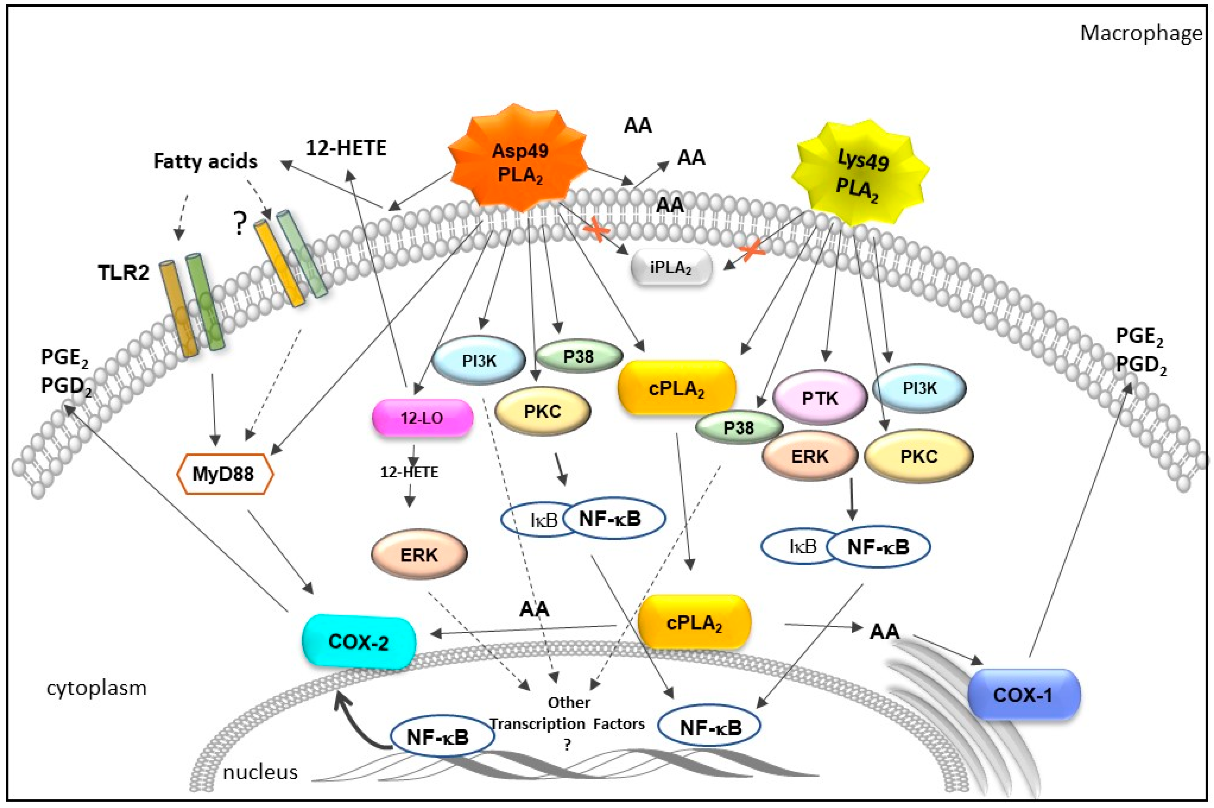
2.2. Influence of Bothrops svPLA2s on Pathways of Arachidonic Acid Metabolism

It is well established that sPLA2s play key modulatory roles in numerous cellular processes in physiological and pathological conditions by regulating the release of AA from membrane phospholipids [27,171]. It has long been recognized that the AA-derived lipid mediators are potent mediators of inflammation [83]. The AA is rapidly metabolized by several enzyme complexes, including cyclooxygenases (COX), lipoxygenases (LOX), and cytochrome P450 (CYP450). These enzymatic pathways promote the synthesis of oxygenated and bioactive products, generically called eicosanoids, which include prostaglandins (PG), leukotrienes (LT), hydroperoxyeicosatetraenoic acids (HPETEs), hydroxyeicosatetraenoic acids (HETEs), epoxides (EETs), and lipoxins (LX) [172,173,174,175,176,177,178]. A summary of the cascades involved in biosynthesis of eicosanoids is shown in Figure 2.
It is important to emphasize that COX-1 is a constitutive isoform present in most tissues and is responsible for generating PGs for diverse physiological functions [179,180,181,182]. In contrast, COX-2 is upregulated by inflammatory cytokines and growth factors [183,184] and is constitutively expressed in some tissues [185,186].
Regarding the production of inflammatory eicosanoids by Bothrops svPLA2, studies have demonstrated that the intraperitoneal injection of MT-III [187] and MT-II [162] in mice induced an early and transient release of PGD2, followed by a rapid and sustained release of PGE2. Likewise, in mice injected with BatroxPLA2 [140], from B. atrox snake venom, and BJ-PLA2-I from B. jararaca [141], an early release of PGE2 was observed. The in vivo experimental models of previous studies have revealed that B. asper sPLA2s induce the release of other eicosanoids, such as thromboxane A2 (TXA2) and LTB4 [123]. Moreover, an Asp49 svPLA2 from B. atrox venom [140] stimulated the production of LTB4, lipoxin, and PGE2.
PGE2 and PGD2 are important modulators of vasodilation, and PGE2 can potentiate an increase in vascular permeability, promoted by mediators of this phenomenon, with a consequent formation of edema [188,189]. Studies using pharmacological treatment with non-steroidal anti-inflammatory compounds were crucial in demonstrating the participation of these COXs-derived lipid mediators on edema [126,190] and hyperalgesia [135], induced by B. asper sPLA2s. In addition, studies demonstrating that MT-III and MT-II upregulated COX-2 protein expression in peritoneal leukocytes without altering the constitutive expression of COX-1 evidenced the ability of these venom PLA2s to influence downstream cyclooxygenase isozymes and suggested this as a mechanism by which these sPLA2s induced the production of prostaglandins [162,163]. Moreover, these findings suggested that the catalytic activity of these bothropic PLA2s did not contribute to the induction of PG biosynthesis, since MT-II, devoid of catalytic activity, caused the same effect.
In this regard, studies have demonstrated that the IκB phosphorylation inhibitor TPCK effectively prevented both MT-II- or MT-III-induced COX-2 expression, suggesting that the activation of NF-κB was critical for the induction of COX-2 expression by these bothropic svPLA2s. The involvement of NF-κB as the mechanism underlying this venom sPLA2s-induced upregulation of COX-2 expression was further confirmed by results that revealed the inhibition of the NF-κB nuclear translocation site, markedly reduced svPLA2s-induced COX-2 expression and, as a consequence, reduced PGE2 production by macrophages in culture [162,164].
Studies employing mouse resident peritoneal macrophages or neutrophils in culture revealed that viperids sPLA2s induced a marked release of PGE2 in cell supernatants, accompanied by the release of AA [162,165,166,191]. These data support the results in vivo and serve as evidence that immune innate leukocytes, such as resident macrophages and neutrophils, are important sources of PGs under in vivo stimuli by sPLA2 from Bothrops spp. snake venoms. Interestingly, studies have demonstrated that the incubation of resident peritoneal macrophages with MT-II or MT-III significantly increased the concentration of AA [162,164]. Although the release of AA induced by the Asp49 sPLA2 was approximately 20 times greater than that induced by MT-II, it demonstrated that the catalytic activity of viperid sPLA2s was not an essential requirement for inducing COX-2 expression and PGE2 production. According to Kini and Evans (1995) [192], in mechanisms independent on catalytic activity, as in the case of MT-II, the interaction of sPLA2s to acceptor regions can cause the biological effect or interfere with the interaction of target proteins with their physiological ligands. Furthermore, some effects may result from combinations of both enzymatic and non-enzymatic mechanisms [192], leading to the activation of several signaling pathways; this should be considered when interpreting the effects of group IIA Lys49 svPLA2s. In this context, since crosstalk among sPLA2, cPLA2, and iPLA2s has been demonstrated to occur in several physiological and inflammatory conditions [193,194,195,196], the contribution of prey/victim cPLA2 and/or iPLA2 to the increased production of PGs and upregulation of COX-2 protein expression induced by svPLA2 variants was evaluated in diverse in vitro experimental models [162,165,187]. Thus, the pharmacological treatment of cells with the cPLA2 inhibitor but not the iPLA2 inhibitor decreased the release of AA and the production of PGE2 and PGD2 induced by svPLA2. In contrast, these pretreatments did not modify the MT-III-induced COX-2 expression but reduced the COX-2 expression induced by MT-II. These results demonstrate that cPLA2 is required for distinct actions of MT-II in the PG biosynthetic pathway in macrophages [162,187]. This is consistent with the reported functional cooperation between intracellular PLA2s and GIIA sPLA2 for PG biosynthetic responses in several other cell systems [171,197,198,199]. The role of cPLA2 as a key enzyme in supplying AA for COX-2-dependent PGs production is well established [171,200,201]. Taken together, the available data demonstrate that the Asp49 svPLA2s are functionally coupled with cPLA2, since prior activation of cPLA2 is required for MT-III to act with downstream enzymes for PG biosynthesis in macrophages and neutrophils [165,187]. Interestingly, the association of Lys49 sPLA2 with cPLA2, in addition to being important for the supply of AA for the production of PG, appears to modulate the transcription and protein expression of COX-2 inflammatory isoform. The mechanisms involved in the coupling between the venom GIIA sPLA2s and mammalian cPLA2 have yet to be investigated. One possibility is that GIIA svPLA2s activate cPLA2 by distinct signaling cascades that mimic the transducing mechanism conveyed by physiological activators of cPLA2, such as MAPKs, since this enzyme family is likewise important for the activation of NF-κB [202]. In line with this concept, the Asp49 svPLA2, MT-III, in addition to Lys49 PLA2, MT-II, from B. asper snake venom, were revealed to stimulate the phosphorylation of protein kinases, including the MAPKs, such as p38MAPK and ERK1/2 [167,169,171]. Moreover, other protein kinases, including PI3K, PKC, and PTK, were reported to be phosphorylated in macrophages stimulated by both MT-III and MT-II [162,164]. In this regard, studies have demonstrated that MT-III (Asp49 sPLA2) stimulates PKC and p38MAPK pathways to positively modulate PGE2 production and COX-2 expression via NF-κB, while MT-II (Lys49 PLA2) displays similar effects by activating PKC, ERK1/2, and PTK in murine peritoneal macrophages [167,169,171]. Since PTK is involved in the activation of MAPKs, which, in turn, are essential for cPLA2 activation, this signaling protein might be involved in the activation of cPLA2 by MT-II [202,203,204]. Furthermore, another pathway implicated in the release of PGE2 and the expression of COX-2 induced by MT-III was demonstrated to be independent of NF-κB activation. This pathway involved the activation of ERK1/2 by the 12-HETE pathway, the main product of 12-LO [166]. The involvement of another transcription factor in these MT-III-induced effects was suggested by the authors. Together, these findings reveal the variety and complexity of the mechanisms involved in the effects of svPLA2 leading to the generation of lipid mediators. The signaling molecules and pathways acting in an innate immune cell (macrophage) upon stimulus either by Asp49 or Lys49 svPLA2 are summarized in Figure 3.
Although the participation of the M-type PLA2 receptor or another type of interaction of Bothrops sPLA2 with membrane sites for the stimulation of signal transduction pathways has not yet been demonstrated, a study revealed for the first time the involvement of the TLRs in the inflammatory response induced by MT-III (Asp49-PLA2) from B. asper in macrophages [205]. The involvement of TLR2 and MyD88 adapter molecules was demonstrated to be critical in producing PGs, COX-2 protein expression, and cytokines IL-1β and IL-10 induced by this svPLA2. An indirect mechanism for the activation of TLRs through the release of DAMPs was suggested by the authors, since the analysis of the fatty acids released by the hydrolysis of membrane phospholipids by MT-III revealed high levels of oleic and palmitic acids. In this context, it is known that arachidonic, oleic, and palmitic acids produced by membrane cleavage by sPLA2s are important bioactive mediators involved in the induction and release of COX-2 and PGE2, respectively, through the activation of intracellular signaling mechanisms in several cell types [206,207,208,209].

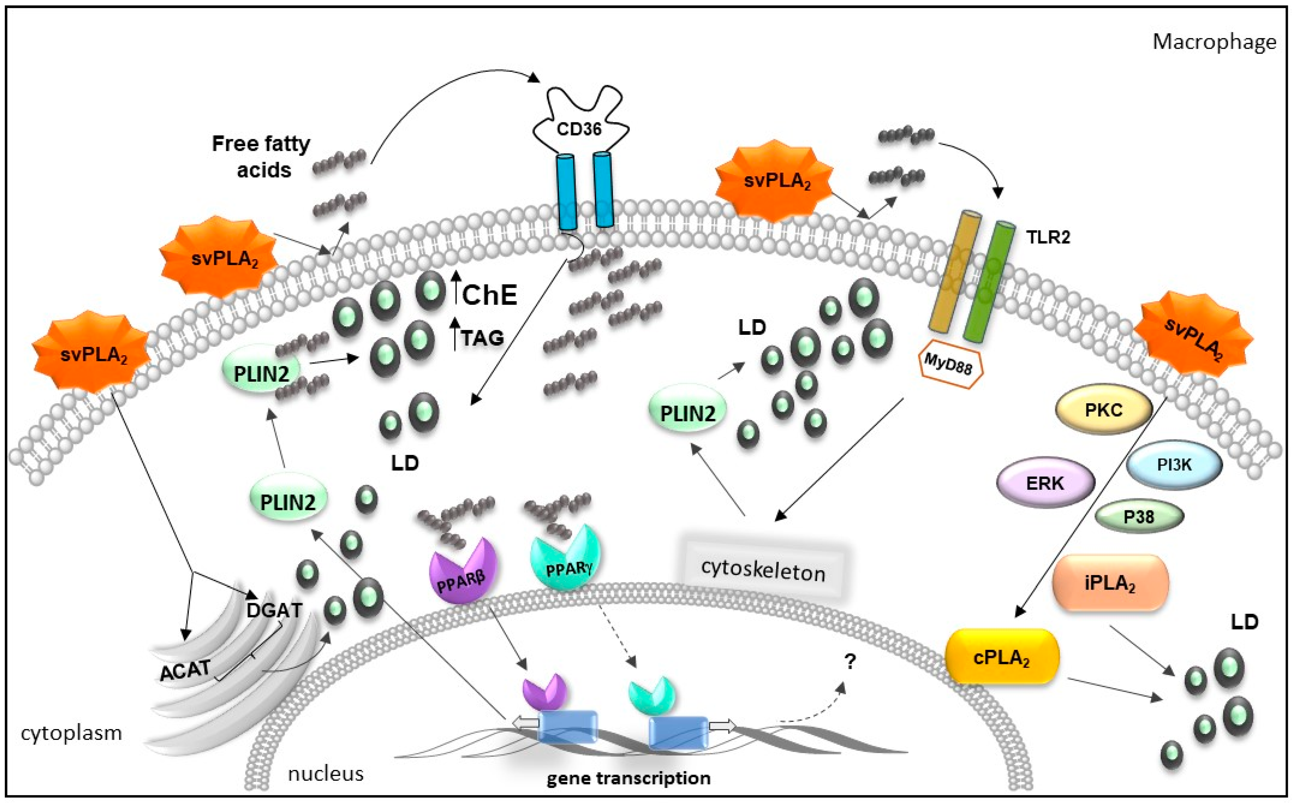
2.3. Bothrops svPLA2s Trigger Lipid Accumulation in Immunocompetent Cells

As mentioned, the high enzymatic activity exhibited by ophidian sPLA2 provides a microenvironment rich in free fatty acids that exert stimulating effects in immunocompetent cells, leading to the biosynthesis of lipid mediators. Moreover, excess free fatty acids exert cytotoxic effects that trigger the activation of cellular mechanisms capable of converting free fatty acids into metabolites of lower toxicity, known as neutral lipids (triacylglycerol and cholesterol—energetic body reserve) [210]. In recent decades, numerous studies have demonstrated the activation of intracellular metabolic pathways responsible for the metabolization of free fatty acids in neutral lipids and the consequent formation of dynamic organelles called lipid droplets (LDs) [211,212]. These organelles are composed of a hydrophobic neutral lipid core surrounded by a phospholipid monolayer membrane, which contains numerous proteins related to cellular activation in addition to structural proteins, such as perilipin 2 (PLIN2), which plays an important role in LD assembly and the formation of foam macrophage [213], marker cells in metabolic diseases, such as atherosclerosis and obesity [214]. LDs are commonly present in adipocytes due to the already-established role of these cells in the supply of energy in mammalian organisms [215]. In addition to the relevance regarding lipid homeostasis, the direct relationship between increased LD formation and inflammatory processes was evidenced by numerous studies [216,217,218]. In this sense, mammalian group IIA sPLA2s have been identified as potential plasma biomarkers for diseases related to lipid imbalance, such as atherosclerotic cardiovascular disease and obesity [219]. Alongside a marked inflammatory reaction, these metabolic diseases are characterized by lipid accumulation in immunocompetent cells [220,221]. In line with the ability of svPLA2s to elicit an inflammatory response characterized by a high level of inflammatory mediators and free fatty acids, it was demonstrated that MT-III (Asp49 PLA2), isolated from B. asper venom, induced LD formation enriched by PLIN2 protein in mice peritoneal macrophages [170]. This effect was likewise observed in rat vascular smooth muscle cells isolated from the thoracic aorta stimulated by MT-III [168]. Moreover, the ability of MT-II, a Lys49 PLA2 homologue devoid of catalytic activity from B. asper venom, to directly activate macrophages to form LDs was reported [167]. This effect was reproduced by a synthetic peptide corresponding to the C-terminal sequence 115–129 of MT-II, evidencing the critical role of C-terminus for the MT-II-induced effect [167]. Similarly, BaTX-I, a catalytically inactive Lys49 variant; BaTX-II, a catalytically active Asp49; and BaPLA2, an acidic catalytically active Asp49 PLA2, isolated from B. atrox snake venom, increased the number of LDs on murine macrophages cell line J774A.1 [152]. The formation of LDs upon stimulus by MT-III was likewise demonstrated in human monocytes of peripheral blood [222].
Considering the above information, LD formation induced by svPLA2s in phagocytes proved to be inherent to the action of sPLA2s regardless of the catalytic activity. The capability of Bothrops svPLA2s to induce the formation of LDs is related to the activation of PRRs of an innate immune response, kinase proteins, and intracellular PLA2 signaling pathways involved in cellular metabolism, proliferation, and differentiation [170,205]. Hence, by using gene knockout mice cells and a pharmacological approach, the participation of TLR2, MYD 88 adaptor protein, and CD36 in LD formation in macrophages in culture has been reported [170,205]. In addition, the upregulation of CD36 receptors was observed in these cells. Considering the participation of the MYD 88 adaptor molecule, the involvement of other TLRs has yet to be investigated. In line with the reported Asp49 svPLA2 action on PRRs, the upregulation of both SRA-1, from the scavenger receptor family, and LOX-1, an LDL receptor, was demonstrated in mouse aortic smooth muscle cells (VSMCs) [168]. These findings demonstrate that the inflammatory response elicited by Asp49 svPLA2 likewise involved the upregulation of PRRs associated with lipid uptake in immunocompetent cells. This fact indicates that svPLA2s can be useful tools in studies aiming to understand the diseases associated with lipid imbalance.
In addition to providing the synthesis of mediators, TLR2 activation elicited by MT-III action was related to cytoskeleton activation [205], a critical step in the transport of structural proteins into LDs, such as PLIN2. Cytoskeletal activation involves the activation of kinase proteins [223]. Consistent with this information, the participation of kinase proteins in LD formation induced by both Asp49 and Lys49 svPLA2s from B. asper venom has been demonstrated through a pharmacological approach and the detection of phosphorylated kinase proteins. Yet, MT-II- and MT-III-induced LD biogenesis is dependent on the activation of PKC, PI3K, p38MAPK, and ERK1/2 signaling pathways in mice peritoneal macrophages [224]. It is well known that PKC regulates a variety of processes associated with lipid droplet biology, such as adipocyte differentiation [225], magnolol-induced lipolysis [226], cholesterol-induced targeting of caveolin to lipid droplets [227], and the expression of the PAT family [228]. Hence, the activation of the PKC signaling pathway in macrophages stimulated by the Bothrops PLA2s MT-II and MT-III in peritoneal macrophages may be implicated in an increase in PLIN2 protein expression, since LD formation induced by both svPLA2s has always been accompanied by an increase in PLIN2 protein expression in macrophages.
PI3K/AKT is a classical pathway involved in insulin resistance, cell growth, and lipid metabolism associated with the inhibition of cholesterol efflux leading to LD formation [229]. In this sense, the participation of the PIK3 signaling pathway in MT-III-induced LDs in macrophages and vascular smooth muscle cells stimulated with MT-III has been demonstrated [167,224]. In the case of vascular smooth muscle cells, the activation of PI3K was related to the uptake of fatty acids to LDs by macropinocytosis [167]. Furthermore, the findings that MT-III increases phagocytic activity and upregulates macrophage markers in VSMCs reinforce the importance of this class of enzymes as inducers of factors implicated in the formation of foamy cells in both mononuclear phagocytic cells and VSMCs, which are key elements in the development of metabolic diseases.
The MAPK signaling pathway has been revealed to mediate the activation of intracellular PLA2s in physiologic and inflammatory contexts [230,231]. It has been demonstrated that the ERK1/2 signaling pathway is implicated in LD formation via the activation of phospholipase D (PLD) and phosphorylation of dynein [232,233]. Consistent with this information, the critical role of ERK1/2 for LD formation induced by MT-II and MT-III in macrophages and VSMCs was reported [167,224]. Regarding p38 MAPK, the literature evidenced its importance in the development of atherosclerosis [234] and the apoptosis of foam macrophages. Macrophage death is a feature of atherosclerotic plaque linked to necrosis and plaque destabilization [235]. Interestingly, although MT-III has been demonstrated to activate apoptotic pathways, including the p38 MAPK signaling pathway, DGAT, ACAT, cPLA2, and LD formation in macrophages, no change in cell viability was observed. Further studies may clarify this lack of apoptotic effect.
It is known that the MAP kinases signaling pathway is implicated in intracellular PLA2 activation, including the Ca2+-dependent cytosolic group IVA PLA2 (cPLA) and the Ca2+-independent group VIA PLA2 (iPLA2) involved in both physiological and pathophysiological conditions [236,237]. The biogenesis of LDs in CHO-K1 cells submitted to an enriched environment of fatty acids demonstrated ERK, p38, and JNK signaling pathway activation, with JNK cascade being responsible for cPLA phosphorylation in this event. Of note, cPLA2 was likewise implicated in LD biogenesis stimulated by MT-III associated with the activation of ERK and the p38 MAPK signaling pathway [224]. Considering the ability of cPLA2 to mobilize cell membrane fatty acids [238], the aforementioned activation should amplify the action of MT-III and provide a greater substrate for the metabolization and formation of LDs. Moreover, the biogenesis of LDs induced by the svPLA2s MT-III and MT-II is dependent on the activation of iPLA2 signaling pathways [167,224]. This signaling pathway was associated with the processing of fatty acids into triacylglycerol, a relevant component in the constitution of LDs [214]. Hence, the crosstalk already evidenced between intracellular PLA2 and svPLA2 to elicit inflammatory conditions [169] might contribute to elucidating mechanisms related to the formation of LDs.
The peroxisome proliferator-activated receptors (PPARs) are transcription factors belonging to the family of nuclear receptors that regulate glucose homeostasis, inflammation, and lipid metabolism. Three proteins, encoded by distinct genes, have been identified: PPAR-α, PPAR-β/δ, and PPAR-γ, which control gene expression by binding to PPREs in the promoters [239]. The activation of PPARs is a tightly regulated process implicated in the control of lipid homeostasis, which involves the biogenesis of LDs and protein expression involved in lipid uptake, including PRRs and structural protein PLIN2, and enzymes implicated in neutral lipid synthesis (triacylglycerol and cholesterol) [240]. PPARs have been demonstrated to increase in foam macrophages [241]. In this sense, it was revealed that MT-III induced the upregulation of the transcription factors PPAR-γ and PPAR-β/δ, in addition to the translocation of these factors to the nucleus of mouse peritoneal macrophages. The pharmacological blockage of the PPAR-β/δ transcription factor abolished the increase in PLIN2 and CD36 protein expression induced by MT-III. Moreover, the PPAR-γ blockage caused a reduction in LD formation and abolished CD36 receptor protein expression induced by MT-III. Since an increased expression of CD36 and PLIN2 is related to macrophage differentiation into foam cells [242,243], these findings suggest that MT-III induces foam cell formation by this route. In addition, MT-III caused an increase in the levels of triacylglycerol and cholesterol due to the uptake of free fatty acids. These effects were mediated by DGAT and ACAT enzymes, which are involved in the synthesis of triacylglycerol and cholesterol, respectively [244]. In agreement with this study, a significant increase in triacylglycerol and cholesterol levels was observed in human monocytes under MT-III stimulation [222]. This effect was dependent on fatty acid reacylation. Moreover, the fatty acid composition of triacylglycerol and cholesterol induced by MT-III was compatible with fatty acids released by the enzymatic action of this svPLA2 on cell membranes. According to the above information, the mechanisms triggered by MT-III, in both mice peritoneal macrophages and human monocytes, align with macrophages differentiation into foamy cells, a cell type characteristic of inflammatory metabolic diseases, such as atherosclerosis [245]. Similarly, MT-III could stimulate LD formation in VSCMs. This lipid accumulation was likewise mediated by the activation of transcription factors PPAR-γ and PPAR-β/δ and DGAT and ACAT enzymes. Moreover, it is noteworthy that VSCMs under stimulation of MT-III exhibited an increase in the protein levels of PRRs, SRA-1 (scavenger receptor type 1), and LOX-1 (lectin-like oxidized low-density lipoprotein receptor-1). Interestingly, the blockage of these receptors did not alter the formation of LDs induced by MT-III, but the upregulation of LOX-1 was associated with an increased uptake of acetylated-low density lipoprotein (acLDL) in VSMCs stimulated by this svPLA2. This higher uptake of acLDL by VSMCs identifies new pathways involved in the accumulation of lipids triggered by a sPLA2 that is not directly linked to the reacylation of free fatty acids. In addition, lipid accumulation induced by MT-III in VSCMs was related to the expression of ATP-binding cassette transporters ABCA1 and ABCG1, responsible for the efflux of cholesterol of macrophage-derived foam cells [242]. Although the signaling pathway by which MT-III induces an increased expression of the factors implicated in lipid homeostasis has not been fully elucidated, these studies have broadened the knowledge about the actions of svPLA2s on the formation of LDs and the synthesis of lipid mediators and provided new insights into the actions of group IIA sPLA2s in diseases related to lipid imbalance. The pathways and factors involved in lipid accumulation in an innate immune cell (macrophage) upon stimulus by svPLA2s are summarized in Figure 4.
Another aspect related to the metabolism of free fatty acids is the biosynthesis of lipid mediators [246,247,248]. On the one hand, it is known that the synthesis of eicosanoids is closely related to the triggering of the inflammatory process induced by svPLA2s, regardless of the catalytic activity on the cell membranes [13]. In this sense, the immunofluorescence approach has demonstrated that LDs stimulated by svPLA2s synthesize PGE2 [167,249]. On the other hand, some eicosanoids are implicated in the resolution of inflammatory processes, such as PGJ2. This mediator was co-located in LDs in macrophage peritoneal mice stimulated by MT-II [161], indicating, for the first time, that the LDs not only are related to the production of inflammatory mediators but also might play a role in regulating this process.
Adipose tissue is the principal organ responsible for balancing energy metabolism in the mammalian body. An imbalance in adipose tissue functions is linked to the triggering of the inflammatory process observed in metabolic diseases, including obesity [250,251]. Recently, it has been demonstrated that MT-III activated proinflammatory mechanisms in 3T3-L1 preadipocytes, including the biosynthesis of PGE2 and PGI2, lipid mediators implicated in preadipocytes differentiation into mature adipocytes, and IL-6 and MCP-1 [169]. In these cells, PGE2 production induced by MT-III was dependent on cPLA2 activation, the upregulation of COX-2 and mPGES-1, and the engagement of the PGE2 EP4 receptor. In addition, the release of IL-6 and MCP-1 was dependent on EP4 or EP3 activation, respectively. These data indicate that the production of PGE2 is critical for the activation of proinflammatory pathways associated with cytokine production in preadipocytes stimulated by this svPLA2. Furthermore, MT-III upregulated the gene expression of the adipokines leptin and adiponectin in preadipocytes [169]. These mediators have been described as regulating appetite and satiety, glucose and lipid metabolism, inflammation, and immune functions [252,253,254,255]. Although the mechanisms related to the release of adipokines and the ability of MT-III to induce lipid accumulation in adipocytes have not yet been investigated, these data offer new directions for investigating the actions triggered by svPLA2s and mammalian GIIA sPLA2s.

3. Conclusions

The existing literature demonstrates that the svPLA2s trigger a cascade of inflammatory events including edema formation, leukocyte recruitment into tissues, release of a complex network of inflammatory mediators, and increased oxidative stress in experimental animal models that mimic the inflammatory responses elicited by viperid snake venoms, especially those from the Bothrops genus, in the victims. The catalytic activity of the svPLA2s is not strictly required by these proteins for the triggering of all the inflammatory responses, since the catalytically inactive Lys49 PLA2 variants can display inflammatory events that are qualitatively similar to those of Asp49 PLA2s. In addition to cell migration, the svPLA2s can activate distinct functions of immunocompetent cells that include phagocytosis, the respiratory burst, NET formation, production of cytokines, chemokines and multiple reactive cleavage products such as lysophospholipids, polyunsaturated fatty acids and eicosanoids, as well as formation of LDs. The highly complex network of mediators, particularly lipid mediators, modulates a variety of inflammatory events triggered by this class of snake venom toxins. The effects triggered by svPLA2s in inflammatory cells that lead to generation of lipid mediators has been associated with the activation of distinct signaling pathways of inflammatory kinase proteins by mechanisms dependent and independent of NF-kB. Moreover, the inflammatory response elicited by svPLA2s in leukocytes also involves upregulation of PRRs of innate immune response, the crosstalk between the svPLA2 and intracellular PLA2s, and upregulation of factors implicated in lipid homeostasis. Although much has been learned regarding the inflammatory actions of svPLA2s, many knowledge gaps still exist and need to be addressed. There is still considerable work to be done before we fully understand the complex interactions that occur among svPLA2s and immunocompetent cells and tissues that lead to inflammation. The cell acceptors and/or receptors involved in the actions of svPLA2s in these cells and the signaling pathways elicited and how they interact with each other remain to be clarified. In addition, the actual types and subtypes of receptors activated by the principal mediators produced by svPLA2s and the mechanisms involved in coupling between the svPLA2s and endogenous PLA2s have yet to be investigated. Recently, the stimulatory activity of a svPLA2 on adipose tissue cells leading to increased biosynthesis of PGE2 and other inflammatory mediators including adipokines was demonstrated. This information offers new directions for investigating the actions triggered by svPLA2s and mammalian GIIA sPLA2 and gives insights into the potential role of the adipocytes as target cells for viperid snake venoms. Finally, a deeper and comprehensive understanding of the mechanisms underlying the inflammatory actions of svPLA2s will give new insights into (i) the actions of group IIA sPLA2s in diseases related to lipid imbalance and inflammation and (ii) a better understanding of the pathophysiology of Bothrops envenomation. Within this frame, the acquired knowledge might pave the way for the development of novel therapeutic approaches aimed at counteracting the prominent inflammation caused by Bothrops snakebite envenoming.

Author Contributions

Conceptualization, C.T., V.M., E.L. and C.M.F.; writing—original draft, C.T., V.M., E.L., C.M.F., P.M.J. and R.M.-M.; writing—review and editing, C.T., V.M., E.L., C.M.F., P.M.J. and R.M.-M.; visualization, V.M. and R.M.-M.; supervision, C.T. All authors have read and agreed to the published version of the manuscript.

Funding

This research was funded by Fundação Butantan, São Paulo, Brazil. E.L. is a recipient of a postdoctural fellowship from Coordenação de Aperfeicçoamento de Pessoal de Nível Superior (CAPES). C.T. is a recipient of a Conselho Nacional de Desenvolvimento Científico e Tecnológico fellowship (PQ-CNPq), grant number 310930/201.7. R.M.M. is recipient of a doctoral fellowship from Fundação Butantan, grant FB 001/0708/003.434/2021.

Institutional Review Board Statement

Not applicable.

Informed Consent Statement

Not applicable.

Data Availability Statement

Not applicable.

Conflicts of Interest

The authors declare no conflict of interest.

References

  1. Teixeira, C.F.P.; Cury, Y.; Oga, S.; Jancar, S. Hyperalgesia induced by Bothrops jararaca venom in rats: Role of eicosanoids and platelet activating factor (PAF). Toxicon 1994, 32, 419–426. [Google Scholar] [CrossRef]
  2. Teixeira, C.; Cury, Y.; Moreira, V.; Picolo, G.; Chaves, F. Inflammation induced by Bothrops asper venom. Toxicon 2009, 54, 67–76. [Google Scholar] [CrossRef]
  3. Mamede, C.C.N.; de Sousa Simamoto, B.B.; da Cunha Pereira, D.F.; de Oliveira Costa, J.; Ribeiro, M.S.M.; de Oliveira, F. Edema, hyperalgesia and myonecrosis induced by Brazilian bothropic venoms: Overview of the last decade. Toxicon 2020, 187, 10–18. [Google Scholar] [CrossRef] [PubMed]
  4. Gutiérrez, J.M.; Rucavado, A.; Chaves, F.; Díaz, C.; Escalante, T. Experimental pathology of local tissue damage induced by Bothrops asper snake venom. Toxicon 2009, 54, 958–975. [Google Scholar] [CrossRef] [PubMed]
  5. Ministério da Saúde Guia de Vigilância Epidemiológica; Ministério da Saúde: Brasília, Brazil, 2019; ISBN 9788533416321.
  6. Echeverría, S.; Leiguez, E.; Guijas, C.; do Nascimento, N.G.; Acosta, O.; Teixeira, C.; Leiva, L.C.; Rodríguez, J.P. Evaluation of pro-inflammatory events induced by Bothrops alternatus snake venom. Chem. Biol. Interact. 2018, 281, 24–31. [Google Scholar] [CrossRef] [PubMed]
  7. Jorge, R.J.B.; Monteiro, H.S.A.; Gonçalves-Machado, L.; Guarnieri, M.C.; Ximenes, R.M.; Borges-Nojosa, D.M.; Luna, K.P.D.O.; Zingali, R.B.; Corrêa-Netto, C.; Gutiérrez, J.M.; et al. Venomics and antivenomics of Bothrops erythromelas from five geographic populations within the Caatinga ecoregion of northeastern Brazil. J. Proteom. 2015, 114, 93–114. [Google Scholar] [CrossRef] [PubMed]
  8. Nicolau, C.A.; Carvalho, P.C.; Junqueira-de-Azevedo, I.L.M.; Teixeira-Ferreira, A.; Junqueira, M.; Perales, J.; Neves-Ferreira, A.G.C.; Valente, R.H. An in-depth snake venom proteopeptidome characterization: Benchmarking Bothrops jararaca. J. Proteom. 2017, 151, 214–231. [Google Scholar] [CrossRef]
  9. Sousa, L.F.; Portes-Junior, J.A.; Nicolau, C.A.; Bernardoni, J.L.; Nishiyama, M.Y., Jr.; Amazonas, D.R.; Freitas-de-Sousa, L.A.; Mourão, R.H.; Chalkidis, H.M.; Valente, R.H.; et al. Functional proteomic analyses of Bothrops atrox venom reveals phenotypes associated with habitat variation in the Amazon. J. Proteom. 2017, 159, 32–46. [Google Scholar] [CrossRef] [PubMed]
  10. De Farias, I.B.; de Morais-Zani, K.; Serino-Silva, C.; Sant’Anna, S.S.; da Rocha, M.M.; Grego, K.F.; Andrade-Silva, D.; Serrano, S.M.T.; Tanaka-Azevedo, A.M. Functional and proteomic comparison of Bothrops jararaca venom from captive specimens and the Brazilian Bothropic Reference Venom. J. Proteom. 2018, 174, 36–46. [Google Scholar] [CrossRef] [PubMed]
  11. Mora-Obando, D.; Salazar-Valenzuela, D.; Pla, D.; Lomonte, B.; Guerrero-Vargas, J.A.; Ayerbe, S.; Gibbs, H.L.; Calvete, J.J. Venom variation in Bothrops asper lineages from North-Western South America. J. Proteom. 2020, 229, 103945. [Google Scholar] [CrossRef] [PubMed]
  12. Gutiérrez, J.M.; Lomonte, B. Phospholipases A2: Unveiling the secrets of a functionally versatile group of snake venom toxins. Toxicon 2013, 62, 27–39. [Google Scholar] [CrossRef] [PubMed]
  13. Bickler, P.E. Amplification of Snake Venom Toxicity by Endogenous Signaling Pathways. Toxins 2020, 12, 68. [Google Scholar] [CrossRef] [PubMed] [Green Version]
  14. Hiu, J.J.; Yap, M.K.K. Cytotoxicity of snake venom enzymatic toxins: Phospholipase A2 and L-amino acid oxidase. Biochem. Soc. Trans. 2020, 48, 719–731. [Google Scholar] [CrossRef] [PubMed] [Green Version]
  15. Gutiérrez, J.M.; Chaves, F.; Bolaños, R.; Cerdas, L.; Rojas, E.; Arroyo, O.; Portilla, E. Neutralizacion de los efectos locales del veneno de Bothrops asper por un antiveneno polivalente. Toxicon 1981, 19, 493–500. [Google Scholar] [CrossRef]
  16. Gutiérrez, J.M.; Rojas, G.; Bogarín, G.; Lomonte, B. Evaluation of the neutralizing ability of antivenoms for the treatment of snake bite envenoming in Central America. Envenomings Treat. 1996, 223–231. [Google Scholar]
  17. Gutiérrez, J.; Avila, C.; Rojas, E.; Cerdas, L. An alternative in vitro method for testing the potency of the polyvalent antivenom produced in Costa Rica. Toxicon 1988, 26, 411–413. [Google Scholar] [CrossRef]
  18. Cardoso, J.L.; Fan, H.W.; França, F.O.; Jorge, M.T.; Leite, R.P.; Nishioka, S.A.; Avila, A.; Sano-Martins, I.S.; Tomy, S.C.; Santoro, M.L. Randomized comparative trial of three antivenoms in the treatment of envenoming by lance-headed vipers (Bothrops jararaca) in São Paulo, Brazil. Q. J. Med. 1993, 86, 315–325. [Google Scholar] [PubMed]
  19. Lewin, M.; Samuel, S.; Merkel, J.; Bickler, P. Varespladib (LY315920) appears to be a potent, broad-spectrum, inhibitor of snake venom phospholipase A2 and a possible pre-referral treatment for envenomation. Toxins 2016, 8, 248. [Google Scholar] [CrossRef] [Green Version]
  20. Wang, Y.; Zhang, J.; Zhang, D.; Xiao, H.; Xiong, S.; Huang, C. Exploration of the inhibitory potential of varespladib for snakebite envenomation. Molecules 2018, 23, 391. [Google Scholar] [CrossRef] [PubMed] [Green Version]
  21. Schaloske, R.H.; Dennis, E.A. The phospholipase A2 superfamily and its group numbering system. Biochim. Biophys. Acta-Mol. Cell Biol. Lipids 2006, 1761, 1246–1259. [Google Scholar] [CrossRef] [PubMed]
  22. Six, D.A.; Dennis, E.A. The expanding superfamily of phospholipase A2 enzymes: Classification and characterization. Biochim. Biophys. Acta-Mol. Cell Biol. Lipids 2000, 1488, 1–19. [Google Scholar] [CrossRef]
  23. Dennis, E.A.; Cao, J.; Hsu, Y.H.; Magrioti, V.; Kokotos, G. Phospholipase A2 enzymes: Physical structure, biological function, disease implication, chemical inhibition, and therapeutic intervention. Chem. Rev. 2011, 111, 6130–6185. [Google Scholar] [CrossRef] [Green Version]
  24. Murakami, M.; Sato, H.; Taketomi, Y. Updating Phospholipase A2 Biology. Biomolecules 2020, 10, 1457. [Google Scholar] [CrossRef] [PubMed]
  25. Lee, G.-H.; Fujita, M.; Nakanishi, H.; Miyata, H.; Ikawa, M.; Maeda, Y.; Murakami, Y.; Kinoshita, T. PGAP6, a GPI-specific phospholipase A2, has narrow substrate specificity against GPI-anchored proteins. J. Biol. Chem. 2020, 295, 14501–14509. [Google Scholar] [CrossRef]
  26. Aloulou, A.; Rahier, R.; Arhab, Y.; Noiriel, A.; Abousalham, A. Phospholipases: An Overview. In Methods in Molecular Biology; Sandoval, G., Ed.; Springer: New York, NY, USA, 2018; Volume 1835, pp. 69–105. ISBN 978-1-4939-8671-2. [Google Scholar]
  27. Quach, N.D.; Arnold, R.D.; Cummings, B.S. Secretory phospholipase A2 enzymes as pharmacological targets for treatment of disease. Biochem. Pharmacol. 2014, 90, 338–348. [Google Scholar] [CrossRef] [Green Version]
  28. Harris, J.B.; Scott-Davey, T. Secreted phospholipases A2 of snake venoms: Effects on the peripheral neuromuscular system with comments on the role of phospholipases A2 in disorders of the CNS and their uses in industry. Toxins 2013, 5, 2533–2571. [Google Scholar] [CrossRef] [PubMed] [Green Version]
  29. Triggiani, M.; Granata, F.; Frattini, A.; Marone, G. Activation of human inflammatory cells by secreted phospholipases A2. Biochim. Biophys. Acta Mol. Cell Biol. Lipids 2006, 1761, 1289–1300. [Google Scholar] [CrossRef] [PubMed]
  30. Valentin, E.; Lambeau, G. What can venom phospholipases A2 tell us about the functional diversity of mammalian secreted phospholipases A2? Biochimie 2000, 82, 815–831. [Google Scholar] [CrossRef]
  31. Kudo, I.; Murakami, M. Phospholipase A2 enzymes. Prostaglandins Other Lipid Mediat. 2002, 68–69, 3–58. [Google Scholar] [CrossRef]
  32. Kini, R.M. Excitement ahead: Structure, function and mechanism of snake venom phospholipase A2 enzymes. Toxicon 2003, 42, 827–840. [Google Scholar] [CrossRef] [PubMed]
  33. Lambeau, G.; Barhanin, J.; Schweitz, H.; Qar, J.; Lazdunski, M. Identification and properties of very high affinity brain membrane-binding sites for a neurotoxic phospholipase from the taipan venom. J. Biol. Chem. 1989, 264, 11503–11510. [Google Scholar] [CrossRef]
  34. Lambeau, G.; Schmid-Alliana, A.; Lazdunski, M.; Barhanin, J. Identification and purification of a very high affinity binding protein for toxic phospholipases A2 in skeletal muscle. J. Biol. Chem. 1990, 265, 9526–9532. [Google Scholar] [CrossRef]
  35. Silliman, C.C.; Moore, E.E.; Zallen, G.; Gonzalez, R.; Johnson, J.L.; Elzi, D.J.; Meng, X.; Hanasaki, K.; Ishizaki, J.; Arita, H.; et al. Presence of the M-type sPLA 2 receptor on neutrophils and its role in elastase release and adhesion. Am. J. Physiol. Physiol. 2002, 283, C1102–C1113. [Google Scholar] [CrossRef] [PubMed] [Green Version]
  36. Granata, F.; Petraroli, A.; Boilard, E.; Bezzine, S.; Bollinger, J.; Del Vecchio, L.; Gelb, M.H.; Lambeau, G.; Marone, G.; Triggiani, M. Activation of Cytokine Production by Secreted Phospholipase A 2 in Human Lung Macrophages Expressing the M-Type Receptor. J. Immunol. 2005, 174, 464–474. [Google Scholar] [CrossRef] [Green Version]
  37. Hanasaki, K.; Arita, H. Phospholipase A2 receptor: A regulator of biological functions of secretory phospholipase A2. Prostaglandins Other Lipid Mediat. 2002, 68–69, 71–82. [Google Scholar] [CrossRef]
  38. Ezekowitz, R.A.B.; Stahl, P.D. The structure and function of vertebrate mannose lectin-like proteins. J. Cell Sci. 1988, 1988, 121–133. [Google Scholar] [CrossRef] [Green Version]
  39. Taylor, M.E.; Conary, J.T.; Lennartz, M.R.; Stahl, P.D.; Drickamer, K. Primary structure of the mannose receptor contains multiple motifs resembling carbohydrate-recognition domains. J. Biol. Chem. 1990, 265, 12156–12162. [Google Scholar] [CrossRef]
  40. Gantzel, R.H.; Kjær, M.B.; Laursen, T.L.; Kazankov, K.; George, J.; Møller, H.J.; Grønbæk, H. Macrophage Activation Markers, Soluble CD163 and Mannose Receptor, in Liver Fibrosis. Front. Med. 2021, 7, 615599. [Google Scholar] [CrossRef]
  41. Gordon, S.; Clarke, S.; Greaves, D.; Doyle, A. Molecular immunobiology of macrophages: Recent progress. Curr. Opin. Immunol. 1995, 7, 24–33. [Google Scholar] [CrossRef]
  42. Stoy, N. Macrophage Biology and Pathobiology in the Evolution of Immune Responses: A Functional Analysis. Pathobiology 2001, 69, 179–211. [Google Scholar] [CrossRef]
  43. Hernández, M.; Burillo, S.L.; Crespo, M.S.; Nieto, M.L. Secretory Phospholipase A2 Activates the Cascade of Mitogen-activated Protein Kinases and Cytosolic Phospholipase A2 in the Human Astrocytoma Cell Line 1321N1. J. Biol. Chem. 1998, 273, 606–612. [Google Scholar] [CrossRef] [PubMed] [Green Version]
  44. Kinoshita, E.; Handa, N.; Hanada, K.; Kajiyama, G.; Sugiyama, M. Activation of MAP kinase cascade induced by human pancreatic phospholipase A2 in a human pancreatic cancer cell line. FEBS Lett. 1997, 407, 343–346. [Google Scholar] [CrossRef] [Green Version]
  45. Kundu, G.C.; Mukherjee, A.B. Evidence that porcine pancreatic phospholipase A2 via its high affinity receptor stimulates extracellular matrix invasion by normal and cancer cells. J. Biol. Chem. 1997, 272, 2346–2353. [Google Scholar] [CrossRef] [Green Version]
  46. Triggiani, M.; Calabrese, C.; Granata, F.; Gentile, M.; Marone, G. Metabolism of Lipid Mediators in Human Eosinophils. In Human Eosinophils; KARGER: Basel, Switzerland, 2000; pp. 77–98. [Google Scholar]
  47. Hara, S.; Kudo, I.; Matsuta, K.; Miyamoto, T.; Inoue, K. Amino Acid Composition and NH2-Terminal Amino Acid Sequence of Human Phospholipase A2 Purified from Rheumatoid Synovial Fluid1. J. Biochem. 1988, 104, 326–328. [Google Scholar] [CrossRef] [PubMed]
  48. Touqui, L.; Alaoui-El-Azher, M. Mammalian Secreted Phospholipases A2 and Their Pathophysiolo-gical Significance in Inflammatory Diseases. Curr. Mol. Med. 2001, 1, 739–754. [Google Scholar] [CrossRef] [PubMed]
  49. Liu, N.J.; Chapman, R.; Lin, Y.; Mmesi, J.; Bentham, A.; Tyreman, M.; Abraham, S.; Stevens, M.M. Point of care testing of phospholipase A2 group IIA for serological diagnosis of rheumatoid arthritis. Nanoscale 2016, 8, 4482–4485. [Google Scholar] [CrossRef] [PubMed]
  50. Feldman, M.; Ginsburg, I. A Novel Hypothetical Approach to Explain the Mechanisms of Pathogenicity of Rheumatic Arthritis. Mediterr. J. Rheumatol. 2021, 32, 112–117. [Google Scholar] [CrossRef] [PubMed]
  51. Aufenanger, J.; Samman, M.; Quintel, M.; Fassbender, K.; Zimmer, W.; Bertsch, T. Pancreatic phospholipase A2 activity in acute pancreatitis: A prognostic marker for early identification of patients at risk. Clin. Chem. Lab. Med. 2002, 40, 293–297. [Google Scholar] [CrossRef] [PubMed] [Green Version]
  52. Zhang, K.J.; Zhang, D.L.; Jiao, X.L.; Dong, C. Effect of phospholipase A2 silencing on acute experimental pancreatitis. Eur. Rev. Med. Pharmacol. Sci. 2013, 17, 3279–3284. [Google Scholar]
  53. Schröder, T.; Kivilaakso, E.; Kinnunen, P.K.J.; Lempinen, M. Serum Phospholipase A 2 in Human Acute Pancreatitis. Scand. J. Gastroenterol. 1980, 15, 633–636. [Google Scholar] [CrossRef]
  54. Vadas, P.; Pruzanski, W. Role of secretory phospholipases A2 in the pathobiology of disease. Lab. Investig. 1986, 55, 391–404. [Google Scholar] [PubMed]
  55. Ahmad, N.S.; Tan, T.L.; Arifin, K.T.; Ngah, W.Z.W.; Yusof, Y.A.M. High sPLA2-IIA level is associated with eicosanoid metabolism in patients with bacterial sepsis syndrome. PLoS ONE 2020, 15, e0230285. [Google Scholar] [CrossRef] [Green Version]
  56. Haapamäki, M.M.; Grönroos, J.M.; Nurmi, H.; Irjala, K.; Alanen, K.A.; Nevalainen, T.J. Phospholipase A2 in serum and colonic mucosa in ulcerative colitis. Scand. J. Clin. Lab. Investig. 1999, 59, 279–287. [Google Scholar] [CrossRef]
  57. Haapamäki, M.M.; Grönroos, J.M.; Nurmi, H.; Alanen, K.; Nevalainen, T.J. Gene Expression of Group Ii Phospholipase A2 in Intestine in Crohn’s Disease. Am. J. Gastroenterol. 1999, 94, 713–720. [Google Scholar] [CrossRef]
  58. Woodruff, T.M.; Arumugam, T.V.; Shiels, I.A.; Newman, M.L.; Ross, P.A.; Reid, R.C.; Fairlie, D.P.; Taylor, S.M. A potent and selective inhibitor of group IIa secretory phospholipase A2 protects rats from TNBS-induced colitis. Int. Immunopharmacol. 2005, 5, 883–892. [Google Scholar] [CrossRef] [PubMed]
  59. Stadel, J.M.; Hoyle, K.; Naclerio, R.M.; Roshak, A.; Chilton, F.H. Characterization of phospholipase A2 from human nasal lavage. Am. J. Respir. Cell Mol. Biol. 1994, 11, 108–113. [Google Scholar] [CrossRef]
  60. Kim, D.K.; Fukuda, T.; Thompson, B.T.; Cockrill, B.; Hales, C.; Bonventre, J.V. Bronchoalveolar lavage fluid phospholipase A2 activities are increased in human adult respiratory distress syndrome. Am. J. Physiol. Cell. Mol. Physiol. 1995, 269, L109–L118. [Google Scholar] [CrossRef] [PubMed]
  61. Letsiou, E.; Htwe, Y.M.; Dudek, S.M. Secretory Phospholipase A2 Enzymes in Acute Lung Injury. Cell Biochem. Biophys. 2021, 79, 609–617. [Google Scholar] [CrossRef] [PubMed]
  62. Granata, F.; Staiano, R.I.; Loffredo, S.; Petraroli, A.; Genovese, A.; Marone, G.; Triggiani, M. The role of mast cell-derived secreted phospholipases A2 in respiratory allergy. Biochimie 2010, 92, 588–593. [Google Scholar] [CrossRef]
  63. Sun, C.Q.; Zhong, C.Y.; Sun, W.W.; Xiao, H.; Zhu, P.; Lin, Y.Z.; Zhang, C.L.; Gao, H.; Song, Z.Y. Elevated Type II Secretory Phospholipase A2 Increases the Risk of Early Atherosclerosis in Patients with Newly Diagnosed Metabolic Syndrome. Sci. Rep. 2016, 6, 34929. [Google Scholar] [CrossRef] [PubMed] [Green Version]
  64. Menschikowski, M.; Hagelgans, A.; Siegert, G. Secretory phospholipase A2 of group IIA: Is it an offensive or a defensive player during atherosclerosis and other inflammatory diseases? Prostaglandins Other Lipid Mediat. 2006, 79, 1–33. [Google Scholar] [CrossRef] [PubMed]
  65. Cunningham, T.J.; Yao, L.; Oetinger, M.; Cort, L.; Blankenhorn, E.P.; Greenstein, J.I. Secreted phospholipase A2 activity in experimental autoimmune encephalomyelitis and multiple sclerosis. J. Neuroinflamm. 2006, 3, 26. [Google Scholar] [CrossRef] [PubMed] [Green Version]
  66. Mirtti, T.; Laine, V.J.O.; Hiekkanen, H.; Hurme, S.; Rowe, O.; Nevalainen, T.J.; Kallajoki, M.; Alanen, K. Group IIA phospholipase A2 as a prognostic marker in prostate cancer: Relevance to clinicopathological variables and disease-specific mortality. Apmis 2009, 117, 151–161. [Google Scholar] [CrossRef] [PubMed]
  67. Dong, Z.; Liu, Y.; Scott, K.F.; Levin, L.; Gaitonde, K.; Bracken, R.B.; Burke, B.; Zhai, Q.J.; Wang, J.; Oleksowicz, L.; et al. Secretory phospholipase A2-IIa is involved in prostate cancer progression and may potentially serve as a biomarker for prostate cancer. Carcinogenesis 2010, 31, 1948–1955. [Google Scholar] [CrossRef]
  68. Wang, M.; Hao, F.Y.; Wang, J.G.; Xiao, W. Group IIa secretory phospholipase A2 (sPLA2IIa) and progression in patients with lung cancer. Eur. Rev. Med. Pharmacol. Sci. 2014, 18, 2648–2654. [Google Scholar] [PubMed]
  69. Chen, J.; Ye, L.; Sun, Y.; Takada, Y. A Concise Update on the Relevance of Secretory Phospholipase A2 Group IIA and its Inhibitors with Cancer. Med. Chem. 2017, 13, 606–615. [Google Scholar] [CrossRef] [PubMed]
  70. Schalkwijk, C.; Pfeilschifter, J.; Märki, F.; van den Bosch, H. Interleukin-1β, tumor necrosis factor and forskolin stimulate the synthesis and secretion of group II phospholipase A2 in rat mesangial cells. Biochem. Biophys. Res. Commun. 1991, 174, 268–275. [Google Scholar] [CrossRef]
  71. Divchev, D.; Schieffer, B. The secretory phospholipase A2 group IIA: A missing link between inflammation, activated renin-angiotensin system, and atherogenesis? Vasc. Health Risk Manag. 2008, 4, 597–604. [Google Scholar] [CrossRef] [Green Version]
  72. Leistad, L.; Feuerherm, A.J.; Faxvaag, A.; Johansen, B. Multiple phospholipase A2 enzymes participate in the inflammatory process in osteoarthritic cartilage. Scand. J. Rheumatol. 2011, 40, 308–316. [Google Scholar] [CrossRef] [PubMed]
  73. Beck, S.; Lambeau, G.; Scholz-Pedretti, K.; Gelb, M.H.; Janssen, M.J.W.; Edwards, S.H.; Wilton, D.C.; Pfeilschifter, J.; Kaszkin, M. Potentiation of Tumor Necrosis Factor α-induced Secreted Phospholipase A2 (sPLA2)-IIA Expression in Mesangial Cells by an Autocrine Loop Involving sPLA2 and Peroxisome Proliferator-activated Receptor α Activation. J. Biol. Chem. 2003, 278, 29799–29812. [Google Scholar] [CrossRef] [Green Version]
  74. Wu, Y.; Li, Y.; Shang, M.; Jian, Y.; Wang, C.; Bardeesi, A.S.A.; Li, Z.; Chen, T.; Zhao, L.; Zhou, L.; et al. Secreted phospholipase A2 of Clonorchis sinensis activates hepatic stellate cells through a pathway involving JNK signalling. Parasites Vectors 2017, 10, 147. [Google Scholar] [CrossRef] [Green Version]
  75. Sarate, R.M.; Chovatiya, G.L.; Ravi, V.; Khade, B.; Gupta, S.; Waghmare, S.K. sPLA 2 -IIA Overexpression in Mice Epidermis Depletes Hair Follicle Stem Cells and Induces Differentiation Mediated Through Enhanced JNK/c-Jun Activation. Stem Cells 2016, 34, 2407–2417. [Google Scholar] [CrossRef] [PubMed] [Green Version]
  76. Baek, S.-H.; Lim, J.-H.; Park, D.-W.; Kim, S.-Y.; Lee, Y.-H.; Kim, J.-R.; Kim, J.-H. Group IIA secretory phospholipase A2 stimulates inducible nitric oxide synthase expression via ERK and NF-κB in macrophages. Eur. J. Immunol. 2001, 31, 2709–2717. [Google Scholar] [CrossRef]
  77. Beck, G.C.; Yard, B.A.; Schulte, J.; Haak, M.; van Ackern, K.; van der Woude, F.J.; Kaszkin, M. Secreted phospholipases A2 induce the expression of chemokines in microvascular endothelium. Biochem. Biophys. Res. Commun. 2003, 300, 731–737. [Google Scholar] [CrossRef]
  78. Medzhitov, R. Origin and physiological roles of inflammation. Nature 2008, 454, 428–435. [Google Scholar] [CrossRef] [PubMed]
  79. Barton, G.M. A calculated response: Control of inflammation by the innate immune system. J. Clin. Investig. 2008, 118, 413–420. [Google Scholar] [CrossRef]
  80. Kruger, P.; Saffarzadeh, M.; Weber, A.N.R.; Rieber, N.; Radsak, M.; von Bernuth, H.; Benarafa, C.; Roos, D.; Skokowa, J.; Hartl, D. Neutrophils: Between Host Defence, Immune Modulation, and Tissue Injury. PLOS Pathog. 2015, 11, e1004651. [Google Scholar] [CrossRef] [PubMed] [Green Version]
  81. Bonecchi, R. Chemokines and chemokine receptors: An overview. Front. Biosci. 2009, 14, 540. [Google Scholar] [CrossRef] [Green Version]
  82. Branco, A.C.C.C.; Yoshikawa, F.S.Y.; Pietrobon, A.J.; Sato, M.N. Role of Histamine in Modulating the Immune Response and Inflammation. Mediat. Inflamm. 2018, 2018, 9524075. [Google Scholar] [CrossRef] [PubMed]
  83. Bennett, M.; Gilroy, D.W. Lipid Mediators in Inflammation. Microbiol. Spectr. 2016, 4, 4–6. [Google Scholar] [CrossRef]
  84. Krieglstein, C. Adhesion molecules and their role in vascular disease. Am. J. Hypertens. 2001, 14, S44–S54. [Google Scholar] [CrossRef] [Green Version]
  85. Kameritsch, P.; Renkawitz, J. Principles of Leukocyte Migration Strategies. Trends Cell Biol. 2020, 30, 818–832. [Google Scholar] [CrossRef]
  86. Rosales, C.; Uribe-Querol, E. Phagocytosis: A Fundamental Process in Immunity. Biomed Res. Int. 2017, 2017, 9042851. [Google Scholar] [CrossRef] [PubMed] [Green Version]
  87. Gordon, S. Phagocytosis: An Immunobiologic Process. Immunity 2016, 44, 463–475. [Google Scholar] [CrossRef] [Green Version]
  88. Collin, M.; Ehlers, M. The carbohydrate switch between pathogenic and immunosuppressive antigen-specific antibodies. Exp. Dermatol. 2013, 22, 511–514. [Google Scholar] [CrossRef]
  89. Medzhitov, R. TLR-mediated innate immune recognition. Semin. Immunol. 2007, 19, 1–2. [Google Scholar] [CrossRef] [Green Version]
  90. Aderem, A.; Ulevitch, R.J. Toll-like receptors in the induction of the innate immune response. Nature 2000, 406, 782–787. [Google Scholar] [CrossRef] [PubMed]
  91. Kawai, T.; Akira, S. TLR signaling. Semin. Immunol. 2007, 19, 24–32. [Google Scholar] [CrossRef]
  92. Motta, V.; Soares, F.; Sun, T.; Philpott, D.J. NOD-Like Receptors: Versatile Cytosolic Sentinels. Physiol. Rev. 2015, 95, 149–178. [Google Scholar] [CrossRef] [Green Version]
  93. Corridoni, D.; Simmons, A. Innate immune receptors for cross-presentation: The expanding role of NLRs. Mol. Immunol. 2019, 113, 6–10. [Google Scholar] [CrossRef]
  94. McGettrick, A.F.; O’Neill, L.A. Localisation and trafficking of Toll-like receptors: An important mode of regulation. Curr. Opin. Immunol. 2010, 22, 20–27. [Google Scholar] [CrossRef]
  95. Kawai, T.; Akira, S. The role of pattern-recognition receptors in innate immunity: Update on toll-like receptors. Nat. Immunol. 2010, 11, 373–384. [Google Scholar] [CrossRef] [PubMed]
  96. Broz, P.; Dixit, V.M. Inflammasomes: Mechanism of assembly, regulation and signalling. Nat. Rev. Immunol. 2016, 16, 407–420. [Google Scholar] [CrossRef] [PubMed]
  97. Rathinam, V.A.K.; Vanaja, S.K.; Fitzgerald, K.A. Regulation of inflammasome signaling. Nat. Immunol. 2012, 13, 333–342. [Google Scholar] [CrossRef] [Green Version]
  98. Strowig, T.; Henao-Mejia, J.; Elinav, E.; Flavell, R. Inflammasomes in health and disease. Nature 2012, 481, 278–286. [Google Scholar] [CrossRef] [PubMed]
  99. Schroder, K.; Tschopp, J. The Inflammasomes. Cell 2010, 140, 821–832. [Google Scholar] [CrossRef] [Green Version]
  100. Malik, A.; Kanneganti, T.-D. Inflammasome activation and assembly at a glance. J. Cell Sci. 2017, 130, 3955–3963. [Google Scholar] [CrossRef] [Green Version]
  101. Lin, Y.-Z.; Yao, S.; Veach, R.A.; Torgerson, T.R.; Hawiger, J. Inhibition of Nuclear Translocation of Transcription Factor NF-κB by a Synthetic Peptide Containing a Cell Membrane-permeable Motif and Nuclear Localization Sequence. J. Biol. Chem. 1995, 270, 14255–14258. [Google Scholar] [CrossRef] [PubMed] [Green Version]
  102. Lawrence, T. The nuclear factor NF-kappaB pathway in inflammation. Cold Spring Harb. Perspect. Biol. 2009, 1, a001651. [Google Scholar] [CrossRef] [Green Version]
  103. Kwiatkowska, K.; Sobota, A. Signaling pathways in phagocytosis. Bioessays 1999, 21, 422–431. [Google Scholar] [CrossRef]
  104. Kitaura, J.; Eto, K.; Kinoshita, T.; Kawakami, Y.; Leitges, M.; Lowell, C.A.; Kawakami, T. Regulation of Highly Cytokinergic IgE-Induced Mast Cell Adhesion by Src, Syk, Tec, and Protein Kinase C Family Kinases. J. Immunol. 2005, 174, 4495–4504. [Google Scholar] [CrossRef] [Green Version]
  105. Yang, Q.; Langston, J.C.; Tang, Y.; Kiani, M.F.; Kilpatrick, L.E. The role of tyrosine phosphorylation of protein kinase C delta in infection and inflammation. Int. J. Mol. Sci. 2019, 20, 1498. [Google Scholar] [CrossRef] [Green Version]
  106. Fruman, D.A.; Chiu, H.; Hopkins, B.D.; Bagrodia, S.; Cantley, L.C.; Abraham, R.T. The PI3K Pathway in Human Disease. Cell 2017, 170, 605–635. [Google Scholar] [CrossRef] [Green Version]
  107. Stephenson, J.D.; Shepherd, V.L. Purification of the human alveolar macrophage mannose receptor. Biochem. Biophys. Res. Commun. 1987, 148, 883–889. [Google Scholar] [CrossRef]
  108. Ezekowitz, R.A.; Sastry, K.; Bailly, P.; Warner, A. Molecular characterization of the human macrophage mannose receptor: Demonstration of multiple carbohydrate recognition-like domains and phagocytosis of yeasts in Cos-1 cells. J. Exp. Med. 1990, 172, 1785–1794. [Google Scholar] [CrossRef]
  109. Greenberg, S. Modular components of phagocytosis. J. Leukoc. Biol. 1999, 66, 712–717. [Google Scholar] [CrossRef] [PubMed] [Green Version]
  110. Fernández, N.; Alonso, S.; Valera, I.; Vigo, A.G.; Renedo, M.; Barbolla, L.; Crespo, M.S. Mannose-Containing Molecular Patterns Are Strong Inducers of Cyclooxygenase-2 Expression and Prostaglandin E 2 Production in Human Macrophages. J. Immunol. 2005, 174, 8154–8162. [Google Scholar] [CrossRef] [Green Version]
  111. McNally, A.K.; DeFife, K.M.; Anderson, J.M. Interleukin-4-induced macrophage fusion is prevented by inhibitors of mannose receptor activity. Am. J. Pathol. 1996, 149, 975–985. [Google Scholar]
  112. Newton, R.; Holden, N. Inhibitors of p38 Mitogen-Activated Protein Kinase. BioDrugs 2003, 17, 113–129. [Google Scholar] [CrossRef]
  113. Janssen, W.J.; Henson, P.M. Cellular Regulation of the Inflammatory Response. Toxicol. Pathol. 2012, 40, 166–173. [Google Scholar] [CrossRef] [Green Version]
  114. Buckley, C.D.; Gilroy, D.W.; Serhan, C.N. Proresolving Lipid Mediators and Mechanisms in the Resolution of Acute Inflammation. Immunity 2014, 40, 315–327. [Google Scholar] [CrossRef] [Green Version]
  115. Serhan, C.N.; Savill, J. Resolution of inflammation: The beginning programs the end. Nat. Immunol. 2005, 6, 1191–1197. [Google Scholar] [CrossRef] [PubMed]
  116. Feehan, K.T.; Gilroy, D.W. Is Resolution the End of Inflammation? Trends Mol. Med. 2019, 25, 198–214. [Google Scholar] [CrossRef]
  117. Netea, M.G.; Balkwill, F.; Chonchol, M.; Cominelli, F.; Donath, M.Y.; Giamarellos-Bourboulis, E.J.; Golenbock, D.; Gresnigt, M.S.; Heneka, M.T.; Hoffman, H.M.; et al. A guiding map for inflammation. Nat. Immunol. 2017, 18, 826–831. [Google Scholar] [CrossRef] [PubMed] [Green Version]
  118. Kini, R.M.; Chan, Y.M. Accelerated Evolution and Molecular Surface of Venom Phospholipase A2 Enzymes. J. Mol. Evol. 1999, 48, 125–132. [Google Scholar] [CrossRef] [PubMed]
  119. Krizaj, I.; Bieber, A.L.; Ritonja, A.; Gubensek, F. The primary structure of ammodytin L, a myotoxic phospholipase A2 homologue from Vipera ammodytes venom. Eur. J. Biochem. 1991, 202, 1165–1168. [Google Scholar] [CrossRef] [PubMed]
  120. van den Berg, B.; Tessari, M.; de Haas, G.H.; Verheij, H.M.; Boelens, R.; Kaptein, R. Solution structure of porcine pancreatic phospholipase A2. EMBO J. 1995, 14, 4123–4131. [Google Scholar] [CrossRef]
  121. Díaz, C.; Gutiérrez, J.; Lomonte, B.; Gené, J. The effect of myotoxins isolated from Bothrops snake venoms on multilamellar liposomes: Relationship to phospholipase A2, anticoagulant and myotoxic activities. Biochim. Biophys. Acta-Biomembr. 1991, 1070, 455–460. [Google Scholar] [CrossRef]
  122. Lomonte, B.; Gutiérrez, J.M. Phospholipases A2 from viperidae snake venoms: How do they induce skeletal muscle damage? Acta Chim. Slov. 2011, 58, 647–659. [Google Scholar]
  123. Zuliani, J.P.; Fernandes, C.M.; Zamuner, S.R.; Gutiérrez, J.M.; Teixeira, C.F.P. Inflammatory events induced by Lys-49 and Asp-49 phospholipases A2 isolated from Bothrops asper snake venom: Role of catalytic activity. Toxicon 2005, 45, 335–346. [Google Scholar] [CrossRef]
  124. Teixeira, C.F.P.; Landucci, E.C.T.; Antunes, E.; Chacur, M.; Cury, Y. Inflammatory effects of snake venom myotoxic phospholipases A2. Toxicon 2003, 42, 947–962. [Google Scholar] [CrossRef]
  125. Landucci, E.C.T.; De Castro, R.C.; Toyama, M.; Giglio, J.R.; Marangoni, S.; De Nucci, G.; Antunes, E. Inflammatory oedema induced by the Lys-49 phospholipase A2 homologue piratoxin-I in the rat and rabbit. Effect of polyanions and p-bromophenacyl bromide. Biochem. Pharmacol. 2000, 59, 1289–1294. [Google Scholar] [CrossRef]
  126. Chaves, F.; León, G.; Alvarado, V.H.; Gutiérrez, J.M. Pharmacological modulation of edema induced by Lys-49 and Asp-49 myotoxic phospholipases A2 isolated from the venom of the snake Bothrops asper (Terciopelo). Toxicon 1998, 36, 1861–1869. [Google Scholar] [CrossRef]
  127. Daniele, J.J.; Bianco, I.D.; Fidelio, G.D. Kinetic and Pharmacological Characterization of Phospholipases A2 from Bothrops neuwiedii Venom. Arch. Biochem. Biophys. 1995, 318, 65–70. [Google Scholar] [CrossRef]
  128. Landucci, E.C.; Castro, R.C.; Pereira, M.F.; Cintra, A.C.; Giglio, J.R.; Marangoni, S.; Oliveira, B.; Cirino, G.; Antunes, E.; De Nucci, G. Mast cell degranulation induced by two phospholipase A2 homologues: Dissociation between enzymatic and biological activities. Eur. J. Pharmacol. 1998, 343, 257–263. [Google Scholar] [CrossRef]
  129. Ketelhut, D.F.; Homem de Mello, M.; Veronese, E.L.; Esmeraldino, L.; Murakami, M.; Arni, R.; Giglio, J.; Cintra, A.C.; Sampaio, S. Isolation, characterization and biological activity of acidic phospholipase A2 isoforms from Bothrops jararacussu snake venom. Biochimie 2003, 85, 983–991. [Google Scholar] [CrossRef]
  130. Cogo, J.C.; Lilla, S.; Souza, G.H.M.F.; Hyslop, S.; de Nucci, G. Purification, sequencing and structural analysis of two acidic phospholipases A2 from the venom of Bothrops insularis (jararaca ilhoa). Biochimie 2006, 88, 1947–1959. [Google Scholar] [CrossRef]
  131. de Castro, R.; Landucci, E.C.; Toyama, M.; Giglio, J.; Marangoni, S.; De Nucci, G.; Antunes, E. Leucocyte recruitment induced by type II phospholipases A2 into the rat pleural cavity. Toxicon 2000, 38, 1773–1785. [Google Scholar] [CrossRef]
  132. Kanashiro, M.M.; Rita de Cássia, M.E.; Petretski, J.H.; Prates, M.V.; Alves, E.W.; Machado, O.L.; da Silva, W.D.; Kipnis, T.L. Biochemical and biological properties of phospholipases A2 from Bothrops atrox snake venom. Biochem. Pharmacol. 2002, 64, 1179–1186. [Google Scholar] [CrossRef]
  133. Metcalfe, D.D.; Baram, D.; Mekori, Y.A. Mast cells. Physiol. Rev. 1997, 77, 1033–1079. [Google Scholar] [CrossRef] [PubMed]
  134. Metz, M.; Maurer, M. Mast cells—Key effector cells in immune responses. Trends Immunol. 2007, 28, 234–241. [Google Scholar] [CrossRef]
  135. Chacur, M.; Longo, I.; Picolo, G.; Gutiérrez, J.M.; Lomonte, B.; Guerra, J.L.; Teixeira, C.F.P.; Cury, Y. Hyperalgesia induced by Asp49 and Lys49 phospholipases A2 from Bothrops asper snake venom: Pharmacological mediation and molecular determinants. Toxicon 2003, 41, 667–678. [Google Scholar] [CrossRef]
  136. Gambero, A.; Landucci, E.C.T.; Toyama, M.H.; Marangoni, S.; Giglio, J.R.; Nader, H.B.; Dietrich, C.P.; De Nucci, G.; Antunes, E. Human neutrophil migration in vitro induced by secretory phospholipases A2: A role for cell surface glycosaminoglycans. Biochem. Pharmacol. 2002, 63, 65–72. [Google Scholar] [CrossRef]
  137. Gambero, A.; Thomazzi, S.M.; Cintra, A.C.O.; Landucci, E.C.T.; De Nucci, G.; Antunes, E. Signalling pathways regulating human neutrophil migration induced by secretory phospholipases A2. Toxicon 2004, 44, 473–481. [Google Scholar] [CrossRef]
  138. Zuliani, J.P.; Gutiérrez, J.M.; Teixeira, C. Signaling pathways involved in zymosan phagocytosis induced by two secreted phospholipases A2 isolated from Bothrops asper snake venom in macrophages. Int. J. Biol. Macromol. 2018, 113, 575–582. [Google Scholar] [CrossRef]
  139. de Freitas Oliveira, C.; da Silva Lopes, D.; Mendes, M.M.; Homsi-Brandeburgo, M.I.; Hamaguchi, A.; de Alcântara, T.M.; Clissa, P.B.; de Melo Rodrigues, V. Insights of local tissue damage and regeneration induced by BnSP-7, a myotoxin isolated from Bothrops (neuwiedi) pauloensis snake venom. Toxicon 2009, 53, 560–569. [Google Scholar] [CrossRef] [PubMed]
  140. Menaldo, D.L.; Bernardes, C.P.; Zoccal, K.F.; Jacob-Ferreira, A.L.; Costa, T.R.; Del Lama, M.P.F.M.; Naal, R.M.Z.G.; Frantz, F.G.; Faccioli, L.H.; Sampaio, S.V. Immune cells and mediators involved in the inflammatory responses induced by a P-I metalloprotease and a phospholipase A2 from Bothrops atrox venom. Mol. Immunol. 2017, 85, 238–247. [Google Scholar] [CrossRef]
  141. Cedro, R.C.A.; Menaldo, D.L.; Costa, T.R.; Zoccal, K.F.; Sartim, M.A.; Santos-Filho, N.A.; Faccioli, L.H.; Sampaio, S.V. Cytotoxic and inflammatory potential of a phospholipase A2 from Bothrops jararaca snake venom. J. Venom. Anim. Toxins Incl. Trop. Dis. 2018, 24, 1–14. [Google Scholar] [CrossRef] [PubMed] [Green Version]
  142. Arend, W.P.; Gabay, C. Cytokines in the rheumatic diseases. Rheum. Dis. Clin. N. Am. 2004, 30, 41–67. [Google Scholar] [CrossRef] [Green Version]
  143. David, B.A.; Kubes, P. Exploring the complex role of chemokines and chemoattractants in vivo on leukocyte dynamics. Immunol. Rev. 2019, 289, 9–30. [Google Scholar] [CrossRef]
  144. de Oliveira, S.; Rosowski, E.E.; Huttenlocher, A. Neutrophil migration in infection and wound repair: Going forward in reverse. Nat. Rev. Immunol. 2016, 16, 378–391. [Google Scholar] [CrossRef] [PubMed] [Green Version]
  145. Corasolla Carregari, V.; Stuani Floriano, R.; Rodrigues-Simioni, L.; Winck, F.V.; Baldasso, P.A.; Ponce-Soto, L.A.; Marangoni, S. Biochemical, Pharmacological, and Structural Characterization of New Basic Bbil-TX from Bothriopsis bilineata Snake Venom. Biomed Res. Int. 2013, 2013, 612649. [Google Scholar] [CrossRef] [Green Version]
  146. Moura, A.A.D.; Kayano, A.M.; Oliveira, G.A.; Setúbal, S.S.; Ribeiro, J.G.; Barros, N.B.; Nicolete, R.; Moura, L.A.; Fuly, A.L.; Nomizo, A.; et al. Purification and Biochemical Characterization of Three Myotoxins from Bothrops mattogrossensis Snake Venom with Toxicity against Leishmania and Tumor Cells. Biomed Res. Int. 2014, 2014, 195356. [Google Scholar] [CrossRef] [Green Version]
  147. Boeno, C.N.; Paloschi, M.V.; Lopes, J.A.; Pires, W.L.; Setúbal, S.D.S.; Evangelista, J.R.; Soares, A.M.; Zuliani, J.P. Inflammasome Activation Induced by a Snake Venom Lys49-Phospholipase A2 Homologue. Toxins 2019, 12, 22. [Google Scholar] [CrossRef] [Green Version]
  148. Ranéia e Silva, P.A.; de Lima, D.S.; Mesquita Luiz, J.P.; Câmara, N.O.S.; Alves-Filho, J.C.F.; Pontillo, A.; Bortoluci, K.R.; Faquim-Mauro, E.L. Inflammatory effect of Bothropstoxin-I from Bothrops jararacussu venom mediated by NLRP3 inflammasome involves ATP and P2X7 receptor. Clin. Sci. 2021, 135, 687–701. [Google Scholar] [CrossRef] [PubMed]
  149. Setúbal, S.S.; Pontes, A.S.; Furtado, J.L.; Xavier, C.V.; Silva, F.L.; Kayano, A.M.; Izidoro, L.F.M.; Soares, A.M.; Calderon, L.A.; Stábeli, R.G.; et al. Action of two phospholipases A2 purified from Bothrops alternatus snake venom on macrophages. Biochemistry 2013, 78, 194–203. [Google Scholar] [CrossRef]
  150. Rueda, A.Q.; Rodríguez, I.G.; Arantes, E.C.; Setúbal, S.S.; Calderon, L.D.A.; Zuliani, J.P.; Stábeli, R.G.; Soares, A.M. Biochemical Characterization, Action on Macrophages, and Superoxide Anion Production of Four Basic Phospholipases A 2 from Panamanian Bothrops asper Snake Venom. Biomed Res. Int. 2013, 2013, 789689. [Google Scholar] [CrossRef] [Green Version]
  151. Zuliani, J.P.; Gutiérrez, J.M.; e Silva, L.L.C.; Sampaio, S.C.; Lomonte, B.; de Fátima Pereira Teixeira, C. Activation of cellular functions in macrophages by venom secretory Asp-49 and Lys-49 phospholipases A2. Toxicon 2005, 46, 523–532. [Google Scholar] [CrossRef]
  152. Furtado, J.L.; Oliveira, G.A.; Pontes, A.S.; Setúbal, S.D.S.; Xavier, C.V.; Lacouth-Silva, F.; Lima, B.F.; Zaqueo, K.D.; Kayano, A.M.; Calderon, L.A.; et al. Activation of J77A.1 macrophages by three phospholipases A2 isolated from Bothrops atrox snake venom. Biomed Res. Int. 2014, 2014, 683123. [Google Scholar] [CrossRef] [PubMed] [Green Version]
  153. Brinkmann, V.; Zychlinsky, A. Beneficial suicide: Why neutrophils die to make NETs. Nat. Rev. Microbiol. 2007, 5, 577–582. [Google Scholar] [CrossRef] [PubMed]
  154. Castanheira, F.V.S.; Kubes, P. Neutrophils and NETs in modulating acute and chronic inflammation. Blood 2019, 133, 2178–2185. [Google Scholar] [CrossRef]
  155. Setúbal, S.D.S.; Pontes, A.S.; Nery, N.M.; Rego, C.M.A.; Santana, H.M.; de Lima, A.M.; Boeno, C.N.; Paloschi, M.V.; Soares, A.M.; Zuliani, J.P. Human neutrophils functionality under effect of an Asp49 phospholipase A2 isolated from Bothrops atrox venom. Toxicon X 2020, 6, 100032. [Google Scholar] [CrossRef]
  156. Bon, C.; Choumet, V.; Delot, E.; Faure, G.; Robbe-Vincent, A.; Saliou, B. Different Evolution of Phospholipase A 2 Neurotoxins (Beta-Neurotoxins) from Elapidae and Viperidae Snakes. Ann. N. Y. Acad. Sci. 1994, 710, 142–148. [Google Scholar] [CrossRef] [PubMed]
  157. Rossetto, O.; Morbiato, L.; Caccin, P.; Rigoni, M.; Montecucco, C. Presynaptic enzymatic neurotoxins. J. Neurochem. 2006, 97, 1534–1545. [Google Scholar] [CrossRef] [PubMed]
  158. Sampaio, S.C.; Brigatte, P.; Sousa-E-Silva, M.C.C.; Dos-Santos, E.C.; Rangel-Santos, A.C.; Curi, R.; Cury, Y. Contribution of crotoxin for the inhibitory effect of Crotalus durissus terrificus snake venom on macrophage function. Toxicon 2003, 41, 899–907. [Google Scholar] [CrossRef]
  159. Sampaio, S.C.; Rangel-Santos, A.C.; Peres, C.M.; Curi, R.; Cury, Y. Inhibitory effect of phospholipase A2 isolated from Crotalus durissus terrificus venom on macrophage function. Toxicon 2005, 45, 671–676. [Google Scholar] [CrossRef] [PubMed]
  160. Freitas, A.P.; Favoretto, B.C.; Clissa, P.B.; Sampaio, S.C.; Faquim-Mauro, E.L. Crotoxin Isolated from Crotalus durissus terrificus Venom Modulates the Functional Activity of Dendritic Cells via Formyl Peptide Receptors. J. Immunol. Res. 2018, 2018, 7873257. [Google Scholar] [CrossRef] [Green Version]
  161. Giannotti, K.C.; Leiguez, E.; De Carvalho, A.E.Z.; Nascimento, N.G.; Matsubara, M.H.; Fortes-Dias, C.L.; Moreira, V.; Teixeira, C. A snake venom group IIA PLA2 with immunomodulatory activity induces formation of lipid droplets containing 15-d-PGJ2 in macrophages. Sci. Rep. 2017, 7, 4098. [Google Scholar] [CrossRef] [PubMed]
  162. Moreira, V.; De Castro Souto, P.C.M.; Ramirez Vinolo, M.A.; Lomonte, B.; María Gutiérrez, J.; Curi, R.; Teixeira, C. A catalytically-inactive snake venom Lys49 phospholipase A2 homolog induces expression of cyclooxygenase-2 and production of prostaglandins through selected signaling pathways in macrophages. Eur. J. Pharmacol. 2013, 708, 68–79. [Google Scholar] [CrossRef]
  163. Moreira, V.; Gutiérrez, J.M.; Amaral, R.B.; Zamunér, S.R.; de Fátima Pereira Teixeira, C. Effects of Bothrops asper snake venom on the expression of cyclooxygenases and production of prostaglandins by peritoneal leukocytes in vivo, and by isolated neutrophils and macrophages in vitro. Prostaglandins Leukot. Essent. Fat. Acids 2009, 80, 107–114. [Google Scholar] [CrossRef]
  164. Moreira, V.; Lomonte, B.; Vinolo, M.A.R.; Curi, R.; Gutiérrez, J.M.; Teixeira, C. An asp49 phospholipase A2 from snake venom induces cyclooxygenase-2 expression and prostaglandin E2 production via activation of NF- κ B, p38MAPK, and PKC in macrophages. Mediat. Inflamm. 2014. [Google Scholar] [CrossRef] [Green Version]
  165. Moreira, V.; Gutiérrez, J.M.; Amaral, R.B.; Lomonte, B.; Purgatto, E.; Teixeira, C. A phospholipase A2 from Bothrops asper snake venom activates neutrophils in culture: Expression of cyclooxygenase-2 and PGE2 biosynthesis. Toxicon 2011, 57, 288–296. [Google Scholar] [CrossRef]
  166. Moreira, V.; Gutiérrez, J.M.; Lomonte, B.; Vinolo, M.A.R.; Curi, R.; Lambeau, G.; Teixeira, C. 12-HETE is a regulator of PGE2 production via COX-2 expression induced by a snake venom group IIA phospholipase A2 in isolated peritoneal macrophages. Chem. Biol. Interact. 2020, 317, 108903. [Google Scholar] [CrossRef] [PubMed]
  167. Cristina Giannotti, K.; Leiguez, E.; Moreira, V.; Nascimento, N.G.; Lomonte, B.; Gutiérrez, J.M.; Lopes De Melo, R.; Teixeira, C. A Lys49 phospholipase A2, isolated from Bothrops asper snake venom, induces lipid droplet formation in macrophages which depends on distinct signaling pathways and the C-terminal region. Biomed Res. Int. 2013, 2013, 807982. [Google Scholar] [CrossRef] [Green Version]
  168. Giannotti, K.C.; Weinert, S.; Viana, M.N.; Leiguez, E.; Araujo, T.L.S.; Laurindo, F.R.M.; Lomonte, B.; Braun-Dullaeus, R.; Teixeira, C. A secreted phospholipase A2 induces formation of smooth muscle foam cells which transdifferentiate to macrophage-like state. Molecules 2019, 24, 3244. [Google Scholar] [CrossRef] [Green Version]
  169. Leiguez, E.; Motta, P.; Maia Marques, R.; Lomonte, B.; Sampaio, S.V.; Teixeira, C. A Representative GIIA Phospholipase A2 Activates Preadipocytes to Produce Inflammatory Mediators Implicated in Obesity Development. Biomolecules 2020, 10, 1593. [Google Scholar] [CrossRef] [PubMed]
  170. Leiguez, E.; Giannotti, K.C.; Do Nascimento Viana, M.; Matsubara, M.H.; Fernandes, C.M.; Gutiérrez, J.M.; Lomonte, B.; Teixeira, C. A snake venom-secreted phospholipase A2 induces foam cell formation depending on the activation of factors involved in lipid homeostasis. Mediat. Inflamm. 2018, 2018, 2547918. [Google Scholar] [CrossRef] [Green Version]
  171. Murakami, M.; Nakatani, Y.; Atsumi, G.I.; Inoue, K.; Kudo, I. Regulatory functions of phospholipase A2. Crit. Rev. Immunol. 2017, 37, 121–180. [Google Scholar] [CrossRef] [PubMed]
  172. Yokomizo, T.; Ogawa, Y.; Uozumi, N.; Kume, K.; Izumi, T.; Shimizu, T. cDNA Cloning and Mutagenesis Study of Leukotriene B4 12-Hydroxydehydrogenase. Adv. Exp. Med. Biol. 1997, 151–156. [Google Scholar] [CrossRef]
  173. Sarau, H.M.; Foley, J.J.; Schmidt, D.B.; Martin, L.D.; Webb, E.F.; Tzimas, M.N.; Breton, J.J.; Chabot-Fletcher, M.; Underwood, D.C.; Hay, D.W.P.; et al. In vitro and in vivo pharmacological characterization of SB 201993, an eicosanoid-like LTB4receptor antagonist with anti-inflammatory activity. Prostaglandins Leukot. Essent. Fat. Acids 1999, 61, 55–64. [Google Scholar] [CrossRef]
  174. Serhan, C.N.; Takano, T.; Maddox, J.F. Aspirin-Triggered 15-Epi-Lipoxin A4 and Stable Analogs of Lipoxin A4 are Potent Inhibitors of Acute Inflammation. Adv. Exp. Med. Biol. 1999, 133–149. [Google Scholar] [CrossRef]
  175. Chen, J.-K.; Wang, D.-W.; Falck, J.R.; Capdevila, J.; Harris, R.C. Transfection of an Active Cytochrome P450 Arachidonic Acid Epoxygenase Indicates That 14,15-Epoxyeicosatrienoic Acid Functions as an Intracellular Second Messenger in Response to Epidermal Growth Factor. J. Biol. Chem. 1999, 274, 4764–4769. [Google Scholar] [CrossRef] [Green Version]
  176. Calder, P.C. Eicosanoids. Essays Biochem. 2020, 64, 423–441. [Google Scholar] [CrossRef] [PubMed]
  177. Samuelsson, B.; Dahlén, S.-E.; Lindgren, J.Å.; Rouzer, C.A.; Serhan, C.N. Leukotrienes and Lipoxins: Structures, Biosynthesis, and Biological Effects. Science 1987, 237, 1171–1176. [Google Scholar] [CrossRef] [PubMed]
  178. Rocca, B.; FitzGerald, G.A. Cyclooxygenases and prostaglandins: Shaping up the immune response. Int. Immunopharmacol. 2002, 2, 603–630. [Google Scholar] [CrossRef]
  179. O’Neill, G.P.; Ford-Hutchinson, A.W. Expression of mRNA for cyclooxygenase-1 and cyclooxygenase-2 in human tissues. FEBS Lett. 1993, 330, 157–160. [Google Scholar] [CrossRef] [Green Version]
  180. Merlie, J.P.; Fagan, D.; Mudd, J.; Needleman, P. Isolation and characterization of the complementary DNA for sheep seminal vesicle prostaglandin endoperoxide synthase (cyclooxygenase). J. Biol. Chem. 1988, 263, 3550–3553. [Google Scholar] [CrossRef]
  181. Funk, C.D.; Funk, L.B.; Kennedy, M.E.; Pong, A.S.; Fitzgerald, G.A. Human platelet/erythroleukemia cell prostaglandin G/H synthase: cDNA cloning, expression, and gene chromosomal assignment. FASEB J. 1991, 5, 2304–2312. [Google Scholar] [CrossRef]
  182. Pannunzio, A.; Coluccia, M. Cyclooxygenase-1 (COX-1) and COX-1 inhibitors in cancer: A review of oncology and medicinal chemistry literature. Pharmaceuticals 2018, 11, 101. [Google Scholar] [CrossRef] [Green Version]
  183. Pruzanski, W.; Stefanski, E.; Vadas, P.; Kennedy, B.P.; van den Bosch, H. Regulation of the cellular expression of secretory and cytosolic phospholipases A2, and cyclooxygenase-2 by peptide growth factors. Biochim. Biophys. Acta-Mol. Cell Res. 1998, 1403, 47–56. [Google Scholar] [CrossRef] [Green Version]
  184. Martínez-Colón, G.J.; Moore, B.B. Prostaglandin E2 as a Regulator of Immunity to Pathogens. Pharmacol. Ther. 2018, 185, 135–146. [Google Scholar] [CrossRef]
  185. Minghetti, L. Cyclooxygenase-2 (COX-2) in Inflammatory and Degenerative Brain Diseases. J. Neuropathol. Exp. Neurol. 2004, 63, 901–910. [Google Scholar] [CrossRef] [Green Version]
  186. Zidar, N.; Odar, K.; Glavac, D.; Jerse, M.; Zupanc, T.; Stajer, D. Cyclooxygenase in normal human tissues—Is COX-1 really a constitutive isoform, and COX-2 an inducible isoform? J. Cell. Mol. Med. 2009, 13, 3753–3763. [Google Scholar] [CrossRef] [Green Version]
  187. Moreira, V.; Gutiérrez, J.M.; Soares, A.M.; Zamunér, S.R.; Purgatto, E.; de Fátima Pereira Teixeira, C. Secretory phospholipases A2 isolated from Bothrops asper and from Crotalus durissus terrificus snake venoms induce distinct mechanisms for biosynthesis of prostaglandins E2 and D2 and expression of cyclooxygenases. Toxicon 2008, 52, 428–439. [Google Scholar] [CrossRef]
  188. Gerritsen, M.E. Physiological and pathophysiological roles of eicosanoids in the microcirculation. Cardiovasc. Res. 1996, 32, 720–732. [Google Scholar] [CrossRef]
  189. Kida, T.; Sawada, K.; Kobayashi, K.; Hori, M.; Ozaki, H.; Murata, T. Diverse effects of prostaglandin e2on vascular contractility. Heart Vessel 2014, 29, 390–395. [Google Scholar] [CrossRef]
  190. Gutiérrez, J.; Lomonte, B.; Chaves, F.; Moreno, E.; Cerdas, L. Pharmacological activities of a toxic phospholipase a isolated from the venom of the snake Bothrops asper. Comp. Biochem. Physiol. Part C Comp. Pharmacol. 1986, 84, 159–164. [Google Scholar] [CrossRef]
  191. Moreira, V.; Zamuner, S.R.; Wallace, J.L.; de Fátima PereiraTeixeira, C. Bothrops jararaca and Crotalus durissus terrificus venoms elicit distinct responses regarding to production of prostaglandins E2 and D2, and expression of cyclooxygenases. Toxicon 2007, 49, 615–624. [Google Scholar] [CrossRef] [PubMed]
  192. Kini, R.M.; Evans, H.J. The role of enzymatic activity in inhibition of the extrinsic tenase complex by phospholipase A2 isoenzymes from Naja nigricollis venom. Toxicon 1995, 33, 1585–1590. [Google Scholar] [CrossRef]
  193. Thommesen, L.; Sjursen, W.; Gåsvik, K.; Hanssen, W.; Brekke, O.L.; Skattebøl, L.; Holmeide, A.K.; Espevik, T.; Johansen, B.; Laegreid, A. Selective inhibitors of cytosolic or secretory phospholipase A2 block TNF-induced activation of transcription factor nuclear factor-kappa B and expression of ICAM-1. J. Immunol. 1998, 161, 3421–3430. [Google Scholar] [PubMed]
  194. Anthonsen, M.W.; Solhaug, A.; Johansen, B. Functional Coupling between Secretory and Cytosolic Phospholipase A2 Modulates Tumor Necrosis Factor-α- and Interleukin-1β-induced NF-κB Activation. J. Biol. Chem. 2001, 276, 30527–30536. [Google Scholar] [CrossRef] [Green Version]
  195. Balsinde, J.; Balboa, M.A.; Dennis, E.A. Functional coupling between secretory phospholipase A2 and cyclooxygenase-2 and its regulation by cytosolic group IV phospholipase A2. Proc. Natl. Acad. Sci. USA 1998, 95, 7951–7956. [Google Scholar] [CrossRef] [Green Version]
  196. Peng, Z.; Chang, Y.; Fan, J.; Ji, W.; Su, C. Phospholipase A2 superfamily in cancer. Cancer Lett. 2021, 497, 165–177. [Google Scholar] [CrossRef] [PubMed]
  197. Naraba, H.; Murakami, M.; Matsumoto, H.; Shimbara, S.; Ueno, A.; Kudo, I.; Oh-ishi, S. Segregated coupling of phospholipases A2, cyclooxygenases, and terminal prostanoid synthases in different phases of prostanoid biosynthesis in rat peritoneal macrophages. J. Immunol. 1998, 160, 2974–2982. [Google Scholar] [PubMed]
  198. Kuwata, H.; Nakatani, Y.; Murakami, M.; Kudo, I. Cytosolic Phospholipase A2 Is Required for Cytokine-induced Expression of Type IIA Secretory Phospholipase A2 That Mediates Optimal Cyclooxygenase-2-dependent Delayed Prostaglandin E2 Generation in Rat 3Y1 Fibroblasts. J. Biol. Chem. 1998, 273, 1733–1740. [Google Scholar] [CrossRef] [Green Version]
  199. Murakami, M.; Shimbara, S.; Kambe, T.; Kuwata, H.; Winstead, M.V.; Tischfield, J.A.; Kudo, I. The Functions of Five Distinct Mammalian Phospholipase A2s in Regulating Arachidonic Acid Release. J. Biol. Chem. 1998, 273, 14411–14423. [Google Scholar] [CrossRef] [PubMed] [Green Version]
  200. Murakami, M.; Das, S.; Kim, Y.-J.; Cho, W.; Kudo, I. Perinuclear localization of cytosolic phospholipase A 2 α is important but not obligatory for coupling with cyclooxygenases. FEBS Lett. 2003, 546, 251–256. [Google Scholar] [CrossRef] [Green Version]
  201. Ghosh, M.; Stewart, A.; Tucker, D.E.; Bonventre, J.V.; Murphy, R.C.; Leslie, C.C. Role of Cytosolic Phospholipase A 2 in Prostaglandin E 2 Production by Lung Fibroblasts. Am. J. Respir. Cell Mol. Biol. 2004, 30, 91–100. [Google Scholar] [CrossRef]
  202. Belich, M.P.; Salmerón, A.; Johnston, L.H.; Ley, S.C. TPL-2 kinase regulates the proteolysis of the NF-κB-inhibitory protein NF-κB1 p105. Nature 1999, 397, 363–368. [Google Scholar] [CrossRef]
  203. Kifor, O.; MacLeod, R.J.; Diaz, R.; Bai, M.; Yamaguchi, T.; Yao, T.; Kifor, I.; Brown, E.M. Regulation of MAP kinase by calcium-sensing receptor in bovine parathyroid and CaR-transfected HEK293 cells. Am. J. Physiol. Physiol. 2001, 280, F291–F302. [Google Scholar] [CrossRef]
  204. Aoki, K.; Zubkov, A.Y.; Parent, A.D.; Zhang, J.H. Mechanism of ATP-Induced [Ca2+] i Mobilization in Rat Basilar Smooth Muscle Cells. Stroke 2000, 31, 1377–1385. [Google Scholar] [CrossRef] [Green Version]
  205. Leiguez, E.; Giannotti, K.C.; Moreira, V.; Matsubara, M.H.; Gutíerrez, J.M.; Lomonte, B.; Rodriǵuez, J.P.; Balsinde, J.; Teixeira, C. Critical role of TLR2 and MyD88 for functional response of macrophages to a group IIA-secreted phospholipase A2from snake venom. PLoS ONE 2014, 9, e93741. [Google Scholar] [CrossRef] [Green Version]
  206. Soto, M.E.; Guarner-Lans, V.; Herrera-Morales, K.Y.; Pérez-Torres, I. Participation of arachidonic acid metabolism in the aortic aneurysm formation in patients with Marfan syndrome. Front. Physiol. 2018, 9, 77. [Google Scholar] [CrossRef] [PubMed] [Green Version]
  207. Rikitake, Y.; Hirata, K.; Kawashima, S.; Takeuchi, S.; Shimokawa, Y.; Kojima, Y.; Inoue, N.; Yokoyama, M. Signaling Mechanism Underlying COX-2 Induction by Lysophosphatidylcholine. Biochem. Biophys. Res. Commun. 2001, 281, 1291–1297. [Google Scholar] [CrossRef] [PubMed]
  208. Hughes-Fulford, M.; Tjandrawinata, R.R.; Li, C.-F.; Sayyah, S. Arachidonic acid, an omega-6 fatty acid, induces cytoplasmic phospholipase A 2 in prostate carcinoma cells. Carcinogenesis 2005, 26, 1520–1526. [Google Scholar] [CrossRef]
  209. Ruipérez, V.; Casas, J.; Balboa, M.A.; Balsinde, J. Group V Phospholipase A 2 -Derived Lysophosphatidylcholine Mediates Cyclooxygenase-2 Induction in Lipopolysaccharide-Stimulated Macrophages. J. Immunol. 2007, 179, 631–638. [Google Scholar] [CrossRef] [Green Version]
  210. Gubern, A.; Barceló-Torns, M.; Casas, J.; Barneda, D.; Masgrau, R.; Picatoste, F.; Balsinde, J.; Balboa, M.A.; Claro, E. Lipid droplet biogenesis induced by stress involves triacylglycerol synthesis that depends on Group VIA phospholipase A2. J. Biol. Chem. 2009, 284, 5697–5708. [Google Scholar] [CrossRef] [PubMed] [Green Version]
  211. Guijas, C.; Pérez-Chacón, G.; Astudillo, A.M.; Rubio, J.M.; Gil-de-Gómez, L.; Balboa, M.A.; Balsinde, J. Simultaneous activation of p38 and JNK by arachidonic acid stimulates the cytosolic phospholipase A2-dependent synthesis of lipid droplets in human monocytes. J. Lipid Res. 2012, 53, 2343–2354. [Google Scholar] [CrossRef] [PubMed] [Green Version]
  212. Jarc, E.; Kump, A.; Malavašič, P.; Eichmann, T.O.; Zimmermann, R.; Petan, T. Lipid droplets induced by secreted phospholipase A2 and unsaturated fatty acids protect breast cancer cells from nutrient and lipotoxic stress. Biochim. Biophys. Acta-Mol. Cell Biol. Lipids 2018, 1863, 247–265. [Google Scholar] [CrossRef]
  213. Arrese, E.L.; Saudale, F.Z.; Soulages, J.L. Lipid droplets as signaling platforms linking metabolic and cellular functions. Lipid Insights 2014, 7, 7–16. [Google Scholar] [CrossRef] [PubMed]
  214. Onal, G.; Kutlu, O.; Gozuacik, D.; Dokmeci Emre, S. Lipid Droplets in Health and Disease. Lipids Health Dis. 2017, 16, 128. [Google Scholar] [CrossRef] [Green Version]
  215. Welte, M.A. Expanding roles for lipid droplets. Curr. Biol. 2015, 25, R470–R481. [Google Scholar] [CrossRef] [PubMed] [Green Version]
  216. Bosch, M.; Sánchez-Álvarez, M.; Fajardo, A.; Kapetanovic, R.; Steiner, B.; Dutra, F.; Moreira, L.; López, J.A.; Campo, R.; Marí, M.; et al. Mammalian lipid droplets are innate immune hubs integrating cell metabolism and host defense. Science 2020, 370, 6514. [Google Scholar] [CrossRef]
  217. Karagiannis, F.; Masouleh, S.K.; Wunderling, K.; Surendar, J.; Schmitt, V.; Kazakov, A.; Michla, M.; Hölzel, M.; Thiele, C.; Wilhelm, C. Lipid-Droplet Formation Drives Pathogenic Group 2 Innate Lymphoid Cells in Airway Inflammation. Immunity 2020, 52, 620–634.e6. [Google Scholar] [CrossRef] [PubMed]
  218. Marschallinger, J.; Iram, T.; Zardeneta, M.; Lee, S.E.; Lehallier, B.; Haney, M.S.; Pluvinage, J.V.; Mathur, V.; Hahn, O.; Morgens, D.W.; et al. Lipid-droplet-accumulating microglia represent a dysfunctional and proinflammatory state in the aging brain. Nat. Neurosci. 2020, 23, 194–208. [Google Scholar] [CrossRef] [PubMed]
  219. Garces, F.; López, F.; Nĩo, C.; Fernandez, A.; Chacin, L.; Hurt-Camejo, E.; Camejo, G.; Apitz-Castro, R. High plasma phospholipase A 2 activity, inflammation markers, and LDL alterations in obesity with or without type 2 diabetes. Obesity 2010, 18, 2023–2029. [Google Scholar] [CrossRef]
  220. Tall, A.R.; Yvan-Charvet, L. Cholesterol, inflammation and innate immunity. Nat. Rev. Immunol. 2015, 15, 104–116. [Google Scholar] [CrossRef] [Green Version]
  221. Schaftenaar, F.; Frodermann, V.; Kuiper, J.; Lutgens, E. Atherosclerosis: The interplay between lipids and immune cells. Curr. Opin. Lipidol. 2016, 27, 209–215. [Google Scholar] [CrossRef]
  222. Rodríguez, J.P.; Leiguez, E.; Guijas, C.; Lomonte, B.; Gutiérrez, J.M.; Teixeira, C.; Balboa, M.A.; Balsinde, J. A lipidomic perspective of the action of group iia secreted phospholipase a2 on human monocytes: Lipid droplet biogenesis and activation of cytosolic phospholipase a2α. Biomolecules 2020, 10, 891. [Google Scholar] [CrossRef]
  223. Moujaber, O.; Stochaj, U. The Cytoskeleton as Regulator of Cell Signaling Pathways. Trends Biochem. Sci. 2020, 45, 96–107. [Google Scholar] [CrossRef]
  224. Leiguez, E.; Zuliani, J.P.; Cianciarullo, A.M.; Fernandes, C.M.; Gutierrez, J.M.; Teixeira, C. A group IIA-secreted phospholipase A2 from snake venom induces lipid body formation in macrophages: The roles of intracellular phospholipases A2 and distinct signaling pathways. J. Leukoc. Biol. 2011, 90, 155–166. [Google Scholar] [CrossRef]
  225. Yu, Y.H.; Liao, P.R.; Guo, C.J.; Chen, C.H.; Mochly-Rosen, D.; Chuang, L.M. PKC-ALDH2 pathway plays a novel role in adipocyte differentiation. PLoS ONE 2016, 11, e0161993. [Google Scholar] [CrossRef] [PubMed]
  226. Huang, S.H.; Shen, W.J.; Yeo, H.L.; Wang, S.M. Signaling pathway of magnolol-stimulated lipolysis in sterol ester-loaded 3T3-L1 preadipocyes. J. Cell. Biochem. 2004, 91, 1021–1029. [Google Scholar] [CrossRef]
  227. Le Lay, S.; Hajduch, E.; Lindsay, M.R.; Le Lièpvre, X.; Thiele, C.; Ferré, P.; Parton, R.G.; Kurzchalia, T.; Simons, K.; Dugail, I. Cholesterol-induced caveolin targeting to lipid droplets in adipocytes: A role for caveolar endocytosis. Traffic 2006, 7, 549–561. [Google Scholar] [CrossRef] [Green Version]
  228. Than, N.G.; Sumegi, B.; Bellyei, S.; Berki, T.; Szekeres, G.; Janaky, T.; Szigeti, A.; Bohn, H.; Than, G.N. Lipid droplet and milk lipid globule membrane associated placental protein 17b (PP17b) is involved in apoptotic and differentiation processes of human epithelial cervical carcinoma cells. Eur. J. Biochem. 2003, 270, 1176–1188. [Google Scholar] [CrossRef]
  229. Zhong, W.; Fan, B.; Cong, H.; Wang, T.; Gu, J. Oleic acid-induced perilipin 5 expression and lipid droplets formation are regulated by the PI3K/PPARα pathway in HepG2 cells. Appl. Physiol. Nutr. Metab. 2019, 44, 840–848. [Google Scholar] [CrossRef] [PubMed]
  230. Han, X.; Wang, T.; Zhang, J.; Liu, X.; Li, Z.; Wang, G.; Song, Q.; Pang, D.; Ouyang, H.; Tang, X. Apolipoprotein CIII regulates lipoprotein-associated phospholipase A2 expression via the MAPK and NFκB pathways. Biol. Open 2015, 4, 661–665. [Google Scholar] [CrossRef] [PubMed] [Green Version]
  231. Hu, S.-B.; Zou, Q.; Lv, X.; Zhou, R.L.; Niu, X.; Weng, C.; Chen, F.; Fan, Y.W.; Deng, Z.Y.; Li, J. 9t18:1 and 11t18:1 activate the MAPK pathway to regulate the expression of PLA2 and cause inflammation in HUVECs. Food Funct. 2020, 11, 649–661. [Google Scholar] [CrossRef] [PubMed]
  232. Andersson, L.; Boström, P.; Ericson, J.; Rutberg, M.; Magnusson, B.; Marchesan, D.; Ruiz, M.; Asp, L.; Huang, P.; Frohman, M.A.; et al. PLD1 and ERK2 regulate cytosolic lipid droplet formation. J. Cell Sci. 2006, 119, 2246–2257. [Google Scholar] [CrossRef] [Green Version]
  233. Boström, P.; Andersson, L.; Li, L.; Perkins, R.; Højlund, K.; Borén, J.; Olofsson, S.O. The assembly of lipid droplets and its relation to cellular insulin sensitivity. Biochem. Soc. Trans. 2009, 37, 981–985. [Google Scholar] [CrossRef] [PubMed] [Green Version]
  234. Reustle, A.; Torzewski, M. Role of p38 MAPK in atherosclerosis and aortic valve sclerosis. Int. J. Mol. Sci. 2018, 19, 3761. [Google Scholar] [CrossRef] [PubMed] [Green Version]
  235. Kavurma, M.M.; Rayner, K.J.; Karunakaran, D. The walking dead: Macrophage inflammation and death in atherosclerosis. Curr. Opin. Lipidol. 2017, 28, 91–98. [Google Scholar] [CrossRef] [Green Version]
  236. Chakraborti, S. Phospholipase A2 isoforms: A perspective. Cell. Signal. 2003, 15, 637–665. [Google Scholar] [CrossRef]
  237. Hooks, S.B.; Cummings, B.S. Role of Ca2+-independent phospholipase A2 in cell growth and signaling. Biochem. Pharmacol. 2008, 76, 1059–1067. [Google Scholar] [CrossRef] [Green Version]
  238. Kita, Y.; Shindou, H.; Shimizu, T. Cytosolic phospholipase A 2 and lysophospholipid acyltransferases. Biochim. Biophys. Acta-Mol. Cell Biol. Lipids 2019, 1864, 838–845. [Google Scholar] [CrossRef] [PubMed]
  239. Grygiel-Górniak, B. Peroxisome proliferator-activated receptors and their ligands: Nutritional and clinical implications—A review. Nutr. J. 2014, 13, 17. [Google Scholar] [CrossRef] [Green Version]
  240. Engin, A.B.; Engin, A. Obesity and Lipotoxicity; Springer: Berlin/Heidelberg, Germany, 2017. [Google Scholar] [CrossRef]
  241. Mei, C.-L.; He, P.; Cheng, B.; Liu, W.; Wang, Y.-F.; Wan, J.-J. Chlamydia pneumoniae induces macrophage-derived foam cell formation via PPAR α and PPAR γ-dependent pathways. Cell Biol. Int. 2009, 33, 301–308. [Google Scholar] [CrossRef] [PubMed]
  242. Chistiakov, D.A.; Melnichenko, A.A.; Myasoedova, V.A.; Grechko, A.V.; Orekhov, A.N. Mechanisms of foam cell formation in atherosclerosis. J. Mol. Med. 2017, 95, 1153–1165. [Google Scholar] [CrossRef]
  243. Son, S.H.; Goo, Y.H.; Chang, B.H.; Paul, A. Perilipin 2 (PLIN2)-deficiency does not increase cholesterol-induced toxicity in macrophages. PLoS ONE 2012, 7, e33063. [Google Scholar] [CrossRef] [Green Version]
  244. Turkish, A.; Sturley, S.L. Regulation of Triglyceride Metabolism. I. Eukaryotic neutral lipid synthesis: “Many ways to skin ACAT or a DGAT”. Am. J. Physiol.-Gastrointest. Liver Physiol. 2007, 292, 953–957. [Google Scholar] [CrossRef]
  245. Yu, X.H.; Fu, Y.C.; Zhang, D.W.; Yin, K.; Tang, C.K. Foam cells in atherosclerosis. Clin. Chim. Acta 2013, 424, 245–252. [Google Scholar] [CrossRef] [PubMed] [Green Version]
  246. Cruz, A.L.S.; Barreto, E.D.A.; Fazolini, N.P.B.; Viola, J.P.B.; Bozza, P.T. Lipid droplets: Platforms with multiple functions in cancer hallmarks. Cell Death Dis. 2020, 11, 105. [Google Scholar] [CrossRef] [PubMed] [Green Version]
  247. Dias, S.S.G.; Soares, V.C.; Ferreira, A.C.; Sacramento, C.Q.; Fintelman-Rodrigues, N.; Temerozo, J.R.; Teixeira, L.; Nunes da Silva, M.A.; Barreto, E.; Mattos, M.; et al. Lipid droplets fuel SARS-CoV-2 replication and production of inflammatory mediators. PLoS Pathog. 2020, 16, e1009127. [Google Scholar] [CrossRef]
  248. Jarc, E.; Petan, T. A twist of FATe: Lipid droplets and inflammatory lipid mediators. Biochimie 2020, 169, 69–87. [Google Scholar] [CrossRef]
  249. De Carvalho, A.E.Z.; Giannotti, K.; Junior, E.L.; Matsubara, M.; Dos Santos, M.C.; Fortes-Dias, C.L.; Teixeira, C. Crotalus durissus ruruima Snake Venom and a Phospholipase A2 Isolated from This Venom Elicit Macrophages to Form Lipid Droplets and Synthesize Inflammatory Lipid Mediators. J. Immunol. Res. 2019, 2019, 2745286. [Google Scholar] [CrossRef] [Green Version]
  250. Juge-Aubry, C.E.; Henrichot, E.; Meier, C.A. Adipose tissue: A regulator of inflammation. Best Pract. Res. Clin. Endocrinol. Metab. 2005, 19, 547–566. [Google Scholar] [CrossRef] [PubMed]
  251. Oikonomou, E.K.; Antoniades, C. The role of adipose tissue in cardiovascular health and disease. Nat. Rev. Cardiol. 2019, 16, 83–99. [Google Scholar] [CrossRef]
  252. La Cava, A.; Matarese, G. The weight of leptin in immunity. Nat. Rev. Immunol. 2004, 4, 371–379. [Google Scholar] [CrossRef]
  253. Tsatsanis, C.; Zacharioudaki, V.; Androulidaki, A.; Dermitzaki, E.; Charalampopoulos, I.; Minas, V.; Gravanis, A.; Margioris, A.N. Adiponectin induces TNF-α and IL-6 in macrophages and promotes tolerance to itself and other pro-inflammatory stimuli. Biochem. Biophys. Res. Commun. 2005, 335, 1254–1263. [Google Scholar] [CrossRef] [PubMed]
  254. Yamaguchi, N.; Argueta, J.G.M.; Masuhiro, Y.; Kagishita, M.; Nonaka, K.; Saito, T.; Hanazawa, S.; Yamashita, Y. Adiponectin inhibits Toll-like receptor family-induced signaling. FEBS Lett. 2005, 579, 6821–6826. [Google Scholar] [CrossRef] [Green Version]
  255. Martínez-Sánchez, N. There and back again: Leptin actions in white adipose tissue. Int. J. Mol. Sci. 2020, 21, 6039. [Google Scholar] [CrossRef] [PubMed]
Figure 1. Scheme of inflammatory activities of svPLA2s. The svPLA2s induce inflammatory events, characterized by activation of innate immune cells and endothelial cells and release of several inflammatory mediators that interfere in the vascular dynamic. svPLA2s induce mast cells degranulation and activation of resident macrophages with release of inflammatory mediators such as prostaglandins (PGs), histamine, serotonin, and substance P, which lead to vasodilation, increase of vascular permeability, culminating in edema formation and pain. In addition, svPLA2s activate phagocytosis by macrophages and increase the local production of oxygen reactive species (ROS). Furthermore, svPLA2s, along with vascular alterations and produced inflammatory mediators, increase the expression of adhesion molecules such as LFA, CD-18 and L-selectin. These adhesion molecules, in turn, promote chemotaxis and leukocyte migration. The svPLA2s induce production of myeloperoxidase (MPO) and release of NETs by neutrophils. Both neutrophils and macrophages release proinflammatory mediators such as platelet-activating factor (PAF), IL-8, LTB4, IL-1β, IL-6, and TNF-α. These last three mediators are involved in the upregulation of COX-2 isoform, and release of PGs, thus amplifying the inflammatory response induced by svPLA2s.
Toxins 13 00868 g001
Figure 2. Scheme of arachidonic acid metabolism by several enzymatic pathways leading to production of bioactive lipid mediators. Abbreviations: (PLA2) phospholipase A2, (Lyso-PAF) lysophospholipid-platelet-activating factor, (COX-1) cyclooxygenase-1, (COX-2) cyclooxygenase-2, (5-LO) 5-lipoxygenase, (15-LO) 15-lipoxygenase, (12-LO) 12-lipoxygenase, cytochrome P450 (CYP450), (PGG2) prostaglandin G2, (PGH2) prostaglandin H2, (TXA2) thromboxane A2, (PGE2) prostaglandin E2, (PGD2) prostaglandin D2, (PGJ2) prostaglandin J2, (PGF) prostaglandin F2 alpha, (PGI2) prostacyclin, (15-HPETE) 15-hydroperoxyeicosatetraenoic, (15-HETE) 15-hydroxyeicosatetraenoic acid, (12-HPETE) 12-hydroperoxyeicosatetraenoic, (12-HETE) 12-hydroxyeicosatetraenoic acid, (5-HPETE) 5-hydroperoxyeicosatetraenoic, (5-HETE) 5-hydroxyeicosatetraenoic acid, (LTA4) leukotriene A4, (LTB4) leukotriene B4, (LTC4) leukotriene C4, (LTD4) leukotriene D4, (LTE4) leukotriene E4, (LXA4) lipoxin A4, (LXB4), lipoxin B4, (19-HETE) 19-hydroxyeicosatetraenoic acid, (20-HETE) 20-hydroxyeicosatetraenoic acid, (EETs) epoxyeicosatrienoic acids.
Toxins 13 00868 g002
Figure 3. Schematic representation of signaling pathways stimulated by Asp49 and Lys49 PLA2s from B. asper snake venom to produce prostaglandins in macrophages. Asp49 PLA2 induces AA and fatty acids release from macrophage membrane. Free fatty acids can act as DAMPs and activate TLR2 or other TLRs (still unknown), via activation of adapter protein MyD88, leading to COX-2 protein expression and release of PGs, Asp49PLA2 upregulates the 12-LO pathway, culminating the release of 12-HETE. 12-HETE, in turn, activates the ERK1/2 pathway, leading to COX-2 protein expression and PG release, independent on NF-κB translocation. Asp49PLA2 also activates the signaling protein PI3K leading to COX-2 expression and production of PGs independent on NF-κB activation. Asp49 PLA2 also activates PKC and p38 MAPK pathways promoting COX-2 expression and production of PGs via NF-κB activation. In addition, Asp49PLA2 provides AA for activation of COX-1 activity which is followed by production of proinflammatory PGs. Asp49 and Lys49 PLA2s both produce PGs by pathways independent on iPLA2. Although both sPLA2 from bothropic venom produce PGs via crosstalk with cPLA2, only Lys49PLA2 induces COX-2 expression dependent on this cytosolic PLA2. Lys49PLA2 activates signaling pathways mediated by p38 MAPK, PTK, PKC, and ERK1/2. All these kinase pathways, except for p38 MAPK, are involved in NF-κB activation and COX-2 protein expression and PG production. Full arrows indicate actions already studied and demonstrated. Dotted arrows indicate hypothesized or unknown effects.
Toxins 13 00868 g003
Figure 4. Schematic representation of the mechanisms and factors involved in lipid accumulation induced by svPLA2s, in macrophages. svPLA2 acts on membrane phospholipids generating free fatty acids, which are ligands of and may activate the TLR2, CD36, and cytoplasmic transcription receptors and factors PPARs. svPLA2 induces the activation of transcription factors PPAR-γ and PPAR-δ/β and increases protein expression of PPARs and CD36. PPAR-γ, PPAR-β/δ, TLR2/MyD88, and CD36 receptors, as well as DGAT and ACAT enzymes are involved in the lipid droplets formation stimulated by MT-III. PPAR-β/δ, but not PPAR-γ, is implicated in upregulation of PLIN2 protein expression, induced by MT-III. Moreover, TLR2 and the Myd88 adaptor molecules participate in the recruitment of the PLIN2 protein via cytoskeleton activation stimulated by MT-III. In addition, LD formation induced by svPLA2 is dependent on activation of PKC, PI3K, p38MAP, ERK, cPLA2, and iPLA2 signaling pathways.
Toxins 13 00868 g004
Table 1. Inflammatory activities of basic and acidic svPLA2s isoforms.
PLA2OriginBasic or AcidType of PLA2 VariantInflammatory Activity/
Experimental Model
Refs.
Piratoxin-IB. pirajaiBasicLys49Increase in vascular permeability (in vivo)
Mast cell degranulation, neutrophil chemotaxis (in vitro)
[125,136]
P-1B. neuwiediiAcidicndEdema (in vivo)[126]
P-2B. neuwiediiAcidicndEdema (in vivo)[126]
SIIISPIIAB. jararacussuAcidicAsp49Edema (in vivo)[129]
SIIISPIIBB. jararacussuAcidicAsp49Edema (in vivo)[129]
SIIISPIIIAB. jararacussuAcidicAsp49Edema (in vivo)[129]
SIIISPIIIBB. jararacussuAcidicAsp49Edema (in vivo)[129]
BintTX-IB. insularisAcidicAsp49Edema (in vivo)[130]
Bothropstoxin-I
(BthTX-I)
B. jararacussuBasicLys49Edema, leukocyte
migration, mast cell degranulation (in vivo)
Neutrophil chemotaxis, activation of inflammasome (in vitro)
[128,131,136,147,148]
Bothropstoxin-II
(BthTX-II)
B. jararacussuBasicAsp49Edema, leukocyte
migration, mast cell degranulation (in vivo)
Neutrophil chemotaxis (in vitro)
[128,131,136]
Myotoxin-II
(MT-II)
B. asperBasicLys49Increase in vascular
permeability, leukocyte migration, release of mediators, hyperalgesia, eicosanoid production, COX-2 expression (in vivo)
Phagocytosis, H2O2 production COX-2 expression, lipid droplet formation (in vitro)
[123,126,135,138,161,162,163,164,165,166,167]
Myotoxin-III (MT-III)B. asperBasicAsp49Increase in vascular
permeability, leukocyte migration, release of mediators, hyperalgesia, eicosanoid production; COX-2 expression (in vivo)
Phagocytosis, H2O2 production, COX-2 expression, lipid droplet formation, preadipocyte activation (in vitro)
[123,126,135,138,162,163,164,165,166,168,169,170]
BnSP-7B. pauloensisBasicLys49Edema (in vivo)[139]
BatroxPLA2B. atroxAcidicAsp49Leukocyte chemotaxis,
mediators release (in vivo)
Mast cell degranulation (in vitro)
[140]
BJ-PLA2-IB. jararacaAcidicAsp49Leukocyte migration, mediators release (in vivo)[141]
Bbil-TXB. bilineataBasicndNeutrophil migration, mediators release (in vivo)[145]
BmaTX-IB. mattogrossensisBasicLys49Mediator release (in vitro)[146]
BmaTX-IIB. mattogrossensisBasicLys49Mediator release (in vitro)[146]
BaltTX-IB. alternatusBasicLys49Phagocytosis,
superoxide production (in vitro)
[149]
BaltTX-IIB. alternatusBasicAsp49Superoxide production (in vitro)[149]
pMTX-IIB. asperBasicLys49Phagocytosis,
superoxide production (in vitro)
[150]
pMTX-IIIB. asperBasicAsp49Phagocytosis,
superoxide production (in vitro)
[150]
pMTX-IVB. asperBasicLys49Phagocytosis,
superoxide production (in vitro)
[150]
BaTX-IB. atroxBasicLys49Superoxide production,
lipid droplet formation (in vitro)
[155]
BaTX-IIB. atroxBasicAsp49Superoxide and H2O2 production, MPO release, NET formation, lipid droplet formation (in vitro)[155]
BaPLA2B. atroxAcidicAsp49Superoxide production,
lipid droplet formation (in vitro)
[140,155]
BaPLA2IB. atroxBasicndMast cell degranulation, edema (in vivo)[132]
BaPLA2IIIB. atroxNeutralndMast cell
Degranulation, edema (in vivo)
[132]
nd, not described.
Publisher’s Note: MDPI stays neutral with regard to jurisdictional claims in published maps and institutional affiliations.

