Three-Finger Toxins from Brazilian Coral Snakes: From Molecular Framework to Insights in Biological Function


Abstract
:1. Introduction
2. Results
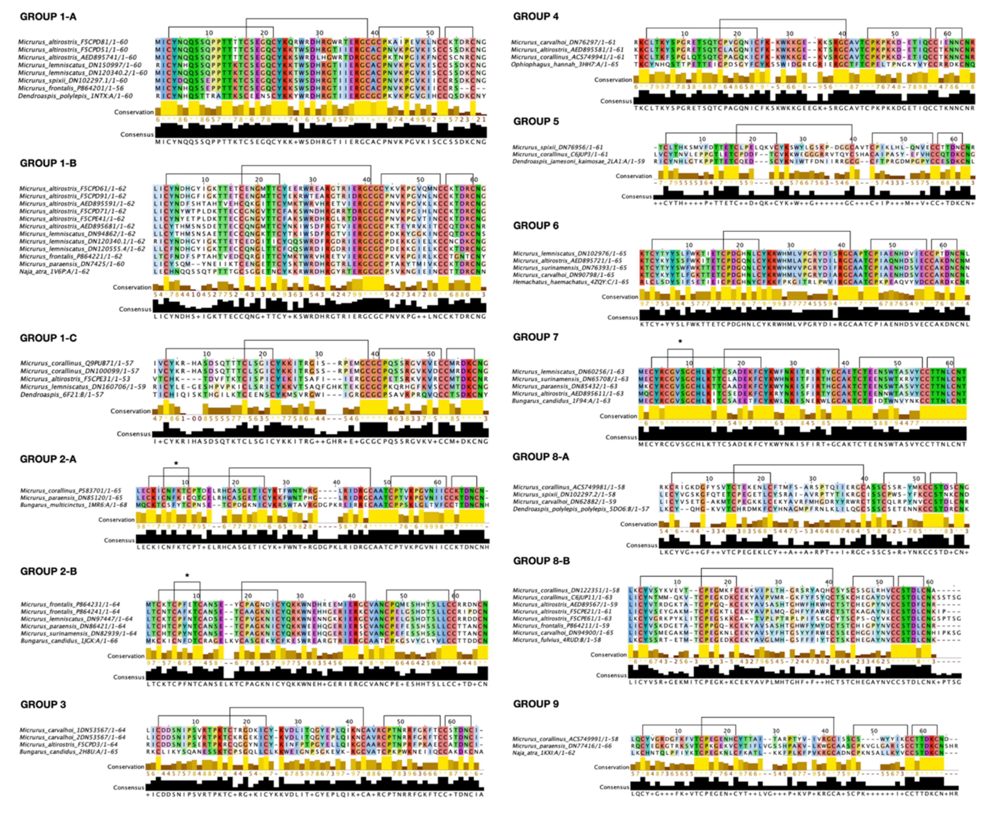
2.1. Three-Finger Toxins Classification Based on Protein Threading
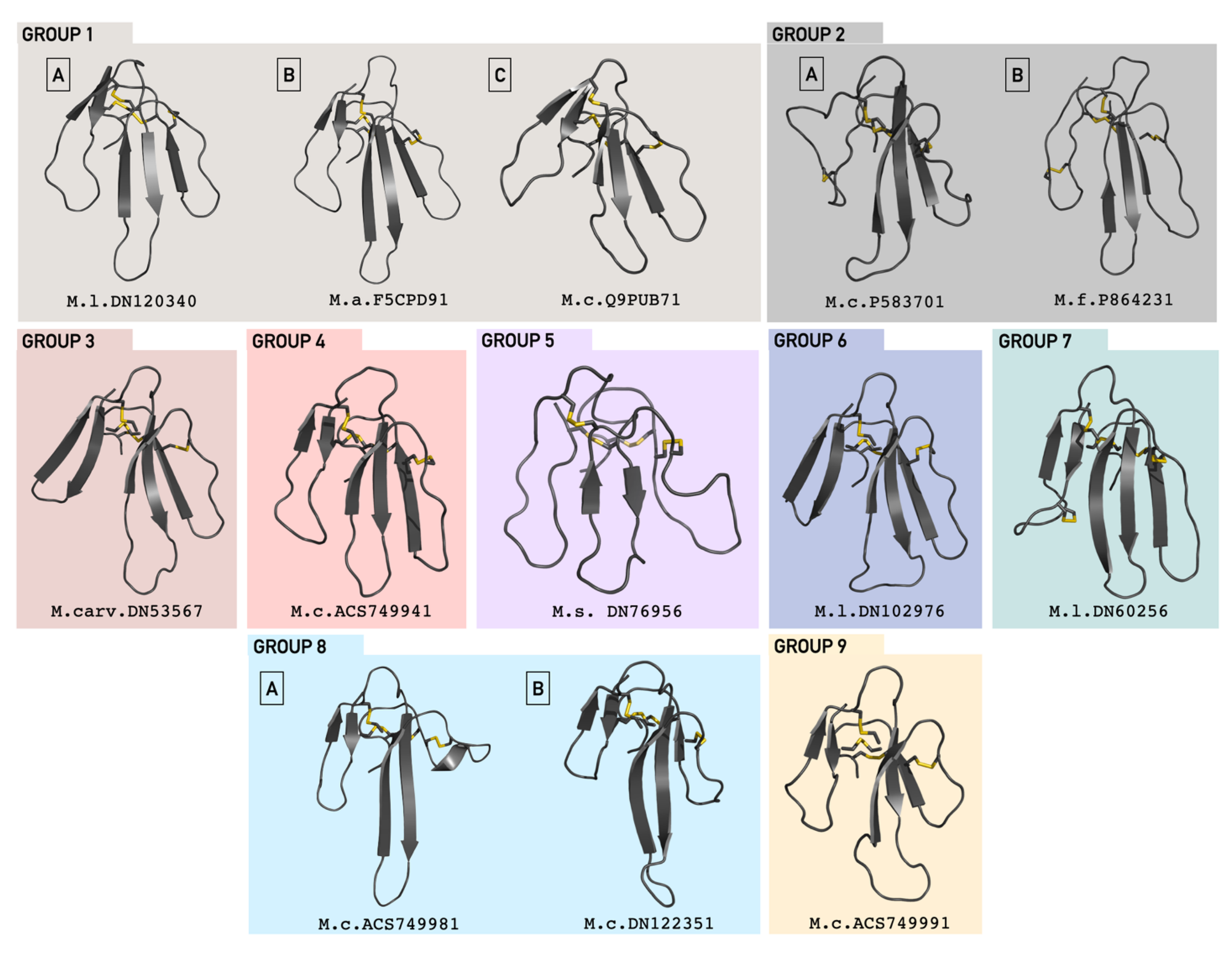
2.2. Structural Representative Model
2.3. Phylogenetic Analysis
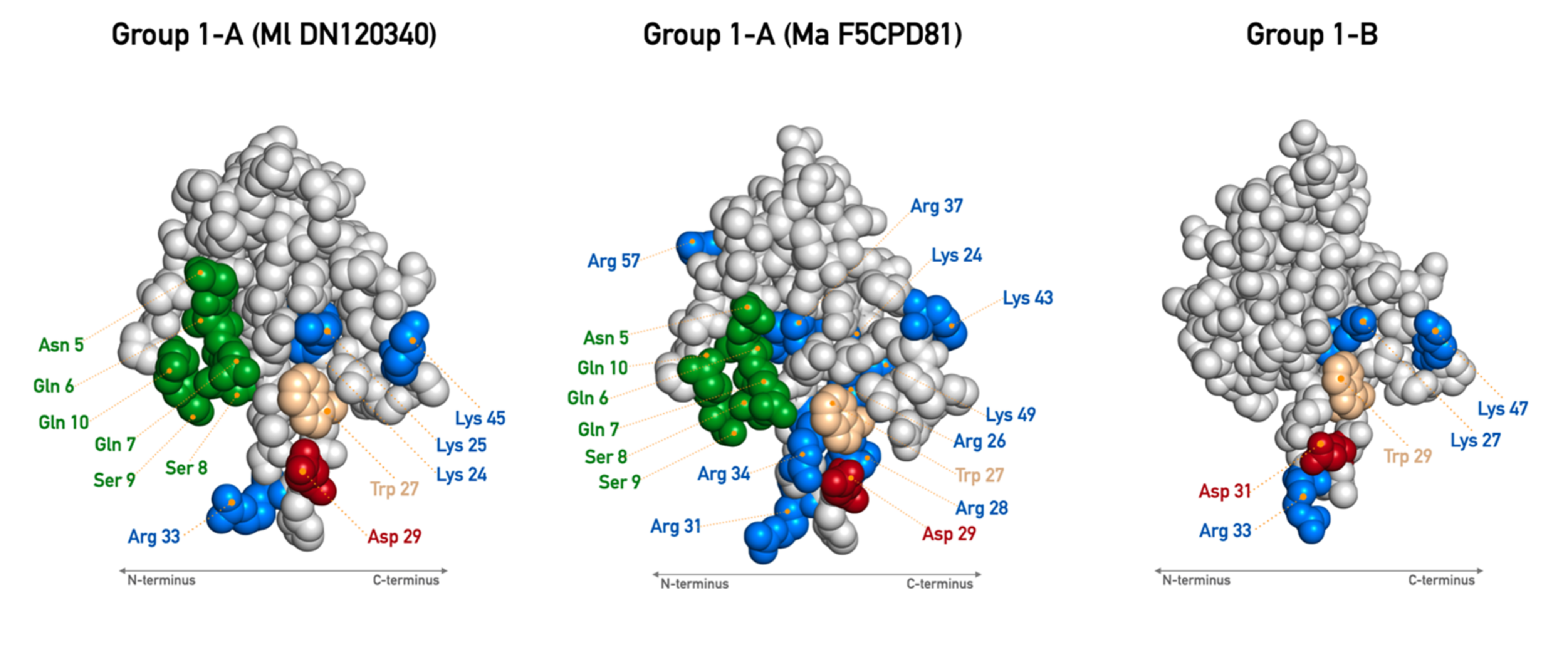
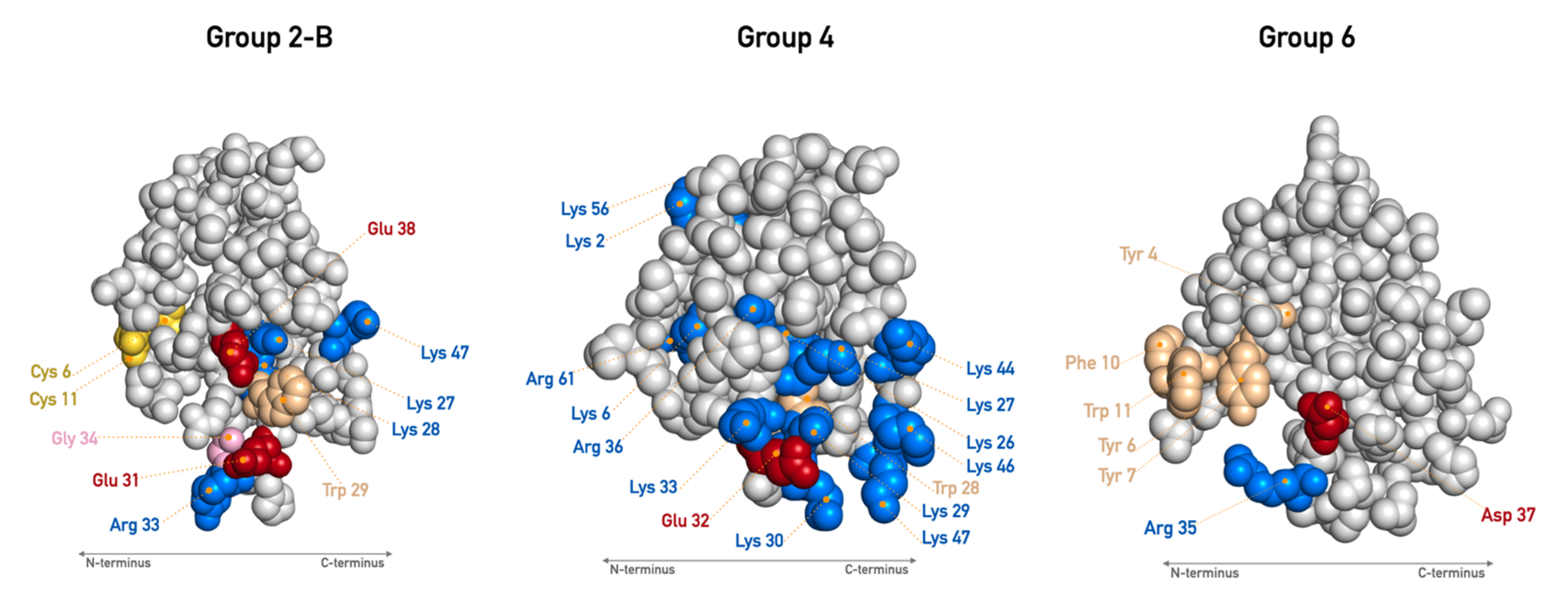
2.4. Biological Insights Annotation
3. Discussion
3.1. Brazilian Micrurus Potent Weapons—Molecular and Biological Insights
3.1.1. Subgroup 1-A
3.1.2. Subgroup 1-B
3.1.3. Subgroup 1-C
3.1.4. Subgroup 2-A
3.1.5. Subgroup 2-B
3.1.6. Group 3
3.1.7. Group 4
3.1.8. Group 5
3.1.9. Group 6
3.1.10. Group 7
3.1.11. Group 8
3.1.12. Group 9
3.2. Undiscovered Biotechnological Potential of 3FTxs
4. Conclusions
5. Methods
5.1. Sequence Database
5.2. Toxin Classification Based on Homology Predictions
5.3. Homology Model Building
5.4. Analysis of Primary Structure and Sequence Alignment
5.5. Electrostatic Surface Potential Calculation
5.6. Phylogenic Analysis
5.7. Group Refinement and Biological Insights Annotation
Supplementary Materials
Author Contributions
Funding
Institutional Review Board Statement
Informed Consent Statement
Data Availability Statement
Acknowledgments
Conflicts of Interest
References
- Uetz, P.; Freed, P.; Hošek, J. The Reptile Database. Available online: http://www.reptile-database.org (accessed on 1 April 2021).
- Corrêa-Netto, C.; De LM Junqueira-de-Azevedo, I.; Silva, D.A.; Ho, P.L.; Leitão-de-Araújo, M.; Alves, M.L.M.; Sanz, L.; Foguel, D.; Zingali, R.B.; Calvete, J.J. Snake venomics and venom gland transcriptomic analysis of Brazilian coral snakes, Micrurus altirostris and M. corallinus. J. Proteom. 2011, 74, 1795–1809. [Google Scholar] [CrossRef] [PubMed]
- Aird, S.D.; Da Silva, N.J.; Qiu, L.; Villar-Briones, A.; Saddi, V.A.; De C Telles, M.P.; Grau, M.L.; Mikheyev, A.S. Coralsnake venomics: Analyses of venom gland transcriptomes and proteomes of six Brazilian taxa. Toxins 2017, 9, 187. [Google Scholar] [CrossRef]
- Sanz, L.; Quesada-Bernat, S.; Ramos, T.; Casais-e-Silva, L.L.; Corrêa-Netto, C.; Silva-Haad, J.J.; Sasa, M.; Lomonte, B.; Calvete, J.J. New insights into the phylogeographic distribution of the 3FTx/PLA 2 venom dichotomy across genus Micrurus in South America. J. Proteom. 2019, 200, 90–101. [Google Scholar] [CrossRef] [PubMed]
- Gutiérrez, J.M.; Calvete, J.J.; Habib, A.G.; Harrison, R.A.; Williams, D.J.; Warrell, D.A. Snakebite envenoming. Nat. Rev. Dis. Prim. 2017, 3, 17063. [Google Scholar] [CrossRef] [PubMed]
- Williams, H.F.; Layfield, H.J.; Vallance, T.; Patel, K.; Bicknell, A.B.; Trim, S.A.; Vaiyapuri, S. The urgent need to develop novel strategies for the diagnosis and treatment of snakebites. Toxins 2019, 11, 363. [Google Scholar] [CrossRef] [PubMed] [Green Version]
- Kini, R.M.; Doley, R. Structure, function and evolution of three-finger toxins: Mini proteins with multiple targets. Toxicon 2010, 56, 855–867. [Google Scholar] [CrossRef]
- Doley, R.; MacKessy, S.P.; Kini, R.M. Role of accelerated segment switch in exons to alter targeting (ASSET) in the molecular evolution of snake venom proteins. BMC Evol. Biol. 2009, 9, 1–14. [Google Scholar] [CrossRef] [PubMed] [Green Version]
- Dashevsky, D.; Fry, B.G. Ancient Diversification of Three-Finger Toxins in Micrurus Coral Snakes. J. Mol. Evol. 2018, 86, 58–67. [Google Scholar] [CrossRef]
- Vital Brazil, O. Coral snake venoms: Mode of action and pathophysiology of experimental envenomation (1). Rev. Inst. Med. Trop. Sao Paulo 1987, 29, 119–126. [Google Scholar] [CrossRef] [PubMed] [Green Version]
- Nirthanan, S.; Gwee, M.C.E. Three-Finger α-Neurotoxins and the Nicotinic Acetylcholine Receptor, Forty Years On. J. Pharmacol. Sci. 2004, 94, 1–17. [Google Scholar] [CrossRef] [PubMed] [Green Version]
- Kessler, P.; Marchot, P.; Silva, M.; Servent, D. The three-finger toxin fold: A multifunctional structural scaffold able to modulate cholinergic functions. J. Neurochem. 2017, 142, 7–18. [Google Scholar] [CrossRef]
- Kudryavtsev, D.S.; Shelukhina, I.V.; Son, L.V.; Ojomoko, L.O.; Kryukova, E.V.; Lyukmanova, E.N.; Zhmak, M.N.; Dolgikh, D.A.; Ivanov, I.A.; Kasheverov, I.E.; et al. Neurotoxins from snake venoms and α-Conotoxin ImI inhibit functionally active Ionotropic γ-aminobutyric acid (GABA) receptors. J. Biol. Chem. 2015, 290, 22747–22758. [Google Scholar] [CrossRef] [PubMed] [Green Version]
- Hannan, S.; Mortensen, M.; Smart, T.G. Snake neurotoxin α-bungarotoxin is an antagonist at native GABAA receptors. Neuropharmacology 2015, 93, 28–40. [Google Scholar] [CrossRef] [PubMed] [Green Version]
- Rosso, J.P.; Schwarz, J.R.; Diaz-Bustamante, M.; Céard, B.; Gutiérrez, J.M.; Kneussel, M.; Pongs, O.; Bosmans, F.; Bougis, P.E. MmTX1 and MmTX2 from coral snake venom potently modulate receptor activity modulate GABAa receptor activity. Proc. Natl. Acad. Sci. USA 2015, 112, E891–E900. [Google Scholar] [CrossRef] [PubMed] [Green Version]
- Diochot, S.; Baron, A.; Salinas, M.; Douguet, D.; Scarzello, S.; Dabert-Gay, A.S.; Debayle, D.; Friend, V.; Alloui, A.; Lazdunski, M.; et al. Black mamba venom peptides target acid-sensing ion channels to abolish pain. Nature 2012, 490, 552–555. [Google Scholar] [CrossRef] [PubMed]
- Rouget, C.; Quinton, L.; Maïga, A.; Gales, C.; Masuyer, G.; Malosse, C.; Chamot-Rooke, J.; Thai, R.; Mourier, G.; De Pauw, E.; et al. Identification of a novel snake peptide toxin displaying high affinity and antagonist behaviour for the α 2-adrenoceptors. Br. J. Pharmacol. 2010, 161, 1361–1374. [Google Scholar] [CrossRef] [Green Version]
- Maïga, A.; Mourier, G.; Quinton, L.; Rouget, C.; Gales, C.; Denis, C.; Lluel, P.; Sénard, J.M.; Palea, S.; Servent, D.; et al. G protein-coupled receptors, an unexploited animal toxin targets: Exploration of green mamba venom for novel drug candidates active against adrenoceptors. Toxicon 2012, 59, 487–496. [Google Scholar] [CrossRef]
- Blanchet, G.; Upert, G.; Mourier, G.; Gilquin, B.; Gilles, N.; Servent, D. New α-adrenergic property for synthetic MTβ and CM-3 three-finger fold toxins from black mamba. Toxicon 2013, 75, 160–167. [Google Scholar] [CrossRef] [PubMed]
- Koivula, K.; Rondinelli, S.; Näsman, J. The three-finger toxin MTα is a selective α2B-adrenoceptor antagonist. Toxicon 2010, 56, 440–447. [Google Scholar] [CrossRef]
- Rivera-Torres, I.O.; Jin, T.B.; Cadene, M.; Chait, B.T.; Poget, S.F. Discovery and characterisation of a novel toxin from Dendroaspis angusticeps, named Tx7335, that activates the potassium channel KcsA. Sci. Rep. 2016, 6, 1–11. [Google Scholar] [CrossRef] [PubMed] [Green Version]
- Yang, D.C.; Deuis, J.R.; Dashevsky, D.; Dobson, J.; Jackson, T.N.W.; Brust, A.; Xie, B.; Koludarov, I.; Debono, J.; Hendrikx, I.; et al. The snake with the scorpion’s sting: Novel three-finger toxin sodium channel activators from the venom of the long-glanded blue coral snake (calliophis bivirgatus). Toxins 2016, 8, 303. [Google Scholar] [CrossRef] [PubMed]
- Abd El-Aziz, T.M.; Al Khoury, S.; Jaquillard, L.; Triquigneaux, M.; Martinez, G.; Bourgoin-Voillard, S.; Sève, M.; Arnoult, C.; Beroud, R.; De Waard, M. Actiflagelin, a new sperm activator isolated from Walterinnesia aegyptia venom using phenotypic screening. J. Venom. Anim. Toxins Incl. Trop. Dis. 2018, 24, 1–11. [Google Scholar] [CrossRef] [Green Version]
- Nguyen, T.T.N.; Folch, B.; Létourneau, M.; Vaudry, D.; Truong, N.H.; Doucet, N.; Chatenet, D.; Fournier, A. Cardiotoxin-I: An Unexpectedly Potent Insulinotropic Agent. ChemBioChem 2012, 13, 1805–1812. [Google Scholar] [CrossRef] [PubMed]
- Conlon, J.M.; Attoub, S.; Musale, V.; Leprince, J.; Casewell, N.R.; Sanz, L.; Calvete, J.J. Isolation and characterization of cytotoxic and insulin-releasing components from the venom of the black-necked spitting cobra Naja nigricollis (Elapidae). Toxicon X 2020, 6, 100030. [Google Scholar] [CrossRef] [PubMed]
- Sanz, L.; De Freitas-Lima, L.N.; Quesada-Bernat, S.; Graça-de-Souza, V.K.; Soares, A.M.; Calderón, L. de A.; Calvete, J.J.; Caldeira, C.A.S. Comparative venomics of Brazilian coral snakes: Micrurus frontalis, Micrurus spixii spixii, and Micrurus surinamensis. Toxicon 2019, 166, 39–45. [Google Scholar] [CrossRef]
- Coelho Da Silva, D.; Araújo De Medeiros, W.A.; De Fátima Correia Batista, I.; Pimenta, D.C.; Lebrun, I.; Francis Abdalla, F.M.; Lopes Sandoval, M.R. Characterization of a new muscarinic toxin from the venom of the Brazilian coral snake Micrurus lemniscatus in rat hippocampus. Life Sci. 2011, 89, 931–938. [Google Scholar] [CrossRef] [PubMed] [Green Version]
- Silveira De Oliveira, J.; Silva, Á.R.D.B.P.D.; Soares, M.B.; Stephano, M.A.; De Oliveira Dias, W.; Raw, I.; Ho, P.L. Cloning and characterization of an α-neurotoxin-type protein specific for the coral snake Micrurus corallinus. Biochem. Biophys. Res. Commun. 2000, 267, 887–891. [Google Scholar] [CrossRef] [PubMed]
- Moreira, K.G.; Prates, M.V.; Andrade, F.A.C.; Silva, L.P.; Beirão, P.S.L.; Kushmerick, C.; Naves, L.A.; Bloch, C. Frontoxins, three-finger toxins from Micrurus frontalis venom, decrease miniature endplate potential amplitude at frog neuromuscular junction. Toxicon 2010, 56, 55–63. [Google Scholar] [CrossRef]
- Söding, J. Protein homology detection by HMM-HMM comparison. Bioinformatics 2005, 21, 951–960. [Google Scholar] [CrossRef] [Green Version]
- Meier, A.; Söding, J. Automatic Prediction of Protein 3D Structures by Probabilistic Multi-template Homology Modeling. PLoS Comput. Biol. 2015, 11, e1004343. [Google Scholar] [CrossRef] [PubMed]
- Hildebrand, A.; Remmert, M.; Biegert, A.; Söding, J. Fast and accurate automatic structure prediction with HHpred. Proteins Struct. Funct. Bioinform. 2009, 77, 128–132. [Google Scholar] [CrossRef] [PubMed] [Green Version]
- Zimmermann, L.; Stephens, A.; Nam, S.Z.; Rau, D.; Kübler, J.; Lozajic, M.; Gabler, F.; Söding, J.; Lupas, A.N.; Alva, V. A Completely Reimplemented MPI Bioinformatics Toolkit with a New HHpred Server at its Core. J. Mol. Biol. 2018, 430, 2237–2243. [Google Scholar] [CrossRef]
- Waterhouse, A.M.; Procter, J.B.; Martin, D.M.A.; Clamp, M.; Barton, G.J. Jalview Version 2-A multiple sequence alignment editor and analysis workbench. Bioinformatics 2009, 25, 1189–1191. [Google Scholar] [CrossRef] [PubMed] [Green Version]
- Leão, L.I.; Ho, P.L.; Junqueira-de-Azevedo, I. de L.M. Transcriptomic basis for an antiserum against Micrurus corallinus (coral snake) venom. BMC Genom. 2009, 10, 1–14. [Google Scholar] [CrossRef] [PubMed] [Green Version]
- Nirthanan, S. Snake three-finger α-neurotoxins and nicotinic acetylcholine receptors: Molecules, mechanisms and medicine. Biochem. Pharmacol. 2020, 181, 114168. [Google Scholar] [CrossRef] [PubMed]
- Tsetlin, V. Snake venom a -neurotoxins and other ‘three-finger’ proteins. Eur. J. Biochem. 1999, 286, 281–286. [Google Scholar] [CrossRef] [PubMed]
- Low, B.W.; Preston, H.S.; Sato, A.; Rosen, L.S.; Searl, J.E.; Rudko, A.D.; Richardson, J.S. Three dimensional structure of erabutoxin b neurotoxic protein: Inhibitor of acetylcholine receptor. Proc. Natl. Acad. Sci. USA 1976, 73, 2991–2994. [Google Scholar] [CrossRef] [Green Version]
- Maeda, N.; Tamiya, N. The primary structure of the toxin Laticauda semifasciata III, a weak and reversibly acting neurotoxin from the venom of a sea snake, Laticauda semifasciata. Biochem. J. 1974, 141, 389–400. [Google Scholar] [CrossRef] [PubMed] [Green Version]
- Tsetlin, V.I.; Karlsson, E.; Arseniev, A.S.; Utkin, Y.N.; Surin, A.M.; Pashkov, V.S.; Pluzhnikov, K.A.; Ivanov, V.T.; Bystrov, V.F.; Ovchinnikov, Y.A. EPR And fluorescence study of interaction of Naja naja oxiana neurotoxin II and its derivatives with acetylcholine receptor protein from Torpedo marmorata. FEBS Lett. 1979, 106, 47–52. [Google Scholar] [CrossRef] [Green Version]
- Tsetlin, V.I.; Karlsson, E.; Utkin, Y.N.; Pluzhnikov, K.A.; Surin, A.M.; Kondakov, V.V.; Bystrov, V.F.; Ivanov, V.T. Printed in Great Britain. Tetrahedron Lett. 1982, 23, 3431–3434. [Google Scholar]
- Trémeau, O.; Lemaire, C.; Drevet, P.; Pinkasfeld, S.; Ducancel, F.; Boulain, J.C.; Ménez, A. Genetic engineering of snake toxins: The functional site of erabutoxin A, as delineated by site-directed mutagenesis, includes variant residues. J. Biol. Chem. 1995, 270, 9362–9369. [Google Scholar] [CrossRef] [PubMed] [Green Version]
- Teixeira-Clerc, F.; Ménez, A.; Kessler, P. How do short neurotoxins bind to a muscular-type nicotinic acetylcholine receptor? J. Biol. Chem. 2002, 277, 25741–25747. [Google Scholar] [CrossRef] [PubMed] [Green Version]
- Lesovoy, D.M.; Bocharov, E.V.; Lyukmanova, E.N.; Kosinsky, Y.A.; Shulepko, M.A.; Dolgikh, D.A.; Kirpichnikov, M.P.; Efremov, R.G.; Arseniev, A.S. Specific membrane binding of neurotoxin II can facilitate its delivery to acetylcholine receptor. Biophys. J. 2009, 97, 2089–2097. [Google Scholar] [CrossRef] [Green Version]
- Cheng, Y.; Meng, Q.; Wang, W.; Wang, J. Structure-function relationship of three neurotoxins from the venom of Naja kaouthia: A comparison between the NMR-derived structure of NT2 with its homologues, NT1 and NT3. Biochim. Biophys. Acta Protein Struct. Mol. Enzymol. 2002, 1594, 353–363. [Google Scholar] [CrossRef]
- Low, B.W.; Corfield, P.W.R. Erabutoxin b: Structure/function relationships following initial protein refinement at 0.140-nm resolution. Eur. J. Biochem. 1986, 161, 579–587. [Google Scholar] [CrossRef] [PubMed]
- Zinn-Justin, S.; Roumestand, C.; Gilquin, B.; Bontems, F.; Ménez, A.; Toma, F. Three-Dimensional Solution Structure of a Curaremimetic Toxin from Naja nigricollis Venom: A Proton NMR and Molecular Modeling Study. Biochemistry 1992, 31, 11335–11347. [Google Scholar] [CrossRef] [PubMed]
- Murakami, M.; Kini, R.; Arni, R. Crystal Structure of Bucain, a Three-Fingered Toxin from the Venom of the Malayan Krait (Bungarus candidus). Protein Pept. Lett. 2009, 16, 1473–1477. [Google Scholar] [CrossRef]
- Labhardt, A.M.; Hunziker-Kwik, E.-H.; Wüthrich, K.; Baklouti, F.; Baudin-Chich, V.; Kister, J.; Marden, M.; Teyssier, G.; Poyart, C.; Delaunay, J.; et al. Secondary structure determination for α-neurotoxin from Dendroaspis polylepis polylepis based on sequence-specific 1H-nuclear-magnetic-resonance assignments. Eur. J. Biochem. 1988, 177, 295–305. [Google Scholar] [CrossRef]
- Tu, X.; Huang, Q.; Lou, X.; Tenga, M.; Niu, L. Purification, N-terminal sequencing, crystallization and preliminary X-ray diffraction analysis of atratoxin, a new short-chain α-neurotoxin from the venom of Naja naja atra. Acta Crystallogr. Sect. D Biol. Crystallogr. 2002, 58, 839–842. [Google Scholar] [CrossRef] [PubMed]
- Lou, X.; Liu, Q.; Tu, X.; Wang, J.; Teng, M.; Niu, L.; Schuller, D.J.; Huang, Q.; Hao, Q. The atomic resolution crystal structure of atratoxin determined by single wavelength anomalous diffraction phasing. J. Biol. Chem. 2004, 279, 39094–39104. [Google Scholar] [CrossRef] [PubMed] [Green Version]
- Shiu, J.H.; Chen, C.Y.; Chang, L.S.; Chen, Y.C.; Chen, Y.C.; Lo, Y.H.; Liu, Y.C.; Chuang, W.J. Solution structure of γ-bungarotoxin: The functional significance of amino acid residues flanking the RGD motif in integrin binding. Proteins Struct. Funct. Genet. 2004, 57, 839–849. [Google Scholar] [CrossRef] [PubMed]
- Nirthanan, S.; Gopalakrishnakone, P.; Gwee, M.C.E.; Khoo, H.E.; Kini, R.M. Non-conventional toxins from Elapid venoms. Toxicon 2003, 41, 397–407. [Google Scholar] [CrossRef]
- Aird, S.D.; Womble, G.C.; Yates, J.R.; Griffin, P.R. Primary structure of γ-bungarotoxin, a new postsynaptic neurotoxin from venom of Bungarus multicinctus. Toxicon 1999, 37, 609–625. [Google Scholar] [CrossRef]
- Lyukmanova, E.N.; Shenkarev, Z.O.; Shulepko, M.A.; Paramonov, A.S.; Chugunov, A.O.; Janickova, H.; Dolejsi, E.; Dolezal, V.; Utkin, Y.N.; Tsetlin, V.I.; et al. Structural insight into specificity of interactions between nonconventional three-finger weak toxin from Naja kaouthia (WTX) and muscarinic acetylcholine receptors. J. Biol. Chem. 2015, 290, 23616–23630. [Google Scholar] [CrossRef] [Green Version]
- Starkov, V.G.; Polyak, Y.L.; Vulfius, E.A.; Kryukova, E.V.; Tsetlin, V.I.; Utkin, Y.N. New weak toxins from the cobra venom. Russ. J. Bioorganic Chem. 2009, 35, 10–18. [Google Scholar] [CrossRef] [PubMed]
- Corfield, P.W.; Lee, T.J.; Low, B.W. The crystal structure of erabutoxin a at 2.0-A resolution. J. Biol. Chem. 1989, 264, 9239–9242. [Google Scholar] [CrossRef]
- Nirthanan, S.; Charpantier, E.; Gopalakrishnakone, P.; Gwee, M.C.E.; Khoo, H.E.; Cheah, L.S.; Bertrand, D.; Manjunatha Kini, R. Candoxin, a novel toxin from Bungarus candidus, is a reversible antagonist of muscle (αβγδ) but a poorly reversible antagonist of neuronal α7 nicotinic acetylcholine receptors. J. Biol. Chem. 2002, 277, 17811–17820. [Google Scholar] [CrossRef] [PubMed] [Green Version]
- Roy, A.; Zhou, X.; Chong, M.Z.; D’Hoedt, D.; Foo, C.S.; Rajagopalan, N.; Nirthanan, S.; Bertrand, D.; Sivaraman, J.; Manjunatha Kini, R. Structural and functional characterization of a novel homodimeric three-finger neurotoxin from the venom of Ophiophagus hannah (King cobra). J. Biol. Chem. 2010, 285, 8302–8315. [Google Scholar] [CrossRef] [PubMed] [Green Version]
- Cheng, C.H.; Chen, Y.C.; Shiu, J.H.; Chang, Y.T.; Chang, Y.S.; Huang, C.H.; Chen, C.Y.; Chuang, W.J. Dynamics and functional differences between dendroaspin and rhodostomin: Insights into protein scaffolds in integrin recognition. Protein Sci. 2012, 21, 1872–1884. [Google Scholar] [CrossRef] [PubMed] [Green Version]
- Conte, L.L.; Chothia, C.; Janin, J. The atomic structure of protein-protein recognition sites. J. Mol. Biol. 1999, 285, 2177–2198. [Google Scholar] [CrossRef] [Green Version]
- Keskin, O.; Ma, B.; Nussinov, R. Hot regions in protein-protein interactions: The organization and contribution of structurally conserved hot spot residues. J. Mol. Biol. 2005, 345, 1281–1294. [Google Scholar] [CrossRef]
- Barnwal, B.; Jobichen, C.; Girish, V.M.; Foo, C.S.; Sivaraman, J.; Kini, R.M. Ringhalexin from Hemachatus haemachatus: A novel inhibitor of extrinsic tenase complex. Sci. Rep. 2016, 6, 1–13. [Google Scholar] [CrossRef] [Green Version]
- Choudhury, M.; McCleary, R.; Kini, R.M.; Velmurugan, D. Orphan Three-Finger Toxins Bind at Tissue Factor–Factor VIIa Interface to Inhibit Factor X Activation: Identification of Functional Site by Docking. TH Open 2018, 02, e303–e314. [Google Scholar] [CrossRef] [PubMed] [Green Version]
- Kuhn, P.; Deacon, A.M.; Comoso, S.; Rajaseger, G.; Kini, R.M.; Uson, I.; Kolatkar, P.R. The atomic resolution structure of bucandin, a novel toxin isolated from the Malayan krait, determined by direct methods. Acta Crystallogr. Sect. D Biol. Crystallogr. 2000, 56, 1401–1407. [Google Scholar] [CrossRef] [PubMed] [Green Version]
- Torres, A.M.; Kini, R.M.; Selvanayagam, N.; Kuchel, P.W. NMR structure of bucandin, a neurotoxin from the venom of the Malayan krait (bungarus candidus). Biochem. J. 2001, 360, 539–548. [Google Scholar] [CrossRef] [PubMed]
- Sun, D.; Yu, Y.; Xue, X.; Pan, M.; Wen, M.; Li, S.; Qu, Q.; Li, X.; Zhang, L.; Li, X.; et al. Cryo-EM structure of the ASIC1a-mambalgin-1 complex reveals that the peptide toxin mambalgin-1 inhibits acid-sensing ion channels through an unusual allosteric effect. Cell Discov. 2018, 4, 1–11. [Google Scholar] [CrossRef] [PubMed] [Green Version]
- Foo, C.S.; Jobichen, C.; Hassan-Puttaswamy, V.; Dekan, Z.; Tae, H.S.; Bertrand, D.; Adams, D.J.; Alewood, P.F.; Sivaraman, J.; Nirthanan, S.; et al. Fulditoxin, representing a new class of dimeric snake toxins, defines novel pharmacology at nicotinic ACh receptors. Br. J. Pharmacol. 2020, 177, 1822–1840. [Google Scholar] [CrossRef]
- Sun, Y.J.; Wu, W.G.; Chiang, C.M.; Hsin, A.Y.; Hsiao, C.D. Crystal structure of cardiotoxin V from Taiwan cobra venom: pH-dependent conformational change and a novel membrane-binding motif identified in the three-finger loops of p-type cardiotoxin. Biochemistry 1997, 36, 2403–2413. [Google Scholar] [CrossRef]
- Kalia, J.; Milescu, M.; Salvatierra, J.; Wagner, J.; Klint, J.K.; King, G.F.; Olivera, B.M.; Bosmans, F. From foe to friend: Using animal toxins to investigate ion channel function. J. Mol. Biol. 2015, 427, 158–175. [Google Scholar] [CrossRef] [PubMed] [Green Version]
- Clark, G.C.; Casewell, N.R.; Elliott, C.T.; Harvey, A.L.; Jamieson, A.G.; Strong, P.N.; Turner, A.D. Friends or Foes? Emerging Impacts of Biological Toxins. Trends Biochem. Sci. 2019, 44, 365–379. [Google Scholar] [CrossRef] [Green Version]
- Pennington, M.W.; Czerwinski, A.; Norton, R.S. Peptide therapeutics from venom: Current status and potential. Bioorganic Med. Chem. 2018, 26, 2738–2758. [Google Scholar] [CrossRef] [PubMed]
- Utkin, Y.N. Last decade update for three-finger toxins: Newly emerging structures and biological activities. World J. Biol. Chem. 2019, 10, 17–27. [Google Scholar] [CrossRef] [PubMed]
- Webb, B.; Sali, A. Comparative protein structure modeling using MODELLER. Curr. Protoc. Bioinform. 2016, 2016, 5–6. [Google Scholar] [CrossRef] [Green Version]
- Shen, M.; Sali, A. Statistical potential for assessment and prediction of protein structures. Protein Sci. 2006, 15, 2507–2524. [Google Scholar] [CrossRef] [PubMed] [Green Version]
- Drozdetskiy, A.; Cole, C.; Procter, J.; Barton, G.J. JPred4: A protein secondary structure prediction server. Nucleic Acids Res. 2015, 43, W389–W394. [Google Scholar] [CrossRef] [PubMed]
- Edgar, R.C. MUSCLE: Multiple sequence alignment with high accuracy and high throughput. Nucleic Acids Res. 2004, 32, 1792–1797. [Google Scholar] [CrossRef] [PubMed] [Green Version]
- Kumar, S.; Stecher, G.; Li, M.; Knyaz, C.; Tamura, K. MEGA X: Molecular evolutionary genetics analysis across computing platforms. Mol. Biol. Evol. 2018, 35, 1547–1549. [Google Scholar] [CrossRef] [PubMed]


Publisher’s Note: MDPI stays neutral with regard to jurisdictional claims in published maps and institutional affiliations. |
© 2021 by the authors. Licensee MDPI, Basel, Switzerland. This article is an open access article distributed under the terms and conditions of the Creative Commons Attribution (CC BY) license (https://creativecommons.org/licenses/by/4.0/).
Share and Cite
Kleiz-Ferreira, J.M.; Cirauqui, N.; Trajano, E.A.; Almeida, M.d.S.; Zingali, R.B. Three-Finger Toxins from Brazilian Coral Snakes: From Molecular Framework to Insights in Biological Function. Toxins 2021, 13, 328. https://doi.org/10.3390/toxins13050328
Kleiz-Ferreira JM, Cirauqui N, Trajano EA, Almeida MdS, Zingali RB. Three-Finger Toxins from Brazilian Coral Snakes: From Molecular Framework to Insights in Biological Function. Toxins. 2021; 13(5):328. https://doi.org/10.3390/toxins13050328
Chicago/Turabian StyleKleiz-Ferreira, Jessica Matos, Nuria Cirauqui, Edson Araujo Trajano, Marcius da Silva Almeida, and Russolina Benedeta Zingali. 2021. "Three-Finger Toxins from Brazilian Coral Snakes: From Molecular Framework to Insights in Biological Function" Toxins 13, no. 5: 328. https://doi.org/10.3390/toxins13050328
APA StyleKleiz-Ferreira, J. M., Cirauqui, N., Trajano, E. A., Almeida, M. d. S., & Zingali, R. B. (2021). Three-Finger Toxins from Brazilian Coral Snakes: From Molecular Framework to Insights in Biological Function. Toxins, 13(5), 328. https://doi.org/10.3390/toxins13050328