Article

FfCOX17 is Involved in Fumonisins Production, Growth, Asexual Reproduction, and Fungicide Sensitivity in Fusarium fujikuroi

College of Plant Protection, Nanjing Agricultural University, Nanjing 210095, China
*
Authors to whom correspondence should be addressed.
Toxins 2022, 14(7), 427; https://doi.org/10.3390/toxins14070427
Submission received: 8 May 2022 / Revised: 18 June 2022 / Accepted: 20 June 2022 / Published: 22 June 2022
(This article belongs to the Section Mycotoxins)

Abstract

:
Fusarium fujikuroi, a causal agent of Rice Bakanae Disease, produces secondary metabolites such as gibberellin, pigments bikaverin, and mycotoxins fumonisins. Fumonisins produced by F. fujikuroi pose a severe threat to human and animal health. The copper chaperone protein plays a critical role in different growth stages of plants, fungi, and yeasts, but their functions and regulation in fumonisin biosynthesis are still unclear. Here, a copper chaperone protein, FfCOX17, was identified in F. fujikuroi. The FfCOX17 deletion mutant (∆FfCOX17) exhibited decreased vegetative growth and asexual reproduction. The transcriptional level of the FfFUM2 gene was significantly induced in ∆FfCOX17, and the fumonisin production in ∆FfCOX17 mutants was significantly increased compared to wild-type F. fujikuroi, but the pathogenicity of ∆FfCOX17 mutants was unaffected, which may be caused by the no significantly changed gibberellin content. ∆FfCOX17 showed decreased sensitivity to oxidative stress, osmotic stress, and increased sensitivity to cell wall stress, heat shock stress, and high concentration glucose. In addition, ∆FfCOX17 also showed increased sensitivity to fungicide fluazinam and fludioxonil, and decreased sensitivity to phenamacril and prochloraz. Taken together, this study suggested that FfCOX17 is critical for fumonisin production, vegetative growth, asexual reproduction, and fungicide sensitivity, but is not required for the virulence function of F. fujikuroi on rice.
Key Contribution: FfCOX17 plays an important role in fumonisins production, vegetative growth, asexual reproduction, and fungicide sensitivity in F. fujikuroi, which provides new insight into the role of COX17 homologs in filamentous fungi.

1. Introduction

Fusarium fujikuroi Nirenberg (teleomorph: Gibberella fujikuroi mating population C) belongs to the G. fujikuroi (Sawada) Wollenweber species complex [1] and is an important pathogenic fungus of Rice Bakanae Disease (RBD) [2]. RBD leads to abnormal growth of rice, yellowing of leaves, reduction of tillers, and empty grains of rice. The majority of these symptoms are caused by gibberellin (GAs), which is a plant hormone [3,4]. In addition to GAs, F. fujikuroi produces other secondary metabolites, including mycotoxin fumonisins (FUM), fusarins (FUS), fusaric acid (FU), pigment bikaverin (BIK), and apicidin F (APF), which seriously threaten the yield and quality of rice [5,6,7]. Fumonisins are currently considered one of the most important fungal toxins in agriculture, as they are not only responsible for animal diseases, but are also associated with some human disease epidemiology [8,9,10]. Fumonisins are polyketide-derived metabolites that can inhibit ceramide synthase, a key enzyme in sphingolipid metabolism, and induce apoptosis [8,10]. The fumonisin biosynthetic genes are clustered in F. fujikuroi, including 16 FUM genes [11]. FUM synthesis appears to be regulated by various environmental factors, such as pH and nutrient sources, at the transcriptional level [12,13]. A previous study has shown that the mitogen-activated protein kinase (MAPK) signaling pathway is involved in the regulation of FUM production in F. verticillioides [14]. Carbon sources have also been shown to regulate FUM biosynthesis in F. proliferatum [15]. Recent studies suggested that post-translational modifications play a key role in the production of FUM in F. verticillioides [16]. So far, the molecular mechanisms underlying FUM regulation have not yet been elucidated.
The copper chaperone protein COX17 is a factor that promotes the binding of copper to cytochrome c oxidase (CcO) and can transfer heavy metals from the cytoplasm to the mitochondrial membrane space [17]. In S. cerevisiae, the mutation of COX17 causes special defects of cytochrome c oxidase, resulting in respiratory deficiency [18]. Mammalian copper chaperone Cox17p has an essential role in the activation of cytochrome c oxidase and embryonic development [19,20,21]. In Arabidopsis, the deletion or mutation of COX17 results in a deficiency in cell growth and stress response [22,23]. The deletion mutant of the COX17 homologous gene in the fungus A. fumigatus, ΔAfCOX17, shows significant defects in mycelial growth [24]. However, the function and regulation of COX17 in F. fujikuroi remain unknown.
In this study, FfCOX17, encoding a cytochrome c oxidase copper chaperone, was identified in F. fujikuroi. We confirmed that FfCOX17 plays a vital role in FUM production, FfFUM2 gene expression, vegetative growth, asexual development, stress responses, and sensitivity to fungicides. These findings will provide a basis for exploring the regulation of FfCOX17 on mycotoxins FUM production in F. fujikuroi.

2. Results

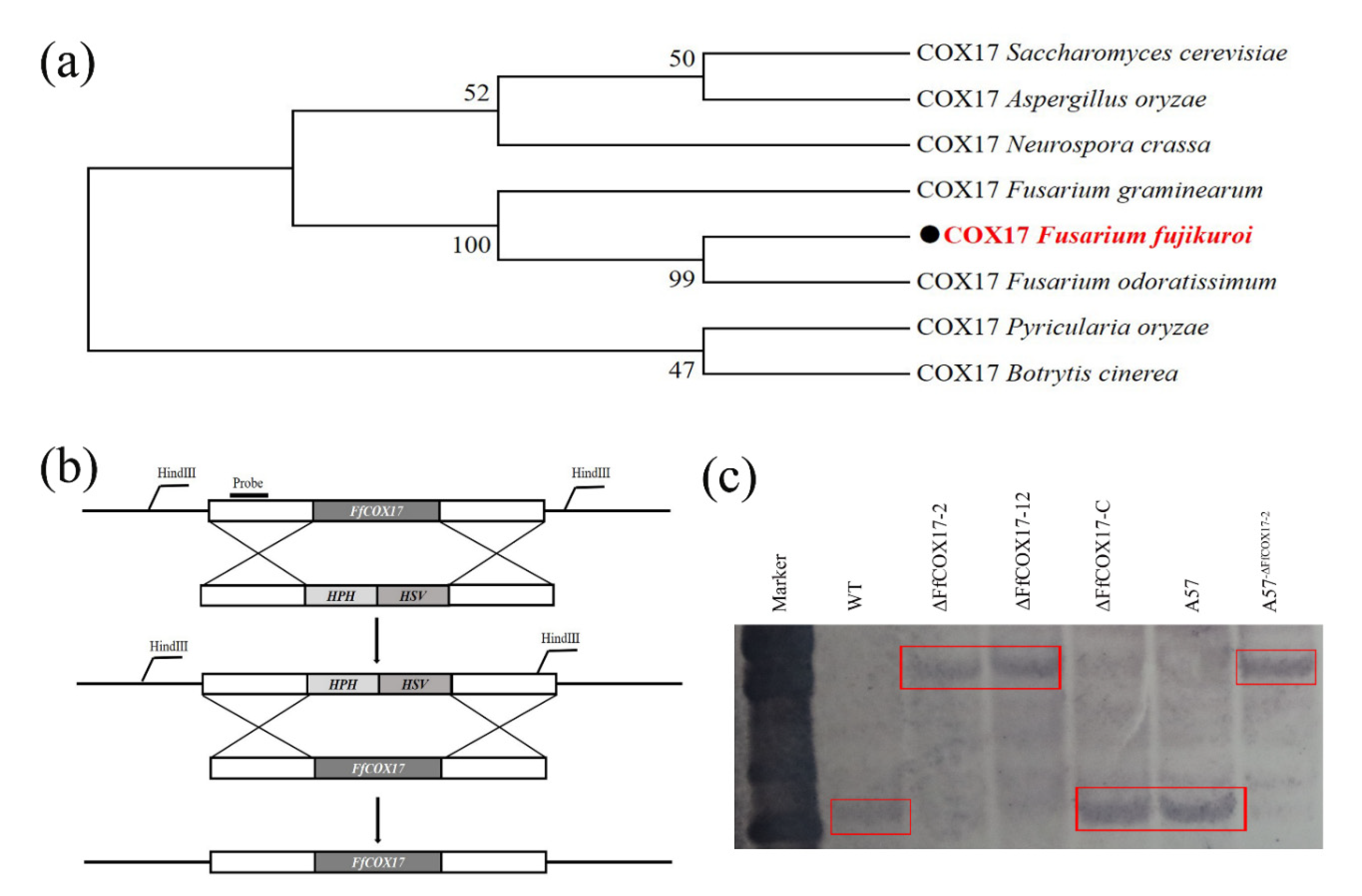
2.1. Identification, Deletion, and Complementation of FfCOX17

The cytochrome c oxidase copper chaperone FfCOX17 (FFUJ_01072) was identified from the F. fujikuroi genome database (http://fungi.ensembl.org/Fusarium_fujikuroi_gca_001023065/Info/Index (accessed on 13 July 2020) by BLASTP using the S. cerevisiae COX17 as a query. FfCOX17 is a 348 bp gene with two introns and three exons and encodes a protein with seventy-six amino acids. Phylogenetic tree analysis showed that FfCOX17 was relatively conserved in Fusarium spp and other species (Figure 1a). To investigate the role of the FfCOX17 in F. fujikuroi, two independent FfCOX17 deletion mutants (ΔFfCOX17-2 and ΔFfCOX17-12) were obtained by homologous recombination (Figure 1b), and these mutants were verified by PCR and further confirmed by Southern blotting (Figure 1c). To confirm whether the observed phenotypes of ΔFfCOX17 were caused by knockout, the complemented mutant (ΔFfCOX17-C) was generated by transformation.

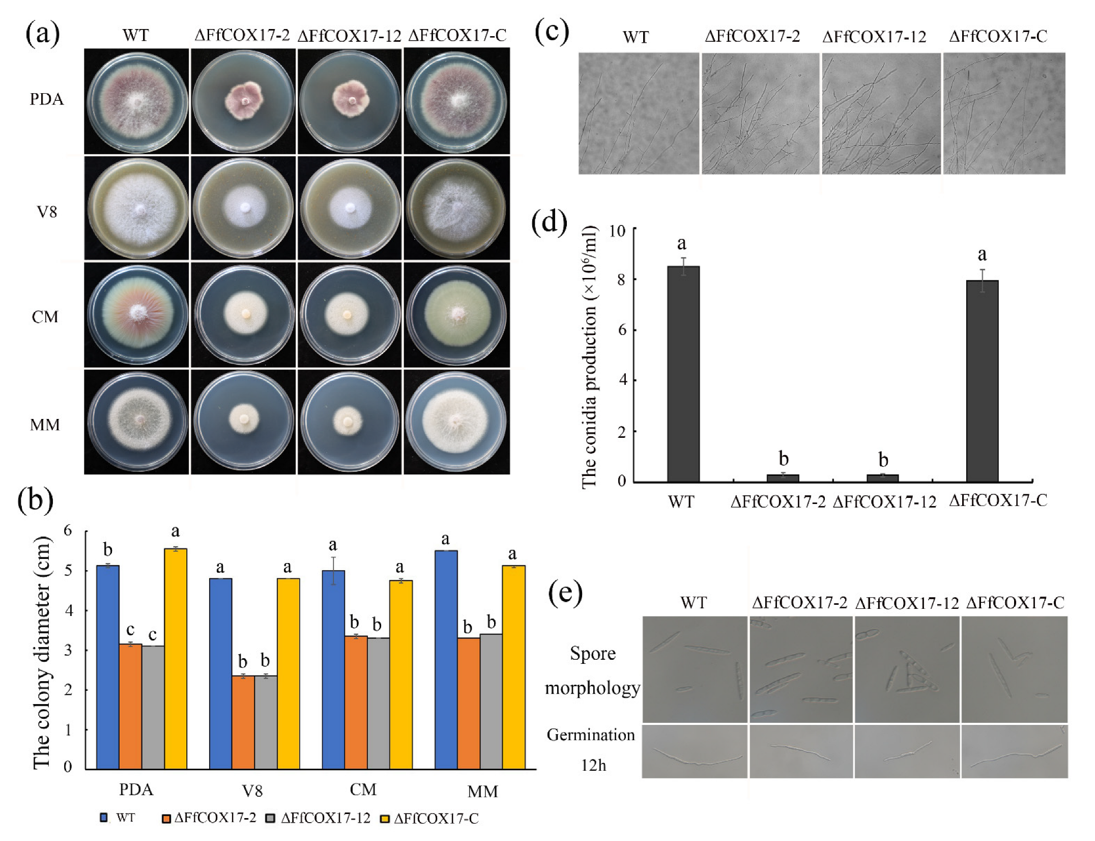
2.2. FfCOX17 Is Involved in Vegetative Growth and Asexual Reproduction

The deletion mutants of FfCOX17 were used to confirm the function of FfCOX17 in F. fujikuroi. The growth rate of ΔFfCOX17-2 and ΔFfCOX17-12 on PDA, V8, CM, and MM medium was significantly lower than that of the wild-type strain (Figure 2a,b, Table 1). The growth defect of ΔFfCOX17 was restored in the complement strain ΔFfCOX17-C, indicating that FfCOX17 is involved in vegetative growth in F. fujikuroi. Microscopic examination showed that the hyphal tips of ΔFfCOX17 were dense, and the apical branches of hyphae increased compared with the wild-type strain (Figure 2c).
In ΔFfCOX17 mutants, the number of conidia was significantly decreased, and the conidia were not typical sickle-shaped (Figure 2d,e, Table 1). The germ tube length at 12 h after germination was shorter compared with wild-type strains (Figure 2e), which suggested that FfCOX17 is required for asexual reproduction.

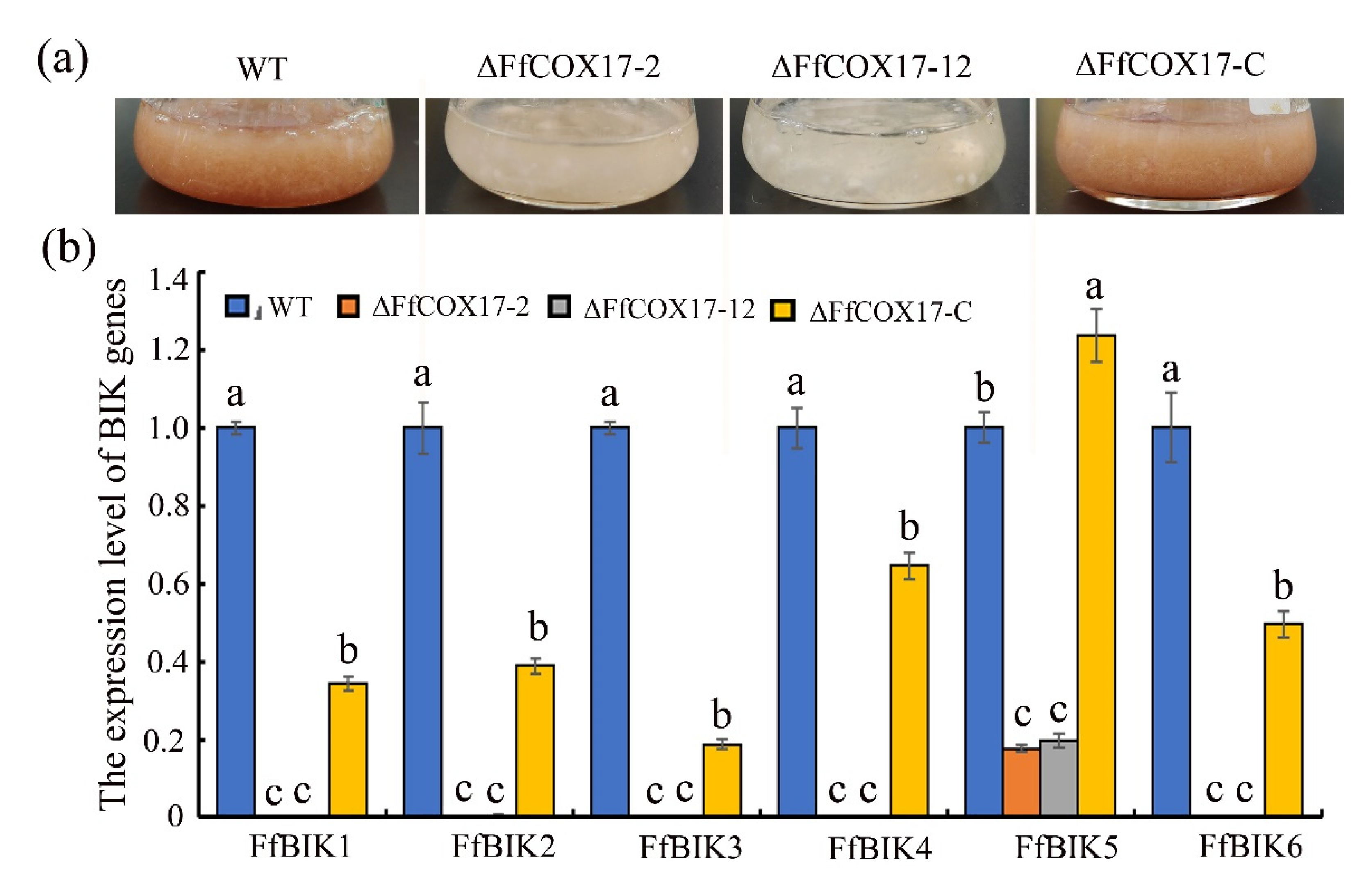
2.3. FfCOX17 Regulates the Expression Level of the BIK Synthesis-Related Genes

As shown in Figure 3a, the ∆FfCOX17 mutants were not able to produce pigment when cultured in ICI liquid medium (containing 6 mM Gln), and the expression levels of all six BIK biosynthetic genes were downregulated in the ∆FfCOX17 mutants (Figure 3b). Interestingly, we found that the expression of FfBIK1, FfBIK2, FfBIK3, FfBIK4, and FfBIK6 genes are rarely detected in ∆FfCOX17 mutants, and the expression level of FfBIK5 decreased by about 80% (Figure 3b), which suggested that FfCOX17 positively regulates the expression of BIK synthesis related genes in F. fujikuroi.

2.4. FfCOX17 Negatively Regulates FUM Biosynthesis in F. fujikuroi

To determine the role of FfCOX17 in F. fujikuroi FUM biosynthesis, the FUM content was measured in the wild-type, ΔFfCOX17 mutants, and complemented strain. ΔFfCOX17 mutants exhibited a significant increase in FUM production compared to that in wild-type and complemented strain (Figure 4a). Next, we quantified the transcriptional changes of FfFUM2 (FFUJ_09248) gene. Figure 4b showed that the expression levels of the FfFUM2 gene in ∆FfCOX17 mutants were significantly increased, suggesting that FfCOX17 controls the FUM production by regulating the transcription level of the FfFUM2 gene.

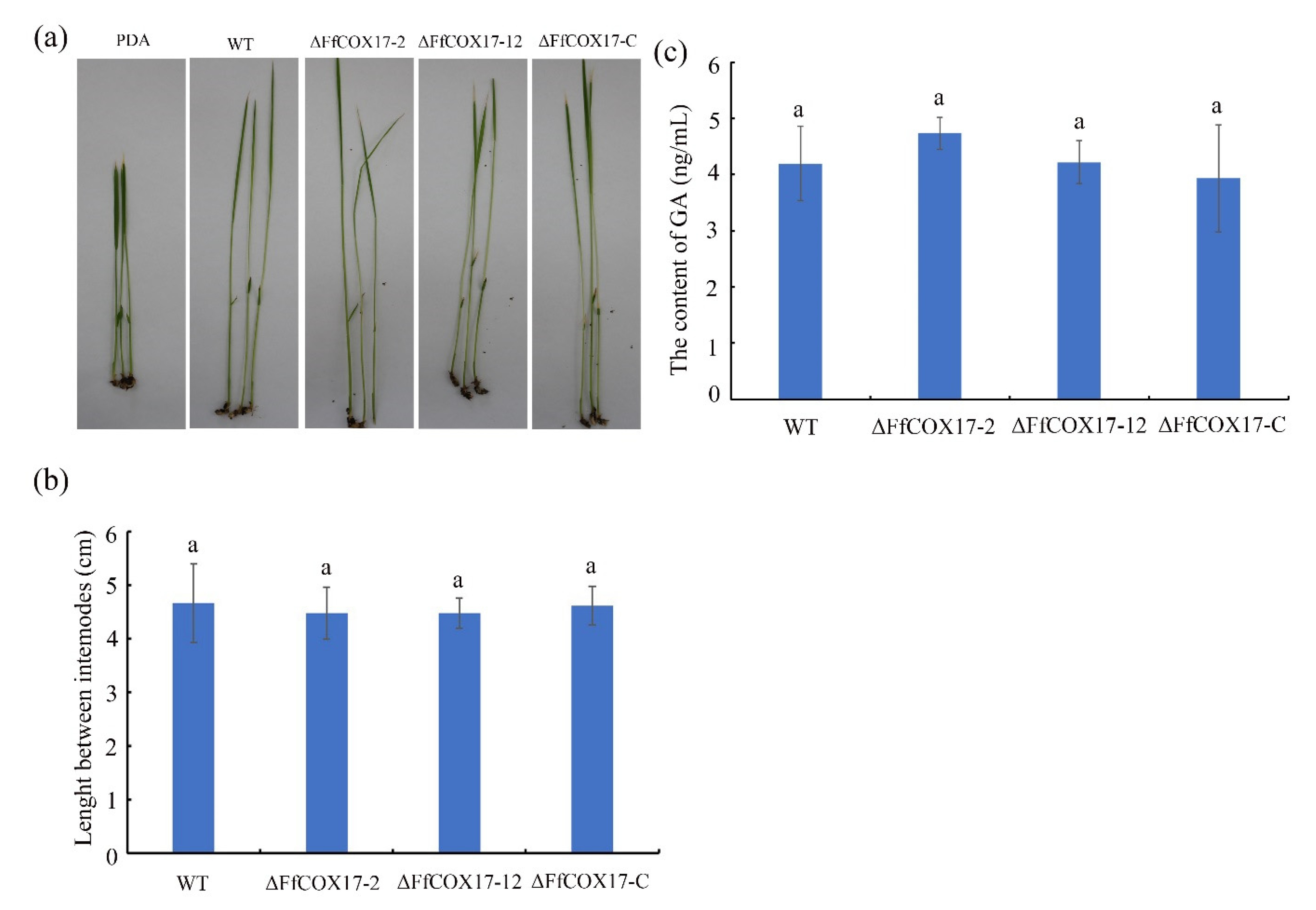
2.5. FfCOX17 Is Not Required for Pathogenicity

The rice seedlings infection assay was performed to assess the role of FfCOX17 in the pathogenicity of F. fujikuroi. The ∆FfCOX17 mutants caused similar lesion lengths compared with the wild-type strain (Figure 5a,b, Table 1), which indicated that FfCOX17 is not essential for plant infection by F. fujikuroi. To further confirm whether the pathogenicity is related to GA production, the GA content was measured using a GA ELISA detection kit. The GA content of the wild-type strain is 4.95 ng/mL, and the GA content of ∆FfCOX17 mutants is 4.73 ng/mL and 4.22 ng/mL, respectively, suggesting that the GA content in ∆FfCOX17 mutants was similar to the wild-type strain (Figure 5c).

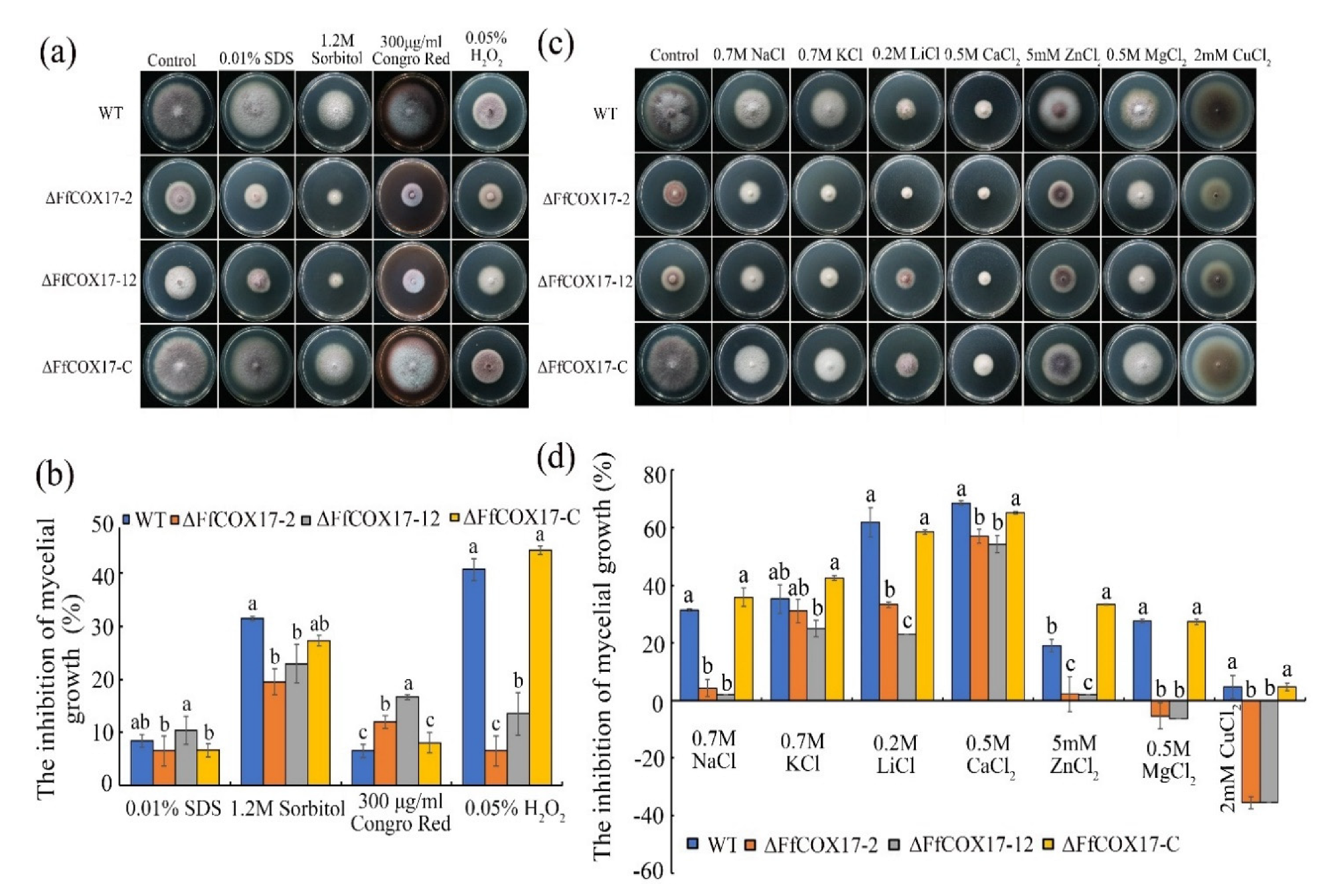
2.6. Sensitivity of the ∆FfCOX17 to Different Stresses

Environmental stress factors play an important role in the process of pathogen infection. As shown in Figure 6, ∆FfCOX17 displayed decreased sensitivity to 1.2 M Sorbitol, 0.05% H2O2, 2 mM CuCl2, 0.7 M NaCl, 0.2 M LiCl, 0.5 M CaCl2, 5 mM ZnCl2 and 0.5 M MgCl2, but significantly increased sensitivity to 300 μg/mL Congo Red (Figure 6a–d). The sensitivity of ΔFfCOX17 mutants to heat shock was also detected at different temperatures, and the results indicated that ΔFfCOX17 displayed increased sensitivity at 15 °C and 30 °C (Figure 7a,b). ΔFfCOX17 exhibited increased sensitivity to 40 g/L glucose, 80 g/L glucose, and decreased sensitivity to 10 g/L glucose (Figure 7c,d). All growth defects of ΔFfCOX17 mutants in response to different stresses were restored by complemented strain ΔFfCOX17-C. These data suggested that FfCOX17 is associated with membrane permeability, cell wall integrity, and sensitivity to environmental factors.

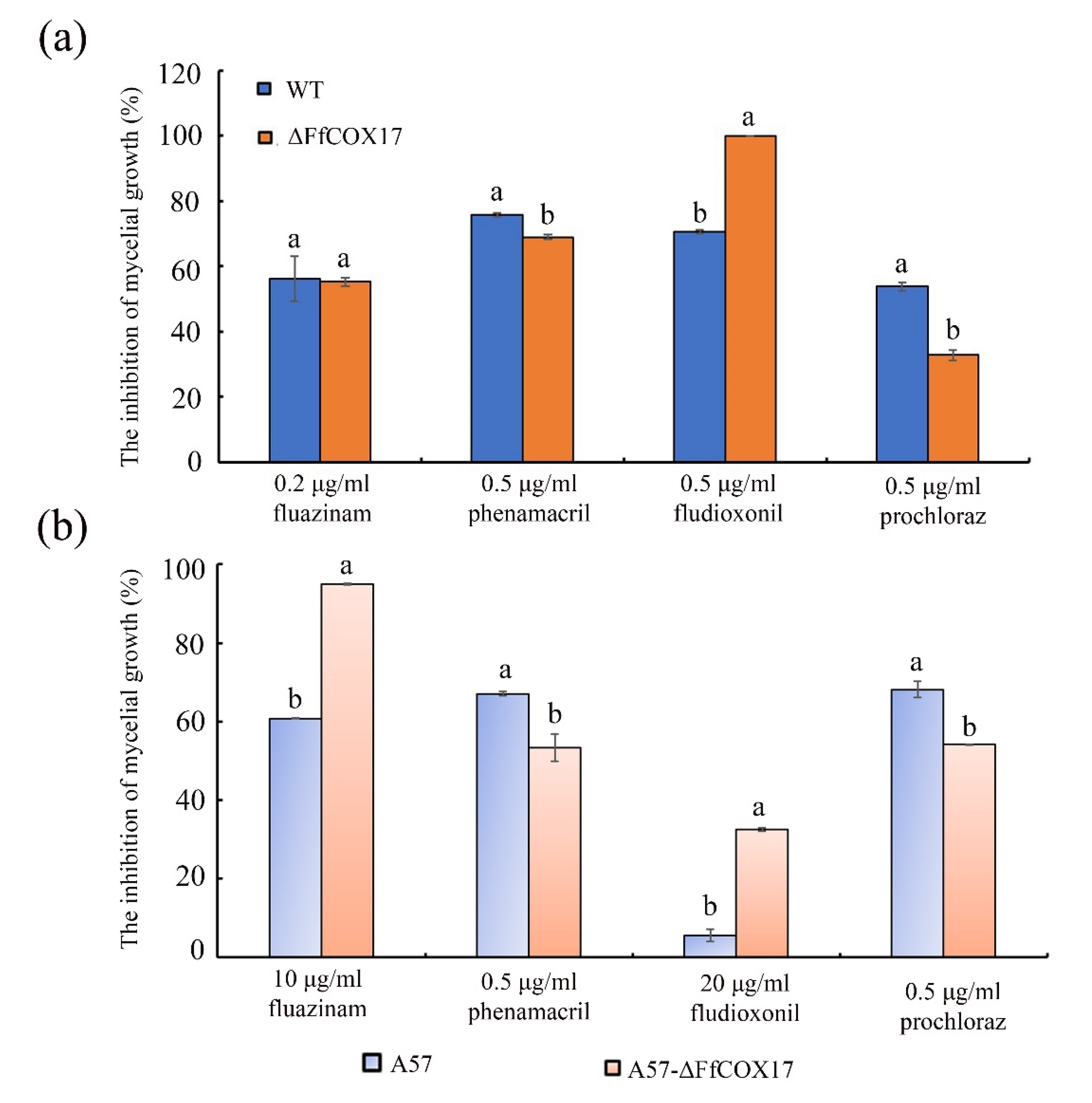
2.7. FfCOX17 Regulates the Sensitivity to Different Fungicides

The sensitivity of F. fujikuroi wild-type strain, fluazinam resistant strain A57, and the ΔFfCOX17 mutants in WT and A57 backgrounds to different fungicides were determined. Under the wild-type strain background, the inhibition rate of WT by 0.5 μg/mL fludioxonil was 70.66% but increased to 100% in the ΔFfCOX17 strains. However, the inhibition rate by 0.5 μg/mL prochloraz was 53.72% in the wild-type strain and decreased to 32.74% in ΔFfCOX17 (Figure 8a). Under the fluazinam resistant strain A57 background, the inhibition rate of A57 mycelium growth by 10 μg/mL fluazinam was 60.80% but increased to 95% in A57-ΔFfCOX17. Similarly, the inhibition rate of A57 by 20 μg/mL fludioxonil was 5.67% and increased to 32.50% in A57-ΔFfCOX17. However, the inhibition rate of A57-ΔFfCOX17 by 0.5 μg/mL phenamacril and 0.5 μg/mL prochloraz decreased compared to that of A57 (Figure 8b).

2.8. Subcellular Localization of GFP-FfCOX17 Fusion Protein

To determine the subcellular localization of FfCOX17, the GFP-FfCOX17 strain was generated. Figure 9 showed that the green fluorescence signals were visualized in the cytoplasm and mitochondria in mycelium and conidia as GFP signals and red signals of mitochondrial Mito marker partially overlapped, indicating that the FfCOX17 was localized in mitochondria and cytoplasm.

3. Discussion

In this study, the copper chaperone protein FfCOX17 was identified in F. fujikuroi. The growth rate of ΔFfCOX17 on the different mediums was significantly lower than that of the wild-type strain. Beyond that, the conidia production was significantly decreased, and the germ tube length of ΔFfCOX17 after germination for 12 h was shorter than wild-type strains, which indicates that FfCOX17 regulates the vegetative growth and asexual reproduction of F. fujikuroi. In S. cerevisiae and Arabidopsis, the copper chaperone protein COX17 is essential to cell growth and stress response [22,25]. COX17 is involved in CcO assembly in yeast and mammalian cells [26]. Previous studies proved that the deletion or silence of the AtCOX17 gene could lead to the growth defect in Arabidopsis [22]. In yeast, COX17 is located in mitochondria and affects cell respiration [26]. COX17 knockout could cause cell respiratory defects in mice [27]. In A. nidulans, the COX17 deletion mutant significantly reduced the mycelial growth rate and formed a small non-reproducible aconidial colony, indicating that COX17 is a necessary gene in A. nidulans [24]. The above results indicated that the COX17 homologous gene has functional characteristics.
Our results showed that the red pigment of the ∆FfCOX17 decreased significantly in the ICI medium. Polyketide synthase gene BIK has been proved to be a factor in the formation of red pigment of mycelial and a total of six genes were involved in BIK synthesis in F. fujikuroi [28]. Interestingly, the expression levels of the six BIK genes were significantly decreased in ∆FfCOX17 relative to the wild-type strain, suggesting that FfCOX17 could regulate the pigment formation of F. fujikuroi by reducing the expression levels of BIK cluster genes. Filamentous fungi produce a variety of secondary metabolites and play different roles in cell physiological and biochemical processes [29,30,31]. Fumonisins could cause several animal diseases and are associated with some human diseases, which can inhibit ceramide synthase [10]. Previous studies indicated that deletion of FvSEC4, FvDIM5, and FvCPSA led to increased production of fumonisin [7,16,32]. In this study, ∆FfCOX17 was found to increase FUM content compared to the wide-type strain. Furthermore, the expression level of the FfFUM2 gene was significantly increased in ∆FfCOX17, which suggested that FfCOX17 regulates the FUM content by increasing the expression levels of the FfFUM2 gene. RBD caused by F. fujikuroi results in abnormal elongation of plants, reduction of tillers, sterility, or empty grains, and most of these symptoms are caused by the production of plant hormone GA [6]. The content of GA and pathogenicity in the ∆FfCOX17 mutant strain did not change compared with the control strain.
In yeast, COX17 can transport copper between the mitochondrial inner membrane and cytoplasm [33]. Silencing of the AtCOX17 gene resulted in decreased response to salt stress, and AtCOX17 is necessary for stress response gene expression levels in Arabidopsis [22]. In yeast and mammals, COX17 protein contains six conserved cysteine residues, which are involved in redox reaction and metal binding and transport, respectively [26]. Mammalian COX17 exists in three oxidation states, COX170S-S, COX172S-S, and COX173S-S, respectively. COX170S-S combines with Cu+; COX172S-S binds to Cu+ or Zn2+; COX173S-S does not bind to any metal [34,35]. The ∆FfCOX17 mutants showed decreased sensitivity to metal ion such as 5 mM ZnCl2, 2 mM CuCl2, 0.5 M MgCl2 and 0.7 M NaCl. In addition, the ∆FfCOX17 mutants displayed decreased sensitivity to oxidative stress factors such as 0.05% H2O2 and increased sensitivity to cell wall-damaging agents 300 μg/mL Congo Red, which indicated the cell wall integrity of ∆FfCOX17 was destroyed. In B. cinerea and S. sclerotiorum, there was positive cross-resistance between fludioxonil and fluazinam [36,37]. Fludioxonil can induce glycerol biosynthesis and interfere with osmotic signal transduction in C. albicans [38]. Deletion of FfCOX17 increased the sensitivity of F. fujikuroi to fluazinam and fludioxonil, but significantly decreased sensitivity to phanamacril and procloraz. ∆FfCOX17 displayed decreased sensitivity to osmotic stress factor 0.7M NaCl and 1.2M Sorbitol, which may be due to the osmotic pathway being disturbed, increasing the sensitivity of F. fujikuroi to fungicides fluazinam and fludioxonil. The results indicated that fluazinam or fludioxonil could be combined with phenamacril or procloraz as an effective fungicide strategy to control RBD.

4. Conclusions

In summary, we identified the copper chaperone protein FfCOX17 in F. fujikuroi, and a localization study found that FfCOX17 is located in mitochondria and cytoplasm. FfCOX17 deletion mutants showed a decrease in vegetative growth and asexual reproduction, sensitivity to oxidative stress, osmotic stress, and increased sensitivity to cell wall stress, heat shock stress, and high concentration glucose. In addition, ∆FfCOX17 also showed increased sensitivity to fungicide fluazinam and fludioxonil and decreased sensitivity to phenamacril and prochloraz. Interestingly, the transcriptional level of the FfFUM2 gene was significantly upregulated in ∆FfCOX17, and the fumonisin production in the ∆FfCOX17 mutants was significantly increased, but the FfCOX17 is not related to virulence. Future studies will focus on analyzing the molecular mechanism of FfCOX17 negatively regulating FUM production.

5. Materials and Methods

5.1. Fungal Strains, Media, and Culture Conditions

The wild-type strain WT of F. fujikuroi was collected from rice fields in Jiangsu Province of China in 2019. Briefly, the disease sample of RBD was randomly collected and disinfected in a sodium hypochlorite solution (5% available chlorine) for 45 s. Then, they were rinsed thrice with sterile water and dried. The disinfested disease sample was placed on a potato dextrose agar (PDA) plate containing 100 μg/mL streptomycin sulfate. The PDA plate was incubated at 25 °C for 5 days. Purified strains of F. fujikuroi were obtained by the single spore method. The strain was maintained on PDA slants at 4 °C. Finally, the wild-type strain A was verified by ITS sequencing and morphology. WT strain and fluazinam-resistant strain A57 (induced in the laboratory) were used as parental strains to obtain the deletion mutants of FfCOX17, and the complementary strain was obtained from the ΔFfCOX17 mutant.
PDA contains 200 g of potato, 20 g glucose, 16 g of agar, and 1 L of water. Complete medium (CM) was made from 50 mL nitrate salts (NaNO3 120 g, KCl 10.4 g, MgSO4·7H2O 10.4 g, KH2PO4 30.4 g per liter of distilled water), 1 mL trace element (ddH2O 80 mL, ZnSO4·7H2O 2.2 g, H3BO3 1.1 g, MnCl2·4H2O 0.5 g, FeSO4·7H2O 0.5 g, CoCl2·6H2O 0.17 g, CuSO4·5H2O 0.16 g, Na2MoO4·2H2O 0.15 g, Na4EDTA 5 g, pH 6.5 per liter of distilled water), 10 g glucose, 2 g peptone, 1 g yeast extract, 1 g casein hydrolysate, 1 mL 1% thiamine, 50 μL 0.05% biotin solution, 15 g agar, pH 6.5 per liter of distilled water. Minimal medium (MM) consisted of 50 mL nitrate salts, 1 mL trace element, 10 g glucose, 1 mL 1% thiamine, 50 μL 0.05% biotin solution, 18 g agar, pH6.5 per liter of distilled water. V8 medium was made from 200 mL V8 juice, 2 g CaCO3, and 20 g agar per liter of distilled water. YEPD medium consisted of 20 g glucose, 10 g peptone, and 3 g yeast extract per liter of distilled water. Spore production was measured after incubation for 7 days in carboxymethylcellulose (CMC) liquid medium (15 g carboxymethyl cellulose, 0.5 g NH4NO3, 0.5 g KH2PO4, 0.25 g FeSO4·7H2O, 0.5 g yeast extract per liter of distilled water).

5.2. Construction of Vectors, Fungal Transformation and Generation of Gene Deletion

To investigate the functions of FfCOX17 in F. fujikuroi, we generated two independent FfCOX17 deletion mutants. A gene replacement carrier ΔFfCOX17 carrying the hygromycin resistance gene (hph) and herpes simplex virus thymidine kinase gene (F2du), an upstream fragment (5’) of FfCOX17, and downstream fragment (3’) of FfCOX17 were amplified from the genome DNA of WT with primers listed in Table S1, the 3490 bp hph-hsv (hph and F2du) fragment was amplified from the hph-hsv plasmid DNA with primers hph-hsv-UF/hph-hsv-UR, the three fragments were fused by single point PCR split-marker approach [29]. The fusion product was amplified using primers FfCOX17-UF/FfCOX17-DR and added to the protoplast of the wild-type strain. We used a 50 μL polymerase chain reaction (PCR) system including 25 μL LAmp Master Mix (Vazyme Biotech Co., Ltd, Nanjing), 2 μL forward primer, 2 μL reverse primer, 1 μL total DNA, and 20 μL water. Reaction procedure: predenaturation at 94 °C for 5 min; 35 cycles: denaturation at 94 °C for 30 s; annealing at 56 °C for 30 s, extension at 72 °C for 30 s/kb; thoroughly extend at 72 °C for 7 min. The protoplasts were prepared from F. fujikuroi hyphae according to previous research [39]. All of the transformants were verified by PCR with different primers (Table S1) and further verified by Southern blotting.
To construct the FfCOX17-GFP fusion vector, the GFP fusion fragment of FfCOX17 was amplified using primers FfCOX17-RP27-GFP-F/FfCOX17-GFP-R and cloned into Pyf11 plasmid vector (XhoI digestion) using 2MultiF Seamless Assembly Mix (ABclonal Technology Co., Ltd, Wuhan), and then transferred to E. coli (DH5α) for amplification. The FfCOX17-GFP fusion vector was added to the protoplast of the wild-type strain to obtain the FfCOX17-GFP strain. The fluorescence signal (wavelength range of green fluorescence is 460 nm~550 nm) was taken under a confocal microscope (Leica TCS SP8).

5.3. Test for Vegetative Growth and Asexual Reproduction

The wild-type strain, deletion mutants ΔFfCOX17 (ΔFfCOX17-2 and ΔFfCOX17-12) and complement strain ΔFfCOX17-C were cultured on a PDA medium for 6 days, a 5 mm plug was cut from the colony margin and placed on PDA, V8, CM, and MM medium at 25 °C for 7 days. Each treatment had three replicates, and the diameter of each plate was measured after seven days of culture.
For asexual reproduction, three mycelial plugs (diameter: 5 mm) of different strains were taken from the colony’s edge which was cultured on a PDA medium for 6 days and then transferred into a 250 mL flask containing 100 mL CMC liquid medium. All of the flasks were shaken at 25 °C, 175 rpm for 7 days. The number of conidia in the CMC liquid medium of each strain was determined by hemocytometer. The experiments were performed three times with three replicates for each treatment.

5.4. Pathogenicity Assays

The seeds of rice variety Ninggeng 7 were prepared, and the surface was disinfected. Briefly, the peeled rice seeds were sterilized with 75% ethanol for one minute, rinsed with sterile water three times, soaked with sodium hypochlorite (4% available chlorine) for 10 min, and rinsed with sterile water three times. The sterilized seeds were transferred into water agar plates (15 g/L agar) cultured for 4 days at low temperature (4 °C), and then transferred to a 28 °C light incubator (alternating light and dark for 12 h) for germination for 3 days. Place the germinated seeds in 3 × 20 cm test tubes (filled with 25% vermiculite), the mycelial plug (5 mm in diameter) of different strains cultured on a PDA medium for 6 days was transferred into the test tubes and add 3 mL Gamborg B5 solution (3.16 g/L) (Duchefa Biochemie B.V. Holland) to each test tube. The mycelial plug of the PDA plate was added as a control. The test tubes were placed in a light incubator at 28 °C for 12 h-light and 12 h-dark cycle conditions for 7 days. Finally, the length of the seedling was measured from the stem base to the second root nodule.

5.5. FUM and GA Content Assay

To determine the content of FUM and GA, three mycelial plugs were cut from the colony margin of cultured on a PDA medium for 6 days and transferred into the conical flask containing 100 mL ICI liquid medium (containing 6 mM Gln) [6], the flasks were shaken at 28 °C, 175 rpm for one week in darkness. After 7 days of culture, the culture solution was collected for the determination of FUM or GA content. The 50 μL sample solution and standards were added to the microwell plate, respectively, joining 50 μL anti-FUM antibody conjugate (or 50 μL anti-GA antibody conjugate), gently mixing for a few seconds, 37 °C warm bath 30 min, wash 5 times, add the chromogen solution at 37 °C and incubate it again for 10 min, add the stop solution, detect the absorbance at 450 nm, and the FUM or GA content was calculated according to the standard curve.

5.6. Quantitative RT-PCR (qPCR)

For gene expression, three mycelial plugs were cut from the colony margin of cultured on a PDA medium for 6 days and placed in the conical flask containing 100 mL ICI liquid medium (containing 6 mM Gln), the flasks were shaken at 28 °C, 175 rpm for 48 h in darkness. RNA samples were isolated from 48 h hyphal with RNAsimple Total RNA Kit (Tiangen Biotech CO., Ltd, Beijing, China). The first-strand cDNA was synthesized by HiScript II RT SuperMix for qPCR (+gDNA wiper) (Vazyme Biotech Co., Ltd, Nanjing, China). qPCR was performed with ChamQ SYBR qPCR Master Mix (Vazyme Biotech Co., Ltd, Nanjing, China) by CFX Connect Real-Time System (Bio-Rad, USA) [40]. To quantify mRNA levels of FfFUM (FUM2, FFUJ_09248), FfBIK1 (FFUJ_06742), FfBIK2 (FFUJ_06743), FfBIK3 (FFUJ_06744), FfBIK4 (FFUJ_06745), FfBIK5 (FFUJ_06746), and FfBIK6 (FFUJ_06747), we used primers FfFUM2-DL-F/FfFUM2-DL-R, FfBIK1-DL-F/FfBIK1-DL-R, FfBIK2-DL-F/FfBIK2-DL-R, FfBIK3-DL-F/FfBIK3-DL-R, FfBIK4-DL-F/FfBIK4-DL-R, FfBIK5-DL-F/FfBIK5-DL-R, and FfBIK6-DL-F/FfBIK6-DL-R, respectively (Table S1). The actin gene (FFUJ_05652) was used as an internal reference gene. The relative expression level of different genes was calculated according to the reference gene using the 2−ΔΔCt method.

5.7. Sensitivity of the ΔFfCOX17 Mutants to Different Stress

To determine the sensitivity of ΔFfCOX17 to different stresses, the mycelia plug (diameter 5 mm) was taken from the edge of the colony, which was cultured on a PDA medium for 6 days and placed on the PDA plate amended with different metal cation (0.7 M NaCl, 0.7 M KCl, 0.2 M LiCl, 0.5 M CaCl2, 5 mM ZnCl2, 0.5 M MgCl2 or 2 mM CuCl2), 300 μg/mL Congo Red (cell wall stress factor), 0.01% SDS (cell membrane stress factor), or 0.05% H2O2 (oxidative stress). In addition, some mycelial plugs were transferred into the PDA plate containing 10 g/L glucose, 20 g/L glucose, 40 g/L glucose, and 80 g/L glucose. All PDA plates were cultured in the incubator under dark conditions for 7 days. For the sensitivity of ΔFfCOX17 to heat shock, the mycelia plugs were placed on the PDA plate and incubated at 15 °C, 25 °C, or 30 °C incubators for 7 days in darkness. The colony diameter of each treatment was measured and the inhibition rate was calculated using the formula: inhibition rate = (the diameter of the treatment − the diameter of control)/(the diameter of control − 0.5) × 100. Each treatment had three repetitions, and the experiment was repeated three times independently.

5.8. Determination of the Sensitivity of F. fujikuroi to Different Fungicides

The wild-type strain, ΔFfCOX17 mutants, and complemented strain ΔFfCOX17-C were used to determine the sensitivity of F. fujikuroi to different fungicides. A 5 mm diameter mycelial plug was cut from the edge of the 6 days PDA colony and transferred onto the PDA plates amended with 0.2 μg/mL fluazinam, 0.5 μg/mL phenamacril, 0.5 μg/mL fludioxonil, and 0.5 μg/mL prochloraz (sensitive strain and ΔFfCOX17 mutant), or 10 μg/mL fluazinam, 0.5 μg/mL phenamacril, 20 μg/mL fludioxonil and 0.5 μg/mL prochloraz (fluazinam-resistant strain and ΔFfCOX17 mutant). The colony diameter was measured after it was incubated at 25 °C for 7 days in darkness and used to calculate the mycelial growth inhibition.

5.9. Statistical Analysis

The experimental data were analyzed using SPSS statistical software. Statistical analysis was performed using one-way variance (ANOVA), followed by the Tukey multiple comparison test. The level of significance was set at p < 0.05. All of the experiments were performed three times with three replicates for each treatment. Adobe Photoshop CS5 was used to draw pictures, and Excel, PowerPoint, and other office software were used to sort out relevant data and draw basic charts.

Supplementary Materials

The following are available online at: https://www.mdpi.com/article/10.3390/toxins14070427/s1, Table S1: Primers used in this study.

Author Contributions

Conceptualization, M.Z. and Y.H.; Experiments and data analysis, X.M., Z.W.; and F.C.; Writing—original draft preparation X.M., M.Z. and Y.H. All authors have read and agreed to the published version of the manuscript.

Funding

The study was supported by the National Natural Science Foundation of China (31972307), Jiang Su Key Research and development program (BE2021361), and “the Six Talent Peaks” Project in Jiangsu Province (NY-040).

Institutional Review Board Statement

Not applicable.

Informed Consent Statement

Not applicable.

Data Availability Statement

Strains and plasmids are available upon request. This research contains the original method of this study, and further inquiries can be directly contacted by the corresponding authors.

Conflicts of Interest

The authors declare no conflict of interest.

References

  1. Janevska, S.; Güldener, U.; Sulyok, M.; Tudzynski, B.; Studt, L. Set1 and Kdm5 are antagonists for H3K4 methylation and regulators of the major conidiation-specific transcription factor gene ABA1 in Fusarium fujikuroi. Environ. Microbiol. 2018, 20, 3343–3362. [Google Scholar] [CrossRef] [PubMed] [Green Version]
  2. Zhang, Y.; Mao, C.X.; Zhai, X.Y.; Jamieson, P.A.; Zhang, C. Mutation in cyp51b and overexpression of cyp51a and cyp51b confer multiple resistant to DMIs fungicide prochloraz in Fusarium fujikuroi. Pest Manag. Sci. 2021, 77, 824–833. [Google Scholar] [CrossRef] [PubMed]
  3. Matic, S.; Gullino, M.L.; Spadaro, D. The puzzle of bakanae disease through interactions between Fusarium fujikuroi and rice. Front. Biosci. 2017, 9, 333–344. [Google Scholar] [CrossRef] [Green Version]
  4. Bao, W.X.; Inagaki, S.; Tatebayashi, S.; Sultana, S.; Shimizu, M.; Kageyama, K.; Suga, H. Expression difference of P450-1 and P450-4 between G- and F-groups of Fusarium fujikuroi. Eur. J. Plant Pathol. 2021, 159, 27–36. [Google Scholar] [CrossRef]
  5. Proctor, R.H.; Plattner, R.D.; Brown, D.W.; Seo, J.A.; Lee, Y.W. Discontinuous Distribution of the Fumonisin Biosynthetic Gene Cluster in Fusarium. Mycol. Res. 2004, 108, 815–822. [Google Scholar] [CrossRef] [PubMed]
  6. Wiemann, P.; Brown, D.W.; Kleigrewe, K.; Jin, W.B.; Keller, N.P.; Humpf, H.U.; Tudzynski, B. FfVel1 and FfLae1, components of a velvet-like complex in Fusarium fujikuroi, affect differentiation, secondary metabolism and virulence. Mol. Microbiol. 2010, 77, 972–994. [Google Scholar] [CrossRef] [Green Version]
  7. Deng, Q.; Wu, H.X.; Gu, Q.; Tang, G.F.; Liu, W.D. Glycosyltransferase FvCpsA Regulates Fumonisin Biosynthesis and Virulence in Fusarium verticillioides. Toxins 2021, 13, 718. [Google Scholar] [CrossRef]
  8. Myung, K.; Li, S.J.; Butchko, R.A.E.; Busman, M.; Proctor, R.H.; Abbas, H.K.; Calvo, A.M. FvVE1 Regulates Biosynthesis of Fumonisins and Fusarins in Fusarium verticillioides. J. Agric. Food Chem. 2009, 57, 5089–5094. [Google Scholar] [CrossRef] [Green Version]
  9. Gelderblom, W.C.A.; Jaskiewicz, K.; Marasas, W.F.O.; Thiel, P.G.; Horak, M.J.; Vleggaar, R.; Kriek, N.P.J. Fumonisins-novel mycotoxins with cancer-promoting activity produced by Fusarium moniliforme. Appl. Environ. Microbiol. 1988, 54, 1806–1811. [Google Scholar] [CrossRef] [Green Version]
  10. Desai, K.; Sullards, M.C.; Allegood, J.; Wang, E.; Schmelz, E.M.; Hartl, M.; Humpf, H.U.; Liotta, D.C.; Peng, Q.; Merrill, A.H., Jr. Fumonisins and fumonisin analogs as inhibitors of ceramide synthase and inducers of apoptosis. Biochim. Biophys. Acta 2002, 1585, 188–192. [Google Scholar] [CrossRef]
  11. Sultana, S.; Kitajim, M.; Kobayash, H.; Nakagawa, H.; Shimizu, M.; Kageyama, K.; Suga, H. A Natural Variation of Fumonisin Gene Cluster Associated with Fumonisin Production Difference in Fusarium fujikuroi. Toxins 2019, 11, 200. [Google Scholar] [CrossRef] [PubMed] [Green Version]
  12. Woloshuk, C.P.; Shim, W.B. Aflatoxins, fumonisins, and trichothecenes: A convergence of knowledge. FEMS Microbiol. Rev. 2013, 37, 94–109. [Google Scholar] [CrossRef] [PubMed] [Green Version]
  13. Fanelli, F.; Iversen, A.; Logrieco, A.F.; Mulè, G. Relationship between fumonisin production and FUM gene expression in Fusarium verticillioides under different environmental conditions. Food Addit. Contam. 2013, 30, 365–371. [Google Scholar] [CrossRef] [PubMed]
  14. Zhang, Y.; Choi, Y.E.; Zou, X.; Xu, J.R. The FvMK1 mitogen-activated protein kinase gene regulates conidiation, pathogenesis, and fumonisin production in Fusarium verticillioides. Fungal Genet. Biol. 2011, 48, 71–79. [Google Scholar] [CrossRef]
  15. Wu, Y.; Li, T.T.; Gong, L.; Wang, Y.; Jiang, Y.M. Effects of Different Carbon Sources on Fumonisin Production and FUM Gene Expression by Fusarium proliferatum. Toxins 2019, 11, 289. [Google Scholar] [CrossRef] [Green Version]
  16. Gu, Q.; Ji, T.T.; Sun, X.; Huang, H.; Zhang, H.; Lu, X.; Wu, L.; Huo, R.; Wu, H.; Gao, X.W. Histone H3 lysine 9 methyltransferase FvDim5 regulates fungal development, pathogenicity and osmotic stress responses in Fusarium verticillioides. FEMS Microbiol. Lett. 2017, 364. [Google Scholar] [CrossRef] [Green Version]
  17. Punter, F.A.; Glerum, D. Mutagenesis reveals a specific role for Cox17p in copper transport to cytochrome oxidase. J. Biol. Chem. 2003, 278, 30875–30880. [Google Scholar] [CrossRef] [Green Version]
  18. Babcock, G.T.; Wikström, M.J.N. Oxygen activation and the conservation of energy in cell respiration. Nature 1992, 356, 301–309. [Google Scholar] [CrossRef]
  19. Takahashi, Y.; Kako, K.; Kashiwabara, S.; Takehara, A.; Inada, Y.; Arai, H.; Nakada, K.; Kodama, H.; Hayashi, J.; Baba, T. Mammalian copper chaperone Cox17p has an essential role in activation of cytochrome C oxidase and embryonic development. Mol. Cell. Biol. 2002, 22, 7614–7621. [Google Scholar] [CrossRef] [Green Version]
  20. Shoubridge, E. Cytochrome c oxidase deficiency. Am. J. Med. Genet. 2001, 106, 46–52. [Google Scholar] [CrossRef]
  21. Banci, L.; Bertini, I.; Ciofi-Baffoni, S.; Hadjiloi, T.; Martinelli, M.; Palumaa, P. Mitochondrial copper (I) transfer from Cox17 to Sco1 is coupled to electron transfer. Proc. Natl. Acad. Sci. USA 2008, 105, 6803–6808. [Google Scholar] [CrossRef] [PubMed] [Green Version]
  22. Garcia, L.; Welchen, E.; Gey, U.; Arce, A.L.; Steinebrunner, I.; Gonzalez, D.H. The cytochrome c oxidase biogenesis factor AtCOX17 modulates stress responses in Arabidopsis. Plant Cell Environ. 2016, 39, 628–644. [Google Scholar] [CrossRef] [PubMed]
  23. Balandin, T.; Castresana, C. AtCOX17, an Arabidopsis homolog of the yeast copper chaperone COX17. Plant Physiol. 2002, 129, 1852–1857. [Google Scholar] [CrossRef] [Green Version]
  24. Anabosia, D.; Meira, Z.; Shadkchana, Y.; Handelmana, M.; Abou-Kandila, A.; Yapb, A.; Urlingsa, D.; Golda, M.S.; Krappmannc, S.; Haasb, H.; et al. Transcriptional response of Aspergillus fumigatus to copper and the role of the Cu chaperones. Virulence 2021, 12, 2186–2200. [Google Scholar] [CrossRef] [PubMed]
  25. Leary, S.C.; Winge, D.R.; Cobine, P. “Pulling the plug” on cellular copper: The role of mitochondria in copper export. Biochim. Biophys. Acta 2009, 1793, 146–153. [Google Scholar] [CrossRef] [PubMed] [Green Version]
  26. Voronova, A.; Meyer-Klaucke, W.; Meyer, T.; Rompel, A.; Krebs, B.; Kazantseva, J.; Sillard, R.; Palumaa, P. Oxidative switches in functioning of mammalian copper chaperone Cox17. Biochem. J. 2007, 408, 139–148. [Google Scholar] [CrossRef] [Green Version]
  27. Maxfield, A.B.; Heaton, D.N.; Winge, D. Cox17 is functional when tethered to the mitochondrial inner membrane. J. Biol. Chem. 2004, 279, 5072–5080. [Google Scholar] [CrossRef] [PubMed] [Green Version]
  28. Wiemann, P.; Willmann, A.; Straeten, M.; Kleigrewe, K.; Beyer, M.; Humpf, H.U.; Tudzynski, B. Biosynthesis of the red pigment bikaverin in Fusarium fujikuroi: Genes, their function and regulation. Mol. Microbiol. 2009, 72, 931–946. [Google Scholar] [CrossRef]
  29. Michielse, C.B.; Studt, L.; Janevska, S.; Sieber, C.M.; Arndt, B.; Espino, J.J.; Humpf, H.U.; Güldener, U.; Tudzynski, B. The global regulator FfSge 1 is required for expression of secondary metabolite gene clusters but not for pathogenicity in Fusarium fujikuroi. Environ. Microbiol. 2015, 17, 2690–2708. [Google Scholar] [CrossRef]
  30. Brakhage, A. Regulation of fungal secondary metabolism. Nat. Rev. Microbiol. 2013, 11, 21–32. [Google Scholar] [CrossRef]
  31. Niehaus, E.M.; Rindermann, L.; Janevska, S.; Münsterkötter, M.; Güldener, U.; Tudzynski, B. biotechnology: Analysis of the global regulator Lae1 uncovers a connection between Lae1 and the histone acetyltransferase HAT1 in Fusarium fujikuroi. Appl. Microbiol. Biotechnol. 2018, 102, 279–295. [Google Scholar] [CrossRef] [PubMed]
  32. Yan, H.; Huang, J.; Zhang, H.; Shim, W.B. A Rab GTPase protein FvSec4 is necessary for fumonisin B1 biosynthesis and virulence in Fusarium verticillioides. Curr. Genet. 2019, 66, 205–216. [Google Scholar] [CrossRef] [PubMed]
  33. Palumaa, P. Copper chaperones. The concept of conformational control in the metabolism of copper. FEBS Lett. 2013, 587, 1902–1910. [Google Scholar] [CrossRef] [Green Version]
  34. Palumaa, P.; Kangur, L.; Voronova, A.; Sillard, R. Mechanism of metal binding by Cox17, a copper chaperone for cytochrome c oxidase. Biochem. J. 2004, 382, 307–314. [Google Scholar] [CrossRef] [PubMed] [Green Version]
  35. Arnesano, F.; Balatri, E.; Banci, L.; Bertini, I.; Winge, D.R. Folding studies of Cox17 reveal an important interplay of cysteine oxidation and copper binding. Structure 2005, 13, 713–722. [Google Scholar] [CrossRef] [PubMed] [Green Version]
  36. Shao, W.Y.; Ren, W.C.; Zhang, Y.; Hou, Y.P.; Duan, Y.B.; Wang, J.X.; Zhou, M.G.; Chen, C.J. Baseline sensitivity of natural pop-ulations and characterization of resistant strains of Botrytis cinerea to fluazinam. Australas. Plant Pathol. 2015, 44, 375–383. [Google Scholar] [CrossRef]
  37. Mao, X.W.; Li, J.S.; Chen, Y.L.; Song, X.S.; Duan, Y.B.; Wang, J.X.; Chen, C.J.; Zhou, M.G.; Hou, Y.P. Resistance risk assessment for fluazinam in Sclerotinia sclerotiorum. Pestic. Biochem. Physiol. 2018, 144, 27–35. [Google Scholar] [CrossRef]
  38. Ochiai, N.; Fujimura, M.; Oshima, M.; Motoyama, T.; Ichiishi, A.; Yamada-Okabe, H.; Yamaguchi, I. Effects of iprodione and fludioxonil on glycerol synthesis and hyphal development in Candida albicans. Biosci. Biotechnol. Biochem. 2002, 66, 2209–2215. [Google Scholar] [CrossRef] [Green Version]
  39. Zheng, Z.T.; Gao, T.; Zhang, Y.; Hou, Y.P.; Wang, J.X.; Zhou, M.G. FgFim, a key protein regulating resistance to the fungicide JS 399-19, asexual and sexual development, stress responses and virulence in Fusarium graminearum. Mol. Plant Pathol. 2014, 15, 488–499. [Google Scholar] [CrossRef]
  40. Zhu, Y.Y.; Zhang, Y.S.; Liu, N.; Ren, W.C.; Hou, Y.P.; Duan, Y.B.; Song, X.S.; Zhou, M.G. The Dis1/Stu2/XMAP215 Family Gene FgStu2 Is Involved in Vegetative Growth, Morphology, Sexual and Asexual Reproduction, Pathogenicity and DON Production of Fusarium graminearum. Front. Microbiol. 2020, 11, 545015. [Google Scholar] [CrossRef]
Figure 1. Identification, deletion, and complementation of FfCOX17; (a) Phylogenetic analysis of COX17 from S. cerevisiae, A. oryzae, N. crasse, F. graminearun, F. fujikuroi, F. odoratissimum, P. oryzae, and B. cinerea. (b) The ΔFfCOX17 knockout vector was constructed via a homologous replacement strategy. (c) Southern blotting analysis of wild-type strain WT, ΔFfCOX17-2, ΔFfCOX17-12, and ΔFfCOX17-C using a 500 bp FfCOX17 upstream fragment as a probe, and genomic DNA digested with HindIII.
Toxins 14 00427 g001
Figure 2. The effects of FfCOX17 on vegetative growth and asexual reproduction; (a) Colony morphology of the wild-type strain WT and the mutant strains of FfCOX17 on PDA, V8, CM, and MM medium at 25 °C for 7 days. WT: the wild-type strain; ΔFfCOX17-2 and ΔFfCOX17-12: the deletion mutant of FfCOX17; ΔFfCOX17-C: complemented strains of FfCOX17 deletion mutant; (b) The diameter of mycelial growth of wild-type strain WT, ΔFfCOX17-2, and ΔFfCOX17-12 on PDA, V8, CM and MM media at 25 °C for 7 days. (c) The hyphal tip morphology of the mycelium of wild-type strain WT, ΔFfCOX17-2, ΔFfCOX17-12, and complement strain. (d) The conidia production of wild-type strain WT, ΔFfCOX17-2, ΔFfCOX17-12, and complement strain cultured in CMC liquid medium for 7 days. (e) The spore morphology and germination of WT, ΔFfCOX17-2, ΔFfCOX17-12, and complement strain were cultured in CMC liquid medium. Values in each column with the same letter are not significantly different (Tukey; p = 0.05).
Toxins 14 00427 g002
Figure 3. The effects of FfCOX17 on the expression level of BIK biosynthesis-related genes; (a) the pigment changes of wild-type, ΔFfCOX17-2, ΔFfCOX17-12 and complement strain in ICI liquid medium at 25 °C for 7 days (175 rpm). (b) The relative expression levels of BIK genes in the mutant strains of ΔFfCOX17 on ICI liquid medium at 25 °C for 48 h (175 rpm). FfBIK1 (FFUJ_06742), FfBIK2 (FFUJ_06743), FfBIK3 (FFUJ_06744), FfBIK4 (FFUJ_06745), FfBIK5 (FFUJ_06746), FfBIK6 (FFUJ_06747). Values in each column with the same letter are not significantly different (Tukey; p = 0.05).
Toxins 14 00427 g003
Figure 4. The effects of FfCOX17 on FUM production. (a) The content of FUM of wild-type strain, ΔFfCOX17 deletion mutants, and complement strain. (b) The expression levels of the FfFUM gene (FUM2, FFUJ_09248) in wild-type strain WT, ΔFfCOX17-2, ΔFfCOX17-12, and complement strain cultured in ICI liquid medium for 48 h. Values in each column with the same letter are not significantly different (Tukey; p = 0.05).
Toxins 14 00427 g004
Figure 5. The effects of FfCOX17 on pathogenicity of Fusarium fujikuroi. (a) Pathogenicity of wild-type, ΔFfCOX17-2, ΔFfCOX17-12 and complemented strain on rice seedlings. (b) Rice seedling lesion length of wild-type WT, ΔFfCOX17-2, ΔFfCOX17-12 and complemented strain. (c) The GA content of different strains. Values in each column with the same letter are not significantly different (Tukey; p = 0.05).
Toxins 14 00427 g005
Figure 6. Sensitivity of the ΔFfCOX17 to different stresses and metal cation; (a) Growth phenotype of wild-type WT, ΔFfCOX17-2, ΔFfCOX17-12 and complement strain on PDA amended with 0.01% SDS, 1.2 M Sorbitol, 300 μg/mL Congo Red, and 0.05% H2O2 after 7 days of incubation at 25 °C. (b) The inhibition of mycelial growth of wild-type WT, ΔFfCOX17-2, ΔFfCOX17-12 and complement strain to ΔFfCOX17 to 0.01% SDS, 1.2 M Sorbitol, 300 μg/mL Congo Red, and 0.05% H2O2. (c) The colony morphology of wild-type WT, ΔFfCOX17-2, ΔFfCOX17-12 and complement strain on PDA containing 0.7 M NaCl, 0.7 M KCl, 0.2 M LiCl, 0.5 M CaCl2, 5 mM ZnCl2, 0.5 M MgCl2 or 2 mM CuCl2 after 7 days of incubation at 25 °C. (d) The inhibition of mycelial growth of the ΔFfCOX17 to metal cation 0.7 M NaCl, 0.7 M KCl, 0.2 M LiCl, 0.5 M CaCl2, 5 mM ZnCl2, 0.5 M MgCl2 or 2 mM CuCl2. Values in each column with the same letter are not significantly different (Tukey; p = 0.05).
Toxins 14 00427 g006
Figure 7. Sensitivity of the ΔFfCOX17 to heat shock and different concentration glucose; (a) The colony morphology of wild-type, ΔFfCOX17-2, ΔFfCOX17-12 and complement strain to different temperatures (15 °C and 30 °C). (b) The inhibition of mycelial growth of wild-type WT, ΔFfCOX17-2, ΔFfCOX17-12, and complement strain at different temperatures (15 °C and 30 °C). (c) Sensitivity of wild-type WT, ΔFfCOX17-2, ΔFfCOX17-12 and complement strain to different concentration glucose. (d) The inhibition of mycelial growth of wild-type, ΔFfCOX17-2, ΔFfCOX17-12 and complement strain to 10 g/L Glucose, 40 g/L Glucose, and 80 g/L Glucose. Values in each column with the same letter are not significantly different (Tukey; p = 0.05).
Toxins 14 00427 g007
Figure 8. The sensitivity of ΔFfCOX17 to different fungicides. (a) The inhibition of mycelial growth of wild-type WT and ΔFfCOX17 mutants to 0.2 μg/mL fluazinam, 0.5 μg/mL phenamacril, 0.5 μg/mL fludioxonil and 0.5 μg/mL prochloraz. (b) The sensitivity of fluazinam-resistant strain A57 and A57-ΔFfCOX17 mutant to 10 μg/mL fluazinam, 0.5 μg/mL phenamacril, 20 μg/mL fludioxonil and 0.5 μg/mL prochloraz. Values in each column with the same letter are not significantly different (Tukey; p = 0.05).
Toxins 14 00427 g008
Figure 9. Subcellular localization of FfCOX17 in mycelial and conidia.
Toxins 14 00427 g009
Table 1. Phenotypes of the FfCOX17 deletion mutant (ΔFfCOX17-2 and ΔFfCOX17-12), the parental strain (WT), and the complemented strain (ΔFfCOX17-C) in terms of growth, conidiation, and virulence.
StrainVegetative Growth (cm)Conidiation
(×106/mL)
Pathogenicity
PDAV8CMMMLesion Length (cm)
WT5.13 ± 0.05 b4.80 a5.0 ± 0.35 a5.50 a8.50 ± 0.35 a4.67 ± 0.73 a
ΔFfCOX17-23.15 ± 0.06 c2.35 ± 0.06 b3.35 ± 0.06 b3.30 b0.28 ± 0.10 b4.48 ± 0.48 a
ΔFfCOX17-123.10 c2.35 ± 0.06 b3.30 b3.40 b0.27 ± 0.06 b4.48 ± 0.28 a
ΔFfCOX17-C5.55 ± 0.06 a4.80 a4.75 ± 0.06 a5.13 ± 0.05 a7.94 ± 0.44 a4.61 ± 0.36 a
Note: Values in each column with the same letter are not significantly different (Tukey; p = 0.05). Abbreviations: PDA, potato dextrose agar; V8, V8 juice agar; CM, complete medium; MM, minimal medium.
Publisher’s Note: MDPI stays neutral with regard to jurisdictional claims in published maps and institutional affiliations.

