Review

Some Examples of Bacterial Toxins as Tools

by
Gudula Schmidt
Institute of Experimental and Clinical Pharmacology and Toxicology, University of Freiburg, Albertstr. 25, 79104 Freiburg, Germany
Toxins 2024, 16(5), 202; https://doi.org/10.3390/toxins16050202
Submission received: 5 March 2024 / Revised: 18 April 2024 / Accepted: 20 April 2024 / Published: 23 April 2024

Abstract

:
Pathogenic bacteria produce diverse protein toxins to disturb the host’s defenses. This includes the opening of epithelial barriers to establish bacterial growth in deeper tissues of the host and to modulate immune cell functions. To achieve this, many toxins share the ability to enter mammalian cells, where they catalyze the modification of cellular proteins. The enzymatic activity is diverse and ranges from ribosyl- or glycosyl-transferase activity, the deamidation of proteins, and adenylate-cyclase activity to proteolytic cleavage. Protein toxins are highly active enzymes often with tight specificity for an intracellular protein or a protein family coupled with the intrinsic capability of entering mammalian cells. A broad understanding of their molecular mechanisms established bacterial toxins as powerful tools for cell biology. Both the enzymatic part and the pore-forming/protein transport capacity are currently used as tools engineered to study signaling pathways or to transport cargo like labeled compounds, nucleic acids, peptides, or proteins directly into the cytosol. Using several representative examples, this review is intended to provide a short overview of the state of the art in the use of bacterial toxins or parts thereof as tools.
Key Contribution: Here, we give a short overview of how representative bacterial toxins have been engineered as tools for cell biology.

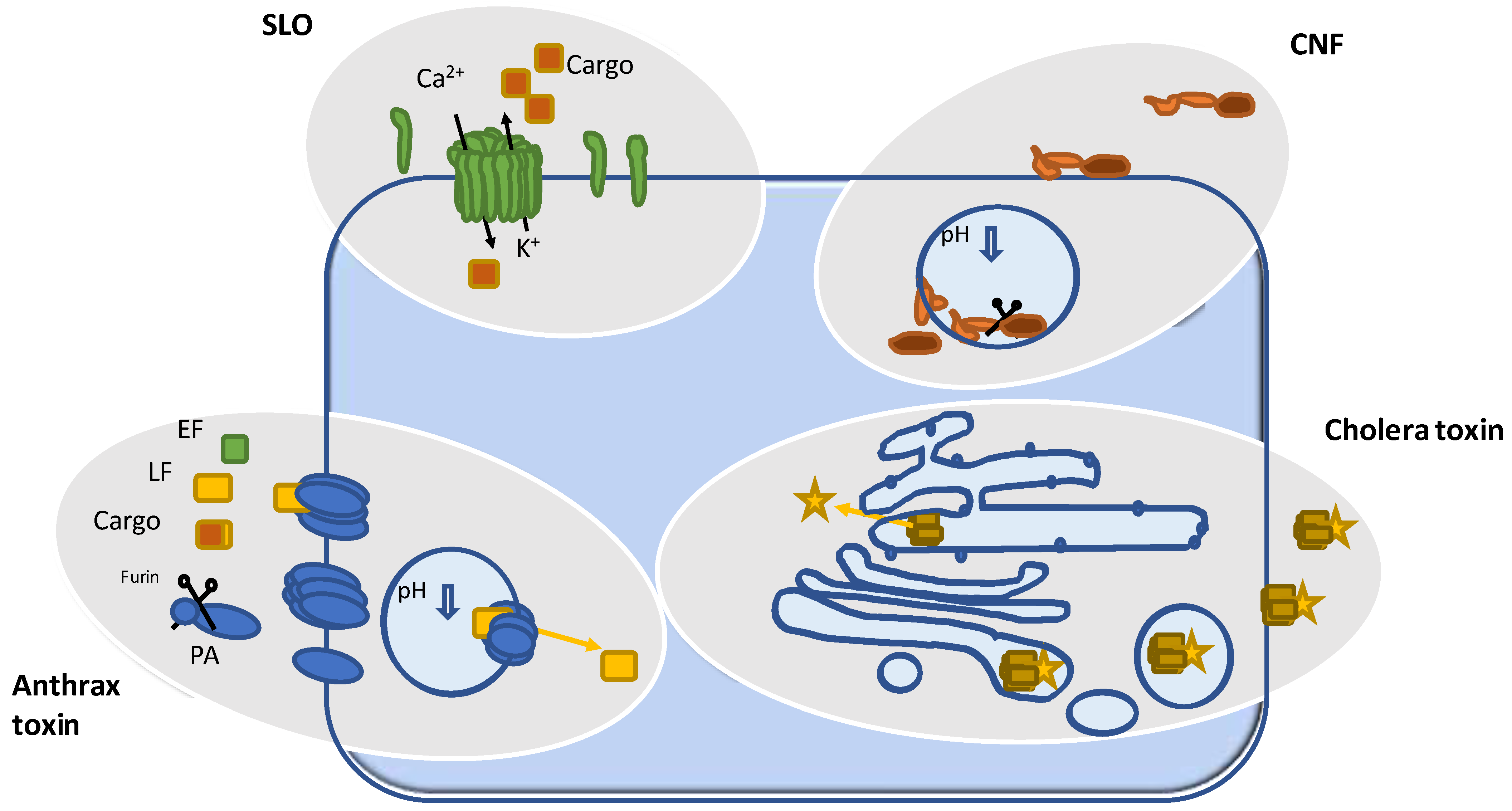
1. Pore Forming Bacterial Toxins

A large number of bacterial toxins do not have catalytic activity but instead form pores in the plasma membrane by oligomerization [1]. Water-soluble monomers are released by pathogenic bacteria, bind to cellular receptors (lipids, glycolipids, glycoproteins, or proteins), and are incorporated into the membrane for oligomerization. This is accompanied by a remarkable conformational change, leading to the formation of water-filled pores, changes in the membrane potential, and eventually cell death [2]. Pore-forming toxins are grouped into two structurally different classes. Prominent examples of alpha pore-forming toxins (the membrane channel is created by membrane-spanning alpha helices) are the family of Cytolysins (Cly A), which target mammalian cells, and the Colicins, which act on other bacteria for competition with food sources (Table 1) [3]. Together with aerolysins and hemolysins, the cholesterol-dependent cytolysins (CDC) belong to the family of β-pore forming toxins. They consist of amphipathic β-strands [4,5] and form pores between 1 nm and 100 nm in diameter [6]. Pore formation itself and changes in the cytosolic ion composition induce survival mechanisms of the targeted cells. For example, the shedding of pore-containing membrane vesicles is induced by the influx of calcium ions. Additionally, endocytosis and lysosomal degradation of the protein toxins as well as activation of autophagy have been described [7].
Stable pores also form in model bilayer membranes called black lipid membranes. This allows the determination of the biophysical properties including ion selectivity and the size of the channels formed [8,9]. Particularly, because of the non-permeable nature of the plasma membrane, toxins that form big pores, like streptolysin O, can be used to load small molecules and even peptides or drugs into the cytoplasm of mammalian cells.
Although streptolysin O treatment is temporally harmful to the target cell, channel formation seems to be reversible. Indeed, it has been shown that several cell lines survive toxin treatment in cell culture experiments [10]. However, the transport of ions or soluble small molecules through water-filled pores is exclusively driven by diffusion (Figure 1). This is different when toxins are used, which form pores aiming at the selective transport of an enzyme into the cytosol of target cells (see Section 2). Besides pore formation, the property of specific binding to cellular receptors has been utilized. For example, Clostridium perfringens Perfringolysin O (PFO) binds to cholesterol. Domain 4 of PFO, which is sufficient for this interaction, has been used as a biosensor for cholesterol nanodomains in cell biology [4,11]. Similarly, non-pore-forming toxins have been used for specific labeling of cellular structures. Vibrio cholerae cholera toxin binds to the ganglioside GM1 and Shigella dysenteriae Shiga toxin binds to the glycolipid Gb3. Both toxins have been utilized to monitor lipid trafficking within cells [12,13].

2. Toxins Forming Pores for Delivery of an Enzyme

Some toxins form pores with the objective of transporting toxic enzymes into mammalian cells. For this purpose, the enzymatically active part (A) binds to the pore formed by a second toxin component (B). The most prominent example of such a two-component AB-type toxin is anthrax toxin [14]. The toxin produced by Bacillus anthracis is composed of a protective antigen (PA), which binds to the surface of mammalian cells, and of two separate enzymes: lethal factor (LF) and edema factor (EF). PA specifically interacts with tumor endothelial marker 8 (TM8, anthrax toxin receptor 1) or with capillary morphogenesis gene 2 (CMG2, anthrax toxin receptor 2) [15,16]. Oligomerization of PA leads to the formation of a heptameric prepore [17]. This process requires cleavage of each PA monomer by the abundant serine protease furin or furin-like proteases on the cell surface [18]. This property has been utilized for toxin engineering. The alteration of the cleavage site in PA toward target sequences for other proteases has been shown to modulate the specificity of the mutated toxin. This allowed turning the toxin’s activity toward cancer cells on which some proteases are highly expressed [19]. Moreover, a mutant of PA that lost its affinity to the natural receptors was re-targeted to other membrane proteins. For example, fusion of EGF or an Her2 affibody to the mutated PA enhanced the specificity of the engineered anthrax toxin pore for EGFR/Her2 expressing tumor cells by the factor of 100 [20,21,22].
Cleavage and subsequent prepore formation allow the binding of LF and/or EF. The protein complex is then taken up by clathrin-dependent endocytosis [23]. Upon acidification of the maturing endosome, the anthrax toxin pore inserts into the endosomal membrane and thereby facilitates the release of LF/EF into the cytosol. Cellular chaperones enable the folding of the proteins to their catalytically active conformation. Lethal factor is a metalloprotease that catalyzes the cleavage of the mitogen-activated protein kinase kinase (MAPKK) and Nlrp1, thereby influencing inflammasome formation. Edema factor enhances the amount of intracellular cAMP by acting as adenylyl cyclase (for review see [24]). Biochemical and structural analysis of anthrax toxin led to the identification of the domains of EF and LF that are essential for PA binding. The fusion of these domains with several protein cargos of choice allowed the transport of foreign proteins through the membrane channel formed by anthrax protective antigen. Besides the endogenous adapter domain of LF/EF, an artificial tag composed of positively charged amino acids was shown to be sufficient for binding to the PA oligomer and for mediating cargo delivery [25]. The combination of all these modifications lastly enabled the use of anthrax toxin for tumor targeting and drug transport into the cytosol. Anthrax toxin-based therapeutic applications have already been established in animal models [26].
Similar to anthrax toxin, Clostridium botulinum C2 toxin is a binary AB-type protein toxin consisting of a pore-forming unit (C2-II) and a second protein with enzymatic activity (C2-I), which is transported through the pentameric channel. Like PA, C2-II has to be cleaved to form the heptameric prepore and to bind C2-I. The protein complex is taken up by receptor-mediated endocytosis. The C2 receptor seems to be a common protein-linked carbohydrate, which complicates re-targeting the toxin to specific cells [27]. Acidification induces channel insertion into the endosomal membrane, allowing the release of C2-I into the cytosol [28]. C2-I is an ADP ribosyltransferase that modifies actin leading to the breakdown of the actin cytoskeleton and cell rounding. Comparable to anthrax toxin, the N-terminal part of C2-I or a polycationic tag sufficiently mediates the binding and transport of cargo proteins through the C2-II pore [29]. Using this method, the C2 toxin also has been engineered to deliver proteins into mammalian cells [30].
It must not be omitted that such pore formation coupled with protein transport across biological membranes is not exclusive for bacterial toxins but is also implemented by mammalian cells. Cytotoxic T lymphocytes release perforins together with granzymes to destroy virus-infected cells and thereby limit reproduction and spreading of the virus [31]. Perforins form pores in the cell membrane through which the apoptosis-inducing protease granzyme can be delivered.

3. The Benefit of Understanding the Enzymatic Activity of Bacterial Toxins

Besides the binary AB-type protein toxins, other toxins with enzymatic activity are composed of at least three independently acting domains located within one single protein: the toxic enzyme, a cell binding domain that also mediates endocytosis, and a third domain allowing delivery of the catalytic part from the endosome into the cytosol. In contrast to the toxins described above, no stable pore is formed in artificial membranes and the question of how the enzymatic parts of those proteins escape the endosome is not yet answered. However, the fusion of cargo proteins to the N-terminal binding/translocation domain of Diphtheria toxin also facilitates the delivery of fluorescent proteins and enzymes [32,33].
The domain composition of single-chained toxins allowed the use of isolated domains for cell biological or even pharmacological purposes. The enzyme domain is usually very efficient and modifies specific proteins or protein families within mammalian cells. With the knowledge about the exact modification sites and the fate of the modified substrates, bacterial toxins have been utilized to answer cell biology questions. The cellular function of the respective target proteins and connected signaling pathways have been studied extensively. Several bacterial toxins with different enzymatic activities are well characterized and have been utilized to study mammalian signaling cascades.

3.1. ADP Ribosyltransferases

Prominent examples of ADP-ribosyl-transferases are Vibrio cholerae cholera toxin and Bordetella pertussis pertussis toxin. Both toxins are composed of a cell-binding pentamer and the catalytic subunit. They are transported in endosomes via the Golgi apparatus to the endoplasmic reticulum (ER). There, the catalytic domains are separated and lose their structure. Export to the cytosol is mediated by the ER-associated degradation (ERAD) pathway and the translocated enzyme is refolded in the cytosol. This uptake mechanism is called “long-trip” uptake to distinguish it from the “short-trip” translocation from acidified endosomes [34,35]. The toxins specifically modify alpha subunits of heterotrimeric G proteins to permanently activate (cholera toxin, Gαs) or inactivate (pertussis toxin, Gαi) them [36]. Accordingly, the toxins have been proven to be invaluable tools to study the function of these important signaling molecules.
A further example is Clostridium botulinum C3 toxin, which selectively modifies and inactivates the small GTPase RhoA, a main regulator of the actin cytoskeleton. Its inactivation by C3 leads to the breakdown of actin filaments and rounding of cells [37]. In cells of neuronal origin, Rho inactivation and Rho-independent effects by the toxin induce the outgrowth of neurites [38,39]; from this emerged the idea of utilizing C3 for the regeneration of neuronal connections after severing neurites due to injury [40]. C3 lacks a binding and translocation domain and therefore requires fusion to other membrane-crossing domains, such as cell-penetrating peptides, to enter most mammalian cells. Alternatively, the toxin can be injected manually. However, binding to the type III intermediate filament protein vimentin has been suggested to be sufficient for cell entry into damaged neurons [41].
Corynebacterium diphtheriae diphtheria toxin (DT) modifies elongation factor 2 (EF-2) by ADP-ribosylation. Modification of EF-2 blocks translation and protein synthesis in target cells, eventually leading to cell death [42]. The catalytic domain of DT has been fused to several other (not lethal) bacterial toxins as a powerful readout system during CRISPR/Cas9-based screens for the identification of cellular receptors [43,44,45]. In addition, DT has also been studied for pharmacological applications for over 50 years and several fusion proteins have been tested for cell specificity and activity. Indeed, two drugs based on DT have been approved: Ontak and Tagraxofusp. Ontak (denileukin diftitox) is an IL-2 peptide fused to a DT catalytic domain deployed to destroy T-cells, whereas Tagraxofusp is DT fused to an IL-3 peptide for selective removal of IL3 receptor-expressing cells [46]. The recombinant protein is established as a first-line therapy against the Blastic Plasmacytoid Dendritic Cell Neoplasm (BPDCN) [47,48].
An additional immunotoxin assembled from DT and single-chain fragments derived from an anti-PD1 antibody was recently developed. It targets programmed death 1 (PD1), a checkpoint receptor effectively depleting PD1-expressing cells in vitro and in mice [49]. An immunotoxin with similar activity but a lower yield of the recombinant protein was produced before anti-PD1-ABD-PE [50]. This fusion protein is based on a different toxin: the ribosyl-transferase Pseudomonas aeruginosa exotoxin A (ExoA). Like diphtheria toxin, ExoA targets an elongation factor and blocks protein synthesis [51]. Overall, one can state that engineered immunotoxins are on the way to becoming effective therapeutic agents.

3.2. The Deamidating Toxins CNF, PMT, and DNT

The AB toxins Bordetella bronchiseptica dermonecrotic toxin (DNT), Pasteurella multocida toxin (PMT), and the cytotoxic necrotizing factors (CNF1-3, CNFY), which are produced by pathogenic Escherichia coli strains and Yersinia pseudotuberculosis, encompass a family of deamidating AB toxins [52]. They catalyze the modification of Rho GTPases (DNT and CNFs) or alpha subunits of heterotrimeric G proteins (PMT). In each case, a specific glutamine of the target is deamidated to glutamic acid. This leads to persistent activation of the G proteins because the inactivation mechanism, the hydrolysis of the bound GTP, is blocked [53,54]. All toxins activate the complete pool of target proteins within mammalian cells and are frequently used to study Rho/Gα-dependent signaling pathways [55,56]. The toxins are taken up by receptor-mediated endocytosis. The T-type calcium channel Cav3.1 has been identified as a cellular receptor for DNT, [43]. PMT has been shown to interact with lipid components of the membrane [57,58] and to bind to the LDL-receptor-related protein 1 (LRP1) [59]. The receptors for CNFs have not been identified so far. All of these toxins are proteolytically cleaved within the endosome for release of the catalytic part into the cytosol. Blocking acidification of the endosomes by Bafilomycin A showed that the pH change is required for the transport of the catalytic domains into the cytosol, a fact known for many AB toxins [60,61,62].
Pasteurella multocida strains that produce PMT cause atrophic rhinitis in affected pigs. The toxin induces bone loss by inhibiting bone-building osteoblasts with simultaneous activation of bone-resorbing osteoclasts [63]. Moreover, PMT modulates the function of several immune cells [64]. In cultivated cells, PMT induces cytoskeletal rearrangements and acts as a strong mitogen. Besides unraveling signaling pathways downstream of G-protein coupled receptors, PMT has been studied as a possible pharmacological agent for treating fibrodysplasia ossificans, a rare human bone disease associated with massive pathological bone formation [65,66].
Activation of Rho proteins by Escherichia coli CNF1 leads to rearrangement of the actin cytoskeleton [67]. In contrast to the inactivating modifications described above, activation by CNF1 seems to be long-lasting. This is probably because there is no need for the cell to destroy an active protein. Although CNF1 is a toxin and induces necrotizing lesions when applied to the skin, it induces several beneficial changes in physiological responses when injected into tissues. For example, local administration of CNF1 into the footpads of mice enhanced analgesia [68]. Also, systemic application reduced pain by up-regulating µ-opioid receptors [69]. The toxin also promises beneficial properties against brain tumors. It promoted senescence and/or death of mouse and human glioblastoma cells [70]. Moreover, CNF1 induced the outgrowth of neurites and stimulated the morphogenesis of dendritic spines to modulate plasticity learning in a mouse model [71,72,73]. Both facts led to the attempt to inject the Rho activator directly into the liquor of glioma-bearing mice since gliomas and especially glioblastomas are extremely aggressive brain tumors. Indeed, the toxin enhanced neuronal function, decreased the tumor volume, preserved the healthy tissue, and increased the survival time of treated mice [74]. Recently, a fusion toxin was constructed encompassing chlorotoxin, a peptide from the venom of the scorpion Leiurus quinquestriatus, and CNF1. Adding the peptide enabled the protein to efficiently cross the blood–brain barrier. It allowed systemic application of the large protein while still showing anti-neoplastic activity in a glioblastoma mouse model [68,70]. Recently, CNF1 was also used to rescue cognitive dysfunction in a mouse model of Rett Syndrome [75]. Proceeding with these promising studies about the usefulness of CNF1 and other bacterial deamidases may pave the way for new and unexpected clinical applications.

3.3. Toxins with Other Enzymatic Activities

The most impressive existing application of a bacterial toxin is the use of botulinum neurotoxin (BoNT, Botox) produced by Clostridium botulinum. It is highly toxic and the causative agent of Botulism, a rare but potentially fatal illness. However, the toxin is frequently used for medical and even cosmetic applications.
Botulinum neurotoxins are a family of Zn2+-metalloproteases [76]. The toxins cleave the three SNARE proteins synaptobrevin, syntaxin, and SNAP-25 in mammalian cells. SNARE proteins are required for the fusion of the vesicular membrane with the plasma membrane and therefore for the release of neurotransmitters into the synaptic cleft. Thus, their cleavage leads to neuro-paralysis [77]. Botulinum neurotoxins bind to at least two receptors present in the presynaptic membrane, a polysialoganglioside and a glycosylated synaptotagmin, situated in the luminal membrane of synaptic vesicles, which leads to high cellular selectivity of the neurotoxins [78,79,80]. As a typical AB toxin, the enzyme domain of Botox is released from the acidified endosome into the cytosol, where it cleaves SNARE proteins leading to paralysis of the affected neuron [81,82]. Among the first clinical applications of Botox were spasmodic illnesses like blepharospasm, cervical dystonia, and limb spasticity. The therapeutic use of Botox is now widespread and ranges from tremors and hyperhidrosis to migraine, depression, and chronic pain. However, the main application of the toxin is its use for cosmetic indications [83].
Bordetella pertussis adenylate cyclase (CyaA) interacts with the integrin CD11b/CD18 (complement receptor 3) present in cells of the innate immune system, including dendritic cells which are highly sensitive to the toxin. In contrast to other AB toxins, CyaA forms a pore directly in the plasma membrane to translocate the invasive adenylate cyclase [84,85]. Factors for this process seem to be the membrane potential and the activity of calpain, which cleaves off the catalytic part of CyaA [86,87]. The intrinsic immune cell specificity of the pore-forming subunit of CyaA is interesting for the artificial delivery of cancer cell antigens into dendritic cells, ultimately stimulating tumor-specific cytotoxic T-cells [88,89].

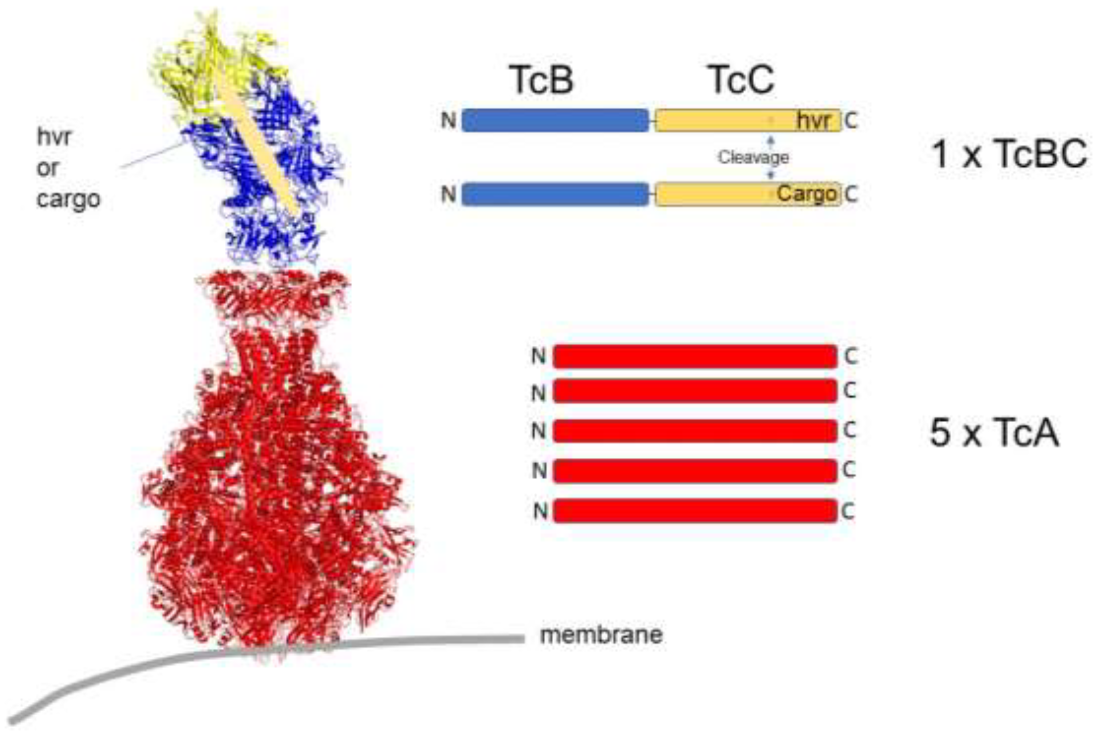
4. Secreted Injection Systems

To reach their targets within cells, protein toxins need to cross the plasma membrane. In contrast to the mode of action of AB toxins, some bacteria directly inject proteins into mammalian cells with the help of a syringe-like apparatus (Type-III secretion systems). This requires direct contact between the bacterium and the target cell. Bacteria with type-III secretion systems include Salmonella and Yersinia species, which likewise have been used for the injection of heterologous proteins into mammalian cells [90]. A functionally similar injection apparatus is synthesized by the insect pathogen Photorhabdus luminescens. A noteworthy difference to the type-III secretion systems is that here, the whole machinery is composed of three isolated proteins (TcA, TcB, and TcC), which are secreted by the bacteria [91,92]. The photorhabdus toxin complex (PTC) assembles autonomously in body fluids or in a cell culture medium (Figure 2). This attribute allowed recombinant expression in non-pathogenic bacteria in sufficient amounts for purification [93,94]. Structural analysis of the large (1.4 MDa) toxin complex (TC) by cryo-electron microscopy showed that the A component forms a pentamer with a needle-like structure surrounded by a shell building up the injection apparatus. TcB and TcC monomers assemble into a cocoon-like structure enclosing the protein to be injected (BC). The enzymatically active part of PTC is located within the C-terminus of TcC. It is proteolytically cleaved inside the cocoon by an intrinsic protease and is loaded into the syringe after BC binds to the A-pentamer [95,96]. Similar to Bacillus anthracis, Photorhabdus luminescens expresses several C components with diverse enzymatic activity. It was possible to exchange this part for diverse cargo proteins that are similar in size and charge. Using such engineered PTCs as transport units for the delivery of heterologous proteins into cells allowed the loading of more than 105 cargo proteins into each cell with a coverage close to 100% of injected cells [94,97].
Recently, a second extracellular injection system with a phage-tail like architecture has been isolated from Photorhabdus asymbiotica [98]. Photorhabdus virulence cassette (PVC) is the gene cluster encoding more than 20 different proteins. These assemble into a contractile injection system composed of a base plate complex, a phage tail-like structure, and a cap. The recombinant nanosyringe could also be manipulated for the injection of several cargo proteins [99]. Compared to PTC, the charge and size of the cargo is less restricted. Proteins up to 130 kDa have been successfully injected. However, the much larger size of the injection system may be a disadvantage when considering tissue penetration.

5. Conclusions

The broad knowledge about bacterial protein toxins, their mode of action, cellular receptors, membrane insertion motifs, pathways into the cytosol, substrate specificity, and selectivity together with structural information has allowed their application as cell-biological and even pharmacological tools. Because of their ability to enter mammalian cells, bacterial toxins have been engineered to transport heterologous cargo into cells. Moreover, some of them have been re-targeted toward specific cells and tissues.
Without a doubt, the tight substrate specificity of the bacterial toxins with enzymatic activity is extremely useful for the analysis of target protein-dependent signaling pathways and functions. One advantage of toxin utilization is that there is no need for over-expressing dominant negative or constitutively activated mutant signaling proteins. This initially preserves their physiological pool without the need for overexpression. However, degradation and re-expression of the toxin-modified proteins have been described [100,101,102]. A further benefit of using toxins is that, depending on the toxin concentration, up to 100% of cells can be targeted. This improves experimental methodologies. For clinical applications, the formation of antibodies against foreign proteins must be expected and may limit the benefit following repeated toxin exposure. Remarkably, the frequently used Botulinum Neurotoxin BoNT/A1 appears to be a poor antigen. Very few patients develop neutralizing antibodies, even when the treatment is continued for years [103,104]. Such promising examples of bacterial toxins successfully used as drugs enhance the probability of developing further toxin-based reagents for clinical applications.

Funding

This work was funden by the Deutsche Forschungsgemeinschaft grant DFG SCHM 12-1.

Institutional Review Board Statement

Not applicable.

Informed Consent Statement

Not applicable.

Data Availability Statement

Not applicable.

Acknowledgments

G.S. was supported by the Deutsche Forschungsgemeinschaft grant DFG SCHM 12-1.

Conflicts of Interest

The author declares no conflict of interest.

References

  1. Bhakdi, S.; Valeva, A.; Walev, I.; Weller, U.; Palmer, M. Bacterial Toxins—Tools in Cell Biology and Pharmacology; Aktories, K., Ed.; Chapman & Hall: London, UK, 1997; pp. 241–257. [Google Scholar]
  2. Peraro, M.D.; van der Goot, F.G. Pore-forming toxins: Ancient, but never really out of fashion. Nat. Rev. Microbiol. 2016, 14, 77–92. [Google Scholar] [CrossRef]
  3. Madden, J.C.; Ruiz, N.; Caparon, M. Cytolysin-Mediated Translocation (CMT): A Functional Equivalent of Type III Secretion in Gram-Positive Bacteria. Cell 2001, 104, 143–152. [Google Scholar] [CrossRef]
  4. Gilbert, R.J. Cholesterol-dependent cytolysins. Adv. Exp. Med. Biol. 2010, 677, 56–66. [Google Scholar]
  5. Feil, S.C.; Ascher, D.B.; Kuiper, M.J.; Tweten, R.K.; Parker, M.W. Structural Studies of Streptococcus pyogenes Streptolysin O Provide Insights into the Early Steps of Membrane Penetration. J. Mol. Biol. 2014, 426, 785–792. [Google Scholar] [CrossRef]
  6. Ahnert-Hilger, G.; Pahner, I.; Höltje, M. Handbook of Experimental Pharmacology; Aktories, K., Just, I., Eds.; Springer: Berlin/Heidelberg, Germany, 2000; pp. 557–575, Ch. 24. [Google Scholar]
  7. Verma, P.; Gandhi, S.; Lata, K.; Chattopadhyay, K. Pore-forming toxins in infection and immunity. Biochem. Soc. Trans. 2021, 49, 455–465. [Google Scholar] [CrossRef]
  8. Benz, R.; Hardie, K.R.; Hughes, C. Pore formation in artificial membranes by the secreted hemolysins of Proteus vulgaris and Morganella morganii. Eur. J. Biol. Inorg. Chem. 1994, 220, 339–347. [Google Scholar] [CrossRef]
  9. Benz, R.; Schmid, A.; E Hancock, R. Ion selectivity of gram-negative bacterial porins. J. Bacteriol. 1985, 162, 722–727. [Google Scholar] [CrossRef]
  10. Tweten, R.K.; Parker, M.W.; Johnson, A.E. The cholesterol-dependent cytolysins. Curr. Top. Microbiol. Immunol. 2001, 257, 15–33. [Google Scholar]
  11. Das, A.; Goldstein, J.L.; Anderson, D.D.; Brown, M.S.; Radhakrishnan, A. Use of mutant 125 I-perfringolysin O to probe transport and organization of cholesterol in membranes of animal cells. Proc. Natl. Acad. Sci. USA 2013, 110, 10580–10585. [Google Scholar] [CrossRef]
  12. Orlandi, P.; Curran, P.; Fishman, P. Brefeldin A blocks the response of cultured cells to cholera toxin. Implications for intracellular trafficking in toxin action. J. Biol. Chem. 1993, 268, 12010–12016. [Google Scholar] [CrossRef]
  13. Bergan, J.; Lingelem, A.B.D.; Simm, R.; Skotland, T.; Sandvig, K. Shiga toxins. Toxicon 2012, 60, 1085–1107. [Google Scholar] [CrossRef]
  14. Beauregard, K.E.; Wimer-Mackin, S.; Collier, R.J.; Lencer, W.I. Anthrax toxin entry into polarized epithelial cells. Infect. Immun. 1999, 67, 3026–3030. [Google Scholar] [CrossRef]
  15. Bradley, K.A.; Mogridge, J.; Mourez, M.; Collier, R.J.; Young, J.A.T. Identification of the cellular receptor for anthrax toxin. Nature 2001, 414, 225–229. [Google Scholar] [CrossRef]
  16. Escuyer, V.; Collier, R.J. Anthrax protective antigen interacts with a specific receptor on the surface of CHO-K1 cells. Infect. Immun. 1991, 59, 3381–3386. [Google Scholar] [CrossRef]
  17. Collier, R.J.; Young, J.A. Anthrax toxin. Annu. Rev. Cell Dev. Biol. 2003, 19, 45–70. [Google Scholar] [CrossRef]
  18. Ezzell, J.W.; Abshire, T.G. Serum protease cleavage of Bacillus anthracis protective antigen. J. Gen. Microbiol. 1992, 138, 543–549. [Google Scholar] [CrossRef]
  19. Bachran, C.; Leppla, S.H. Tumor Targeting and Drug Delivery by Anthrax Toxin. Toxins 2016, 8, 197. [Google Scholar] [CrossRef]
  20. Mechaly, A.; McCluskey, A.J.; Collier, R.J. Changing the receptor specificity of anthrax toxin. mBio 2012, 3, 10–128. [Google Scholar] [CrossRef]
  21. McCluskey, A.J.; Olive, A.J.; Starnbach, M.N.; Collier, R.J. Targeting HER2-positive cancer cells with receptor-redirected anthrax protective antigen. Mol. Oncol. 2013, 7, 440–451. [Google Scholar] [CrossRef]
  22. Zahaf, N.-I.; Lang, A.E.; Kaiser, L.; Fichter, C.D.; Lassmann, S.; McCluskey, A.; Augspach, A.; Aktories, K.; Schmidt, G. Targeted delivery of an ADP-ribosylating bacterial toxin into cancer cells. Sci. Rep. 2017, 7, 41252. [Google Scholar] [CrossRef]
  23. Abrami, L.; Bischofberger, M.; Kunz, B.; Groux, R.; van der Goot, F.G. Endocytosis of the anthrax toxin is mediated by clathrin, actin and unconventional adaptors. PLoS Pathog. 2010, 6, e1000792. [Google Scholar] [CrossRef]
  24. Liu, W.; Nestorovich, E.M. Anthrax toxin channel: What we know based on over 30 years of research. Biochim. Biophys. Acta (BBA) Biomembr. 2021, 1863, 183715. [Google Scholar] [CrossRef]
  25. Collier, R.J. Membrane translocation by anthrax toxin. Mol. Asp. Med. 2009, 30, 413–422. [Google Scholar] [CrossRef]
  26. Friebe, S.; Van der Goot, F.G.; Bürgi, J. The Ins and Outs of Anthrax Toxin. Toxins 2016, 8, 69. [Google Scholar] [CrossRef]
  27. Eckhardt, M.; Barth, H.; Blöcker, D.; Aktories, K. Binding of Clostridium botulinum C2 toxin to asparagine-linked complex and hybrid carbohydrates. J. Biol. Chem. 2000, 275, 2328–2334. [Google Scholar] [CrossRef]
  28. Barth, H.; Blöcker, D.; Behlke, J.; Bergsma-Schutter, W.; Brisson, A.; Benz, R.; Aktories, K. Cellular uptake of Clostridium botulinum C2 toxin requires oligomerization and acidification. J. Biol. Chem. 2000, 275, 18704–18711. [Google Scholar] [CrossRef]
  29. Heber, S.; Borho, J.; Stadler, N.; Wondany, F.; König, I.; Michaelis, J.; Papatheodorou, P.; Barth, H.; Fellermann, M. The Clostridium botulinum C2 Toxin Subunit C2IIa Delivers Enzymes with Positively Charged N-Termini into the Cytosol of Target Cells. Toxins 2023, 15, 390. [Google Scholar] [CrossRef]
  30. Barth, H.; Stiles, B.G. Binary actin-ADP-ribosylating toxins and their use as molecular trojan horses for drug delivery into eukaryotic cells. Curr. Med. Chem. 2008, 15, 459–469. [Google Scholar] [CrossRef]
  31. Voskoboinik, I.; Whisstock, J.C.; Trapani, J.A. Perforin and granzymes: Function, dysfunction and human pathology. Nat. Rev. Immunol. 2015, 15, 388–400. [Google Scholar] [CrossRef]
  32. Olsnes, S.; Klingenberg, O.; Munoz, R.; Falnes, P.O.; Wiedlocha, A. Bacterial Toxins: Tools in Cell Biology and Pharmacology; Aktories, K., Ed.; Chapman & Hall: London, UK, 1997; pp. 273–294. [Google Scholar]
  33. Auger, A.; Park, M.; Nitschke, F.; Minassian, L.M.; Beilhartz, G.L.; Minassian, B.A.; Melnyk, R.A. Efficient Delivery of Structurally Diverse Protein Cargo into Mammalian Cells by a Bacterial Toxin. Mol. Pharm. 2015, 12, 2962–2971. [Google Scholar] [CrossRef]
  34. Aktories, K.; Jakobs, K.H.; Robbel, L. Further contributions to an improved pertussis vaccine: I. Molecular mechanisms of pertussis toxin. Behring Inst. Res. Commun. 1984, 76, 106–109. [Google Scholar]
  35. Merritt, E.A.; Hol WG, J. AB5 toxins. Curr. Opin. Struct. Biol. 1995, 5, 165–171. [Google Scholar] [CrossRef]
  36. Gierschik, P.; Jakobs, K.H. Handbook of Experimental Pharmacology; Herken, H., Hucho, F., Eds.; Springer: Berlin/Heidelberg, Germany, 1992; Volume 102, pp. 807–839, Ch. 23. [Google Scholar]
  37. Aktories, K.; Braun, U.; Rösener, S.; Just, I.; Hall, A. The rho gene product expressed in E. Coli is a substrate of botulinum ADP-ribosyltransferase C3. Biochem. Biophys. Res. Commun. 1989, 158, 209–213. [Google Scholar] [CrossRef]
  38. Auer, M.; Schweigreiter, R.; Hausott, B.; Thongrong, S.; Höltje, M.; Just, I.; Bandtlow, C.; Klimaschewski, L. Rho-independent stimulation of axon outgrowth and activation of the ERK and Akt signaling pathways by C3 transferase in sensory neurons. Front. Cell. Neurosci. 2012, 6, 43. [Google Scholar] [CrossRef]
  39. Jeon, C.; Moon, M.; Kim, J.; Kim, H.; Kim, J.; Li, Y.; Jin, J.; Kim, P.; Kim, H.; Meier, K.E.; et al. Control of neurite outgrowth by RhoA inactivation. J. Neurochem. 2012, 120, 684–698. [Google Scholar] [CrossRef]
  40. Höltje, M.; Djalali, S.; Hofmann, F.; Münster-Wandowski, A.; Hendrix, S.; Boato, F.; Dreger, S.C.; Große, G.; Henneberger, C.; Grantyn, R.; et al. A 29-amino acid fragment of Clostridium botulinum C3 protein enhances neuronal outgrowth, connectivity, and reinnervation. FASEB J. 2008, 23, 1115–1126. [Google Scholar] [CrossRef]
  41. Rohrbeck, A.; Schröder, A.; Hagemann, S.; Pich, A.; Höltje, M.; Ahnert-Hilger, G.; Just, I. Vimentin mediates uptake of C3 exoenzyme. PLoS ONE 2014, 9, e101071. [Google Scholar] [CrossRef]
  42. Chung, D.W.; Collier, R. The mechanism of ADP-ribosylation of elongation factor 2 catalyzed by fragment a from diphtheria toxin. Biochim. Biophys. Acta (BBA) Enzym. 1977, 483, 248–257. [Google Scholar] [CrossRef]
  43. Teruya, S.; Hiramatsu, Y.; Nakamura, K.; Fukui-Miyazaki, A.; Tsukamoto, K.; Shinoda, N.; Motooka, D.; Nakamura, S.; Ishigaki, K.; Shinzawa, N.; et al. Bordetella Dermonecrotic Toxin Is a Neurotropic Virulence Factor That Uses CaV3.1 as the Cell Surface Receptor. mBio 2020, 11, e03146-19. [Google Scholar] [CrossRef]
  44. Schoellkopf, J.; Mueller, T.; Hippchen, L.; Mueller, T.; Reuten, R.; Backofen, R.; Orth, J.; Schmidt, G. Genome wide CRISPR screen for Pasteurella multocida toxin (PMT) binding proteins reveals LDL Receptor Related Protein 1 (LRP1) as crucial cellular receptor. PLoS Pathog. 2022, 18, e1010781. [Google Scholar] [CrossRef]
  45. Xu, Y.; Viswanatha, R.; Sitsel, O.; Roderer, D.; Zhao, H.; Ashwood, C.; Voelcker, C.; Tian, S.; Raunser, S.; Perrimon, N.; et al. CRISPR screens in Drosophila cells identify Vsg as a Tc toxin receptor. Nature 2022, 610, 349–355. [Google Scholar] [CrossRef] [PubMed]
  46. Fuentes, A.C.; Szwed, E.; Spears, C.D.; Thaper, S.; Dang, L.H.; Dang, N.H. Denileukin Diftitox (Ontak) as Maintenance Therapy for Peripheral T-Cell Lymphomas: Three Cases with Sustained Remission. Case Rep. Oncol. Med. 2015, 2015, 123756. [Google Scholar] [CrossRef] [PubMed]
  47. Beziat, G.; Ysebaert, L. Tagraxofusp for the Treatment of Blastic Plasmacytoid Dendritic Cell Neoplasm (BPDCN): A Brief Report on Emerging Data. OncoTargets Ther. 2020, 13, 5199–5205. [Google Scholar] [CrossRef]
  48. FitzGerald, D.J. Targeted diphtheria toxin to treat BPDCN. Blood 2014, 124, 310–312. [Google Scholar] [CrossRef]
  49. Zhang, T.; Dong, S.; Zhai, Y.; Naatz, L.; Zhou, Z.; Chen, M. Diphtheria toxin-derived, anti-PD-1 immunotoxin, a potent and practical tool to selectively deplete PD-1+ cells. Protein Sci. 2023, 32, e4741. [Google Scholar] [CrossRef] [PubMed]
  50. Zhao, P.; Wang, P.; Dong, S.; Zhou, Z.; Cao, Y.; Yagita, H.; He, X.; Zheng, S.G.; Fisher, S.J.; Fujinami, R.S.; et al. Depletion of PD-1-positive cells ameliorates autoimmune disease. Nat. Biomed. Eng. 2019, 3, 292–305. [Google Scholar] [CrossRef] [PubMed]
  51. Iglewski, B.H.; Liu, P.V.; Kabat, D. Mechanism of action of Pseudomonas aeruginosa exotoxin Aiadenosine diphosphate-ribosylation of mammalian elongation factor 2 in vitro and in vivo. Infect. Immun. 1977, 15, 138–144. [Google Scholar] [CrossRef] [PubMed]
  52. Hoffmann, C.; Schmidt, G. CNF and DNT. Rev. Physiol. Biochem. Pharmacol. 2004, 152, 49–63. [Google Scholar] [CrossRef] [PubMed]
  53. Schmidt, G.; Sehr, P.; Wilm, M.; Selzer, J.; Mann, M.; Aktories, K. Gln 63 of Rho is deamidated by Escherichia coli cytotoxic necrotizing factor-1. Nature 1997, 387, 725–729. [Google Scholar] [CrossRef]
  54. Flatau, G.; Lemichez, E.; Gauthier, M.; Chardin, P.; Paris, S.; Fiorentini, C.; Boquet, P. Toxin-induced activation of the G protein p21 Rho by deamidation of glutamine. Nature 1997, 387, 729–733. [Google Scholar] [CrossRef]
  55. Wilson, B.A.; Ho, M. Pasteurella multocida toxin as a tool for studying G(q) signal transduction. Rev. Physiol Biochem. Pharmacol. 2004, 152, 93–109. [Google Scholar] [PubMed]
  56. Kubatzky, K.F. Pasteurella multocida toxin—lessons learned from a mitogenic toxin. Front. Immunol. 2022, 13, 1058905. [Google Scholar] [CrossRef] [PubMed]
  57. Wilson, B.A.; Ho, M. Pasteurella multocida Toxin Interaction with Host Cells: Entry and Cellular Effects. Curr. Top. Microbiol. Immunol. 2012, 361, 93–111. [Google Scholar] [CrossRef]
  58. Brothers, M.C.; Ho, M.; Maharjan, R.; Clemons, N.C.; Bannai, Y.; Waites, M.A.; Faulkner, M.J.; Kuhlenschmidt, T.B.; Kuhlenschmidt, M.S.; Blanke, S.R.; et al. Membrane interaction of Pasteurella multocida toxin involves sphingomyelin. FEBS J. 2011, 278, 4633–4648. [Google Scholar] [CrossRef] [PubMed]
  59. Kowarschik, S.; Schöllkopf, J.; Müller, T.; Tian, S.; Knerr, J.; Bakker, H.; Rein, S.; Dong, M.; Weber, S.; Grosse, R.; et al. Yersinia pseudotuberculosis cytotoxic necrotizing factor interacts with glycosaminoglycans. FASEB J. 2021, 35, e21647. [Google Scholar] [CrossRef]
  60. Umata, T.; Moriyama, Y.; Futai, M.; Mekada, E. The cytotoxic action of diphtheria toxin and its degradation in intact Vero cells are inhibited by bafilomycin A1, a specific inhibitor of vacuolar-type H(+)-ATPase. J. Biol. Chem. 1990, 265, 21940–21945. [Google Scholar] [CrossRef] [PubMed]
  61. Knust, Z.; Blumenthal, B.; Aktories, K.; Schmidt, G. Cleavage of Escherichia coli cytotoxic necrotizing factor 1 is required for full biologic activity. Infect. Immun. 2009, 77, 1835–1841. [Google Scholar] [CrossRef]
  62. Bergmann, S.; Jehle, D.; Schwan, C.; Orth, J.H.C.; Aktories, K. Pasteurella multocida Toxin as a Transporter of Non-Cell-Permeating Proteins. Infect. Immun. 2013, 81, 2459–2467. [Google Scholar] [CrossRef]
  63. Mohamadzadeh, M. Microbial Toxins: Current Research and Future Trends. Expert Rev. Anti-Infect. Ther. 2009, 7, 695–696. [Google Scholar] [CrossRef]
  64. Hildebrand, D.; Walker, P.; Dalpke, A.; Heeg, K.; Kubatzky, K.F. Pasteurella multocida Toxin-induced Pim-1 expression disrupts suppressor of cytokine signalling (SOCS)-1 activity. Cell. Microbiol. 2010, 12, 1732–1745. [Google Scholar] [CrossRef]
  65. Verma, A.K.; Aga, P.; Singh, S.K.; Singh, R. The stone man disease: Fibrodysplasia ossificans progressiva: Imaging revisited. BMJ Case Rep. 2012, 2012, bcr2012006422. [Google Scholar] [CrossRef] [PubMed]
  66. Strack, J.; Heni, H.; Gilsbach, R.; Hein, L.; Aktories, K.; Orth, J.H.C. Noncanonical G-protein-dependent modulation of osteoclast differentiation and bone resorption mediated by Pasteurella multocida toxin. mBio 2014, 5, e02190-14. [Google Scholar] [CrossRef] [PubMed]
  67. Boquet, P. The cytotoxic necrotizing factor 1 (CNF1) from Escherichia coli. Toxicon 2001, 39, 1673–1680. [Google Scholar] [CrossRef] [PubMed]
  68. Colarusso, A.; Maroccia, Z.; Parrilli, E.; Germinario, E.A.P.; Fortuna, A.; Loizzo, S.; Ricceri, L.; Tutino, M.L.; Fiorentini, C.; Fabbri, A. Cnf1 Variants Endowed with the Ability to Cross the Blood–Brain Barrier: A New Potential Therapeutic Strategy for Glioblastoma. Toxins 2020, 12, 291. [Google Scholar] [CrossRef] [PubMed]
  69. Pavone, F.; Luvisetto, S.; Marinelli, S.; Straface, E.; Fabbri, A.; Falzano, L.; Fiorentini, C.; Malorni, W. The Rac GTPase-activating bacterial protein toxin CNF1 induces analgesia up-regulating μ-opioid receptors. Pain 2009, 145, 219–229. [Google Scholar] [CrossRef]
  70. Vannini, E.; Mori, E.; Tantillo, E.; Schmidt, G.; Caleo, M.; Costa, M. CTX-CNF1 Recombinant Protein Selectively Targets Glioma Cells in Vivo. Toxins 2021, 13, 194. [Google Scholar] [CrossRef]
  71. Diana, G.; Valentini, G.; Travaglione, S.; Falzano, L.; Pieri, M.; Zona, C.; Meschini, S.; Fabbri, A.; Fiorentini, C. Enhancement of learning and memory after activation of cerebral Rho GTPases. Proc. Natl. Acad. Sci. USA 2007, 104, 636–641. [Google Scholar] [CrossRef]
  72. Malchiodi-Albedi, F.; Paradisi, S.; Di Nottia, M.; Simone, D.; Travaglione, S.; Falzano, L.; Guidotti, M.; Frank, C.; Cutarelli, A.; Fabbri, A.; et al. CNF1 Improves astrocytic ability to support neuronal growth and differentiation in vitro. PLoS ONE 2012, 7, e34115. [Google Scholar] [CrossRef]
  73. Loizzo, S.; Rimondini, R.; Travaglione, S.; Fabbri, A.; Guidotti, M.; Ferri, A.; Campana, G.; Fiorentini, C. CNF1 Increases brain energy level, counteracts neuroinflammatory markers and rescues cognitive deficits in a murine model of alzheimer’s disease. PLoS ONE 2013, 8, e65898. [Google Scholar] [CrossRef]
  74. Vannini, E.; Olimpico, F.; Middei, S.; Ammassari-Teule, M.; de Graaf, E.L.; McDonnell, L.; Schmidt, G.; Fabbri, A.; Fiorentini, C.; Baroncelli, L.; et al. Electrophysiology of glioma: A Rho GTPase-activating protein reduces tumor growth and spares neuron structure and function. Neuro-Oncology 2016, 18, 1634–1643. [Google Scholar] [CrossRef]
  75. Urbinati, C.; Cosentino, L.; Germinario, E.A.P.; Valenti, D.; Vigli, D.; Ricceri, L.; Laviola, G.; Fiorentini, C.; Vacca, R.A.; Fabbri, A.; et al. Treatment with the Bacterial Toxin CNF1 Selectively Rescues Cognitive and Brain Mitochondrial Deficits in a Female Mouse Model of Rett Syndrome Carrying a MeCP2-Null Mutation. Int. J. Mol. Sci. 2021, 22, 6739. [Google Scholar] [CrossRef] [PubMed]
  76. Montecucco, C.; Schiavo, G. Tetanus and botulism neurotoxins: A new group of zinc proteases. Trends Biochem. Sci. 1993, 18, 324–327. [Google Scholar] [CrossRef]
  77. Schiavo, G.G.; Benfenati, F.; Poulain, B.; Rossetto, O.; de Laureto, P.P.; DasGupta, B.R.; Montecucco, C. Tetanus and botulinum-B neurotoxins block neurotransmitter release by proteolytic cleavage of synaptobrevin. Nature 1992, 359, 832–835. [Google Scholar] [CrossRef]
  78. Montecucco, C.; Papini, E.; Schiavo, G. Bacterial protein toxins penetrate cells via a four-step mechanism. FEBS Lett. 1994, 346, 92–98. [Google Scholar] [CrossRef]
  79. Montecucco, C.; Schiavo, G. Structure and function of tetanus and botulinum neurotoxins. Q. Rev. Biophys. 1995, 28, 423–472. [Google Scholar] [CrossRef]
  80. Montecucco, C. Protein toxins and membrane transport. Curr. Opin. Cell Biol. 1998, 10, 530–536. [Google Scholar] [CrossRef] [PubMed]
  81. McNew, J.A.; Parlati, F.; Fukuda, R.; Johnston, R.J.; Paz, K.; Paumet, F.; Söllner, T.H.; Rothman, J.E. Compartmental specificity of cellular membrane fusion encoded in SNARE proteins. Nature 2000, 407, 153–159. [Google Scholar] [CrossRef]
  82. Schiavo, G.; Matteoli, M.; Montecucco, C.; Kasai, H.; Takahashi, N.; Tokumaru, H.; Martin, J.H.; Engber, D.; Meng, Z.; Banerjee, A.; et al. Neurotoxins affecting neuroexocytosis. Physiol. Rev. 2000, 80, 717–766. [Google Scholar] [CrossRef]
  83. Carruthers, A.; Bruce, S.; Cox, S.E.; Kane, M.A.; Lee, E.; Gallagher, C.J. OnabotulinumtoxinA for Treatment of Moderate to Severe Crow’s Feet Lines: A Review. Aesthetic Surg. J. 2016, 36, 591–597. [Google Scholar] [CrossRef] [PubMed]
  84. Benz, R.; Maier, E.; Ladant, D.; Ullmann, A.; Sebo, P. Adenylate cyclase toxin (CyaA) of Bordetella pertussis. Evidence for the formation of small ion-permeable channels and comparison with HlyA of Escherichia coli. J. Biol. Chem. 1994, 269, 27231–27239. [Google Scholar] [CrossRef] [PubMed]
  85. Vojtova-Vodolanova, J.; Basler, M.; Osicka, R.; Knapp, O.; Maier, E.; Cerny, J.; Benada, O.; Benz, R.; Sebo, P. Oligomerization is involved in pore formation by Bordetella adenylate cyclase toxin. FASEB J. 2009, 23, 2831–2843. [Google Scholar] [CrossRef] [PubMed]
  86. Veneziano, R.; Rossi, C.; Chenal, A.; Devoisselle, J.-M.; Ladant, D.; Chopineau, J. Bordetella pertussis adenylate cyclase toxin translocation across a tethered lipid bilayer. Proc. Natl. Acad. Sci. USA 2013, 110, 20473–20478. [Google Scholar] [CrossRef] [PubMed]
  87. Bumba, L.; Masin, J.; Fiser, R.; Sebo, P. Bordetella Adenylate Cyclase Toxin Mobilizes Its β2 Integrin Receptor into Lipid Rafts to Accomplish Translocation across Target Cell Membrane in Two Steps. PLoS Pathog. 2010, 6, e1000901. [Google Scholar] [CrossRef] [PubMed]
  88. Sebo, P.; Osicka, R.; Masin, J. Adenylate cyclase toxin-hemolysin relevance for pertussis vaccines. Expert Rev. Vaccines 2014, 13, 1215–1227. [Google Scholar] [CrossRef] [PubMed]
  89. Svedova, M.; Masin, J.; Fiser, R.; Cerny, O.; Tomala, J.; Freudenberg, M.; Tuckova, L.; Kovar, M.; Dadaglio, G.; Adkins, I.; et al. Pore-formation by adenylate cyclase toxoid activates dendritic cells to prime CD8+ and CD4+ T cells. Immunol. Cell Biol. 2016, 94, 322–333. [Google Scholar] [CrossRef] [PubMed]
  90. Cornelis, G.R. Type III secretion: A bacterial device for close combat with cells of their eukaryotic host. Philos. Trans. R. Soc. Lond. 2000, 355, 681–693. [Google Scholar] [CrossRef] [PubMed]
  91. Daborn, P.J.; Waterfield, N.; Blight, M.A.; Ffrench-Constant, R.H. measuring virulence factor expression by the pathogenic bacterium Photorhabdus luminescens in culture and during insect infection. J. Bacteriol. 2001, 183, 5834–5839. [Google Scholar] [CrossRef] [PubMed]
  92. Meusch, D.; Gatsogiannis, C.; Efremov, R.G.; Lang, A.E.; Hofnagel, O.; Vetter, I.R.; Aktories, K.; Raunser, S. Mechanism of Tc toxin action revealed in molecular detail. Nature 2014, 508, 61–65. [Google Scholar] [CrossRef]
  93. Ffrench-Constant, R.; Waterfield, M.D. DNA Sequences from TCD Genomic Region of Photorhabdus luminescens. U.S. Patent US 7,777,100 B2, 17 August 2010. [Google Scholar]
  94. Gatsogiannis, C.; Lang, A.E.; Meusch, D.; Pfaumann, V.; Hofnagel, O.; Benz, R.; Aktories, K.; Raunser, S. A syringe-like injection mechanism in Photorhabdus luminescens toxins. Nature 2013, 495, 520–523. [Google Scholar] [CrossRef]
  95. Ost, G.S.; Ng’Ang’A, P.N.; Lang, A.E.; Aktories, K. Photorhabdus luminescens Tc toxin is inhibited by the protease inhibitor MG132 and activated by protease cleavage resulting in increased binding to target cells. Cell. Microbiol. 2019, 21, e12978. [Google Scholar] [CrossRef]
  96. Gatsogiannis, C.; Merino, F.; Prumbaum, D.; Roderer, D.; Leidreiter, F.; Meusch, D.; Raunser, S. Membrane insertion of a Tc toxin in near-atomic detail. Nat. Struct. Mol. Biol. 2016, 23, 884–890. [Google Scholar] [CrossRef] [PubMed]
  97. Ng’ang´a, P.N.; Ebner, J.K.; Plessner, M.; Aktories, K.; Schmidt, G. Engineering Photorhabdus luminescens toxin complex (PTC) into a recombinant injection nanomachine. Life Sci. Alliance 2019, 2, e201900485. [Google Scholar] [CrossRef] [PubMed]
  98. Jiang, F.; Li, N.; Wang, X.; Cheng, J.; Huang, Y.; Yang, Y.; Yang, J.; Cai, B.; Wang, Y.-P.; Jin, Q.; et al. Cryo-EM Structure and Assembly of an Extracellular Contractile Injection System. Cell 2019, 177, 370–383.e15. [Google Scholar] [CrossRef] [PubMed]
  99. Jiang, F.; Shen, J.; Cheng, J.; Wang, X.; Yang, J.; Li, N.; Gao, N.; Jin, Q. N-terminal signal peptides facilitate the engineering of PVC complex as a potent protein delivery system. Sci. Adv. 2022, 8, eabm2343. [Google Scholar] [CrossRef] [PubMed]
  100. Gerhard, R.; Tatge, H.; Genth, H.; Thum, T.; Borlak, J.; Fritz, G.; Just, I. Clostridium difficile toxin a induces expression of the stress-induced early gene product RhoB. J. Biol. Chem. 2005, 280, 1499–1505. [Google Scholar] [CrossRef] [PubMed]
  101. Barth, H.; Olenik, C.; Sehr, P.; Schmidt, G.; Aktories, K.; Meyer, D.K. Neosynthesis and activation of Rho by escherichia coli cytotoxic necrotizing factor (CNF1) reverse cytopathic effects of ADP-ribosylated Rho. J. Biol. Chem. 1999, 274, 27407–27414. [Google Scholar] [CrossRef]
  102. Pop, M.; Aktories, K.; Schmidt, G. Isotype-specific degradation of Rac activated by the cytotoxic necrotizing factor 1 (CNF1). J. Biol. Chem. 2004, 279, 35840–35848. [Google Scholar] [CrossRef]
  103. Bellows, S.; Jankovic, J. Immunogenicity Associated with Botulinum Toxin Treatment. Toxins 2019, 11, 491. [Google Scholar] [CrossRef]
  104. Carr, W.W.; Jain, N.; Sublett, J.W. Immunogenicity of Botulinum Toxin Formulations: Potential Therapeutic Implications. Adv. Ther. 2021, 38, 5046–5064. [Google Scholar] [CrossRef]
Figure 1. Toxins used as tools. SLO: The pore-forming toxin Streptolysin O binds to cholesterol-rich domains and inserts into the plasma membrane of mammalian cells. It oligomerizes to form water-filled pores through which ions and small molecules can enter cells by diffusion. CNF: Cytotoxic necrotizing factors are taken up by receptor-mediated endocytosis. Following proteolytic cleavage, the catalytic domain is released into the cytosol. This process requires acidification of the endosomes. In the cytosol, CNFs activate Rho proteins by deamidation. Cholera toxin: The toxin is composed of a cell-binding pentamer and an associated ADP-ribosyl-transferase. Toxin-containing endosomes are transported to the ER via the Golgi apparatus. From here, the transferase is released into the cytosol by the outwards transporter of misfolded proteins (endoplasmic-reticulum-associated protein degradation, ERAD). Cholera toxin was mainly used to study this transport route. Anthrax toxin: Anthrax protective antigen (PA) binds to the surface of mammalian cells. Cleavage by furin is required for oligomerization to a heptameric prepore. This allows the binding of lethal factor (LF), edema factor (EF), or an engineered cargo. The complex is then taken up by receptor-mediated endocytosis and released from the acidified endosome into the cytosol.
Toxins 16 00202 g001
Figure 2. Photorhabdus luminescens toxin complex (PTC) is composed of three isolated proteins (TcA, TcB, and TcC), which are secreted by the bacteria. The A component (red) forms a pentamer with a needle-like structure surrounded by a shell (Adapted from [92]). TcB (blue) and TcC (yellow) monomers assemble into a cocoon-like structure enclosing the protein to be injected. The enzymatically active part of PTC (hypervariable region, hvr) is located within the C-terminus of TcC. It is proteolytically cleaved inside the cocoon by an intrinsic protease, as indicated. The engineered protein TcBC is a fusion of TcB and TcC. The catalytic hvr can be exchanged by diverse cargo proteins. x = times.
Toxins 16 00202 g002
Table 1. Selection of secreted bacterial toxins that target mammalian cells: Pore-forming toxins (top), toxins with enzymatic activity (bottom).
Pore-forming toxins (selection)
ToxinBacteriumClassReceptor
AerolysinAeromonas spp.βCD52
Cytolysin AEscherichia coli, Salmonella entericaαCholesterol
ε-ToxinClostridium perfringensβHAVCR1
Hemolysin α/γStaphylococcus aureusβPhosphatidylcholin/ADAM10
Hemolysin BLBacillus cereusαCholesterol
ListeriolysinListeria monocytogenesβCholesterol
Perfringolysin OClostridium perfringensβCholesterol
Streptolysin OStreptococcus pyogenesβCholesterol
Toxins with enzymatic activity (selection)
ToxinBacteriumEnzymatic ActivitySubstrateProtein Receptor
Anthrax toxin (EF)Bacillus anthracisAdenylylcyclaseATPTM8, CMG2
Anthrax toxin (LF)Bacillus anthracisProteaseMAPKK, Nlrp1TM8, CMG2
Botulinum neurotoxinClostridium botulinumProteaseSNARE proteinsSynaptotagmin
C2 toxinClostridium botulinumADP-RibosyltransferaseActin
C3 toxinClostridium botulinumADP-RibosyltransferaseRhoAVimentin
Cholera toxinVibrio choleraeADP-RibosyltransferaseGα s
Cytotoxic necrotizing factors 1,2,3Escherichia coliDeamidaseRho GTPases
Cytotoxic necrotizing factor YYersinia pseudotuberculosisDeamidaseRho GTPases
CyaABordetella pertussisAdenylylcyclaseATPCD11b/CD18
Dermonecrotic toxinBordetella bronchisepticaTransglutaminaseRho GTPasesCav3.1
Diphtheria toxinCorynebacterium diphtheriaeADP-RibosyltransferaseElongation-factor
Pertussis toxinBordetella pertussisADP-RibosyltransferaseGα i
Pasteurella multocida toxinPasteurella multocidaDeamidaseGα subunitsLRP1
Shiga toxinShigella dysenteriae
Escherichia coli
DepurinaseRNA
Toxin AClostridoides difficileGlucosyltransferaseRho GTPasesLRP1
Toxin BClostridoides difficileGlucosyltransferaseRho GTPasesLRP1
Abbreviations: ADAM10: disintegrin and metalloprotease domain-containing protein 10, Cav3.1: T-type calcium channel, CD: cluster of differentiation, CMG2: capillary morphogenesis gene 2, HAVCR1: hepatitis A virus cellular receptor, LRP1: LDL-receptor related protein 1, MAPKK: mitogen-activated protein kinase kinase, Nlrp1: NACHT, LRR, FIIND, CARD domain, and PYD domains-containing protein 1, TM8: tumor endothelial marker 8.
Disclaimer/Publisher’s Note: The statements, opinions and data contained in all publications are solely those of the individual author(s) and contributor(s) and not of MDPI and/or the editor(s). MDPI and/or the editor(s) disclaim responsibility for any injury to people or property resulting from any ideas, methods, instructions or products referred to in the content.

