Article

Rhein Inhibits Cell Development and Aflatoxin Biosynthesis via Energy Supply Disruption and ROS Accumulation in Aspergillus flavus

1
College of Biological Engineering, Henan University of Technology, Zhengzhou 450001, China
2
Department of Health Professional Technologies, Faculty of Allied Health Sciences, The University of Lahore, Lahore 54570, Pakistan
*
Author to whom correspondence should be addressed.
Toxins 2024, 16(7), 285; https://doi.org/10.3390/toxins16070285
Submission received: 15 May 2024 / Revised: 16 June 2024 / Accepted: 21 June 2024 / Published: 23 June 2024
(This article belongs to the Special Issue Aspergillus flavus and Aflatoxins (Volume III))

Abstract

:
Aspergillus flavus and its carcinogenic secondary metabolites, aflatoxins, not only cause serious losses in the agricultural economy, but also endanger human health. Rhein, a compound extracted from the Chinese herbal medicine Rheum palmatum L. (Dahuang), exhibits good anti-inflammatory, anti-tumor, and anti-oxidative effects. However, its effect and underlying mechanisms against Aspergillus flavus have not yet been fully illustrated. In this study, we characterized the inhibition effect of rhein on A. flavus mycelial growth, sporulation, and aflatoxin B1 (AFB1) biosynthesis and the potential mechanism using RNA-seq analysis. The results indicate that A. flavus mycelial growth and AFB1 biosynthesis were significantly inhibited by 50 μM rhein, with a 43.83% reduction in colony diameter and 87.2% reduction in AFB1 production. The RNA-seq findings demonstrated that the differentially expressed genes primarily participated in processes such as spore formation and development, the maintenance of cell wall and membrane integrity, management of oxidative stress, the regulation of the citric acid cycle, and the biosynthesis of aflatoxin. Biochemical verification experiments further confirmed that 50 μM rhein effectively disrupted cell wall and membrane integrity and caused mitochondrial dysfunction through disrupting energy metabolism pathways, leading to decreased ATP synthesis and ROS accumulation, resulting in impaired aflatoxin biosynthesis. In addition, a pathogenicity test showed that 50 μM rhein inhibited A. flavus spore growth in peanut and maize seeds by 34.1% and 90.4%, while AFB1 biosynthesis was inhibited by 60.52% and 99.43%, respectively. In conclusion, this research expands the knowledge regarding the antifungal activity of rhein and provides a new strategy to mitigate A. flavus contamination.
Key Contribution: Rhein affects the growth and spore formation of A. flavus and blocks aflatoxin biosynthesis.

1. Introduction

Aspergillus flavus has been found to contaminate peanuts, maize, and wheat during cultivation, storage, processing, and transportation [1,2]. A. flavus also produces highly carcinogenic secondary metabolites called aflatoxins, of which aflatoxin B1 is classified as a grade 1 carcinogen [3,4]. The consumption of aflatoxin-contaminated food can cause poisoning, which may lead to hepatocellular carcinoma, seriously endangering the life and health of both humans and livestock [5]. Studies have shown that over 120 different countries have established maximum permissible levels of aflatoxin contamination in food products [6]. It is therefore important to develop rapid, efficient, and safe measures to address the problems associated with AFB1 contamination, given the serious threat it poses to human health.
Biological, physical, and chemical methods have been employed to prevent the contamination of A. flavus in food [7]. Biological measures, such as endophytic Aspergillus fumigatus and lactic acid bacteria, are efficient in inhibiting the growth of A. flavus and lessening mycotoxin production [8,9]. Physical methods, such as γ-irradiation of maize artificially inoculated with A. flavus, also resulted in a reduction in aflatoxins. In recent years, compounds from natural plants, including cinnamaldehyde, citral, and eugenol, have shown excellent antifungal performance against A. flavus and aflatoxin biosynthesis, owing to their eco-friendly properties [10]. Therefore, it is imperative to develop natural compounds to effectively prevent A. flavus growth and aflatoxin contamination.
Rhein, an anthraquinone compound derived from the natural plant Rhubard, also known as a kind of traditional Chinese herbal medicine, exhibits various effects, including anti-inflammatory, anti-tumor, antioxidant, anti-diabetic, cardioprotective, neuroprotective, hepatoprotective, and nephroprotective properties [11,12,13]. For example, rhein inhibits colon cancer cell growth by targeting the mammalian target of rapamycin (mTOR) pathway and human kidney cancer cells through the mitogen-activated protein kinase (MAPK)/nuclear factor kappa-light-chain enhancer of activated B cells (NF-κB) signaling pathway [14,15]. Additionally, studies have demonstrated that rhein can inhibit Propionibacterium acnes growth through inhibiting NADH dehydrogenase-2 activity [16]. Previous reports have also shown that rhein and its derivatives exhibit insecticidal and antifungal activity against Saprolegnia sp., Phytophthora capsici, Candida albicans, Cryptococcus neoformans, and Trichophyton mentagrophytes [17,18,19,20,21,22,23]. However, the effects and underlying mechanisms of rhein against A. flavus mycelium growth and aflatoxin biosynthesis have not yet been investigated.
In the current study, we investigated the effects of rhein on A. flavus mycelium growth, spore formation, and AFB1 biosynthesis. Rhein was found to significantly diminish the pathogenicity of A. flavus on both maize and peanut seeds. An analysis of the transcriptomic data showed that differential gene expression was involved in conidium formation and development (wetA, BrlA, AbaA), cell wall and membrane structure, oxidative stress response, the citrate cycle, and aflatoxin biosynthesis (aflA, aflC, aflO, aflQ). Our study provides new strategies for the prevention of aflatoxin contamination.

2. Results

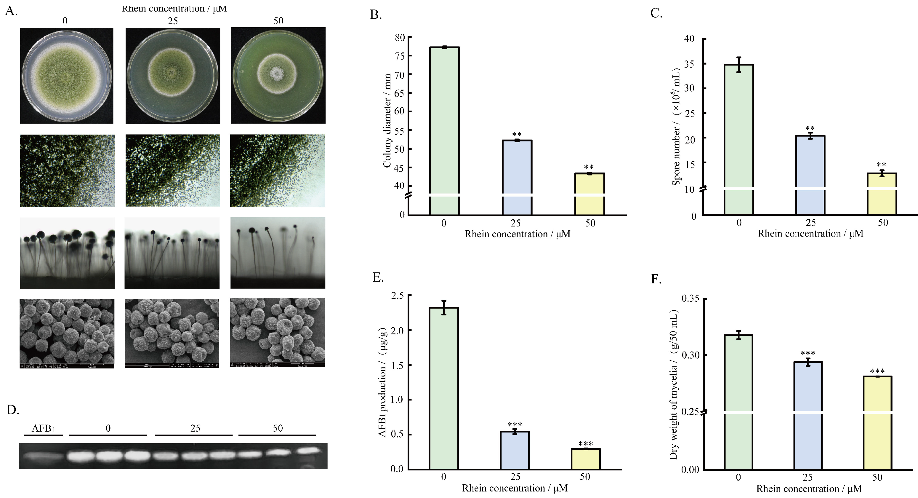
2.1. Effect of Rhein on A. flavus Growth, Spore Formation, and AFB1 Production

To examine the antifungal effects of rhein on mycelium growth and AFB1 production, 2 μL of A. flavus spore solution (1 × 10⁶ spores/mL) was inoculated into PDA medium with varying concentrations of rhein (0, 25, or 50 μM), then incubated at 30 °C for 5 days. The results demonstrate that A. flavus mycelium growth was significantly inhibited by treatment with 25 and 50 µM rhein. Additionally, the mycelium became dense and the colony diameter was reduced by 32.34% and 43.83%, respectively. Regarding the formation of A. flavus spores, it was observed that the number of spores and conidiophores decreased significantly with increasing rhein concentration, where the number of spores was significantly reduced by 41.32% and 63.18% under the 25 and 50 μM rhein treatments, respectively. Electron microscopy analysis revealed that the surface of the conidia without rhein treatment was regular and the shape was plentiful, while the morphology of rhein-treated A. flavus spores appeared shrunken and irregular (Figure 1A–C). TLC and HPLC analysis showed that, compared to the control, AFB1 production decreased by 76.5% and 87.2% after treatment with 25 and 50 μM rhein, respectively, suggesting a dose-dependent inhibition of AFB1 biosynthesis (Figure 1D,E). Additionally, the dry weight of mycelia also decreased significantly with increasing rhein concentration (Figure 1F). In summary, rhein showed significant inhibitory effects against A. flavus mycelium growth, spore formation, and AFB1 yield; moreover, 50 μM rhein showed better inhibition of A. flavus mycelium growth, sporulation, and AFB1. Based on these results, the A. flavus treated with 50 μM rhein was selected for RNA-seq analysis.

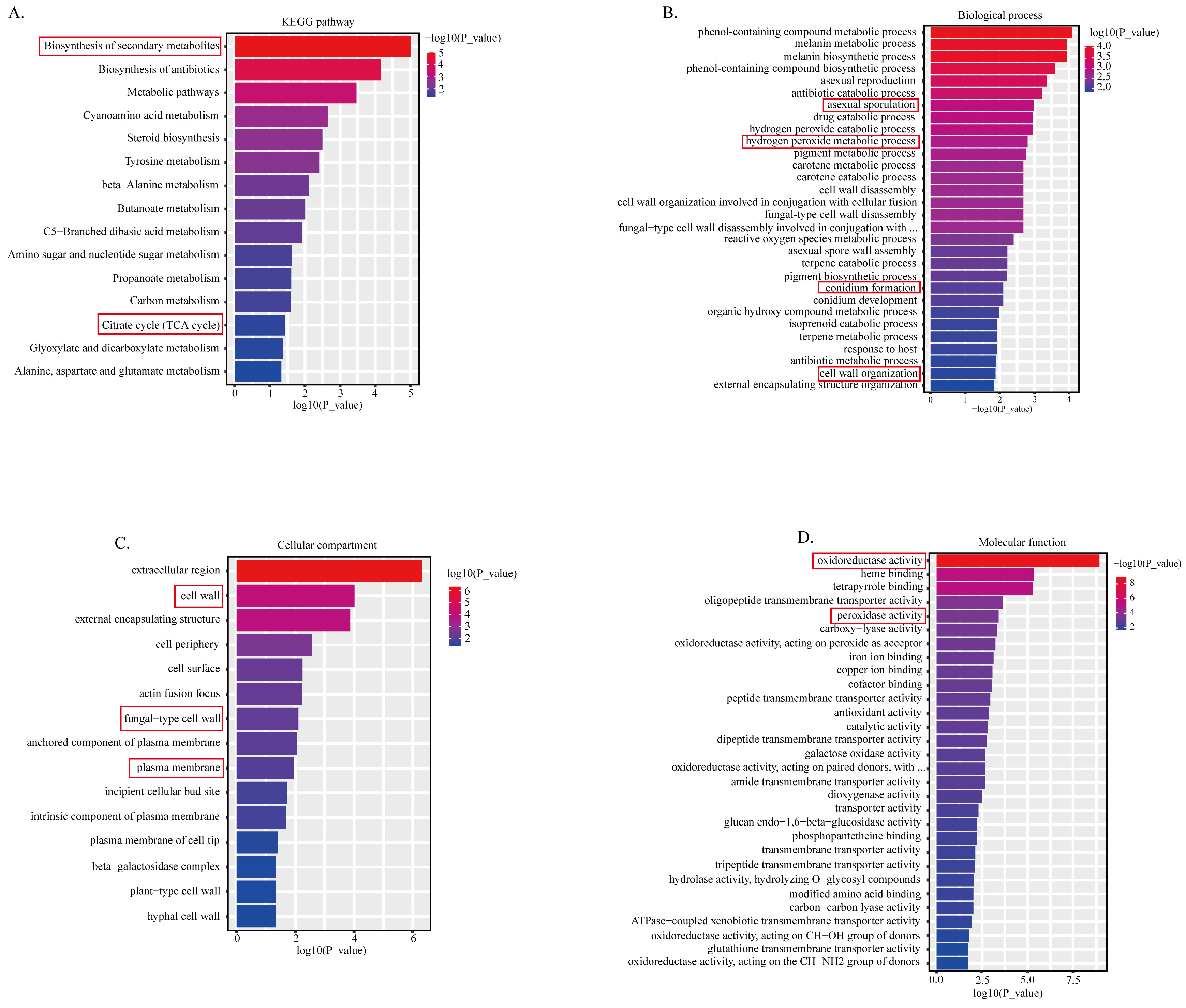
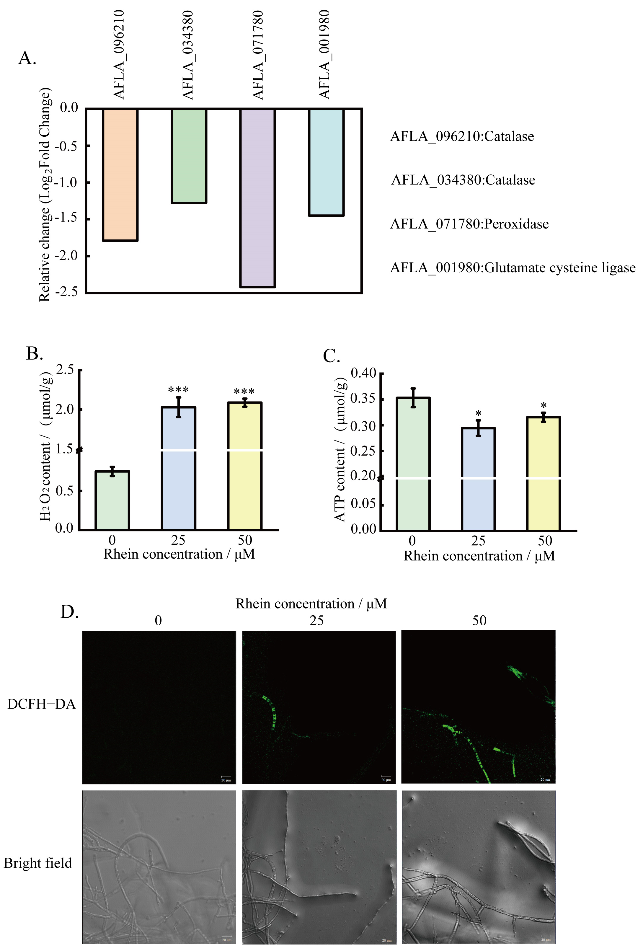
2.2. Transcriptomic Analysis

The A. flavus treated with 50 μM rhein was selected for RNA-seq analysis with triple biological replicates in order to explicate the underlying mechanism through which rhein affects A. flavus mycelium growth and aflatoxin biosynthesis. The results show 633 differentially expressed genes (DEGs) in rhein-treated A. flavus, with 150 DEGs upregulated and 483 DEGs downregulated compared to the control. To further elucidate the function of the DEGs, enrichment analysis was conducted. The results reveal that the DEGs were primarily linked to the biosynthetic pathway of secondary metabolites, metabolic pathways, and the citric acid cycle, as indicated by the KEGG enrichment analysis (Figure 2A). GO enrichment analysis revealed that DEGs were associated with conidium formation and development, the cell wall and membrane, the hydrogen peroxide metabolic process, and oxidoreductase activity (Figure 2B–D).
To better elucidate the underlying mechanism, the representative DEGs were classified into four categories, including conidium formation and development, cell wall and cell membrane, glycolysis, citrate cycle, oxidative stress, and aflatoxin biosynthesis (Table 1).

2.3. Effect of Rhein on the Hydrophobicity of A. flavus

To verify the differential expression of conidial hydrophobin genes, the effect of rhein on the hydrophobicity of A. flavus was investigated. The results demonstrate that bromophenol blue solution and sterile water fully penetrated the surface of the rhein-treated A. flavus mycelium into the medium as compared to the control. The mycelial surface was observed using a stereoscopic microscope, and at the same magnification, the sterile water droplets on the surface of A. flavus colonies treated with rhein were found to be smaller (Figure 3). These results show that treatment with rhein inhibits the hydrophobicity of the cell surface of the mycelia.

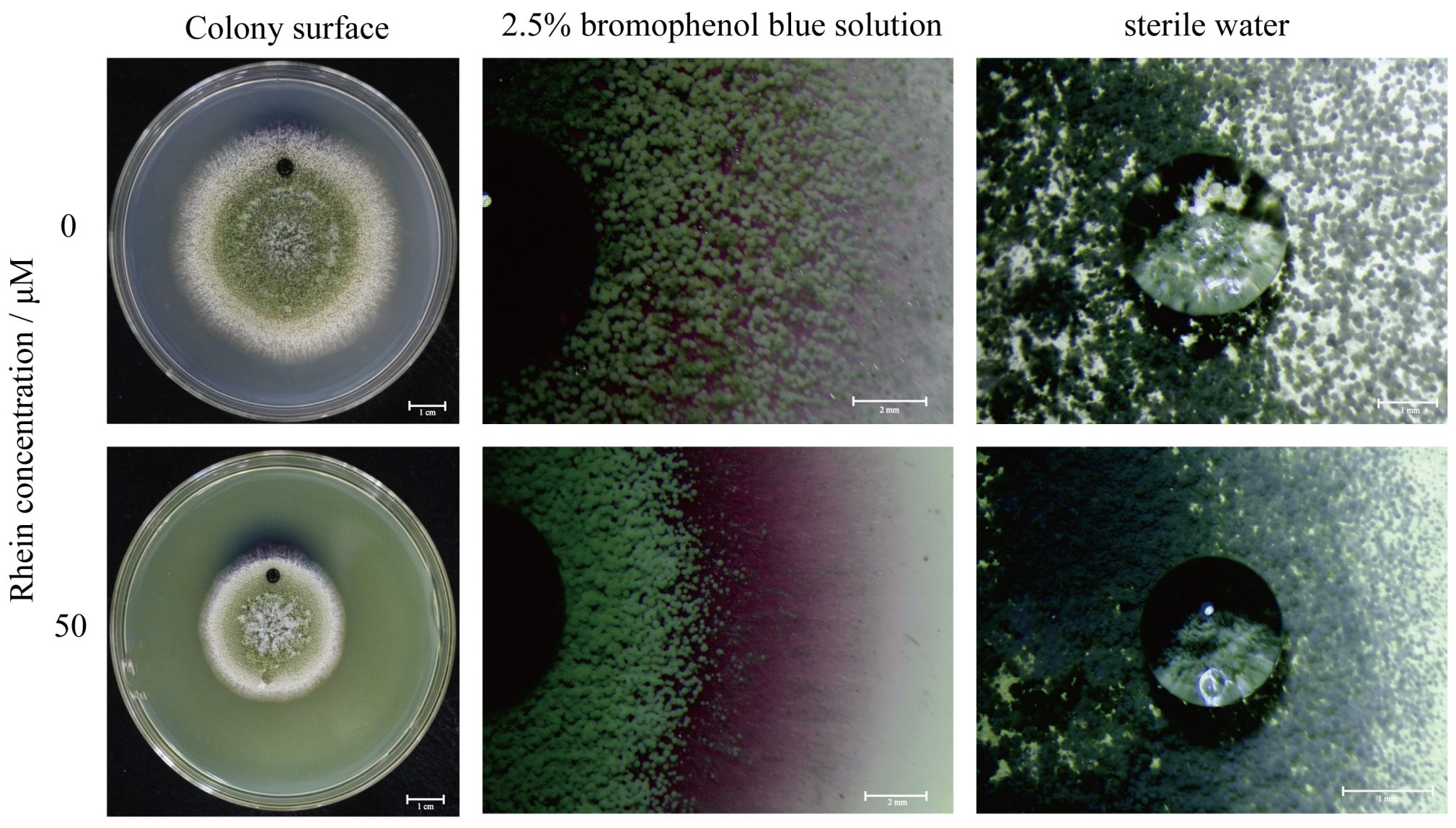
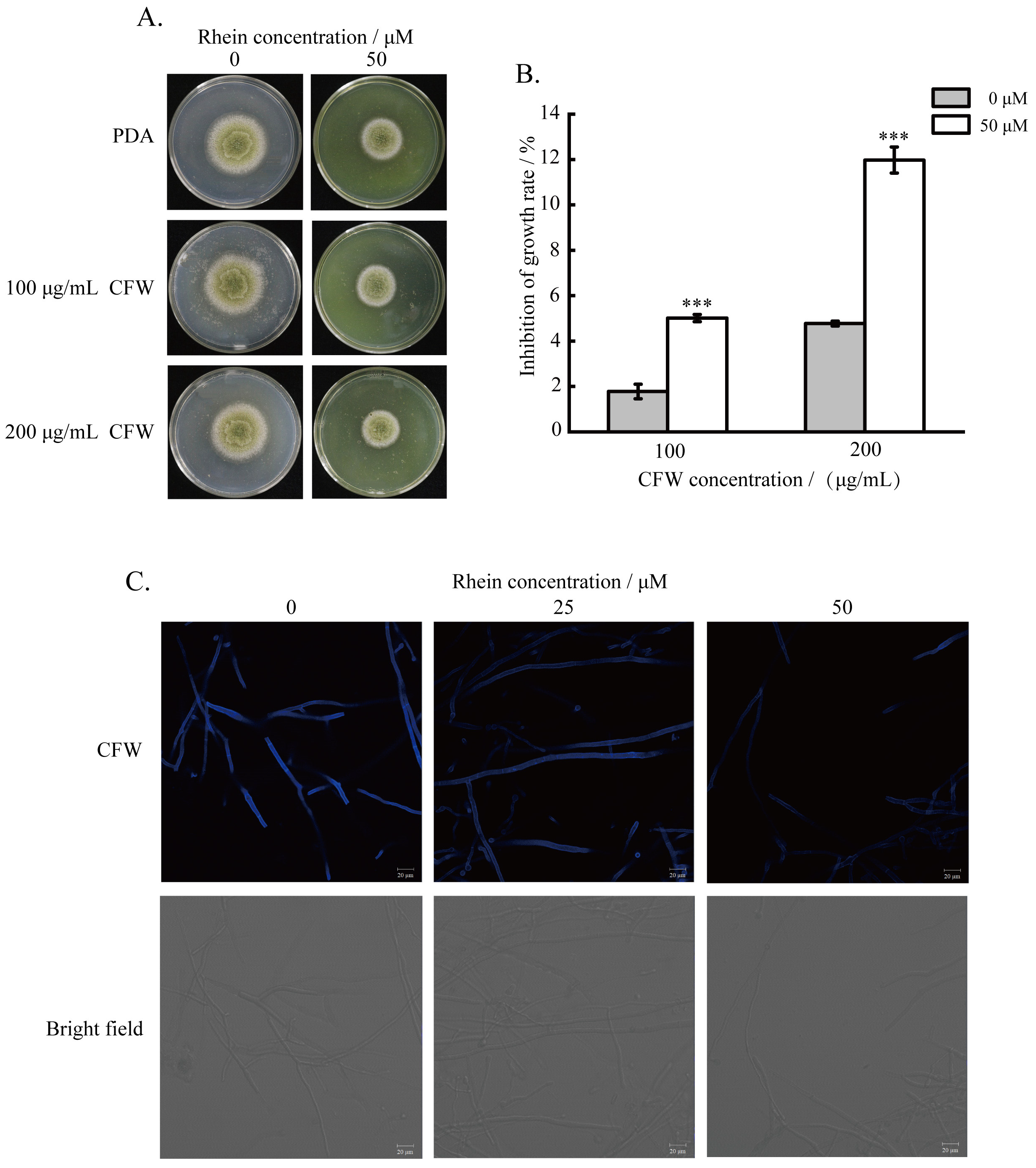
2.4. Effect of the Rhein on the Integrity of A. flavus Cell Wall

To study the effect of rhein on A. flavus cell wall integrity, the strains were cultured for 4 days under the reagent CFW. The results unveil that A. flavus strains were more sensitive to CFW after rhein treatment than the controls (Figure 4A,B). Additionally, the integrity of the cell wall was examined through CFW staining, showing a decrease in CFW fluorescence intensity with increasing rhein concentration (Figure 4C). These results suggest that rhein disrupts A. flavus cell wall synthesis.

2.5. Effect of Rhein on the Integrity of Cell Membrane

Different concentrations of SDS were introduced into PDA medium to examine the effect of rhein on the integrity of A. flavus cell membranes. The results demonstrate that the inhibition rate of rhein-treated A. flavus reached 11.2% and 39.1% under the conditions of 100 and 200 μg/mL SDS, respectively. Rhein-treated A. flavus was more sensitive than the control group (Figure 5A,B). Additionally, PI staining revealed that the red fluorescence intensity gradually increased with the concentration of rhein (Figure 5C). These results reveal that the addition of rhein disrupts the cell membrane of A. flavus.
To validate the analysis of RNA-seq data, the intracellular ROS accumulation, H2O2, and ATP contents were measured. The results demonstrate that, accompanied by the downregulated expression of catalase, peroxidase, and glutamate cysteine ligase-related genes (Figure 6A), the intracellular H2O2 content of A. flavus increased (Figure 6B), which was in agreement with the results of the transcriptome analysis. The intracellular ATP content of A. flavus also decreased slightly after rhein treatment (Figure 6C). In addition, the results of DCFH-DA staining demonstrate that A. flavus mycelia treated with rhein exhibited stronger green fluorescence than those untreated with rhein (Figure 6D), indicating that rhein induced the accumulation of ROS.

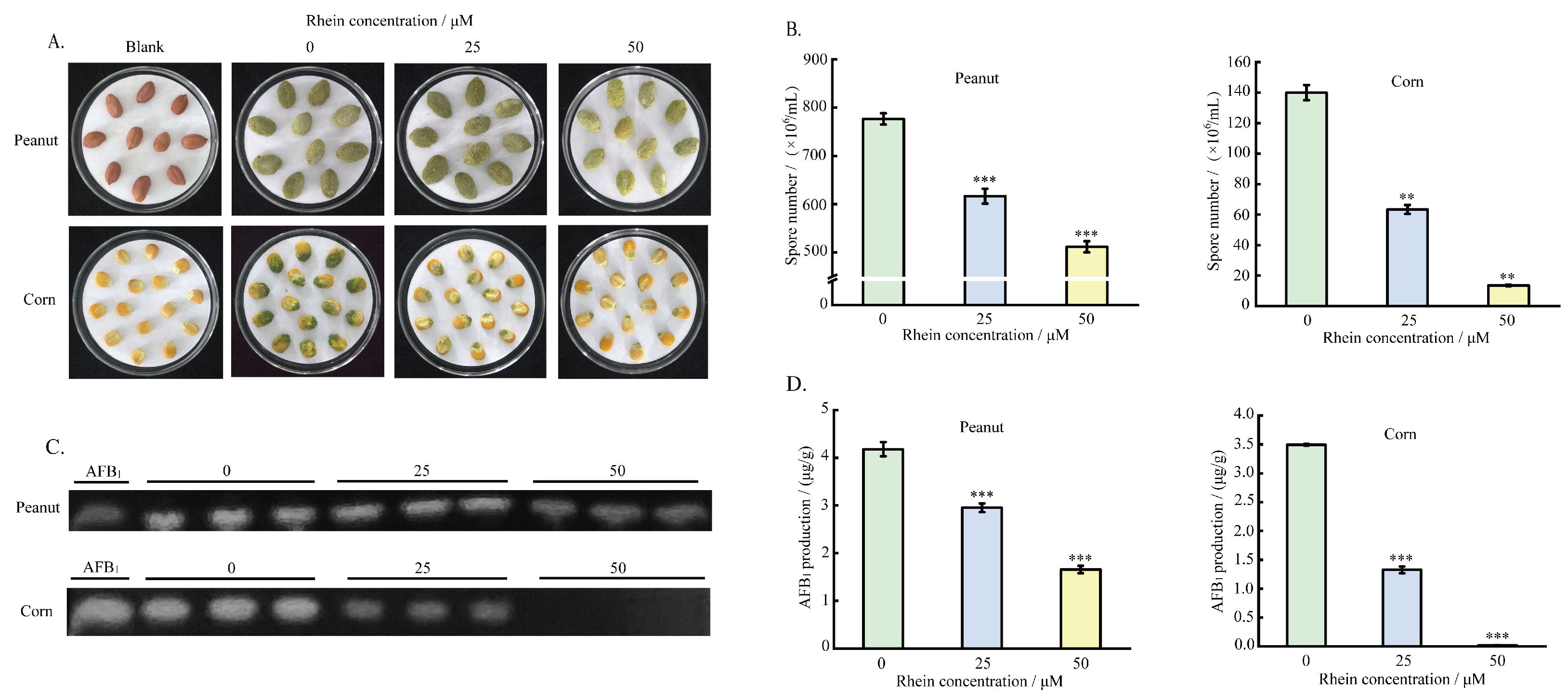
2.6. Effect of the Rhein on A. flavus Pathogenicity on Crop Seeds
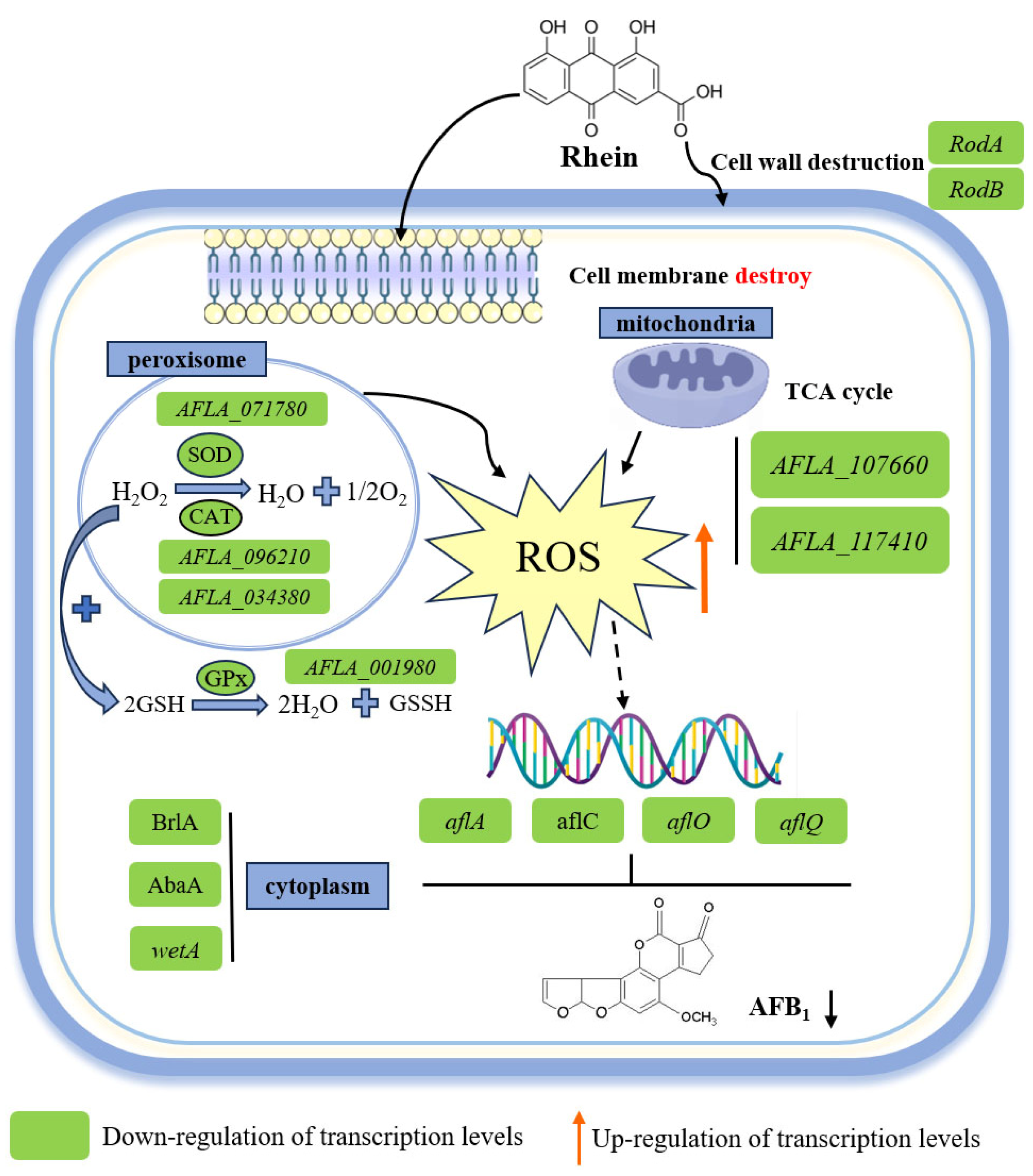
To investigate the effect of rhein on the sporogenesis and the AFB1 yield of A. flavus on crop seeds, different concentrations of rhein were incubated for 5 days with maize and peanut seeds infected with A. flavus spore suspension. The results indicate that the colonization ability of A. flavus on peanut and maize seeds was reduced after treatment with rhein (Figure 7A). The number of spores on the surface of the seeds was reduced by 90.4% and 34.1%, respectively, in comparison to the control group, following treatment with 50 μM of rhein (Figure 7B). Additionally, the TLC and HPLC results demonstrate the inhibition of AFB1 production. With the 50 μM rhein treatment, the AFB1 yield of A. flavus on peanuts and maize decreased by 60.52% and 99.43%, respectively (Figure 7C,D). These results clearly demonstrate that rhein significantly inhibits the pathogenicity of A. flavus on maize and peanut seeds.

3. Discussion

Developing safe and effective antifungal drugs is urgently needed to reduce the threat posed by A. flavus to cereals and food. In our study, the effects of rhein, a compound from traditional Chinese medicine, on A. flavus mycelium growth, sporulation, and AFB1 biosynthesis were characterized. The results demonstrate that rhein disrupted A. flavus cell wall and membrane integrity, thereby inhibiting its growth and sporulation. Additionally, an RNA-seq analysis and subsequent validation demonstrated that mitochondrial dysfunction and the accumulation of intracellular ROS might lead to the inhibition of aflatoxin biosynthesis.
Rhein treatment significantly inhibited cell growth and development in A. flavus. Various regulators and transcription factors regulate the A. flavus spore-forming process. It has been reported that conidia formation and maturation in A. flavus are mainly governed by BrlA, AbaA, and WetA [24]. The AbaA gene plays a role in conidial cell differentiation and is activated by BrlA at metaphase [25,26], while the wetA gene, activated by AbaA in anaphase, participates in synthesizing key conidial cell wall components [27]. In our study, rhein treatment substantially reduced the A. flavus spore count, and the expression of genes related to conidial growth, such as AFLA_052030 (wetA), AFLA_082850 (BrlA), AFLA_029620 (AbaA), and AFLA_044790, was significantly downregulated (Table 1). These findings suggest that rhein may inhibit spore formation in A. flavus. In addition, RodA and RodB are conidial surface proteins, with RodA playing a role in the surface rod structure and RodB contributing to the conidial cell wall structure in Aspergillus fumigatus [28,29]. In our study, the bromophenol blue solution diffused rapidly and was absorbed on the surface of rhein-treated mycelium (Figure 3). Furthermore, the expression of genes related to hydrophobic proteins (AFLA_098380, AFLA_014260) was significantly downregulated. This is in agreement with the results of the hydrophobicity assay in the Afper1 gene deletion strain studied by Lv et al. [30]. These results suggest that rhein treatment may have reduced the hydrophobicity of the mycelium surface.
The cell wall and membrane of the fungi are dynamic structures which are essential for maintaining cell viability and integrity, making them promising targets for antifungal treatment [31,32,33,34,35]. In this study, CFW staining showed that rhein disrupted cell wall synthesis, and genes involved in cell wall protein synthesis (AFLA_061960) were also downregulated. However, transcriptome data analysis demonstrated that gene expression of chitin synthetase was upregulated, suggesting that upregulated chitin synthetase might contribute to compensating for the destroyed cell wall after rhein treatment. In addition, PI staining demonstrated that the red fluorescence intensity was increased, suggesting cell membrane damage. Similar phenomena have been observed in A. flavus cell membranes when treated with honokiol [36], (E)-2-hexenal [37], and phenyllactic acid [38]. Moreover, the gene expression levels of AFLA_007630 and AFLA_039810, which are involved in membrane protein synthesis, were significantly downregulated. This suggests that rhein may induce impaired synthesis of the cell wall and membrane, leading to the disruption of cell components. These results also indicate that the antifungal activity of rhein may target the integrity of the cell membrane and cell wall.
It is well known that mitochondria are organelles that generate ATP through oxidative phosphorylation reactions, providing energy to the cell [39]. Oxidative phosphorylation occurs during the citric acid cycle (TCA) in the mitochondria [40]. Previous studies have indicated that mitochondrial dysfunction leads to the accumulation of ROS by reducing ATP levels [41]. In our study, genes associated with the TCA cycle, including citrate synthetase and succinyl-CoA synthetase, were significantly downregulated after rhein treatment (Table 1). This finding mirrors the genetic changes observed in A. flavus treated with estragole [42]. In addition, A. flavus treated with rhein exhibited a remarkable decrease in intracellular ATP levels, which aligns with the findings of Duan et al. [43] regarding A. flavus treated with 1-octanol. These results reveal that rhein affects the changes in enzymes in other metabolic pathways, such as the TCA cycle, leading to mitochondrial dysfunction and ultimately the accumulation of ROS.
Peroxisomes are an important component of eukaryotic cells, participating in various cellular physiological processes and being capable of rapidly producing and removing H2O2 and O2, thereby regulating the dynamic changes in ROS levels [44,45,46]. In this study, the genes encoding catalase and peroxidase (AFLA_096210, AFLA_034380) were significantly downregulated after treatment with rhein (Figure 6A). Subsequently, experimental verification showed that the intracellular H2O2 content was increased in A. flavus treated with rhein. These results suggest that H2O2 accumulation disrupts oxidation reduction (redox) homeostasis. In addition, glutathione biosynthesis is also involved in oxidative stress, with glutathione ligase reported as the key enzyme catalyzing glutathione biosynthesis [47]. Elevated levels of ROS also promote mitochondrial and metabolic dysfunction, resulting in oxidative stress [48]. Our transcriptome data show that the transcriptional level of the glutathione ligase gene (AFLA_001980) was remarkably downregulated (Table 1). We also assessed the intracellular ROS level in A. flavus using DCFH-DA staining and noted the dose-dependent increase in fluorescence intensity with rhein treatment. These results suggest that rhein triggered oxidative stress, resulting in a significant accumulation of ROS, consistent with the intracellular ROS level observed in A. flavus treated with methyl 2-methylbutyrate [49]. Therefore, we speculate that rhein treatment might cause changes in the intracellular peroxisomes of A. flavus, which disrupted the redox balance and subsequently led to ROS accumulation.
Aflatoxin biosynthesis involves a complex regulatory network composed of multiple genes and enzymatic reactions, regulated by the pathway-specific transcription factor aflR and several structural genes, including aflA, aflB, aflC, aflO, aflQ, aflR, and aflS. Additionally, it is also governed by environmental conditions, such as light, nutrient sources, and oxidative stress [50,51,52,53]. According to previous reports, three proteins, aflA, aflB, and aflC, are jointly involved in the synthesis of norsolorinic acid (NOR) in the biosynthesis pathway of aflatoxin [5]. AflO, which encodes O-methyltransferase, converts demethylsterigmatocystin (DMST) to a new compound, sterigmatocystin (ST), and aflQ is a P-450 monooxygenase that converts O-methylsterigmatocystin (OMST) to AFB1 [3]. In our study, the production of AFB1 by rhein-treated A. flavus growing on both PDA medium and grain was significantly decreased. Analysis of transcriptome data revealed that the genes involved in the pathway for the aflatoxin synthesis (aflA, aflC, aflO, and aflQ) were all remarkedly downregulated after rhein treatment. In A. flavus, deletion of catalase CTA1 contributes to the intracellular ROS accumulation and downregulation of aflC, aflD, and aflQ, consequently leading to impaired AFB1 biosynthesis [54]. Additionally, the accumulated intracellular ROS in A. flavus treated with curcumin also leads to a reduction in aflatoxin production [55]. In our study, the downregulated expression levels of aflA, aflC, aflO, and aflQ and decreased AFB1 biosynthesis may be attributed to the ROS accumulation triggered by rhein treatment.

4. Conclusions

In summary, rhein inhibited the growth and development of A. flavus by interfering with cell wall and membrane synthesis. Additionally, rhein might disrupt energy metabolism, cause mitochondrial dysfunction, and decrease ATP synthesis, leading to ROS accumulation, thereby inhibiting AFB1 biosynthesis (Figure 8). Furthermore, rhein treatment effectively inhibited mycelial growth and the production of AFB1 in both peanut and maize seeds. Our findings indicate that rhein can be considered as a potential novel agent for preventing fungal infections in food and feedstuffs.

5. Materials and Methods

5.1. Strains, Chemicals, and Medium

The model strain A. flavus NRRL 3357, a kind gift from Prof. Zhumei He (Sun Yat-sen University, China), was used in this study. Rhein (CAS:478-43-3, 98.5% purity) was obtained from J&K Scientific Ltd. (Beijing, China). Peanut and maize seeds were sourced from local markets. Potato dextrose agar medium (PDA) was selected for fungal growth, conidial production, and aflatoxin B1 (AFB1) biosynthesis. Rhein was dissolved in DMSO to form a solution with a final concentration of 10 mg/mL.

5.2. Effect of Rhein on Phenotype, Spore, and Aflatoxin B1 Production of A. flavus

The effects of rhein on A. flavus colony size, spore morphology, and aflatoxin B1 yield were examined with PDA medium. Rhein at different concentrations (25 and 50 μM) was added to PDA medium to create treatment groups according to previously described methods [56], while the same volume of DMSO was added to the control group. The spore solution (1 × 10⁶ spores/mL) was inoculated, and the colony diameter was recorded after culturing at 30 °C for 5 days. The morphology of spores and conidial pedicles was observed using a light microscope, stereoscopic microscope, and electron microscope.
AFB1 was extracted and detected using the methods described in a previous study [42]. In brief, 25 μM or 50 μM of rhein were introduced into PDA medium, and 2 μL of spore solution was inoculated followed by incubation for 5 days at 30 °C. AFB1 was then extracted by adding chloroform and distilled water (volume ratio of 1:1). Finally, AFB1 was analyzed through HPLC and TLC.
To evaluate the impact of rhein on mycelium production, 25 or 50 μM of rhein was introduced into liquid medium containing 50 mL DPY. Subsequently, A. flavus spores were inoculated to achieve a concentration of 1 × 10⁶ spores/mL. After shaking for 3 days at 30 °C, the mycelium was filtered and dried for 6 h, then weighed.

5.3. RNA-seq Analysis

The A. flavus spore suspension and 50 μM rhein were grown in PDA medium for 60 h at 30 °C, with DMSO replacing rhein in the control group. The TRIzol method by Thermo Fisher, Shanghai, China, was employed to extract total RNA from the mycelia with three biological replicates, and the quality and integrity of RNA were assessed. Subsequently, Shanghai Yuanxin Biotechnology Co., Ltd. (Shanghai, China) constructed the cDNA library for RNA-seq analysis. Each sample with three biological replicates, |log2fold change| > 1, and p value < 0.05 was defined as a differentially expressed gene. KEGG enrichment analysis and GO were performed using Goatools and KOBAS software, respectively. Raw transcriptome data were entered into the CNCB database under CRA accession number CRA015824.

5.4. Effect of Rhein on Hydrophobicity of A. flavus

Hydrophobicity was determined using the method of a previous study with slight modifications [57]. A. flavus spores were point-inoculated on PDA medium supplemented with rhein (50 μM), then incubated for a duration of 4 days at 30 °C. Subsequently, the colony surface was treated with sterile water and 2.5% bromophenol blue solution for 12 h and then photographed.

5.5. Effect of Rhein on A. flavus Cell Wall

A. flavus spore suspension was inoculated into PDA medium supplemented with rhein (50 μM) and fluorescent brightener 28 (CFW, 100 and 200 μg/mL) and incubated at 30 °C for 3 days. Moreover, the A. flavus colony diameter was recorded and photographed to evaluate the effect of rhein on A. flavus cell walls.
A. flavus spore suspension and rhein (25 and 50 μM) were added to 50 mL DPY medium and incubated for 48 h. Following the staining method described in a previous study [58], the mycelia were collected, washed, and then stained with Calcofluor white stain (CFW) for 1 min in the dark on slides. Finally, confocal laser scanning microscopy (CLSM) was utilized to observe the mycelia.

5.6. Effect of Rhein on A. flavus Cell Membrane

A. flavus spore was point-inoculated onto PDA supplemented with 50 μM rhein and different concentrations of sodium dodecyl sulphate (SDS, 100, and 200 μg/mL), and then it was incubated at 30 °C for 4 days. Then, the colony diameters were assessed and the relative inhibition rate was calculated to evaluate the effect of rhein on inhibition of A. flavus cell membranes.
Similarly, A. flavus mycelia were treated as described above. A. flavus spore suspension and rhein (25 and 50 μM) were added to 50 mL DPY medium and allowed to grow for 48 h. The mycelia were then collected and washed. Subsequently, propidium iodide (PI) staining was carried out at 37 °C for 30 min. Finally, the mycelia were collected and examined through CLSM.

5.7. Determination of A. flavus Intracellular ROS, H2O2, and ATP Content

A. flavus spore suspension and various rhein concentrations (25 and 50 μM) were added to 50 mL DPY medium and allowed to grow for 48 h at 30 °C. The mycelia were treated as described above, then stained with DCFH-DA and incubated in the dark at 37 °C for 30 min. Finally, samples were visualized using CLSM.
A. flavus spore was point-inoculated on PDA medium with cellophane with or without rhein (50 μM) and incubated at 30 °C for 60 h. Subsequently, A. flavus mycelia were ground into powder and collected. The H2O2 and ATP contents were determined with reference to the biochemical kit (Solarbio Science, Beijing, China).

5.8. Effect of Rhein on A. flavus Mycelium Growth and AFB1 Yield on Peanuts and Maize

Different concentrations of rhein (25 and 50 μM) and A. flavus spore suspension were added into 50 mL sterile water until a final concentration of 1 × 10⁶ spores/mL was achieved. Then, sterilized peanuts and corn were placed in the water and shaken for 1 h. The treated peanuts and corn were then evenly placed in a dish covered with filter paper and cultured at 30 °C in the dark for 5 days. To maintain appropriate moisture levels, sterile water was added periodically during this incubation period. The infected peanut and corn seeds were placed in triangular flasks after 5 days of incubation, and the spores were counted after 30 min of thorough agitation after adding 20 mL of sterile water. AFB1 was extracted and determined as described above.

5.9. Statistical Analysis

In this study, all experiments were performed in triplicate and presented as mean ± SD, followed by significance analysis using SAS software (version 9.2), with statistical significance considered at p < 0.05.

Author Contributions

Methodology, X.W.; validation, R.D.; data curation, Y.L. (Yang Lei) and S.W.; writing—original draft preparation, X.W.; writing—review and editing, K.I.S., N.L. and Y.L. (Yangyong Lv); project administration, Y.H. All authors have read and agreed to the published version of the manuscript.

Funding

This work was funded by the Natural Science Foundation of Henan Province (222300420037), the Natural Science Foundation of China (31972176), and the Cultivation Programme for Young Backbone Teachers in Henan University of Technology (21420114).

Institutional Review Board Statement

Not applicable.

Informed Consent Statement

Not applicable.

Data Availability Statement

The raw transcriptome read data are available in the CNCB database under accession number CRA015824.

Conflicts of Interest

The authors declare no conflicts of interest.

References

  1. Li, D.; Qin, L.; Wang, Y.; Xie, Q.; Li, N.; Wang, S.; Yuan, J. AflSte20 regulates morphogenesis, stress response, and aflatoxin biosynthesis of Aspergillus flavus. Toxins 2019, 11, 730. [Google Scholar] [CrossRef] [PubMed]
  2. Zhang, F.; Zhong, H.; Han, X.; Guo, Z.; Yang, W.; Liu, Y.; Yang, K.; Zhuang, Z.; Wang, S. Proteomic profile of Aspergillus flavus in response to water activity. Fungal Biol. 2015, 119, 114–124. [Google Scholar] [CrossRef] [PubMed]
  3. Caceres, I.; Khoury, A.A.; Khoury, R.E.; Lorber, S.; Oswald, I.P.; Khoury, A.E.; Atoui, A.; Puel, O.; Bailly, J.D. Aflatoxin biosynthesis and genetic regulation: A review. Toxins 2020, 12, 150. [Google Scholar] [CrossRef]
  4. Hu, Y.; Yang, G.; Zhang, D.; Liu, Y.; Li, Y.; Lin, G.; Guo, Z.; Wang, S.; Zhuang, Z. The PHD transcription factor Rum1 regulates morphogenesis and aflatoxin biosynthesis in Aspergillus flavus. Toxins 2018, 10, 301. [Google Scholar] [CrossRef] [PubMed]
  5. Khan, R.; Ghazali, F.M.; Mahyudin, N.A.; Samsudin, N.I.P. Aflatoxin biosynthesis, genetic regulation, toxicity, and control strategies: A review. J Fungi 2021, 7, 606. [Google Scholar] [CrossRef]
  6. Eom, T.J.; Moon, H.; Yu, J.H.; Park, H.S. Characterization of the velvet regulators in Aspergillus flavus. J. Microbiol. 2018, 56, 893–901. [Google Scholar] [CrossRef] [PubMed]
  7. Gong, A.; Song, M.; Zhang, J. Current strategies in controlling Aspergillus flavus and aflatoxins in grains during storage: A review. Sustainability 2024, 16, 3171. [Google Scholar] [CrossRef]
  8. Abdelaziz, A.M.; El-Wakil, D.A.; Attia, M.S.; Ali, O.M.; AbdElgawad, H.; Hashem, A.H. Inhibition of Aspergillus flavus growth and aflatoxin production in Zea mays L. using Endophytic Aspergillus fumigatus. J Fungi 2022, 8, 482. [Google Scholar] [CrossRef]
  9. Ben Taheur, F.; Mansour, C.; Kouidhi, B.; Chaieb, K. Use of lactic acid bacteria for the inhibition of Aspergillus flavus and Aspergillus carbonarius growth and mycotoxin production. Toxicon 2019, 166, 15–23. [Google Scholar] [CrossRef]
  10. Liang, D.; Xing, F.; Selvaraj, J.N.; Liu, X.; Wang, L.; Hua, H.; Zhou, L.; Zhao, Y.; Wang, Y.; Liu, Y. Inhibitory effect of cinnamaldehyde, citral, and eugenol on aflatoxin biosynthetic gene expression and aflatoxin B1 biosynthesis in Aspergillus flavus. J. Food Sci. 2015, 80, M2917–M2924. [Google Scholar] [CrossRef]
  11. Wu, C.; Cao, H.; Zhou, H.; Sun, L.; Xue, J.; Li, J.; Bian, Y.; Sun, R.; Dong, S.; Liu, P.; et al. Research progress on the antitumor effects of rhein: Literature review. Anticancer Agents Med. Chem. 2017, 17, 1624–1632. [Google Scholar] [CrossRef] [PubMed]
  12. Li, G.M.; Chen, J.R.; Zhang, H.Q.; Cao, X.Y.; Sun, C.; Peng, F.; Yin, Y.P.; Lin, Z.; Yu, L.; Chen, Y.; et al. Update on pharmacological activities, security, and pharmacokinetics of rhein. Evid. Based Complement Altern. Med. 2021, 2021, 4582412. [Google Scholar] [CrossRef] [PubMed]
  13. Sun, H.; Luo, G.; Chen, D.; Xiang, Z. A comprehensive and system review for the pharmacological mechanism of action of rhein, an active anthraquinone ingredient. Front. Pharmacol. 2016, 7, 247. [Google Scholar] [CrossRef] [PubMed]
  14. Zhang, H.; Yi, J.K.; Huang, H.; Park, S.; Park, S.; Kwon, W.; Kim, E.; Jang, S.; Kim, S.Y.; Choi, S.K.; et al. Rhein suppresses colorectal cancer cell growth by inhibiting the mTOR pathway in vitro and in vivo. Cancers 2021, 13, 2176. [Google Scholar] [CrossRef] [PubMed]
  15. Ma, Y.L.; Chen, F.; Shi, J. Rhein inhibits malignant phenotypes of human renal cell carcinoma by impacting on MAPK/NF-κB signaling pathways. Onco Targets Ther. 2018, 11, 1385–1394. [Google Scholar] [CrossRef] [PubMed]
  16. Nguyen, A.T.; Kim, K.Y. Rhein inhibits the growth of Propionibacterium acnes by blocking NADH dehydrogenase-2 activity. J. Med. Microbiol. 2020, 69, 689–696. [Google Scholar] [CrossRef] [PubMed]
  17. Yao, J.Y.; Lin, L.Y.; Yuan, X.M.; Ying, W.L.; Xu, Y.; Pan, X.Y.; Ge, P.H. Antifungal activity of Rhein and aloe-emodin from Rheum palmatum on fish pathogenic Saprolegnia sp. J. World Aquacult. Soc. 2017, 48, 137–144. [Google Scholar] [CrossRef]
  18. Wang, B.; Yang, J.; Zhao, X.; Feng, X.; Xu, S.; Li, P.; Li, L.; Chen, Y. Antifungal activity of the botanical compound rhein against Phytophthora capsici and the underlying mechanisms. Pest Manag. Sci. 2024, 80, 1228–1239. [Google Scholar] [CrossRef] [PubMed]
  19. Agarwal, S.K.; Singh, S.S.; Verma, S.; Kumar, S. Antifungal activity of anthraquinone derivatives from Rheum emodi. J. Ethnopharmacol. 2000, 72, 43–46. [Google Scholar] [CrossRef]
  20. Chewchinda, S.; Wuthi-udomlert, M.; Gritsanapan, W. HPLC quantitative analysis of rhein and antidermatophytic activity of cassia fistula pod pulp extracts of various storage conditions. Biomed. Res. Int. 2013, 2013, 821295. [Google Scholar] [CrossRef]
  21. Zhu, X.; Chen, S.; Zheng, Y.; Zhang, Y.; Hsiang, T.; Huang, R.; Qi, J.; Gan, T.; Chang, Y.; Li, J. Antifungal and insecticidal activities of rhein derivatives: Synthesis, characterization and preliminary structure-activity relationship studies. Nat. Prod. Res. 2022, 36, 4140–4146. [Google Scholar] [CrossRef] [PubMed]
  22. Shang, X.F.; Zhao, Z.M.; Li, J.C.; Yang, G.Z.; Liu, Y.Q.; Dai, L.X.; Zhang, J.Y. Insecticidal and antifungal activities of Rheum palmatum L. anthraquinones and structurally related compounds. Ind. Crop. Prod. 2019, 137, 508–520. [Google Scholar] [CrossRef]
  23. Chen, S.; Wang, M.; Yu, L.; Shi, J.; Zhang, Y.; Tian, Y.; Li, L.; Zhu, X.; Li, J. Rhein-amino acid easter caonjugates as potential antifungal agents: Synthesis and biological evaluation. Molecules 2023, 28, 2074. [Google Scholar] [CrossRef] [PubMed]
  24. Cho, H.J.; Son, S.H.; Chen, W.; Son, Y.E.; Lee, I.; Yu, J.H.; Park, H.S. Regulation of conidiogenesis in Aspergillus flavus. Cells 2022, 11, 2796. [Google Scholar] [CrossRef]
  25. Andrianopoulos, A.; Timberlake, W.E. The Aspergillus nidulans abaA gene encodes a transcriptional activator that acts as a genetic switch to control development. Mol. Cell Biol. 1994, 14, 2503–2515. [Google Scholar] [CrossRef] [PubMed]
  26. Mead, M.E.; Borowsky, A.T.; Joehnk, B.; Steenwyk, J.L.; Shen, X.X.; Sil, A.; Rokas, A. Recurrent loss of abaA, a master regulator of asexual development in filamentous fungi, correlates with changes in genomic and morphological traits. Genome Biol. Evol. 2020, 12, 1119–1130. [Google Scholar] [CrossRef] [PubMed]
  27. Wu, M.Y.; Mead, M.E.; Kim, S.C.; Rokas, A.; Yu, J.H. WetA bridges cellular and chemical development in Aspergillus flavus. PLoS ONE 2017, 12, e0179571. [Google Scholar] [CrossRef]
  28. Pedersen, M.H.; Borodina, I.; Moresco, J.L.; Svendsen, W.E.; Frisvad, J.C.; Søndergaard, I. High-yield production of hydrophobins RodA and RodB from Aspergillus fumigatus in Pichia pastoris. Appl. Microbiol. Biotechnol. 2011, 90, 1923–1932. [Google Scholar] [CrossRef]
  29. Grünbacher, A.; Throm, T.; Seidel, C.; Gutt, B.; Röhrig, J.; Strunk, T.; Vincze, P.; Walheim, S.; Schimmel, T.; Wenzel, W.; et al. Six hydrophobins are involved in hydrophobin rodlet formation in Aspergillus nidulans and contribute to hydrophobicity of the spore surface. PLoS ONE 2014, 9, e94546. [Google Scholar] [CrossRef]
  30. Lv, Y.; Yang, H.; Wang, J.; Wei, S.; Zhai, H.; Zhang, S.; Hu, Y. Afper1 contributes to cell development and aflatoxin biosynthesis in Aspergillus flavus. Int. J. Food Microbiol. 2022, 377, 109828. [Google Scholar] [CrossRef]
  31. Gow, N.A.R.; Latge, J.P.; Munro, C.A. The fungal cell wall: Structure, biosynthesis, and function. Microbiol. Spectr. 2017, 5. [Google Scholar] [CrossRef] [PubMed]
  32. Garcia-Rubio, R.; de Oliveira, H.C.; Rivera, J.; Trevijano-Contador, N. The fungal cell wall: Candida, cryptococcus, and Aspergillus Species. Front. Microbiol. 2020, 10, 2993. [Google Scholar] [CrossRef]
  33. Sant, D.G.; Tupe, S.G.; Ramana, C.V.; Deshpande, M.V. Fungal cell membrane-promising drug target for antifungal therapy. J. Appl. Microbiol. 2016, 121, 1498–1510. [Google Scholar] [CrossRef] [PubMed]
  34. Li, Q.; Zhu, X.; Xie, Y.; Liang, J. Antifungal properties and mechanisms of three volatile aldehydes (octanal, nonanal and decanal) on Aspergillus flavus. Grain Oil Sci. Technol. 2021, 4, 131–140. [Google Scholar] [CrossRef]
  35. Bowman, S.M.; Free, S.J. The structure and synthesis of the fungal cell wall. Bioessays 2006, 28, 799–808. [Google Scholar] [CrossRef] [PubMed]
  36. Zhang, W.; Li, B.; Lv, Y.; Wei, S.; Zhang, S.; Hu, Y. Transcriptomic analysis shows the antifungal mechanism of honokiol against Aspergillus flavus. Int. J. Food Microbiol. 2023, 384, 109972. [Google Scholar] [CrossRef] [PubMed]
  37. Ma, W.; Zhao, L.; Zhao, W.; Xie, Y. (E)-2-Hexenal, as a potential natural antifungal compound, inhibits Aspergillus flavus spore germination by disrupting mitochondrial energy metabolism. J. Agric. Food Chem. 2019, 67, 1138–1145. [Google Scholar] [CrossRef] [PubMed]
  38. Li, M.; Yao, B.; Meng, X. Inhibitory effect and possible mechanism of phenyllactic acid on Aspergillus flavus spore germination. J. Basic Microbiol. 2022, 62, 1457–1466. [Google Scholar] [CrossRef]
  39. Kowalczyk, P.; Sulejczak, D.; Kleczkowska, P.; Bukowska-Ośko, I.; Kucia, M.; Popiel, M.; Wietrak, E.; Kramkowski, K.; Wrzosek, K.; Kaczyńska, K. Mitochondrial oxidative stress-a causative factor and therapeutic target in many diseases. Int. J. Mol. Sci. 2021, 22, 13384. [Google Scholar] [CrossRef]
  40. Chen, Y.; Zhou, Z.; Min, W. Mitochondria, oxidative stress and innate immunity. Front. Physiol. 2018, 9, 1487. [Google Scholar] [CrossRef]
  41. van Hameren, G.; Campbell, G.; Deck, M.; Berthelot, J.; Gautier, B.; Quintana, P.; Chrast, R.; Tricaud, N. In vivo real-time dynamics of ATP and ROS production in axonal mitochondria show decoupling in mouse models of peripheral neuropathies. Acta Neuropathol. Commun. 2019, 7, 86. [Google Scholar] [CrossRef] [PubMed]
  42. Liang, L.; Zhang, W.; Hao, J.; Wang, Y.; Wei, S.; Zhang, S.; Hu, Y.; Lv, Y. Estragole inhibits growth and aflatoxin biosynthesis of Aspergillus flavus by affecting reactive oxygen species homeostasis. Microbiol. Spectr. 2023, 11, e0134823. [Google Scholar] [CrossRef] [PubMed]
  43. Duan, W.Y.; Qin, Y.L.; Zhang, S.B.; Zhai, H.C.; Lv, Y.Y.; Wei, S.; Hu, Y.S. Inhibitory mechanisms of plant volatile 1-octanol on the germination of Aspergillus flavus spores. Food Biophys. 2024, 19, 96–108. [Google Scholar] [CrossRef]
  44. Del Río, L.A.; López-Huertas, E. ROS Generation in Peroxisomes and its Role in Cell Signaling. Plant Cell Physiol. 2016, 57, 1364–1376. [Google Scholar] [CrossRef] [PubMed]
  45. Sandalio, L.M.; Rodríguez-Serrano, M.; Romero-Puertas, M.C.; del Río, L.A. role of peroxisomes as a source of reactive oxygen species (ROS) signaling molecules. Subcell Biochem. 2013, 69, 231–255. [Google Scholar] [CrossRef]
  46. Pellegrino, E.; Aylan, B.; Bussi, C.; Fearns, A.; Bernard, E.M.; Athanasiadi, N.; Santucci, P.; Botella, L.; Gutierrez, M.G. Peroxisomal ROS control cytosolic Mycobacterium tuberculosis replication in human macrophages. J. Cell Biol. 2023, 222, e202303066. [Google Scholar] [CrossRef] [PubMed]
  47. Krzywanski, D.M.; Dickinson, D.A.; Iles, K.E.; Wigley, A.F.; Franklin, C.C.; Liu, R.M.; Kavanagh, T.J.; Forman, H.J. Variable regulation of glutamate cysteine ligase subunit proteins affects glutathione biosynthesis in response to oxidative stress. Arch. Biochem. Biophys. 2004, 423, 116–125. [Google Scholar] [CrossRef] [PubMed]
  48. Newsholme, P.; Cruzat, V.F.; Keane, K.N.; Carlessi, R.; de Bittencourt, P.I., Jr. Molecular mechanisms of ROS production and oxidative stress in diabetes. Biochem. J. 2016, 473, 4527–4550. [Google Scholar] [CrossRef]
  49. Wei, S.; Zhang, Y.; Wu, M.; Lv, Y.; Zhang, S.; Zhai, H.; Hu, Y. Mechanisms of methyl 2-methylbutyrate suppression on Aspergillus flavus growth and aflatoxin B1 biosynthesis. Int. J. Food Microbiol. 2024, 409, 110462. [Google Scholar] [CrossRef]
  50. Kenne, G.J.; Gummadidala, P.M.; Omebeyinje, M.H.; Mondal, A.M.; Bett, D.K.; McFadden, S.; Bromfield, S.; Banaszek, N.; Velez-Martinez, M.; Mitra, C.; et al. Activation of aflatoxin biosynthesis alleviates total ROS in Aspergillus parasiticus. Toxins 2018, 10, 57. [Google Scholar] [CrossRef]
  51. Tai, B.; Chang, J.; Liu, Y.; Xing, F. Recent progress of the effect of environmental factors on Aspergillus flavus growth and aflatoxins production on foods. Food Qual. Safety 2020, 4, 21–28. [Google Scholar] [CrossRef]
  52. Meneely, J.P.; Kolawole, O.; Haughey, S.A.; Miller, S.J.; Krska, R.; Elliott, C.T. The challenge of global aflatoxins legislation with a focus on peanuts and peanut products: A systematic review. Expo Health 2023, 15, 467–487. [Google Scholar] [CrossRef]
  53. Tumukunde, E.; Xie, R.; Wang, S. Updates on the functions and molecular mechanisms of the genes involved in Aspergillus flavus development and biosynthesis of aflatoxins. J Fungi 2021, 7, 666. [Google Scholar] [CrossRef] [PubMed]
  54. Zhu, Z.; Yang, M.; Bai, Y.; Ge, F.; Wang, S. Antioxidant-related catalase CTA1 regulates development, aflatoxin biosynthesis, and virulence in pathogenic fungus Aspergillus flavus. Environ. Microbiol. 2020, 22, 2792–2810. [Google Scholar] [CrossRef] [PubMed]
  55. Zhang, D.; Yang, Y.; Yao, B.; Hu, T.; Ma, Z.; Shi, W.; Ye, Y. Curcumin inhibits Aspergillus flavus infection and aflatoxin production possibly by inducing ROS burst. Food Res. Int. 2023, 167, 112646. [Google Scholar] [CrossRef]
  56. Chen, B.; Ye, F.; Yu, L.; Jia, G.; Huang, X.; Zhang, X.; Peng, S.; Chen, K.; Wang, M.; Gong, S.; et al. Development of cell-active N6-methyladenosine RNA demethylase FTO inhibitor. J. Am. Chem. Soc. 2012, 134, 17963–17971. [Google Scholar] [CrossRef]
  57. Fasoyin, O.E.; Wang, B.; Qiu, M.; Han, X.; Chung, K.R.; Wang, S. Carbon catabolite repression gene creA regulates morphology, aflatoxin biosynthesis and virulence in Aspergillus flavus. Fungal Genet. Biol. 2018, 115, 41–51. [Google Scholar] [CrossRef]
  58. Zhang, W.; Li, B.; Lv, Y.; Wei, S.; Zhang, S.; Hu, Y. Synergistic effects of combined cinnamaldehyde and nonanal vapors against Aspergillus flavus. Int. J. Food Microbiol. 2023, 402, 110277. [Google Scholar] [CrossRef]
Figure 1. Effects of rhein (0, 25, 50 μM) on A. flavus morphology and AFB1 biosynthesis. (A) Observation of colony and conidial morphology; (B) colony diameter; (C) the number of spores; (D) AFB1 production according to TLC analysis; (E) AFB1 production according to HPLC analysis; and (F) dry weight of mycelia. ** represents p < 0.01, *** represents p < 0.001.
Toxins 16 00285 g001
Figure 2. Transcriptome analysis of rhein-treated and untreated A. flavus mycelia. (A) KEGG pathway enrichment; (B) enrichment of biological processes; (C) analysis of the enrichment of cellular components in DEGs; and (D) analysis of DEGs for molecular function enrichment. The red boxes indicated the related items in Table 1 and the subsequent biochemical verification experiments.
Toxins 16 00285 g002
Figure 3. Determination of the hydrophobicity of A. flavus treated with or without rhein.
Toxins 16 00285 g003
Figure 4. Effects of rhein on A. flavus cell wall. (A) The sensitivity of A. flavus to CFW after treatment with rhein; (B) the CFW inhibition of A. flavus mycelial growth; (C) A. flavus mycelia treated with rhein (0, 25, 50 μM) stained with CFW by CLSM. *** represents p < 0.001.
Toxins 16 00285 g004
Figure 5. Effects of rhein on A. flavus cell membrane. (A) The sensitivity to SDS of A. flavus treated with rhein; (B) the SDS inhibition of A. flavus mycelial growth; (C) A. flavus mycelial treated with rhein (0, 25, 50 μM) stained with PI by CLSM. ** represents p < 0.01, *** represents p < 0.001.2.6. Effects of Rhein on Intracellular ROS, H2O2, and ATP in A. flavus.
Toxins 16 00285 g005
Figure 6. Effects of rhein on A. flavus intracellular ROS. (A) Expression levels of differential genes associated with oxidative stress in A. flavus after rhein treatment; (B) H2O2 content; (C) ATP content; (D) A. flavus mycelial treated with rhein (0, 25, 50 μM) stained with DCFH-DA by CLSM. * represents p < 0.05, *** represents p < 0.001.
Toxins 16 00285 g006
Figure 7. Effect of rhein on A. flavus pathogenicity to crop seeds. (A) Growth of A. flavus on seeds with or without rhein treatment; (B) number of A. flavus spores on seeds; (C) TLC; and (D) HPLC analysis of AFB1 yield. ** represents p < 0.01, *** represents p < 0.001.
Toxins 16 00285 g007
Figure 8. Mechanistic diagram of rhein inhibiting A. flavus growth and AFB1 biosynthesis.
Toxins 16 00285 g008
Table 1. The representative DEGs in the control compared to the rhein treatment.
Gene IDlog2(FC)Gene Functional Description
Conidium formation and development
AFLA_052030−1.55Developmental regulatory protein wetA
AFLA_082850−2.47C2H2 type conidiation transcription factor BrlA
AFLA_029620−2.06Transcription factor AbaA
AFLA_098380−1.85Conidial hydrophobin RodA
AFLA_014260−1.44Conidial hydrophobin RodB
AFLA_044790−1.63Conidiation-specific family protein
Cell wall and membrane
AFLA_052780−1.60Cell wall glucanase (Scw4)
AFLA_061960−1.94Cell wall protein
AFLA_1372001.56Chitin synthase
AFLA_025900−2.71Chitin deacetylase
AFLA_007630−1.52Integral membrane protein
AFLA_039810−1.23DUF1212 domain membrane protein
Oxidative stress
AFLA_096210−1.79Catalase
AFLA_034380−1.28Catalase
AFLA_071780−2.42Peroxidase family protein
AFLA_001980−1.45Glutamate cysteine ligase
Glycolysis, citrate cycle, and aflatoxin biosynthesis
AFLA_1014701.05Glyceraldehyde-3-phosphate dehydrogenase
AFLA_036370−1.29Phosphoenolpyruvate carboxykinase
AFLA_107660−2.13Succinyl-CoA synthetase
AFLA_117410−4.26Citrate synthase
AFLA_117420−1.75Fatty acid synthase subunit alpha (aflA)
AFLA_081850−1.23Norsolorinic acid synthase (aflC)
AFLA_065570−1.35O-methyltransferase (aflO)
AFLA_0731301.28O-methylsterigmatocystin oxidoreductase ordA-like (aflQ)
Disclaimer/Publisher’s Note: The statements, opinions and data contained in all publications are solely those of the individual author(s) and contributor(s) and not of MDPI and/or the editor(s). MDPI and/or the editor(s) disclaim responsibility for any injury to people or property resulting from any ideas, methods, instructions or products referred to in the content.

