Article

Protective Effect of Lipoic Acid on Oxidative Stress and Tissue Damage Induced by Aflatoxin B1 in Young Laying Hens

1
Department of Animal Nutrition and Feed Science, College of Animal Science and Technology, Huazhong Agricultural University, Wuhan 430070, China
2
Faculty of Veterinary and Animal Science, Muhammad Nawaz Shareef University of Agriculture, Multan 60000, Pakistan
3
China Grain Reserves (Sichuan) Quality Inspection Center Co., Ltd., Chengdu 610052, China
*
Author to whom correspondence should be addressed.
Toxins 2025, 17(4), 184; https://doi.org/10.3390/toxins17040184
Submission received: 14 March 2025 / Revised: 3 April 2025 / Accepted: 4 April 2025 / Published: 6 April 2025

Abstract

:
The aim of this study is to investigate the alleviating effect of lipoic acid on oxidative stress and tissue damage induced by aflatoxin B1 (AFB1) in young laying hens. The experiment was divided into a control group, an AFB1 group, and three lipoic acid treatment groups. The AFB1 group and three lipoic acid treatment groups were given diets supplemented with 90 μg/kg of AFB1. The additional amounts of lipoic acid were 20, 100, and 500 mg/kg, respectively, with a feeding period of 4 weeks. The experimental results showed that AFB1 significantly increased the levels of alanine aminotransferase (ALT), aspartate aminotransferase (AST), alkaline phosphatase (ALP) and malondialdehyde (MDA) in the serum and significantly decreased the levels of total protein (TP), albumin (ALB), total superoxide dismutase (T-SOD), glutathione peroxidase (GSH-Px) and catalase (CAT) (p < 0.05). In addition, AFB1 damaged the structure of the liver, spleen, and ovarian tissues. Lipoic acid reduced the levels of ALT, AST, ALP, and MDA in the serum and increased the levels of TP, ALB, T-SOD, GSH-Px, and CAT (p < 0.05). Meanwhile, lipoic acid also protected mitochondrial structure and alleviated liver, spleen, and ovarian tissue damage caused by AFB1. In summary, lipoic acid can alleviate oxidative stress and tissue damage caused by AFB1 in young laying hens.
Key Contribution:
This study investigated the protective effects of lipoic acid on oxidative stress and the liver, spleen, and ovarian tissue damage induced by aflatoxin B1 in young laying hens.

1. Introduction

Aflatoxin B1 (AFB1) is a very common fungal toxin in feed, with high toxicity and a wide contamination range, posing a serious threat to the health of livestock and poultry [1,2]. Under the regulatory provisions of China’s Feed Hygiene Standard GB 13078-2017, the maximum permissible level of AFB1 in feed raw materials is strictly limited to 50 µg/kg. As typical feed-dependent economic animals, laying hens are prone to health damage through long-term intake of AFB1-contaminated feed [3]. Youth is a critical stage in the growth and development of laying hens and determines their later egg-laying performance [4]. Therefore, the developmental status of young laying hens will directly affect the economic benefits of breeding [4]. AFB1 in feed can harm the health of young laying hens [1,2]. After ingestion, AFB1 can cause damage to multiple systems in young laying hens, ranging from impaired growth performance, such as slow weight gain and a reduced feed conversion rate, to oxidative damage and damage to important organs such as the liver and spleen, leading to immune suppression [5,6]. At the same time, it increases the susceptibility of the chicken flock to various diseases, seriously affecting the growth and development of young laying hens and their overall health [7]. After AFB1 enters the cell, it leads to the production of a large amount of reactive oxygen species to attack unsaturated fatty acids on the mitochondrial membrane, causing lipid peroxidation, disrupting the integrity of the mitochondrial membrane structure, and further leading to mitochondrial dysfunction and cell apoptosis [8,9].
Lipoic acid is a naturally occurring dithiol compound, which is similar to the vitamin B class of compounds [10]. It is a strong antioxidant, usually in the form of lipoic acid, and its antioxidant capacity is stronger than that of common antioxidants such as vitamin C and vitamin E [11]. In the body, lipoic acid is synthesized by the lipoic acid synthase system in mitochondria [12]. It can directly eliminate reactive oxygen species in the body through oxidation-reduction or convert reactive oxygen species into less aggressive products, reducing the damage of reactive oxygen species to mitochondria [13]. When laying hens suffer from AFB1 poisoning, mitochondrial damage can also lead to a decrease in the synthesis ability of lipoic acid and an increase in the production of reactive oxygen species [9,14]. Therefore, it is reasonable to speculate that supplementation with lipoic acid in feed can alleviate or eliminate the harm of AFB1 to young laying hens.
To date, there have been no reports on the use of lipoic acid to alleviate AFB1 poisoning in young laying hens. Considering the significant harm done by AFB1 to laying hens [3,6], this study used young laying hens as experimental animals. The effects of different treatments on the production performance, organ weight, antioxidant enzyme content, serum biochemical indicators, liver, spleen, and ovarian tissue structure damage, and nuclear and mitochondrial structure damage in the livers of young laying hens were determined. The effects of AFB1 on oxidative stress and tissue damage in young laying hens, as well as the alleviating effect of lipoic acid on AFB1 poisoning symptoms, were explored. This study can provide a theoretical basis for the prevention of AFB1 poisoning in young laying hens during the breeding production process.

2. Results

2.1. Changes in Production Performance and Organ Weight

The initial weight of each group of young laying hens was similar, about 1.23 kg/hen. Adding 90 μg/kg AFB1 to the diet had no significant effect on the weight, daily feed intake, and heart and spleen weight of young laying hens (p > 0.05) (Table 1). The liver weight of the AFB1 group showed an increasing trend (p > 0.05) (Table 1). Compared with the AFB1 group, adding 100 mg/kg of lipoic acid to the diet during the fourth week significantly reduced liver weight (p < 0.05) (Table 1). When 20 mg/kg or 500 mg/kg of lipoic acid was added to the diet, there was a decreasing trend in liver weight (p > 0.05) (Table 1).

2.2. Changes in Serum Biochemical Indicators

AFB1 significantly increased the levels of ALT, AST, and ALP in the serum of young laying hens while significantly reducing the levels of TP and ALB in the serum (p < 0.05) (Table 2). Compared with the AFB1 group, when 100 or 500 mg/kg of lipoic acid was added to the diet, the levels of ALT, AST, and ALP in the serum of young laying hens significantly decreased, while the levels of TP and ALB significantly increased (p < 0.05) (Table 2). Compared with the control group, the serum total bile acid (TBA) content in the AFB1 group showed an upward trend (p > 0.05) (Table 2).

2.3. Changes in Serum Antioxidant Enzyme Levels

AFB1 significantly reduced the levels of T-SOD, GSH-Px, and CAT in serum and significantly increased the level of MDA (p < 0.05) (Table 3). Compared with the AFB1 group, adding 20 mg/kg of lipoic acid to the diet had no significant effect on the content of serum antioxidant enzymes (p > 0.05) (Table 3). When the dose of lipoic acid was 100 mg/kg or above, the levels of T-SOD and GSH-Px in serum significantly increased, while the level of MDA significantly decreased (p < 0.05) (Table 3). When the dose of lipoic acid was 500 mg/kg, the content of CAT in serum significantly increased (p < 0.05) (Table 3).

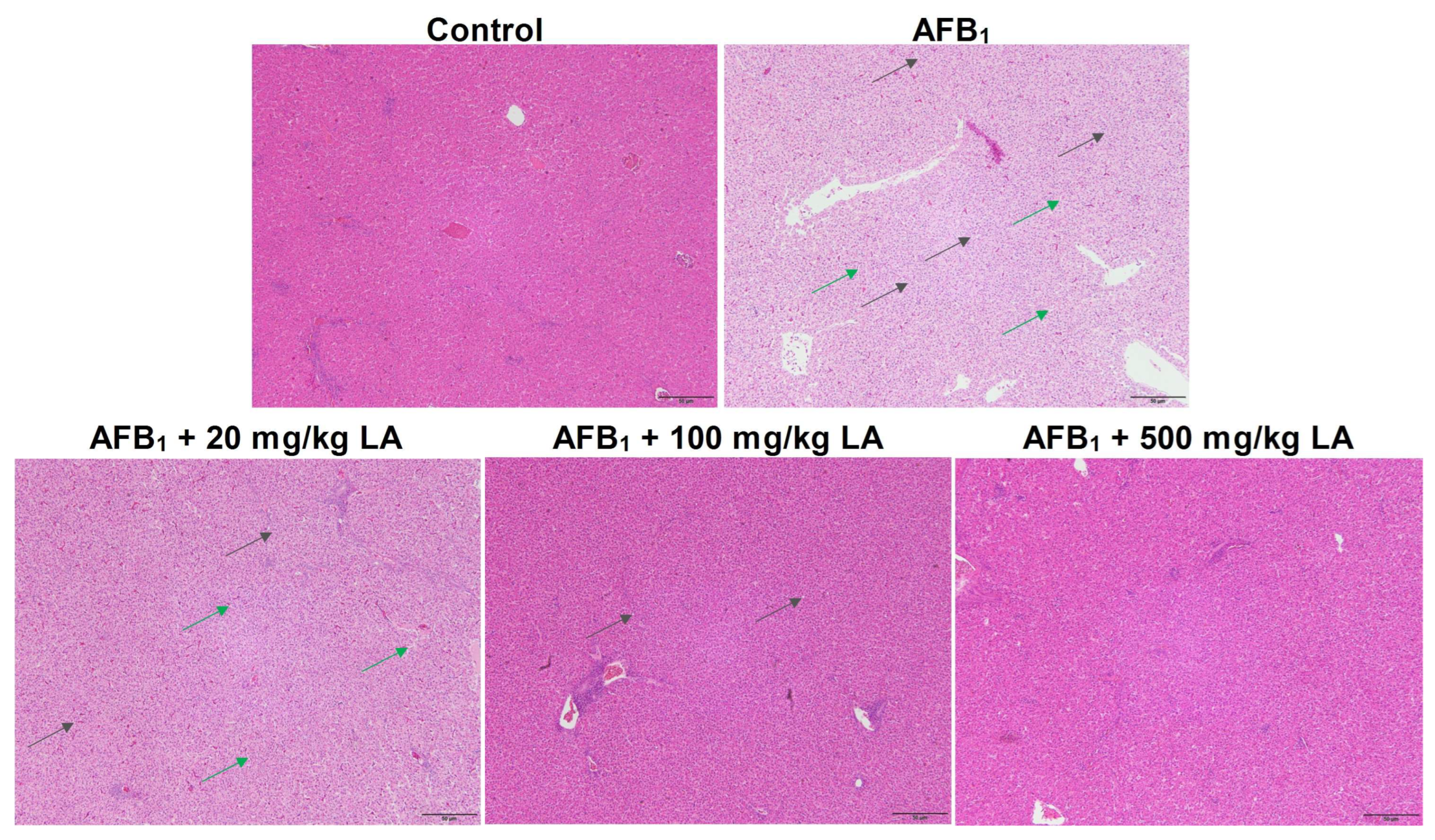
2.4. Changes in Liver Tissue Structure

The hierarchy of liver tissue structure in the control group was clear, with liver cells of regular morphology arranged close to each other and no symptoms such as fat degeneration or increased tissue gaps observed (Figure 1). There were symptoms of increased interstitial space and fatty degeneration in the liver tissue of the AFB1 group, and there were many circular vacuoles formed due to fatty degeneration on the slices (Figure 1). Adding 20 mg/kg of lipoic acid to the diet had a poor relief effect on liver AFB1 poisoning symptoms (Figure 1). When the amount of added lipoic acid reached 100 mg/kg, the symptoms of enlarged liver tissue gaps disappeared, and when the amount of added lipoic acid reached 500 mg/kg, the symptoms of hepatic steatosis disappeared (Figure 1).

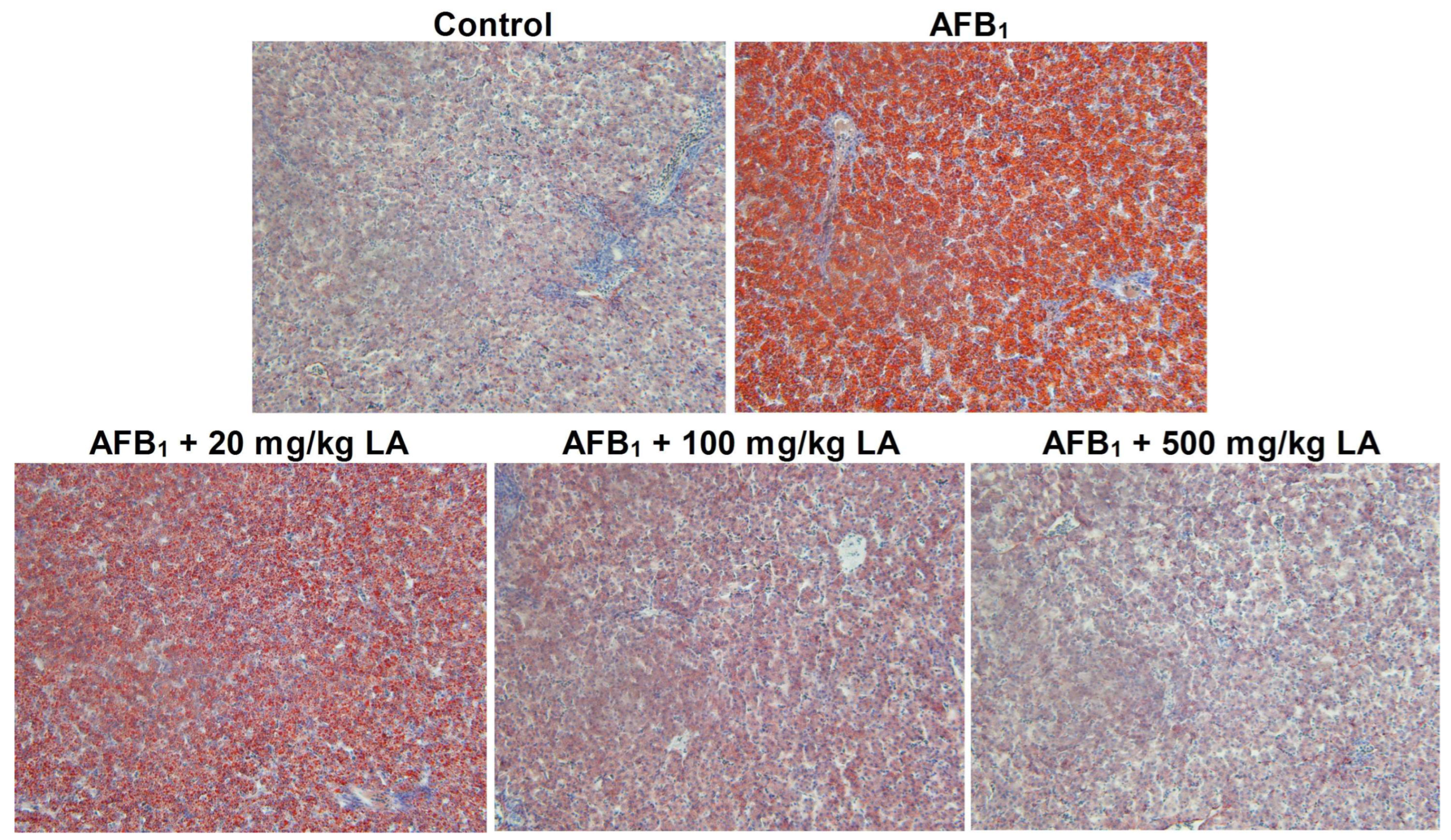
2.5. The Condition of Hepatic Steatosis

The livers of the control group were normal without any fat degeneration (Figure 2). There were obvious signs of steatosis in the livers of the AFB1 group, and a large amount of lipids were stained orange-red in the field of view (Figure 2). Compared with the AFB1 group, adding lipoic acid to the diet can alleviate the fat degeneration caused by AFB1. When the amount of lipoic acid added reached 500 mg/kg, the symptoms of fat degeneration disappeared (Figure 2).

2.6. Changes in Liver Ultrastructure

The contour of the nucleus and mitochondria in the control group liver is clear, the nuclear membrane of the nucleus is intact, the mitochondrial matrix is full, and the color of the matrix is darker (Figure 3). The nuclear membrane of AFB1 group liver cells shrinks, the mitochondria swell and enlarge, mitochondrial cristae break, and the matrix decreases and becomes partially blank (Figure 3). Adding lipoic acid to the diet can alleviate the nuclear and mitochondrial structural damage caused by AFB1. When the dose of lipoic acid is 100 mg/kg, the symptoms of nuclear membrane wrinkling disappear, and the symptoms of mitochondrial swelling and mitochondrial cristae damage are relieved (Figure 3). When the additional amount of lipoic acid is 500 mg/kg, the structure of the nucleus and mitochondria returns to normal (Figure 3).

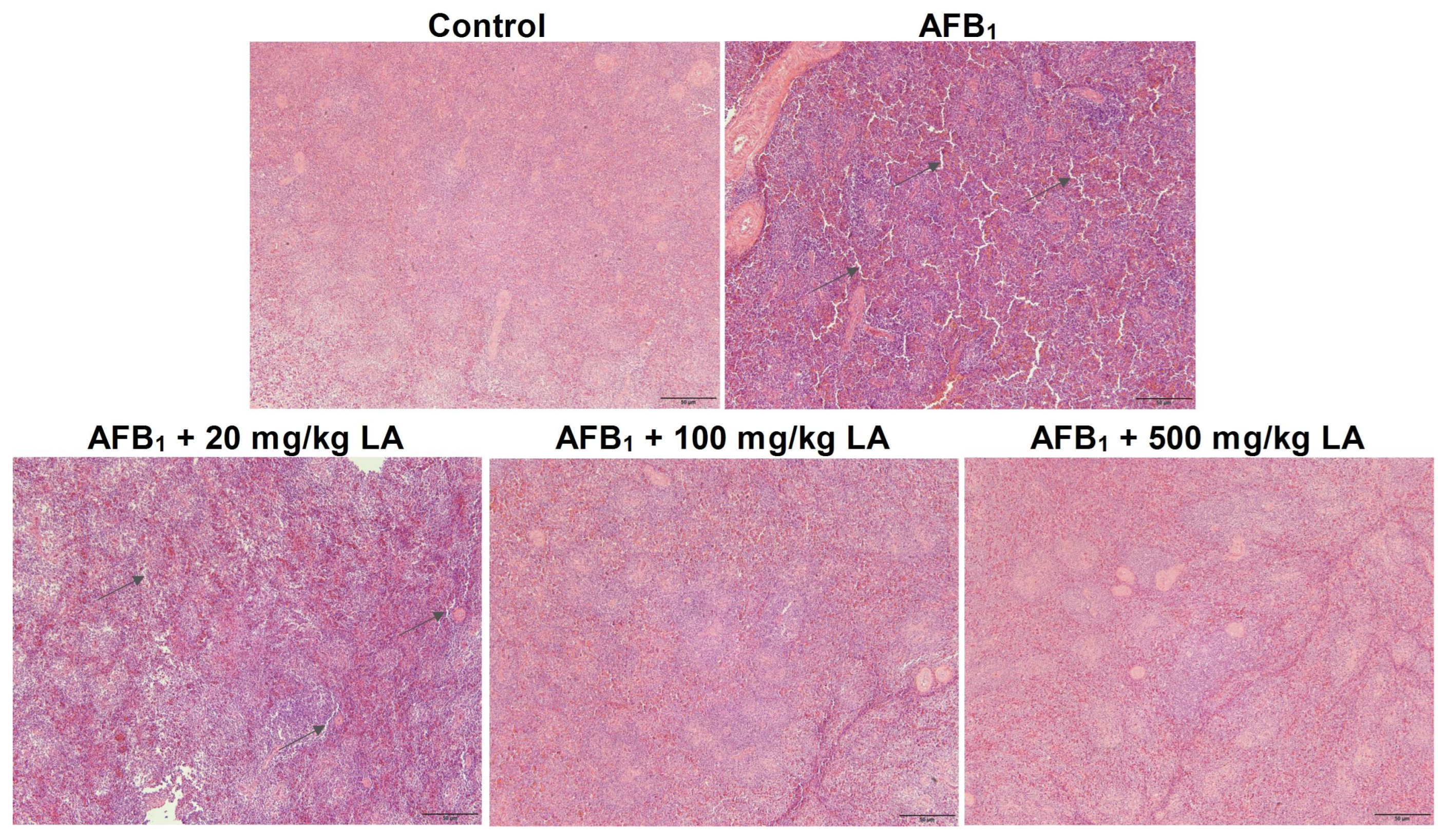
2.7. Changes in the Structure of Spleen Tissue

The structural hierarchy of white and red pulp in the spleen of the control group of young laying hens is clear, and the structure of cells and tissues is normal (Figure 4). There was a symptom of increased tissue space in the spleen of the AFB1 group (Figure 4). Adding lipoic acid to the diet can alleviate spleen damage caused by AFB1. When the amount of lipoic acid added reaches 100 mg/kg or 500 mg/kg, the symptoms of enlarged spleen tissue gaps caused by AFB1 disappear (Figure 4).

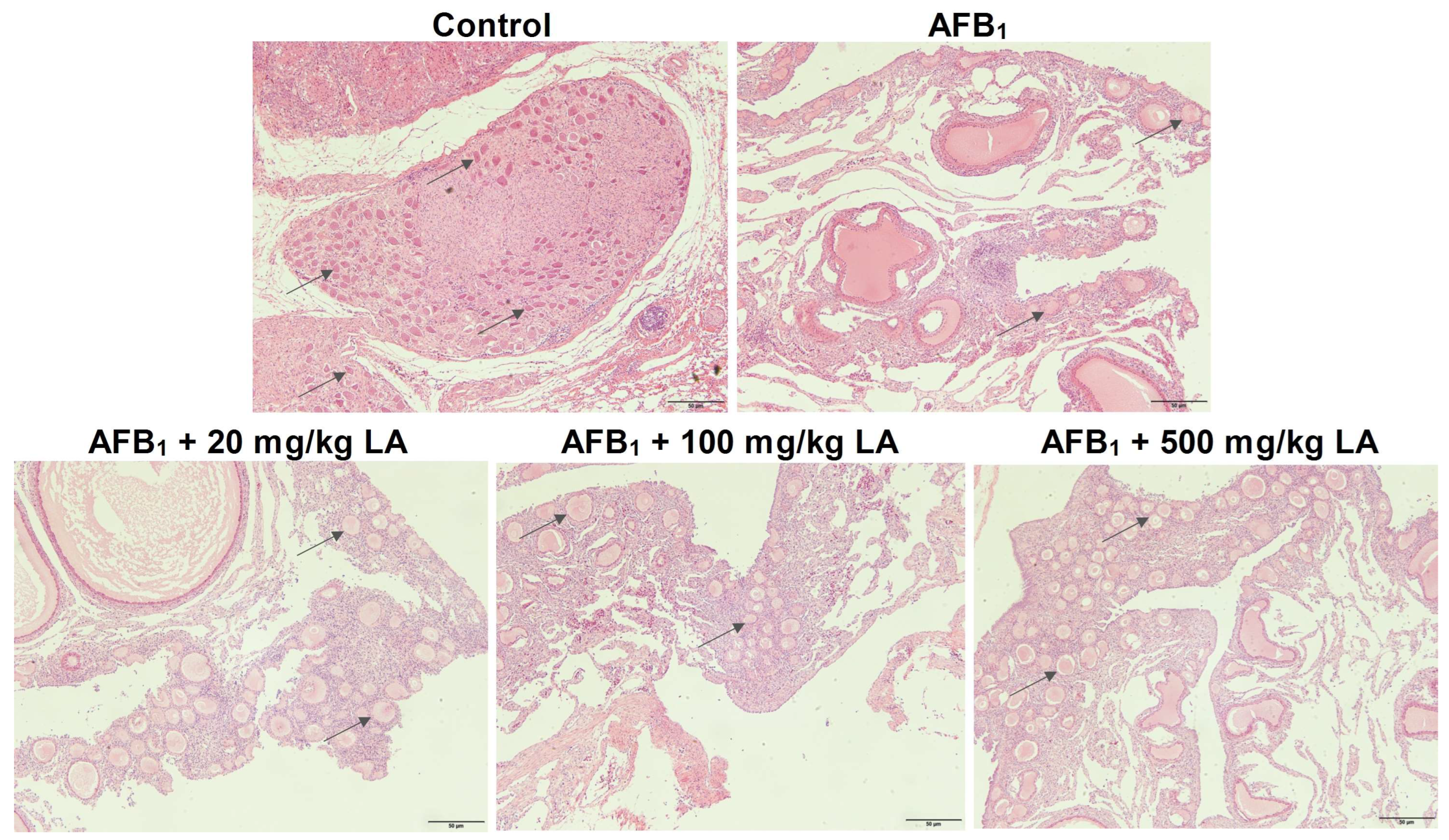
2.8. Changes in Ovarian Tissue Structure

The tissue structure of the control group’s ovaries is normal, and many follicles can be seen in the ovarian cortex (Figure 5). Compared with the control group, the number of follicles in the ovarian cortex of the AFB1 group decreased significantly, indicating that AFB1 caused damage to ovarian tissue (Figure 5). As the amount of added lipoic acid gradually increased, there was a significant increase in the number of follicles in the ovarian cortex, indicating that lipoic acid can alleviate ovarian tissue damage caused by AFB1 (Figure 5).

3. Discussion

The production performance and health status of laying hens are closely related to the breeding efficiency. Numerous studies have shown that AFB1 can adversely affect animal production performance [3,6,9,15,16]. Karimi Torshizi and Sedaghat (2023) reported that adding 500 μg/kg AFB1 to the diet significantly reduced the feed intake, egg production rate, and egg weight of laying hens and significantly increased the feed conversion rate [3]. Seifi et al. (2022) reported that adding 1000 μg/kg AFB1 to the diet significantly reduced the egg production rate and eggshell thickness of laying hens [17]. Yu et al. (2024) reported that adding 90 μg/kg AFB1 to the diet can have adverse effects on the production performance and organ health of meat ducks [15]. However, this study found that adding 90 μg/kg AFB1 to the diet had no significant effect on the production performance and organ weight of young laying hens, which may be due to their relative insensitivity to AFB1 [18]. Ducks are more sensitive to AFB1, while chickens are relatively insensitive to AFB1 [18]. Therefore, adding 90 μg/kg of AFB1 to the diet may have strong toxicity to meat ducks, but it may only be a low-dose toxicity to laying hens. In previous reports, the production performance of laying hens was only affected when the addition of AFB1 in the diet reached 500 or 1000 μg/kg [3,17]. Meanwhile, Raj et al. (2023) reported that adding 50 μg/kg AFB1 to the diet had no significant effect on the production performance of laying hens aged 246–259 days [19]. The results of this study are similar to the report, which further indicates that young laying hens have a high tolerance to AFB1.
Serum biochemical indicators can reflect the health status of the liver, and AFB1 can damage the health of animal livers [9,20,21]. This study found that the levels of ALT, AST, and ALP in the serum of young laying hens in the AFB1 group increased, while the levels of TP and ALB decreased, indicating that AFB1 also caused damage to the livers of young laying hens. This study also found that adding lipoic acid to the diet can alleviate the abnormal serum biochemical indicators caused by AFB1. Li et al. (2014) reported that adding 300 mg/kg of lipoic acid to the diet can alleviate the inflammation caused by AFB1 in broiler chickens, while increasing the levels of TP and ALB in the serum [22]. The results of this study are similar to the report, indicating that lipoic acid can alleviate liver damage in young laying hens caused by AFB1.
Antioxidant indicators can reflect the body’s ability to resist oxidative stress [23]. Numerous studies have shown that AFB1 can cause damage to the liver’s antioxidant system [24,25,26]. This study found that AFB1 reduces the levels of antioxidant enzymes T-SOD, GSH-Px, and CAT in serum, while increasing the level of lipid peroxidation product MDA, indicating that AFB1 caused oxidative stress in young laying hens. This study also found that adding lipoic acid to the diet can increase the content of antioxidant enzymes in serum and reduce the content of MDA. Maciejczyk et al. (2022) reported that lipoic acid can enhance the antioxidant capacity in the hypothalamus of rats, inhibiting oxidative stress and inflammation [27]. Skibska et al. (2023) reported that lipoic acid can increase the levels of GSH, SOD, and free thiol groups in rat kidneys, reduce the level of hydrogen peroxide and alleviate lipopolysaccharide induced oxidative stress [28]. Yang et al. (2023) reported that adding lipoic acid to the diet can increase the levels of SOD, CAT, and GSH Px in sheep serum, enhancing their antioxidant capacity [29]. The results of this study are similar to those reported above, indicating that lipoic acid can enhance the antioxidant capacity of young laying hens and alleviate oxidative damage caused by AFB1.
The liver is a target organ of AFB1; therefore, when animals suffer from AFB1 poisoning, the damage to the liver is extremely severe [15,16,18]. This study found that AFB1 caused fat degeneration and increased tissue space in the liver of young laying hens, indicating that AFB1 also caused tissue damage to the liver of young laying hens. This result is consistent with previous reports. This study also found that lipoic acid can alleviate liver damage in young laying hens caused by AFB1. Li et al. (2014) reported that adding 300 mg/kg of lipoic acid to the diet can alleviate AFB1-induced liver damage in broiler chickens [22]. Longhitano et al. (2024) reported that lipoic acid can increase the antioxidant level of liver cells and alleviate oxidative stress damage in liver cells [30]. The results of this study are consistent with previous reports. The important reason for the relief of liver damage symptoms may be related to the increased antioxidant capacity of young laying hens by lipoic acid [13,31].
AFB1 can disrupt the structure of the nucleus and mitochondria, causing cell apoptosis [9,16]. In this study, it was also found that AFB1 can cause structural damage to the nuclei and mitochondria in the livers of young laying hens. At the same time, this study also found that lipoic acid can relieve symptoms such as mitochondrial swelling, decreased matrix, and damaged mitochondrial cristae caused by AFB1. Longhitano et al. (2023) reported that lipoic acid can not only restore the damaged membrane potential of mitochondria in steatosis model cells but also improve the symptoms of mitochondrial cristae fragmentation [32]. Mozaffarian et al. (2022) reported that adding 40 μM or more of lipoic acid to the culture medium can alleviate mitochondrial membrane damage caused by arsenic [33]. In addition, Song et al. (2022) reported that adding 100 mg/kg of lipoic acid to the diet can alleviate mitochondrial structural damage in heart cells of chronic sleep-deprived (CSD) mice [34]. The results of this study are similar to those reported above, indicating that lipoic acid can relieve mitochondrial damage in the livers of young laying hens caused by AFB1. In young laying hens, lipoic acid is synthesized by mitochondria and can clear reactive oxygen species [12,35]. AFB1 causes damage to mitochondrial structure, which inevitably inhibits the synthesis process of lipoic acid [12,35]. By adding lipoic acid to the diet, the insufficient synthesis of lipoic acid in mitochondria is compensated for, and the antioxidant capacity of the liver is improved. Therefore, the damaged mitochondrial structure in the liver is relieved.
The spleen and ovaries are important immune and reproductive organs in laying hens, respectively. This study found that AFB1 caused damage to the tissue structure of the spleen and ovaries of young laying hens. Zhu et al. (2017) found that adding 600 μg/kg AFB1 to the diet can cause spleen congestion in one-day-old broiler chickens and increase the number of apoptotic cells in the spleen [36]. Wu et al. (2023) reported that adding 2000 μg/kg AFB1 to the diet increased the number of blocked follicles in the ovaries of rats and damaged ovarian health [37]. This study also found that adding lipoic acid to the diet can partially alleviate the structural damage of the spleen and ovarian tissues caused by AFB1. The reason may be that lipoic acid enhances the antioxidant capacity of spleen and ovarian tissues, or it may be that lipoic acid accelerates the metabolic process of AFB1 in the liver, reducing the toxic effects of AFB1 on the body [13,18,31]. Further research is needed to determine the specific causes. Damage to ovarian structure may affect the egg laying performance of laying hens in the later stage, so special attention should be paid to the prevention and control of AFB1 in laying hen feed during production.

4. Conclusions

In summary, adding 90 μg/kg AFB1 to the diet does not significantly affect the production performance of young laying hens, but AFB1 can damage the structure of mitochondria in the livers of laying hens and impair the health of liver, spleen, and ovarian tissues. Adding lipoic acid to the diet can enhance the antioxidant capacity of young laying hens, protect mitochondrial structure, and alleviate symptoms such as abnormal serum biochemical indicators, oxidative stress, and damage to the liver, spleen, and ovarian tissues caused by AFB1.

5. Materials and Methods

5.1. Experimental Design and Sample Collection Methods

This experiment was approved by the Ethics Committee of Huazhong Agricultural University with approval number HZAUCH-2025-0001. A total of 160 105-day-old Jingfen No. 6 young laying hens with similar appearance and weight were selected for the experiment. All young laying hens were randomly divided into five groups (n = 8 repetitions/group, 4 hens/repetitions). The AFB1 group and three lipoic acid treatment groups were all fed a basic diet supplemented with 90 μg/kg AFB1. The basic diets of the three lipoic acid treatment groups were also supplemented with 20, 100, and 500 mg/kg lipoic acid, respectively. The formula and nutritional levels of the basic diets are shown in Supplementary Table S1. All young laying hens were raised in egg cages at the animal experimental base of Huazhong Agricultural University. During the experiment, all young laying hens were free to feed and drink, and the feeding period was 4 weeks. During the feeding period, the feed intake and weights of the young laying hens were recorded. After the fourth week, one chicken was randomly selected from each replicate, and blood was collected through the Infrawinged vein. After serum precipitation, the serum was separated using a low-temperature high-speed centrifuge (Eppendorf AG, Hamburg, Germany) at 3000 r/min. The serum was then stored in a −80 °C freezer for the determination of serum biochemical and antioxidant indicators. After the blood collection was completed, the young laying hens were euthanized by the acute blood loss method, and the abdominal cavity of the hens was opened for sampling. Livers, spleens, and hearts were weighed, and the weights were recorded. Small samples of liver, spleen, and ovary tissues were collected for tissue section preparation.

5.2. Determination of Growth Performance

During the experiment, the feeding amount and remaining feed amount of each replicate were weighed and recorded, and the average daily feed intake was calculated based on the feeding amount and remaining feed amount. Livers, spleens, and hearts were weighed, and the weights were recorded. Small samples of liver, spleen, and ovary tissues were collected for tissue section preparation.

5.3. Determination of Organ Weight

During slaughtering and sampling, the intact liver, spleen, and heart were removed; the weights of the liver, spleen, and heart were accurately weighed, and the data were recorded.

5.4. Determination of Serum Biochemical Indicators

The levels of serum biochemical indicators were measured using a fully automated biochemical analyzer (Beckman, Brea, CA, USA) and the appropriate test kits (Shenzhen Mindray Biomedical Technology Co., Ltd., Nanjing, China), and the methods for detecting each indicator were the same as those described in the kit instructions. The serum was stored at −80 °C until use.

5.5. Determination of Serum Antioxidant Indicators

The levels of antioxidant indicators were detected using commercial reagent kits from Nanjing Jiancheng Biotechnology Co., Ltd. (Nanjing, China), and the methods for detecting each indicator were the same as those described in the kit instructions.

5.6. H&E Staining of the Liver, Spleen, and Ovary

Fresh liver, spleen, and ovarian tissue blocks were immersed in a 4% paraformaldehyde solution for 24 h, and then the tissue blocks were dehydrated in a dehydrator (Junjie Electronics Co., Ltd., Wuhan, China). The dehydrated tissue blocks were made into wax blocks using paraffin and then sliced into sections with a thickness of about 4 μm using a microtome (Leica Instruments Co., Ltd., Shanghai, China). After transferring the slices onto a glass slide, the cell nucleus was first stained with haematoxylin staining solution, followed by staining of the cytoplasm with eosin staining solution. After the staining was completed, the slices were dehydrated and air dried. The surface of the slice was covered with a cover glass and sealed with neutral gum. Histomorphological analysis was performed using a motorized bright-field microscope (BX53, Olympus, Tokyo, Japan) with integrated cellSens imaging software (version 1.12).

5.7. Liver Oil Red O Staining

After being dehydrated and embedded, the tissue blocks were sliced into sections with a thickness of about 8 μm using a cryomicrotome (Thermo, Waltham, MA, USA). After reheating and drying the slices, they were fixed with fixative and then stained with Oil Red O staining solution. After diluting the background with 75% alcohol, the cell nucleus was stained with haematoxylin staining solution. The surface of the slice was covered with a cover glass and sealed with neutral gum. Detection analysis was performed using a motorized bright-field microscope (BX53, Olympus, Tokyo, Japan) with integrated cellSens imaging software.

5.8. Observation of Liver Ultrastructure

Fresh liver tissue blocks were soaked in 2.5% glutaraldehyde solution for 24 h and washed three times with a phosphate-buffer solution. The cleaned tissue blocks were then fixed in osmium acid solution for 2 h and washed three times with the phosphate-buffer solution. After dehydrating the tissue block with alcohol, the epoxy resin was then allowed to infiltrate the tissue block. After embedding the tissue block with pure Spurr resin, the tissue block was sliced into ultra-thin sections of approximately 70 nm using an ultramicrotome (Leica Instruments Co., Ltd., Shanghai, China) with a diamond knife. The sections are floated on the water surface, picked up and fixed with copper grids, then stained with uranyl acetate and lead citrate, and finally air-dried at room temperature. Ultrastructural analysis was conducted at 80 kV accelerating voltage using a field-emission transmission electron microscope (Hitachi HT7800, Hitachi High-Tech, Tokyo, Japan) equipped with an AMT XR111 digital imaging system. Calibrated magnifications (×10,000) were applied for systematic observation of epoxy resin-embedded sections (70–90 nm thickness).

5.9. Statistical Analysis

After preliminary sorting of the experimental data using Excel software, SPSS Statistics 25 (IBM Corp. Armonk, NY, USA) was used for statistical analysis. Using one-way ANOVA and Tukey’s test to analyze between-group differences.

Supplementary Materials

The following supporting information can be downloaded at: https://www.mdpi.com/article/10.3390/toxins17040184/s1, Table S1: Composition of basal diets and nutrient levels.

Author Contributions

Conceptualization, Y.C. and H.W.; methodology, Y.C. and H.W.; software, H.W.; validation, Y.C., X.X., Y.X. and Y.J.; formal analysis, Y.Z.; investigation, S.A.R.; resources, Q.Y.; data curation, Y.C.; writing—original draft preparation, Y.C.; writing—review and editing, Y.C.; visualization, X.X.; supervision, D.Q.; project administration, D.Q.; funding acquisition, D.Q. All authors have read and agreed to the published version of the manuscript.

Funding

This research was funded by the National Key R&D Program of China. (Grant No.: 2023YFD1301005).

Institutional Review Board Statement

All animal procedures used in this study were approved by the Institutional Animal Care and Use Committee of Huazhong Agricultural University, China (approval number ID: HZAUCH-2025-0001).

Informed Consent Statement

Not applicable.

Data Availability Statement

The data presented in this study are available on request from the corresponding author (the data are not publicly available due to privacy or ethical restrictions).

Acknowledgments

We thank the National Key Research and Development Program for the support of this research.

Conflicts of Interest

Author Yi Xue was employed by the company of China Grain Reserves (Sichuan) Quality Inspection Center. The remaining authors declare that the research was conducted in the absence of any commercial or financial relationships that could be construed as a potential conflict of interest.

References

  1. Wang, P.; Wang, Y.; Feng, T.; Yan, Z.; Zhu, D.; Lin, H.; Iqbal, M.; Deng, D.; Kulyar, M.F.; Shen, Y. Hedyotis diffusa alleviate aflatoxin B1-induced liver injury in ducks by mediating Nrf2 signaling pathway. Ecotox. Environ. Safe. 2023, 249, 114339. [Google Scholar] [CrossRef] [PubMed]
  2. Choi, H.; Garavito-Duarte, Y.; Gormley, A.R.; Kim, S.W. Aflatoxin B1: Challenges and Strategies for the Intestinal Microbiota and Intestinal Health of Monogastric Animals. Toxins 2025, 17, 43. [Google Scholar] [CrossRef] [PubMed]
  3. Karimi, T.M.; Sedaghat, A. A consortium of detoxifying bacteria mitigates the aflatoxin B1 toxicosis on performance, health, and blood constituents of laying hens. Poult. Sci. 2023, 102, 102601. [Google Scholar] [CrossRef] [PubMed]
  4. Nazir, U.; Fu, Z.; Zheng, X.; Zafar, M.H.; Yang, Z.; Wang, Z.; Yang, H. Transcriptomic analysis of ileal adaptations and growth responses in growing hens supplemented with alanyl-glutamine dipeptide. Poult. Sci. 2024, 103, 104479. [Google Scholar] [CrossRef]
  5. Qing, H.; Huang, S.; Zhan, K.; Zhao, L.; Zhang, J.; Ji, C.; Ma, Q. Combined Toxicity Evaluation of Ochratoxin A and Aflatoxin B1 on Kidney and Liver Injury, Immune Inflammation, and Gut Microbiota Alteration Through Pair-Feeding Pullet Model. Front. Immunol. 2022, 13, 920147. [Google Scholar] [CrossRef]
  6. Pandey, I.; Chauhan, S.S. Studies on production performance and toxin residues in tissues and eggs of layer chickens fed on diets with various concentrations of aflatoxin AFB1. Br. Poult. Sci. 2007, 48, 713–723. [Google Scholar] [CrossRef]
  7. Chen, X.; Naehrer, K.; Applegate, T.J. Interactive effects of dietary protein concentration and aflatoxin B1 on performance, nutrient digestibility, and gut health in broiler chicks. Poult. Sci. 2016, 95, 1312–1325. [Google Scholar] [CrossRef]
  8. Niu, Y.J.; Wu, J.; Ren, W.; Liu, G.; Wu, G.; Peng, Y.; Zheng, D.; Jin, K.; Zuo, Q.; Li, G.; et al. Aflatoxin B1 impairs the growth and development of chicken PGCs through oxidative stress and mitochondrial dysfunction. Ecotoxicol. Environ. Saf. 2025, 290, 117727. [Google Scholar] [CrossRef]
  9. Wang, Y.; Wu, J.; Wang, L.; Yang, P.; Liu, Z.; Rajput, S.A.; Hassan, M.; Qi, D. Epigallocatechin Gallate and Glutathione Attenuate Aflatoxin B1-Induced Acute Liver Injury in Ducklings via Mitochondria-Mediated Apoptosis and the Nrf2 Signalling Pathway. Toxins 2022, 14, 876. [Google Scholar] [CrossRef]
  10. Tripathi, A.K.; Ray, A.K.; Mishra, S.K.; Bishen, S.M.; Mishra, H.; Khurana, A. Molecular and Therapeutic Insights of Alpha-Lipoic Acid as a Potential Molecule for Disease Prevention. Rev. Bras. Farmacogn. 2023, 33, 272–287. [Google Scholar] [CrossRef]
  11. Superti, F.; Russo, R. Alpha-Lipoic Acid: Biological Mechanisms and Health Benefits. Antioxidants 2024, 13, 1228. [Google Scholar] [CrossRef] [PubMed]
  12. Padmalayam, I. Targeting mitochondrial oxidative stress through lipoic acid synthase: A novel strategy to manage diabetic cardiovascular disease. Cardiovasc. Hematol. Agents Med. Chem. 2012, 10, 223–233. [Google Scholar] [CrossRef] [PubMed]
  13. Theodosis-Nobelos, P.; Papagiouvannis, G.; Tziona, P.; Rekka, E.A. Lipoic acid. Kinetics and pluripotent biological properties and derivatives. Mol. Biol. Rep. 2021, 48, 6539–6550. [Google Scholar] [CrossRef] [PubMed]
  14. Shi, D.; Liao, S.; Guo, S.; Li, H.; Yang, M.; Tang, Z. Protective effects of selenium on aflatoxin B1-induced mitochondrial permeability transition, DNA damage, and histological alterations in duckling liver. Biol. Trace Elem. Res. 2015, 163, 162–168. [Google Scholar] [CrossRef]
  15. Yu, A.; Wang, H.; Cheng, Q.; Rajput, S.A.; Qi, D. The Effects of Aflatoxin B1 on Liver Cholestasis and Its Nutritional Regulation in Ducks. Toxins 2024, 16, 239. [Google Scholar] [CrossRef]
  16. Chu, Y.; Yu, A.; Wang, H.; Rajput, S.A.; Yu, Q.; Qi, D. Biological Mechanisms of Aflatoxin B1-Induced Bile Metabolism Abnormalities in Ducklings. Animals 2024, 14, 2996. [Google Scholar] [CrossRef]
  17. Seifi, S.; Sadighara, P.; Mohajer, A. Protective effects of Aloe vera powder supplementation on some quantitative and qualitative characteristics of egg, histopathological changes and serum biochemistry of laying hens fed by Aflatoxin B1. Vet. Res. Forum. 2022, 13, 507–512. [Google Scholar] [CrossRef]
  18. Wu, K.; Liu, M.; Wang, H.; Rajput, S.A.; Shan, Y.; Qi, D.; Wang, S. The Mechanism Underlying the Extreme Sensitivity of Duck to Aflatoxin B1. Oxid. Med. Cell. Longev. 2021, 2021, 1–8. [Google Scholar] [CrossRef]
  19. Raj, J.; Farkas, H.; Jakovcevic, Z.; Vasiljevic, M.; Kumar, R.; Asrani, R.K. Effects of supplemented multicomponent mycotoxin detoxifying agent in laying hens fed aflatoxin B1 and T2-toxin contaminated feeds. Poult. Sci. 2023, 102, 102795. [Google Scholar] [CrossRef]
  20. Wang, X.; Wang, Y.; Wang, Q.; Dai, C.; Li, J.; Huang, P.; Li, Y.; Ding, X.; Huang, J.; Hussain, T.; et al. Effect of dietary protein on growth performance, and serum biochemical index in late pregnant Hu ewes and their offspring. Anim. Biotechnol. 2023, 34, 97–105. [Google Scholar] [CrossRef]
  21. Mojgani, N.; Razmgah, N.; Torshizi, M.A.K.; Sanjabi, M.R. Effects of three Bacillus specious on hatchability, growth performance and serum biochemistry in Japanese quails fed diet contaminated with Aflatoxin B1. Acta Sci. Anim. Sci. 2020, 42, e50184. [Google Scholar] [CrossRef]
  22. Li, Y.; Ma, Q.G.; Zhao, L.H.; Guo, Y.Q.; Duan, G.X.; Zhang, J.Y.; Ji, C. Protective Efficacy of Alpha-lipoic Acid against AflatoxinB1-induced Oxidative Damage in the Liver. Asian-Australas. J. Anim. Sci. 2014, 27, 907–915. [Google Scholar] [CrossRef] [PubMed]
  23. Barnes, P.J. Oxidative stress-based therapeutics in COPD. Redox Biol. 2020, 33, 101544. [Google Scholar] [CrossRef] [PubMed]
  24. Sang, R.; Ge, B.; Li, H.; Zhou, H.; Yan, K.; Wang, W.; Cui, Q.; Zhang, X. Taraxasterol alleviates aflatoxin B1-induced liver damage in broiler chickens via regulation of oxidative stress, apoptosis and autophagy. Ecotoxicol. Environ. Saf. 2023, 251, 114546. [Google Scholar] [CrossRef]
  25. Hatipoglu, D.; Keskin, E. The effect of curcumin on some cytokines, antioxidants and liver function tests in rats induced by Aflatoxin B1. Heliyon 2022, 8, e9890. [Google Scholar] [CrossRef]
  26. Yan, J.; Chen, L.; Zhang, L.; Zhang, Z.; Zhao, Y.; Wang, Y.; Ou, J. New Insights into the Persistent Effects of Acute Exposure to AFB1 on Rat Liver. Front. Microbiol. 2022, 13, 911757. [Google Scholar] [CrossRef]
  27. Maciejczyk, M.; Zebrowska, E.; Nesterowicz, M.; Zendzian-Piotrowska, M.; Zalewska, A. Alpha-Lipoic Acid Strengthens the Antioxidant Barrier and Reduces Oxidative, Nitrosative, and Glycative Damage, as well as Inhibits Inflammation and Apoptosis in the Hypothalamus but Not in the Cerebral Cortex of Insulin-Resistant Rats. Oxid. Med. Cell. Longev. 2022, 2022, 7450514. [Google Scholar] [CrossRef]
  28. Skibska, B.; Kochan, E.; Stanczak, A.; Lipert, A.; Skibska, A. Antioxidant and Anti-inflammatory Effects of alpha-Lipoic Acid on Lipopolysaccharide-induced Oxidative Stress in Rat Kidney. Arch. Immunol. Ther. Exp. 2023, 71, 16. [Google Scholar] [CrossRef]
  29. Yang, X.; Zhang, H.; Pang, F.; Zhang, L.; Fu, T.; Wang, L.; Liu, K.; Gao, T. Effects of alpha-lipoic acid on growth performance, antioxidant capacity, and immune function in sheep. J. Anim. Sci. 2023, 101, skad092. [Google Scholar] [CrossRef]
  30. Longhitano, L.; Distefano, A.; Musso, N.; Bonacci, P.; Orlando, L.; Giallongo, S.; Tibullo, D.; Denaro, S.; Lazzarino, G.; Ferrigno, J.; et al. (+)-Lipoic acid reduces mitochondrial unfolded protein response and attenuates oxidative stress and aging in an in vitro model of non-alcoholic fatty liver disease. J. Transl. Med. 2024, 22, 82. [Google Scholar] [CrossRef]
  31. Shanaida, M.; Lysiuk, R.; Mykhailenko, O.; Hudz, N.; Abdulsalam, A.; Gontova, T.; Oleshchuk, O.; Ivankiv, Y.; Shanaida, V.; Lytkin, D.; et al. Alpha-lipoic Acid: An Antioxidant with Anti-aging Properties for Disease Therapy. Curr. Med. Chem. 2025, 32, 23–54. [Google Scholar] [CrossRef] [PubMed]
  32. Longhitano, L.; Distefano, A.; Amorini, A.M.; Orlando, L.; Giallongo, S.; Tibullo, D.; Lazzarino, G.; Nicolosi, A.; Alanazi, A.M.; Saoca, C.; et al. (+)-Lipoic Acid Reduces Lipotoxicity and Regulates Mitochondrial Homeostasis and Energy Balance in an In Vitro Model of Liver Steatosis. Int. J. Mol. Sci. 2023, 24, 14491. [Google Scholar] [CrossRef] [PubMed]
  33. Mozaffarian, F.; Dehghani, M.A.; Vanani, A.R.; Mahdavinia, M. Protective Effects of Alpha Lipoic Acid Against Arsenic Induced Oxidative Stress in Isolated Rat Liver Mitochondria. Biol. Trace Elem. Res. 2022, 200, 1190–1200. [Google Scholar] [CrossRef] [PubMed]
  34. Song, F.; Lin, J.; Zhang, H.; Guo, Y.; Mao, Y.; Liu, Z.; Li, G.; Wang, Y. Long-Term Sleep Deprivation-Induced Myocardial Remodeling and Mitochondrial Dysfunction in Mice Were Attenuated by Lipoic Acid and N-Acetylcysteine. Pharmaceuticals 2022, 16, 51. [Google Scholar] [CrossRef]
  35. Dieckmann, C.L. A hub for regulation of mitochondrial metabolism: Fatty acid and lipoic acid biosynthesis. Iubmb Life 2024, 76, 332–344. [Google Scholar] [CrossRef]
  36. Zhu, P.; Zuo, Z.; Zheng, Z.; Wang, F.; Peng, X.; Fang, J.; Cui, H.; Gao, C.; Song, H.; Zhou, Y.; et al. Aflatoxin B1 affects apoptosis and expression of death receptor and endoplasmic reticulum molecules in chicken spleen. Oncotarget 2017, 8, 99531–99540. [Google Scholar] [CrossRef]
  37. Wu, K.; Liu, M.; Wang, H.; Rajput, S.A.; Al, Z.O.; Wang, S.; Qi, D. Effect of zearalenone on aflatoxin B1-induced intestinal and ovarian toxicity in pregnant and lactating rats. Ecotoxicol. Environ. Saf. 2023, 258, 114976. [Google Scholar] [CrossRef]
Figure 1. Changes in liver tissue structure of young laying hens fed by AFB1 and lipoic acid (LA) after the 4 week treatment. Green arrow: increased tissue spaces; black arrow: circular vacuoles formed by fat degeneration; the magnification is 100×.
Toxins 17 00184 g001
Figure 2. Changes in liver fatty degeneration of young laying hens fed by AFB1 and lipoic acid (LA) after the 4 week treatment. Red indicates the condition of fat degeneration; the magnification is 200×.
Toxins 17 00184 g002
Figure 3. Changes in nuclear and mitochondrial structures in the liver of young laying hens fed by AFB1 and lipoic acid (LA) after the 4 week treatment. Red arrow: mitochondrion (AFB1 causes mitochondria to swell and mitochondrial cristae to break); green arrow: nucleus (AFB1 causes nuclear membrane shrinking); the magnification is 10,000×.
Toxins 17 00184 g003
Figure 4. Changes in the spleen tissue structure of young laying hens fed by AFB1 and lipoic acid (LA) after the 4 week treatment. Black arrow: increased tissue spaces (organizational damage); the magnification is 100×.
Toxins 17 00184 g004
Figure 5. Changes in ovarian tissue structure of young laying hens fed by AFB1 and lipoic acid (LA) after the 4 week treatment. Black arrow: ovarian follicle (AFB1 reduces the number of ovarian follicles); the magnification is 100×.
Toxins 17 00184 g005
Table 1. Changes in the production performance and organ weight of young laying hens fed by AFB1 and lipoic acid (LA) after the 4 week treatment 1.
ItemsControlAFB1AFB1 +
20 mg/kg LA
AFB1 +
100 mg/kg LA
AFB1 +
500 mg/kg LA
Week 2
Weight (kg)1.42 ± 0.031.37 ± 0.041.41 ± 0.061.40 ± 0.091.39 ± 0.10
Daily feed
intake (g)
93.80 ± 2.73 a89.82 ± 2.38 ab87.28 ± 6.96 b89.91 ± 3.19 ab88.77 ± 4.81 b
Week 4
Weight (kg)1.49 ± 0.031.47 ± 0.061.49 ± 0.091.49 ± 0.091.44 ± 0.09
Daily feed
intake (g)
94.00 ± 3.7192.80 ± 4.7690.03 ± 10.0891.62 ± 3.2789.24 ± 5.27
Heart (g)6.28 ± 1.136.93 ± 1.416.34 ± 0.826.87 ± 1.356.35 ± 0.81
Liver (g)34.43 ± 6.69 ab39.71 ± 8.45 a35.10 ± 5.47 ab32.03 ± 4.85 b33.12 ± 4.34 ab
Spleen (g)3.15 ± 0.593.06 ± 0.602.81 ± 0.473.26 ± 0.403.02 ± 0.43
1 Values are expressed as means ± SD (n = 8), and different lowercase letters indicate significant differences between groups (p < 0.05).
Table 2. Changes in serum biochemical indicators of young laying hens fed by AFB1 and lipoic acid (LA) after the 4 week treatment.
BioindicatorsControlAFB1AFB1 +
20 mg/kg LA
AFB1 +
100 mg/kg LA
AFB1 +
500 mg/kg LA
ALT
(U/L)
1.43 ± 0.27 d3.36 ± 0.55 a3.39 ± 0.44 a2.61 ± 0.38 b1.96 ± 0.37 c
AST
(U/L)
195.78 ± 19.37 c293.73 ± 18.44 a281.29 ± 16.10 a250.90 ± 18.60 b211.11 ± 13.72 c
ALP
(U/L)
288.23 ± 47.06 c474.99 ± 40.13 a470.80 ± 45.79 a381.59 ± 30.33 b315.33 ± 61.57 c
TP
(g/L)
47.79 ± 6.17 a16.69 ± 3.70 d17.53 ± 3.98 d26.80 ± 5.51 c36.48 ± 4.31 b
ALB
(g/L)
22.44 ± 3.87 a10.35 ± 1.97 d11.18 ± 2.85 d14.24 ± 1.91 c17.71 ± 3.02 b
TBA
(μmol/L)
37.39 ± 4.9341.01 ± 7.1940.88 ± 7.2139.20 ± 9.1636.49 ± 5.85
Values are expressed as means ± SD (n = 8), and different lowercase letters indicate significant differences between groups (p < 0.05).
Table 3. Changes in serum antioxidant enzyme levels of young laying hens fed by AFB1 and lipoic acid (LA) after the 4 week treatment.
Antioxidant EnzymesControlAFB1AFB1 +
20 mg/kg LA
AFB1 +
100 mg/kg LA
AFB1 +
500 mg/kg LA
T-SOD
(U/mL)
89.02 ± 7.88 a60.06 ± 6.03 d60.18 ± 4.52 d67.48 ± 4.19 c77.64 ± 5.80 b
MDA
(nmol/mL)
7.80 ± 0.53 c16.36 ± 1.36 a15.78 ± 1.57 a12.69 ± 1.13 b8.80 ± 1.69 c
GSH-Px
(U/mL)
1862.84 ± 63.03 a1371.62 ± 137.74 c1397.30 ± 81.93 c1642.57 ± 126.95 b1775.68 ± 61.16 a
CAT
(U/mL)
3.34 ± 0.46 a1.82 ± 0.56 c1.91 ± 0.52 c2.25 ± 0.35 bc2.58 ± 0.33 b
Values are expressed as means ± SD (n = 8), and different lowercase letters indicate significant differences between groups (p < 0.05).
Disclaimer/Publisher’s Note: The statements, opinions and data contained in all publications are solely those of the individual author(s) and contributor(s) and not of MDPI and/or the editor(s). MDPI and/or the editor(s) disclaim responsibility for any injury to people or property resulting from any ideas, methods, instructions or products referred to in the content.

