Article

A Novel Device for Micro-Droplets Generation Based on the Stepwise Membrane Emulsification Principle

1
School of Integrated Circuit Science and Engineering, Beihang University, Beijing 100191, China
2
Department of Mechanical Engineering, University of Saskatchewan, Saskatoon, SK S7N 5A9, Canada
3
Department of Electrical and Computer Engineering, University of Saskatchewan, Saskatoon, SK S7N 5A9, Canada
4
Synchrotron Laboratory for Micro and Nano Devices, Canadian Light Source Incorporated, Saskatoon, SK S7N 2V3, Canada
5
School of Mechanical and Power Engineering, East China University of Science and Technology, Shanghai 201620, China
*
Authors to whom correspondence should be addressed.
Micromachines 2024, 15(9), 1118; https://doi.org/10.3390/mi15091118
Submission received: 14 July 2024 / Revised: 30 August 2024 / Accepted: 30 August 2024 / Published: 31 August 2024

Abstract

:
This paper presents a novel design of the device to generate microspheres or micro-droplets based on the membrane emulsification principle. Specifically, the novelty of the device lies in a proposed two-layer or stepwise (by generalization) membrane structure. An important benefit of the stepwise membrane is that it can be fabricated with the low-cost material (SU-8) and using the conventional lithography technology along with a conventional image-based alignment technique. The experiment to examine the effectiveness of the proposed membrane was conducted, and the result shows that microspheres with the size of 2.3 μm and with the size uniformity of 0.8 μm can be achieved, which meets the requirements for most applications in industries. It is noted that the traditional membrane emulsification method can only produce microspheres of around 20 μm. The main contribution of this paper is thus the new design principle of membranes (i.e., stepwise structure), which can be made by the cost-effective fabrication technique, for high performance of droplets production.

1. Introduction

There are many applications for microspheres, e.g., cosmetics, electronics, pharmaceuticals, food processing, biomedicine, and drug delivery [1,2,3,4,5,6,7,8,9,10,11,12]. Microfluidic devices based on the emulsification principle with a membrane and/or micro-channel structure are increasingly of interest due to their high throughputs and high ability to control internal morphology. The requirements for any device that generates microspheres include (1) the size of the microspheres (the smaller, the better), (2) the uniformity of the microspheres in terms of size (the more uniform, the better), (3) the flexibility—i.e., the capability of making microspheres of different batch sizes with one device (the larger the range of the batch sizes, the better), and (4) the efficacy of the microspheres generation process (mass production or not). For comparison of various microsphere generation methods, refer to Lei et al. [13], Wu et al. [14], Arun K. Chattopadhyay and K.L. Mittal [15], and Solans et al. [16]. The general idea for all these devices that generate microspheres is to let two fluids interact with each other so that droplets may form in one of the two fluids [13], see Figure 1. The fluid out of which droplets are formed is called the dispersed phase, and the fluid that interacts with the dispersed phase is called the continuous phase (Figure 1).
Nakashima and Shimizu in the 1980s introduced a unique approach for generating emulsion by using a membrane of numerous uniform micro-pores [17]. The dispersed phase was forced by a low pressure to flow through the pores of the microporous membrane to interact directly with the continuous phase [18]. Emulsified droplets were formed from the dispersed phase and detached at the end of the pores with a drop-by-drop mechanism, as described in Figure 1. By this technique, the size of the droplet is mainly determined by the size of the pores in the porous membrane. Practically, the size of the generated droplet is about 2 to 10 times larger than size of the pore in the membrane [19]. Therefore, to obtain microspheres of small sizes, the challenge is to construct a membrane with many pores of small sizes (preferably in nanometers). There is a dilemma with the structure of the membrane. On one hand, a small pore membrane usually leads to a thin membrane with conventional membrane materials, which is fragile, while on the other hand, a thick membrane with small pores means that the membrane contains the high-aspect-ratio geometric feature, which is a challenge for microfabrication.
To tackle the above dilemma, the conventional idea in the literature is to seek unconventional materials, which can be made into a thin yet ductile and strong membrane. In connection with this, materials such as Shirasu Porous Glass (SPG) [17], silicon wafer [20], ceramic [21], stainless steel [22], and polymer [23] have been proposed in the literature. Among them, SPG was the most promising. However, fabrication of an SPG membrane is complex with high cost [17]. Moreover, the chance of contamination of an SPG membrane is high due to tortuous pore structures. Indeed, the poor rigidity and stability of a polymer membrane are the major shortcomings for membrane emulsification. For ceramic membranes, it is difficult to achieve well-defined circular openings, and there is a high chance of delamination during cleaning [21]. For stainless steel membranes, though they have excellent structural rigidity, small size holes on the membrane are difficult to achieve. Numerous other materials, e.g., silicon [20], etc., have been reported as membrane materials, yet difficulties in fabrication and high costs are still a challenge.
In the study reported in this paper, we propose a completely different idea from the above-mentioned literature; rather than focusing on the special types of materials, we focused on the structure of the membrane. Specifically, we propose a membrane with a stepwise structure such that we expect that a conventional material such as SU-8 can be used while the aspect ratio remains adequate to the conventional micro-fabrication technology.

2. Materials and Methods

2.1. Design

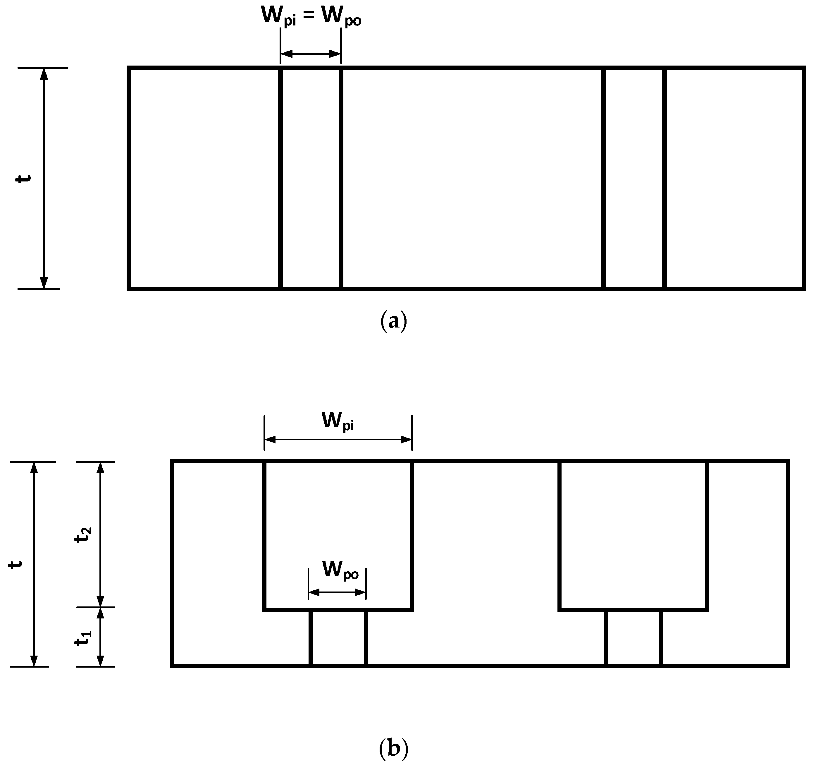
Figure 2 illustrates the concept of the stepwise structure, where Figure 2a illustrates a one-layer structure of the membrane, while Figure 2b illustrates a stepwise structure of the membrane. The design as illustrated in Figure 2a has two problems. The first problem is the difficulty in fabrication of such a “deep well” feature with the current photolithography (even with the expensive deep x-ray lithography process) [24]. The second problem is the difficulty of driving the dispersed phase fluid to flow through the long path channel [22]. It may be clear that the design concept of Figure 2a is not suitable to our constraint, namely use of a conventional photolithography (UV light as light source) process, and therefore, the design concept goes to one as illustrated in Figure 2b.
From Figure 2b the hole in the membrane is divided into several holes in different layers which have different thicknesses; specifically, the hole size is reduced stepwise from the top layer to the bottom layer with the smallest hole size being in the bottom layer. As such, the aspect ratio, an important parameter challenge in the photolithography process, corresponds to the smallest hole in the bottom layer, i.e., t1/wpo, and the design should be adequate to the conventional photolithography process at our facility.
There were two design constraints for the embodiment design of the stepwise membrane in our study: (1) the membrane should be subject to the 120 kPa pressure estimated according to [20], and (2) the smallest hole size should be around 1 micron to generate microspheres of around 1 micron. The dimensions of the stepwise membrane in Figure 2b were determined based on a trial-end-error process along with a finite element simulation to meet these constraints. Depending on the photolithography process used, an adequate aspect ratio is noted, but the smaller the aspect ratio, the less challenging the photolithography process. In our case, the aspect ratio should be less than 5, and we chose 1 only. It is noted that the smallest hole size is also restricted by the resolution of a particular photolithography process. A size of 1 micron is about the limit. Thus, in Figure 2b, the membrane has two layers: the upper layer with the thickness (t2) and with the square pore size (wpi) and the bottom layer with the thickness (t1) and the square pore size (wpo). The dispersed fluid will enter through the inlet pores (wpi) in the upper layer and exit through the narrow outlet pores (wpo) in the bottom layer. Further, the pores in the bottom layer connect with the continuous phase fluid, which meets the dispersed phase fluid at the holes in the membrane. In this design, the thick upper layer was expected to withstand the pressure, i.e., meeting the constraint (1), and the smallest pore size in the bottom layer was expected to generate small (around 1 micron) microspheres, i.e., meeting the constraint (2).
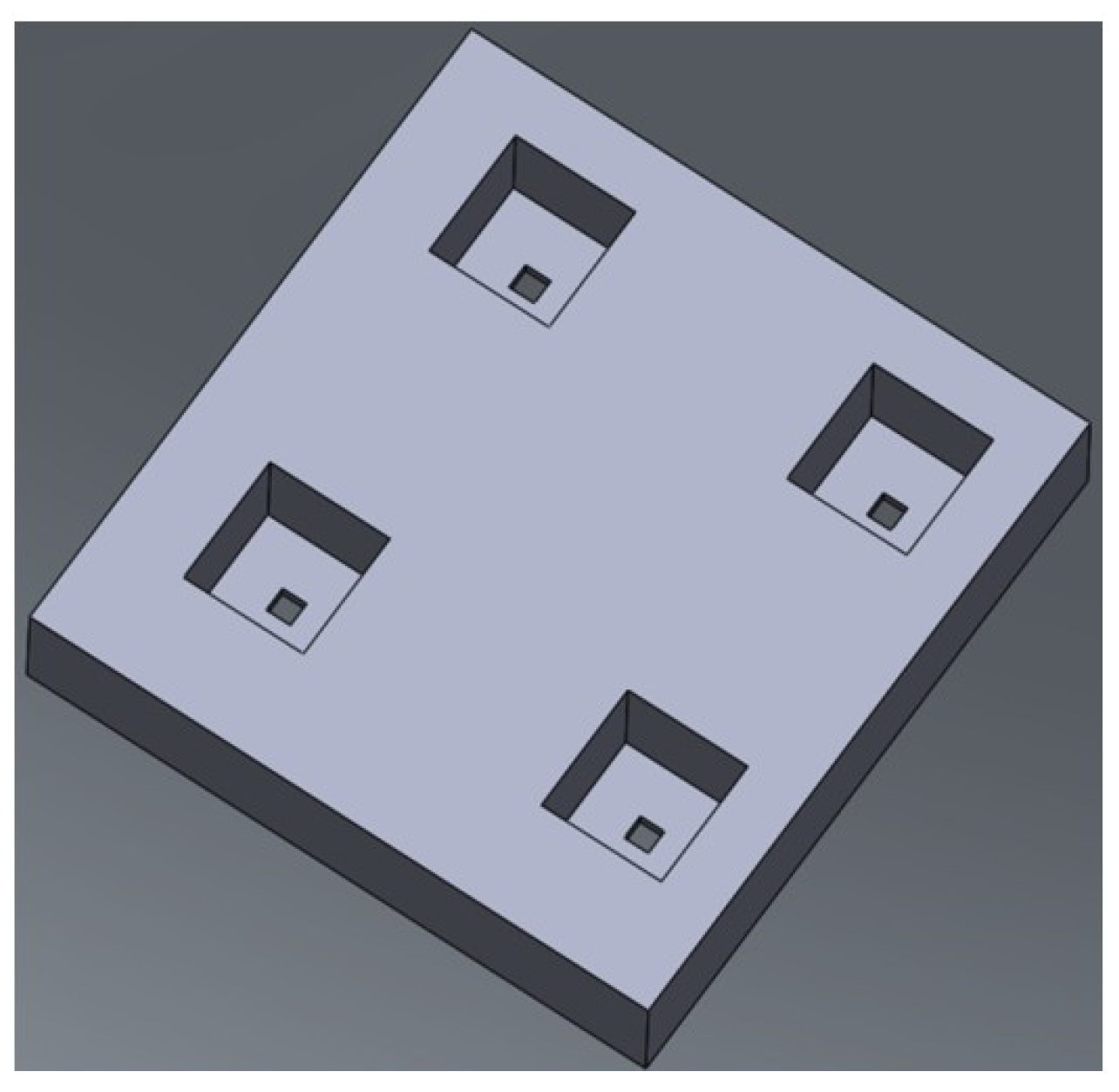
For the study presented in this paper, the geometry of the stepwise membrane was determined based on the simulation alongside the experience as follows (Figure 2b): wpo = t1 = 1 µm and wpi = t2 = 20 µm. The overall thickness (t) of the membrane was 21 µm (Figure 2a). Figure 3 shows the three-dimensional view of the stepwise membrane. As a result of this design, the aspect ratio of the upper layer was 20:20 (1:1) and that of the bottom layer was 1:1. Therefore, the conventional photolithography process could readily fabricate the device.

2.2. Fabrication

In this study, the materials (SU-8 2000 series) were used as the photoresist. SU-8 2000 series are the improved formulations of SU-8 and were suitable for the structure with its thick film (from 0.5 to >200 μm) and high aspect ratio (>10:1) [25,26]. The details of the photolithography process for fabricating the stepwise membrane are presented in the next sections.

2.2.1. Chromium Sputtering

The glass substrate had the thickness of 100 μm, and it was sputtered with a layer of chromium with the thickness of 120 nm. The chromium layer was non-transparent so that the pores and the alignment marks were visible through the transparent glass substrate and SU-8 layer.

2.2.2. SU-8 Spin Coating (1 μm)

SU-8 2001 was dynamically dispensed on the chromium, 1500 revolutions per minute (rpm) for 3 s (s) and then 4000 rpm for 30 s. The membrane was then soft baked on a hotplate at 95 °C for 1 min.

2.2.3. Mark Alignment

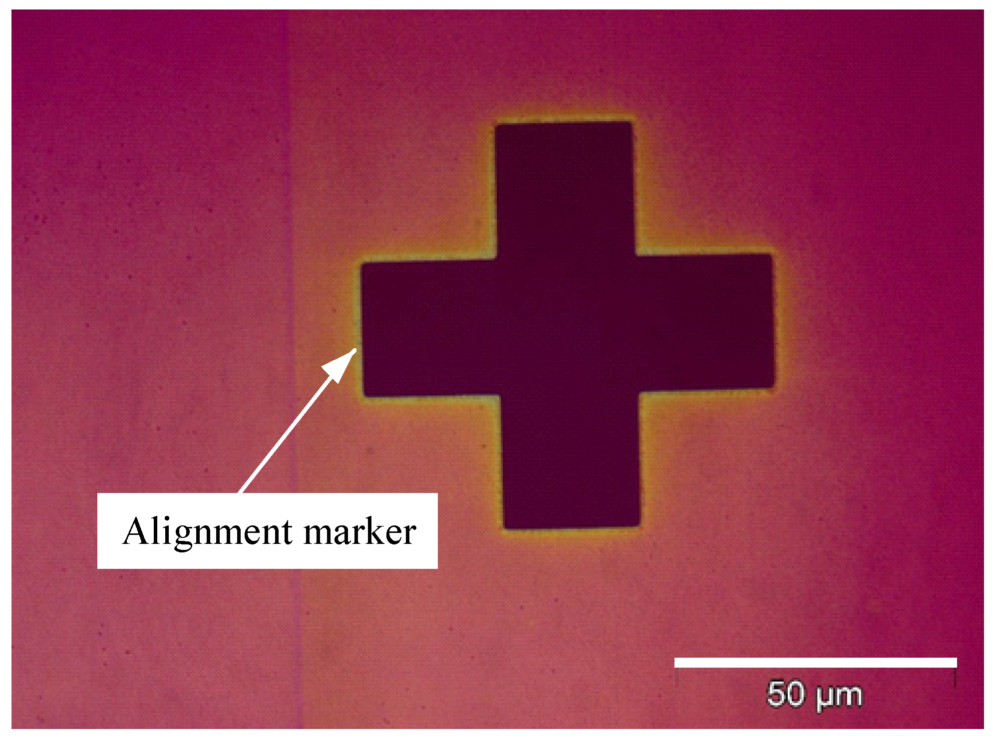
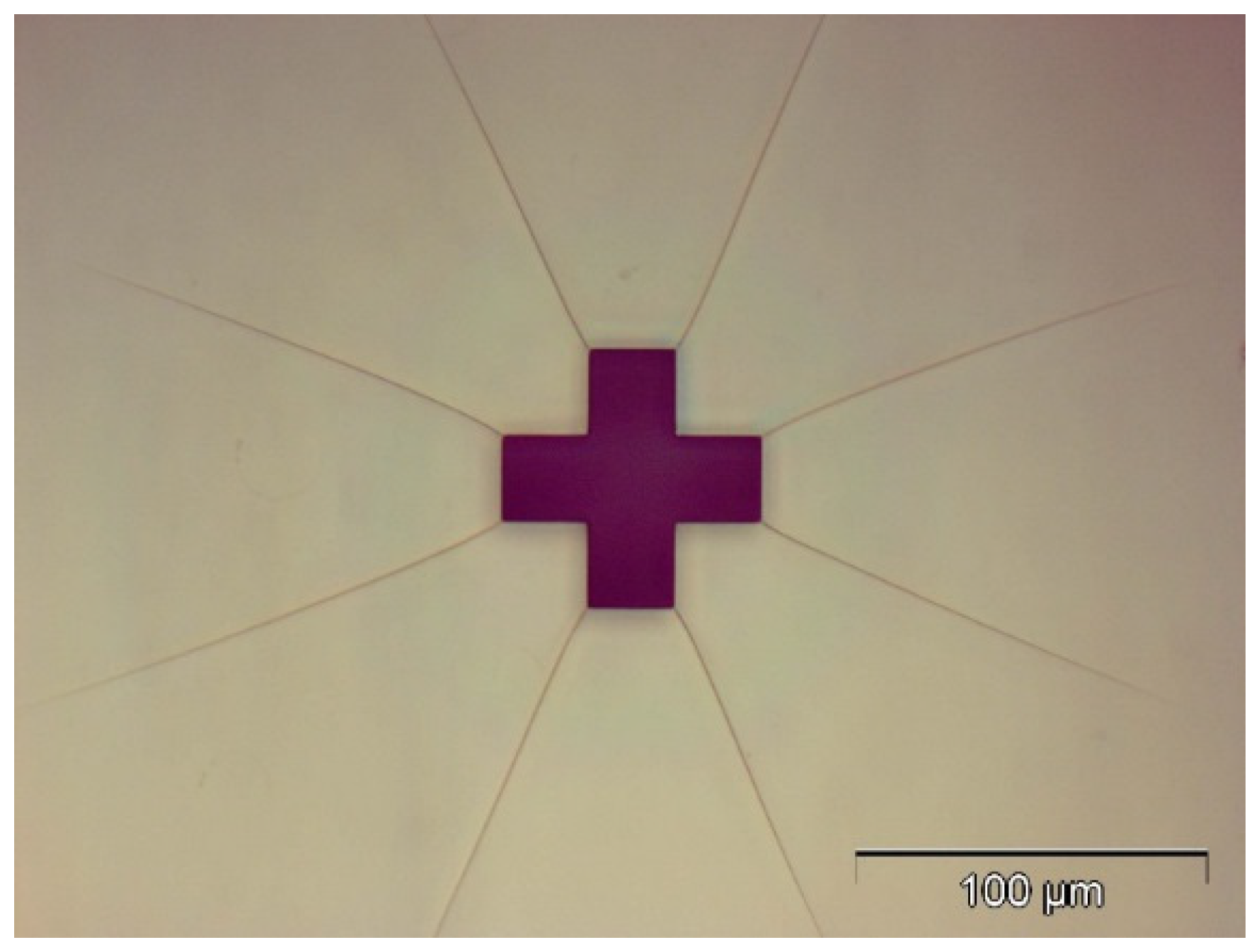
Two alignment crosses were used as markers to help the alignment of two layers of SU-8, as shown in Figure 4.

2.2.4. Exposure

The membrane was exposed under the laser with 12,000 m J / c m 2 . The laser power was 100 mW and the wavelength was 355 nm (DWL 66+, Heidelberg Instruments, Heidelberg, Germany). The direct write (maskless) lithography was used.

2.2.5. Post-Exposure Bake (PEB)

The membrane was baked on the same hotplate at 95 °C for 1 min.

2.2.6. Development

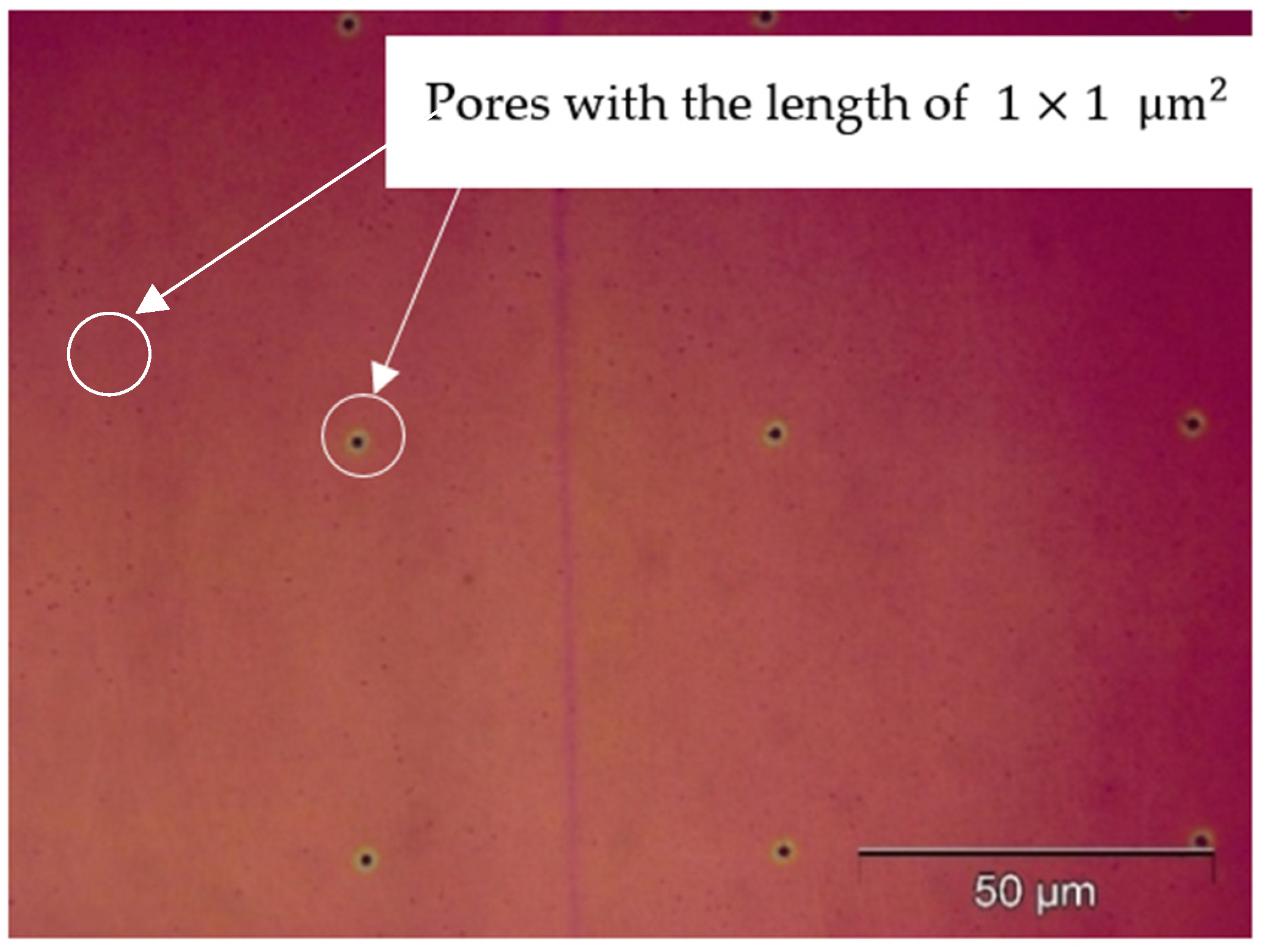
The membrane was developed in propylene glycol methyl ether acetate (PGMEA) for 1 min, and then it was rinsed off by isopropyl alcohol (IPA) for 30 s. At last, the membrane was dried by nitrogen ( N 2 ). The opaque region was removed. Pores with the length of 1 × 1 μm2 were obtained, as shown in Figure 5.

2.2.7. Oxygen Descum Plasma Cleaning

The flow rates of oxygen and argon were 50 and 20 standard cubic centimeters per minute (SCCM), respectively. The power for plasma cleaning was 150 W, and the time was 5 min (Diener Electronic, Germany).

2.2.8. Chromium Etching

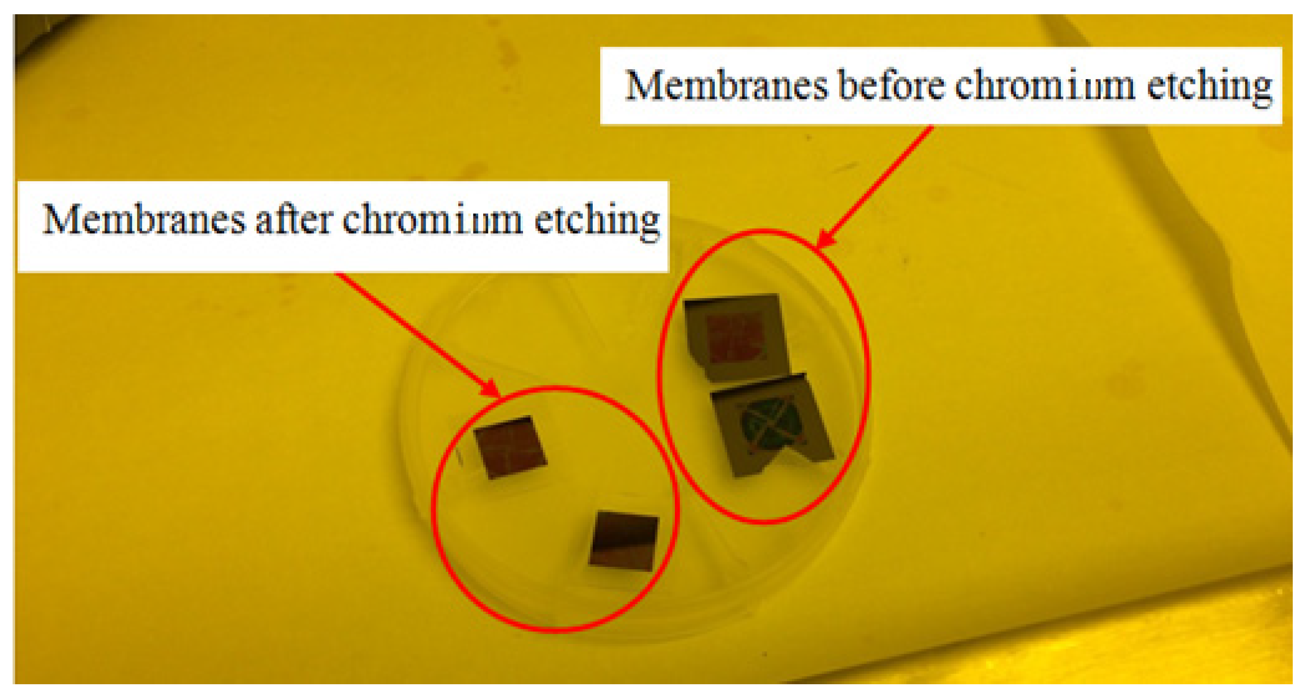
Chromium in the pore region was etched off by etching in Cr-etch for 45 s (MicroChemicals GmbH, Germany) and then rinsed with distilled (DI) water. The membranes before and after chromium etching are shown in Figure 6.

2.2.9. SU-8 Spin Coating (20 μm)

SU-8 2025 was dynamically spin coated onto the membrane at 500 rpm for 1 s. Then the spin speed was manually accelerated to 4000 rpm over 10 s and kept for 30 s. The membrane was then soft baked on the same hotplate at 65 °C for 1 min. Then, the temperature was ramped to 95 °C and kept for 5 min.

2.2.10. Mark Alignment

Two alignment crosses were used as marks to help the alignment of two layers of SU-8. As shown in Figure 7, the alignment mark of the first layer was covered by the alignment mark of the second layer perfectly.

2.2.11. Exposure

The membrane was exposed under the laser with 12,000 m J / c m 2 . The laser power was 100 mW and the wavelength was 355 nm (DWL 66+, Heidelberg Instruments, Germany). The direct write (maskless) lithography was used.

2.2.12. Post-Exposure Bake (PEB)

The membrane was baked on the same hotplate at 65 °C for 1 min. Then the temperature was ramped to 95 °C and kept for 5 min at 95 °C.

2.2.13. Development

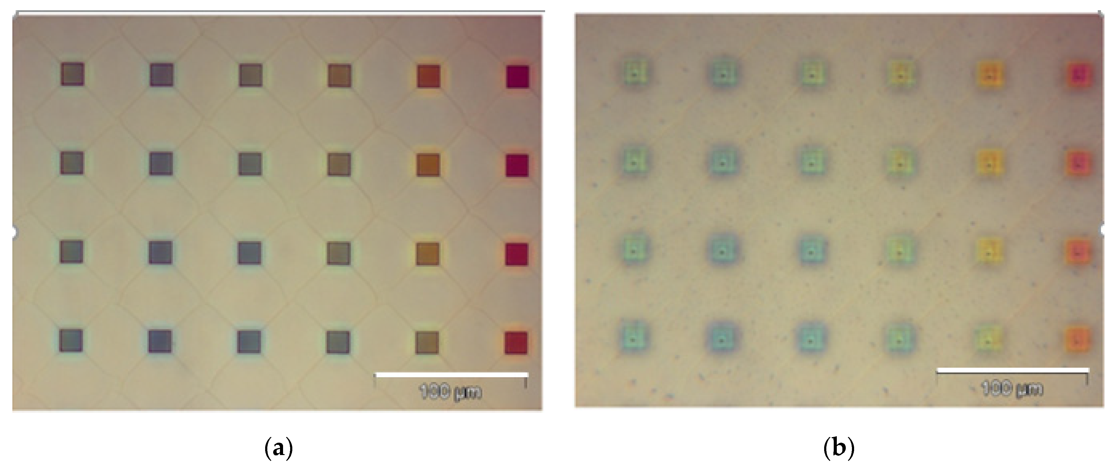
The membrane was developed in PGMEA for 4 min and then rinsed with IPA for 30 s. At last, the membrane was dried by N 2 . The opaque region was removed. Pores with 20 × 20   μ m 2 were obtained. The optical images of the top view and bottom view of the pores with 20 × 20   μ m 2 are presented in Figure 8.

2.2.14. Glued to an Acrylonitrile Butadiene Styrene (ABS) Tube

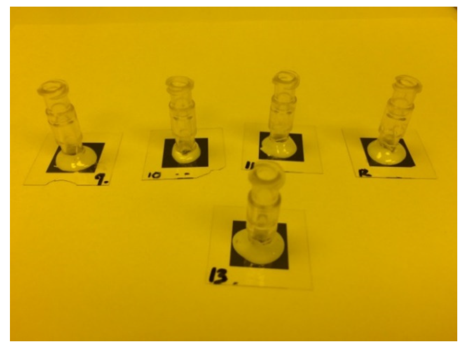
Epoxy 907 AB glues (Miller-Stephenson Chemical Incorporate, Danbury, CT, USA) were mixed for 3 min and then left to sit for 30 min. Next, the mixed glues were put around the edges of ABS tubing with 1 cm diameter (Taobao Company, Hangzhou, China), and then the tubing was placed on the top of samples, as shown in Figure 9. Twelve hours were needed, to wait for the best adhesive property.

2.2.15. Substrate and Chromium Removing

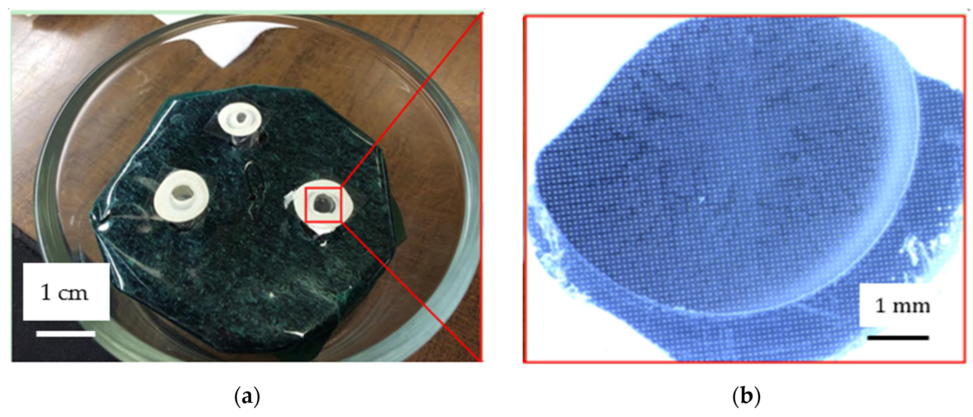
On the first day, the membrane was immersed in 5% hydrofluoric (HF) acid for 8 h. On the second day, the membrane was etched by 5% HF acid for another 5 h to fully remove the glass substrate. Then the chromium layer was etched off by etching in Cr-etch for 3 to 4 min and then rinsed with distilled (DI) water and dried by N 2 . Three pieces of membrane were obtained for the microsphere generation, as shown in Figure 10a. Two membranes failed during the gluing step because the glue covered these two membranes. The optical image of one piece of membrane is shown in Figure 10b. The diameter of the membrane emulsification device was 0.5 cm.
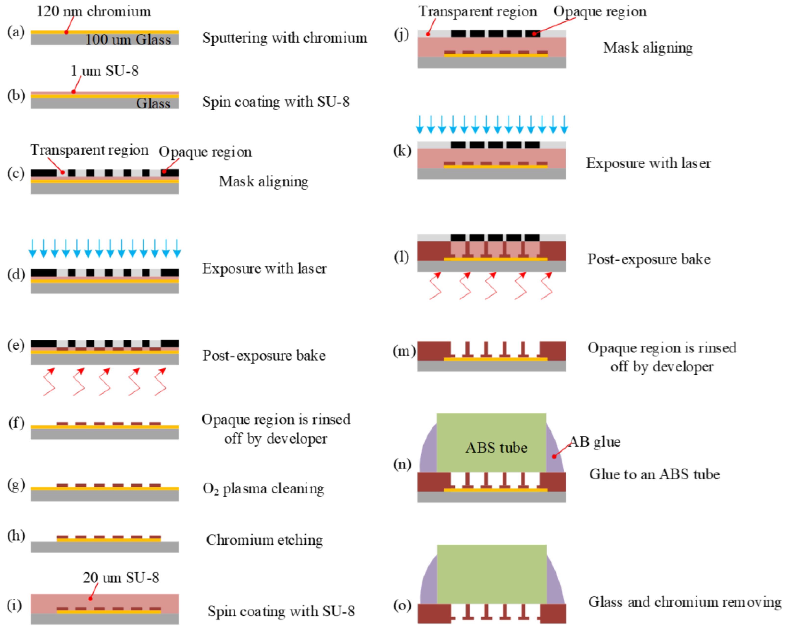
To sum up, the fabrication process of the porous membrane is shown in Figure 11.

2.3. Experiment

Paraffin oil (Sigma-Aldrich Corporation, St. Louis, MO, USA) with 2% Span 80 surfactant (Sigma-Aldrich Corporation, USA) was used as the continuous phase fluid and distilled (DI) water as the dispersed phase fluid in this experiment. Figure 12 shows the experimental set-up. A magnetic stirrer (Corning PC 210, Corning Incorporated, USA) was used to provide the shear force. A syringe pump (Harvard Apparatus, Holliston, MA, USA) controlling the flow rate of the dispersed phase was connected to the ABS tube by a syringe (6 mL, Covidien Limited, Dublin, Ireland) and tubing (1.34 mm PTFE, Adtech Polymer Engineering Limited, Stroud, UK). The syringe pump was calibrated before the experiment. The inverted microscope (Olympus IX70, Olympus Corporation, Tokyo, Japan) was used to observe the microsphere generation process, and a computer with image processing software was used for post-processing. The diameter of one single microsphere was measured multiple times to eliminate the possible error during the measuring process. The experiment was performed at room temperature (20 °C ± 2 °C) at the Intelligent Systems Laboratory at the University of Saskatchewan.
The minimum pressure ( P m i n ) that ensures the dispersed phase can be pressed through the porous membrane is given by
P m i n = 4 γ c o s θ d p
where γ is the interfacial tension between the continuous and dispersed phases; θ is the contact angle between the dispersed phase and the membrane surface in the continuous phase, as shown in Figure 1; d p is the pore diameter. In this work, γ = 3.65 × 10 3 N · m 1 [27] and the contact angle θ between the dispersed phase and the membrane surface in the continuous phase (Figure 1) was assumed to be zero because the wall was set to be a non-wetting boundary. Therefore, at the minimum pressure P m i n = 4 × 3.65 × 10 3 1 × 10 6 = 1.46   k P a , the corresponding flow rate of the dispersed phase is about 5   μ L · h 1 . In this work, the flow rate of the dispersed phase was set to be ten times larger than the minimum flow rate, namely 50   μ L · h 1 . The agitation speeds of the magnetic stirrer were 60, 80, and 100 rpm. An amount of 1 mL continuous fluid with microspheres generated was carefully extracted from the beaker to a clean glass petri dish for observation. The image processing software (“Analyze Particles” Function, ImageJ, Original version, National Institutes of Health, Stapleton, NY, USA) was used to improve the resolution of microscopic images and measure the diameter of microspheres on the petri dish [28].

3. Results and Discussion

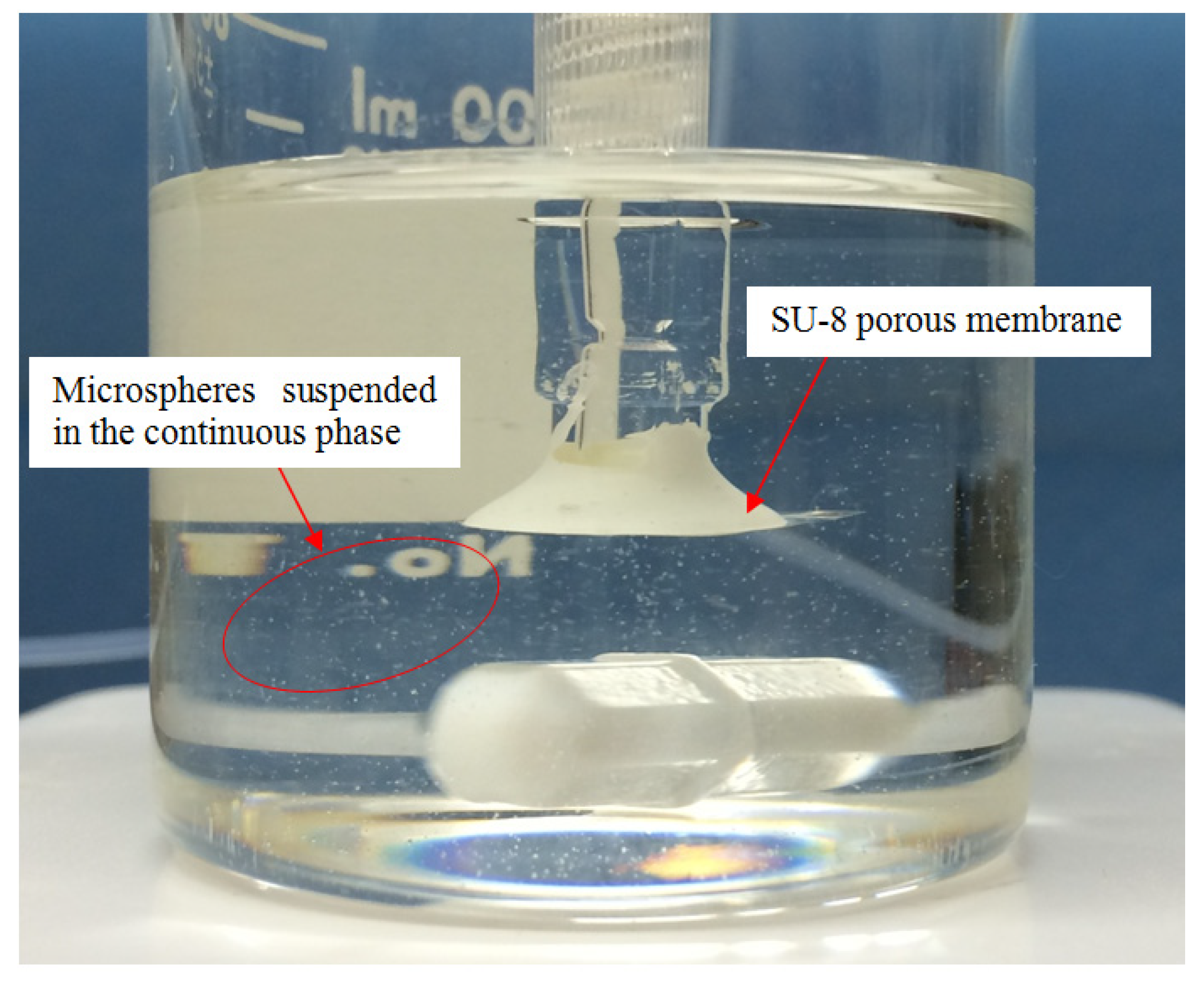
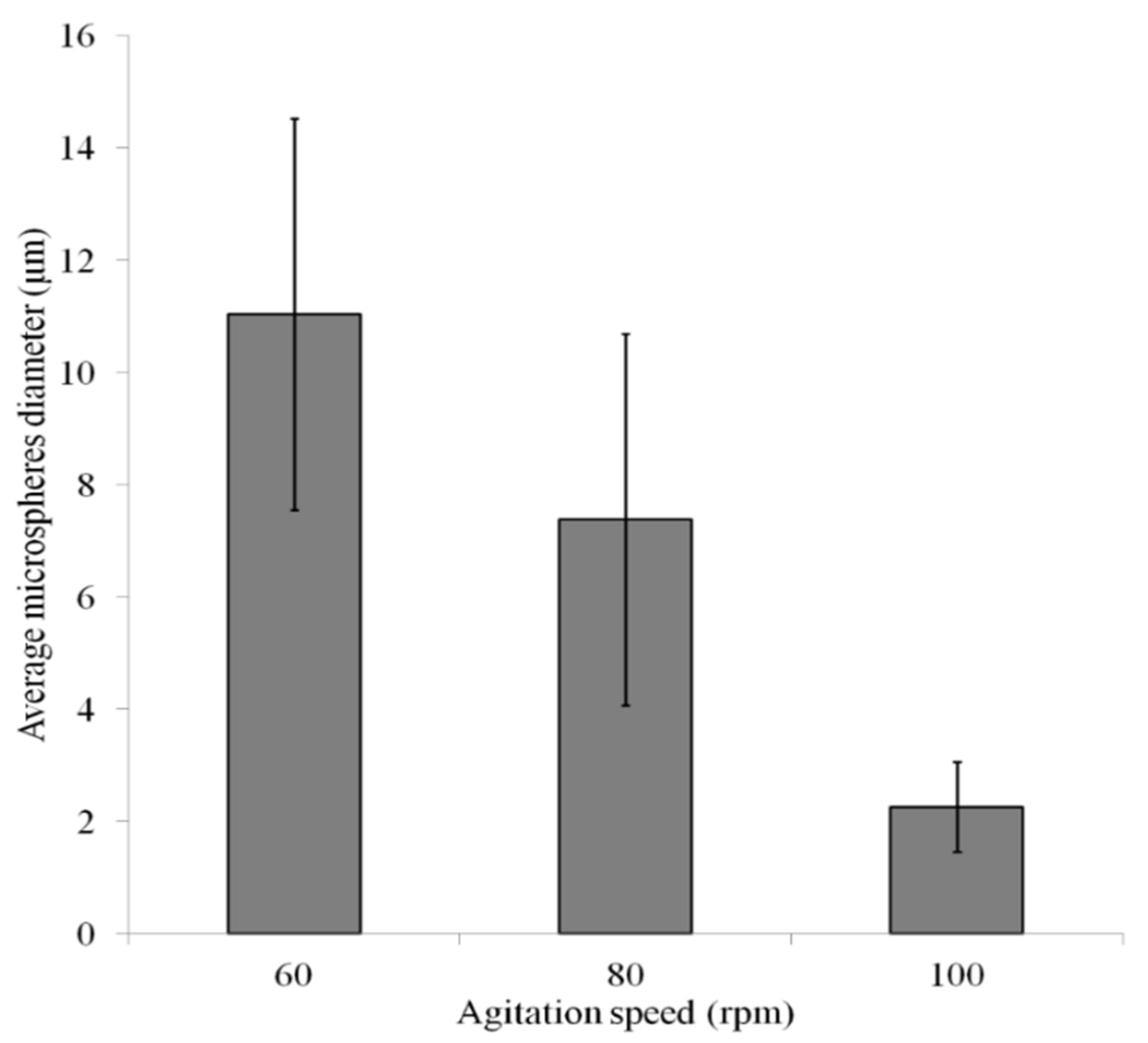
From Figure 13, there were lots of white dots in the continuous phase, which were microspheres generated and suspended in the continuous phase. Figure 14a,b, and c are the optical images of the microspheres generated by the SU-8 membrane under the agitation speeds of 60, 80, and 100 rpm, respectively. Figure 15 presents the average diameters of the microspheres along with the standard deviations for the different agitation speeds. It can be seen from Figure 15 that the diameters of the microspheres were 11.0 ± 3.5 μm, 7.4 ± 3.3 μm, and 2.3 ± 0.8 μm at agitation speeds of 60 rpm, 80 rpm, and 100 rpm, respectively. Thus, it can be concluded that the higher the agitation speed, the smaller the diameter of the microspheres. This phenomenon can be explained by the fact that higher agitation speed results in higher shear stress.
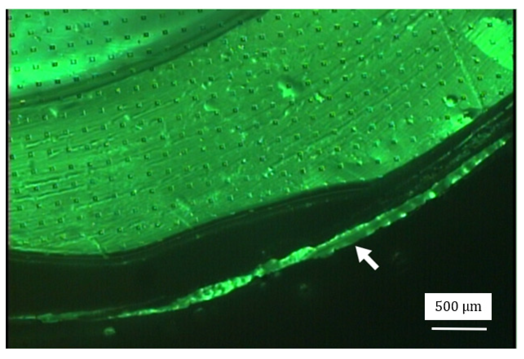
To test the strength of the designed SU-8 porous membrane in this study, the flow rate of the dispersed phase was increased gradually from 50 μL · h −1 by steps of 100 μL · h−1. The first membrane failure happened when the flow rate of the dispersed phase was 4500 μL · h −1 and the agitation speed was 100 rpm, as shown in Figure 16 (white arrow). It is noted that this flow rate was comparable to other membranes found in the literature. For example, Dragosavac et al. [29] produced microspheres using a nickel membrane with the flow rate of the dispersed phase at 3000 μL · h−1; the minimum diameter of the microspheres was 100 μm. Van der Graaf et al. [30] produced microspheres using a silicon membrane with the flow rate of the dispersed phase at 1.23   μ L · h 1 ; the minimum diameter of the microspheres was 35   μ m . Piacentini et al. [31] produced microspheres using a nickel membrane with the flow rate of the dispersed phase at 6000   μ L · h 1 ; the minimum diameter of the microspheres was 40   μ m . Vladisavljevic and Williams [32] produced microspheres using a stainless steel membrane with the flow rate of the dispersed phase at 1.2 × 10 7   μ L · h 1 ; the minimum diameter of the microspheres was 100   μ m . In the previous work of our group, Song [20] produced microspheres using a silicon membrane with the flow rate of the dispersed phase at 1.4 × 10 6   μ L · h 1 ; the minimum diameter of the microspheres was 1.6   μ m . In that work, the dispersed phase fluid was found to be difficult to push through the holes, because the height (t) of the holes with respect to the diameter (w) of the holes was too large, see Figure 2a. To make the membrane work, a high pressure was needed. However, it was found that the membrane was fragile and easily fractured. Addressing that problem was one of the motivations for the idea of the stepwise membrane presented in this paper. With the design in Figure 2b, the two flow paths (Path 1: t1 with respect to wpo; Path 2: t2 with respect to wpi) are short, especially Path 1.
Thus, compared with the other membranes in the literature, as previously mentioned, the small diameter of the microspheres with good size uniformity (i.e., 2.3 ± 0.8   μ m ) was achieved (the flow rate of the dispersed phase was 50 μL · h 1 ; the agitation speed was 100 rpm) with the developed SU-8 membrane. It is noted that microgel variation could range down to 10~15% in the literature [33], when the diameters of the microgels are larger than 100 μm. The diameters of the microspheres in our work were about 3 μm, which means very small differences cause larger variation. Thus, error rather than variation was used to evaluate the size uniformity of the microspheres. It is noted that the flow rate of the dispersed phase in this experiment can certainly be increased, as the maximal flow rate of the dispersed phase for the developed SU-8 membrane was 4500 μL · h 1 under the agitation speed of 100 rpm (notice: the breakage of the membrane was due to the pressure on the membrane, which is further a function of the flow rate of the dispersed phase and the agitation speed). With a far lower flow rate of the dispersed phase and the agitation speed of 100 rpm, the agitation speed can be increased, which implies further improved performance of the microsphere generation process with the SU-8 membrane in terms of small size and high uniformity of microspheres.

4. Conclusions and Future Work

This paper presents a study on the design, fabrication, and testing of a new emulsification membrane. The novelty of this membrane was the stepwise structure, which ensured that the size of the pores was small while the strength of the membrane was high. The membrane was made of the material SU-8, taken as the photoresist in the photolithography microfabrication process. The membrane had two layers. The first layer was the thickness of 1 µm, and the diameter of the pores in this layer was 1 µm. The second layer had the thickness of 20 µm, and the diameter of pores in this layer was 20 µm. The experiment was conducted to show the generation of microspheres with this novel membrane, which showed improved performance of the microsphere generation process with the SU-8 membrane in terms of the small size and the high uniformity of the microspheres. The experimental result showed that the new membrane is very promising in terms of the size of the microspheres as well as the size uniformity; particularly, the diameter and the uniformity of the microspheres were 11.0 ± 3.5   μ m , 7.4 ± 3.3   μ m , and 2.3 ± 0.8   μ m , under the agitation speeds of 60, 80, and 100 rpm, respectively. The stepwise membrane, as developed in this work, is cost effective, because the material of SU-8 photoresist is inexpensive and easy to manufacture, specifically fabricated with the conventional photolithography process, in comparison with the material of SPG along with its fabrication process [34].
To the best of our knowledge, this is the first study in the literature to use the SU-8 photoresist to make a membrane for microsphere generation. There are other factors that may influence the microsphere generation, such as surface roughness, pore alignment, interfacial tension between the continuous phase and dispersed phase, and so forth. These factors are worthy of future work. There is also interest in optimizing the design of the stepwise membrane.

Author Contributions

Conceptualization, W.Z., H.Z. and L.L.; methodology, S.A., G.W. and L.L.; investigation, L.L.; resources, L.L.; data curation, L.L.; writing—original draft preparation, L.L.; writing—review and editing, W.Z. and H.Z.; visualization, L.L.; supervision, W.Z.; project administration, W.Z.; funding acquisition, W.Z. and L.L. All authors have read and agreed to the published version of the manuscript.

Funding

This research was funded by the Natural Sciences and Engineering Research Council of Canada (NSERC) through its Create grant program (565429-2022) to Wenjun (Chris) Zhang and the National Natural Science Foundation of China (Grant No. 52205585) to Lei Lei.

Data Availability Statement

The raw data supporting the conclusions of this article will be made available by the authors on request.

Acknowledgments

The fabrication of the SU-8 membrane was performed in part at the Synchrotron Laboratory for Micro and Nano Devices (SyLMAND) at the Canadian Light Source (CLS). The CLS is supported by the Canada Foundation for Innovation, Natural Sciences and Engineering Research Council of Canada, the University of Saskatchewan, the Government of Saskatchewan, Western Economic Diversification Canada, the National Research Council Canada, and the Canadian Institutes of Health Research.

Conflicts of Interest

The authors declare no conflicts of interest. The funders had no role in the design of the study; in the collection, analyses, or interpretation of data; in the writing of the manuscript; or in the decision to publish the results.

Nomenclatures

SPGShirasu Porous Glass
t1thickness of the bottom layer
t2thickness of the upper layer
wpisize of the square pore for the upper layer
wposize of the square pore for the bottom layer
rpmrevolutions per minute
ssecond
minminute
hhour
PEBpost-exposure bake
PGMEA propylene glycol methyl ether acetate
IPA isopropyl alcohol
N 2 nitrogen
DIdistilled water
ABSacrylonitrile butadiene styrene
HFhydrofluoric
P m i n minimum pressure that ensures the dispersed phase can be pressed through the porous membrane
γ interfacial tension between the continuous and dispersed phases
θ the contact angle between the dispersed phase and the membrane surface in the continuous phase
d p pore diameter

References

  1. Zhu, J.; Wang, M.; Zhang, H.; Yang, S.; Song, K.-Y.; Yin, R.; Zhang, W. Effects of Hydrophilicity, Adhesion Work, and Fluid Flow on Biofilm Formation of PDMS in Microfluidic Systems. ACS Appl. Bio Mater. 2020, 3, 8386–8394. [Google Scholar] [CrossRef]
  2. Yin, R.; He, J.; Bai, M.; Huang, C.; Wang, K.; Zhang, H.; Yang, S.-M.; Zhang, W. Engineering synthetic artificial pancreas using chitosan hydrogels integrated with glucose-responsive microspheres for insulin delivery. Mater. Sci. Eng. C-Mater. Biol. Appl. 2019, 96, 374–382. [Google Scholar] [CrossRef] [PubMed]
  3. Gholamipour-Shirazi, A.; Carvalho, M.S.; Fossum, J.O. Controlled microfluidic emulsification of oil in a clay nanofluid: Role of salt for Pickering stabilization. Eur. Phys. J.-Spec. Top. 2016, 225, 757–765. [Google Scholar] [CrossRef]
  4. Perez-Moral, N.; Watt, S.; Wilde, P. Comparative study of the stability of multiple emulsions containing a gelled or aqueous internal phase. Food Hydrocoll. 2014, 42, 215–222. [Google Scholar] [CrossRef]
  5. Santos, J.; Arriaga, L.R.; Calero, N.; Lee, H.; Munoz, J.; David, D.A. Influence of the formulation on double emulsions containing ecological ingredients prepared by a microfluidic technique. Afinidad 2018, 75, 182–188. [Google Scholar]
  6. Yin, R.; Wang, K.; Du, S.; Chen, L.; Nie, J.; Zhang, W. Design of genipin-crosslinked microgels from concanavalin A and glucosyloxyethyl acrylated chitosan for glucose-responsive insulin delivery. Carbohydr. Polym. 2014, 103, 369–376. [Google Scholar] [CrossRef]
  7. Yin, D.; Zhang, H.; Yang, C.; Zhang, W.; Yang, S. A More Biomimetic Cell Migration Assay with High Reliability and Its Applications. Pharmaceuticals 2022, 15, 695. [Google Scholar] [CrossRef]
  8. Sugiyama, H.; Osaki, T.; Takeuchi, S.; Toyota, T. Perfusion Chamber for Observing a Liposome-Based Cell Model Prepared by a Water-in-Oil Emulsion Transfer Method. ACS Omega 2020, 5, 19429–19436. [Google Scholar] [CrossRef]
  9. Farahinia, A.; Zhang, W.J.; Badea, I. Novel microfluidic approaches to circulating tumor cell separation and sorting of blood cells: A review. J. Sci.-Adv. Mater. Devices 2021, 6, 303–320. [Google Scholar] [CrossRef]
  10. Jia, Y.H.; Zhang, H.B.; Yang, S.B.; Xi, Z.H.; Tang, T.T.; Yin, R.X.; Zhang, W.J. Electrospun PLGA membrane incorporated with andrographolide-loaded mesoporous silica nanoparticles for sustained antibacterial wound dressing. Nanomedicine 2018, 13, 2881–2899. [Google Scholar] [CrossRef] [PubMed]
  11. Yang, C.; Yin, D.; Zhang, H.B.; Badea, I.; Yang, S.M.; Zhang, W.J. Cell Migration Assays and Their Application to Wound Healing Assays—A Critical Review. Micromachines 2024, 15, 720. [Google Scholar] [CrossRef]
  12. Farahinia, A.; Zhang, W.; Badea, I. Recent Developments in Inertial and Centrifugal Microfluidic Systems along with the Involved Forces for Cancer Cell Separation: A Review. Sensors 2023, 23, 5300. [Google Scholar] [CrossRef]
  13. Lei, L.; Bergstrom, D.J.; Zhang, B.; Zhang, H.; Yin, R.; Song, K.Y.; Zhang, W. Micro/Nanospheres Generation by Fluid-Fluid Interaction Technology: A Literature Review. Recent Pat. Nanotechnol. 2017, 11, 15–33. [Google Scholar] [CrossRef] [PubMed]
  14. Wu, J.; Yadavali, S.; Lee, D.; Issadore, D.A. Scaling up the throughput of microfluidic droplet-based materials synthesis: A review of recent progress and outlook. Appl. Phys. Rev. 2021, 8, 031304. [Google Scholar] [CrossRef]
  15. Chattopadhyay, A.K.; Mittal, K.L. Surfactants in Solution; CRC Press: Boca Raton, FL, USA, 2020. [Google Scholar]
  16. Solans, C.; Morales, D.; Homs, M. Spontaneous emulsification. Curr. Opin. Colloid Interface Sci. 2016, 22, 88–93. [Google Scholar] [CrossRef]
  17. Nakashima, T.; Shimizu, M. Porous glass from calcium alumino boro-silicate glass. Bull. Ceram. Soc. Jpn. 1986, 21, 408–412. [Google Scholar]
  18. Charcosset, C.; Limayem, I.; Fessi, H. The membrane emulsification process—A review. J. Chem. Technol. Biotechnol. Int. Res. Process Environ. Clean Technol. 2010, 79, 209–218. [Google Scholar] [CrossRef]
  19. Lei, L. Modeling and Optimization of the Microsphere Generation. Ph.D. Thesis, University of Saskatchewan, Saskatoon, SK, Canada, 2016. [Google Scholar]
  20. Song, K.Y. Design and Fabrication of Novel Microfluidic Systems for Microsphere Generation. Ph.D. Thesis, University of Saskatchewan, Saskatoon, SK, Canada, 2011. [Google Scholar]
  21. Reis, D.R.; Zin, G.; Lemos-Senna, E.; Ambrosi, A.; Di Luccio, M. A modified premix method for the emulsification of spearmint essential oil (Mentha spicata) by ceramic membranes. Surf. Interfaces 2021, 26, 101328. [Google Scholar] [CrossRef]
  22. Othman, R.; Vladisavljevic, G.T.; Nagy, Z.K.; Holdich, R.G. Encapsulation and Controlled Release of Rapamycin from Polycaprolactone Nanoparticles Prepared by Membrane Micromixing Combined with Antisolvent Precipitation. Langmuir 2016, 32, 10685–10693. [Google Scholar] [CrossRef]
  23. Laouini, A.; Jaafar-Maalej, C.; Sfar, S.; Charcosset, C.; Fessi, H. Liposome preparation using a hollow fiber membrane contactor-Application to spironolactone encapsulation. Int. J. Pharm. 2011, 415, 53–61. [Google Scholar] [CrossRef]
  24. Madou, M.J. Fundamentals of Microfabrication; CRC Press: Boca Raton, FL, USA, 2003. [Google Scholar] [CrossRef]
  25. Aktary, M.; Jensen, M.O.; Westra, K.L.; Brett, M.J.; Freeman, M.R. High-resolution pattern generation using the epoxy novolak SU-8 2000 resist by electron beam lithography. J. Vac. Sci. Technol. B 2003, 21, 5–7. [Google Scholar] [CrossRef]
  26. Tobing, L.Y.M.; Mueller, A.D.; Tong, J.C.; Zhang, D.H. Nanobridges formed through electron beam image reversal lithography for plasmonic mid-infrared resonators with high aspect ratio nanogaps. Nanotechnology 2019, 30, 425302. [Google Scholar] [CrossRef] [PubMed]
  27. Lei, L.; Zhang, H.; Bergstrom, D.J.; Anthony, T.; Song, K.-Y.; Zhang, W. Experimental and simulation study of flow patterns in the combined flow focusing and T-junction device. J. Micromech. Microeng. 2020, 30, 055001. [Google Scholar] [CrossRef]
  28. Schneider, C.A.; Rasband, W.S.; Eliceiri, K.W. NIH Image to ImageJ: 25 years of image analysis. Nat. Methods 2012, 9, 671–675. [Google Scholar] [CrossRef]
  29. Dragosavac, M.M.; Holdich, R.G.; Vladisavljević, G.T.; Sovilj, M.N. Stirred cell membrane emulsification for multiple emulsions containing unrefined pumpkin seed oil with uniform droplet size. J. Membr. Sci. 2012, 392–393, 122–129. [Google Scholar] [CrossRef]
  30. Van der Graaf, S.; Schroen, C.; Van der Sman, R.G.M.; Boom, R.M. Influence of dynamic interfacial tension on droplet formation during membrane emulsification. J. Colloid Interface Sci. 2004, 277, 456–463. [Google Scholar] [CrossRef]
  31. Piacentini, E.; Giorno, L.; Dragosavac, M.M.; Vladisavljevic, G.T.; Holdich, R.G. Microencapsulation of oil droplets using cold water fish gelatine/gum arabic complex coacervation by membrane emulsification. Food Res. Int. 2013, 53, 362–372. [Google Scholar] [CrossRef]
  32. Vladisavljevic, G.T.; Kobayashi, I.; Nakajima, M. Production of uniform droplets using membrane, microchannel and microfluidic emulsification devices. Microfluid. Nanofluid. 2012, 13, 151–178. [Google Scholar] [CrossRef]
  33. Muir, V.G.; Qazi, T.H.; Shan, J.; Groll, J.; Burdick, J.A. Influence of Microgel Fabrication Technique on Granular Hydrogel Properties. ACS Biomater. Sci. Eng. 2021, 7, 4269–4281. [Google Scholar] [CrossRef]
  34. Mugabi, J.; Jeong, J.H. Review of the technological advances for the preparation of colloidal dispersions at high production throughput using microporous membrane systems. Colloid Polym. Sci. 2024, 302, 463–485. [Google Scholar] [CrossRef]
Figure 1. A side view of a membrane emulsification device.
Micromachines 15 01118 g001
Figure 2. The membrane for microspheres generation. (a) The single-layer structure of the pores on the membrane emulsification device; (b) the stepwise structure of the pores (not to scale).
Micromachines 15 01118 g002
Figure 3. The stepwise pore structure of the new membrane emulsification device (not to scale).
Micromachines 15 01118 g003
Figure 4. The optical image of one alignment on the first layer.
Micromachines 15 01118 g004
Figure 5. The optical image of pores with 1 × 1 μm2.
Micromachines 15 01118 g005
Figure 6. The membranes before and after chromium etching.
Micromachines 15 01118 g006
Figure 7. The optical image of one alignment on the second layer.
Micromachines 15 01118 g007
Figure 8. The optical images of (a) the top view and (b) bottom view of the pores with 20 × 20 μm2.
Micromachines 15 01118 g008
Figure 9. The membranes glued to ABS tubes. The diameter of the tubes is 1 cm.
Micromachines 15 01118 g009
Figure 10. The fabricated stepwise membranes for microsphere generation. (a) Three membranes; (b) the magnified optical image of the porous membrane.
Micromachines 15 01118 g010
Figure 11. The schematic illustration of the fabrication procedure of the membrane with multilayer structure (not drawn to scale). (a) Sputtering with chrome; (b) Spin coating with SU-8; (c) Mask aligning; (d) Exposure with laser; (e) Post exposure bake; (f) Opaque region is rinsed off by developer; (g) O2 plasma cleaning; (h) Chrome etching; (i) Spin coating with SU-8; (j) Mask aligning; (k) Exposure with laser; (l) Post exposure bake; (m) Opaque region is rinsed off by developer; (n) Glue to an ABS tube; (o) Glass and chrome removing.
Micromachines 15 01118 g011
Figure 12. Experimental set-up for the membrane emulsification process.
Micromachines 15 01118 g012
Figure 13. White dots are microspheres generated using the SU-8 membrane. Agitation speed: 100 rpm. Distance between the agitator and the membrane: 1 cm.
Micromachines 15 01118 g013
Figure 14. The optical images of the microspheres generated by the SU-8 membrane with the agitation speeds of 60 rpm (a), 80 rpm (b), and 100 rpm (c). Microspheres are highlighted in red circles in (c).
Micromachines 15 01118 g014
Figure 15. Average microsphere diameters with standard deviations for different agitation speeds.
Micromachines 15 01118 g015
Figure 16. An optical image of the failure of the SU-8 membrane.
Micromachines 15 01118 g016
Disclaimer/Publisher’s Note: The statements, opinions and data contained in all publications are solely those of the individual author(s) and contributor(s) and not of MDPI and/or the editor(s). MDPI and/or the editor(s) disclaim responsibility for any injury to people or property resulting from any ideas, methods, instructions or products referred to in the content.