Article

Highly Sensitive Amperometric Sensor Based on Laccase-Mimicking Metal-Based Hybrid Nanozymes for Adrenaline Analysis in Pharmaceuticals

1
Institute of Cell Biology, National Academy of Sciences of Ukraine, 79005 Lviv, Ukraine
2
Faculty of Veterinary Hygiene, Ecology and Law, Stepan Gzhytskyi National University of Veterinary Medicine and Biotechnologies, 79000 Lviv, Ukraine
3
Department of Biology and Chemistry, Drohobych Ivan Franko State Pedagogical University, 82100 Drohobych, Ukraine
*
Authors to whom correspondence should be addressed.
Catalysts 2021, 11(12), 1510; https://doi.org/10.3390/catal11121510
Submission received: 19 November 2021 / Revised: 7 December 2021 / Accepted: 9 December 2021 / Published: 11 December 2021
(This article belongs to the Special Issue Enzymes in Materials Science)

Abstract

:
Nanozymes are nanomaterials which exhibit artificial enzymatic activities and are considered as alternatives to natural enzymes. They are characterized by good catalytic activity and high stability, as well as ease and low cost of preparation. In this study, the mimetics of laccase or “nanolaccases” (NLacs) were synthesized by a simple method of chemical reduction of transition metal salts. The NLacs were tested for their catalytic activity in solution and on the electrode surface. The most effective NLacs, namely nAuCePt and nPtFe, were found to possess excellent laccase-like activities capable of oxidizing the endocrine hormone adrenaline (AD). These NLacs were characterized in detail and used for the development of amperometric sensors for AD determination. The amperometric sensors containing the best NLacs, as well as a natural fungal laccase, were constructed. The most effective nAuCePt-containing sensor had good specificity in relation to AD and improved analytical characteristics. It possessed a 384-fold higher sensitivity than adrenaline (230,137 A·M−1·m−2), a 64-fold lower limit of detection (0.025 µM), and a broader linear range (0.085–45 µM) in comparison with the sensor based on natural laccase. The constructed nAuCePt-containing sensor was successfully used for AD analysis in pharmaceutical formulation.

Graphical Abstract

1. Introduction

The detection of biologically active toxic pharmaceutical compounds and their metabolites in surface waters cause great concern for humanity [1,2]. Several chemicals have already been found in raw urban wastewaters at concentrations up to μg·L−1, which are dangerous for health [3].
Pharmaceutical manufacturing has become a significant source of harmful chemicals as pollutants of the environment. Even after treatment facilities, the concentrations of toxic compounds in the wastewaters of these factories may be 10–1000 times higher than safe values. In addition, households and hospitals make a significant contribution to the excretion of pharmaceuticals into urban wastewaters [4].
Among the chemicals detected in terrestrial and aquatic environments, some anti-inflammatories and analgesics, catecholamines and psychiatric drugs are the most dangerous for humans and wildlife [5]. As the influence of these contaminants in the environment is clearly recognized, early detection and quantification of the mentioned pharmaceuticals is necessary, not only for controlling the quality of water, especially drinking water, but also for the diagnosis and monitoring of various health conditions [4,6].
The catecholamines—noradrenaline, adrenaline (also called, epinephrine) and dopamine—are key neurotransmitters in the sympathetic nervous system, as they stimulate adrenergic receptors in a wide variety of cells [7]. High concentrations of adrenaline (AD) correlate well with glycogenolysis in the liver, hypoglycemia, myocardial infarction, lipolysis in the adipose tissue, and the heart contraction rate [8]. AD is an important biomarker for Parkinson’s disease [9] and other malignances [10]. Thus, AD and other catecholamines are secreted in excessive amounts by pheochromocytomas (tumors of the adrenal glands).
A variety of analytical techniques for the determination of AD has been reported—among them, liquid chromatography [11], spectrophotometry [12], capillary electrophoresis [13], fluorometry [14], high-pressure liquid chromatography, and circular dichroism [15]—which are characterized by low selectivity, high costs, and time consumption. Within the last few years, electrochemical methods for the analysis of pharmaceuticals, especially catecholamines, have become very popular. However, only a limited number of papers have reported the electrochemical detection of AD. Electrochemical approaches for AD analysis are preferable to laborious instrumental methods due to the simplicity of procedures and instrumentation, fast response, sensitivity, and low cost [16].
To construct amperometric biosensors, laccase preparations and different nanomaterials were used [17,18,19,20,21,22,23,24,25,26,27,28]. The proposed laccase-based sensors have been utilized to monitor the level of AD in pharmaceuticals, even though the practical application of such biosensors is often limited due to the impact of environmental compartments on the enzyme in the recognition layer [17,18].
Artificial enzymes with pseudo-laccase activity, especially nano-size laccase-like nanozymes (NZs) or “nanolaccases (NLacs)”, have preferential properties compared to natural enzymes. Thus, the search for new effective NLacs, which are promising for construction of electrochemical sensors, appears to be an urgent task [19,20,21,22,23,24,25,26,27,28].
In the current work, we report the synthesis of NLacs, their characterization, and their application in the development of amperometric sensors for the direct measurement of AD. The most effective sensors contain metal-hybrid NZs on the surfaces of graphite electrodes. The proposed sensor was successfully tested for AD determination in a pharmaceutical product.

2. Results and Discussion

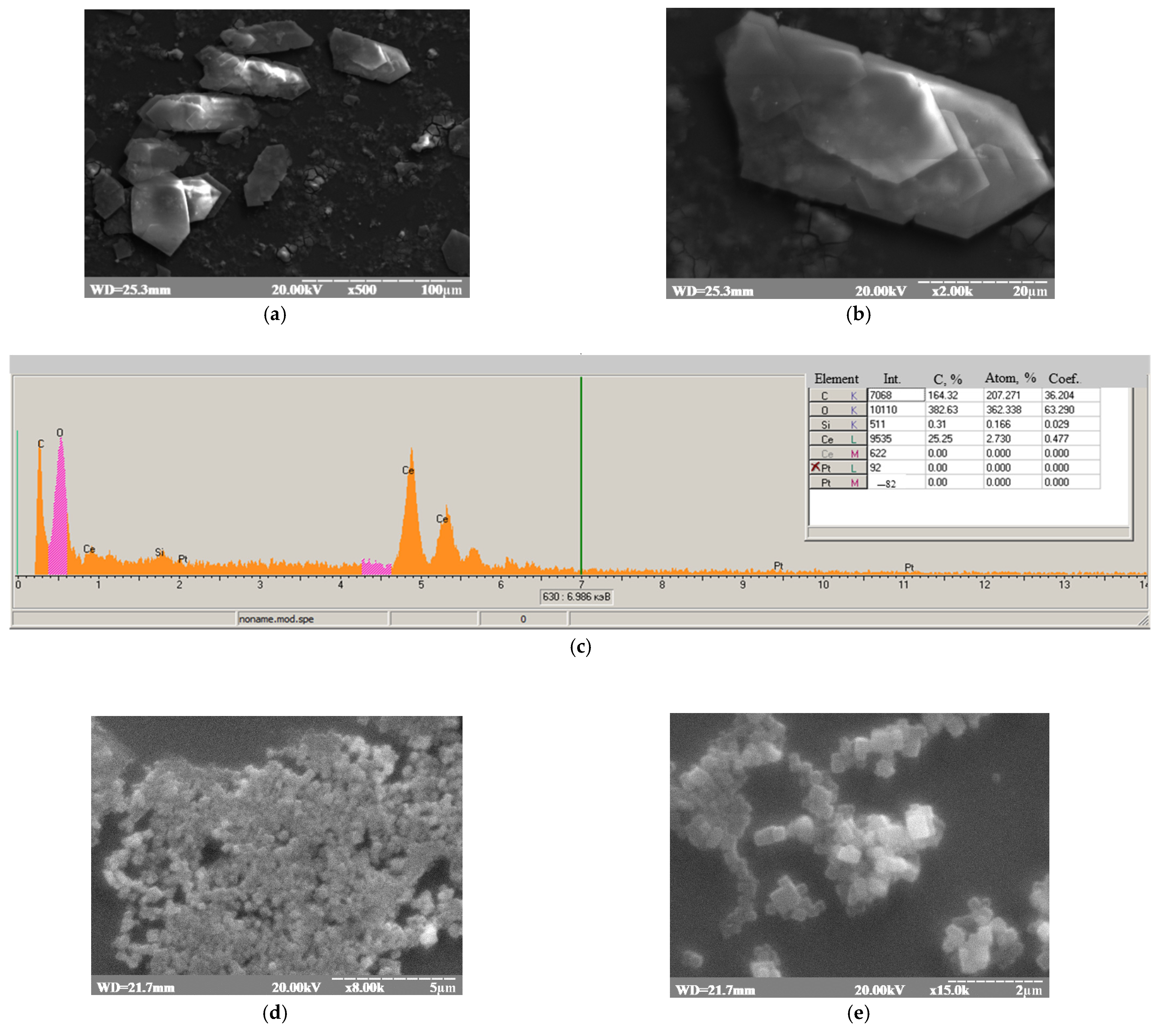
2.1. Synthesis and Characterization of the NLacs

A number of metal-based composite materials were synthesized using the method of chemical reduction. The synthesized NZs were screened for their ability to oxidize ABTS in the solution (Table 1). A natural laccase preparation from Trametes gibbosa 1525 was used for comparison. It was shown that several tested NZs, especially nAuCePt and nFePt, possessed significant laccase-like activities. The most catalytically active NZs in the solution were characterized by scanning electron microscopy (SEM).
Figure 1 presents the overall morphology of the formed particles. According to SEM images, nAuCePt have the shape of prisms, with sizes varying from 20 to 100 µm (Figure 1a–c). The obtained nPtFe have the shape of cubes, with sizes varying from 4 to 10 µm (Figure 1d–f). It was shown that the characteristic peaks on the XRM images corresponded to Ce0, Pt0, Au0, and Fe0 (Figure 1c,f). It was demonstrated (the data are not shown) that the sizes of all the studied compounds do not satisfy the nanoscale criterion of being less than 100 nm in all three dimensions. In some cases, they are nanoscale only in one dimension (plate), while in the others, they exceed the criterion. This is most likely due to the aggregation of the initially formed NZs. To take this factor into account, we classify NZs as materials whose nanoscale is confirmed by physical methods for at least one dimension.

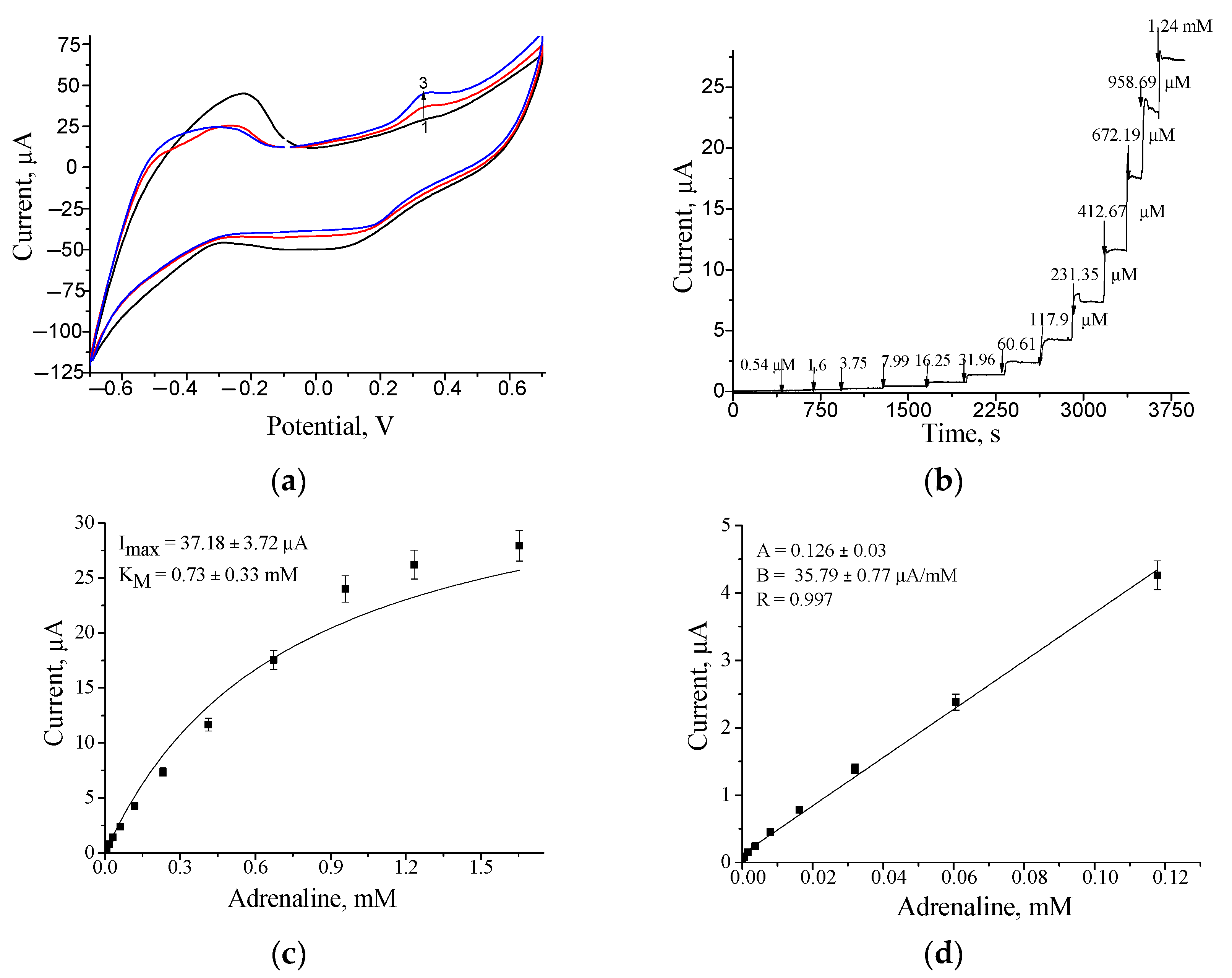
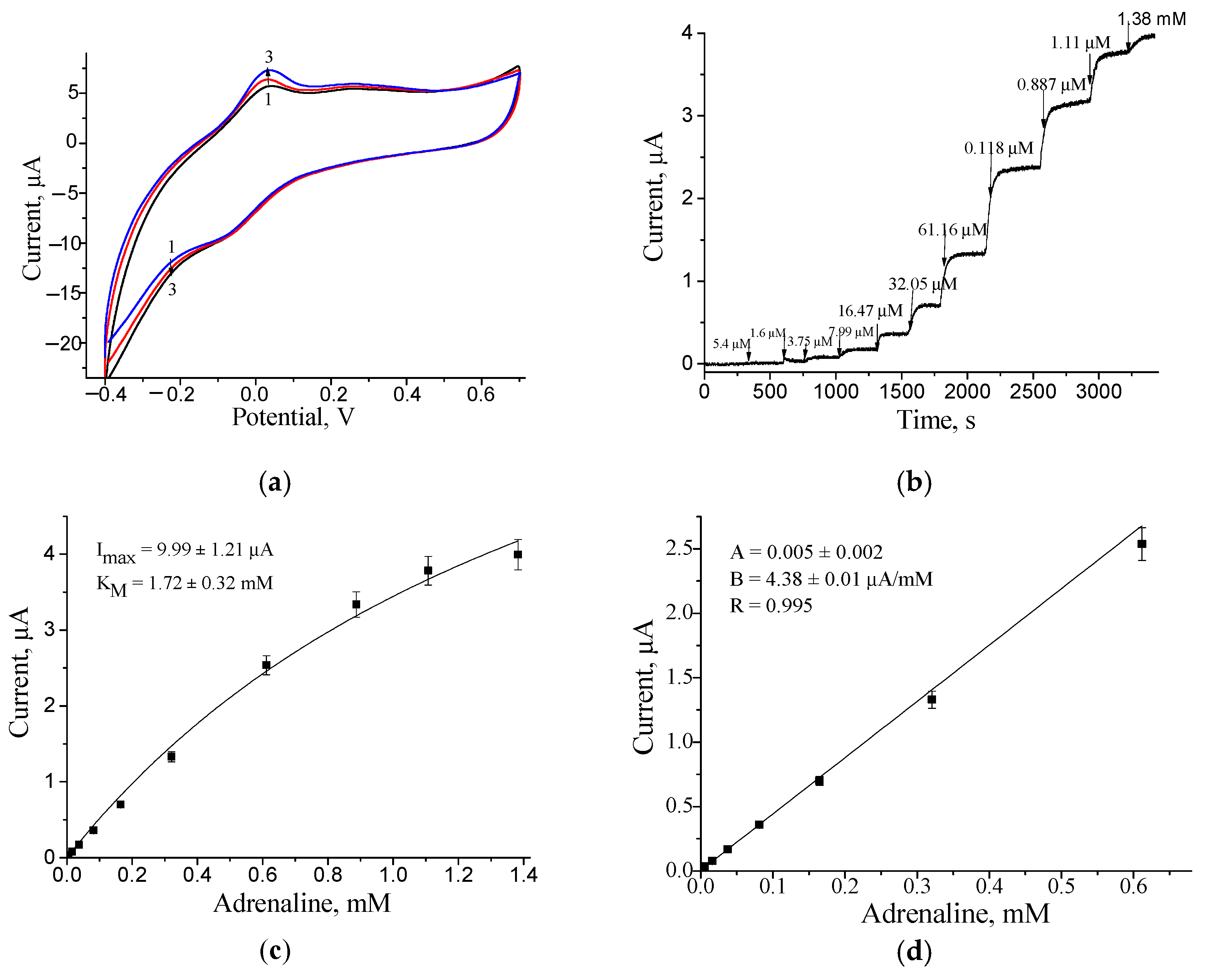
2.2. The Development and Characterization of the NZ-Modified Electrodes

Here, we proposed a new sensor for AD determination based on metal-hybrid NLacs immobilized on a graphite surface of the electrode (further marked as NZ/GE). Figure 2 shows a scheme of the chemical processes between AD (also called epinephrine) and NLacs. The synthesized NLacs (for example, nAuCePt) catalyze the oxidation of AD to adrenochrome. The last compound is reduced on the surface of GE under the potential of 50 mV. The resulting cathodic currents correlate directly with the concentration of AD in the sample solution.
The nanozymes nAuCePt and nPtFe were deposited on the top of a GE rod electrode (marked as nAuCePt/GE and nFePt/GE, respectively), as described in Section 3.6, and their electro-catalytic activities toward AD oxidation were compared with laccase/GE. Figure 3, Figure 4 and Figure 5 demonstrate the electrochemical characteristics of the developed sensors as current responses upon AD addition, namely cyclic voltamperograms (CV) with a scan rate of 50 mV·s−1, chronoamperograms, and calibration curves under optimal working potentials, which were determined from the CV.
Following the chronoamperograms for the constructed amperometric sensors (Figure 3b, Figure 4b and Figure 5b), we plotted the calibration curves for AD determination as illustrated in Figure 3c, Figure 4c and Figure 5c. The linear ranges for AD determination and sensitivities were calculated for all obtained modified electrodes (Figure 3d, Figure 4d and Figure 5d).
The CV for the nAuCePt/GE (Figure 3a) demonstrated that the cathodic reduction peak, contributed by AD decomposition, arose at potentials of approximately −50 mV (vs. Ag/AgCl). The CV for the nPtFe/GE (Figure 4a) demonstrated that the anodic oxidation peak arose at the potential of approximately +250 mV. The chronamperometric dependences and calibration graphs for AD determination are shown in Figure 3 and Figure 4b–d respectively for both types of NZs. As a control measure, the unmodified GE was tested, and no signal was detected upon AD addition (the data not shown).
Figure 3c, Figure 4c and Figure 5c present the maximal current responses on AD at substrate saturation (Imax) and the apparent Michaelis–Menten constants (KMapp) to AD. nAuCePt/GE was shown to have the highest Imax (27-fold increase compared to that of laccase/GE) and the lowest KMapp among the sensors reported here.
The analytical characteristics of the developed electrodes on AD in comparison with the known sensors are summarized in Table 2. As demonstrated in Table 2, for AD detection, not only laccase preparations but also various nanomaterials may be used [17,18,19,20,21,22,23,24,25,26,27,28] Some of the reported sensors exhibit excellent analytical properties, having low limits of detection (2 nM [27] or 9 nM [26]) and wide linear ranges (for example, from 0.7 µM to 1200 µM [22]). However, the main disadvantages of these sensors are insufficient sensitivity [20] and high working potentials (+400 mV) [27]. The nAuCePt/GE proposed by us was shown to possess the highest sensitivity to AD in comparison with the reported (bio)sensors, this value showing a 384-fold increase compared to that of laccase/GE (Table 2).
As a result of our study, we can conclude that nAuCePt and nAuPtPd possess excellent sensitivities and wide linear-ranges for AD detection and may, thus, be considered as promising mimetics of laccase in amperometric sensors.

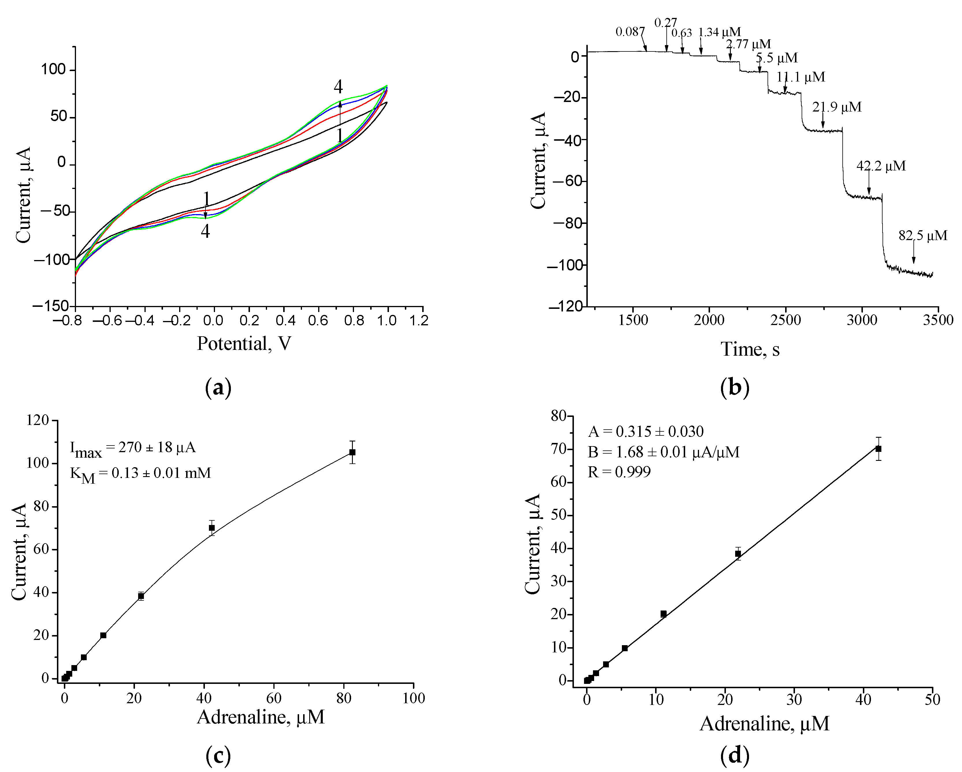
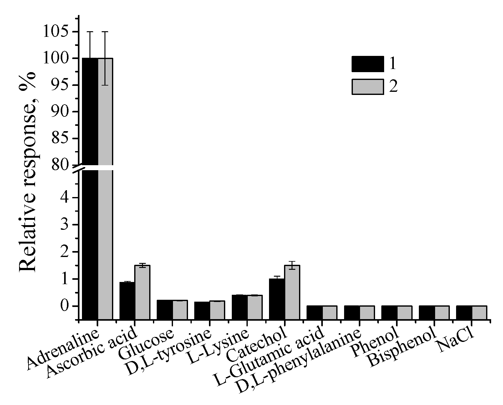
2.3. Characterization of the Most Effective nAuCePt-Based Electrode and Its Application

In general, the specificity of a recognition element in relation to the target analyte is a valuable characteristic for its use in sensors. The results of the selectivity test for the constructed nAuCePt/GE are presented in Figure 6. The NZ-based electrode was shown to be highly specific to AD. In the case of adding structurally similar compounds as well as solutions of glucose and sodium chloride, the signals of the sensor are insignificant (less than 5%). The latter compounds are often used as solubilizers in pharmaceuticals.
To experience nAuCePt/GE as an AD-selective sensor for determination of AD concentration, a commercial pharmaceutical product was analyzed under optimal conditions. The tested sample in the ampoule was the solution for the injection of “Adrenaline (Epinephrine)”. For the analysis of AD in the sample of the pharmaceutical, the method called the “Standard Addition Test” (SAT) was used for two dilutions (Figure 7). The principles of the graphical analytical method SAT, as well as the algorithm for the calculation of an analyte’s concentration from the parameters of linear regression, were described earlier [29].
The values of AD concentration in the commercial sample, which were estimated using the developed sensor and declared by the manufacturer, are presented in Table 3. Both concentrations of AD correlate well (Table 3), with an error of less than 5%.

3. Materials and Methods

3.1. Reagents

Gold(III) chloride solution (HAuCl4), Cerium(III) hydrocarbonate (Ce(HCO3)4), chloroplatinic acid (H2PtCl6), palladium chloride (PdCl3), copper(II) sulfate (CuSO4), iron(III) chloride (FeCl3·4H2O), Cobalt(II) chloride hexahydrate (CoCl2·6H2O), adrenaline, o-dianisidine, ascorbic acid, sodium borohydride (NaBH4), catechol, glucose, D,L-tyrosine, L-Lysine, L-glutamic acid, D,L-phenylalanine, phenol, bisphenol, NaCl, Nafion (5% solution in 90% ethanol), 2,2′-Azino-bis(3-ethylbenzthiazoline-6-sulfonic acid) (ABTS) and all other reagents and solvents used in this work were purchased from Sigma-Aldrich (Steinheim, Germany). All solutions were prepared using ultrapure water obtained with the Milli-Q® IQ 7000 Water (Merck KGaA, Darmstadt, Germany).

3.2. Isolation and Purification of Laccase

Laccase was isolated from the fungus Trametes gibbosa 1525. The cells were cultivated at 28 °C in YPD medium supplemented with 1% sucrose and 0.05 mM Cu2+ for 5 days. To remove the mycelia, the fungal culture was passed through filter paper, and a cultural liquid was used for enzyme isolation. Laccase with activity of ≥10 U·mg−1 was purified according to the described method [30].
The laccase activity was determined in kinetic mode spectrophotometrically at 420 nm (Shimadzu, Kyoto, Japan) as described earlier [30]. As a substrate, 0.5 mM ABTS in 50 mM sodium acetate buffer solution, with a pH of 4.5, was used.

3.3. Synthesis and Characterization of Laccase-like NZs

The tri-metallic composites of nCoPtPd, nFePtPd, and nAuPtPd were obtained according to the protocol, which was proposed for nNiPtPd synthesis [31], using NaBH4 as a reducing agent.
To obtain nPtFe, solutions of 50 mM H2PtCl6 and 50 mM FeCl3 were mixed (1:1 ratio by volume), then 100 mM NaBH4 was added up to 5 mM while stirring for 20 min. nPtFe were shown to be formed in the reaction mixture after a 24 h incubation without stirring.
nAuCePt were obtained by the reduction of metal ions from the appropriate salts with ascorbic acid. The following scheme was used for this NZ synthesis: 5 mL 10 mM HAuCl4, 5 mL 10 mM Ce(HCO3)4 and 1 mL 100 mM ascorbic acid were mixed. After vigorous stirring for 5 min, 5 mL 10 mM H2PtCl6 and 0.1 mL 100 mM ascorbic acid were added and the reaction mixture was stirred for 20 min.
All formed NZs were collected by centrifugation, washed with water, and stored as a suspension in water until use at +4 °C, as described earlier [32,33].
The pseudo-laccase activity was determined in the same way as for natural laccase [30]. Morphological analyses of the samples were performed using scanning electron microscopy (SEM) as described in [32].

3.4. Apparatus, Measurements, and Statistical Analysis

The amperometric sensors were evaluated using constant-potential amperometry. The amperometric experiments were carried out in triplicates, using a three-electrode configuration with a graphite rod as a working electrode, a Pt-wire as a counter electrode and an Ag/AgCl/KCl (3 M) as a reference electrode. The analytical characteristics of the sensors were estimated as described earlier [29,31,32,33].

3.5. Functionalization and Characterization of the Electrodes

To construct the NZ-based amperometric sensor, the surface of the graphite electrode (GE) was modified with 5 µL of NLac solution (1 mg/mL) or 5 µL of enzyme solution (12 U/mL). The dried film was covered with 5 µL of 0.5% Nafion solution. After drying at room temperature, the modified GE was washed with 50 mM of acetic buffer, with a pH of 4.5. The electrochemical properties of the functionalized GEs were studied by cyclic voltammetry (CV) and chronoamperometry, and the graphs of the dependences of the amperometric signals in increasing concentrations of AD were compared. The most electroactive NLac/GE was chosen and tested as an amperometric sensor for AD determination.

3.6. Determination of AD in Pharmaceutical Formulation

nAuCePt/GE, as the most effective amperometric sensor on AD, was tested on the real sample of the pharmaceutical formulation, “Adrenaline (Epinephrine) solution for injection, ampoule” (“Darnytsia”, Kyiv, Ukraine). Each assay, being performed for two dilutions of the sample, was repeated 3 times. The analytical results were statistically processed using the OriginPro 2021 software (OriginLab, One Roundhouse Plaza, Suite 303, Northampton, MA 01060, USA). The manufacturer has declared the following composition of this pharmaceutical product: 1.8 mg/mL of adrenaline tartrate in water for injections. The tested solution additionally contained sodium metabisulfite (E223) and sodium chloride.

4. Conclusions

In the current research, a number of hybrid metal-based nanoparticles were synthesized by chemical methods and screened for their ability to oxidize AD in solution. The best catalytically active nanoparticles were chosen as artificial laccases. Their structure and electrochemical behavior on electrodes were characterized. The nAuCePt, as the most effective laccase-like nanozyme, was studied in more detail. It was successfully used as a mimetic of laccase in an amperometric sensor for AD analysis in pharmaceuticals. Due to high sensitivity and specificity, the developed nAuCePt-based sensor may be promising in the diagnosis of some diseases, including tumors of the adrenal glands or mental disorders.

Author Contributions

Conceptualization and methodology, O.D., N.S. and G.G.; investigation, N.S. and O.D.; resources, data curation, N.S., G.G. and M.G.; writing—original draft preparation, N.S., O.D. and M.G.; writing—review and editing, N.S., G.G. and M.G.; supervision, M.G.; project administration, M.G.; funding acquisition, M.G. All authors have read and agreed to the published version of the manuscript.

Funding

This research was partially funded by NAS of Ukraine (The program “Smart sensor devices of a new generation based on modern materials and technologies”, projects #13 and #10/3), by the National Research Foundation of Ukraine (project 2020.02/0100 “Development of new nanozymes as catalytic elements for enzymatic kits and chemo/biosensors”), and by the Ministry of Education and Science of Ukraine (Ukrainian-Lithuanian R&D, project 0120U103398; 0121U109539; 0121U109543).

Acknowledgments

We would like to thank Nina A. Bisko (M.G. Kholodny Institute of Botany, NAS of Ukraine) for kindly providing the fungal strain used in this study. We are highly appreciative of our colleagues from the Institute of Cell Biology, NAS of Ukraine, namely Maksym D. Lootsik for discussion and critical remarks, and Roman Ya. Serkiz for assistance with SEM experiments.

Conflicts of Interest

The authors declare no conflict of interest.

Abbreviations

ABTS2:2′-Azinobis-(3-ethylbenzthiazoline-6-sulphonate)
ADAdrenaline
CVCyclic voltammetry
GCEGlassy-carbon electrode
GEGraphite electrode
ImaxMaximal current response on tested analyte at substrate saturation
KMappApparent Michaelis–Menten constant
LODLimit of detection
LRLinear range
NPsNanoparticles
NZNanozyme
Me1/Me2Core/shell Nanozyme; where Me1—core and Me2—shell
LaccaseNatural laccase from Trametes gibbosa 1525
SATStandard addition test
SEM-XRM Scanning electron microscopy coupled with X-ray microanalysis
PtE Platinum-rod electrode
PPy-PVSPolypyrrole–polyvinylsulphonate;
GCE Glassy-carbon electrode;
IrOx/SPEIridium-oxide modified screen-printed electrode;
Pt-BMI.PF6PtNPs in 1-butyl-3-methylimidazolium hexafluorophosphate;
CPECarbon-paste electrode
EBNBH2,2-[1,2-ethanediylbis(nitriloethylidyne)]-bis-hydroquinone double-wall carbon-nanotube;
GCEGlassy-carbon electrode
MWCNT/CFECarbon film electrode (CFE) modified with multiwalled carbon-nanotubes (MWCNTs)
AuNPs/PANi/GCEGold nanoparticles/polyaniline Langmuir-modified glassy-carbon electrode.

References

  1. Akerman-Sanchez, G.; Rojas-Jimenez, K. Fungi for the bioremediation of pharmaceutical-derived pollutants: A bioengineering approach to water treatment. Environ. Adv. 2021, 4, 100071. [Google Scholar] [CrossRef]
  2. Massima Mouele, E.S.; Tijani, J.O.; Badmus, K.O.; Pereao, O.; Babajide, O.; Zhang, C.; Shao, T.; Sonin, E.; Tarasenko, V.; Fatoba, O.O.; et al. Removal of Pharmaceutical Residues from Water and Wastewater Using Dielectric Barrier Discharge Methods—A Review. Int. J. Environ. Res. Public Health 2021, 18, 1683. [Google Scholar] [CrossRef]
  3. Rebollar-Pérez, G.; Campos-Terán, J.; Ornelas-Soto, N.; Méndez-Albores, A.; Torres, E. Biosensors based on oxidative enzymes for detection of environmental pollutants. Biocatalysis 2016, 1, 118–129. [Google Scholar] [CrossRef]
  4. USGS.gov. Available online: https://toxics.usgs.gov/highlights/PMFs.html (accessed on 18 November 2021).
  5. Rana, R.S.; Singh, P.; Kandari, V.; Singh, R.; Dobhal, R.; Gupta, S. A review on characterization and bioremediation of pharmaceutical industries’ wastewater: An Indian perspective. Appl. Water Sci. 2017, 7, 1–12. [Google Scholar] [CrossRef] [Green Version]
  6. Taylor, D. The Pharmaceutical Industry and the Future of Drug Development. In Pharmaceuticals in the Environment; Hester, R.E., Harrison, R.M., Eds.; Royal Society of Chemistry: London, UK, 2015; Volume 41, pp. 1–33. Available online: www.rsc.org (accessed on 10 December 2021).
  7. McCorry, L.K. Physiology of the Autonomic Nervous System. Am. J. Pharm. Educ. 2007, 71, 78. [Google Scholar] [CrossRef] [Green Version]
  8. Peaston, A.E.; Evsikov, A.V.; Graber, J.H.; Vries, W.N.; Holbrook, A.E.; Solter, D.; Knowles, B.B. Retrotransposons regulate host genes in mouse oocytes and preimplantation embryos. Dev. Cell. 2004, 7, 597–606. [Google Scholar] [CrossRef] [PubMed]
  9. Emamzadeh, F.N.; Surguchov, A. Parkinson’s Disease: Biomarkers, Treatment, and Risk Factors. Front. Neurosci. 2018, 12, 612. [Google Scholar] [CrossRef]
  10. Zuber, S.M.; Kantorovich, V.; Pacak, K. Hypertension in Pheochromocytoma: Characteristics and Treatment. Endocrinol. Metab. Clin. N. Am. 2011, 40, 295–311. [Google Scholar] [CrossRef] [PubMed] [Green Version]
  11. Thomas, A.; Geyer, H.; Mester, H.J.; Schänzer, W.; Zimmermann, E.; Thevis, M. Quantitative Determination of Adrenaline and Noradrenaline in Urine Using Liquid Chromatography-Tandem Mass Spectrometry. Chromatographia 2006, 64, 587–591. [Google Scholar] [CrossRef]
  12. Bibire, N.; Christopoulos, L.; Apostu, M.; Dorneanu, V. Quantitative determination of adrenaline by visible spectrophotometric method. Rev. Med. Chir. Soc. Med. Nat. Iasi. 2007, 111, 779–782. [Google Scholar] [PubMed]
  13. Liu, C.; Zhang, J.; Zhang, X.; Zhao, L.; Shuang, L. Enantiomeric separation of adrenaline, noradrenaline, and isoprenaline by capillary electrophoresis using streptomycin-modified gold nanoparticles. Mikrochim. Acta 2018, 185, 227. [Google Scholar] [CrossRef]
  14. Menon, S.; Jesny, S.; Sivasankaran, U.; Girish, K.K. Fluorometric Determination of Epinephrine: A Green Approach. Anal. Sci. 2016, 32, 999–1001. [Google Scholar] [CrossRef] [Green Version]
  15. A Kojło, A.; Calatayud, J.M. Spectrophotometric determination of adrenaline with an oxidative column in a FIA assembly. J. Pharm. Biomed. Anal. 1990, 8, 663–666. [Google Scholar] [CrossRef]
  16. Beitollahi, H.; Safaei, M.; Tajik, S. Voltammetric and amperometric sensors for determination of epinephrine: A short review (2013–2017). J. Electrochem. Sci. Eng. 2019, 9, 27–43. [Google Scholar] [CrossRef]
  17. Quan, D.; Shin, W. Amperometric Detection of Catechol and Catecholamines by Immobilized Laccase from DeniLite. Electroanalysis 2004, 16, 1576–1582. [Google Scholar] [CrossRef]
  18. Arslan, F.; Durmus, S.; Colak, O.; Arslan, H. A New Laccase-Based Biosensor for Epinephrine Determination. Gu. J. Sci. 2015, 28, 1–9. [Google Scholar]
  19. Zou, L.; Li, Y.; Cao, S.; Ye, B. Gold nanoparticles/polyaniline Langmuir-Blodgett Film modified glassy carbon electrode as voltammetric sensor for detection of epinephrine and uric acid. Talanta 2013, 117, 333–337. [Google Scholar] [CrossRef] [PubMed]
  20. Salimi, A.; Alizadeh, V.; Compton, R.G. Disposable amperometric sensor for neurotransmitters based on screen-printed electrodes modified with a thin iridium oxide film. Anal. Sci. 2005, 21, 1275–1280. [Google Scholar] [CrossRef] [Green Version]
  21. Brondani, D.; Scheeren, C.W.; Dupont, J.; Vieira, I.C. Biosensor based on platinum nanoparticles dispersed in ionic liquid and laccase for determination of adrenaline. Sens. Actuators B Chem. 2009, 140, 252–259. [Google Scholar] [CrossRef]
  22. Beitollahi, H.; Ardakani, M.M.; Ganjipour, B.; Naeimi, H. Novel 2,2-[1,2-ethanediylbis(nitriloethylidyne)]-bis-hydroquinone double-wall carbon nanotube paste electrode for simultaneous determination of epinephrine, uric acid and folic acid. Biosen. Bioelectron. 2008, 24, 362–368. [Google Scholar] [CrossRef] [PubMed]
  23. Ghica, M.E.; Brett, C.M.A. Simple and efficient epinephrine sensor based on carbon nanotube modified carbon film electrodes. Anal. Let. 2013, 46, 1379–1393. [Google Scholar] [CrossRef]
  24. Ni, J.A.; Ju, H.X.; Chen, H.Y.; Leech, D. Amperometric determination of epinephrine with an osmium complex and Nafion double-layer membrane modied electrode. Anal. Chim. Acta 1999, 378, 151–157. [Google Scholar] [CrossRef]
  25. Agboola, B.O.; Ozoemena, K.I. Efficient electrocatalytic detection of epinephrine at gold electrodes with self-assembled metallo-octacarboxyphthalocyanine complexes. Electroanalysis 2008, 20, 1696–1707. [Google Scholar] [CrossRef]
  26. Shankar, S.S.; Shereema, R.M.; Rakhi, R.B. Electrochemical Determination of Adrenaline using MXene/Graphite composite paste electrodes. ACS Appl. Mater. Interfaces 2018, 50, 43343–43351. [Google Scholar] [CrossRef]
  27. Xu, H.; Wang, X.; Chen, R.; Yu, Z. Voltammetric Determination of Epinephrine in the Presence of Uric Acid Based on Aminated Graphene and Ag NPs Hybrid Membrane Modified Electrode. Chem. Res. Chin. Univ. 2014, 30, 205–210. [Google Scholar] [CrossRef]
  28. Turkušić, E.; Redžić, S.; Kahrović, E.; Zahirović, A. Electrochemical Determination of Adrenaline at Ru(III) Schiff Base Complex Modified Carbon Electrodes. Croat. Chem. Acta 2017, 90, 345–352. [Google Scholar] [CrossRef]
  29. Stasyuk, N.; Gayda, G.; Andriy Zakalskiy, A.; Zakalska, O.; Serkiz, R.; Gonchar, M. Amperometric biosensors based on oxidases and PtRu nanoparticles as artificial peroxidase. Food Chem. 2019, 285, 213–220. [Google Scholar] [CrossRef]
  30. Demkiv, O.M.; Gayda, G.Z.; Broda, D.; Gonchar, M.V. Extracellular laccase from Monilinia fructicola: Isolation, primary characterization and application. Cell Biol. Intern. 2021, 45, 536–548. [Google Scholar] [CrossRef]
  31. Stasyuk, N.; Gayda, G.; Demkiv, O.; Darmohray, L.; Gonchar, M.; Nisnevitch, M. Amperometric biosensors for L-arginine determination based on L-arginine oxidase and peroxidase-like nanozymes. Appl. Sci. 2021, 11, 7024. [Google Scholar] [CrossRef]
  32. Gayda, G.Z.; Demkiv, O.M.; Gurianov, Y.; Serkiz, R.Y.; Klepach, H.M.; Gonchar, M.V.; Nisnevitch, M. “Green” Prussian Blue Analogues as Peroxidase Mimetics for Amperometric Sensing and Biosensing. Biosensors 2021, 11, 193. [Google Scholar] [CrossRef]
  33. Demkiv, O.; Stasyuk, N.; Serkiz, R.; Gayda, G.; Nisnevitch, M.; Gonchar, M. Peroxidase-Like Metal-Based Nanozymes: Synthesis, Catalytic Properties, and Analytical Application. Appl. Sci. 2021, 11, 777. [Google Scholar] [CrossRef]
Figure 1. Characteristics of the nAuCePt (ac) and nPtFe (df) NZs: SEM images (a,b,d,e); X-ray spectral microanalysis (c,f).
Catalysts 11 01510 g001aCatalysts 11 01510 g001b
Figure 2. The principal scheme of the AD determination.
Catalysts 11 01510 g002
Figure 3. Characteristics of nAuCePt/GE: CV as current responses upon AD addition up to 0 mM (1, black line), 4 µM (2, red line), 8 µM (3, blue line), and 16 mM (4, green line) concentrations (a); chronamperometric response (b), dependence of amperometric signal on concentration of AD (c) and calibration graph (d) under working potential −50 mV.
Catalysts 11 01510 g003
Figure 4. Characteristics of nPtFe/GE: CV as current responses upon AD addition up to 0 mM (1, black line), 4 µM (2, red line) and 8 µM (3, blue line), concentrations (a); chronamperometric response (b), dependence of amperometric signal on concentration of AD (c) and calibration graph (d) under working potential +250 mV.
Catalysts 11 01510 g004
Figure 5. Characteristics of laccase/GE: CV as current responses upon AD addition up to 0 mM (1, black line), 4 µM (2, red line) and 8 µM (3, blue line) concentrations (a); chronamperometric response (b), dependence of amperometric signal on concentration of AD (c) and calibration graph (d) under working potential +100 mV.
Catalysts 11 01510 g005
Figure 6. Selectivity for nAuCePt/GE. The test was performed with 2 mM (1) and 4 mM (2) solutions of different analytes under the working potential of −50 mV. The amperometric signal on AD being the highest was chosen as 100%.
Catalysts 11 01510 g006
Figure 7. Determination of AD concentration in the sample of the pharmaceutical “Adrenaline (Epinephrine)” solution (for injection in the ampoule) using the nAuCePt-based chemosensor with the SAT method.
Catalysts 11 01510 g007
Table 1. Laccase-like activities of the synthesized NZs in solution.
NoNZSpecific Activity, U·mg−1
1nAuCePt2.90 ± 0.25
2nAuCe0.85 ± 0.15
3nPtFe1.40 ± 0.10
4nFePtPd0.70 ± 0.05
5nAuPtPd0.30 ± 0.02
6nCoPtPd0.40 ± 0.02
7Laccase12.0 ± 0.11
Table 2. Analytical characteristics of the developed laccase-like NZ/GE as chemosensors for adrenaline in comparison with the known sensors for AD assay.
Sensing ElementPotential,
mV
Sensitivity,
A·M−1·m−2
KMapp, mMLinear Range, µMLOD,
µM
Reference
Laccase/PtNPs/PtE 1+503570up to 550.4[17]
Laccase/PPy-PVS 2/PtE–2208000 327 × 10−60.1–1.0
1–10
0.01[18]
AuNPs/PANi/GCE 40.4–400.08[19]
IrOx/SPE 5300.1–150.03[20]
Graphite powder: laccase:Nujol: Pt-BMI.PF6 6/CPE 70.1–2130.029[21]
Graphite powder–carbon nanotube–EBNBH 8 complex–paraffin/GCE 9+2650.7–12000.216[22]
MWCNT/CFE 10–100420up to 1002[23]
Os-(PVP)10/Nafion/GCE3160.512–113[24]
MWCNT-CoTSPc/GCE 618603.0–150.450[25]
MXene/GPE+1000.02–10
10–100
0.009[26]
Aminated graphene/AgNPs/GCE+4000.916–1840.002[27]
Na[RuL2]/GCE+10027–13635[28]
SP/Na[RuL2]/cellulose acetate/CE-27–2721.3
SP/Na[RuL2]/MWCNTs/CE-54–2720.35
nAuCePt/GE–50230,1370.130.085–450.025Current work
nAuPtPd/GE+25010,1370.110.530–320.5
nPtFe/GE+25049000.730.530–1200.9
nFePtPd/GE+25030000.881.65–530.9
nCoPtPd/GE+25029500.661.65–610.9
Laccase/GE+1006001.725–6001.6
1 PtE—platinum-rod electrode; 2 PPy-PVS—polypyrrole–polyvinylsulphonate; 3 calculated from the calibration graphs; 4 AuNPs/PANi/GCE—Gold nanoparticles/polyaniline Langmuir-modified glassy-carbon; 5 IrOx/SPE—iridium-oxide modified screen-printed electrode; 6 Pt-BMI.PF6—PtNPs in 1-butyl-3-methylimidazolium hexafluorophosphate; 7 CPE—carbon-paste electrode; 8 EBNBH—2,2-[1,2-ethanediylbis(nitriloethylidyne)]-bis-hydroquinone double-wall carbon-nanotube; 9 GCE—glassy-carbon electrode; 10 MWCNT/CFE—carbon-film electrode (CFE) modified with multiwalled carbon-nanotubes (MWCNTs).
Table 3. Results of AD estimation in pharmaceutical “Adrenaline”.
Commercial SampleConcentration of ADCV,
%
Producer
EstimatedDeclared, %
mM%
Adrenaline (Epinephrine) solution for injection, ampoule5.57 ± 0.250.1850.1802.7Pharmaceutical factory “Darnytsia”, Kyiv, Ukraine
Publisher’s Note: MDPI stays neutral with regard to jurisdictional claims in published maps and institutional affiliations.

