Article

Electrocatalytic Properties of Co Nanoconical Structured Electrodes Produced by a One-Step or Two-Step Method †

by
Katarzyna Skibinska
1,*,
Karolina Kolczyk-Siedlecka
1,
Dawid Kutyla
1,
Anna Jedraczka
1,
Beata Leszczyńska-Madej
1,
Mateusz M. Marzec
2 and
Piotr Zabinski
1,*
1
Faculty of Non-Ferrous Metals, AGH University of Science and Technology, al. Mickiewicza 30, 30-059 Krakow, Poland
2
Academic Centre for Materials and Nanotechnology (ACMiN), AGH University of Science and Technology, al. Mickiewicza 30, 30-059 Krakow, Poland
*
Authors to whom correspondence should be addressed.
This paper is an extended version of paper published in 1st International Electronic Conference on Catalysis Sciences in 2020 (ECCS 2020), 10–30 November 2020.
Catalysts 2021, 11(5), 544; https://doi.org/10.3390/catal11050544
Submission received: 13 April 2021 / Revised: 22 April 2021 / Accepted: 23 April 2021 / Published: 24 April 2021

Abstract

:
One-dimensional (1D) nanostructures, such as nanotubes, nanopores, nanodots and nanocones, are characterized by better catalytic properties than bulk material due to their large active surface area and small geometrical size. These structures can be produced by several methods of synthesis including the one- and two-step methods. In the one-step method, a crystal modifier is added to the solution in order to limit the horizontal direction of structures growing during electrodeposition. In this work, NH4Cl was used as a crystal modifier. Another way of production of 1D nanocones is the electrodeposition of metal in porous anodic alumina oxide (AAO) templates, called the two-step method. In this case, the AAO template was obtained using a two-step anodization process. Nanocones obtained by the two-step method show smaller geometrical size. In this work, cobalt nanoconical structures were obtained from an electrolyte containing CoCl2 and H3BO3. The electrocatalytic properties of materials fabricated by one-step and two-step methods were measured in 1 M NaOH and compared with bulk material electrodeposited from the same electrolyte. There were several microshell structures in the case of Co deposits obtained by the one-step method. To solve this problem, different conditions of synthesis Co cones by the one-step method were applied. The electrocatalytic activity of these samples was checked as well.

1. Introduction

One-dimensional (1D) nanostructures are characterized by two nanometric dimensions in three perpendicular directions [1]. In recent years shape control of micro- and nanomaterials has been developed due to their unique properties. 1D materials can be synthesized using several methods such as electrodeposition with crystal modifiers [2], electrodepositon in a template produced by two-step anodization [3,4], hydrothermal reduction [5], sol-gel [6], laser ablation [7], nanoimprint lithography [8], etc. The electrodeposition method is a simple and low-cost process for coating fabrication. It also allows obtaining composites [9]. The coatings morphology can be controlled by changing process parameters [10,11]. It is possible to cover large areas and complex-shaped surfaces using this method.
Synthesis of different-shaped materials during a single electrodeposition process is called the one-step method [12,13,14]. It is connected with the addition of a crystal modifier, which is also called a capping reagent. The higher amount of crystal modifier allows obtaining sharper cones [13].
In the case of the one-step method, the addition of a crystal modifier causes blocking a horizontal direction of structures growth and promoting a parallel one. This effect is connected with the screw dislocation-driven crystal growth [13]. There are several examples of used crystal modifiers such as ethylenediamine dihydrochloride (EDA·2HCl) [15,16,17], CaCl2·2H2O [13] or NH4Cl [18]. This method allows covering a large area quickly. Another advantage of this method is that it avoids the use of chromic acid, which is dangerous for the environment. In the case of cobalt, shell-like materials were obtained instead of conical ones [19]. It was also noticed that the addition of H3BO3 has a significant influence on the micro- and nanocone structure due to the formation of complexes with Ni ions [12] that act as a capping reagent. Change of current density during the electrodeposition process allows to the production of microcones and the synthesis of nanoconical structures on their surface [12,20].
Two-step methods connect two processes: fabrication of a porous anodic alumina oxide (AAO) template and electrodeposition of a metal or alloy on this template. Pre-produced AAO templates obtained by a two-step anodization method are used in this process, Firstly, long-period anodization (the first step of the two-step anodization process) is performed to produce an Al2O3 layer, which has to be removed in a mixed solution of chromic and phosphoric acid. Alternating cycles of short-period anodization and pore widening processes are called the second-step of two-step anodization. Two-step methods allow one to obtain round-ended nanocones. The advantage of this method is the possibility to control the geometrical features of the synthesized structures. Produced 1D structures show better electrocatalytic properties than bulk materials due to their small geometrical size [21,22].
Nowadays, the issue of renewable energy sources is more and more important due to technological development and thus the increase in energy demand. Shaped structures, especially nanosized ones, are characterized by greater electrocatalytic activity due to their small geometric size and large active surface area [14,23]. They could this be used in solar water splitting systems [24]. Sharp-ended cones ensure also superhydrophobic properties of the materials [17,19]. These types of materials are characterized by excellent anti-corrosion and self-cleaning properties [25].
Cobalt is an element from the Fe-group metals which are characterized by ferromagnetic properties. Obtaining conical structures allows them to be applied as magnetic devices and memory sensors. Cobalt coatings are also characterized by superhydrophobic properties [26].
In this work, Co cones are obtained from the same base electrolyte using two different methods. However, the applied conditions did not allow one to obtain sufficient shape of cones electrodeposited from the solution containing crystal modifier (one-step method A). Cobalt structures were synthesized again from an electrolyte with a different composition (one-step method B). The value of applied current density was also different. The influence of time on the structure quality was investigated using SEM photos. The samples were compared in terms of morphology and electrocatalytic properties. The electrocatalytic activity was measured in 1 M NaOH using the linear scan voltammetry (LSV) and chronopotentiometry (CP) methods. The novelty of this work is connected with the successful synthesis of cobalt-containing conical structures from the electrolyte containing a crystal modifier (one-step method B). It was necessary to apply different conditions (one-step method B), which allow one to obtain cones. An attempt to compare this one-step method with the two-step (with AAO template) one was made. This information is useful due to the need for synthesis of stable catalysts with large active surface area and defined geometrical size.

2. Results and Discussion

2.1. Preliminary Synthesis of Co Cones

In order to confirm obtaining desired structures samples were observed using a JEOL-6000 Plus scanning electron microscope (SEM). Photos are shown in Figure 1.
It can be noticed that all expected types of materials were obtained. However, the synthesized bulk material surface is not a flat one. Cobalt cones fabricated by the one-step method A are not homogeneous. Single cones are connected, which indicates that NH4Cl could not block the horizontal direction growth to an appropriate degree. There are numerous holes and precipitations on the surface of Co nanocones obtained by two-step method. However, their geometrical size is several times smaller. The SEM photo and results of EDS analysis of Co nanocones are shown in Figure 2.
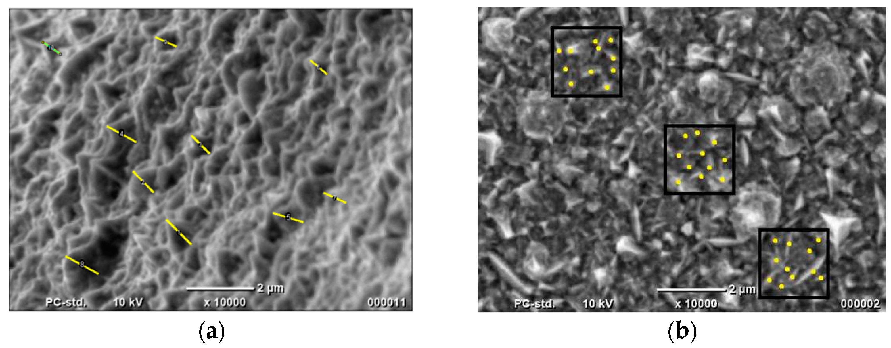
It can be noticed that Co cones were obtained successfully. The chemical composition is as follows: 90.40 at.% Co and 10.60 at.% Cu. The presence of copper is connected with a thin, sputtered copper layer which ensured the conductivity of AAO. To determine appropriately the electrocatalytic properties of samples, the real surface area has to be known. In the case of cones synthesized using one-step method A, the geometrical features were determined using SEM photos as shown in Figure 3.
The height of conical structures was marked by a line at least 10 times and measured as shown in Figure 2a. To determine the number of cones (Figure 2b), several areas were randomly selected, where the ends of the cones were marked with points and the number per 1 μm2 counted.
This allowed us to calculate of the surface of a single cone and then multiply this value by the number of structures. Based on the obtained results (Table 1), the real surface areas were determined for all materials.
The biggest value of the real active surface area is shown by Co cones obtained in one-step method A. However, the determination was approximate in all cases. Firstly, the cones are heterogeneous. Their base is also not a perfectly round one. Nevertheless, this calculation confirmed that the height of Co cones, obtained using one-step method A, is almost tenfold greater than for structures synthesized by the two-step method. The height and the number of nanocones for the two-step method were earlier determined using TEM photos [27,28].
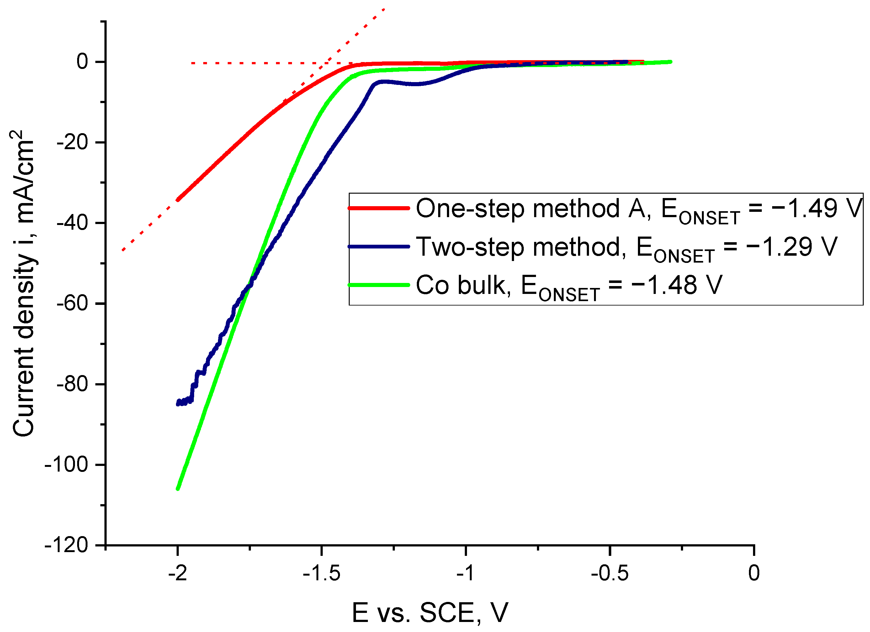
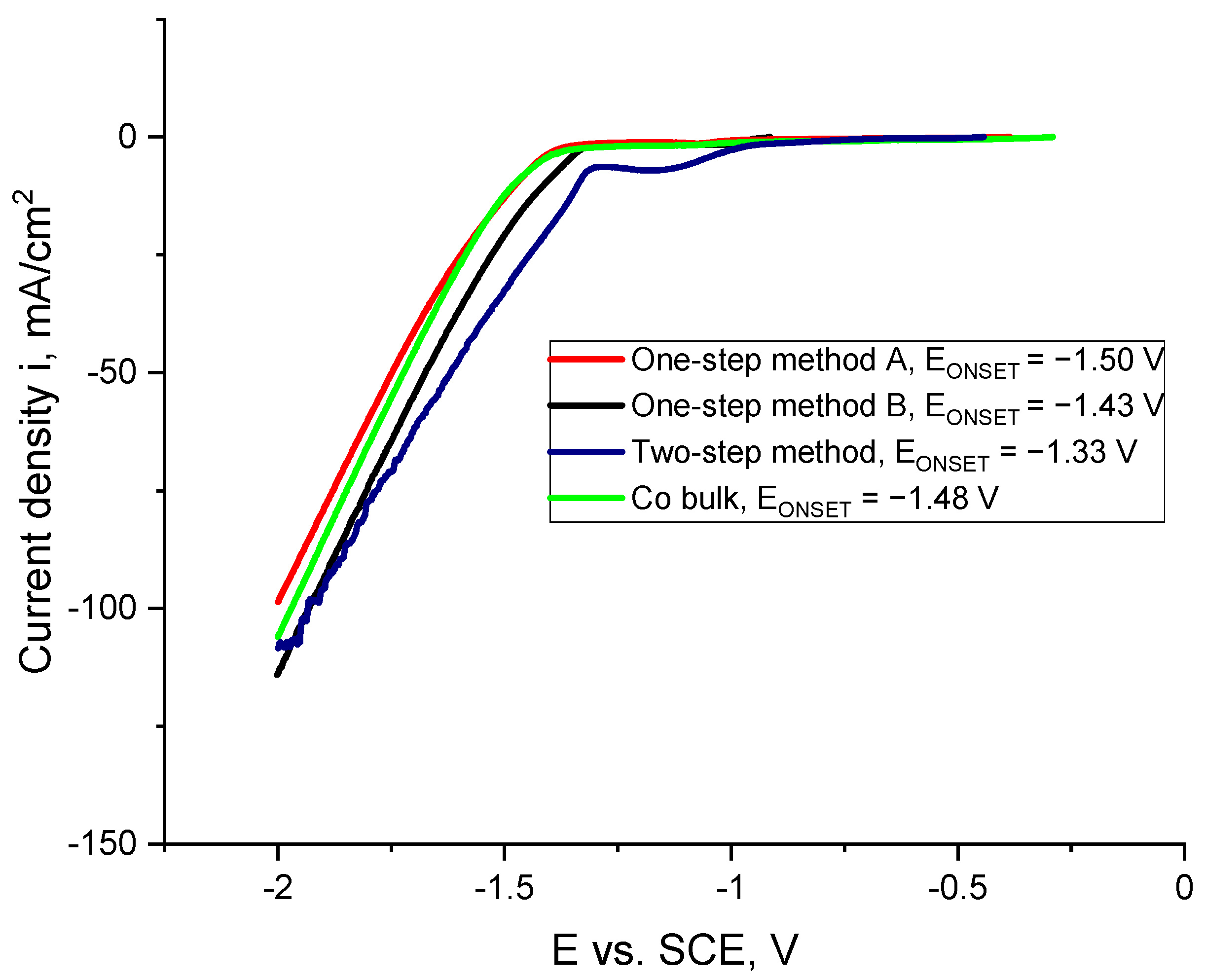
The electrocatalytic properties of samples were measured in 1 M NaOH using the linear scan voltammetry (LSV) technique. The results are shown in Figure 4.
It can be noticed that the Co cones fabricated by one-step methods A show the worst electrocatalytic properties. The sharp character of the curve for Co nanocones is connected with areas on the sample surface blocked by hydrogen bubbles. Two tangents were plotted on Figure 4 (one-step method A). The point of their intersection marked the value of ONSET potential for each sample. The values of determined EONSET are presented in Figure 4.
The hydrogen evolution began the earliest for the Co nanocones obtained using the two-step method. This confirms that the main benefit of nanostructuring is connected with the increase of surface active area and therefore the sample electrocatalytic activity. The noticeable decrease in the case of Co bulk (Figure 4 green curve) is connected with the removal of any oxides from the sample surface. Co nanocones obtained using the AAO template were destroyed during the hydrogen evolution reaction due to the fragile character of the samples.

2.2. Electrodeposition of Co Cones Using Different Conditions—One-Step Method B

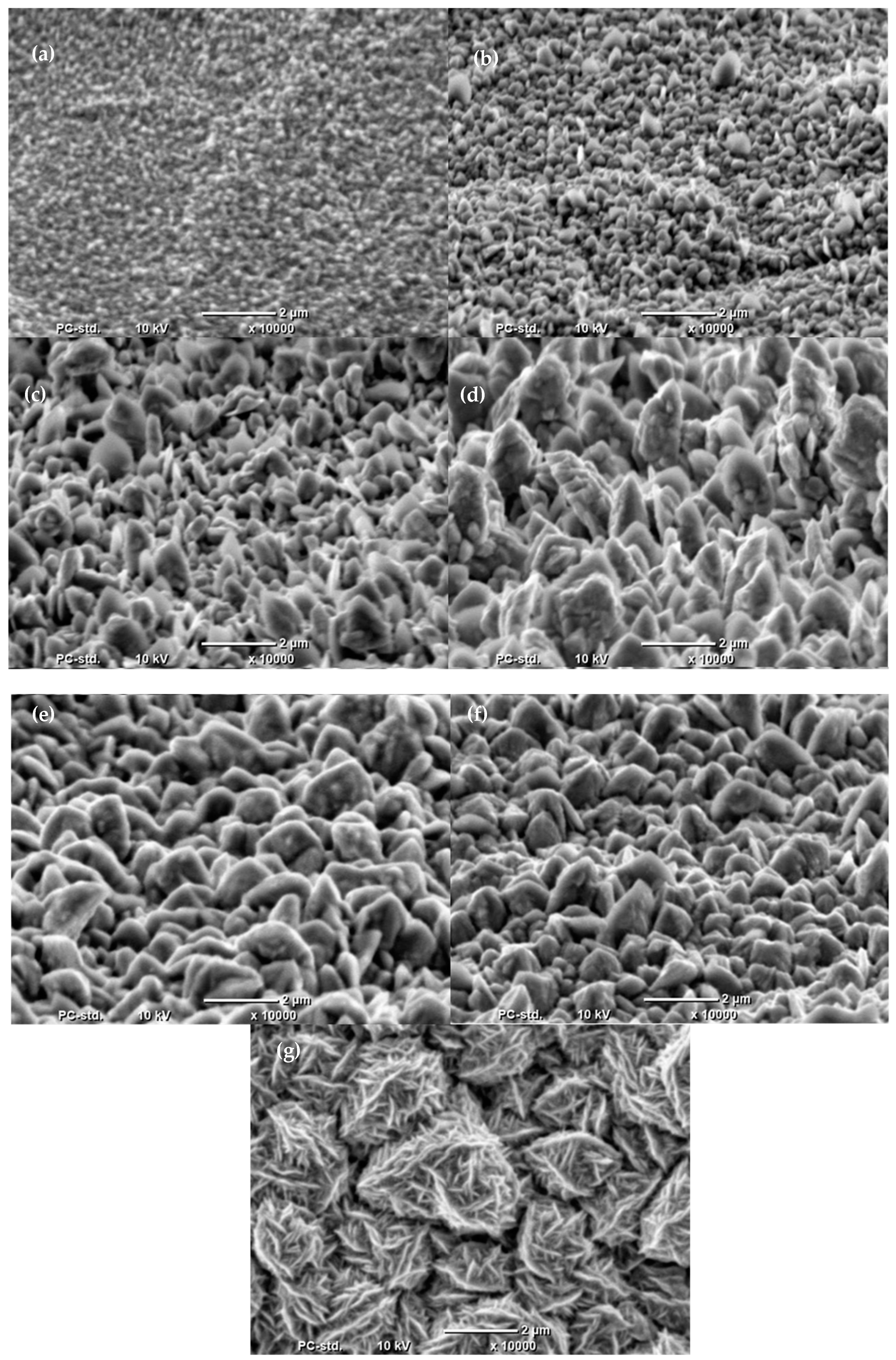
The unsatisfactory shape of cones required us to change the electrodeposition conditions. To achieve this, the influence of electrodeposition time from the electrolyte with new composition on the quality of cones was checked (one-step method B). The results are shown in Figure 5. The obtained results show that there are significant increases in size and active area development of the obtained structures during the electrodeposition time. Deposits synthesized for 40 s (Figure 5d) show a shape closer to a conical one. Further prolongation of electrodeposition time caused the fabrication of shorter cones. All coatings are characterized by structures with not uniform and various values of height. Another prolongation of the electrodeposition time has allowed obtaining complex-shaped structures built by micro-shells, as shown in Figure 5g.
The surface of coatings obtained by one-step method A and B was analyzed using AFM method and compared. The results are shown in Figure 6.
The performed analysis confirmed that the obtained structures are not on the nanoscale in both cases. In the case of one-step method A, the height of the cones is more uniform than for the ones produced by one-step method B. These two methods allowed us to synthesize dense sharp-ended conical structures with high density. The previous calculation of the height of cones obtained by one-step method A gave the height value equal to 0.866 μm. It can be concluded that it is in the range of AFM results. Moreover, to better investigate the sample surface, the roughness of the samples was measured using confocal microscopy. Observations were performed in three different areas of the sample. The first sample was obtained by one-step method A for 20 min at 20 mA/cm2 and the second one by one-step method B for 40 s at 350 mA/cm2. Figure 7 shows these results.
Based on these results, it can be concluded that one-step method A allowed us to obtain a bigger amount of smaller cones than one-step method B. The mean roughness Ra was determined. Its approximate values were as follows: 0.161 μm for Figure 7a and 0.219 μm for Figure 7b. However, due to the measurement accuracy, the approximate value of the maximum profile height Rz was also calculated. It is equal to 0.920 μm for the sample obtained by one-step method A and 1.529 μm for the one obtained by one-step method B. This confirmed that one-step method A allows one to obtain a multitude of small cones. The electrocatalytic properties of these structures were measured as well. The results are shown in Figure 8.
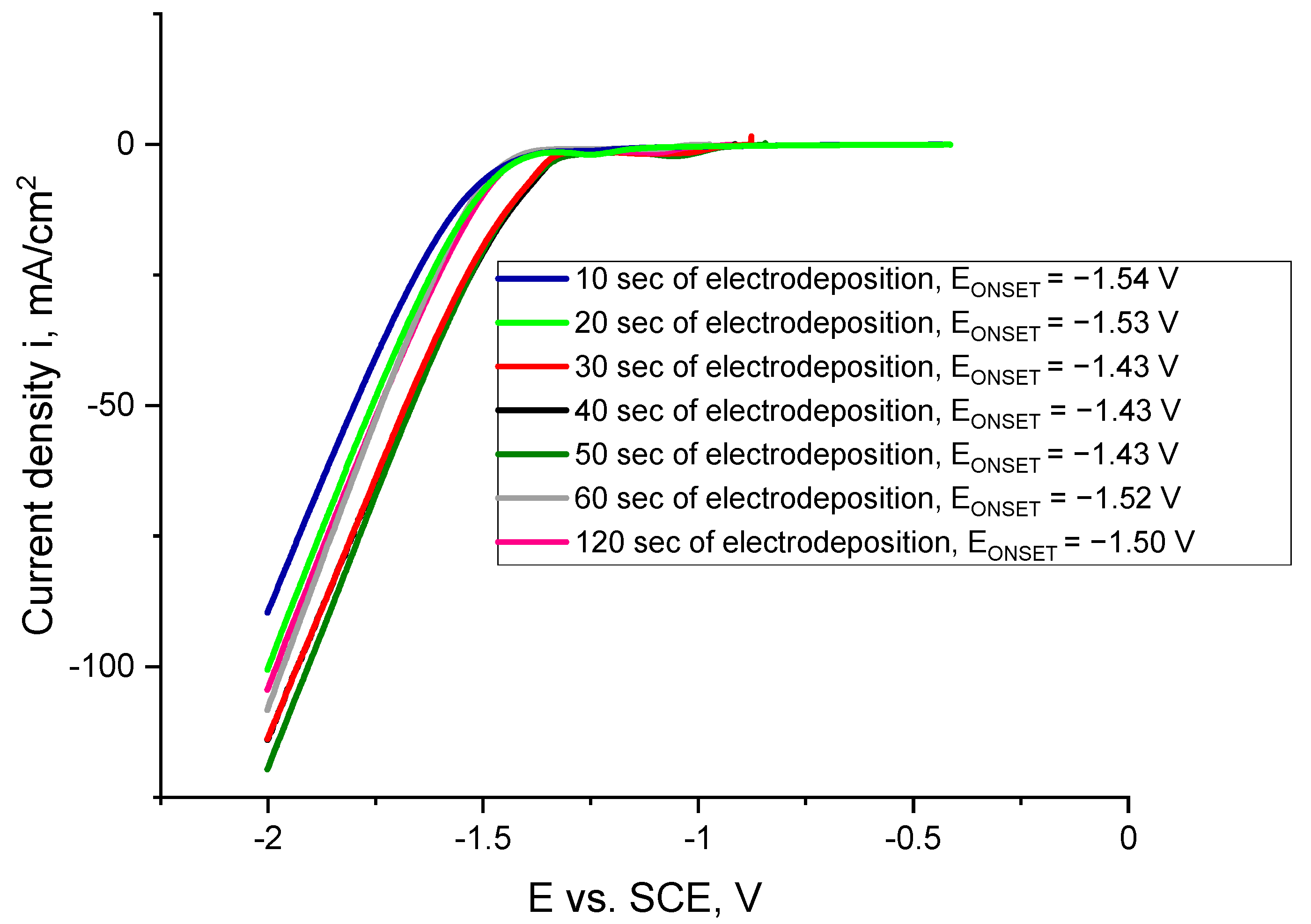
In the case of high compaction of these structures, a problem connected with the determination of active surface area based on SEM photos appeared. Current density is calculated using the Cu substrate surface (2.8 cm2). It can be noticed that there is no influence of the electrodeposition time on the sample properties in the case of hydrogen evolution reaction (HER). For samples obtained after 30, 40 and 50 s this reaction had started at the same potential value.
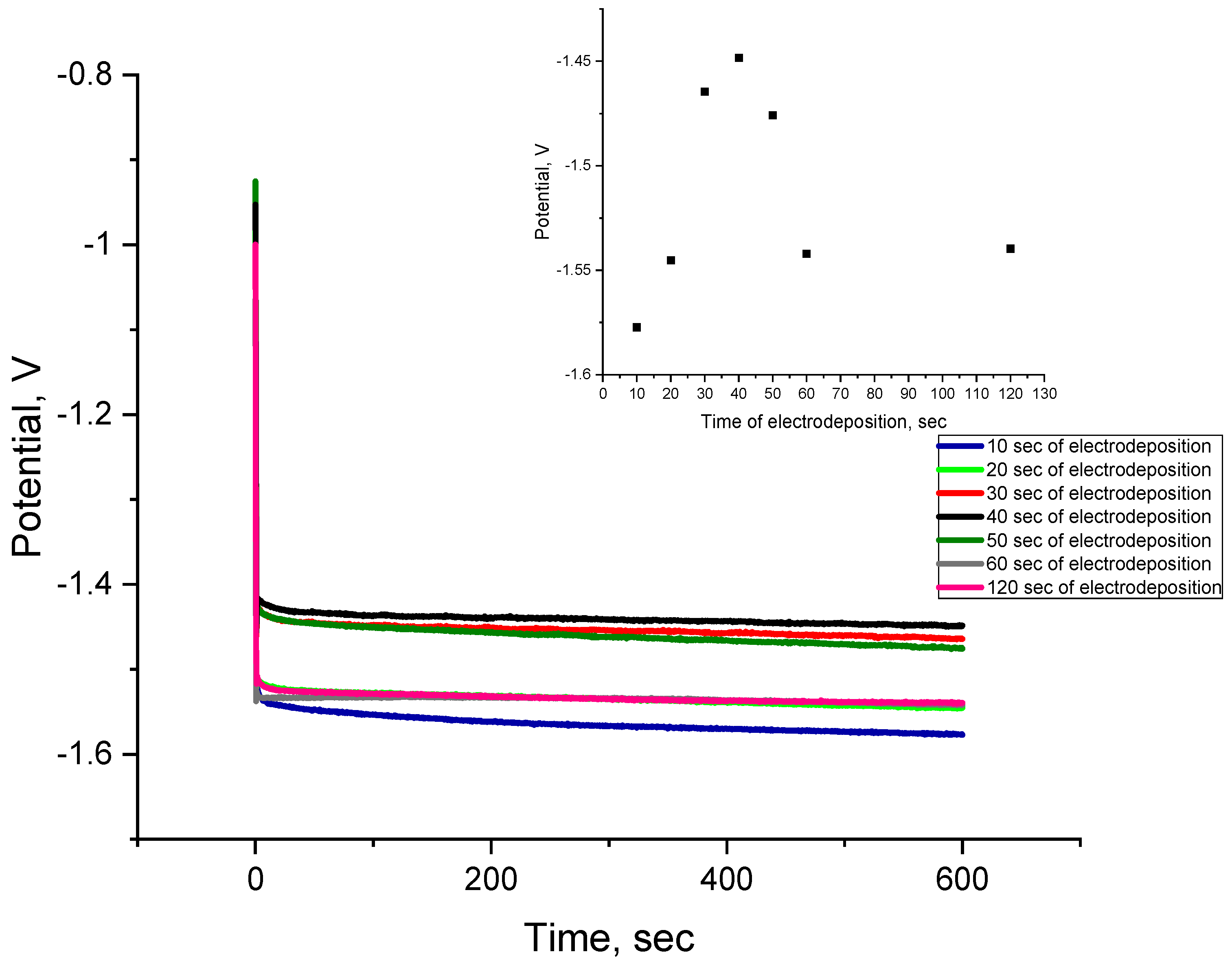
Mechanical stability of cones during electrocatalytic measurements allowed us to perform chronopotentiometry tests for 600 s as well. The results are shown in Figure 9.
The chronopotentiometry results confirm that 40 s of electrodeposition allowed us to obtain the coating characterized by the best electrocatalytic properties. This sample showed the less negative value of potential, which is the response to an applied, constant current.
These results let to conclude that the most appropriate time of electrodeposition, which allows to synthesize the most uniform structures with the highest quality and the best electrocatalytic properties, is 40 s.
To make the results the most comparable with those of cones obtained by the one-step method A, a sample surface area without considering the real one was used. However, it is noticeable that Co cones obtained in new conditions (one-step method B) are the largest ones in comparison with previous ones described in Section 2.1. The results are shown in Figure 10.
The different character of the curves in comparison with Figure 4 is connected with the surface area. In this case, the EONSET values were also determined and showed in Figure 4. The results show that for the sample obtained by one-step method B the hydrogen evolution reaction started at a less negative potential than for the previous sample produced by the same method. It is connected with more developed surface of the deposit.
In the case of different compositions of used electrolytes in one-step method the composition of obtained structures was analyzed using EDS analysis and compared. The results are shown in Table 2.
It can be noticed that all layers obtained using various methods allowed us to obtain Co cones and nanocones. Obviously, the presence of Cl in the samples synthesized by the one-step method is connected with the addition of NH4Cl and the adsorption of Cl ions on the surface of the deposit. The lowest amount of Co was obtained for cones produced by one-step method A. This is interesting due to the fact that this electrolyte contained more Co. The concentration of chloride, in this case, is also two times higher.
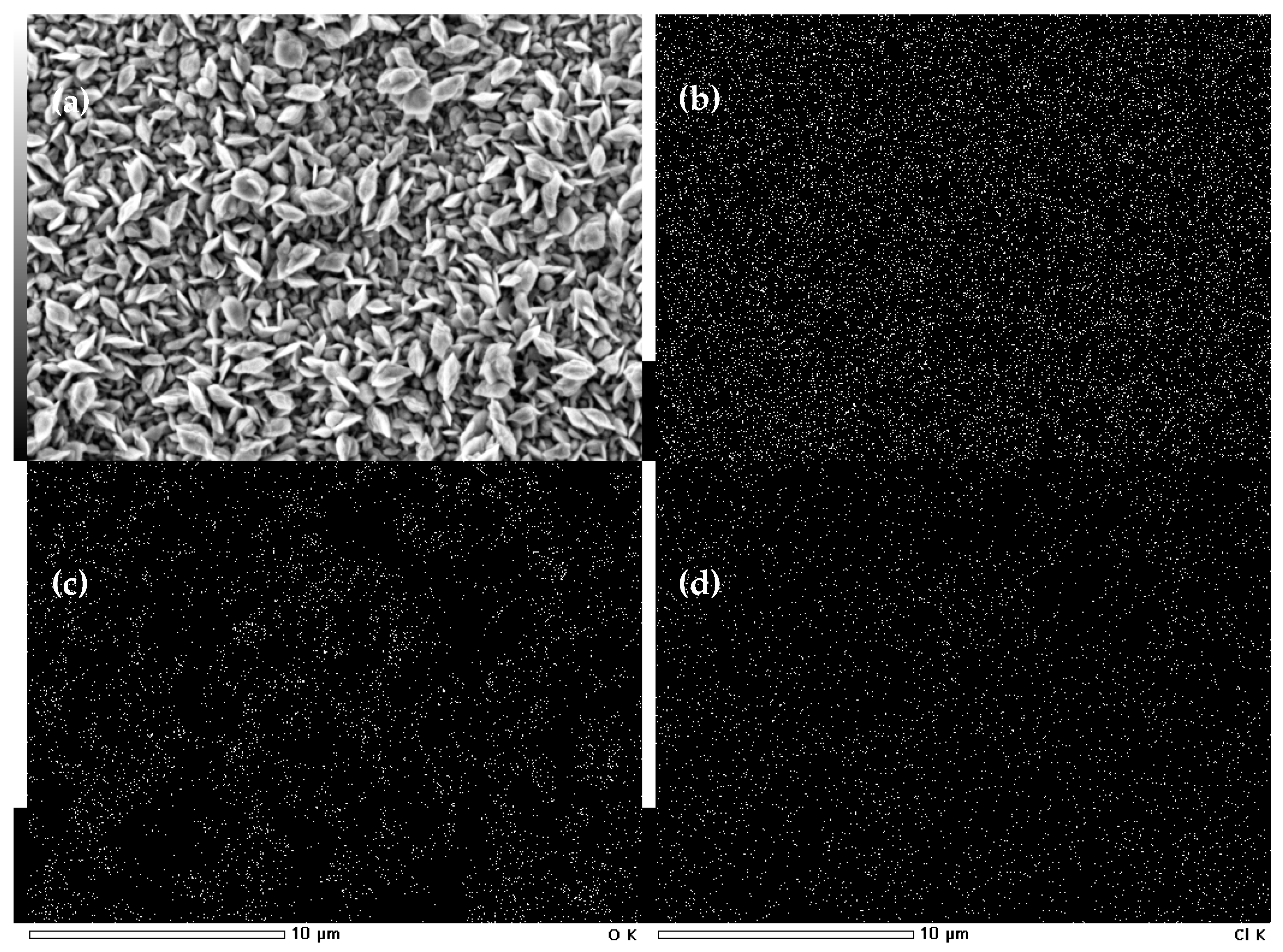
The mapping analysis for the sample obtained by one-step method B was also performed. Results are shown in Figure 11.
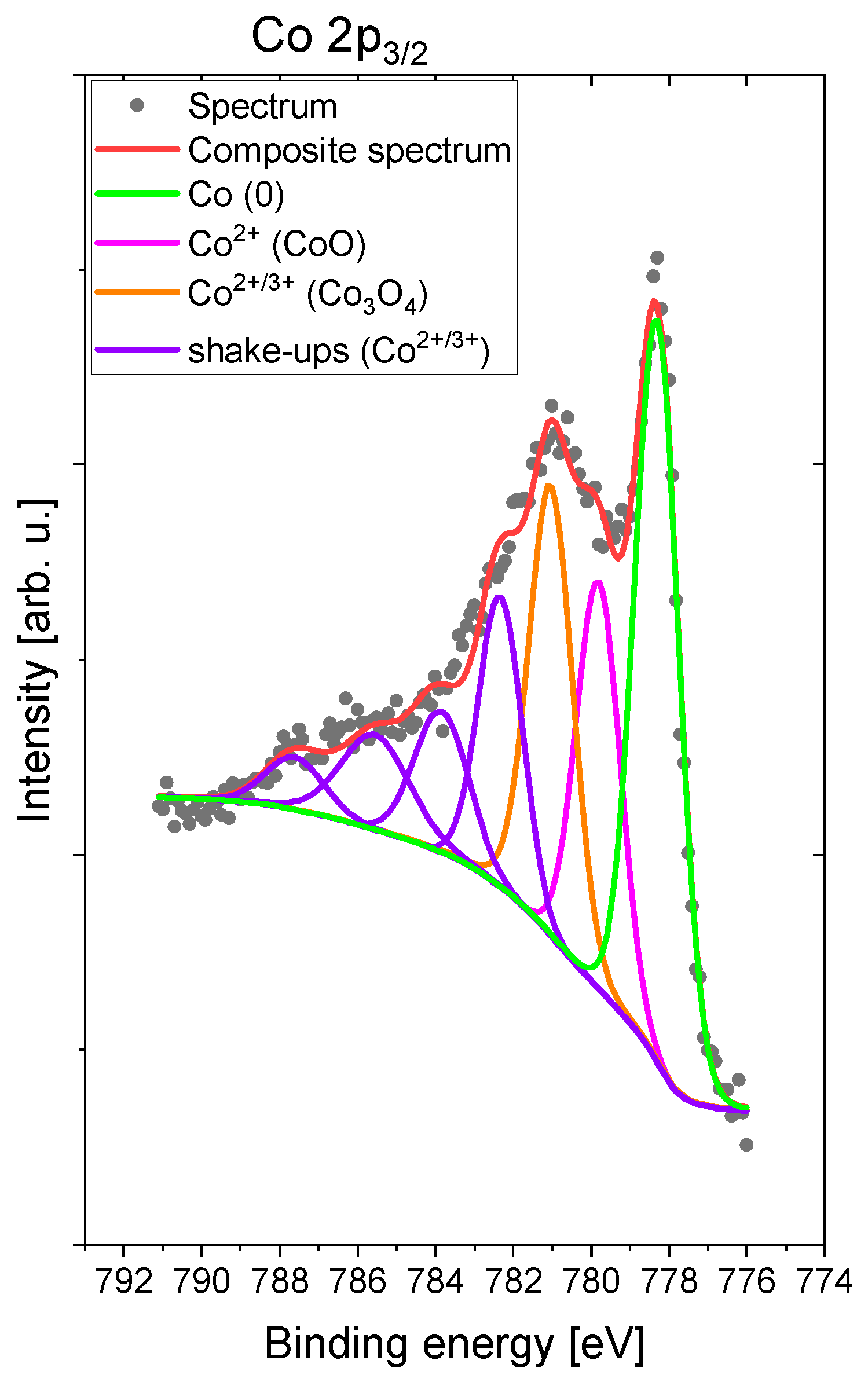
These results indicate that the distribution of Co is uniform for this sample. Also, in the case of chloride, the same tendency is observed. However, in the case of oxygen, the distribution is non-uniform. Some areas characterized by lower content of O are observed, which can be connected with oxidation of more active areas. The surface of this sample was also investigated using X-ray photoelectron spectroscopy (XPS). The result is shown in Figure 12.
For the XPS analyzed sample Co 2p3/2 spectra show the multiplet splitting structure characteristic for first-row transition metal species containing unpaired electrons. From the inspection of line positions the first peak centered at 778.1 eV represents the metallic part of Co, lines at 780 and 781 eV and the very high-intensity shake-up satellites (782–778 eV) indicate that the cobalt is in both Co3+ and Co2+ oxidation states like in Co3O4 and CoO [29,30,31]. The relative concentrations of cobalt species present on analyzed sample surfaces are as follows: 332.5 Co (0), 32.4 CoO and 35.1 Co3O4. This analysis confirms that on the obtained surface there is a mixture of metallic and different oxide states. However, the content of oxides responsible for hydrogen evolution reaction varies.

3. Materials and Methods

3.1. Preliminary Studies on Cobalt Electrodeposition

Fabrication of cobalt cones by the one-step method consisted of the electrodeposition from an electrolyte with the addition of NH4Cl as a capping reagent. A substrate was a copper foil. The electrodeposition was carried out galvanostatically with a Pt sheet as a counter electrode. The used electrolyte has the composition as follows: 200 g/L CoCl2, 100 g/L H3BO3, 100 g/L NH4Cl with neutral pH, the temperature was equal to 60 °C. The applied current density was equal to 20 mA/cm2, the coatings were deposited in 20 min. The electrodeposition process performed in the following conditions was entitled: one-step method A.
In the case of two-step method, the electrodeposition process was performed using an AAO template obtained by two-step anodization. Firstly, long-period anodization of an aluminium AA1050 sample was conducted in 0.3 M oxalic acid at 2 °C at 45 V for 1 h. This process is called the first step of anodization. Then, obtained Al2O3 layer was removed for 1 h at 60 °C using a mixture of 1.6 wt% chromic acid and 6 wt% phosphoric acid. The second step of anodization consisted of alternating short-anodization and pore widening process. Short-anodization was performed in 0.3 M H2C2O4 at 9 °C at 45 V for 25 s and 20 s for the first step and next ones. Pores widening process was carried out in 5 wt% phosphoric acid solution for 12 min at 30 °C. Four alternating cycles were performed to obtain the template.
For two-step method and fabrication of the bulk sample, the same electrodeposition conditions as in one-step method A were applied. In these cases, there was no addition of NH4Cl to the electrolyte.
The free-standing Co nanocones were obtained by removing the template in a dilute NaOH solution. In the case of bulk material, the copper sample was also the substrate. The surface area for all materials was 2.8 cm2.
SEM photos of synthesized materials were taken by 6000 Plus (JEOL, Tokyo, Japan) and SU-70 (Hitachi, Tokyo, Japan) scanning electron microscopes and used for the determination of real active surface area. To compare the compositions of samples, EDS analysis was applied through the same microscope. The sample surface was analyzed by atomic force microscopy (AFM, NtegraAura microscope, NT MDT, Moscow, Russia) equipped with an NSG03 tip. The sample roughness was determined using a confocal microscope (LEXT OLS4100, Olympus, Tokyo, Japan). The XPS analyses were carried out in a VersaProbe II Scanning XPS system (PHI, Chigasaki, Japan) using monochromatic Al Kα (1486.6 eV) X-rays focused to 100 µm spot and scanned over the area of 400 µm × 400 µm. The photoelectron take-off angle was 45° and the pass energy in the analyzer was set to 117.50 eV (0.5 eV step) for survey scans and 46.95 eV (0.1 eV step) to obtain high energy resolution spectra for the C 1s, O 1s, N 1s and Re 4f regions. A dual-beam charge compensation with 7 eV Ar+ ions and 1 eV electrons was used to maintain a constant sample surface potential regardless of the sample conductivity. All XPS spectra were charge referenced to the unfunctionalized, saturated carbon (C–C) C 1s peak at 285.0 eV. The operating pressure in the analytical chamber was less than 3 × 10⁻⁷ mbar. Deconvolution of spectra was carried out using PHI MultiPak software (v.9.9.0.8). Spectrum background was subtracted using the Shirley method.
Electrocatalytic properties in the hydrogen evolution process of produced deposits were measured using a SP-300 potentiostat (Bio-Logic, Seyssinet-Pariset, France). The linear scan voltammetry (LSV) measurements were performed in a range from the open circuit value to −2 V vs a saturated calomel electrode (SCE) with a scan speed equal to 5 mV/s. In the three-electrode cell, the Co layer was the working electrode, a saturated calomel electrode SCE was the reference electrode and a Pt sheet was the counter one. The process was carried out at room temperature in 1 M NaOH. The values of EONSET connected with starting of hydrogen reaction were determined from the figures.
Moreover, Co cones obtained by one-step method in new conditions were tested by chronopotentiometry (CP) measurements for 600 s at a constantly current density of—10 mA/cm2. This measurement allowed to determine voltage value, which can be connected with the overpotential of water splitting reaction.

3.2. One-Step Method B

In this case, the Cu foil was used as a substrate and the electrodeposition process was carried out galvanostatically with a Pt sheet as a counter electrode as well. The differences were in used current density, the temperature of electrolyte and amounts of components in the solutions. This method was called one-step method B. The pH of the solution in both cases was equal to 4. The differences in conditions for both methods are shown in Table 3.

4. Conclusions

There is the possibility to produce cobalt cones by one-step and nanocones by a two-step method using electrolytes with the same composition. Co cones obtained by one-step A shows greater geometrical size, although many microshell structures are visible. SEM photos were used to determine the value of the real active surface area.
Co cones, synthesized by one-step method A, show the worst electrocatalytic properties. This could be due to the assumptions during the determination of the active surface area. For cobalt nanocones produced by two-step anodization, the hydrogen evolution reaction started the earliest.
The Co nanocones layer was destroyed by the hydrogen bubbles produced during this reaction. This can be noticed in the sharp character of the curve.
The change of electrodeposition conditions in one-step method B allowed us to obtain structures with shapes closer to conical ones.
Forty seconds of electrodeposition process ensure the fabrication of deposits with the most uniform cones in the case of height. Further prolongation of the process time caused the synthesis of complex, micro-shell structures.
The results of AFM analysis and surface roughness measurements show that one-step method A allowed synthesizing a multitude of smaller cones in comparison with one-step method B. This confirmed that produced cones were microsized. Cones obtained by the one-step method B show better electrocatalytic properties than previous ones.
All performed experiments allowed us to produce Co deposits with a high atomic percentage of Co. The distribution of Co, Cl on the sample surface is uniform. However, XPS analysis confirms that the sample surface the mixture of metallic and different oxide states.

Author Contributions

K.S. preparation of the AAO templates, electrodeposition of Co by one-step method from two electrolytes, writing—original draft preparation. K.K.-S. SEM analysis. D.K. measurement of electrocatalytic properties. A.J. electrodeposition of Co. B.L.-M. Confocal Microscope analysis. M.M.M. XPS analysis. P.Z. Supervision, writing—review and editing. All authors have read and agreed to the published version of the manuscript.

Funding

This research was funded by the Polish National Centre of Science, grant number UMO-2016/23/G/ST5/04058.

Institutional Review Board Statement

Not applicable.

Informed Consent Statement

Not applicable.

Data Availability Statement

Data sharing not applicable.

Acknowledgments

The authors would like to express their appreciation to Grzegorz Włoch (Faculty of Non-Ferrous Metals, AGH University of Science and Technology) for performing SEM and EDS analysis.

Conflicts of Interest

The authors declare no conflict of interest. The funders had no role in the design of the study; in the collection, analyses, or interpretation of data; in the writing of the manuscript, or in the decision to publish the results.

References

  1. Nazeruddin, G.M.; Prasad, R.N.; Shaikh, Y.I.; Shaikh, A.A. Synergetic effect of Ag-Cu bimetallic nanoparticles on antimicrobial activity. Pharm. Lett. 2014, 6, 129–136. [Google Scholar]
  2. Mathew, A.; Munichandraiah, N.; Mohan Rao, G. Synthesis and magnetic studies of flower-like nickel nanocones. Mater. Sci. Eng. B Solid State Mater. Adv. Technol. 2009, 158, 7–12. [Google Scholar] [CrossRef]
  3. Nagaura, T.; Takeuchi, F.; Yamauchi, Y.; Wada, K.; Inoue, S. Fabrication of ordered Ni nanocones using a porous anodic alumina template. Electrochem. Commun. 2008, 10, 681–685. [Google Scholar] [CrossRef]
  4. Kurowska, E.; Brzózka, A.; Jarosz, M.; Sulka, G.D.; Jaskuła, M. Silver nanowire array sensor for sensitive and rapid detection of H2O2. Electrochim. Acta 2013, 104, 439–447. [Google Scholar] [CrossRef]
  5. Abdel-Aal, E.A.; Malekzadeh, S.M.; Rashad, M.M.; El-Midany, A.A.; El-Shall, H. Effect of synthesis conditions on preparation of nickel metal nanopowders via hydrothermal reduction technique. Powder Technol. 2007, 171, 63–68. [Google Scholar] [CrossRef]
  6. Jia, F.; Zhang, L.; Shang, X.; Yang, Y. Non-aqueous sol-gel approach towards the controllable synthesis of nickel nanospheres, nanowires, and nanoflowers. Adv. Mater. 2008, 20, 1050–1054. [Google Scholar] [CrossRef]
  7. Zavedeev, E.V.; Petrovskaya, A.V.; Simakin, A.V.; Shafeev, G.A. Formation of nanostructures upon laser ablation of silver in liquids. Quantum Electron. 2006, 36, 978–980. [Google Scholar] [CrossRef]
  8. Choi, J.; Wehrspohn, R.B.; Gösele, U. Moiré pattern formation on porous alumina arrays using nanoimprint lithography. Adv. Mater. 2003, 15, 1531–1534. [Google Scholar] [CrossRef]
  9. Mech, K.; Gajewska, M.; Marzec, M.; Szaciłowski, K. On the influence of magnetic field on electrodeposition of Ni–TiO2 composites from a citrate baths. Mater. Chem. Phys. 2020, 255, 123550. [Google Scholar] [CrossRef]
  10. Kwiecińska, A.M.; Kutyła, D.; Kołczyk-Siedlecka, K.; Skibińska, K.; Żabiński, P.; Kowalik, R. Electrochemical analysis of co-deposition cobalt and selenium. J. Electroanal. Chem. 2019, 848, 113278. [Google Scholar] [CrossRef]
  11. Kutyła, D.; Kołczyk-Siedlecka, K.; Kwiecińska, A.; Skibińska, K.; Kowalik, R.; Żabiński, P. Preparation and characterization of electrodeposited Ni-Ru alloys: Morphological and catalytic study. J. Solid State Electrochem. 2019, 23, 3089–3097. [Google Scholar] [CrossRef] [Green Version]
  12. Rahimi, E.; Davoodi, A.; Kiani Rashid, A.R. Characterization of screw dislocation-driven growth in nickel micro-nanostructure electrodeposition process by AFM. Mater. Lett. 2018, 210, 341–344. [Google Scholar] [CrossRef]
  13. Lee, J.M.; Jung, K.K.; Lee, S.H.; Ko, J.S. One-step fabrication of nickel nanocones by electrodeposition using CaCl2·2H2O as capping reagent. Appl. Surf. Sci. 2016, 369, 163–169. [Google Scholar] [CrossRef]
  14. Barati Darband, G.; Aliofkhazraei, M.; Sabour Rouhaghdam, A. Nickel nanocones as efficient and stable catalyst for electrochemical hydrogen evolution reaction. Int. J. Hydrog. Energy 2017, 42, 14560–14565. [Google Scholar] [CrossRef]
  15. Du, Z.; Zhang, S.; Xing, Y.; Wu, X. Nanocone-arrays supported tin-based anode materials for lithium-ion battery. J. Power Sources 2011, 196, 9780–9785. [Google Scholar] [CrossRef]
  16. Xia, Y.; Mo, X.; Ling, H.; Hang, T.; Li, M. Facile fabrication of Au nanoparticles-decorated Ni nanocone arrays as effective surface-enhanced Raman Scattering substrates. J. Electrochem. Soc. 2016, 163, D575–D578. [Google Scholar] [CrossRef]
  17. Tian, F.; Hu, A.; Li, M.; Mao, D. Superhydrophobic nickel films fabricated by electro and electroless deposition. Appl. Surf. Sci. 2012, 258, 3643–3646. [Google Scholar] [CrossRef]
  18. Li, Y.; Zhang, X.; Hu, A.; Li, M. Morphological variation of electrodeposited nanostructured Ni-Co alloy electrodes and their property for hydrogen evolution reaction. Int. J. Hydrog. Energy 2018, 3. [Google Scholar] [CrossRef]
  19. Xiao, H.; Hu, A.; Hang, T.; Li, M. Electrodeposited nanostructured cobalt film and its dual modulation of both superhydrophobic property and adhesiveness. Appl. Surf. Sci. 2015, 324, 319–323. [Google Scholar] [CrossRef]
  20. Hashemzadeh, M.; Raeissi, K.; Ashrafizadeh, F.; Khorsand, S. Effect of ammonium chloride on microstructure, super-hydrophobicity and corrosion resistance of nickel coatings. Surf. Coat. Technol. 2015, 283, 318–328. [Google Scholar] [CrossRef]
  21. Brzózka, A.; Szeliga, D.; Kurowska-Tabor, E.; Sulka, G.D. Synthesis of copper nanocone array electrodes and its electrocatalytic properties toward hydrogen peroxide reduction. Mater. Lett. 2016, 174, 66–70. [Google Scholar] [CrossRef]
  22. Li, Y.; Guo, S. Noble metal-based 1D and 2D electrocatalytic nanomaterials: Recent progress, challenges and perspectives. Nano Today 2019, 28, 100774. [Google Scholar] [CrossRef]
  23. Chaudhari, N.K.; Jin, H.; Kim, B.; Lee, K. Nanostructured materials on 3D nickel foam as electrocatalysts for water splitting. Nanoscale 2017, 9, 12231–12247. [Google Scholar] [CrossRef] [PubMed]
  24. Daulbayev, C.; Sultanov, F.; Bakbolat, B.; Daulbayev, O. 0D, 1D and 2D nanomaterials for visible photoelectrochemical water splitting. A Review. Int. J. Hydrogen Energy 2020, 45, 33325–33342. [Google Scholar] [CrossRef]
  25. Liu, J.; Fang, X.; Zhu, C.; Xing, X.; Cui, G.; Li, Z. Fabrication of superhydrophobic coatings for corrosion protection by electrodeposition: A comprehensive review. Colloids Surfaces A Physicochem. Eng. Asp. 2020, 607, 125498. [Google Scholar] [CrossRef]
  26. Wu, B.; Lu, S.; Xu, W.; Cui, S.; Li, J.; Han, P.F. Study on corrosion resistance and photocatalysis of cobalt superhydrophobic coating on aluminum substrate. Surf. Coat. Technol. 2017, 330, 42–52. [Google Scholar] [CrossRef]
  27. Skibinska, K.; Smola, G.; Bialo, L.; Kutyla, D.; Kolczyk-Siedlecka, K.; Kwiecinska, A.; Wojnicki, M.; Zabinski, P. Influence of Annealing Time of Aluminum AA1050 on the Quality of Cu and Co Nanocones. J. Mater. Eng. Perform. 2020, 29, 8025–8035. [Google Scholar] [CrossRef]
  28. Skibinska, K.; Kołczyk-Siedlecka, K.; Kutyła, D.; Gajewska, M.; Zabinski, P. Synthesis of Co—Fe 1D Nanocone Array Electrodes Using Aluminum Oxide Template. Materials 2021, 14, 1717. [Google Scholar] [CrossRef]
  29. Biesinger, M.C.; Payne, B.P.; Grosvenor, A.P.; Lau, L.W.M.; Gerson, A.R.; Smart, R.S.C. Resolving surface chemical states in XPS analysis of first row transition metals, oxides and hydroxides: Cr, Mn, Fe, Co and Ni. Appl. Surf. Sci. 2011, 257, 2717–2730. [Google Scholar] [CrossRef]
  30. Okamoto, Y.; Adachi, T.; Maezawa, A.; Imanaka, T. Effect of ZnO Addition on Cobalt–Alumina Interaction Species. Bull. Chem. Soc. Jpn. 1991, 64, 236–242. [Google Scholar] [CrossRef] [Green Version]
  31. Dash, K.C.; Folkesson, B.; Larsson, R.; Mohapatra, M. An XPS investigation on a series of schiff base dioxime ligands and cobalt complexes. J. Electron Spectros. Relat. Phenomena 1989, 49, 343–357. [Google Scholar] [CrossRef]
Figure 1. SEM photos of (a) bulk material and Co cones synthesized using (b) one-step method and (c) two-step method. Magnification: ×10,000.
Catalysts 11 00544 g001
Figure 2. (a) SEM photo of Co nanocones and (b) mapping analysis spectra.
Catalysts 11 00544 g002
Figure 3. Determination of (a) cones height and (b) number of cones using SEM photos.
Catalysts 11 00544 g003
Figure 4. Linear sweep voltammetry (LSV) curves of Co bulk and Co nanocones obtained using one-step and two-step method in 1 M NaOH solution.
Catalysts 11 00544 g004
Figure 5. Co cones obtained using one-step method B after (a) 10 s, (b) 20 s, (c) 30 s, (d) 40 s, (e) 50 s, (f) 60 s and (g) 120 s of electrodeposition.
Catalysts 11 00544 g005
Figure 6. AFM analysis of sample obtained by one-step method (a) A for 20 min at 20 mA/cm2 and (b) B for 40 s at 350 mA/cm2.
Catalysts 11 00544 g006
Figure 7. The surface roughness analysis of sample obtained by one-step method A (a) area 1, (b) area 2, (c) area 3 and B (d) area 1, (e) area 2 and (f) area 3.
Catalysts 11 00544 g007
Figure 8. Linear sweep voltammetry (LSV) curves of Co cones obtained after various time of electrodeposition using one-step method B in 1 M NaOH solution.
Catalysts 11 00544 g008
Figure 9. Results of chronopotentiometry measurements for samples electrodeposited for various time.
Catalysts 11 00544 g009
Figure 10. LSV curves of Co bulk and Co cones obtained using one-step in different conditions and two-step method in 1 M NaOH solution.
Catalysts 11 00544 g010
Figure 11. Mapping analysis of obtained coating: (a) sample surface image and distribution of: (b) Co, (c) O and (d) Cl.
Catalysts 11 00544 g011
Figure 12. XPS spectrum of sample obtained by one-step method B.
Catalysts 11 00544 g012
Table 1. Determined values of real active surface area for all samples.
MaterialHeight [nm]Number of Cones per 1 μm2Real Active Surface Area [cm2]
Co cones obtained in one-step method8662.758.05
Co nanocones obtained in two-step method74 ± 1683.57
Co bulk--2.80
Table 2. Comparison of deposit compositions.
SampleComposition of Deposit [% at.]
CoOCl
Co cones obtained by one-step method A90.837.661.51
Co nanocones obtained by two-step method95.594.41-
Co bulk93.286.72-
Co cones obtained by one-step method B96.243.140.62
Table 3. New conditions of the electrodeposition process in one-step method B.
Conditions of the Electrodeposition Process
One-Step Method AOne-Step Method B
Electrolyte composition200 g/L CoCl2, 100 g/L H3BO3, 100 g/L NH4Cl100 g/L CoCl2, 35 g/L H3BO3, 100 g/L NH4Cl
Current density i [mA/cm2]20350
Temperature [°C]6065
Publisher’s Note: MDPI stays neutral with regard to jurisdictional claims in published maps and institutional affiliations.