Article

Purification and Structural Characterization of the Auxiliary Activity 9 Native Lytic Polysaccharide Monooxygenase from Thermoascus aurantiacus and Identification of Its C1- and C4-Oxidized Reaction Products

by
Weishuai Yu
1,†,
Imran Mohsin
2,3,†,
Anastassios C. Papageorgiou
2,3,* and
Duochuan Li
1,*
1
Department of Mycology, Shandong Agricultural University, Taian 271018, China
2
Turku Bioscience Centre, University of Turku, 20521 Turku, Finland
3
Turku Bioscience Centre, Åbo Akademi University, 20521 Turku, Finland
*
Authors to whom correspondence should be addressed.
These authors contributed equally to this work.
Catalysts 2022, 12(2), 139; https://doi.org/10.3390/catal12020139
Submission received: 22 November 2021 / Revised: 12 January 2022 / Accepted: 18 January 2022 / Published: 23 January 2022
(This article belongs to the Special Issue Enzyme Catalysis, Biotransformation and Bioeconomy)

Abstract

:
Auxiliary activity 9 (AA9) lytic polysaccharide monooxygenases (LPMOs) are copper-dependent oxidoreductases that use O2 or H2O2 to perform oxidative cleavage of cellulose in the presence of an electron donor. Combined with cellulases, they can assist in a more efficient cleavage of cellulose. AA9 LPMOs have therefore attracted considerable attention in recent years for use in biotechnological applications. Here, a native AA9 LPMO (nTaAA9A) from the thermophilic fungus Thermoascus aurantiacus was purified and characterized. The enzyme was shown to be active and able to cleave cellulose and xylan to produce C1- and C4-oxidized products. It was also found to retain about 84.3, 63.7, and 35.3% of its activity after incubation for 30 min at 60, 70, and 80 °C, respectively, using quantitative activity determination. The structure was determined to 1.36 Å resolution and compared with that of the recombinant enzyme expressed in Aspergillus oryzae. Structural differences in the glycosylated Asn138 and in solvent-exposed loops were identified.

1. Introduction

Cellulose is a polysaccharide consisting of a linear chain of glucose units connected through 1,4-β-glycosidic bonds. As the most abundant renewable organic compound on earth, cellulose can be turned into economically viable biofuels by enzymatic degradation [1]. A significant milestone in the enzymatic degradation of cellulose was the discovery of lytic polysaccharide monooxygenases (LPMOs) that catalyze the cleavage of 1,4-β-glycosidic bonds in cellulose. Contrary to cellulases which, as glycoside hydrolases (GHs), catalyze the breakage of 1,4-β-glycosidic bonds in cellulose via a hydrolytic mechanism, LPMOs follow an oxidative mechanism [2,3,4]. Extensive studies in LPMOs have led to their further classification as Auxiliary Activity (AA) enzymes in the Carbohydrate Active enZymes (CAZy; www.cazy.org; accessed on 20 November 2021) database [5], where they form eight separate families (AA9–AA11, AA13–AA17) [6,7]. LPMOs of fungal origin are found in the families AA9 (formerly GH61) and AA11.
The discovery of LPMOs has revolutionized the enzymatic degradation of cellulose because LPMOs can cleave crystalline cellulose and allow cellulases, which interact with single cellulose chains, to hydrolyze cellulose more efficiently [4,8,9]. Apart from cellulose degradation, LPMOs have also been suggested to play a role in various other biological processes, such as bacterial pathogenicity [10] and viral virulence [11].
LPMOs are copper-dependent oxidoreductases that employ O2 or H2O2 to carry out oxidative cleavage of cellulose in the presence of an electron donor [4,8,9,12,13,14,15]. LPMOs use a cellulose degradation mechanism different from that of cellulases, as they lack a conserved carboxylate pair and an active site groove [16]. The reaction proceeds through an oxidative step that involves the hydroxylation of crystalline cellulose at the C1 or C4 carbon, leading to the subsequent cleavage of the glycosidic bond.
Crystal structures of ~30 LPMOs are currently known [16], including those of Thermoascus aurantiacus TaAA9A (PDB id 2yet) [17], Aspergillus fumigatus AfuAA9A (PDB id 6h1z) [18], Serratia marcescens SmAA10A (PDB id 2bem) [19], Aspergillus oryzae AoAA13 (PDB id 4opb) [20], and recently, McAA9F (PDB id 7ntl) [21] from the thermophilic fungus Malbranchea cinnamomea. The available LPMO structures have revealed a planar surface suitable for binding crystalline cellulose and the presence of a single copper ion located at the center of the planar surface [4,12]. A remarkable catalytic characteristic of LPMOs is the difference in regioselectivity of cellulose oxidative cleavage. It has been shown that LPMOs are able to cleave cellulose by C1 and C4 oxidation to form non-oxidized and oxidized cello-oligosaccharides [17,22,23].
LPMOs are also capable of breaking down xylan. Fungal AA9 LPMOs, such as LsAA9A from Lentinus similis [12] and MtLPMO9A from Myceliophthora thermophila [24] have been shown to cleave xylan. Xylan-active AA14 LPMO from Pycnoporus coccineus, PcAA14B, and GH30 TtXyn30A from Thermothelomyces thermophila were found to act synergistically with a family GH11 endoxylanase (AnXyn11) in the degradation of xylan-containing substrates, resulting in an increase of the released total oligosaccharides [25]. A synergistic action of keratinases with LPMOs has also been proposed [26], thus offering additional strategies to improve keratinase performance.
Various protein engineering efforts have been carried out to improve activity and thermostability of LPMOs. For example, a tetramutant in AfAA9A_B with remarkable improvement in biomass conversion at elevated temperatures has been reported [18]. Genomic sequencing has shown a number of AA9 LPMOs in thermophilic fungi [27,28,29]. AA9 LPMOs from thermophilic fungi are potentially more thermostable than those from mesophilic fungi; thus, they have received increased attention in recent years [17,28]. The recombinant AA9 LPMO (rTaAA9A) from the thermophilic fungus Thermoascus aurantiacus is well characterized, and its C1- and C4-oxidized products were previously identified based on mass spectrometry analysis [17] and sequence and phylogenetic analysis [27]. Here, we report the purification and characterization of the native TaAA9A (nTaAA9A). The C1- and C4-oxidized products of nTaAA9A were identified, and the thermostability of the enzyme was studied using quantitative activity determination. nTaAA9A reaction products with xylan as a substrate were identified as well. Furthermore, its structure was determined and compared with that of the recombinant enzyme expressed in Aspergillus oryzae.

2. Results and Discussion
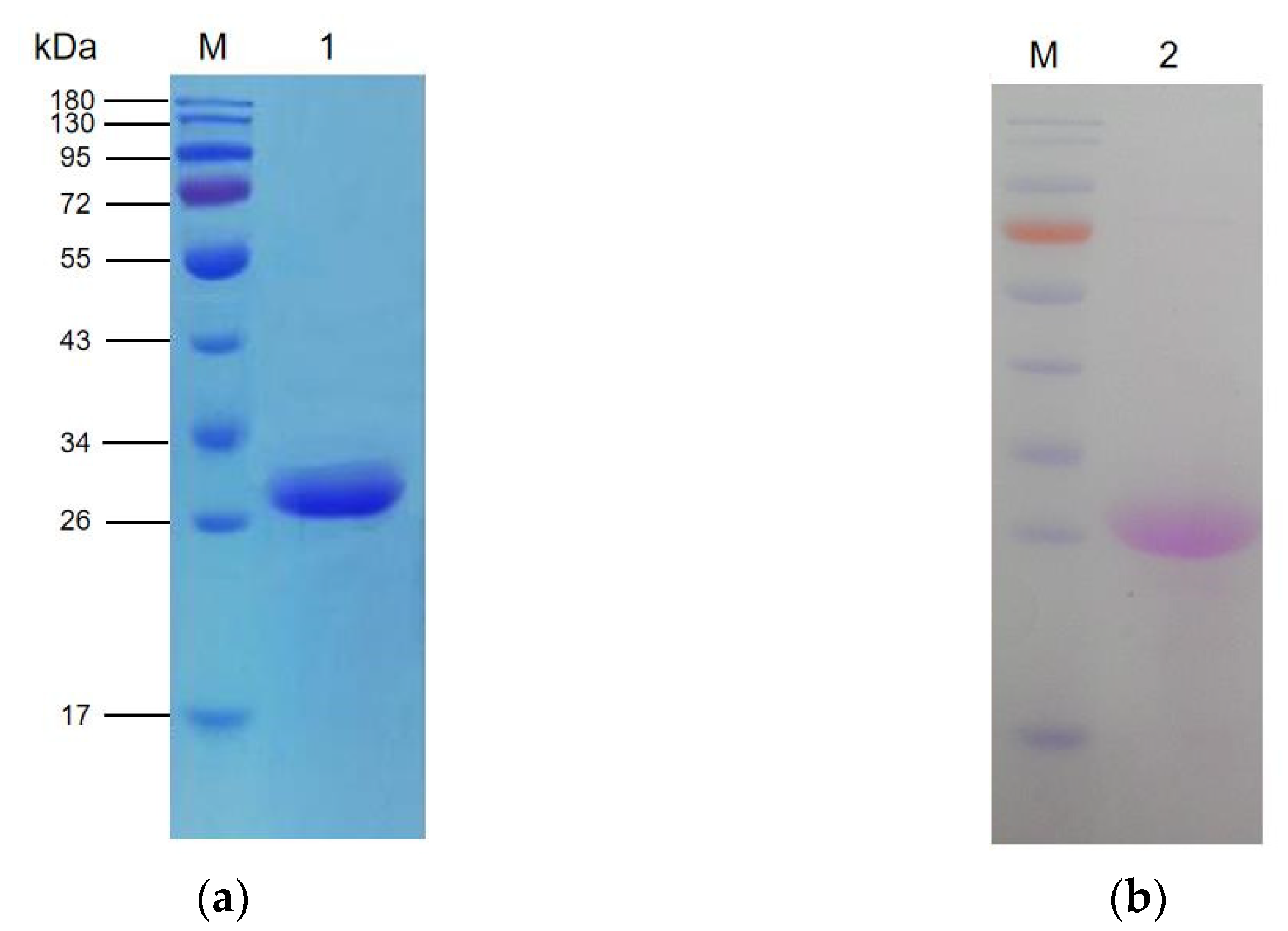
2.1. Purification

A native LPMO was purified to homogeneity from the culture filtrate of T. aurantiacus growing in cellulose-containing medium by ion-exchange chromatography and gel filtration (Figure 1a) and identified as TaAA9A using liquid chromatography–tandem mass spectrometry (LC–MS/MS) (Figure S1a,b). The molecular weight of the purified TaAA9A was estimated to be about 27.42 kDa by SDA-PAGE (Figure 1a), which is higher than that calculated based on the deduced amino acid sequence (24.39 kDa), suggesting glycosylation. Using NetNGlyc 1.0 Server (www.cbs.dtu.dk/services/NetNGlyc/ accessed on 20 November 2021), a putative N-linked glycosylation site (Asn138) in the deduced amino acid sequence of TaAA9A was predicted, indicating that the TaAA9A protein may be N-glycosylated. Further periodic acid-Schiff staining confirmed nTaAA9A’s glycosylation (Figure 1b), in agreement with the predicted results of NetNGlyc 1.0 Server and SDS-PAGE analysis.

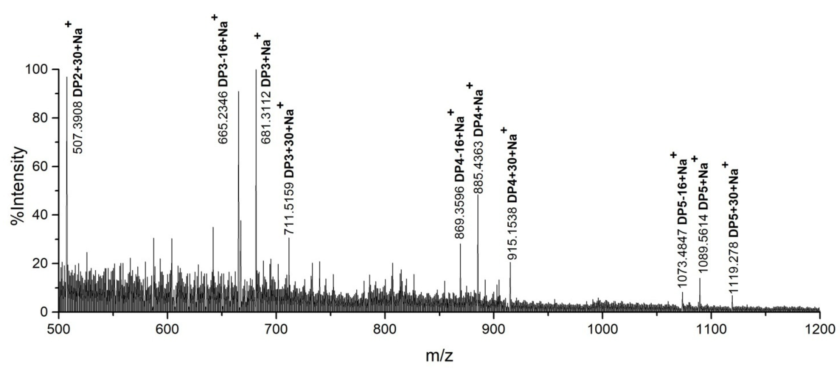
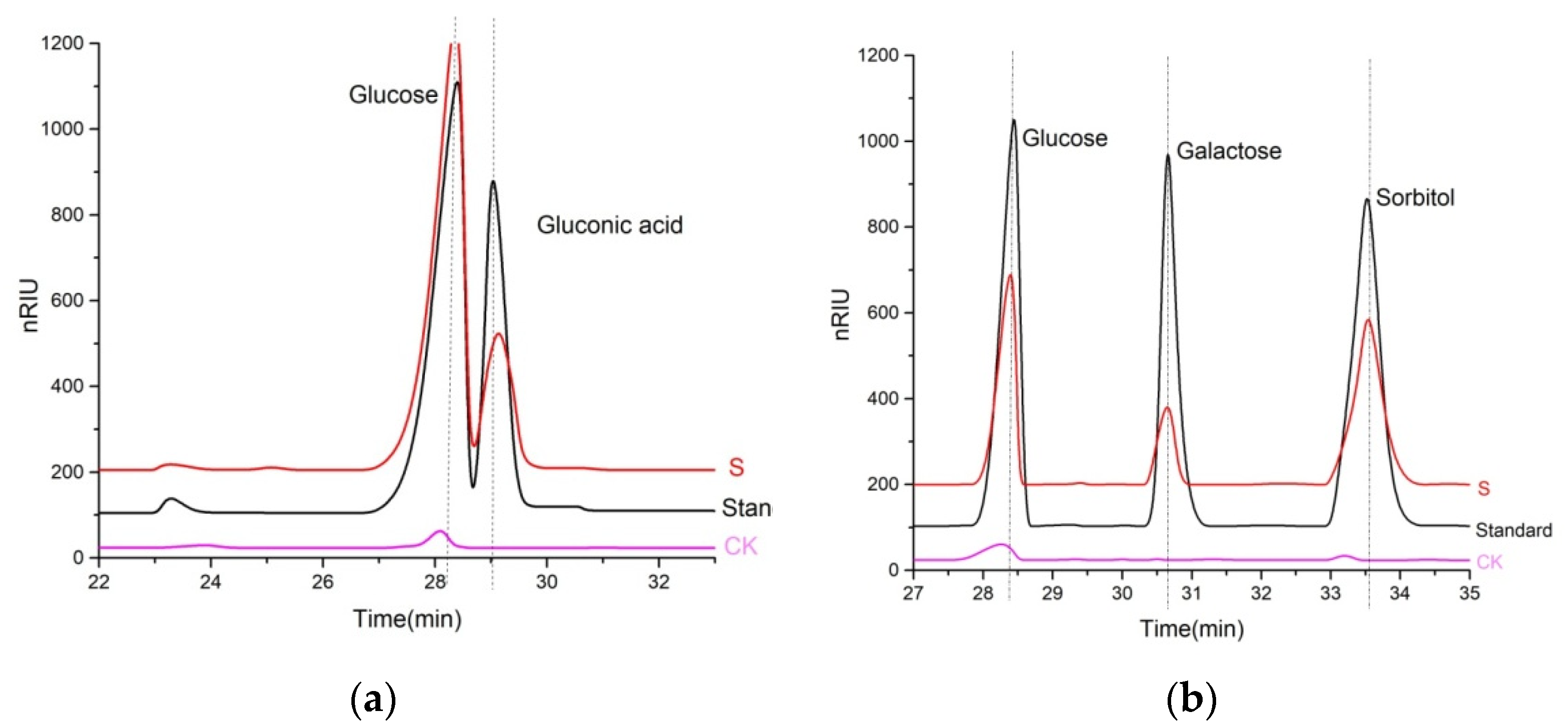
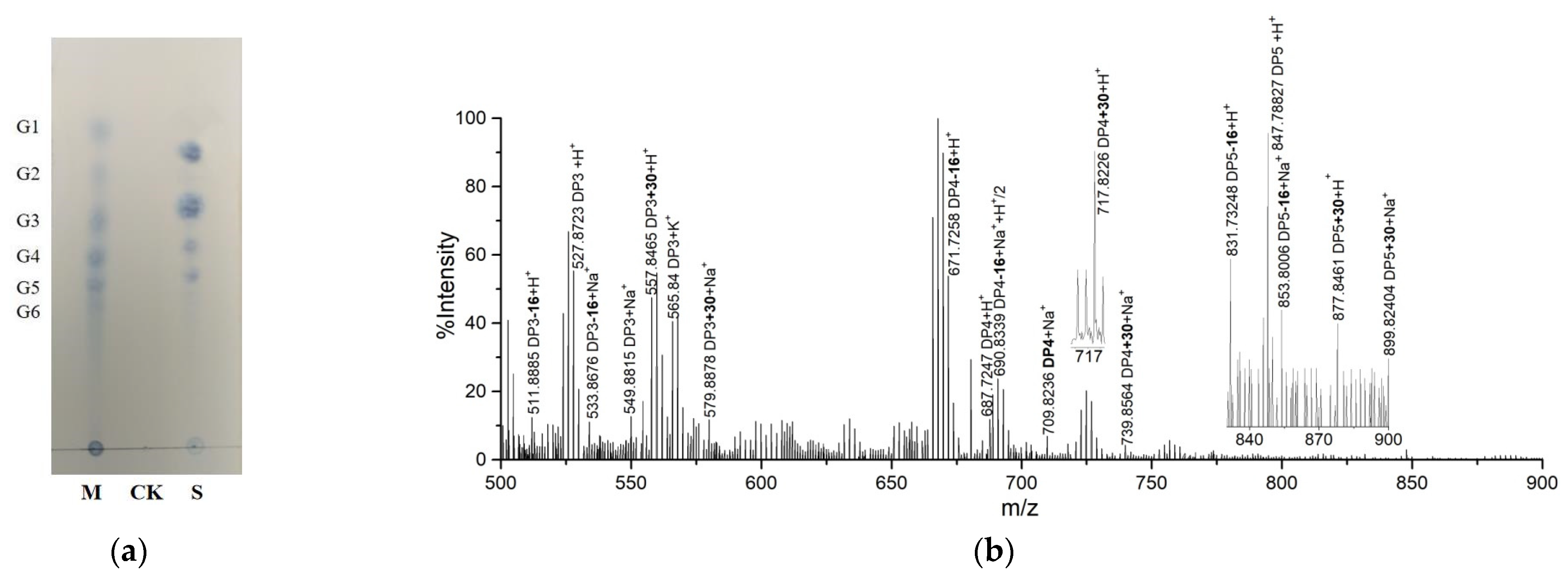
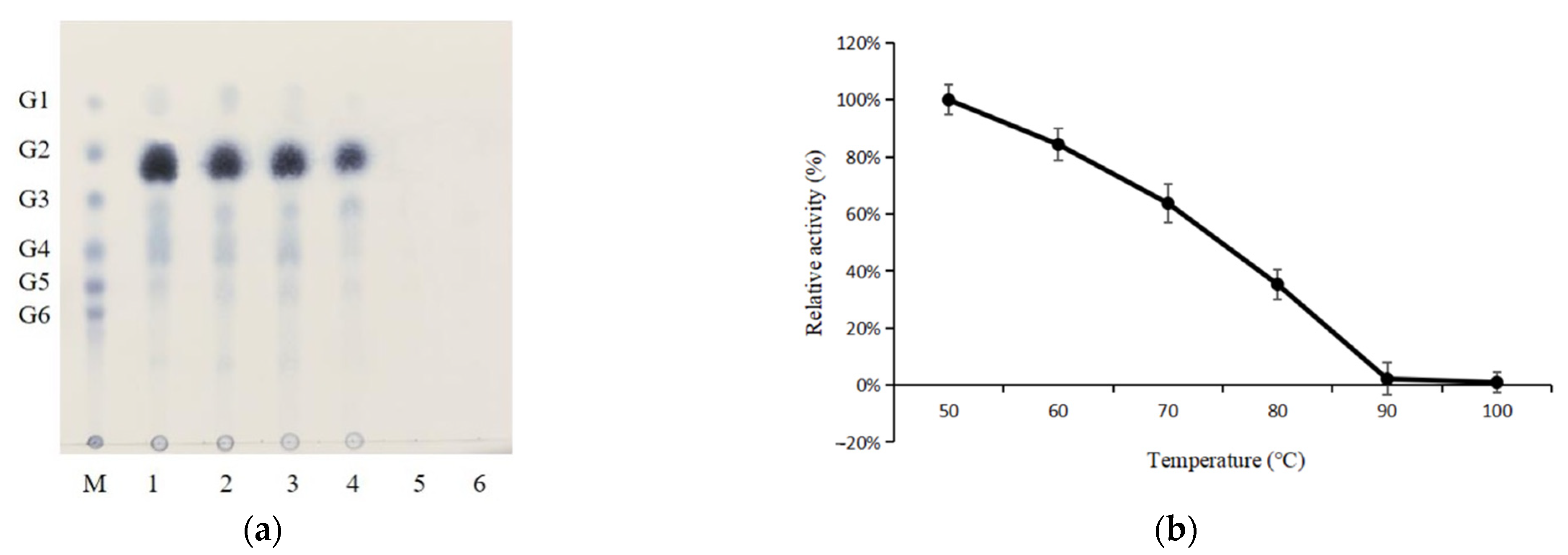
2.2. Product Identification

nTaAA9A reaction products were identified using thin-layer chromatography (TLC), matrix-assisted laser desorption ionization–time-of-flight mass spectrometry (MALDI-TOF MS), and high-performance liquid chromatography–refractive index detector (HPLC-RID). TLC analysis showed that nTaAA9A can cleave cellulose to yield cello-oligosaccharides with various degrees of polymerization (DP) (Figure 2). To demonstrate the presence of C1- and C4-oxidized oligosaccharides, a previously described chemical method [17,30] using methyl iodide to permethylate nTaAA9A products was employed. As expected, molecular ion peaks at m/z DPn + 30 and m/z DPn − 16 corresponding to C1- and C4-oxidized oligosaccharides were observed using MALDI–TOF MS (Figure 3). To further determine the presence of C1- and C4-oxidized oligosaccharides, a chemical method using trifluoroacetic acid (TFA) to hydrolyze nTaAA9A products was applied. Using HPLC–RID analysis, two C1- and C4-oxidized monosaccharides were observed (Figure 4a,b). These results indicate the presence of C1- and C4-oxidized oligosaccharides in nTaAA9A reaction products.
It has been demonstrated that rTaAA9A expressed in Aspergillus oryzae can cleave cellulose to produce C1- and C4-oxidized cello-oligosaccharides [17], using MALDI–TOF MS. In the present study, we show that the native TaAA9A can cleave cellulose to produce C1- and C4-oxidized cello-oligosaccharides using MALDI–TOF MS and HPLC–RID analysis, which further confirms the nature of the C1- and C4-oxidizing activity of nTaAA9A.
The activity of nTaAA9A towards xylan was also investigated. MALDI–TOF MS analysis of nTaAA9A reaction products with xylan as a substrate showed that nTaAA9A can cleave xylan to produce C1- and C4-oxidized xylo-oligosaccharides (Figure 5a,b; Figure S2), similar to LsAA9A from Lentinus similis [12] and MtLPMO9A from Myceliophthora thermophila [24]. Notably, T. aurantiacus can simultaneously secrete three main enzymes on biomass substrates: a GH7 cellobiohydrolase, a GH10 xylanase, and TaAA9A. These three enzymes have been shown to be key players in efficient biomass degradation [31]. It could, therefore, be suggested that TaAA9A may synergistically act with the GH10 xylanase on hemi-cellulose via both oxidative and hydrolytic mechanisms to enhance the degradation of hemi-cellulose [17].

2.3. Structure Quality and Description

The structure of the native TaAA9A was determined to 1.36 Å resolution to final Rwork and Rfree of 0.151 and 0.185, respectively (Table 1). The final model contained 1762 protein atoms and 307 water molecules. The C-terminal Gly residue according to the amino acid sequence was not visible in the electron density map and thus, it was not modelled. Like other crystal structures of various LPMOs, nTaAA9A is characterized by a β-sandwich fold with two twisted antiparallel β-sheets connected through loops of various lengths and conformation. The active site is located on a flat solvent-exposed region of the molecule, in contrast to traditional cellulases that possess a substrate-binding cleft or tunnel. A Cu2+ ion involved in the catalytic reaction was identified at the N-terminal, as previously observed. The Cu2+ ion was refined to a temperature factor of 13.2 Å2 and occupancy of 1.0, suggesting a well-defined tightly bound ion. His1, one of the Cu2+-coordinating residues, was found methylated, as also observed in other LPMO structures. The reason, in general, of this methylation in LPMOs is still unclear, although LPMOs that lack this post-translational modification are still catalytically active [17]. It has been suggested that His1 methylation may convey protection against oxidative damage [17,32]. This posttranslational modification is not always present and is not expected in LPMOs which are produced in P. pastoris [32], as for example in McAA9F [21]. The final structure also contains two N-acetyl-glucosamine (NAG) molecules which were identified based on the electron-density map and built in Asn138.

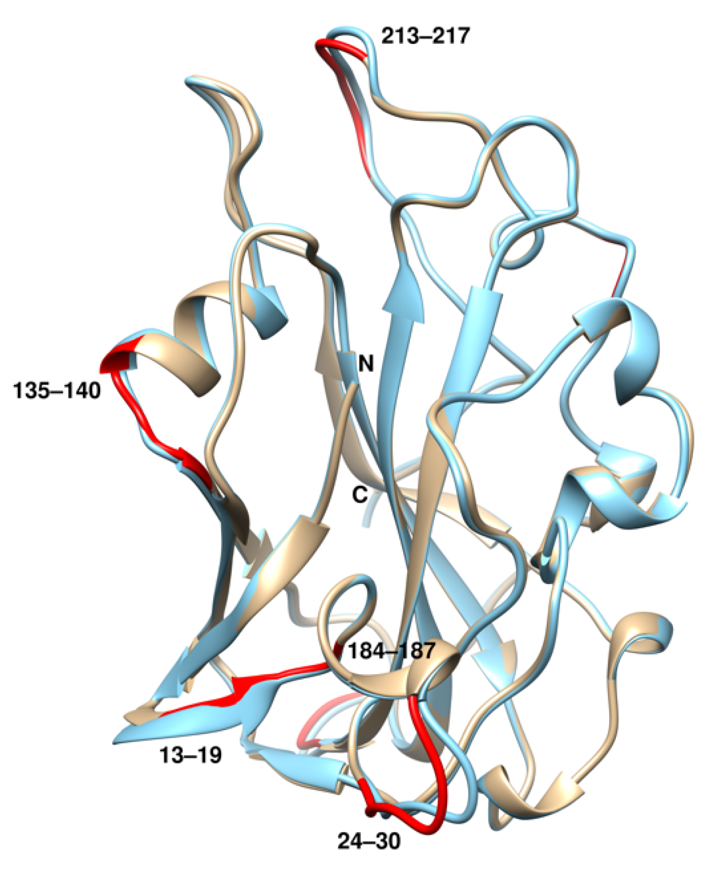
2.4. Structural Comparison with rTaAA9A

Structural superposition resulted in a root-mean-square deviation (rmsd) of 0.43 Å between nTaAA9A and rTaAA9A, suggesting only subtle differences between the two structures. The highest deviations (~0.8–2.4 Å) were found in the regions 9–13, 25–30, 184–187, 202–203, and 213–217 (Figure 6). Also, in Asn138, owing to the different glycosylation in that residue. Asn138 was glycosylated with at least two NAG molecules, as found in the crystal structure. A third glycan was found, but the density was not enough to model it. In rTaAA9A, only one NAG molecule was attached after deglycosylation of the expressed rTaAA9A [17]. Close inspection revealed a different orientation for the side chain of Asn138 and, consequently, the position of the glycan moieties (Figure 7). In nTaAA9A, the two NAG molecules can sit in a shallow groove and make interactions with Asn13, Gln78, and Gln5. In contrast, the NAG molecule in rTaAA9A points outwards and is exposed to the solvent. The subtle changes in surface residues may contribute to the slightly different solvent-accessible area in the native and recombinant TaAA9A (9285Å2 and 9416 Å2, respectively).

2.5. Oligosaccharide Binding

The TaAA9A structure provides insight into the molecular basis of cellulose C1 and C4 oxidation. TaAA9A has an active site containing a copper ion, which is coordinated by two highly conserved His residues (His1 and His86, known as a histidine brace) to create a copper ion-binding site and also a buried highly conserved Tyr residue (Tyr175) that occupies the axial position. Direct structural evidence of LPMO–substrate interaction in Lentinus similis AA9A and Collariella virescens AA9 in the presence of cellohexaose [33] has shown that the copper ion in the active site is close to the C1 and C4 carbon atoms of the oligosaccharides [12]. Superposition of TaAA9A onto CvAA9_A–cellohexaose (rmsd 0.9 Å for 133 equivalent residues; Figure S3) revealed that the active site of nTaAA9A was near to the C1 and C4 carbons of cellohexaose, supporting the C1 and C4 oxidation on cellulose by TaAA9A (Figure 8). An important difference between CvAA9_A, LsAA9A, and TaAA9A is the presence of a long loop (residues 16–31) in the latter. Differences also in the length of the active loops were identified that may play a role in the orientation of the substrate. Individual residues could also affect binding as, for example, Leu41 could clash with the substrate, whereas in CvAA9_A, there is a shorter residue (Thr28). Arg164 belongs to a long external loop (157–164) and makes interactions with BGC-5. In TaAA9A, the equivalent loop is shorter, and interactions with glucose units to provide some stabilization in the binding are therefore not feasible. In general, different loop lengths in AA9s have been implicated in specificity for C1, C4, or C1/C4 oxidation. Tyr212, a highly conserved residue, makes stacking interactions with the flat pyranose ring of the substrate [34] and is likely to support substrate binding in a similar fashion in TaAA9A as well.
The structural superposition also explains difficulties in obtaining structures of the complexes. Clashes with symmetry-related molecules in the crystal lattice usually obstruct ligand binding in LPMOs. Lattice problems have been identified in rTaAA9A crystals and have prevented crystallographic binding studies. Although the nTaAA9 crystals reported here are different from those of the rTaAA9A (space group P21 and two molecules in the asymmetric unit), residues 68–71 and 182–184 of a symmetry-related nTaAA9A clash with three of the glucose moieties of the substrate, whereas the rest of the substrate makes no contacts with symmetry-related molecules.

2.6. Thermostability Properties

Theoretically, AA9 LPMOs from thermophilic fungi should be thermostable. In this study, the thermostability of TaAA9A was investigated by detecting gluconic acid in TaAA9A reaction products hydrolyzed with TFA, using HPLC–RID. The analysis revealed that nTaAA9A exhibits high thermostability (Figure 9), consistent with other thermostable enzymes from thermophilic fungi [35,36]. The enzyme retained about 84.3%, 63.7%, and 35.3% of its activity after incubation for 30 min at 60, 70, and 80 °C, respectively. A cluster of four residues (Val90, Ser131, Leu134, and Trp141; Figure S4) was previously identified in TaAA9A and used to create a thermostable variant of AfuAA9A [18]. The improvement in AfuAA9A thermostability was attributed to the elimination of some unfavorable electrostatic interactions in the enzyme.
Owing to the difficulties in quantitative activity determination of LPMOs, there are only a few reports of their thermostability using activity assay [18,33,37]. So far, the thermostability of only three LPMOs from non-thermophilic fungi, AfuAA9A from Aspergillus fumigatus, LsAA9A from Lentinus similis, and TcAA9A from Talaromyces cellulolyticus, have been measured using differential scanning fluorimetry, differential scanning calorimetry, and activity assay. AfuAA9A and LsAA9A exhibited a melting temperature Tm of 68–69 °C and thermal inflection, Ti, of 71.8 °C, respectively [33], whereas TcAA9A fully lost its activity after incubation at 50 °C for 8 h [37].
Thermostability parameters, such as the number of charged residues, surface area, small-volume aliphatic amino acids, and salt bridges, which have been proposed as indicatives of protein thermostability [38,39], are shown in Table 2. The thermostability, however, is sometimes a combination of different factors and not easily explained by a single parameter. In the absence of thermostability measurements for McAA9F, the temperature-dependent stability was calculated using the SCooP algorithm, a Gibbs–Helmholtz equation-based program [40] which calculates all the thermodynamic quantities associated with the two-fold transition of proteins (e.g., the melting temperature Tm, the standard folding enthalpy Hm measured at Tm, and the standard folding heat capacity Cp). Theoretical measurements were carried out for all enzymes. TaAA9A was found to have a higher Tm than McAA9F, although an accurate measurement of their thermostabilities would need experimental verification under similar assay conditions. Nevertheless, more studies are required to better understand the thermostability issues for this family of enzymes.

3. Materials and Methods

3.1. Strains and Chemicals

Thermoascus aurantiacus strain CGMCC3.17992 from fresh horse dung from China was isolated according to a method described previously [42]. It was deposited in the China General Microbiological Culture Collection Center (CGMCC), a publicly accessible culture collection. A standard cello-oligosaccharide mixture was purchased from Elicityl (Crolles, France). Avicel PH-101, xylan, ascorbate (Vc), glucose, galactose, and gluconic acid were purchased from Sigma-Aldrich.

3.2. Purification and Identification of nTaAA9A from Thermoascus aurantiacus

The native secretory TaAA9A was purified from a T. aurantiacus culture grown at 50 °C for 7 days in cellulose-containing medium [43] supplemented with 0.1 mM CuSO4. After the 7 days of incubation, the mycelium was initially filtered off, and the filtrate was subsequently centrifuged at 10,000× g for 15 min at 4 °C. The resultant supernatant was used for the purification. Ion-exchange chromatography on a DEAE-Sepharose column (GE Healthcare, Chicago, IL, USA) followed by gel filtration on an Enrich SEC650 column (BIO-RAD, Hercules, CA, USA) was employed. Solid ammonium sulphate was initially added to the resultant supernatant, leading to 90% saturation. After 6 h, the resulting precipitate was collected by centrifugation at 10,000× g for 15 min at 4 °C, dissolved, and dialyzed in 50 mM Tris-HCl (pH 8.0) (buffer A). In the subsequent step, the dialyzed sample was loaded on a DEAE-Sepharose column equilibrated with buffer A. nTaAA9A was eluted with a 120 mL linear gradient of NaCl (0–0.3 M in buffer A) at a flow rate of 2 mL/min. Fractions with enzymatic activity were pooled and concentrated by vacuum freeze–drying. In the last step, 0.25 mL of the concentrated sample was applied to a gel filtration Enrich SEC650 column. nTaAA9A was eluted with 50 mL of buffer A at a flow rate of 0.5 mL/min. The purified nTaAA9A was visualized on an SDS-PAGE gel, and the band of interest was cut out. The amino acid sequence of the excised nTaAA9A protein band was determined using LC–MS/MS according to a method previously described [30]. All data were analyzed using MASCOT 2.2 software (Matrix Science). MS/MS spectra were searched against the TaAA9A (ACS05720.1) protein sequence database [17].

3.3. Protein Determination, SDS-PAGE, and Carbohydrate Staining

Protein concentration was measured with the Lowry method [44]. The purity of the nTaAA9A protein was assessed using SDS-PAGE [45]. The carbohydrates in the nTaAA9A enzyme were stained with the Pierce™ Glycoprotein Staining Kit (Thermo Scientific, Waltham, MA, USA).

3.4. nTaAA9A Activity Assay

Phosphoric acid-swollen cellulose (PASC) was prepared as described by Phillips et al. [46]. Activity assays, including the use of xylan as a substrate, were carried out as previously described [43]. nTaAA9A reaction products were identified using TLC, matrix-assisted laser desorption–ionization-time-of-flight mass spectrometry (MALDI–TOF MS), and HPLC–RID analysis.

3.5. TLC and MALDI–TOF MS

Thin-layer chromatography (TLC) was used to analyze nTaAA9A reaction products according to a method previously described [43]. nTaAA9A reaction products were further analyzed using MALDI–TOF MS as described in previous publications [30,43].

3.6. Permethylation and Reduction of nTaAA9A Reaction Products

Permethylation of nTaAA9A reaction products was carried out as described [47], and reduction of TaAA9A reaction products was carried out as previously described [22].

3.7. HPLC–RID

nTaAA9A reaction products and their reduced reaction products were hydrolyzed by TFA as previously described [22] and analyzed by HPLC–RID using an Agilent 1200 series instrument with a refractive index detector (RID). Products were separated using an Aminex HPX-87H column (Bio-Rad) and a 5 mM H2SO4 mobile phase. Glucose, sorbitol, and gluconic acid were annotated based on the elution pattern of standard glucose, sorbitol, and gluconic acid solutions. The flow rate was 0.2 mL/min, and the column was maintained at a temperature of 30 °C.

3.8. Protein Crystallization

The protein was concentrated to ~10 mg/mL in buffer NaOAc 10 mM, NaN3 0.002%, pH 4.8. Crystals were produced at 16 °C using the vapor-diffusion hanging drop method with a well solution of 0.2 M ammonium sulfate, 0.1 M HEPES-NaOH pH 7.5, 25% w/v PEG 3350. The drops consisted of 2 μL of protein solution mixed with 2 μL of the well solution. Crystals appeared after ~3 days, most of them in clusters. For crystallographic data collection, single crystals were carefully separated from the clusters.

3.9. Structure Determination and Validation

X-ray diffraction data were collected at cryogenic temperatures (100 K) in the presence of 10% v/v glycerol as cryoprotectant. Data to 1.86 Å resolution were initially collected at ESRF (Grenoble, France), and the resolution was later extended to 1.36 Å using X-ray data collected at EMBL-Hamburg (beamline P13 at the PETRA III ring). Data processing was carried out with XDS [48], followed by scaling with AIMLESS [49]. Initial phases were obtained with molecular replacement using the structure of rTaAA9A (PDB id 2yet) as search model, leading to a single solution with TFZ = 33.8 in Phaser [50]. Refinement was carried out with PHENIX (v. 1.19.2) [51] using maximum likelihood as the target and simulated annealing with a starting temperature of 1000 K. Water was added at the final stages when the Rfree (calculated using 5% of the data excluded from the refinement) dropped below 30%. The electron difference maps were examined, and a glycosylation site was identified at Asn138 based on the electron density difference map. Validation of the structure was performed with Molprobity [52] and validation tools in Coot [53]. Figures of the structures were created with Chimera [54].

4. Conclusions

In the present study, a native thermostable AA9 LPMO, nTaAA9A, from the thermophilic fungus T. aurantiacus was purified and characterized. nTaAA9A was active and exhibited C1- and C4-oxidizing activity against cellulose and xylan. The enzyme was found to retain significant activity at elevated temperatures. The purified enzyme was found to have a single glycosylation site with at least two NAG molecules. Structural differences were identified with the recombinant rTaAA9A in surface loops and in the glycosylation site. The results will help in exploiting TaAA9A in various biotechnological applications to improve the cleavage of cellulose and xylan.

Supplementary Materials

The following supporting information can be downloaded at: https://www.mdpi.com/article/10.3390/catal12020139/s1, Figure S1: Identification of TaAA9A using LC–MS; Figure S2: Chemical structures of oxidized and non-oxidized xylo-oligosaccharides; Figure S3: Structure-based sequence alignment of nTaAA9A and CvAA9_A; Figure S4: Depiction of the four-residue cluster used for thermostability improvement.

Author Contributions

A.C.P., D.L. designed the experiments. W.Y., I.M. performed the experiments. A.C.P., D.L., W.Y., I.M. analyzed the data. A.C.P., D.L. contributed to the drafting and revision of the manuscript. All authors have read and agreed to the published version of the manuscript.

Funding

This research was funded by the National Natural Science Foundation of China (Grant No. 31571949) and the Ministry of Science and Technology of China (Grant No. 2015BAD15B05).

Data Availability Statement

All data generated or analyzed during this study are included in this published article.

Acknowledgments

We thank Biocenter Finland for instrument support and the staff at the P13 beamline (EMBL-Hamburg, DESY) for help during data collection. Access to synchrotron beamtime was provided by the European Union’s Horizon 2020 iNEXT-Discovery programme (grant agreement No 871037).

Conflicts of Interest

The authors declare no conflict of interest.

References

  1. Service, R.F. Is there a road ahead for cellulosic ethanol? Science 2010, 329, 784–785. [Google Scholar] [CrossRef] [Green Version]
  2. Vaaje-Kolstad, G.; Westereng, B.; Horn, S.J.; Liu, Z.; Zhai, H.; Sørlie, M.; Eijsink, V.G.H. An Oxidative Enzyme Boosting the Enzymatic Conversion of Recalcitrant Polysaccharides. Science 2010, 330, 219–222. [Google Scholar] [CrossRef] [PubMed]
  3. Vaaje-Kolstad, G.; Forsberg, Z.; Loose, J.S.; Bissaro, B.; Eijsink, V.G. Structural diversity of lytic polysaccharide monooxygenases. Curr. Opin. Struct. Biol. 2017, 44, 67–76. [Google Scholar] [CrossRef]
  4. Bissaro, B.; Várnai, A.; Røhr, Å.K.; Eijsink, V.G.H. Oxidoreductases and Reactive Oxygen Species in Conversion of Lignocellulosic Biomass. Microbiol. Mol. Biol. Rev. 2018, 82, e00029-18. [Google Scholar] [CrossRef] [PubMed] [Green Version]
  5. Levasseur, A.; Drula, E.; Lombard, V.; Coutinho, P.M.; Henrissat, B. Expansion of the enzymatic repertoire of the CAZy database to integrate auxiliary redox enzymes. Biotechnol. Biofuels 2013, 6, 41. [Google Scholar] [CrossRef] [PubMed] [Green Version]
  6. Forsberg, Z.; Sørlie, M.; Petrović, D.; Courtade, G.; Aachmann, F.L.; Vaaje-Kolstad, G.; Bissaro, B.; Røhr, Å.K.; Eijsink, V.G. Polysaccharide degradation by lytic polysaccharide monooxygenases. Curr. Opin. Struct. Biol. 2019, 59, 54–64. [Google Scholar] [CrossRef]
  7. Tamburrini, K.C.; Terrapon, N.; Lombard, V.; Bissaro, B.; Longhi, S.; Berrin, J.-G. Bioinformatic Analysis of Lytic Polysaccharide Monooxygenases Reveals the Pan-Families Occurrence of Intrinsically Disordered C-Terminal Extensions. Biomolecules 2021, 11, 1632. [Google Scholar] [CrossRef]
  8. Chylenski, P.; Bissaro, B.; Sørlie, M.; Røhr, Å.K.; Várnai, A.; Horn, S.J.; Eijsink, V.G.H. Lytic Polysaccharide Monooxygenases in Enzymatic Processing of Lignocellulosic Biomass. ACS Catal. 2019, 9, 4970–4991. [Google Scholar] [CrossRef]
  9. Tandrup, T.; Frandsen, K.E.H.; Johansen, K.S.; Berrin, J.-G.; Leggio, L.L. Recent insights into lytic polysaccharide monooxygenases (LPMOs). Biochem. Soc. Trans. 2018, 46, 1431–1447. [Google Scholar] [CrossRef]
  10. Askarian, F.; Uchiyama, S.; Masson, H.; Sørensen, H.V.; Golten, O.; Bunæs, A.C.; Mekasha, S.; Røhr, Å.K.; Kommedal, E.; Ludviksen, J.A.; et al. The lytic polysaccharide monooxygenase CbpD promotes Pseudomonas aeruginosa virulence in systemic infection. Nat. Commun. 2021, 12, 1230. [Google Scholar] [CrossRef]
  11. Chiu, E.; Hijnen, M.; Bunker, R.D.; Boudes, M.; Rajendran, C.; Aizel, K.; Olieric, V.; Schulze-Briese, C.; Mitsuhashi, W.; Young, V.; et al. Structural basis for the enhancement of virulence by viral spindles and their in vivo crystallization. Proc. Natl. Acad. Sci. USA 2015, 112, 3973–3978. [Google Scholar] [CrossRef] [PubMed] [Green Version]
  12. Simmons, T.J.; Frandsen, K.E.H.; Ciano, L.; Tryfona, T.; Lenfant, N.; Poulsen, J.C.; Wilson, L.F.L.; Tandrup, T.; Tovborg, M.; Schnorr, K.; et al. Structural and electronic determinants of lytic polysaccharide monooxygenase reactivity on polysaccharide substrates. Nat. Commun. 2017, 8, 1064. [Google Scholar] [CrossRef] [PubMed]
  13. Wang, B.; Johnston, E.M.; Li, P.; Shaik, S.; Davies, G.J.; Walton, P.H.; Rovira, C. QM/MM Studies into the H2O2-Dependent Activity of Lytic Polysaccharide Monooxygenases: Evidence for the Formation of a Caged Hydroxyl Radical Intermediate. ACS Catal. 2018, 8, 1346–1351. [Google Scholar] [CrossRef] [Green Version]
  14. Wang, B.; Walton, P.H.; Rovira, C. Molecular Mechanisms of Oxygen Activation and Hydrogen Peroxide Formation in Lytic Polysaccharide Monooxygenases. ACS Catal. 2019, 9, 4958–4969. [Google Scholar] [CrossRef] [Green Version]
  15. Paradisi, A.; Johnston, E.M.; Tovborg, M.; Nicoll, C.R.; Ciano, L.; Dowle, A.; McMaster, J.; Hancock, Y.; Davies, G.J.; Walton, P.H. Formation of a Copper(II)–Tyrosyl Complex at the Active Site of Lytic Polysaccharide Monooxygenases Following Oxidation by H2O2. J. Am. Chem. Soc. 2019, 141, 18585–18599. [Google Scholar] [CrossRef] [Green Version]
  16. Frandsen, K.E.H.; Leggio, L.L. Lytic polysaccharide monooxygenases: A crystallographer’s view on a new class of biomass-degrading enzymes. IUCrJ 2016, 3, 448–467. [Google Scholar] [CrossRef] [Green Version]
  17. Quinlan, R.J.; Sweeney, M.D.; Leggio, L.L.; Otten, H.; Poulsen, J.-C.N.; Johansen, K.S.; Krogh, K.B.R.M.; Jørgensen, C.I.; Tovborg, M.; Anthonsen, A.; et al. Insights into the oxidative degradation of cellulose by a copper metalloenzyme that exploits biomass components. Proc. Natl. Acad. Sci. USA 2011, 108, 15079–15084. [Google Scholar] [CrossRef] [Green Version]
  18. Leggio, L.L.; Weihe, C.D.; Poulsen, J.-C.N.; Sweeney, M.; Rasmussen, F.; Lin, J.; De Maria, L.; Wogulis, M. Structure of a lytic polysaccharide monooxygenase from Aspergillus fumigatus and an engineered thermostable variant. Carbohydr. Res. 2018, 469, 55–59. [Google Scholar] [CrossRef]
  19. Bissaro, B.; Isaksen, I.; Vaaje-Kolstad, G.; Eijsink, V.G.H.; Røhr, Å.K. How a Lytic Polysaccharide Monooxygenase Binds Crystalline Chitin. Biochemistry 2018, 57, 1893–1906. [Google Scholar] [CrossRef]
  20. Leggio, L.L.; Simmons, T.J.; Poulsen, J.-C.N.; Frandsen, K.E.H.; Hemsworth, G.R.; Stringer, M.A.; von Freiesleben, P.; Tovborg, M.; Johansen, K.S.; De Maria, L.; et al. Structure and boosting activity of a starch-degrading lytic polysaccharide monooxygenase. Nat. Commun. 2015, 6, 5961. [Google Scholar] [CrossRef] [Green Version]
  21. Mazurkewich, S.; Seveso, A.; Hüttner, S.; Brändén, G.; Larsbrink, J. Structure of a C1/C4-oxidizing AA9 lytic polysaccharide monooxygenase from the thermophilic fungus Malbranchea cinnamomea. Acta Crystallogr. Sect. D Struct. Biol. 2021, D77, 1019–1026. [Google Scholar] [CrossRef] [PubMed]
  22. Beeson, W.T.; Phillips, C.M.; Cate, J.H.D.; Marletta, M.A. Oxidative Cleavage of Cellulose by Fungal Copper-Dependent Polysaccharide Monooxygenases. J. Am. Chem. Soc. 2012, 134, 890–892. [Google Scholar] [CrossRef] [PubMed]
  23. Bey, M.; Zhou, S.; Poidevin, L.; Henrissat, B.; Coutinho, P.M.; Berrin, J.-G.; Sigoillot, J.-C. Cello-Oligosaccharide Oxidation Reveals Differences between Two Lytic Polysaccharide Monooxygenases (Family GH61) from Podospora anserina. Appl. Environ. Microbiol. 2013, 79, 488–496. [Google Scholar] [CrossRef] [PubMed] [Green Version]
  24. Frommhagen, M.; Sforza, S.; Westphal, A.H.; Visser, J.; Hinz, S.W.A.; Koetsier, M.J.; Van Berkel, W.J.H.; Gruppen, H.; Kabel, M.A. Discovery of the combined oxidative cleavage of plant xylan and cellulose by a new fungal polysaccharide monooxygenase. Biotechnol. Biofuels 2015, 8, 101. [Google Scholar] [CrossRef] [PubMed] [Green Version]
  25. Zerva, A.; Pentari, C.; Grisel, S.; Berrin, J.-G.; Topakas, E. A new synergistic relationship between xylan-active LPMO and xylobiohydrolase to tackle recalcitrant xylan. Biotechnol. Biofuels 2020, 13, 142. [Google Scholar] [CrossRef]
  26. Lange, L.; Huang, Y.; Busk, P.K. Microbial decomposition of keratin in nature—A new hypothesis of industrial relevance. Appl. Microbiol. Biotechnol. 2016, 100, 2083–2096. [Google Scholar] [CrossRef] [Green Version]
  27. Vu, V.V.; Beeson, W.T.; Phillips, C.M.; Cate, J.H.D.; Marletta, M.A. Determinants of Regioselective Hydroxylation in the Fungal Polysaccharide Monooxygenases. J. Am. Chem. Soc. 2014, 136, 562–565. [Google Scholar] [CrossRef]
  28. Berka, R.M.; Grigoriev, I.V.; Otillar, R.; Salamov, A.; Grimwood, J.; Reid, I.; Ishmael, N.; John, T.; Darmond, C.; Moisan, M.-C.; et al. Comparative genomic analysis of the thermophilic biomass-degrading fungi Myceliophthora thermophila and Thielavia terrestris. Nat. Biotechnol. 2011, 29, 922–927. [Google Scholar] [CrossRef]
  29. Petrović, D.M.; Várnai, A.; Dimarogona, M.; Mathiesen, G.; Sandgren, M.; Westereng, B.; Eijsink, V.G.H. Comparison of three seemingly similar lytic polysaccharide monooxygenases from Neurospora crassa suggests different roles in plant biomass degradation. J. Biol. Chem. 2019, 294, 15068–15081. [Google Scholar] [CrossRef]
  30. Chen, J.; Guo, X.; Zhu, M.; Chen, C.; Li, D. Polysaccharide monooxygenase-catalyzed oxidation of cellulose to glucuronic acid-containing cello-oligosaccharides. Biotechnol. Biofuels 2019, 12, 42. [Google Scholar] [CrossRef]
  31. McClendon, S.D.; Batth, T.; Petzold, C.J.; Adams, P.D.; Simmons, B.A.; Singer, S.W. Thermoascus aurantiacus is a promising source of enzymes for biomass deconstruction under thermophilic conditions. Biotechnol. Biofuels 2012, 5, 54. [Google Scholar] [CrossRef] [PubMed] [Green Version]
  32. Petrović, D.M.; Bissaro, B.; Chylenski, P.; Skaugen, M.; Sørlie, M.; Jensen, M.S.; Aachmann, F.L.; Courtade, G.; Várnai, A.; Eijsink, V.G.H. Methylation of the N-terminal histidine protects a lytic polysaccharide monooxygenase from auto-oxidative inactivation. Protein Sci. 2018, 27, 1636–1650. [Google Scholar] [CrossRef] [PubMed]
  33. Tandrup, T.; Tryfona, T.; Frandsen, K.E.H.; Johansen, K.S.; DuPree, P.; Leggio, L.L. Oligosaccharide Binding and Thermostability of Two Related AA9 Lytic Polysaccharide Monooxygenases. Biochemistry 2020, 59, 3347–3358. [Google Scholar] [CrossRef] [PubMed]
  34. Harris, P.V.; Welner, D.; McFarland, K.C.; Re, E.; Poulsen, J.-C.N.; Brown, K.; Salbo, R.; Ding, H.; Vlasenko, E.; Merino, S.; et al. Stimulation of Lignocellulosic Biomass Hydrolysis by Proteins of Glycoside Hydrolase Family 61: Structure and Function of a Large, Enigmatic Family. Biochemistry 2010, 49, 3305–3316. [Google Scholar] [CrossRef] [PubMed]
  35. Maheshwari, R.; Bharadwaj, G.; Bhat, M.K. Thermophilic Fungi: Their Physiology and Enzymes. Microbiol. Mol. Biol. Rev. 2000, 64, 461–488. [Google Scholar] [CrossRef] [Green Version]
  36. Li, D.-C.; Li, A.-N.; Papageorgiou, A.C. Cellulases from Thermophilic Fungi: Recent Insights and Biotechnological Potential. Enzym. Res. 2011, 2011, 308730. [Google Scholar] [CrossRef] [Green Version]
  37. Zhang, R.; Liu, Y.; Zhang, Y.; Feng, D.; Hou, S.; Guo, W.; Niu, K.; Jiang, Y.; Han, L.; Sindhu, L.; et al. Identification of a thermostable fungal lytic polysaccharide monooxygenase and evaluation of its effect on lignocellulosic degradation. Appl. Microbiol. Biotechnol. 2019, 103, 5739–5750. [Google Scholar] [CrossRef]
  38. Panja, A.S.; Bandopadhyay, B.; Maiti, S. Protein Thermostability Is Owing to Their Preferences to Non-Polar Smaller Volume Amino Acids, Variations in Residual Physico-Chemical Properties and More Salt-Bridges. PLoS ONE 2015, 10, e0131495. [Google Scholar] [CrossRef]
  39. Trivedi, S.; Gehlot, H.S.; Rao, S.R. Protein thermostability in Archaea and Eubacteria. Genet. Mol. Res. 2006, 5, 816–827. [Google Scholar]
  40. Pucci, F.; Kwasigroch, J.M.; Rooman, M. SCooP: An accurate and fast predictor of protein stability curves as a function of temperature. Bioinformatics 2017, 33, 3415–3422. [Google Scholar] [CrossRef]
  41. Costantini, S.; Colonna, G.; Facchiano, A.M. ESBRI: A web server for evaluating salt bridges in proteins. Bioinformation 2008, 3, 137–138. [Google Scholar] [CrossRef] [PubMed]
  42. Li, D.-C.; Lu, M.; Li, Y.-L.; Lu, J. Purification and characterization of an endocellulase from the thermophilic fungus Chaetomium thermophilum CT2. Enzym. Microb. Technol. 2003, 33, 932–937. [Google Scholar] [CrossRef]
  43. Chen, C.; Chen, J.; Geng, Z.; Wang, M.; Liu, N.; Li, D. Regioselectivity of oxidation by a polysaccharide monooxygenase from Chaetomium thermophilum. Biotechnol. Biofuels 2018, 11, 155. [Google Scholar] [CrossRef] [PubMed]
  44. Lowry, O.H.; Rosebrough, N.J.; Farr, A.L.; Randall, R.J. Protein measurement with the Folin phenol reagent. J. Biol. Chem. 1951, 193, 265–275. [Google Scholar] [CrossRef]
  45. Laemmli, U.K. Cleavage of Structural Proteins during the Assembly of the Head of Bacteriophage T4. Nature 1970, 227, 680–685. [Google Scholar] [CrossRef] [PubMed]
  46. Phillips, C.M.; Beeson, W.T.; Cate, J.H.; Marletta, M.A. Cellobiose Dehydrogenase and a Copper-Dependent Polysaccharide Monooxygenase Potentiate Cellulose Degradation by Neurospora crassa. ACS Chem. Biol. 2011, 6, 1399–1406. [Google Scholar] [CrossRef]
  47. Needs, P.W.; Selvendran, R.R. Avoiding oxidative degradation during sodium hydroxide/methyl iodide-mediated carbohydrate methylation in dimethyl sulfoxide. Carbohydr. Res. 1993, 245, 1–10. [Google Scholar] [CrossRef]
  48. Kabsch, W. XDS. Acta Crystallogr. Sect. D Struct. Biol. 2010, D66, 125–132. [Google Scholar] [CrossRef] [Green Version]
  49. Evans, P.R.; Murshudov, G.N. How good are my data and what is the resolution? Acta Crystallogr. Sect. D Struct. Biol. 2013, D69, 1204–1214. [Google Scholar] [CrossRef]
  50. McCoy, A.J.; Grosse-Kunstleve, R.W.; Adams, P.D.; Winn, M.D.; Storoni, L.C.; Read, R.J. Phaser crystallographic software. J. Appl. Crystallogr. 2007, 40, 658–674. [Google Scholar] [CrossRef] [Green Version]
  51. Adams, P.D.; Afonine, P.V.; Bunkóczi, G.; Chen, V.B.; Davis, I.W.; Echols, N.; Headd, J.J.; Hung, L.-W.; Kapral, G.J.; Grosse-Kunstleve, R.W.; et al. PHENIX: A comprehensive Python-based system for macromolecular structure solution. Acta Crystallogr. Sect. D Struct. Biol. 2010, D66, 213–221. [Google Scholar] [CrossRef] [Green Version]
  52. Chen, V.B.; Arendall, W.B., III; Headd, J.J.; Keedy, D.A.; Immormino, R.M.; Kapral, G.J.; Murray, L.W.; Richardson, J.S.; Richardson, D.C. MolProbity: All-atom structure validation for macromolecular crystallography. Acta Crystallogr. Sect. D Struct. Biol. 2010, D66, 12–21. [Google Scholar] [CrossRef] [PubMed] [Green Version]
  53. Emsley, P.; Cowtan, K. Coot: Model-building tools for molecular graphics. Acta Crystallogr. Sect. D Struct. Biol. 2004, D60, 2126–2132. [Google Scholar] [CrossRef] [PubMed] [Green Version]
  54. Pettersen, E.F.; Goddard, T.D.; Huang, C.C.; Couch, G.S.; Greenblatt, D.M.; Meng, E.C.; Ferrin, T.E. UCSF Chimera—A visualization system for exploratory research and analysis. J. Comput. Chem. 2004, 25, 1605–1612. [Google Scholar] [CrossRef] [PubMed] [Green Version]
Figure 1. SDS-PAGE of the purified native TaAA9A. nTaAA9A was visualized (a) by staining with Coomassie Brilliant Blue; (b) by staining with the Pierce™ Glycoprotein Staining Kit. Lane M, protein markers (kDa); Lanes 1 and 2, nTaAA9A.
Catalysts 12 00139 g001
Figure 2. TLC analysis of nTaAA9A reaction products using PASC as a substrate. nTaAA9A reaction products were formed following incubation of 0.5% PASC with nTaAA9A in 10 mM HOAc–NH4OAc (pH 5.0) and 1 mM ascorbate at 50 °C for 48 h. Lane M, standard cello-oligosaccharides (G1–G6); Lane S, nTaAA9A reaction products; Lane CK, control sample analyzed as above, except that no nTaAA9A was added.
Catalysts 12 00139 g002
Figure 3. Identification of nTaAA9A permethylated reaction products using MALDI–TOF MS. nTaAA9A reaction products upon incubation of 0.5% PASC with nTaAA9A in 10 mM HOAc–NH4OAc (pH 5.0) and 1 mM ascorbate at 50 °C for 48 h, followed by permethylation with methyl iodide. C1-oxidized oligosaccharides (m/z +30), C4-oxidized oligosaccharides (m/z −16), and non-oxidized oligosaccharides (m/z +0).
Catalysts 12 00139 g003
Figure 4. Identification of nTaAA9A reaction products using HPLC–RID. (a) nTaAA9A reaction products after incubation of 0.5% PASC with TaAA9A in 10 mM HOAc–NH4OAc (pH 5.0) and 1 mM ascorbate at 50 °C for 48 h, followed by hydrolysis with TFA. C1-oxidized products, if present, were hydrolyzed by TFA to yield glucose and gluconic acid. The standard used was a mixture of glucose and gluconic acid; S: nTaAA9A reaction products hydrolyzed by TFA; CK, the control sample analyzed as above but without nTaAA9A. (b) TaAA9A reaction products upon incubation of 0.5% PASC with TaAA9A in 10 mM HOAc–NH4OAc (pH 5.0) and 1 mM ascorbate at 50 °C for 48 h, followed by reduction with NaBH4 and by hydrolysis with TFA. If there were C4-oxidized products, they were reduced by NaBH4, followed by hydrolysis with TFA to yield glucose, galactose, and sorbitol. Standard, a mixture of glucose, galactose, and sorbitol; S: nTaAA9A reaction products reduced by NaBH4, followed by hydrolysis with TFA; CK, the control sample was analyzed as above, except without nTaAA9A.
Catalysts 12 00139 g004
Figure 5. Identification of nTaAA9A permethylated reaction products with xylan as a substrate using TLC and MALDI–TOF MS. (a) TLC: nTaAA9A reaction products upon incubation of 0.5% xylan with TaAA9A in 10 mM HOAc–NH4OAc (pH 5.0) and 1 mM ascorbate at 50 °C for 48 h. Lane M, standard cello-oligosaccharides (G1–G6); Lane S, nTaAA9A reaction products; Lane CK, control sample analyzed as above but without nTaAA9A. (b) MALDI–TOF MS: nTaAA9A reaction products after incubation of 0.5% xylan with nTaAA9A in 10 mM HOAc–NH4OAc (pH 5.0) and 1 mM ascorbate at 50 °C for 48 h, followed by permethylation with methyl iodide. C1-oxidized xylo-oligosaccharides (m/z +30), C4-oxidized xylo-oligosaccharides (m/z −16), and non-oxidized xylo-oligosaccharides (m/z +0).
Catalysts 12 00139 g005
Figure 6. Structural comparison of nTaAA9A (brown) with rTaAA9A (cyan). The regions with the highest variations are indicated in red color and labelled.
Catalysts 12 00139 g006
Figure 7. Glycosylation at Asn138. nTaAA9A is depicted in brown, and rTaAA9A in cyan.
Catalysts 12 00139 g007
Figure 8. The active sites of nTaAA9A (brown) and CvAA9_A (pink; PDB id 6yde). Cellohexaose bound to CvAA9_A is shown in stick representation. nTaAA9A loops that show differences with corresponding loops in CvAA9_A are colored in green and labeled. The CvAA9_A residues that make hydrogen bond interactions with cellohexaose are shown as sticks and labelled. Tyr208 (Tyr212 in nTaAA9A) that provides additional stabilizing interactions with the oligosaccharide is also shown. The structural equivalent residues in nTaAA9 are depicted and labeled. CvAA9_A residues Arg67 and Arg164 correspond to gaps in nTaAA9A (Figure S3), and no structural equivalent residues are shown.
Catalysts 12 00139 g008
Figure 9. Determination of nTaAA9A thermostability. (a) TLC analysis of nTaAA9A reaction products. nTaAA9A was initially treated for 30 min at 50, 60, 70, 80, 90, and 100 °C (lanes 1–6, respectively; M: standard cello-oligosaccharides) in 10 mM NH4OAc–HOAc (pH 5.0) without substrate. The treated nTaAA9A was incubated with 0.5% PASC in 10 mM HOAc–NH4OAc (pH 5.0) and 1 mM ascorbate at 50 °C for 48 h. (b) Residual activity of nTaAA9A as a percentage of the maximum activity (100%). The residual nTaAA9A activities were measured using HPLC–RID by detecting gluconic acid in nTaAA9A reaction products after their hydrolysis with TFA. The experiment was carried out in triplicates.
Catalysts 12 00139 g009
Table 1. X-ray data collection and refinement statistics. Numbers in parentheses refer to the outermost resolution shell.
Data Collection
BeamlineP13 (PETRA III, DESY)
Wavelength (Å)0.9762
Resolution (Å)44.63−1.36 (1.41−1.36)
Space groupP212121
Unit cell a, b, c (Å)37.7, 64.2, 88.5
No. of observations559,476 (22,595)
No. of unique reflections43,359 (2631)
Completeness (%)92.7 (58.5)
Multiplicity12.9 (8.6)
Mosaicity (°)0.22
Rmeas0.218 (2.993)
CC1/20.998 (0.251)
Wilson B factor (Å2)22.1
Refinement
No. of reflections used43,220
Rcryst/Rfree0.151/0.185
RMSD in bonds (Å)0.005
RMSD in angles (°)0.867
Number of protein atoms1762
No. of water molecules307
Average B-factor (Å2)17.6
Ramachandran favored/outliers (%)99.1/0.0
Clashscore2.54
PDB id7q1k
Table 2. Comparative statistics of thermostability parameters in AA9 LPMOs #.
ParameternTaAA9AMcAA9F (7ntl)CvAA9_A (6yde)AfuAA9A (6h1z)LsAA9A (5n04)TcAA9A GenBank (GAM42970.1)
Asp + Glu (−) #191634182119
Arg + Lys (+)772410109
Pro/Gly0.841.040.910.571.110.76
Val (%)5.34.18.74.49.810.3
Amino acid residues228222252229235246
SAS (Å2)9285.09034.09649.09363.09456-
Intra-chain salt bridges §323311-
Melting temperature Tm (°C) 56.149.457.557.851.6-
# Amino acid calculations were carried out on ExPASy ProtParam (https://web.expasy.org/protparam/ accessed on 20 November 2021). § Calculated with ESBRI (http://bioinformatica.isa.cnr.it/ESBRI/introduction.html accessed on 20 November 2021) [41]. Calculated with SCooP_v1.0 (http://babylone.ulb.ac.be/SCooP accessed on 20 November 2021) [40].
Publisher’s Note: MDPI stays neutral with regard to jurisdictional claims in published maps and institutional affiliations.

